Three-dimensional structure and stoichiometry of Helmintosporium victoriae190S totivirus Jose ´ R. Casto ´n a, * , Daniel Luque a , Benes L. Trus b,c , Germa ´n Rivas d , Carlos Alfonso d , Jose ´ M. Gonza ´lez a , Jose ´ L. Carrascosa a , Padmanaban Annamalai e , Said A. Ghabrial e a Department of Estructura de Macromole ´culas, Centro Nacional de Biotecnologı ´a, CSIC, Campus Universidad Auto ´noma de Madrid, Darwin no 3, Cantoblanco, E-28049 Madrid, Spain b Imaging Sciences Laboratory, CIT, NIAMS, NIH, DHHS, Bethesda, MD 20892-5624, USA c Laboratory of Structural Biology Research, NIAMS, NIH, DHHS, Bethesda, MD 20892-5624, USA d Centro de Investigaciones Biolo ´gicas, CSIC, 28006 Madrid, Spain e Department of Plant Pathology, University of Kentucky, Lexington, KY 40546, USA Received 9 August 2005; returned to author for revision 28 September 2005; accepted 22 November 2005 Available online 18 January 2006 Abstract Most double-stranded RNA viruses have a characteristic capsid consisting of 60 asymmetric coat protein dimers in a so-called T = 2 organization, a feature probably related to their unique life cycle. These capsids organize the replicative complex(es) that is actively involved in genome transcription and replication. Available structural data indicate that their RNA-dependent RNA polymerase (RDRP) is packaged as an integral capsid component, either as a replicative complex at the pentameric vertex (as in reovirus capsids) or as a fusion protein with the coat protein (as in some totivirus). In contrast with members of the family Reoviridae, there are two well-established capsid arrangements for dsRNA fungal viruses, exemplified by the totiviruses L-A and UmVand the chrysovirus PcV. Whereas L-A and UmV have a canonical T = 2 capsid, the PcV capsid is based on a T = 1 lattice composed of 60 capsid proteins. We used cryo-electron microscopy combined with three-dimensional reconstruction techniques and hydrodynamic analysis to determine the structure at 13.8 A ˚ resolution of Helminthosporium victoriae 190S virus (Hv190SV), a totivirus isolated from a filamentous fungus. The Hv190SV capsid has a smooth surface and is based on a T = 2 lattice with 60 equivalent dimers. Unlike the RDRP of some other totiviruses, which are expressed as a capsid protein-RDRP fusion protein, the Hv190SV RDRP is incorporated into the capsid as a separate, nonfused protein, free or non-covalently associated to the capsid interior. D 2005 Elsevier Inc. All rights reserved. Keywords: Hv190SV; Double-stranded RNA; Virus capsid; Cryo-electron microscopy; Three-dimensional structure Introduction Double-stranded (ds)RNA viruses share numerous general architectural and functional principles (Lawton et al., 2000; Mertens, 2004). With the exception of birnaviruses (Bo ¨ ttcher et al., 1997; Casto ´n et al., 2001) and chrysoviruses (Casto ´n et al., 2003), all dsRNA viruses from the mammalian reoviruses to the bacteriophage f6, including plant and fungal viruses, share a specialized capsid consisting of 120 protein subunits in a so- called T = 2 organization. T = 2 capsids are used as a template to prime the assembly of surrounding capsids (Grimes et al., 1998; Johnson and Reddy, 1998), as organizers of the genome and replicative complex within them (Lawton et al., 2000; Patton et al., 1997), and as molecular sieves to isolate dsRNA molecules or replicative intermediates (Diprose et al., 2001; Lawton et al., 1997). This tendency is followed by numerous well-studied members of the family Reoviridae . There are nonetheless many fungal and protozoal dsRNA viruses that remain to be characterized structurally. L-A virus (Wickner, 2001), the type species of the genus Totivirus in the family Totiviridae , which infects the yeast Saccharomyces cerevisiae , is the best molecularly and structurally characterized totivirus, and has a canonical T = 2 layer (Casto ´n et al., 1997). The 0042-6822/$ - see front matter D 2005 Elsevier Inc. All rights reserved. doi:10.1016/j.virol.2005.11.038 Abbreviations: 3DR, three-dimensional reconstruction; CP, capsid protein; cryo-EM, cryo-electron microscopy; ds, double-stranded; ctf, contrast transfer function; Hv190SV, Helminthosporium victoriae 190S virus ; RDRP, RNA- dependent RNA polymerase. * Corresponding author. Fax: +34 91 5854506. E-mail address: [email protected] (J.R. Casto ´n). Virology 347 (2006) 323 – 332 www.elsevier.com/locate/yviro

Welcome message from author
This document is posted to help you gain knowledge. Please leave a comment to let me know what you think about it! Share it to your friends and learn new things together.
Transcript
lsevier.com/locate/yviro
Virology 347 (200
Three-dimensional structure and stoichiometry of
Helmintosporium victoriae190S totivirus
Jose R. Caston a,*, Daniel Luque a, Benes L. Trus b,c, German Rivas d, Carlos Alfonso d,
Jose M. Gonzalez a, Jose L. Carrascosa a, Padmanaban Annamalai e, Said A. Ghabrial e
a Department of Estructura de Macromoleculas, Centro Nacional de Biotecnologıa, CSIC, Campus Universidad Autonoma de Madrid, Darwin no 3,
Cantoblanco, E-28049 Madrid, Spainb Imaging Sciences Laboratory, CIT, NIAMS, NIH, DHHS, Bethesda, MD 20892-5624, USA
c Laboratory of Structural Biology Research, NIAMS, NIH, DHHS, Bethesda, MD 20892-5624, USAd Centro de Investigaciones Biologicas, CSIC, 28006 Madrid, Spain
e Department of Plant Pathology, University of Kentucky, Lexington, KY 40546, USA
Received 9 August 2005; returned to author for revision 28 September 2005; accepted 22 November 2005
Available online 18 January 2006
Abstract
Most double-stranded RNA viruses have a characteristic capsid consisting of 60 asymmetric coat protein dimers in a so-called T = 2
organization, a feature probably related to their unique life cycle. These capsids organize the replicative complex(es) that is actively involved in
genome transcription and replication. Available structural data indicate that their RNA-dependent RNA polymerase (RDRP) is packaged as an
integral capsid component, either as a replicative complex at the pentameric vertex (as in reovirus capsids) or as a fusion protein with the coat
protein (as in some totivirus). In contrast with members of the family Reoviridae, there are two well-established capsid arrangements for dsRNA
fungal viruses, exemplified by the totiviruses L-A and UmV and the chrysovirus PcV. Whereas L-A and UmV have a canonical T = 2 capsid, the
PcV capsid is based on a T = 1 lattice composed of 60 capsid proteins. We used cryo-electron microscopy combined with three-dimensional
reconstruction techniques and hydrodynamic analysis to determine the structure at 13.8 A resolution of Helminthosporium victoriae 190S virus
(Hv190SV), a totivirus isolated from a filamentous fungus. The Hv190SV capsid has a smooth surface and is based on a T = 2 lattice with 60
equivalent dimers. Unlike the RDRP of some other totiviruses, which are expressed as a capsid protein-RDRP fusion protein, the Hv190SV RDRP
is incorporated into the capsid as a separate, nonfused protein, free or non-covalently associated to the capsid interior.
D 2005 Elsevier Inc. All rights reserved.
Keywords: Hv190SV; Double-stranded RNA; Virus capsid; Cryo-electron microscopy; Three-dimensional structure
Introduction
Double-stranded (ds)RNA viruses share numerous general
architectural and functional principles (Lawton et al., 2000;
Mertens, 2004). With the exception of birnaviruses (Bottcher et
al., 1997; Caston et al., 2001) and chrysoviruses (Caston et al.,
2003), all dsRNA viruses from the mammalian reoviruses to
the bacteriophage f6, including plant and fungal viruses, share
0042-6822/$ - see front matter D 2005 Elsevier Inc. All rights reserved.
doi:10.1016/j.virol.2005.11.038
Abbreviations: 3DR, three-dimensional reconstruction; CP, capsid protein;
cryo-EM, cryo-electron microscopy; ds, double-stranded; ctf, contrast transfer
function; Hv190SV, Helminthosporium victoriae 190S virus; RDRP, RNA-
dependent RNA polymerase.
* Corresponding author. Fax: +34 91 5854506.
E-mail address: [email protected] (J.R. Caston).
a specialized capsid consisting of 120 protein subunits in a so-
called T = 2 organization. T = 2 capsids are used as a template
to prime the assembly of surrounding capsids (Grimes et al.,
1998; Johnson and Reddy, 1998), as organizers of the genome
and replicative complex within them (Lawton et al., 2000;
Patton et al., 1997), and as molecular sieves to isolate dsRNA
molecules or replicative intermediates (Diprose et al., 2001;
Lawton et al., 1997). This tendency is followed by numerous
well-studied members of the family Reoviridae. There are
nonetheless many fungal and protozoal dsRNA viruses that
remain to be characterized structurally. L-A virus (Wickner,
2001), the type species of the genus Totivirus in the family
Totiviridae, which infects the yeast Saccharomyces cerevisiae,
is the best molecularly and structurally characterized totivirus,
and has a canonical T = 2 layer (Caston et al., 1997). The
6) 323 – 332
www.e
J.R. Caston et al. / Virology 347 (2006) 323–332324
totivirus UmV, which infects the corn pathogen Ustilago
maydis, also has a T = 2 layer (Cheng et al., 1994). Furthermore,
Penicillium chrysogenum virus (PcV), a symptomless fungal
virus of the family Chrysoviridae (Ghabrial et al., 2005; Jiang
and Ghabrial, 2004), has an authentic T = 1 capsid with 60
equivalent protein subunits (Caston et al., 2003).
This study addresses the capsid structure of the totivirus
Helminthosporium victoriae 190S virus (Hv190SV). Hv190SV
infects the phytopathogenic filamentous fungus Helminthos-
porium victoriae (teleomorph: Cochliobolus victoriae), the
causal agent of Victoria blight of oats (Sanderlin and Ghabrial,
1978). The Hv190SV genome is a single-segment dsRNA
molecule (5178 bp; (Huang and Ghabrial, 1996)) that encodes
the capsid protein (CP, 772 residues; Mr 81,200; apparent Mr
¨88,000), and a minor virion-associated protein, the RNA-
dependent RNA polymerase (RDRP, 770 residues; Mr 84,300;
apparent Mr ¨92,000). In a major difference from L-A virus,
however, Hv190SV RDRP is expressed as a separate nonfused
protein rather than as a CP-RDRP fusion protein, even though
CP and RDRP open reading frames (ORF) have overlapping
stop and start codons (Soldevila and Ghabrial, 2000). This
implies that the RDRP is incorporated into the capsid interior
as a free protein, thus sharing certain analogies to the PcV
particle (Caston et al., 2003). Although the Hv190SV capsid is
encoded by a single gene, it contains three closely related CP,
p78, p83 and p88 (Ghabrial et al., 1987). p88 is the primary
translation product, and p83 and p78 represent posttranslational
proteolytic processing products of p88 at its C-terminal region
(Huang and Ghabrial, 1996; Huang et al., 1997). In addition,
whereas p88 and p83 are phosphoproteins, p78 is nonpho-
sphorylated (Ghabrial and Havens, 1992). Purified Hv190SV
virion preparations contain two types of particles, 190S-1 and
190S-2, that differ slightly in sedimentation rates and in capsid
composition, and probably represent different stages in the
virus life cycle (Ghabrial, 1994). The 190S-1 capsids contain
p88 and p83 in equimolar amounts, and the 190S-2 capsids are
composed of similar amounts of p88 and p78.
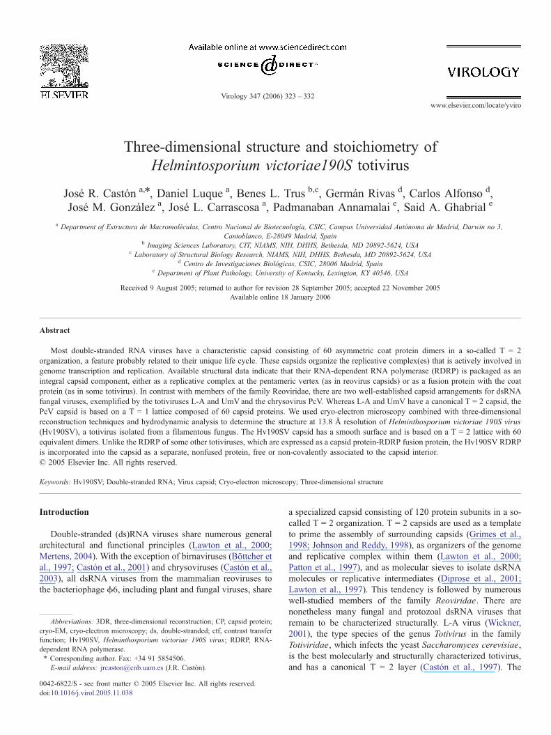
Fig. 1. Analysis of Hv190SVempty particles by SDS-PAGE and cryo-electron micro
vector containing CP coding region (lane 1), and purified Hv190S virions from a nat
SDS-PAGE and stained with Coomassie brilliant blue. Positions of p88, p83 and p78
(lane 3). (B) Cryoelectron micrograph of Hv190SV empty capsids. Scale bar, 50 n
These functional and molecular features make characteriza-
tion of the Hv190SV capsid structure an attractive prospect. We
used cryo-electron microscopy (cryo-EM) combined with
three-dimensional reconstruction (3DR) and complementary
biophysical techniques to determine the structure at 13.8 A
resolution and the protein stoichiometry of the Hv190SV
capsid. We found that Hv190SV, as a representative of
totiviruses that infect filamentous fungi (forming a separate
cluster by phylogenetic analysis), shares the canonical T = 2
capsid with totiviruses that infect yeast and possibly those that
infect parasitic protozoa. Comparative analyses of L-A and
Hv190SV CP nonetheless suggest a new arrangement of the
Hv190SV capsid protein domains.
Results
Virions and virus-like particle purification
Hv190SV virions and empty capsids were purified after two
cycles of differential centrifugation and linear sucrose density
gradient centrifugation (see Materials and methods). Whereas
full virions were purified from a naturally infected fungal
isolate, Hv190SV empty capsids were expressed in a virus-free
fungal host transformed with a CP recombinant vector
(pCB190S-mt; Annamalai and Ghabrial, unpublished results).
In both cases, homogeneous populations of particles were
obtained, as tested in Coomasssie blue stained SDS-PAGE gels
(Fig. 1A). Fractions enriched in Hv190SV particles were
identical and consisted of two major polypeptides (p88 and
p78) in equimolar amounts and a minor polypeptide (p83). As
predicted, no other minor proteins were detected in full virion
preparations, although viral RDRP is known to be present. The
final yield of purified 190SV virions from the naturally
infected isolate is generally low, since two cycles of density
gradient centrifugation are required to remove the chrysovirus
Hv145S, commonly associated with Hv190SV (Sanderlin and
Ghabrial, 1978). A substantially higher yield (20–50 fold) of
scopy. (A) Purified empty particles from a virus-free isolate transformed with a
urally infected fungal isolate (lane 2) were analyzed for protein content by 7.5%
are indicated. The apparent molecular weights of protein standards are indicated
m.
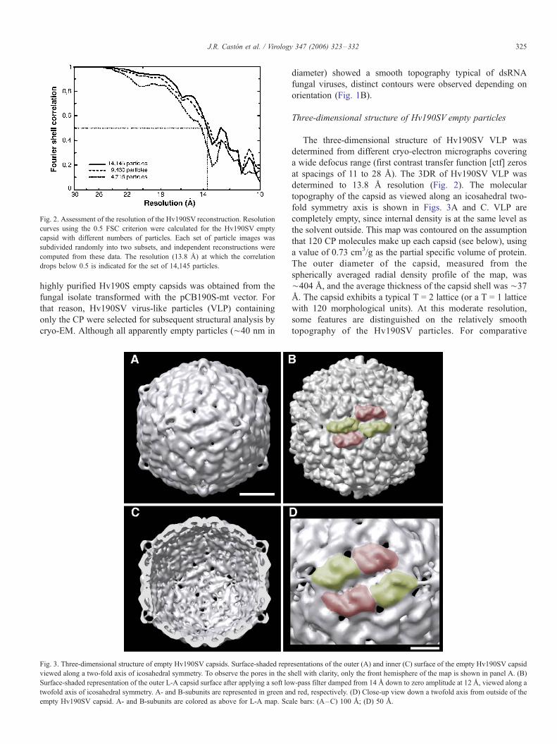
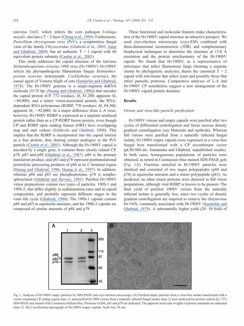
Fig. 2. Assessment of the resolution of the Hv190SV reconstruction. Resolution
curves using the 0.5 FSC criterion were calculated for the Hv190SV empty
capsid with different numbers of particles. Each set of particle images was
subdivided randomly into two subsets, and independent reconstructions were
computed from these data. The resolution (13.8 A) at which the correlation
drops below 0.5 is indicated for the set of 14,145 particles.
J.R. Caston et al. / Virology 347 (2006) 323–332 325
highly purified Hv190S empty capsids was obtained from the
fungal isolate transformed with the pCB190S-mt vector. For
that reason, Hv190SV virus-like particles (VLP) containing
only the CP were selected for subsequent structural analysis by
cryo-EM. Although all apparently empty particles (¨40 nm in
Fig. 3. Three-dimensional structure of empty Hv190SV capsids. Surface-shaded repr
viewed along a two-fold axis of icosahedral symmetry. To observe the pores in the s
Surface-shaded representation of the outer L-A capsid surface after applying a soft lo
twofold axis of icosahedral symmetry. A- and B-subunits are represented in green an
empty Hv190SV capsid. A- and B-subunits are colored as above for L-A map. Sca
diameter) showed a smooth topography typical of dsRNA
fungal viruses, distinct contours were observed depending on
orientation (Fig. 1B).
Three-dimensional structure of Hv190SV empty particles
The three-dimensional structure of Hv190SV VLP was
determined from different cryo-electron micrographs covering
a wide defocus range (first contrast transfer function [ctf] zeros
at spacings of 11 to 28 A). The 3DR of Hv190SV VLP was
determined to 13.8 A resolution (Fig. 2). The molecular
topography of the capsid as viewed along an icosahedral two-
fold symmetry axis is shown in Figs. 3A and C. VLP are
completely empty, since internal density is at the same level as
the solvent outside. This map was contoured on the assumption
that 120 CP molecules make up each capsid (see below), using
a value of 0.73 cm3/g as the partial specific volume of protein.
The outer diameter of the capsid, measured from the
spherically averaged radial density profile of the map, was
¨404 A, and the average thickness of the capsid shell was ¨37
A. The capsid exhibits a typical T = 2 lattice (or a T = 1 lattice
with 120 morphological units). At this moderate resolution,
some features are distinguished on the relatively smooth
topography of the Hv190SV particles. For comparative
esentations of the outer (A) and inner (C) surface of the empty Hv190SV capsid
hell with clarity, only the front hemisphere of the map is shown in panel A. (B)
w-pass filter damped from 14 A down to zero amplitude at 12 A, viewed along a
d red, respectively. (D) Close-up view down a twofold axis from outside of the
le bars: (A–C) 100 A; (D) 50 A.
J.R. Caston et al. / Virology 347 (2006) 323–332326
purposes, a three-dimensional map of L-A virus was calculated
from the atomic structure coordinates (Naitow et al., 2002)
filtered to ¨14 A resolution (Fig. 3B). L-A and Hv190SV
capsids show relatively rougher and smoother outer surfaces,
respectively. From an structural point of view, the Bluetonge
virus core (BTV core), which is also based on a T = 2 lattice
(Grimes et al., 1998), shares a similar smooth outer surface
with the Hv190SV capsid, even though both viruses are
phylogenetically distant (not shown).
Considering the comparison with L-A (and other T = 2
capsids), and favoring the more compact arrangement, we thus
proposed the tentative placement of subunits on this surface
lattice (indexed by colors in Fig. 3D). The outer surface of the
pentamers is formed of five elongated structures subdivided in
two similar bean-like halves (A-subunits in green), with
another five comparable subunits partially intercalated (B-
subunits in red), but with distinct morphology. Numerous holes
may perforate the shell as in the L-A capsid, and the major
holes at the five-fold axis are partially occluded by a density.
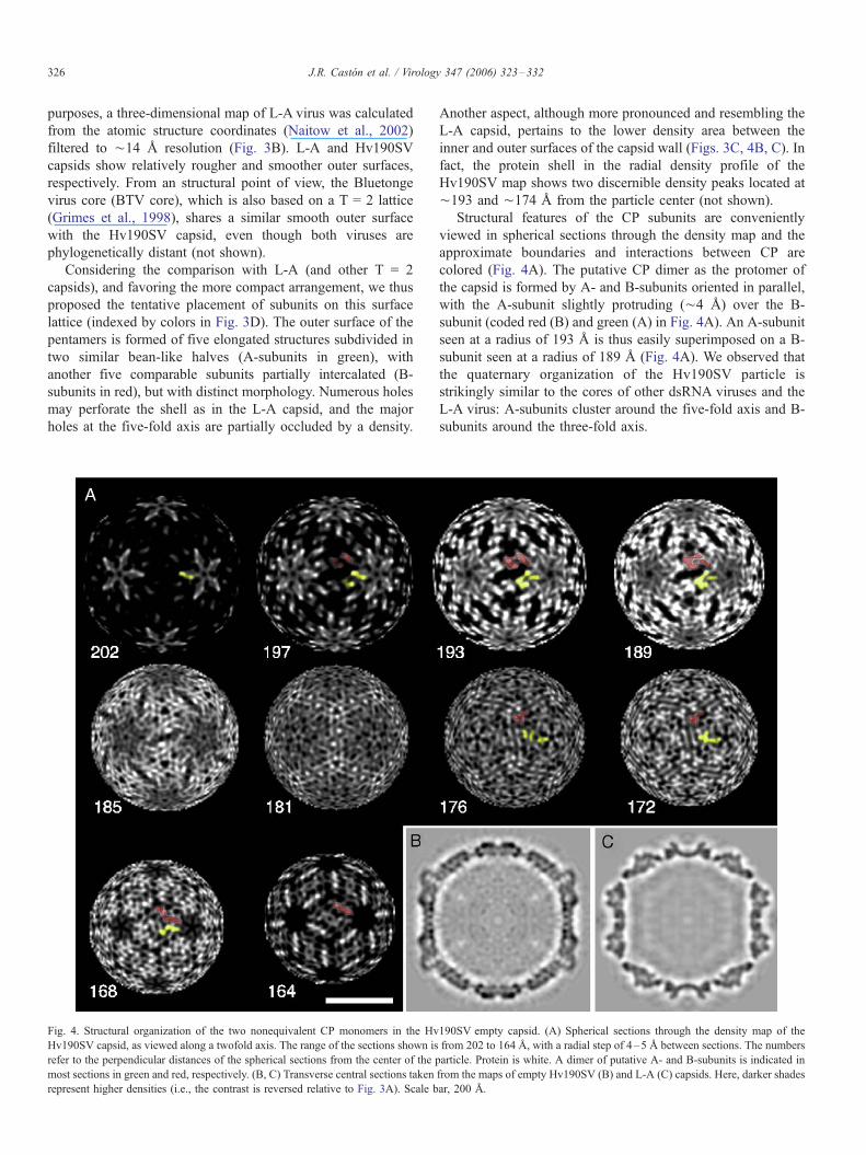
Fig. 4. Structural organization of the two nonequivalent CP monomers in the Hv
Hv190SV capsid, as viewed along a twofold axis. The range of the sections shown is
refer to the perpendicular distances of the spherical sections from the center of the p
most sections in green and red, respectively. (B, C) Transverse central sections taken
represent higher densities (i.e., the contrast is reversed relative to Fig. 3A). Scale b
Another aspect, although more pronounced and resembling the
L-A capsid, pertains to the lower density area between the
inner and outer surfaces of the capsid wall (Figs. 3C, 4B, C). In
fact, the protein shell in the radial density profile of the
Hv190SV map shows two discernible density peaks located at
¨193 and ¨174 A from the particle center (not shown).
Structural features of the CP subunits are conveniently
viewed in spherical sections through the density map and the
approximate boundaries and interactions between CP are
colored (Fig. 4A). The putative CP dimer as the protomer of
the capsid is formed by A- and B-subunits oriented in parallel,
with the A-subunit slightly protruding (¨4 A) over the B-
subunit (coded red (B) and green (A) in Fig. 4A). An A-subunit
seen at a radius of 193 A is thus easily superimposed on a B-
subunit seen at a radius of 189 A (Fig. 4A). We observed that
the quaternary organization of the Hv190SV particle is
strikingly similar to the cores of other dsRNA viruses and the
L-A virus: A-subunits cluster around the five-fold axis and B-
subunits around the three-fold axis.
190SV empty capsid. (A) Spherical sections through the density map of the
from 202 to 164 A, with a radial step of 4–5 A between sections. The numbers
article. Protein is white. A dimer of putative A- and B-subunits is indicated in
from the maps of empty Hv190SV (B) and L-A (C) capsids. Here, darker shades
ar, 200 A.
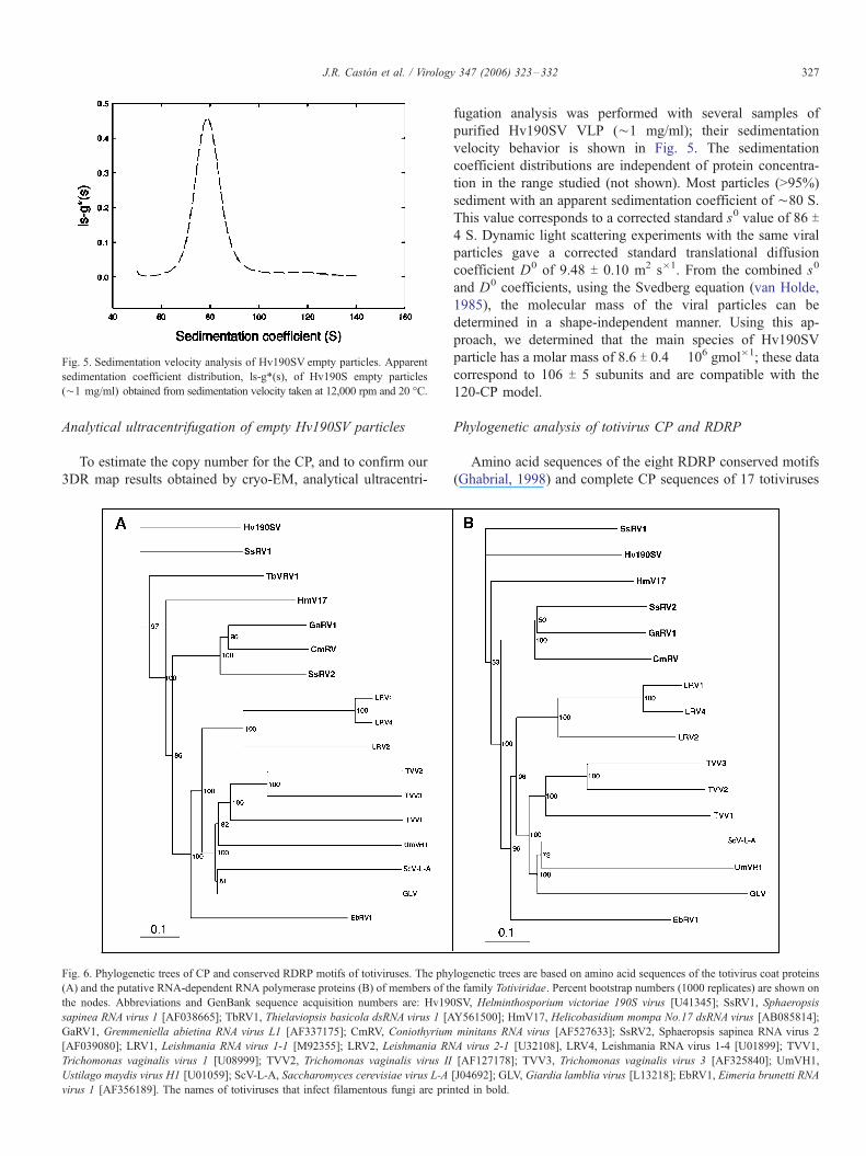
Fig. 5. Sedimentation velocity analysis of Hv190SV empty particles. Apparent
sedimentation coefficient distribution, ls-g*(s), of Hv190S empty particles
(¨1 mg/ml) obtained from sedimentation velocity taken at 12,000 rpm and 20 -C.
J.R. Caston et al. / Virology 347 (2006) 323–332 327
Analytical ultracentrifugation of empty Hv190SV particles
To estimate the copy number for the CP, and to confirm our
3DR map results obtained by cryo-EM, analytical ultracentri-
Fig. 6. Phylogenetic trees of CP and conserved RDRP motifs of totiviruses. The phy
(A) and the putative RNA-dependent RNA polymerase proteins (B) of members of th
the nodes. Abbreviations and GenBank sequence acquisition numbers are: Hv19
sapinea RNA virus 1 [AF038665]; TbRV1, Thielaviopsis basicola dsRNA virus 1 [A
GaRV1, Gremmeniella abietina RNA virus L1 [AF337175]; CmRV, Coniothyrium
[AF039080]; LRV1, Leishmania RNA virus 1-1 [M92355]; LRV2, Leishmania RN
Trichomonas vaginalis virus 1 [U08999]; TVV2, Trichomonas vaginalis virus II
Ustilago maydis virus H1 [U01059]; ScV-L-A, Saccharomyces cerevisiae virus L-A
virus 1 [AF356189]. The names of totiviruses that infect filamentous fungi are prin
fugation analysis was performed with several samples of
purified Hv190SV VLP (¨1 mg/ml); their sedimentation
velocity behavior is shown in Fig. 5. The sedimentation
coefficient distributions are independent of protein concentra-
tion in the range studied (not shown). Most particles (>95%)
sediment with an apparent sedimentation coefficient of ¨80 S.
This value corresponds to a corrected standard s0 value of 86 T4 S. Dynamic light scattering experiments with the same viral
particles gave a corrected standard translational diffusion
coefficient D0 of 9.48 T 0.10 m2 s�1. From the combined s0
and D0 coefficients, using the Svedberg equation (van Holde,
1985), the molecular mass of the viral particles can be
determined in a shape-independent manner. Using this ap-
proach, we determined that the main species of Hv190SV
particle has a molar mass of 8.6 T 0.4 � 106 gmol�1; these data
correspond to 106 T 5 subunits and are compatible with the
120-CP model.
Phylogenetic analysis of totivirus CP and RDRP
Amino acid sequences of the eight RDRP conserved motifs
(Ghabrial, 1998) and complete CP sequences of 17 totiviruses
logenetic trees are based on amino acid sequences of the totivirus coat proteins
e family Totiviridae. Percent bootstrap numbers (1000 replicates) are shown on
0SV, Helminthosporium victoriae 190S virus [U41345]; SsRV1, Sphaeropsis
Y561500]; HmV17, Helicobasidium mompa No.17 dsRNA virus [AB085814];
minitans RNA virus [AF527633]; SsRV2, Sphaeropsis sapinea RNA virus 2
A virus 2-1 [U32108], LRV4, Leishmania RNA virus 1-4 [U01899]; TVV1,
[AF127178]; TVV3, Trichomonas vaginalis virus 3 [AF325840]; UmVH1,
[J04692]; GLV, Giardia lamblia virus [L13218]; EbRV1, Eimeria brunetti RNA
ted in bold.
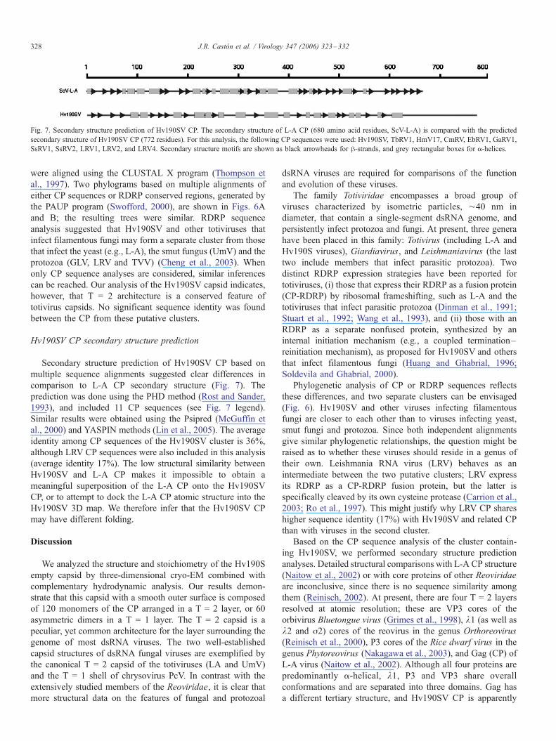
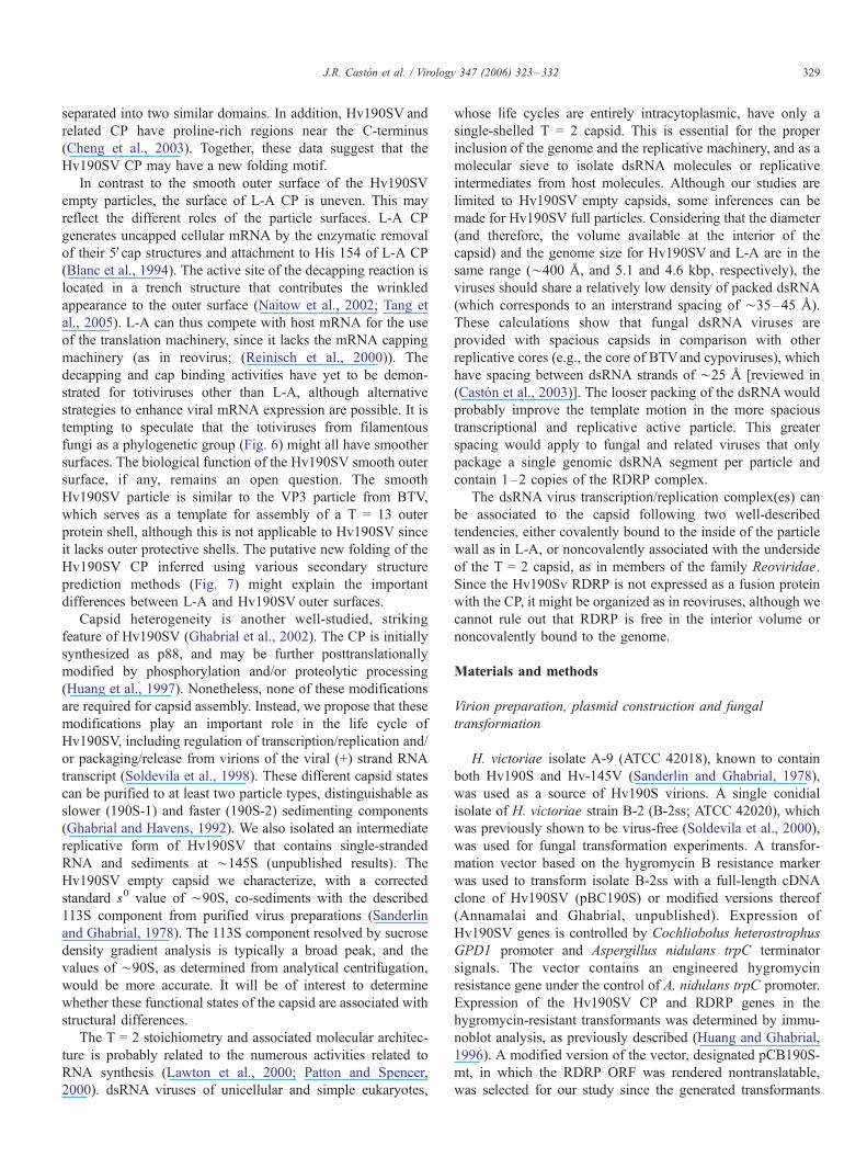
Fig. 7. Secondary structure prediction of Hv190SV CP. The secondary structure of L-A CP (680 amino acid residues, ScV-L-A) is compared with the predicted
secondary structure of Hv190SV CP (772 residues). For this analysis, the following CP sequences were used: Hv190SV, TbRV1, HmV17, CmRV, EbRV1, GaRV1,
SsRV1, SsRV2, LRV1, LRV2, and LRV4. Secondary structure motifs are shown as black arrowheads for h-strands, and grey rectangular boxes for a-helices.
J.R. Caston et al. / Virology 347 (2006) 323–332328
were aligned using the CLUSTAL X program (Thompson et
al., 1997). Two phylograms based on multiple alignments of
either CP sequences or RDRP conserved regions, generated by
the PAUP program (Swofford, 2000), are shown in Figs. 6A
and B; the resulting trees were similar. RDRP sequence
analysis suggested that Hv190SV and other totiviruses that
infect filamentous fungi may form a separate cluster from those
that infect the yeast (e.g., L-A), the smut fungus (UmV) and the
protozoa (GLV, LRV and TVV) (Cheng et al., 2003). When
only CP sequence analyses are considered, similar inferences
can be reached. Our analysis of the Hv190SV capsid indicates,
however, that T = 2 architecture is a conserved feature of
totivirus capsids. No significant sequence identity was found
between the CP from these putative clusters.
Hv190SV CP secondary structure prediction
Secondary structure prediction of Hv190SV CP based on
multiple sequence alignments suggested clear differences in
comparison to L-A CP secondary structure (Fig. 7). The
prediction was done using the PHD method (Rost and Sander,
1993), and included 11 CP sequences (see Fig. 7 legend).
Similar results were obtained using the Psipred (McGuffin et
al., 2000) and YASPIN methods (Lin et al., 2005). The average
identity among CP sequences of the Hv190SV cluster is 36%,
although LRV CP sequences were also included in this analysis
(average identity 17%). The low structural similarity between
Hv190SV and L-A CP makes it impossible to obtain a
meaningful superposition of the L-A CP onto the Hv190SV
CP, or to attempt to dock the L-A CP atomic structure into the
Hv190SV 3D map. We therefore infer that the Hv190SV CP
may have different folding.
Discussion
We analyzed the structure and stoichiometry of the Hv190S
empty capsid by three-dimensional cryo-EM combined with
complementary hydrodynamic analysis. Our results demon-
strate that this capsid with a smooth outer surface is composed
of 120 monomers of the CP arranged in a T = 2 layer, or 60
asymmetric dimers in a T = 1 layer. The T = 2 capsid is a
peculiar, yet common architecture for the layer surrounding the
genome of most dsRNA viruses. The two well-established
capsid structures of dsRNA fungal viruses are exemplified by
the canonical T = 2 capsid of the totiviruses (LA and UmV)
and the T = 1 shell of chrysovirus PcV. In contrast with the
extensively studied members of the Reoviridae, it is clear that
more structural data on the features of fungal and protozoal
dsRNA viruses are required for comparisons of the function
and evolution of these viruses.
The family Totiviridae encompasses a broad group of
viruses characterized by isometric particles, ¨40 nm in
diameter, that contain a single-segment dsRNA genome, and
persistently infect protozoa and fungi. At present, three genera
have been placed in this family: Totivirus (including L-A and
Hv190S viruses), Giardiavirus, and Leishmaniavirus (the last
two include members that infect parasitic protozoa). Two
distinct RDRP expression strategies have been reported for
totiviruses, (i) those that express their RDRP as a fusion protein
(CP-RDRP) by ribosomal frameshifting, such as L-A and the
totiviruses that infect parasitic protozoa (Dinman et al., 1991;
Stuart et al., 1992; Wang et al., 1993), and (ii) those with an
RDRP as a separate nonfused protein, synthesized by an
internal initiation mechanism (e.g., a coupled termination–
reinitiation mechanism), as proposed for Hv190SV and others
that infect filamentous fungi (Huang and Ghabrial, 1996;
Soldevila and Ghabrial, 2000).
Phylogenetic analysis of CP or RDRP sequences reflects
these differences, and two separate clusters can be envisaged
(Fig. 6). Hv190SV and other viruses infecting filamentous
fungi are closer to each other than to viruses infecting yeast,
smut fungi and protozoa. Since both independent alignments
give similar phylogenetic relationships, the question might be
raised as to whether these viruses should reside in a genus of
their own. Leishmania RNA virus (LRV) behaves as an
intermediate between the two putative clusters; LRV express
its RDRP as a CP-RDRP fusion protein, but the latter is
specifically cleaved by its own cysteine protease (Carrion et al.,
2003; Ro et al., 1997). This might justify why LRV CP shares
higher sequence identity (17%) with Hv190SV and related CP
than with viruses in the second cluster.
Based on the CP sequence analysis of the cluster contain-
ing Hv190SV, we performed secondary structure prediction
analyses. Detailed structural comparisons with L-A CP structure
(Naitow et al., 2002) or with core proteins of other Reoviridae
are inconclusive, since there is no sequence similarity among
them (Reinisch, 2002). At present, there are four T = 2 layers
resolved at atomic resolution; these are VP3 cores of the
orbivirus Bluetongue virus (Grimes et al., 1998), k1 (as well as
k2 and j2) cores of the reovirus in the genus Orthoreovirus
(Reinisch et al., 2000), P3 cores of the Rice dwarf virus in the
genus Phytoreovirus (Nakagawa et al., 2003), and Gag (CP) of
L-A virus (Naitow et al., 2002). Although all four proteins are
predominantly a-helical, k1, P3 and VP3 share overall
conformations and are separated into three domains. Gag has
a different tertiary structure, and Hv190SV CP is apparently
J.R. Caston et al. / Virology 347 (2006) 323–332 329
separated into two similar domains. In addition, Hv190SV and
related CP have proline-rich regions near the C-terminus
(Cheng et al., 2003). Together, these data suggest that the
Hv190SV CP may have a new folding motif.
In contrast to the smooth outer surface of the Hv190SV
empty particles, the surface of L-A CP is uneven. This may
reflect the different roles of the particle surfaces. L-A CP
generates uncapped cellular mRNA by the enzymatic removal
of their 5Vcap structures and attachment to His 154 of L-A CP
(Blanc et al., 1994). The active site of the decapping reaction is
located in a trench structure that contributes the wrinkled
appearance to the outer surface (Naitow et al., 2002; Tang et
al., 2005). L-A can thus compete with host mRNA for the use
of the translation machinery, since it lacks the mRNA capping
machinery (as in reovirus; (Reinisch et al., 2000)). The
decapping and cap binding activities have yet to be demon-
strated for totiviruses other than L-A, although alternative
strategies to enhance viral mRNA expression are possible. It is
tempting to speculate that the totiviruses from filamentous
fungi as a phylogenetic group (Fig. 6) might all have smoother
surfaces. The biological function of the Hv190SV smooth outer
surface, if any, remains an open question. The smooth
Hv190SV particle is similar to the VP3 particle from BTV,
which serves as a template for assembly of a T = 13 outer
protein shell, although this is not applicable to Hv190SV since
it lacks outer protective shells. The putative new folding of the
Hv190SV CP inferred using various secondary structure
prediction methods (Fig. 7) might explain the important
differences between L-A and Hv190SV outer surfaces.
Capsid heterogeneity is another well-studied, striking
feature of Hv190SV (Ghabrial et al., 2002). The CP is initially
synthesized as p88, and may be further posttranslationally
modified by phosphorylation and/or proteolytic processing
(Huang et al., 1997). Nonetheless, none of these modifications
are required for capsid assembly. Instead, we propose that these
modifications play an important role in the life cycle of
Hv190SV, including regulation of transcription/replication and/
or packaging/release from virions of the viral (+) strand RNA
transcript (Soldevila et al., 1998). These different capsid states
can be purified to at least two particle types, distinguishable as
slower (190S-1) and faster (190S-2) sedimenting components
(Ghabrial and Havens, 1992). We also isolated an intermediate
replicative form of Hv190SV that contains single-stranded
RNA and sediments at ¨145S (unpublished results). The
Hv190SV empty capsid we characterize, with a corrected
standard s0 value of ¨90S, co-sediments with the described
113S component from purified virus preparations (Sanderlin
and Ghabrial, 1978). The 113S component resolved by sucrose
density gradient analysis is typically a broad peak, and the
values of ¨90S, as determined from analytical centrifugation,
would be more accurate. It will be of interest to determine
whether these functional states of the capsid are associated with
structural differences.
The T = 2 stoichiometry and associated molecular architec-
ture is probably related to the numerous activities related to
RNA synthesis (Lawton et al., 2000; Patton and Spencer,
2000). dsRNA viruses of unicellular and simple eukaryotes,
whose life cycles are entirely intracytoplasmic, have only a
single-shelled T = 2 capsid. This is essential for the proper
inclusion of the genome and the replicative machinery, and as a
molecular sieve to isolate dsRNA molecules or replicative
intermediates from host molecules. Although our studies are
limited to Hv190SV empty capsids, some inferences can be
made for Hv190SV full particles. Considering that the diameter
(and therefore, the volume available at the interior of the
capsid) and the genome size for Hv190SV and L-A are in the
same range (¨400 A, and 5.1 and 4.6 kbp, respectively), the
viruses should share a relatively low density of packed dsRNA
(which corresponds to an interstrand spacing of ¨35–45 A).
These calculations show that fungal dsRNA viruses are
provided with spacious capsids in comparison with other
replicative cores (e.g., the core of BTVand cypoviruses), which
have spacing between dsRNA strands of ¨25 A [reviewed in
(Caston et al., 2003)]. The looser packing of the dsRNAwould
probably improve the template motion in the more spacious
transcriptional and replicative active particle. This greater
spacing would apply to fungal and related viruses that only
package a single genomic dsRNA segment per particle and
contain 1–2 copies of the RDRP complex.
The dsRNA virus transcription/replication complex(es) can
be associated to the capsid following two well-described
tendencies, either covalently bound to the inside of the particle
wall as in L-A, or noncovalently associated with the underside
of the T = 2 capsid, as in members of the family Reoviridae.
Since the Hv190Sv RDRP is not expressed as a fusion protein
with the CP, it might be organized as in reoviruses, although we
cannot rule out that RDRP is free in the interior volume or
noncovalently bound to the genome.
Materials and methods
Virion preparation, plasmid construction and fungal
transformation
H. victoriae isolate A-9 (ATCC 42018), known to contain
both Hv190S and Hv-145V (Sanderlin and Ghabrial, 1978),
was used as a source of Hv190S virions. A single conidial
isolate of H. victoriae strain B-2 (B-2ss; ATCC 42020), which
was previously shown to be virus-free (Soldevila et al., 2000),
was used for fungal transformation experiments. A transfor-
mation vector based on the hygromycin B resistance marker
was used to transform isolate B-2ss with a full-length cDNA
clone of Hv190SV (pBC190S) or modified versions thereof
(Annamalai and Ghabrial, unpublished). Expression of
Hv190SV genes is controlled by Cochliobolus heterostrophus
GPD1 promoter and Aspergillus nidulans trpC terminator
signals. The vector contains an engineered hygromycin
resistance gene under the control of A. nidulans trpC promoter.
Expression of the Hv190SV CP and RDRP genes in the
hygromycin-resistant transformants was determined by immu-
noblot analysis, as previously described (Huang and Ghabrial,
1996). A modified version of the vector, designated pCB190S-
mt, in which the RDRP ORF was rendered nontranslatable,
was selected for our study since the generated transformants
J.R. Caston et al. / Virology 347 (2006) 323–332330
produced only the CP and the yield of the assembled virus
particles was superior to that of other transformants or naturally
infected fungal isolate (Annamalai and Ghabrial, unpublished
results).
For virion or empty virus particle purification, all fungal
isolates or transformants were grown on potato dextrose broth
containing 0.5% yeast extract. The purification procedure
previously described for Penicillium chrysogenum virus (Jiang
and Ghabrial, 2004) was followed with the final purification
step made by rate zonal centrifugation in sucrose density
gradients (100–400 mg ml�1). All steps were carried out in
buffer A (50 mM Tris–HCl buffer, pH 7.8, containing 5 mM
EDTA and 150 mM NaCl). Selected fractions containing the
Hv190SV particles were used for SDS-PAGE and electron
microscopy analyses.
Cryo-electron microscopy
Samples of fractions containing Hv190SV empty capsids
were diluted until a uniform distribution of particles was
observed (when examined by negative staining). Samples (5
Al drops) were applied to one side of a holey carbon film,
washed twice on buffer A drops, blotted, and plunged into a
liquid ethane bath following standard procedures, as described
(Caston et al., 2001; Saugar et al., 2005). Micrographs were
recorded under minimal exposure conditions so that the
specimens imaged received exposures of 6–10 e�/A2, at
nominal magnification of 50,000� (calibrated using the
40.5A axial spacing of bacteriophage T4 tail-sheath) on a
Tecnai G2 electron microscope operating at 200 kV and
equipped with a field emission gun.
Image analysis
General image processing operations were performed using
PIC Software (Trus et al., 1996). Micrographs were assessed
for resolution and astigmatism by computer Fourier analysis,
and defocus values estimated from the positions of the first
zero of the ctf. For the 30 selected micrographs analyzed
(which includes 15,717 particles), defocus values, determined
with Bsoft package (Heymann, 2001), ranged from 0.6 to 3.0
Am (first ctf zeros at spacings of 11–28 A, respectively).
Suitable micrographs were scanned with a Zeiss PhotoScan TD
scanner at 7 Am/pixel and binned to give 21 Am/pixels (4.2A at
the specimen). Particle images were extracted and preprocessed
with X3dp (Conway et al., 1993). Initial estimates of
orientation angles were obtained for a base set of particles by
the Polar Fourier Transform (PFT) algorithm (Baker and
Cheng, 1996), taking as starting model the three-dimensional
map of empty L-A particles, band-limited to 35 A resolution
and appropriately scaled (Cheng et al., 1994). A new density
map was calculated and used for all subsequent orientation and
origin refinements, using a modified version of the PFT
algorithm altered to use both phase and amplitude information
(D.M. Belnap et al., unpublished). Phases were ctf corrected by
simply flipping the phases in the required lobes of the ctf.
Enhancement of the high-resolution Fourier amplitudes was
based on X-ray data from L-A viral capsid (Naitow et al.,
2002). The decay of amplitudes was calculated using the
spatial frequency components from the Hv190SV map (with an
effective resolution of 13.8 A) and the X-ray map of L-A virus.
The decay profile of the Hv190SV map was then adjusted to
match the profile from the X-ray map, and the fitted function
was applied to the Hv190SV map between the frequency range
245 to 12 A. Finally, a soft low-pass filter was applied between
14 and 12A. This is analogous to adjusting the temperature
factor of the two structures to be similar.
Reconstructions were calculated using Fourier–Bessel
techniques (Crowther, 1971). The final reconstruction com-
bined 14,152 images (90% of the total particles), and achieved
resolution by the Fourier shell correlation (FSC) criterion (0.5
threshold) of 13.8 A. Although only two other tests are shown
in Fig. 2 (60%–9430 particles–and 30%–4715 particles),
additional tests were calculated, from 20% to 80% (data not
shown), but no improvement was observed.
The L-A X-ray three-dimensional map was generated by
VIPER (Reddy et al., 2001) (http://mmtsb.scripps.edu/viper/
viper.html), and the resolution of the three-dimensional map
was filtered to 14 A by using the Spider software package. The
central section of the L-A map (Fig. 4C) was made with a ctf
imposed by scaling its one-dimensional power spectrum to the
Hv190SV capsid map section’s one-dimensional power spec-
trum. A two-dimensional FFT was calculated for the central
section of Hv190SV map, and a one-dimensional radial
average was calculated. This one-dimensional radial average
is representative of the power spectrum of the electron
microscope ctf. The same operations were done on the central
section from the filtered L-A X-ray map. The one-dimensional
power spectrum of the Hv190SV map was divided by that of
the X-ray map generating a set of scaling factors, which when
appropriately multiplied times the two-dimensional FFT of the
LA map imposed the electron microscope ctf of HV190SVonto
LA. This scaling method assumes that the signal component of
each power spectrum is roughly the same, and that differences
in the power spectrum represent primarily the electron
microscope’s ctf. The inverse FFT yielded the image shown
in Fig. 4C.
Three-dimensional model images were produced using the
UCSF Chimera package (Pettersen et al., 2004).
Hydrodynamic analysis of Hv190SV empty particles
Sedimentation velocity
The experiments were done using viral particles (loading
concentration ranging from 0.5 to 5 mg/ml) equilibrated in
buffer A. The sedimentation velocity runs were carried out at
12000 rpm and 20 -C in an XL-I analytical ultracentrifuge
(Beckman-Coulter Inc.) with the interference optics detection
system, using an An60Ti rotor and 12 mm double-sector
centrepieces. The sedimentation coefficient distributions were
calculated by least-squares boundary modeling of sedimenta-
tion velocity data using the ls � g*(s) method (Schuck and
Rossmanith, 2000), as implemented in the SEDFIT program.
These experimental sedimentation coefficients were corrected
J.R. Caston et al. / Virology 347 (2006) 323–332 331
to standard conditions (water, 20 -C, and infinite dilution; (van
Holde, 1985)) using the SEDNTERP program (Laue et al.,
1992) to get the corresponding s0 values. The latter program
was also used to calculate the partial specific volume of
Hv190S (0.729 ml/g) from its amino acid composition (Huang
and Ghabrial, 1996).
Dynamic light scattering
The DLS experiments were performed at 20 -C in a
DynaPro-MS/X instrument (Protein Solutions Inc.), using
two concentrations of viral particles (0.5 and 1 mg/ml). The
translational diffusion coefficients (D) of the viral particles
were determined from the scattering data with the DYNAMICS
autocorrelation analysis software (version 6, Protein Solutions
Inc.). These experimental values were corrected for buffer and
concentration (van Holde, 1985) to get the standard D0
coefficients.
Acknowledgments
We thank T. Baker (University of California, San Diego), D.
Belnap (Brigham Young University), J. Conway (IBS, Gre-
noble), J. B. Heymann, and A.C. Steven (NIH) for sharing their
reconstruction software, W. Havens (University of Kentucky)
for technical assistance, A. Valencia (CNB, Madrid) for useful
discussion, and C. Mark for editorial assistance. JRC holds a
contract from the ‘‘Ramon y Cajal’’ program (MCYT). This
work was supported by Grant BMC2002-00996 from the
MCYT, and in part by the Intramural Program of the NIH, CIT.
References
Baker, T.S., Cheng, R.H., 1996. A model-based approach for determining
orientations of biological macromolecules imaged by cryoelectron micros-
copy. J. Struct. Biol. 116 (1), 120–130.
Blanc, A., Ribas, J.C., Wickner, R.B., Sonenberg, N., 1994. His-154 is
involved in the linkage of the Saccharomyces cerevisiae L-A double-
stranded RNA virus Gag protein to the cap structure of mRNAs and is
essential for M1 satellite virus expression. Mol. Cell. Biol. 14 (4),
2664–2674.
Bottcher, B., Kiselev, N., Stel’Mashchuk, V., Perevozchikova, N., Borisov, A.,
Crowther, R., 1997. Three-dimensional structure of infectious bursal
disease virus determined by electron cryomicroscopy. J. Virol. 71 (1),
325–330.
Carrion Jr., R., Ro, Y.T., Patterson, J.L., 2003. Purification, identification,
and biochemical characterization of a host-encoded cysteine protease
that cleaves a Leishmaniavirus gag-pol polyprotein. J. Virol. 77 (19),
10448–10455.
Caston, J.R., Trus, B.L., Booy, F.P., Wickner, R.B., Wall, J.S., Steven, A.C.,
1997. Structure of L-A virus: a specialized compartment for the
transcription and replication of double-stranded RNA. J. Cell Biol. 138
(5), 975–985.
Caston, J.R., Martınez-Torrecuadrada, J.L., Maraver, A., Lombardo, E.,
Rodrıguez, J.F., Casal, J.I., Carrascosa, J.L., 2001. C terminus of Infectious
bursal disease virus major capsid protein VP2 is involved in definition of
the T number for capsid assembly. J. Virol. 75 (22), 10815–10828.
Caston, J.R., Ghabrial, S.A., Jiang, D., Rivas, G., Alfonso, C., Roca, R., Luque,
D., Carrascosa, J.L., 2003. Three-dimensional structure of Penicillium
chrysogenum virus: a double-stranded RNA virus with a genuine T = 1
capsid. J. Mol. Biol. 331 (2), 417–431.
Cheng, R.H., Caston, J.R., Wang, G.-j., Gu, F., Smith, T.J., Baker, T.S.,
Bozarth, R.F., Trus, B.L., Cheng, N., Wickner, R.B., Steven, A.C., 1994.
Fungal virus capsids, cytoplasmic compartments for the replication of
double-stranded RNA, formed as icosahedral shells of asymmetric gag
dimers. J. Mol. Biol. 244 (3), 255–258.
Cheng, J., Jiang, D., Fu, Y., Li, G., Peng, Y., Ghabrial, S.A., 2003. Molecular
characterization of a dsRNA totivirus infecting the sclerotial parasite
Coniothyrium minitans. Virus Res. 93 (1), 41–50.
Conway, J.F., Trus, B.L., Booy, F.P., Newcomb, W.W., Brown, J.C., Steven,
A.C., 1993. The effects of radiation damage on the structure of frozen
hydrated HSV-1 capsids. J. Struct. Biol. 111 (3), 222–233.
Crowther, R.A., 1971. Procedures for three-dimensional reconstruction of
spherical viruses by Fourier synthesis from electron micrographs. Philos.
Trans. R. Soc., Ser. B 261, 221–230.
Dinman, J.D., Icho, T., Wickner, R.B., 1991. A-1 ribosomal frameshift in a
double-stranded RNA virus of yeast forms a gag-pol fusion protein. Proc.
Natl. Acad. Sci. U.S.A. 88 (1), 174–178.
Diprose, J.M., Burroughs, J.N., Sutton, G.C., Goldsmith, A., Gouet, P.,
Malby, R., Overton, I., Zientara, S., Mertens, P.P., Stuart, D.I., Grimes,
J.M., 2001. Translocation portals for the substrates and products of a viral
transcription complex: the bluetongue virus core. EMBO J. 20 (24),
7229–7239.
Ghabrial, S.A., 1994. New developments in fungal virology. Adv. Virus Res.
43, 303–388.
Ghabrial, S.A., 1998. Origin, adaptation and evolutionary pathways of fungal
viruses. Virus Genes 16 (1), 119–131.
Ghabrial, S.A., Havens, W.M., 1992. The Helminthosporium victoriae 190S
mycovirus has two forms distinguishable by capsid protein composition and
phosphorylation state. Virology 188 (2), 657–665.
Ghabrial, S.A., Bibb, J.A., Price, K.H., Havens, W.M., Lesnaw, J.A., 1987. The
capsid polypeptides of the 190S virus of Helminthosporium victoriae.
J. Gen. Virol. 68 (7), 1791–1800.
Ghabrial, S.A., Soldevila, A.I., Havens, W.M., 2002. Molecular genetics of the
viruses infecting the plant pathogenic fungus Helminthosporium victoriae.
In: Tavanntzis, S. (Ed.), dsRNA Genetic Elements. CRC Press, Boca Raton,
pp. 213–236.
Ghabrial, S.A., Jiang, D., Caston, J.R., 2005. Chrysoviridae. In: Fauquet, C.M.,
Mayo, M.A., Maniloff, J., Desselberger, U., Ball, L.A. (Eds.), Virus
Taxonomy, VIIIth Report of the International Committee on Taxonomy of
Viruses. Elsevier/Academic Press, London, pp. 591–595.
Grimes, J.M., Burroughs, J.N., Gouet, P., Diprose, J.M., Malby, R., Zientara, S.,
Mertens, P.P., Stuart, D.I., 1998. The atomic structure of the Bluetongue
virus core. Nature 395 (6701), 470–478.
Heymann, J.B., 2001. Bsoft: image and molecular processing in electron
microscopy. J. Struct. Biol. 133 (2–3), 156–169.
Huang, S., Ghabrial, S.A., 1996. Organization and expression of the double-
stranded RNA genome of Helminthosporium victoriae 190S virus, a
totivirus infecting a plant pathogenic filamentous fungus. Proc. Natl. Acad.
Sci. U.S.A. 93 (22), 12541–12546.
Huang, S., Soldevila, A.I., Webb, B.A., Ghabrial, S.A., 1997. Expression,
assembly, and proteolytic processing of Helminthosporium victoriae 190S
totivirus capsid protein in insect cells. Virology 234 (1), 130–137.
Jiang, D., Ghabrial, S.A., 2004. Molecular characterization of Penicillium
chrysogenum virus: reconsideration of the taxonomy of the genus
Chrysovirus. J. Gen. Virol. 85 (7), 2111–2121.
Johnson, J.E., Reddy, V.S., 1998. Biggest virus molecular structure yet!. Nat.
Struct. Biol. 5 (10), 849–854.
Laue, T.M., Shah, B.D., Ridgeway, T.M., Pelletier, S.L., 1992. Computer-aided
interpretation of analytical sedimentation data for proteins. In: Harding,
S.E., Rowe, A.J., Horton, J.C. (Eds.), Analytical Ultracentrifugation in
Biochemistry and Polymer Science. Royal Society of Chemistry, Cam-
bridge, UK, pp. 90–125.
Lawton, J.A., Estes, M.K., Prasad, B.V., 1997. Three-dimensional visualization
of mRNA release from actively transcribing rotavirus particles. Nat. Struct.
Biol. 4 (2), 118–121.
Lawton, J.A., Estes, M.K., Prasad, B.V., 2000. Mechanism of genome
transcription in segmented dsRNA viruses. Adv. Virus Res. 55, 185–229.
Lin, K., Simossis, V.A., Taylor, W.R., Heringa, J., 2005. A simple and fast
secondary structure prediction method using hidden neural networks.
Bioinformatics 21 (2), 152–159.
J.R. Caston et al. / Virology 347 (2006) 323–332332
McGuffin, L.J., Bryson, K., Jones, D.T., 2000. The PSIPRED protein structure
prediction server. Bioinformatics 16 (4), 404–405.
Mertens, P., 2004. The dsRNA viruses. Virus Res. 101 (1), 3–13.
Naitow, H., Tang, J., Canady, M., Wickner, R.B., Johnson, J.E., 2002. L-Avirus
at 3.4 A resolution reveals particle architecture and mRNA decapping
mechanism. Nat. Struct. Biol. 9 (10), 725–728.
Nakagawa, A., Miyazaki, N., Taka, J., Naitow, H., Ogawa, A., Fujimoto, Z.,
Mizuno, H., Higashi, T., Watanabe, Y., Omura, T., Cheng, R.H., Tsukihara,
T., 2003. The atomic structure of rice dwarf virus reveals the self-assembly
mechanism of component proteins. Structure 11 (10), 1227–1238.
Patton, J.T., Spencer, E., 2000. Genome replication and packaging of
segmented double-stranded RNA viruses. Virology 277 (2), 217–225.
Patton, J.T., Jones, M.T., Kalbach, A.N., He, Y.W., Xiaobo, J., 1997. Rotavirus
RNA polymerase requires the core shell protein to synthesize the double-
stranded RNA genome. J. Virol. 71 (12), 9618–9626.
Pettersen, E.F., Goddard, T.D., Huang, C.C., Couch, G.S., Greenblatt, D.M.,
Meng, E.C., Ferrin, T.E., 2004. UCSF Chimera—a visualization system for
exploratory research and analysis. J. Comput. Chem. 25 (13), 1605–1612.
Reddy, V.S., Natarajan, P., Okerberg, B., Li, K., Damodaran, K.V., Morton,
R.T., Brooks III, C.L., Johnson, J.E., 2001. Virus Particle Explorer
(VIPER), a website for virus capsid structures and their computational
analyses. J. Virol. 75 (24), 11943–11947.
Reinisch, K.M., 2002. The dsRNA Viridae and their catalytic capsids. Nat.
Struct. Biol. 9 (10), 714–716.
Reinisch, K.M., Nibert, M.L., Harrison, S.C., 2000. Structure of the Reovirus
core at 3.6 A resolution. Nature 404 (6781), 960–967.
Ro, Y.T., Scheffter, S.M., Patterson, J.L., 1997. Specific in vitro cleavage of a
Leishmania virus capsid-RNA-dependent RNA polymerase polyprotein by
a host cysteine-like protease. J. Virol. 71 (12), 8983–8990.
Rost, B., Sander, C., 1993. Improved prediction of protein secondary structure
by use of sequence profiles and neural networks. Proc. Natl. Acad. Sci.
U.S.A. 90 (16), 7558–7562.
Sanderlin, R.S., Ghabrial, S.A., 1978. Physicochemical properties of two
distinct types of virus-like particles from Helminthosporium victoriae.
Virology 87 (1), 142–151.
Saugar, I., Luque, D., Ona, A., Rodrıguez, J.F., Carrascosa, J.L., Trus, B.L.,
Caston, J.R., 2005. Structural polymorphism of the major capsid protein of
a double-stranded RNAvirus: an amphipathic a-helix as a molecular switch.
Structure 13 (7), 1007–1017.
Schuck, P., Rossmanith, P., 2000. Determination of the sedimentation
coefficient distribution by least-squares boundary modeling. Biopolymers
54 (5), 328–341.
Soldevila, A.I., Ghabrial, S.A., 2000. Expression of the Totivirus Helminthos-
porium victoriae 190S virus RNA-dependent RNA polymerase from its
downstream open reading frame in dicistronic constructs. J. Virol. 74 (2),
997–1003.
Soldevila, A.I., Huang, S., Ghabrial, S.A., 1998. Assembly of the Hv190S
totivirus capsid is independent of posttranslational modification of the
capsid protein. Virology 251 (2), 327–333.
Soldevila, A.I., Havens, W.M., Ghabrial, S.A., 2000. A cellular protein with an
RNA-binding activity co-purifies with viral dsRNA from mycovirus-
infected Helminthosporium victoriae. Virology 272 (1), 183–190.
Stuart, K.D., Weeks, R., Guilbride, L., Myler, P.J., 1992. Molecular
organization of Leishmania RNA virus 1. Proc. Natl. Acad. Sci. U.S.A.
89 (18), 8596–8600.
Swofford, D.L., 2000. PAUP: Phylogenetic Analysis Using Parsimony and
Other Methods (software). Sinaur Associates, Sunderland, MA.
Tang, J., Naitow, H., Gardner, N.A., Kolesar, A., Tang, L., Wickner,
R.B., Johnson, J.E., 2005. The structural basis of recognition and
removal of cellular mRNA 7-methyl G Fcaps_ by a viral capsid
protein: a unique viral response to host defense. J. Mol. Recognit. 18
(2), 158–168.
Thompson, J.D., Gibson, T.J., Plewniak, F., Jeanmougin, F., Higgins, D.G.,
1997. The CLUSTAL_X windows interface: flexible strategies for multiple
sequence alignment aided by quality analysis tools. Nucleic Acids Res. 25
(24), 4876–4882.
Trus, B.L., Kocsis, E., Conway, J.F., Steven, A.C., 1996. Digital image
processing of electron micrographs: the PIC system—III. J. Struct. Biol.
116 (1), 61–67.
van Holde, K., 1985. Physical Biochemistry. Englewood Cliffs, N.J.
Wang, A.L., Yang, H.M., Shen, K.A., Wang, C.C., 1993. Giardiavirus double-
stranded RNA genome encodes a capsid polypeptide and a gag-pol-like
fusion protein by a translation frameshift. Proc. Natl. Acad. Sci. U.S.A. 90
(18), 8595–8599.
Wickner, R.B., 2001. Viruses of yeast, fungi, and parasitic microorganisms. In:
Knipe, D.M., Howley, P.M., Griffin, D.E., Martin, M.A., Lamb, R.A.,
Roizman, B., Strauss, S.E. (Eds.), Fields. Virology 4th ed. Lippincott
Williams and Wilkins, Philadelphia, pp. 629–658.
Related Documents