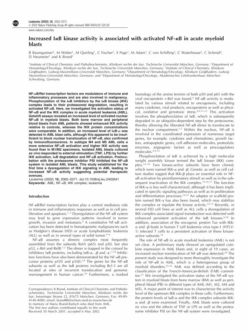
Leukemia (2002) 16, 2062–2071 2002 Nature Publishing Group All rights reserved 0887-6924/02 $25.00 www.nature.com/leu Increased IB kinase activity is associated with activated NF-B in acute myeloid blasts B Baumgartner 1 , M Weber 1 , M Quirling 1 , C Fischer 1 , S Page 1 , M Adam 3 , C von Schilling 2 , C Waterhouse 5 , C Schmid 4 , D Neumeier 1 and K Brand 1 1 Institute of Clinical Chemistry and Pathobiochemistry, Klinikum rechts der Isar, Technische Universita ¨t Mu ¨ nchen, Germany; 2 Department of Hematology/Oncology, Klinikum rechts der Isar, Technische Universita ¨t Mu ¨ nchen, Germany; 3 Institute of Clinical Chemistry, Klinikum Grohadern, Ludwig-Maximillians-Universita ¨t Mu ¨ nchen, Germany; 4 Department of Hematology/Oncology, Klinikum Grohadern, Ludwig- Maximilians-Universita ¨t Mu ¨ nchen, Germany; and 5 Department of Hematology/Oncology, Akademisches Lehrkrankenhaus Mu ¨ nchen- Schwabing, Germany NF-B/Rel transcription factors are modulators of immune and inflammatory processes and are also involved in malignancy. Phosphorylation of the IB inhibitors by the IB kinase (IKK) complex leads to their proteasomal degradation, resulting in activated NF-B. Here, we investigated the activation status of NF-B and the IKK complex in acute myeloid leukemia (AML). Gelshift assays revealed an increased level of activated nuclear NF-B in myeloid blasts. Both bone marrow and peripheral blood blasts from AML patients showed enhanced IKK activity relative to controls, whereas the IKK protein concentrations were comparable. In addition, an increased level of IB- was detected in AML blast cells, although this appeared to be insuf- ficient to block nuclear translocation of NF-B, also confirmed by immunofluorescence. In subtype M4 and M5 AML cells a more extensive NF-B activation and higher IKK activity was found than in M1/M2 specimens. Isolated AML blasts cultured ex vivo responded to external stimulation (TNF, LPS) by further IKK activation, IB degradation and NF-B activation. Preincu- bation with the proteasome inhibitor PSI inhibited the NF-B system in isolated AML blasts. This study established for the first time a dysregulation of IKK signaling in AML leading to increased NF-B activity suggesting potential therapeutic avenues. Leukemia (2002) 16, 2062–2071. doi:10.1038/sj.leu.2402641 Keywords: AML; NF-B; IKK complex; leukemia Introduction NF-B/Rel transcription factors play a central mediatory role in immune and inflammatory responses as well as in cell pro- liferation and apoptosis. 1–3 Dysregulation of the NF-B system may lead to gene expression patterns involved in tumor growth, invasion and metastasis. 4 A constitutive NF-B acti- vation has been detected in hematopoietic malignancies such as Hodgkin‘s disease (HD) or acute lymphoblastic leukemia (ALL) as well as in several types of solid tumor. 4–6 NF-B assumes a dimeric complex most frequently assembled from the subunits RelA (p65) and p50, but also p52, c-Rel and RelB. 1,2 The dimer is trapped in the cytosol by inhibitory IB proteins, including IB-,- and -. 1,7,8 Inhibi- tory functions have also been demonstrated for the NF-B pre- cursor proteins p105 and p100. 2,9 The genes for the NF-B subunits as well as the IB proteins including Bcl-3 are all located at sites of recurrent translocation and genomic rearrangement in human cancer. 10 Furthermore, a marked Correspondence: K Brand, Institute of Clinical Chemistry and Pathobi- ochemistry, Technische Universita ¨t Mu ¨ nchen, Klinikum rechts der Isar, Ismaninger Strasse 22, 81675 Mu ¨ nchen, Germany; Fax: 49-89- 4140-4080; email: [email protected] In memory of Maria Brand-Bartsch who died from AML. The first two authors contributed equally to this work Received 30 March 2001; accepted 6 May 2002 homology of the amino termini of both p50 and p65 with the viral oncoprotein v-Rel was found. 4 NF-B activity is modu- lated by various stimuli related to oncogenesis, including many cytokines, viral products, oncoproteins as well as physi- cal, oxidative and genotoxic stress. 4,9,11,12 This activation involves the phosphorylation of IB, which is subsequently degraded in an ubiquitin-dependent step by the proteasome, thereby allowing the liberated NF-B dimer to translocate to the nuclear compartment. 1,2 Within the nucleus, NF-B is involved in the coordinated expression of numerous target genes, for example, cytokines and chemokines, growth fac- tors, antiapoptotic genes, cell adhesion molecules, proteolytic enzymes, angiogenic factors as well as procoagulatory molecules. 4,9,13 Phosphorylation of IB is achieved by a high molecular weight assembly kinase termed the IB kinase (IKK) com- plex. 14–16 Two kinase-active subunits have been cloned, which were named IKK- and -. Genetic as well as cell cul- ture studies suggest that IKK- plays an essential role in NF- B activation by proinflammatory stimuli as well as in the sub- sequent inactivation of the IKK complex. 14,16,17 The function of IKK- is less well characterized, although it has been impli- cated in specific signaling pathways as well as in proliferation and differentiation processes. 14,15 An adaptor or scaffold pro- tein named IKK- has also been found, which may stabilize the complex or regulate the kinase activity. 14,15 Recently, in several HD cell lines as well as ALL cells a dysregulation of IKK complex-associated signal transduction was detected with enhanced persistent activation of the IB kinases. 6,18 In addition, association of the oncogenic protein Tax with IKK- and - leads in human T cell leukemia virus type-1 (HTLV- 1) infected T cells to a persistent activation of these kinase- active subunits. 19 The role of NF-B in acute myeloid leukemia (AML) is not yet clear. A preliminary study showed an upregulated cyto- kine expression in AML blasts and, in eight of 19 cases, an increased nuclear translocation of NF-B was detected. 20 The present study was designed to more thoroughly investigate the role of NF-B in AML, which is a heterogenous group of myeloid disorders. 21,22 AML was defined according to the classification of the French-American-British (FAB) commit- tee. 21 We investigated the activation status of the NF-B sys- tem in myeloid blasts from bone marrow (BM) as well as peri- pheral blood (PB) in different types of AML (M1, M2, M4 and M5). A major point of interest was to characterize the activity level of the upstream IKK complex in these cells. Furthermore, the protein levels of IB- and the IKK complex subunits IKK- and - were examined. Finally, AML blasts were cultured ex vivo and the effects of TNF or LPS as well as the protea- some inhibitor PSI on the NF-B system were investigated.

Welcome message from author
This document is posted to help you gain knowledge. Please leave a comment to let me know what you think about it! Share it to your friends and learn new things together.
Transcript
Leukemia (2002) 16, 2062–2071 2002 Nature Publishing Group All rights reserved 0887-6924/02 $25.00
www.nature.com/leu
Increased I�B kinase activity is associated with activated NF-�B in acute myeloidblastsB Baumgartner1, M Weber1, M Quirling1, C Fischer1, S Page1, M Adam3, C von Schilling2, C Waterhouse5, C Schmid4,D Neumeier1 and K Brand1
1Institute of Clinical Chemistry and Pathobiochemistry, Klinikum rechts der Isar, Technische Universitat Munchen, Germany; 2Department ofHematology/Oncology, Klinikum rechts der Isar, Technische Universitat Munchen, Germany; 3Institute of Clinical Chemistry, KlinikumGro�hadern, Ludwig-Maximillians-Universitat Munchen, Germany; 4Department of Hematology/Oncology, Klinikum Gro�hadern, Ludwig-Maximilians-Universitat Munchen, Germany; and 5Department of Hematology/Oncology, Akademisches Lehrkrankenhaus Munchen-Schwabing, Germany
NF-�B/Rel transcription factors are modulators of immune andinflammatory processes and are also involved in malignancy.Phosphorylation of the I�B inhibitors by the I�B kinase (IKK)complex leads to their proteasomal degradation, resulting inactivated NF-�B. Here, we investigated the activation status ofNF-�B and the IKK complex in acute myeloid leukemia (AML).Gelshift assays revealed an increased level of activated nuclearNF-�B in myeloid blasts. Both bone marrow and peripheralblood blasts from AML patients showed enhanced IKK activityrelative to controls, whereas the IKK protein concentrationswere comparable. In addition, an increased level of I�B-� wasdetected in AML blast cells, although this appeared to be insuf-ficient to block nuclear translocation of NF-�B, also confirmedby immunofluorescence. In subtype M4 and M5 AML cells amore extensive NF-�B activation and higher IKK activity wasfound than in M1/M2 specimens. Isolated AML blasts culturedex vivo responded to external stimulation (TNF, LPS) by furtherIKK activation, I�B degradation and NF-�B activation. Preincu-bation with the proteasome inhibitor PSI inhibited the NF-�Bsystem in isolated AML blasts. This study established for thefirst time a dysregulation of IKK signaling in AML leading toincreased NF-�B activity suggesting potential therapeuticavenues.Leukemia (2002) 16, 2062–2071. doi:10.1038/sj.leu.2402641Keywords: AML; NF-�B; IKK complex; leukemia
Introduction
NF-�B/Rel transcription factors play a central mediatory rolein immune and inflammatory responses as well as in cell pro-liferation and apoptosis.1–3 Dysregulation of the NF-�B systemmay lead to gene expression patterns involved in tumorgrowth, invasion and metastasis.4 A constitutive NF-�B acti-vation has been detected in hematopoietic malignancies suchas Hodgkin‘s disease (HD) or acute lymphoblastic leukemia(ALL) as well as in several types of solid tumor.4–6
NF-�B assumes a dimeric complex most frequentlyassembled from the subunits RelA (p65) and p50, but alsop52, c-Rel and RelB.1,2 The dimer is trapped in the cytosol byinhibitory I�B proteins, including I�B-�, -� and -�.1,7,8 Inhibi-tory functions have also been demonstrated for the NF-�B pre-cursor proteins p105 and p100.2,9 The genes for the NF-�Bsubunits as well as the I�B proteins including Bcl-3 are alllocated at sites of recurrent translocation and genomicrearrangement in human cancer.10 Furthermore, a marked
Correspondence: K Brand, Institute of Clinical Chemistry and Pathobi-ochemistry, Technische Universitat Munchen, Klinikum rechts derIsar, Ismaninger Strasse 22, 81675 Munchen, Germany; Fax: 49-89-4140-4080; email: [email protected] memory of Maria Brand-Bartsch who died from AML.The first two authors contributed equally to this workReceived 30 March 2001; accepted 6 May 2002
homology of the amino termini of both p50 and p65 with theviral oncoprotein v-Rel was found.4 NF-�B activity is modu-lated by various stimuli related to oncogenesis, includingmany cytokines, viral products, oncoproteins as well as physi-cal, oxidative and genotoxic stress.4,9,11,12 This activationinvolves the phosphorylation of I�B, which is subsequentlydegraded in an ubiquitin-dependent step by the proteasome,thereby allowing the liberated NF-�B dimer to translocate tothe nuclear compartment.1,2 Within the nucleus, NF-�B isinvolved in the coordinated expression of numerous targetgenes, for example, cytokines and chemokines, growth fac-tors, antiapoptotic genes, cell adhesion molecules, proteolyticenzymes, angiogenic factors as well as procoagulatorymolecules.4,9,13
Phosphorylation of I�B is achieved by a high molecularweight assembly kinase termed the I�B kinase (IKK) com-plex.14–16 Two kinase-active subunits have been cloned,which were named IKK-� and -�. Genetic as well as cell cul-ture studies suggest that IKK-� plays an essential role in NF-�B activation by proinflammatory stimuli as well as in the sub-sequent inactivation of the IKK complex.14,16,17 The functionof IKK-� is less well characterized, although it has been impli-cated in specific signaling pathways as well as in proliferationand differentiation processes.14,15 An adaptor or scaffold pro-tein named IKK-� has also been found, which may stabilizethe complex or regulate the kinase activity.14,15 Recently, inseveral HD cell lines as well as ALL cells a dysregulation ofIKK complex-associated signal transduction was detected withenhanced persistent activation of the I�B kinases.6,18 Inaddition, association of the oncogenic protein Tax with IKK-� and -� leads in human T cell leukemia virus type-1 (HTLV-1) infected T cells to a persistent activation of these kinase-active subunits.19
The role of NF-�B in acute myeloid leukemia (AML) is notyet clear. A preliminary study showed an upregulated cyto-kine expression in AML blasts and, in eight of 19 cases, anincreased nuclear translocation of NF-�B was detected.20 Thepresent study was designed to more thoroughly investigate therole of NF-�B in AML, which is a heterogenous group ofmyeloid disorders.21,22 AML was defined according to theclassification of the French-American-British (FAB) commit-tee.21 We investigated the activation status of the NF-�B sys-tem in myeloid blasts from bone marrow (BM) as well as peri-pheral blood (PB) in different types of AML (M1, M2, M4 andM5). A major point of interest was to characterize the activitylevel of the upstream IKK complex in these cells. Furthermore,the protein levels of I�B-� and the IKK complex subunits IKK-� and -� were examined. Finally, AML blasts were culturedex vivo and the effects of TNF or LPS as well as the protea-some inhibitor PSI on the NF-�B system were investigated.
IKK activity in AMLB Baumgartner et al
2063Materials and methods
Harvest of AML blasts and control cells
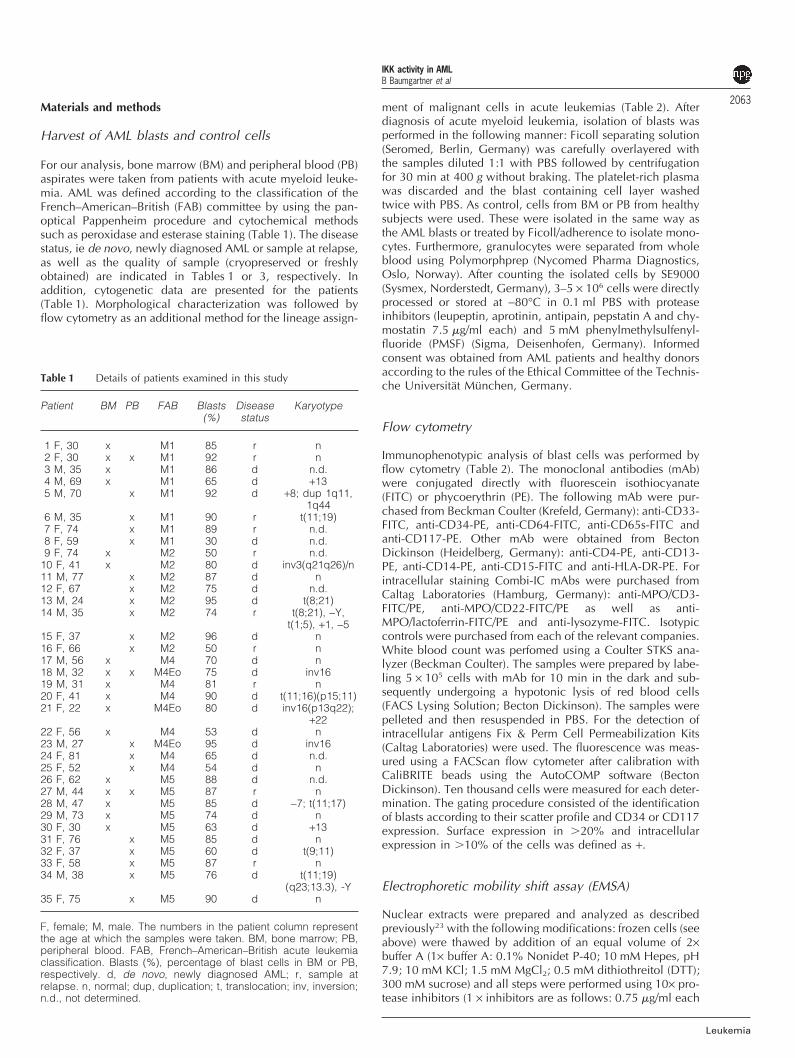
For our analysis, bone marrow (BM) and peripheral blood (PB)aspirates were taken from patients with acute myeloid leuke-mia. AML was defined according to the classification of theFrench–American–British (FAB) committee by using the pan-optical Pappenheim procedure and cytochemical methodssuch as peroxidase and esterase staining (Table 1). The diseasestatus, ie de novo, newly diagnosed AML or sample at relapse,as well as the quality of sample (cryopreserved or freshlyobtained) are indicated in Tables 1 or 3, respectively. Inaddition, cytogenetic data are presented for the patients(Table 1). Morphological characterization was followed byflow cytometry as an additional method for the lineage assign-
Table 1 Details of patients examined in this study
Patient BM PB FAB Blasts Disease Karyotype(%) status
1 F, 30 x M1 85 r n2 F, 30 x x M1 92 r n3 M, 35 x M1 86 d n.d.4 M, 69 x M1 65 d +135 M, 70 x M1 92 d +8; dup 1q11,
1q446 M, 35 x M1 90 r t(11;19)7 F, 74 x M1 89 r n.d.8 F, 59 x M1 30 d n.d.9 F, 74 x M2 50 r n.d.
10 F, 41 x M2 80 d inv3(q21q26)/n11 M, 77 x M2 87 d n12 F, 67 x M2 75 d n.d.13 M, 24 x M2 95 d t(8;21)14 M, 35 x M2 74 r t(8;21), −Y,
t(1;5), +1, −515 F, 37 x M2 96 d n16 F, 66 x M2 50 r n17 M, 56 x M4 70 d n18 M, 32 x x M4Eo 75 d inv1619 M, 31 x M4 81 r n20 F, 41 x M4 90 d t(11;16)(p15;11)21 F, 22 x M4Eo 80 d inv16(p13q22);
+2222 F, 56 x M4 53 d n23 M, 27 x M4Eo 95 d inv1624 F, 81 x M4 65 d n.d.25 F, 52 x M4 54 d n26 F, 62 x M5 88 d n.d.27 M, 44 x x M5 87 r n28 M, 47 x M5 85 d −7; t(11;17)29 M, 73 x M5 74 d n30 F, 30 x M5 63 d +1331 F, 76 x M5 85 d n32 F, 37 x M5 60 d t(9;11)33 F, 58 x M5 87 r n34 M, 38 x M5 76 d t(11;19)
(q23;13.3), -Y35 F, 75 x M5 90 d n
F, female; M, male. The numbers in the patient column representthe age at which the samples were taken. BM, bone marrow; PB,peripheral blood. FAB, French–American–British acute leukemiaclassification. Blasts (%), percentage of blast cells in BM or PB,respectively. d, de novo, newly diagnosed AML; r, sample atrelapse. n, normal; dup, duplication; t, translocation; inv, inversion;n.d., not determined.
Leukemia
ment of malignant cells in acute leukemias (Table 2). Afterdiagnosis of acute myeloid leukemia, isolation of blasts wasperformed in the following manner: Ficoll separating solution(Seromed, Berlin, Germany) was carefully overlayered withthe samples diluted 1:1 with PBS followed by centrifugationfor 30 min at 400 g without braking. The platelet-rich plasmawas discarded and the blast containing cell layer washedtwice with PBS. As control, cells from BM or PB from healthysubjects were used. These were isolated in the same way asthe AML blasts or treated by Ficoll/adherence to isolate mono-cytes. Furthermore, granulocytes were separated from wholeblood using Polymorphprep (Nycomed Pharma Diagnostics,Oslo, Norway). After counting the isolated cells by SE9000(Sysmex, Norderstedt, Germany), 3–5 × 10⁶ cells were directlyprocessed or stored at −80°C in 0.1 ml PBS with proteaseinhibitors (leupeptin, aprotinin, antipain, pepstatin A and chy-mostatin 7.5 �g/ml each) and 5 mM phenylmethylsulfenyl-fluoride (PMSF) (Sigma, Deisenhofen, Germany). Informedconsent was obtained from AML patients and healthy donorsaccording to the rules of the Ethical Committee of the Technis-che Universitat Munchen, Germany.
Flow cytometry
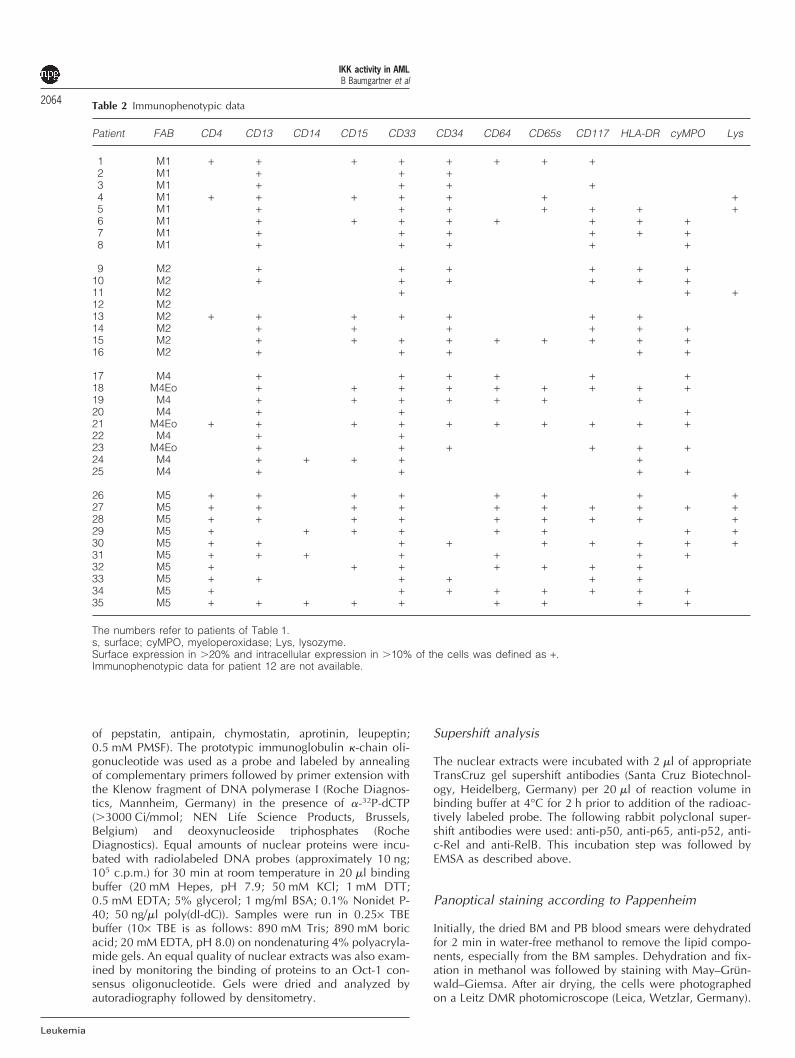
Immunophenotypic analysis of blast cells was performed byflow cytometry (Table 2). The monoclonal antibodies (mAb)were conjugated directly with fluorescein isothiocyanate(FITC) or phycoerythrin (PE). The following mAb were pur-chased from Beckman Coulter (Krefeld, Germany): anti-CD33-FITC, anti-CD34-PE, anti-CD64-FITC, anti-CD65s-FITC andanti-CD117-PE. Other mAb were obtained from BectonDickinson (Heidelberg, Germany): anti-CD4-PE, anti-CD13-PE, anti-CD14-PE, anti-CD15-FITC and anti-HLA-DR-PE. Forintracellular staining Combi-IC mAbs were purchased fromCaltag Laboratories (Hamburg, Germany): anti-MPO/CD3-FITC/PE, anti-MPO/CD22-FITC/PE as well as anti-MPO/lactoferrin-FITC/PE and anti-lysozyme-FITC. Isotypiccontrols were purchased from each of the relevant companies.White blood count was perfomed using a Coulter STKS ana-lyzer (Beckman Coulter). The samples were prepared by labe-ling 5 × 10⁵ cells with mAb for 10 min in the dark and sub-sequently undergoing a hypotonic lysis of red blood cells(FACS Lysing Solution; Becton Dickinson). The samples werepelleted and then resuspended in PBS. For the detection ofintracellular antigens Fix & Perm Cell Permeabilization Kits(Caltag Laboratories) were used. The fluorescence was meas-ured using a FACScan flow cytometer after calibration withCaliBRITE beads using the AutoCOMP software (BectonDickinson). Ten thousand cells were measured for each deter-mination. The gating procedure consisted of the identificationof blasts according to their scatter profile and CD34 or CD117expression. Surface expression in �20% and intracellularexpression in �10% of the cells was defined as +.
Electrophoretic mobility shift assay (EMSA)
Nuclear extracts were prepared and analyzed as describedpreviously23 with the following modifications: frozen cells (seeabove) were thawed by addition of an equal volume of 2×buffer A (1× buffer A: 0.1% Nonidet P-40; 10 mM Hepes, pH7.9; 10 mM KCl; 1.5 mM MgCl2; 0.5 mM dithiothreitol (DTT);300 mM sucrose) and all steps were performed using 10× pro-tease inhibitors (1 × inhibitors are as follows: 0.75 �g/ml each
IKK activity in AMLB Baumgartner et al
2064
Leukemia
Table 2 Immunophenotypic data
Patient FAB CD4 CD13 CD14 CD15 CD33 CD34 CD64 CD65s CD117 HLA-DR cyMPO Lys
1 M1 + + + + + + + +2 M1 + + +3 M1 + + + +4 M1 + + + + + + +5 M1 + + + + + + +6 M1 + + + + + + + +7 M1 + + + + + +8 M1 + + + + +
9 M2 + + + + + +10 M2 + + + + + +11 M2 + + +12 M213 M2 + + + + + + +14 M2 + + + + + +15 M2 + + + + + + + + +16 M2 + + + + +
17 M4 + + + + + +18 M4Eo + + + + + + + + +19 M4 + + + + + + +20 M4 + + +21 M4Eo + + + + + + + + + +22 M4 + +23 M4Eo + + + + + +24 M4 + + + + +25 M4 + + + +
26 M5 + + + + + + + +27 M5 + + + + + + + + + +28 M5 + + + + + + + + +29 M5 + + + + + + + +30 M5 + + + + + + + + +31 M5 + + + + + + +32 M5 + + + + + + +33 M5 + + + + + +34 M5 + + + + + + + +35 M5 + + + + + + + + +
The numbers refer to patients of Table 1.s, surface; cyMPO, myeloperoxidase; Lys, lysozyme.Surface expression in �20% and intracellular expression in �10% of the cells was defined as +.Immunophenotypic data for patient 12 are not available.
of pepstatin, antipain, chymostatin, aprotinin, leupeptin;0.5 mM PMSF). The prototypic immunoglobulin �-chain oli-gonucleotide was used as a probe and labeled by annealingof complementary primers followed by primer extension withthe Klenow fragment of DNA polymerase I (Roche Diagnos-tics, Mannheim, Germany) in the presence of �-32P-dCTP(�3000 Ci/mmol; NEN Life Science Products, Brussels,Belgium) and deoxynucleoside triphosphates (RocheDiagnostics). Equal amounts of nuclear proteins were incu-bated with radiolabeled DNA probes (approximately 10 ng;105 c.p.m.) for 30 min at room temperature in 20 �l bindingbuffer (20 mM Hepes, pH 7.9; 50 mM KCl; 1 mM DTT;0.5 mM EDTA; 5% glycerol; 1 mg/ml BSA; 0.1% Nonidet P-40; 50 ng/�l poly(dI-dC)). Samples were run in 0.25× TBEbuffer (10× TBE is as follows: 890 mM Tris; 890 mM boricacid; 20 mM EDTA, pH 8.0) on nondenaturing 4% polyacryla-mide gels. An equal quality of nuclear extracts was also exam-ined by monitoring the binding of proteins to an Oct-1 con-sensus oligonucleotide. Gels were dried and analyzed byautoradiography followed by densitometry.
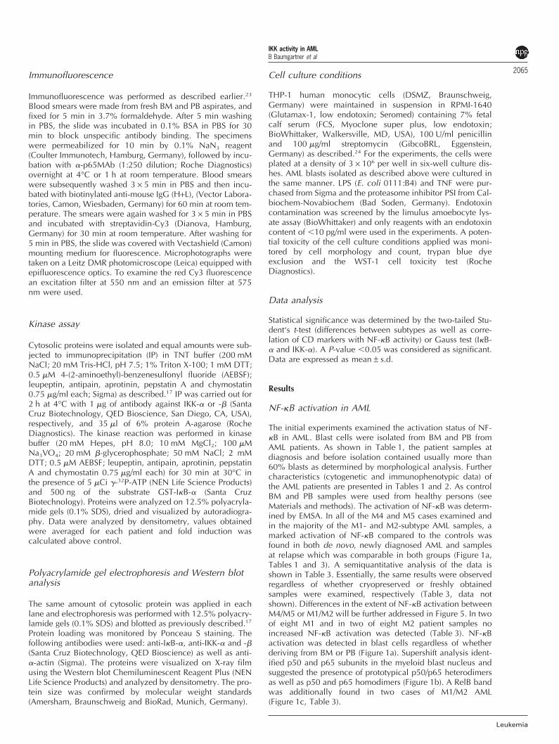
Supershift analysis
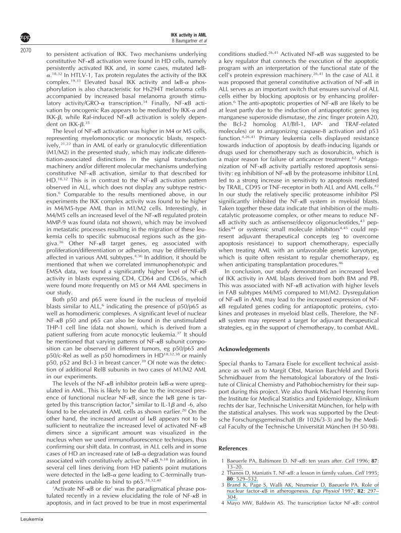
The nuclear extracts were incubated with 2 �l of appropriateTransCruz gel supershift antibodies (Santa Cruz Biotechnol-ogy, Heidelberg, Germany) per 20 �l of reaction volume inbinding buffer at 4°C for 2 h prior to addition of the radioac-tively labeled probe. The following rabbit polyclonal super-shift antibodies were used: anti-p50, anti-p65, anti-p52, anti-c-Rel and anti-RelB. This incubation step was followed byEMSA as described above.
Panoptical staining according to Pappenheim
Initially, the dried BM and PB blood smears were dehydratedfor 2 min in water-free methanol to remove the lipid compo-nents, especially from the BM samples. Dehydration and fix-ation in methanol was followed by staining with May–Grun-wald–Giemsa. After air drying, the cells were photographedon a Leitz DMR photomicroscope (Leica, Wetzlar, Germany).
IKK activity in AMLB Baumgartner et al
2065Immunofluorescence
Immunofluorescence was performed as described earlier.23
Blood smears were made from fresh BM and PB aspirates, andfixed for 5 min in 3.7% formaldehyde. After 5 min washingin PBS, the slide was incubated in 0.1% BSA in PBS for 30min to block unspecific antibody binding. The specimenswere permeabilized for 10 min by 0.1% NaN3 reagent(Coulter Immunotech, Hamburg, Germany), followed by incu-bation with �-p65MAb (1:250 dilution; Roche Diagnostics)overnight at 4°C or 1 h at room temperature. Blood smearswere subsequently washed 3 × 5 min in PBS and then incu-bated with biotinylated anti-mouse IgG (H+L), (Vector Labora-tories, Camon, Wiesbaden, Germany) for 60 min at room tem-perature. The smears were again washed for 3 × 5 min in PBSand incubated with streptavidin-Cy3 (Dianova, Hamburg,Germany) for 30 min at room temperature. After washing for5 min in PBS, the slide was covered with Vectashield (Camon)mounting medium for fluorescence. Microphotographs weretaken on a Leitz DMR photomicroscope (Leica) equipped withepifluorescence optics. To examine the red Cy3 fluorescencean excitation filter at 550 nm and an emission filter at 575nm were used.
Kinase assay
Cytosolic proteins were isolated and equal amounts were sub-jected to immunoprecipitation (IP) in TNT buffer (200 mMNaCl; 20 mM Tris-HCl, pH 7.5; 1% Triton X-100; 1 mM DTT;0.5 �M 4-(2-aminoethyl)-benzenesulfonyl fluoride (AEBSF);leupeptin, antipain, aprotinin, pepstatin A and chymostatin0.75 �g/ml each; Sigma) as described.17 IP was carried out for2 h at 4°C with 1 �g of antibody against IKK-� or -� (SantaCruz Biotechnology, QED Bioscience, San Diego, CA, USA),respectively, and 35 �l of 6% protein A-agarose (RocheDiagnostics). The kinase reaction was performed in kinasebuffer (20 mM Hepes, pH 8.0; 10 mM MgCl2; 100 �MNa3VO4; 20 mM �-glycerophosphate; 50 mM NaCl; 2 mMDTT; 0.5 �M AEBSF; leupeptin, antipain, aprotinin, pepstatinA and chymostatin 0.75 �g/ml each) for 30 min at 30°C inthe presence of 5 �Ci �-32P-ATP (NEN Life Science Products)and 500 ng of the substrate GST-I�B-� (Santa CruzBiotechnology). Proteins were analyzed on 12.5% polyacryla-mide gels (0.1% SDS), dried and visualized by autoradiogra-phy. Data were analyzed by densitometry, values obtainedwere averaged for each patient and fold induction wascalculated above control.
Polyacrylamide gel electrophoresis and Western blotanalysis
The same amount of cytosolic protein was applied in eachlane and electrophoresis was performed with 12.5% polyacry-lamide gels (0.1% SDS) and blotted as previously described.17
Protein loading was monitored by Ponceau S staining. Thefollowing antibodies were used: anti-I�B-�, anti-IKK-� and -�(Santa Cruz Biotechnology, QED Bioscience) as well as anti-�-actin (Sigma). The proteins were visualized on X-ray filmusing the Western blot Chemiluminescent Reagent Plus (NENLife Science Products) and analyzed by densitometry. The pro-tein size was confirmed by molecular weight standards(Amersham, Braunschweig and BioRad, Munich, Germany).
Leukemia
Cell culture conditions
THP-1 human monocytic cells (DSMZ, Braunschweig,Germany) were maintained in suspension in RPMI-1640(Glutamax-1, low endotoxin; Seromed) containing 7% fetalcalf serum (FCS, Myoclone super plus, low endotoxin;BioWhittaker, Walkersville, MD, USA), 100 U/ml penicillinand 100 �g/ml streptomycin (GibcoBRL, Eggenstein,Germany) as described.24 For the experiments, the cells wereplated at a density of 3 × 10⁶ per well in six-well culture dis-hes. AML blasts isolated as described above were cultured inthe same manner. LPS (E. coli 0111:B4) and TNF were pur-chased from Sigma and the proteasome inhibitor PSI from Cal-biochem-Novabiochem (Bad Soden, Germany). Endotoxincontamination was screened by the limulus amoebocyte lys-ate assay (BioWhittaker) and only reagents with an endotoxincontent of �10 pg/ml were used in the experiments. A poten-tial toxicity of the cell culture conditions applied was moni-tored by cell morphology and count, trypan blue dyeexclusion and the WST-1 cell toxicity test (RocheDiagnostics).
Data analysis
Statistical significance was determined by the two-tailed Stu-dent‘s t-test (differences between subtypes as well as corre-lation of CD markers with NF-�B activity) or Gauss test (I�B-� and IKK-�). A P-value �0.05 was considered as significant.Data are expressed as mean ± s.d.
Results
NF-�B activation in AML
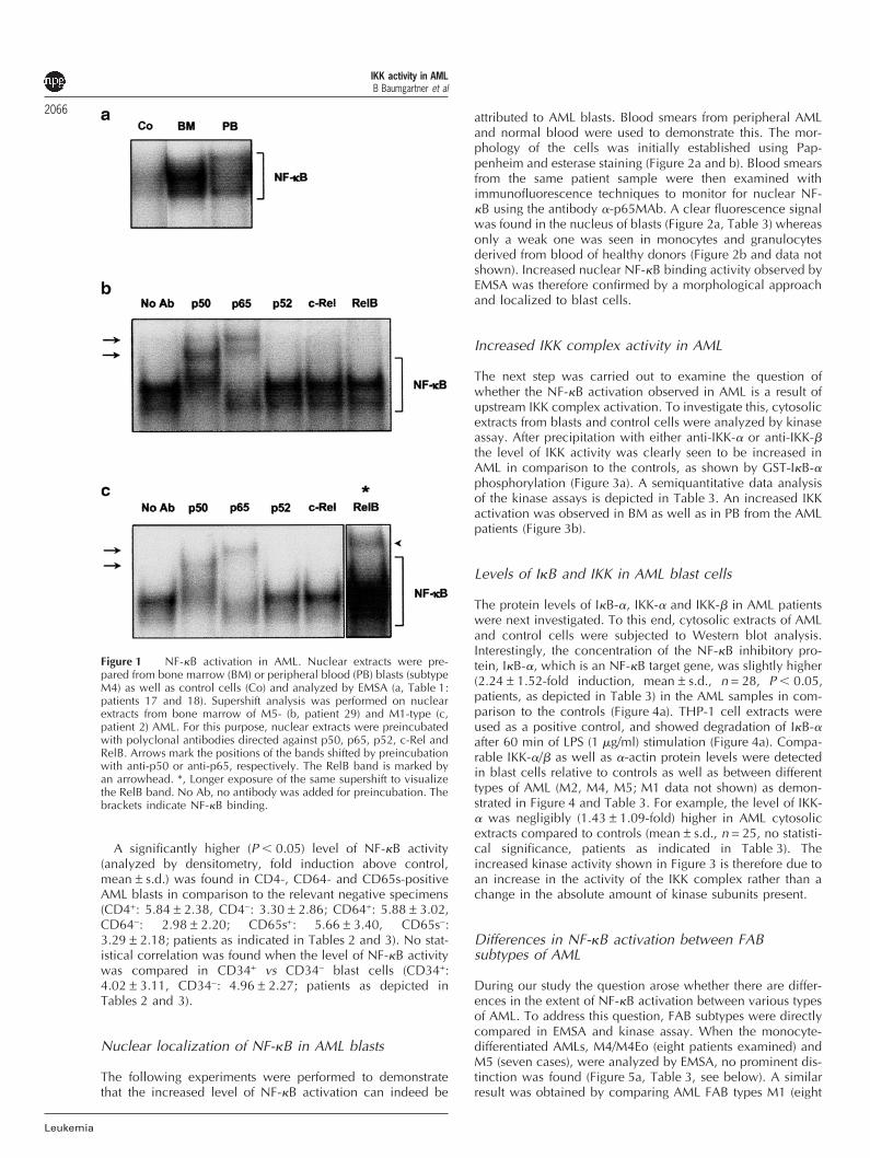
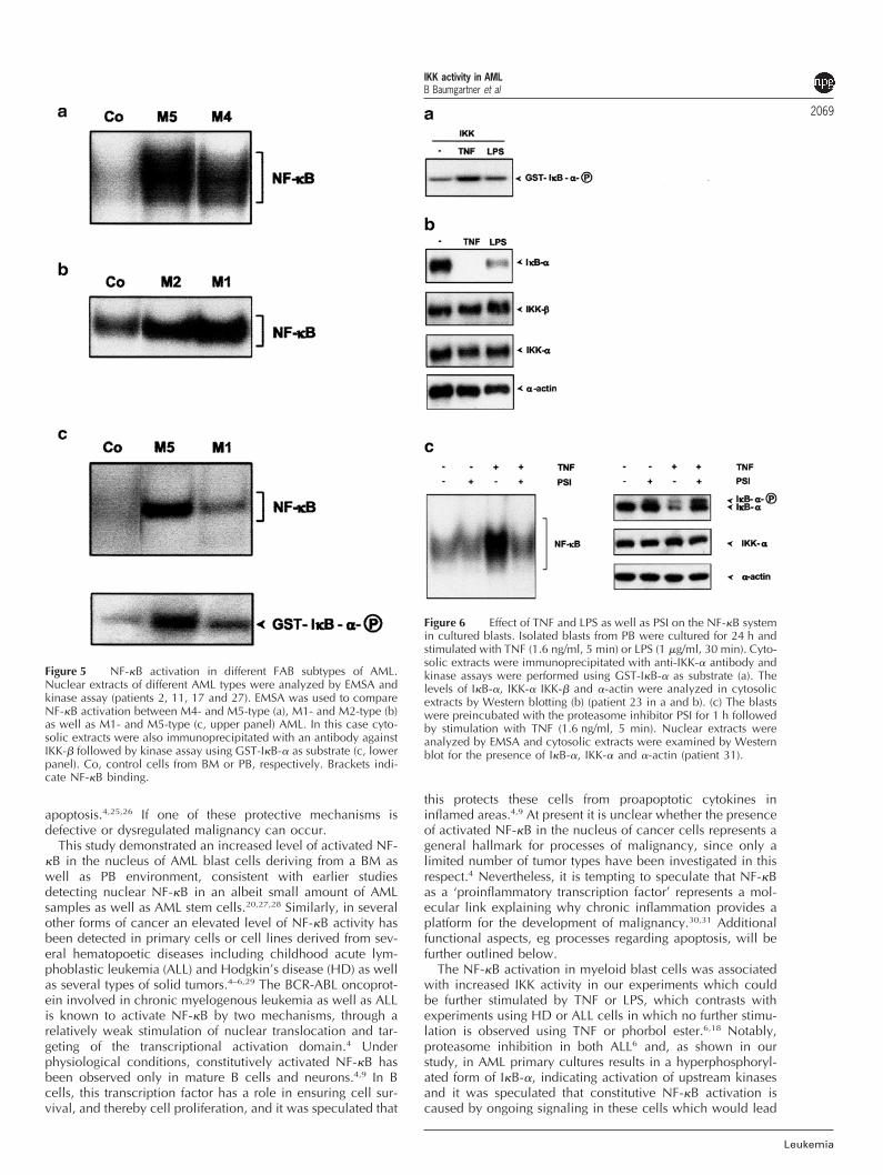
The initial experiments examined the activation status of NF-�B in AML. Blast cells were isolated from BM and PB fromAML patients. As shown in Table 1, the patient samples atdiagnosis and before isolation contained usually more than60% blasts as determined by morphological analysis. Furthercharacteristics (cytogenetic and immunophenotypic data) ofthe AML patients are presented in Tables 1 and 2. As controlBM and PB samples were used from healthy persons (seeMaterials and methods). The activation of NF-�B was determ-ined by EMSA. In all of the M4 and M5 cases examined andin the majority of the M1- and M2-subtype AML samples, amarked activation of NF-�B compared to the controls wasfound in both de novo, newly diagnosed AML and samplesat relapse which was comparable in both groups (Figure 1a,Tables 1 and 3). A semiquantitative analysis of the data isshown in Table 3. Essentially, the same results were observedregardless of whether cryopreserved or freshly obtainedsamples were examined, respectively (Table 3, data notshown). Differences in the extent of NF-�B activation betweenM4/M5 or M1/M2 will be further addressed in Figure 5. In twoof eight M1 and in two of eight M2 patient samples noincreased NF-�B activation was detected (Table 3). NF-�Bactivation was detected in blast cells regardless of whetherderiving from BM or PB (Figure 1a). Supershift analysis ident-ified p50 and p65 subunits in the myeloid blast nucleus andsuggested the presence of prototypical p50/p65 heterodimersas well as p50 and p65 homodimers (Figure 1b). A RelB bandwas additionally found in two cases of M1/M2 AML(Figure 1c, Table 3).
IKK activity in AMLB Baumgartner et al
2066
Leukemia
Figure 1 NF-�B activation in AML. Nuclear extracts were pre-pared from bone marrow (BM) or peripheral blood (PB) blasts (subtypeM4) as well as control cells (Co) and analyzed by EMSA (a, Table 1:patients 17 and 18). Supershift analysis was performed on nuclearextracts from bone marrow of M5- (b, patient 29) and M1-type (c,patient 2) AML. For this purpose, nuclear extracts were preincubatedwith polyclonal antibodies directed against p50, p65, p52, c-Rel andRelB. Arrows mark the positions of the bands shifted by preincubationwith anti-p50 or anti-p65, respectively. The RelB band is marked byan arrowhead. *, Longer exposure of the same supershift to visualizethe RelB band. No Ab, no antibody was added for preincubation. Thebrackets indicate NF-�B binding.
A significantly higher (P � 0.05) level of NF-�B activity(analyzed by densitometry, fold induction above control,mean ± s.d.) was found in CD4-, CD64- and CD65s-positiveAML blasts in comparison to the relevant negative specimens(CD4+: 5.84 ± 2.38, CD4−: 3.30 ± 2.86; CD64+: 5.88 ± 3.02,CD64−: 2.98 ± 2.20; CD65s+: 5.66 ± 3.40, CD65s−:3.29 ± 2.18; patients as indicated in Tables 2 and 3). No stat-istical correlation was found when the level of NF-�B activitywas compared in CD34+ vs CD34− blast cells (CD34+:4.02 ± 3.11, CD34−: 4.96 ± 2.27; patients as depicted inTables 2 and 3).
Nuclear localization of NF-�B in AML blasts
The following experiments were performed to demonstratethat the increased level of NF-�B activation can indeed be
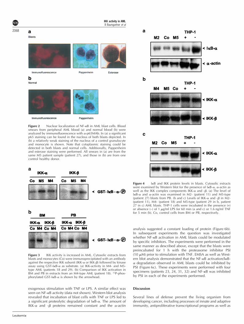
attributed to AML blasts. Blood smears from peripheral AMLand normal blood were used to demonstrate this. The mor-phology of the cells was initially established using Pap-penheim and esterase staining (Figure 2a and b). Blood smearsfrom the same patient sample were then examined withimmunofluorescence techniques to monitor for nuclear NF-�B using the antibody �-p65MAb. A clear fluorescence signalwas found in the nucleus of blasts (Figure 2a, Table 3) whereasonly a weak one was seen in monocytes and granulocytesderived from blood of healthy donors (Figure 2b and data notshown). Increased nuclear NF-�B binding activity observed byEMSA was therefore confirmed by a morphological approachand localized to blast cells.
Increased IKK complex activity in AML
The next step was carried out to examine the question ofwhether the NF-�B activation observed in AML is a result ofupstream IKK complex activation. To investigate this, cytosolicextracts from blasts and control cells were analyzed by kinaseassay. After precipitation with either anti-IKK-� or anti-IKK-�the level of IKK activity was clearly seen to be increased inAML in comparison to the controls, as shown by GST-I�B-�phosphorylation (Figure 3a). A semiquantitative data analysisof the kinase assays is depicted in Table 3. An increased IKKactivation was observed in BM as well as in PB from the AMLpatients (Figure 3b).
Levels of I�B and IKK in AML blast cells
The protein levels of I�B-�, IKK-� and IKK-� in AML patientswere next investigated. To this end, cytosolic extracts of AMLand control cells were subjected to Western blot analysis.Interestingly, the concentration of the NF-�B inhibitory pro-tein, I�B-�, which is an NF-�B target gene, was slightly higher(2.24 ± 1.52-fold induction, mean ± s.d., n = 28, P � 0.05,patients, as depicted in Table 3) in the AML samples in com-parison to the controls (Figure 4a). THP-1 cell extracts wereused as a positive control, and showed degradation of I�B-�after 60 min of LPS (1 �g/ml) stimulation (Figure 4a). Compa-rable IKK-�/� as well as �-actin protein levels were detectedin blast cells relative to controls as well as between differenttypes of AML (M2, M4, M5; M1 data not shown) as demon-strated in Figure 4 and Table 3. For example, the level of IKK-� was negligibly (1.43 ± 1.09-fold) higher in AML cytosolicextracts compared to controls (mean ± s.d., n = 25, no statisti-cal significance, patients as indicated in Table 3). Theincreased kinase activity shown in Figure 3 is therefore due toan increase in the activity of the IKK complex rather than achange in the absolute amount of kinase subunits present.
Differences in NF-�B activation between FABsubtypes of AML
During our study the question arose whether there are differ-ences in the extent of NF-�B activation between various typesof AML. To address this question, FAB subtypes were directlycompared in EMSA and kinase assay. When the monocyte-differentiated AMLs, M4/M4Eo (eight patients examined) andM5 (seven cases), were analyzed by EMSA, no prominent dis-tinction was found (Figure 5a, Table 3, see below). A similarresult was obtained by comparing AML FAB types M1 (eight
IKK activity in AMLB Baumgartner et al
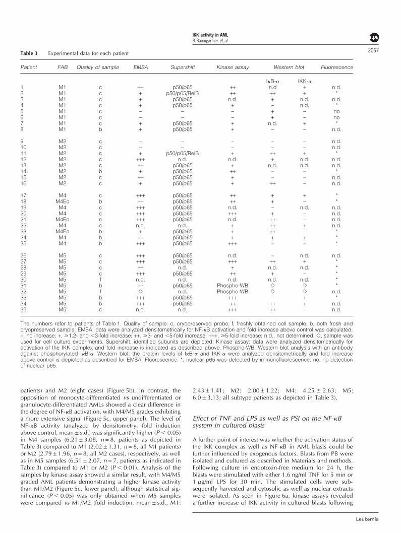
2067Table 3 Experimental data for each patient
Patient FAB Quality of sample EMSA Supershift Kinase assay Western blot Fluorescence
I�B-� IKK-�1 M1 c ++ p50/p65 ++ n.d + n.d.2 M1 c + p50/p65/RelB ++ ++ + *3 M1 c + p50/p65 n.d. + n.d. n.d.4 M1 c + p50/p65 + − n.d. *5 M1 c − − − + − no6 M1 c − − − + − no7 M1 c + p50/p65 + n.d. + *8 M1 b + p50/p65 + − − n.d.
9 M2 c − − − − − n.d.10 M2 c − − − − − n.d.11 M2 c + p50/p65/RelB + ++ + *12 M2 c +++ n.d. n.d. + n.d. n.d.13 M2 c ++ p50/p65 + n.d. n.d. n.d.14 M2 b + p50/p65 ++ − − *15 M2 c ++ p50/p65 + − − n.d.16 M2 c + p50/p65 + ++ − n.d.
17 M4 c +++ p50/p65 ++ + + *18 M4Eo b ++ p50/p65 ++ + − *19 M4 c +++ p50/p65 n.d. − n.d. n.d.20 M4 c +++ p50/p65 +++ + − n.d.21 M4Eo c +++ p50/p65 n.d. ++ − n.d.22 M4 c n.d. n.d. + ++ + n.d.23 M4Eo b + p50/p65 + ++ − *24 M4 b ++ p50/p65 + + + *25 M4 b +++ p50/p65 +++ − − *
26 M5 c +++ p50/p65 n.d. − n.d. n.d.27 M5 c +++ p50/p65 +++ ++ + *28 M5 c ++ n.d. + n.d. n.d. *29 M5 c +++ p50/p65 ++ + − *30 M5 f n.d. n.d. n.d. n.d. n.d. *31 M5 b ++ p50/p65 Phospho-WB � � *32 M5 f � n.d. Phospho-WB � � n.d.33 M5 b +++ p50/p65 +++ − + *34 M5 b +++ p50/p65 ++ ++ + n.d.35 M5 c n.d. n.d. +++ ++ − n.d.
The numbers refer to patients of Table 1. Quality of sample: c, cryopreserved probe; f, freshly obtained cell sample; b, both fresh andcryopreserved sample. EMSA, data were analyzed densitometrically for NF-�B activation and fold increase above control was calculated:−, no increase; +, 1.2- and �3-fold increase; ++, 3- and �5-fold increase; +++, 5-fold increase; n.d., not determined. �, sample wasused for cell culture experiments. Supershift: identified subunits are depicted. Kinase assay: data were analyzed densitometrically foractivation of the IKK complex and fold increase is indicated as described above. Phospho-WB, Western blot analysis with an antibodyagainst phosphorylated I�B-�. Western blot: the protein levels of I�B-� and IKK-� were analyzed densitometrically and fold increaseabove control is depicted as described for EMSA. Fluorescence: *, nuclear p65 was detected by immunofluorescence; no, no detectionof nuclear p65.
patients) and M2 (eight cases) (Figure 5b). In contrast, theopposition of monocyte-differentiated vs undifferentiated orgranulocyte-differentiated AMLs showed a clear difference inthe degree of NF-�B activation, with M4/M5 grades exhibitinga more extensive signal (Figure 5c, upper panel). The level ofNF-�B activity (analyzed by densitometry, fold inductionabove control, mean ± s.d.) was significantly higher (P � 0.05)in M4 samples (6.21 ± 3.08, n = 8, patients as depicted inTable 3) compared to M1 (2.02 ± 1.31, n = 8, all M1 patients)or M2 (2.79 ± 1.96, n = 8, all M2 cases), respectively, as wellas in M5 samples (6.51 ± 2.07, n = 7, patients as indicated inTable 3) compared to M1 or M2 (P � 0.01). Analysis of thesamples by kinase assay showed a similar result, with M4/M5graded AML patients demonstrating a higher kinase activitythan M1/M2 (Figure 5c, lower panel), although statistical sig-nificance (P � 0.05) was only obtained when M5 sampleswere compared vs M1/M2 (fold induction, mean ± s.d., M1:
Leukemia
2.43 ± 1.41; M2: 2.00 ± 1.22; M4: 4.25 2.63; M5:6.0 ± 3.13; all subtype patients as depicted in Table 3).
Effect of TNF and LPS as well as PSI on the NF-�Bsystem in cultured blasts
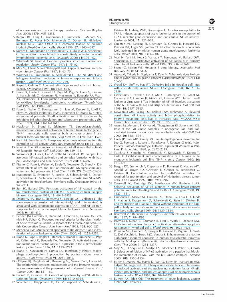
A further point of interest was whether the activation status ofthe IKK complex as well as NF-�B in AML blasts could befurther influenced by exogenous factors. Blasts from PB wereisolated and cultured as described in Materials and methods.Following culture in endotoxin-free medium for 24 h, theblasts were stimulated with either 1.6 ng/ml TNF for 5 min or1 �g/ml LPS for 30 min. The stimulated cells were sub-sequently harvested and cytosolic as well as nuclear extractswere isolated. As seen in Figure 6a, kinase assays revealeda further increase of IKK activity in cultured blasts following
IKK activity in AMLB Baumgartner et al
2068
Leukemia
Figure 2 Nuclear localization of NF-�B in AML blast cells. Bloodsmears from peripheral AML blood (a) and normal blood (b) wereanalyzed by immunofluorescence with �-p65MAb. In (a) a significantp65 staining can be found in the nucleus of both blasts depicted. In(b) a relatively weak staining of the nucleus of a control granulocyteand monocyte is shown. Note that cytoplasmic staining could bedetected in both blasts and normal cells. Additionally, Pappenheimand esterase staining were performed. All smears in (a) are from thesame M5 patient sample (patient 27), and those in (b) are from onecontrol healthy donor.
Figure 3 IKK activity is increased in AML. Cytosolic extracts fromblasts and monocytes (Co) were immunoprecipitated with an antibodyagainst the respective IKK subunit (IKK-� or IKK-�) followed by kinaseassay using GST-I�B-� as substrate. (a) IKK-activity in M4- and M5-type AML (patients 18 and 29). (b) Comparison of IKK activation inBM and PB in extracts from an M4-type AML (patient 18). 32P-phos-phorylated GST-I�B-� is shown by the arrowhead.
exogenous stimulation with TNF or LPS. A similar effect wasseen on NF-�B activity (data not shown). Western blot analysisrevealed that incubation of blast cells with TNF or LPS led toa significant proteolytic degradation of I�B-�. The amount ofIKK-� and -� proteins remained constant and the �-actin
Figure 4 I�B and IKK protein levels in blasts. Cytosolic extractswere examined by Western blot for the presence of I�B-�, �-actin aswell as the IKK complex components IKK-� and -�. (a) The level ofI�B-� and �-actin was examined in M2- (patient 11) and M5-type(patient 27) blasts from PB. (b and c) Levels of IKK-� and -� in M2-(patient 11), M4- (patient 18) and M5-type (patient 29 in b, patient27 in c) AML blasts. THP-1 cells were incubated in the presence (+)or absence (−) of 1 �g/ml LPS for 60 min (a and c) or 1.6 ng/ml TNFfor 5 min (b). Co, control cells from BM or PB, respectively.
analysis suggested a constant loading of protein (Figure 6b).In subsequent experiments the question was investigatedwhether NF-�B activation in AML blasts could be modulatedby specific inhibitors. The experiments were performed in thesame manner as described above, except that the blasts werepreincubated for 1 h with the proteasome inhibitor PSI(10 �M) prior to stimulation with TNF. EMSA as well as West-ern blot analysis demonstrated that the NF-�B activation/I�B-� degradation observed in AML blasts could be inhibited byPSI (Figure 6c). These experiments were performed with fourspecimens (patients 23, 24, 31, 32) and NF-�B was inhibitedby PSI in each of the experiments performed.
Discussion
Several lines of defense prevent the living organism fromdeveloping cancer, including processes of innate and adaptiveimmunity, antiproliferative transcriptional programs as well as
IKK activity in AMLB Baumgartner et al
2069
Figure 5 NF-�B activation in different FAB subtypes of AML.Nuclear extracts of different AML types were analyzed by EMSA andkinase assay (patients 2, 11, 17 and 27). EMSA was used to compareNF-�B activation between M4- and M5-type (a), M1- and M2-type (b)as well as M1- and M5-type (c, upper panel) AML. In this case cyto-solic extracts were also immunoprecipitated with an antibody againstIKK-� followed by kinase assay using GST-I�B-� as substrate (c, lowerpanel). Co, control cells from BM or PB, respectively. Brackets indi-cate NF-�B binding.
apoptosis.4,25,26 If one of these protective mechanisms isdefective or dysregulated malignancy can occur.This study demonstrated an increased level of activated NF-
�B in the nucleus of AML blast cells deriving from a BM aswell as PB environment, consistent with earlier studiesdetecting nuclear NF-�B in an albeit small amount of AMLsamples as well as AML stem cells.20,27,28 Similarly, in severalother forms of cancer an elevated level of NF-�B activity hasbeen detected in primary cells or cell lines derived from sev-eral hematopoetic diseases including childhood acute lym-phoblastic leukemia (ALL) and Hodgkin’s disease (HD) as wellas several types of solid tumors.4–6,29 The BCR-ABL oncoprot-ein involved in chronic myelogenous leukemia as well as ALLis known to activate NF-�B by two mechanisms, through arelatively weak stimulation of nuclear translocation and tar-geting of the transcriptional activation domain.4 Underphysiological conditions, constitutively activated NF-�B hasbeen observed only in mature B cells and neurons.4,9 In Bcells, this transcription factor has a role in ensuring cell sur-vival, and thereby cell proliferation, and it was speculated that
Leukemia
Figure 6 Effect of TNF and LPS as well as PSI on the NF-�B systemin cultured blasts. Isolated blasts from PB were cultured for 24 h andstimulated with TNF (1.6 ng/ml, 5 min) or LPS (1 �g/ml, 30 min). Cyto-solic extracts were immunoprecipitated with anti-IKK-� antibody andkinase assays were performed using GST-I�B-� as substrate (a). Thelevels of I�B-�, IKK-� IKK-� and �-actin were analyzed in cytosolicextracts by Western blotting (b) (patient 23 in a and b). (c) The blastswere preincubated with the proteasome inhibitor PSI for 1 h followedby stimulation with TNF (1.6 ng/ml, 5 min). Nuclear extracts wereanalyzed by EMSA and cytosolic extracts were examined by Westernblot for the presence of I�B-�, IKK-� and �-actin (patient 31).
this protects these cells from proapoptotic cytokines ininflamed areas.4,9 At present it is unclear whether the presenceof activated NF-�B in the nucleus of cancer cells represents ageneral hallmark for processes of malignancy, since only alimited number of tumor types have been investigated in thisrespect.4 Nevertheless, it is tempting to speculate that NF-�Bas a ‘proinflammatory transcription factor’ represents a mol-ecular link explaining why chronic inflammation provides aplatform for the development of malignancy.30,31 Additionalfunctional aspects, eg processes regarding apoptosis, will befurther outlined below.The NF-�B activation in myeloid blast cells was associated
with increased IKK activity in our experiments which couldbe further stimulated by TNF or LPS, which contrasts withexperiments using HD or ALL cells in which no further stimu-lation is observed using TNF or phorbol ester.6,18 Notably,proteasome inhibition in both ALL6 and, as shown in ourstudy, in AML primary cultures results in a hyperphosphoryl-ated form of I�B-�, indicating activation of upstream kinasesand it was speculated that constitutive NF-�B activation iscaused by ongoing signaling in these cells which would lead
IKK activity in AMLB Baumgartner et al
2070
Leukemia
to persistent activation of IKK. Two mechanisms underlyingconstitutive NF-�B activation were found in HD cells, namelypersistently activated IKK and, in some cases, mutated I�B-�.18,32 In HTLV-1, Tax protein regulates the activity of the IKKcomplex.19,33 Elevated basal IKK activity and I�B-� phos-phorylation is also characteristic for Hs294T melanoma cellsaccompanied by increased basal melanoma growth stimu-latory activity/GRO-� transcription.34 Finally, NF-�B acti-vation by oncogenic Ras appears to be mediated by IKK-� andIKK-�, while Raf-induced NF-�B activation is solely depen-dent on IKK-�.35
The level of NF-�B activation was higher in M4 or M5 cells,representing myelomonocytic or monocytic blasts, respect-ively,21,22 than in AML of early or granulocytic differentiation(M1/M2) in the presented study, which may indicate differen-tiation-associated distinctions in the signal transductionmachinery and/or different molecular mechanisms underlyingconstitutive NF-�B activation, similar to that described forHD.18,32 This is in contrast to the NF-�B activation patternobserved in ALL, which does not display any subtype restric-tion.6 Comparable to the results mentioned above, in ourexperiments the IKK complex activity was found to be higherin M4/M5-type AML than in M1/M2 cells. Interestingly, inM4/M5 cells an increased level of the NF-�B regulated proteinMMP-9 was found (data not shown), which may be involvedin metastatic processes resulting in the migration of these leu-kemia cells to specific submucosal regions such as the gin-giva.36 Other NF-�B target genes, eg associated withproliferation/differentiation or adhesion, may be differentiallyaffected in various AML subtypes.4,36 In addition, it should bementioned that when we correlated immunophenotypic andEMSA data, we found a significantly higher level of NF-�Bactivity in blasts expressing CD4, CD64 and CD65s, whichwere found more frequently on M5 or M4 AML specimens inour study.Both p50 and p65 were found in the nucleus of myeloid
blasts similar to ALL,6 indicating the presence of p50/p65 aswell as homodimeric complexes. A significant level of nuclearNF-�B p50 and p65 can also be found in the unstimulatedTHP-1 cell line (data not shown), which is derived from apatient suffering from acute monocytic leukemia.37 It shouldbe mentioned that varying patterns of NF-�B subunit compo-sition can be observed in different tumors, eg p50/p65 andp50/c-Rel as well as p50 homodimers in HD18,32,38 or mainlyp50, p52 and Bcl-3 in breast cancer.39 Of note was the detec-tion of additional RelB subunits in two cases of M1/M2 AMLin our experiments.The levels of the NF-�B inhibitor protein I�B-� were upreg-
ulated in AML. This is likely to be due to the increased pres-ence of functional nuclear NF-�B, since the I�B gene is tar-geted by this transcription factor,9 similar to IL-1� and -6, alsofound to be elevated in AML cells as shown earlier.20 On theother hand, the increased amount of I�B appears not to besufficient to neutralize the increased level of activated NF-�Bdimers since a significant amount was visualized in thenucleus when we used immunofluorescence techniques, thusconfirming our shift data. In contrast, in ALL cells and in somecases of HD an increased rate of I�B-� degradation was foundassociated with constitutively active NF-�B.6,18 In addition, inseveral cell lines deriving from HD patients point mutationswere detected in the I�B-� gene leading to C-terminally trun-cated proteins unable to bind to p65.18,32,40
‘Activate NF-�B or die’ was the paradigmatical phrase pos-tulated recently in a review elucidating the role of NF-�B inapoptosis, and in fact proved to be true in most experimental
conditions studied.26,41 Activated NF-�B was suggested to bea key regulator that connects the execution of the apoptoticprogram with an interpretation of the functional state of thecell’s protein expression machinery.26,41 In the case of ALL itwas proposed that general constitutive activation of NF-�B inALL serves as an important switch that ensures survival of ALLcells either by blocking apoptosis or by enhancing prolifer-ation.6 The anti-apoptotic properties of NF-�B are likely to beat least partly due to the induction of antiapoptotic genes (egmanganese superoxide dismutase, the zinc finger protein A20,the Bcl-2 homolog A1/Bfl-1, IAP- and TRAF-relatedmolecules) or to antagonizing caspase-8 activation and p53function.4,26,41 Primary leukemia cells displayed resistancetowards induction of apoptosis by death-inducing ligands ordrugs used for chemotherapy such as doxorubicin, which isa major reason for failure of anticancer treatment.42 Antago-nization of NF-�B activity partially restored apoptosis sensi-tivity: eg inhibition of NF-�B by the proteasome inhibitor LLnLled to a strong increase in sensitivity to apoptosis mediatedby TRAIL, CD95 or TNF-receptor in both ALL and AML cells.42
In our study the relatively specific proteasome inhibitor PSIsignificantly inhibited the NF-�B system in myeloid blasts.Taken together these data indicate that inhibition of the multi-catalytic proteasome complex, or other means to reduce NF-�B activity such as antisense/decoy oligonucleotides,43 pep-tides44 or systemic small molecule inhibitors4,45 could rep-resent adjuvant therapeutical concepts (eg to overcomeapoptosis resistance) to support chemotherapy, especiallywhen treating AML with an unfavorable genetic karyotype,which is quite often resistant to regular chemotherapy, egwhen anticipating transplantation procedures.46
In conclusion, our study demonstrated an increased levelof IKK activity in AML blasts derived from both BM and PB.This was associated with NF-�B activation with higher levelsin FAB subtypes M4/M5 compared to M1/M2. Dysregulationof NF-�B in AML may lead to the increased expression of NF-�B regulated genes coding for antiapoptotic proteins, cyto-kines and proteases in myeloid blast cells. Therefore, the NF-�B system may represent a target for adjuvant therapeuticalstrategies, eg in the support of chemotherapy, to combat AML.
Acknowledgements
Special thanks to Tamara Eisele for excellent technical assist-ance as well as to Margit Obst, Marion Barchfeld and DorisSchmidbauer from the hematological laboratory of the Insti-tute of Clinical Chemistry and Pathobiochemistry for their sup-port during this project. We also thank Michael Henning fromthe Institute for Medical Statistics and Epidemiology, Klinikumrechts der Isar, Technische Universitat Munchen, for help withthe statistical analyses. This work was supported by the Deut-sche Forschungsgemeinschaft (Br 1026/3-3) and by the Medi-cal Faculty of the Technische Universitat Munchen (H 50-98).
References
1 Baeuerle PA, Baltimore D. NF-�B: ten years after. Cell 1996; 87:13–20.
2 Thanos D, Maniatis T. NF-�B: a lesson in family values. Cell 1995;80: 529–532.
3 Brand K, Page S, Walli AK, Neumeier D, Baeuerle PA. Role ofnuclear factor-�B in atherogenesis. Exp Physiol 1997; 82: 297–304.
4 Mayo MW, Baldwin AS. The transcription factor NF-�B: control
IKK activity in AMLB Baumgartner et al
2071of oncogenesis and cancer therapy resistance. Biochim BiophysActa 2000; 1470: M55–M62.
5 Bargou RC, Leng C, Krappmann D, Emmerich F, Mapara MY,Bommert K, Royer HD, Scheidereit C, Dorken B. High-levelnuclear NF-�B and Oct-2 is a common feature of culturedHodgkin/Reed-Sternberg cells. Blood 1996; 87: 4340–4347.
6 Kordes U, Krappmann D, Heissmeyer V, Ludwig WD, ScheidereitC. Transcription factor NF-�B is constitutively activated in acutelymphoblastic leukemia cells. Leukemia 2000; 14: 399–402.
7 Whiteside ST, Israel A. I kappa B proteins: structure, function andregulation. Semin Cancer Biol 1997; 8: 75–82.
8 May MJ, Ghosh S. Rel/NF-kappaB and I kappa B proteins: an over-view. Semin Cancer Biol 1997; 8: 63–73.
9 Wulczyn FG, Krappmann D, Scheidereit C. The NF-�B/Rel andI�B gene families: mediators of immune response and inflam-mation. J Mol Med 1996; 74: 749–769.
10 Rayet B, Gelinas C. Aberrant rel/nfkb genes and activity in humancancer. Oncogene 1999; 18: 6938–6947.
11 Brand K, Eisele T, Kreusel U, Page M, Page S, Haas M, GerlingA, Kaltschmidt C, Neumann FJ, Mackman N, Baeuerle PA, WalliAK, Neumeier D. Dysregulation of monocytic nuclear factor-�Bby oxidized low-density lipoprotein. Arterioscler Thromb VascBiol 1997; 17: 1901–1909.
12 Page S, Fischer C, Baumgartner B, Haas M, Kreusel U, Loidl G,Hayn M, Ziegler-Heitbrock HWL, Neumeier D, Brand K. 4-Hyd-roxynonenal prevents NF-�B activation and TNF expression byinhibiting I�B phosphorylation and subsequent proteolysis. J BiolChem 1999; 274: 11611–11618.
13 Mackman N, Brand K, Edgington TS. Lipopolysaccharide-mediated transcriptional activation of human tissue factor gene inTHP-1 monocytic cells requires both activator protein 1 andnuclear factor �B binding sites. J Exp Med 1991; 174: 1517–1526.
14 Karin M, Ben-Neriah Y. Phosphorylation meets ubiquitination: thecontrol of NF-�B activity. Annu Rev Immunol 2000; 18: 621–663.
15 Israel A. The IKK complex: an integrator of all signals that activateNF-kappaB? Trends Cell Biol 2000; 10: 129–133.
16 Woronicz JD, Gao X, Cao Z, Rothe M, Goeddel DV. IkappaB kin-ase-beta: NF-kappaB activation and complex formation with Ikap-paB kinase-alpha and NIK. Science 1997; 278: 866–869.
17 Fischer C, Page S, Weber M, Eisele T, Neumeier D, Brand K. Dif-ferent effects of LPS and TNF on monocytic IKK signalsome acti-vation and I�B proteolysis. J Biol Chem 1999; 274: 24625–24632.
18 Krappmann D, Emmerich F, Kordes U, Scharschmidt E, DorkenB, Scheidereit C. Molecular mechanisms of constitutive NF-�B/Relactivation in Hodgkin/Reed–Sternberg cells. Oncogene 1999; 18:943–953.
19 Sun SC, Ballard DW. Persistent activation of NF-kappaB by thetax transforming protein of HTLV-1: hijacking cellular IkappaBkinases. Oncogene 1999; 18: 6948–6958.
20 Dokter WHA, Tuyt L, Sierdsema SJ, Esselink MT, Vellenga E. Thespontaneous expression of interleukin-1� and interleukin-6 isassociated with spontaneous expression of AP-1 and NF-�B tran-scription factor in acute myeloblastic leukemia cells. Leukemia1995; 9: 425–432.
21 Bennett JM, Catovsky D, Daniel MT, Flandrin G, Galton DA, Gral-nick HR, Sultan C. Proposed revised criteria for the classificationof acute myeloid leukemia. A report of the French–American–Bri-tish Cooperative Group. Ann Intern Med 1985; 103: 620–625.
22 McKenna RW. Multifaceted approach to the diagnosis and classi-fication of acute leukemias. Clin Chem 2000; 46: 1252–1259.
23 Brand K, Page S, Rogler G, Bartsch A, Brandl R, Knuechel R, PageM, Kaltschmidt C, Baeuerle PA, Neumeier D. Activated transcrip-tion factor nuclear factor-kappa B is present in the atheroscleroticlesion. J Clin Invest 1996; 97: 1715–1722.
24 Brand K, Mackman N, Curtiss LK. Interferon-� inhibits macro-phage apolipoprotein E production by posttranslational mech-anisms. J Clin Invest 1993; 91: 2031–2039.
25 O’Byrne KJ, Dalgleish AG, Browning MJ, Steward WP, Harris AL.The relationship between angiogenesis and the immune responsein carcinogenesis and the progression of malignant disease. Eur JCancer 2000; 36: 151–169.
26 Barkett M, Gilmore TD. Control of apoptosis by Rel/NF-�B tran-scription factors. Oncogene 1999; 18: 6910–6924.
27 Wuchter C, Krappmann D, Cai Z, Ruppert V, Scheidereit C,
Leukemia
Dorken B, Ludwig W-D, Karawajew L. In vitro susceptibility toTRAIL-induced apoptosis of acute leukemia cells in the context ofTRAIL receptor gene expression and constitutive NF-�B activity.Leukemia 2001; 15: 921–928.
28 Guzman ML, Neering SJ, Upchurch D, Grimes B, Howard DS,Rizzieri DA, Luger SM, Jordan CT. Nuclear factor-�B is constitut-ively activated in primitive human acute myelogenous leukemiacells. Blood 2001; 98: 2301–2307.
29 Mori N, Fujii M, Ikeda S, Yamada Y, Tomonaga M, Ballard DW,Yamamoto, N. Constitutive activation of NF-kappa B in primaryadult T-cell leukemia cells. Blood 1999; 93: 2360–2368.
30 Seeger C, Mason WS. Hepatitis B virus biology. Microbiol MolBiol Rev 2000; 64: 51–68.
31 Asaka M, Takeda H, Sugiyama T, Kato M. What role does Helico-bacter pylori play in gastric cancer? Gastroenterology 1997; 113:56–60.
32 Wood KM, Roff M, Hay RT. Defective I�B� in Hodgkin cell lineswith constitutively active NF-�B. Oncogene 1998; 16: 2131–2139.
33 Geleziunas R, Ferrell S, Lin X, Mu Y, Cunningham ET, Grant M,Connelly MA, Hambor JE, Marcu KB, Greene WC. Human T-cellleukemia virus type 1 Tax induction of NF-�B involves activationof the I�B kinase � (IKK�) and IKK� cellular kinases. Mol Cell Biol1998; 18: 5157–5165.
34 Devalaraja MN, Wang DZ, Ballard DW, Richmond A. Elevatedconstitutive I�B kinase activity and I�B-� phosphorylation inHs294T melanoma cells lead to increased basal MGSA/GRO-�transcription. Cancer Res 1999; 59: 1372–1377.
35 Arsura M, Mercurio F, Oliver AL, Thorgeirsson SS, Sonenshein GE.Role of the I�B kinase complex in oncogenic Ras- and Raf-mediated transformation of rat liver epithelial cells. Mol Cell Biol2000; 20: 5381–5391.
36 Greer JP, Baer MR, Kinney MC. Acute myelogenous leukemia. In:Lee G, Foerster J, Lukens J, Paraskevas F, Rodgers G (eds). Win-trobe‘s Clinical Hematology, 10th edn. Lippincott Williams & Wil-kins: Philadelphia, 1998, pp 2272–2319.
37 Tsuchiya S, Yamabe M, Yamaguchi Y, Kobayashi Y, Konno T,Tada K. Establishment and characterization of a human acutemonocytic leukemia cell line (THP-1). Int J Cancer 1980; 26:171–176.
38 Bargou RC, Emmerich F, Krappmann D, Bommert K, Mapara MY,Arnold W, Royer RD, Grinstein E, Greiner A, Scheidereit C,Dorken B. Constitutive nuclear factor-�B-RelA activation isrequired for proliferation and survival of Hodgkin‘s disease tumorcells. J Clin Invest 1997; 100: 2961–2969.
39 Cogswell PC, Guttridge DC, Funkhouser WK, Baldwin Jr AS.Selective activation of NF-�B subunits in human breast cancer:potential roles for NF-�B2/p52 and for Bcl-3. Oncogene 2000; 19:1123–1131.
40 Emmerich F, Meiser M, Hummel M, Demel G, Foss HD, JundtF, Mathas S, Krappmann D, Scheidereit C, Stein H, Dorken B.Overexpression of I kappa B alpha without inhibition of NF-kap-paB activity and mutations in the I kappa B alpha gene in Reed–Sternberg cells. Blood 1999; 94: 3129–3134.
41 Baichwal VR, Baeuerle PA. Apoptosis: Activate NF-�B or die? CurrBiol 1997; 7: R94–R96.
42 Jeremias I, Kupatt C, Baumann B, Herr I, Wirth T, Debatin KM.Inhibition of nuclear factor �B activation attenuates apoptosisresistance in lymphoid cells. Blood 1998; 91: 4624–4631.
43 Romano MF, Lamberti A, Bisogni R, Tassone P, Pagnini D, StortiG, Del Vecchio L, Turco MC, Venuta S. Enhancement of cytosinearabinoside-induced apoptosis in human myeloblastic leukemiacells by NF-kappa B/Rel-specific decoy oligodeoxynucleotides.Gene Ther 2000; 7: 1234–1237.
44 May MJ, D’Acquisto F, Madge LA, Glockner J, Pober JS, GhoshS. Selective inhibition of NF-�B activation by a peptide that blocksthe interaction of NEMO with the I�B kinase complex. Science2000; 289: 1550–1554.
45 Estrov Z, Manna SK, Harris D, Van Q, Estey EH, Kantarjian HM,Talpaz M, Aggarwal BB. Phenylarsine oxide blocks interleukin-1�-induced activation of the nuclear transcription factor NF-�B,inhibits proliferation, and induces apoptosis of acute myelogenousleukemia cells. Blood 1999; 94: 2844–2853.
46 Burnett AK, Eden OB. The treatment of acute leukemia. Lancet1997; 349: 270–275.
Related Documents