Increased Firing to Cues That Predict Low-Value Reward in the Medial Orbitofrontal Cortex Amanda C. Burton 1,2 , Vadim Kashtelyan 1 , Daniel W. Bryden 1,2 and Matthew R. Roesch 1,2 1 Department of Psychology, 2 Program in Neuroscience and Cognitive Science, University of Maryland, College Park, MD 20742, USA Address correspondence to Matthew R. Roesch. Email: [email protected] Anatomical, imaging, and lesion work have suggested that medial and lateral aspects of orbitofrontal cortex (OFC) play different roles in reward-guided decision-making, yet few single-neuron recording studies have examined activity in more medial parts of the OFC (mOFC) making it difficult to fully assess its involvement in motivated behavior. Previously, we have shown that neurons in lateral parts of the OFC (lOFC) selectively fire for rewards of different values. In that study, we trained rats to respond to different fluid wells for rewards of different sizes or delivered at different delays. Rats preferred large over small reward, and rewards delivered after short compared with long delays. Here, we recorded from single neurons in rat rostral mOFC as they performed the same task. Similar to the lOFC, activity was attenuated for rewards that were delivered after long delays and was enhanced for delivery of larger rewards. However, unlike lOFC, odor-responsive neurons in the mOFC were more active when cues predicted low-value outcomes. These data suggest that odor- responsive mOFC neurons signal the association between environ- mental cues and unfavorable outcomes during decision making. Keywords: discounting, inhibition, orbitofrontal cortex, prediction, reward, single unit, value Introduction Orbitofrontal cortex (OFC) is involved in learning and reward- based decision-making (Kringelbach 2005; Schoenbaum and Roesch 2005; Murray et al. 2007; Wallis 2007; Kable and Glim- cher 2009; Schoenbaum et al. 2009; Padoa-Schioppa 2011). Although OFC is often treated as a unitary structure, anatomical and imaging studies have suggested dissociable functions within subregions of the OFC (Carmichael and Price 1996; Elliott et al. 2000; O’Doherty et al. 2001; Kringelbach and Rolls 2004; McClure et al. 2004, 2007; Hoover and Vertes 2011; Kahnt et al. 2012; Wallis 2012). This dissociation has become clearer as researchers start to apply focal lesions to different aspects of the OFC in rats and primates (Iversen and Mishkin 1970; Noonan et al. 2010; Rygula et al. 2010; Mar et al. 2011; Rudebeck and Murray 2011a, 2011b). For example, work in nonhuman pri- mates has shown that lateral OFC (lOFC) is critical for updating the value of objects during selective satiation, whereas medial OFC (mOFC) appears to be more critical for stopping respond- ing when previously rewarded objects are no longer rewarded during extinction (Rudebeck and Murray 2011a, 2011b). Other primate labs report that lateral, not medial, OFC is critical for reward–credit assignment, whereas mOFC is necessary for normal reward-guided decision-making (Noonan et al. 2010). In rats, a similar story is starting to develop (St Onge and Floresco 2010; Mar et al. 2011; Stopper et al. 2014). For example, a recent report showed that lesions to mOFC and lOFC make rats less and more impulsive, respectively, during performance of a standard delay-discounting task (Mar et al. 2011). In this task, rats chose between a large delayed reward and a small immediate reward. Over several trial blocks, the delay that preceded the large reward increased from 0 to 60 s. In tasks like these, rats initially choose the large reward, but gradually stop selecting it when the delay becomes longer. The delay at which the rat stops selecting the large reward reflects the impulsivity level of the rat. Mar and colleagues found that rats with mOFC lesions were less impulsive after extended postlesion training (i.e., continued to choose the large reward at longer delays compared with controls), whereas rats with lOFC lesions were more impulsive (i.e., selecting the large delayed reward less often than controls). These datasets suggest that models of decision making that include the OFC must be revised to account for the functional dissociation between mOFC and lOFC. Unfortunately, the precise nature of the mOFC’s involvement in decision making is still unclear, in part because few studies have examined activity in mOFC in behaving animals. The differential effects observed after lesions to more medial and lateral subregions suggest that neural correlates related to decision making and reward evaluation in the mOFC must be different than those that have been characterized in more lateral portions (Trem- blay and Schultz 1999; Wallis and Miller 2003; Roesch and Olson 2004, 2005; Schoenbaum and Roesch 2005; Padoa-Schioppa and Assad 2006; Roesch and Olson 2007; Simmons et al. 2007; van Duuren et al. 2007; Wallis 2007; Ken- nerley and Wallis 2009; van Duuren et al. 2009; Bouret and Richmond 2010; Kennerley et al. 2011; Morrison and Salzman 2011; Morrison et al. 2011; Padoa-Schioppa 2011). Alterna- tively, neural processing related to these functions might be similar between these 2 structures and the differential loss of function after lesions might simply reflect the output structures that they project to (Morecraft et al. 1992; Carmichael and Price 1995a, 1995b, 1996; Price et al. 1996; Price 2007; Saleem et al. 2008; Schilman et al. 2008). To address this issue, we recorded from single neurons in the rostral mOFC while rats performed an odor-guided task in which they chose between differently valued rewards. Value was manipulated by independently varying the expected delay to and size of the reward. At the time of reward delivery, reward-responsive neurons showed elevated firing for immedi- ate and larger rewards. Unlike lOFC—and most reward-related brain areas for that matter—odor-responsive neurons in the mOFC fired significantly more strongly for odor cues that pre- dicted a low value. Materials and Methods Subjects Male Long-Evans rats (n = 7) were obtained at 175–200 g from Charles River Labs, Wilmington, MA, USA. Rats were tested at © The Author 2013. Published by Oxford University Press. All rights reserved. For Permissions, please e-mail: [email protected] Cerebral Cortex December 2014;24:3310–3321 doi:10.1093/cercor/bht189 Advance Access publication July 30, 2013 by guest on March 22, 2016 http://cercor.oxfordjournals.org/ Downloaded from

Welcome message from author
This document is posted to help you gain knowledge. Please leave a comment to let me know what you think about it! Share it to your friends and learn new things together.
Transcript
Increased Firing to Cues That Predict Low-Value Reward in the Medial Orbitofrontal Cortex
Amanda C. Burton1,2, Vadim Kashtelyan1, Daniel W. Bryden1,2 and Matthew R. Roesch1,2
1Department of Psychology, 2Program in Neuroscience and Cognitive Science, University of Maryland, College Park,MD 20742, USA
Address correspondence to Matthew R. Roesch. Email: [email protected]
Anatomical, imaging, and lesion work have suggested that medialand lateral aspects of orbitofrontal cortex (OFC) play different rolesin reward-guided decision-making, yet few single-neuron recordingstudies have examined activity in more medial parts of the OFC(mOFC) making it difficult to fully assess its involvement in motivatedbehavior. Previously, we have shown that neurons in lateral parts ofthe OFC (lOFC) selectively fire for rewards of different values. In thatstudy, we trained rats to respond to different fluid wells for rewardsof different sizes or delivered at different delays. Rats preferred largeover small reward, and rewards delivered after short compared withlong delays. Here, we recorded from single neurons in rat rostralmOFC as they performed the same task. Similar to the lOFC, activitywas attenuated for rewards that were delivered after long delaysand was enhanced for delivery of larger rewards. However, unlikelOFC, odor-responsive neurons in the mOFC were more active whencues predicted low-value outcomes. These data suggest that odor-responsive mOFC neurons signal the association between environ-mental cues and unfavorable outcomes during decision making.
Keywords: discounting, inhibition, orbitofrontal cortex, prediction, reward,single unit, value
Introduction
Orbitofrontal cortex (OFC) is involved in learning and reward-based decision-making (Kringelbach 2005; Schoenbaum andRoesch 2005; Murray et al. 2007; Wallis 2007; Kable and Glim-cher 2009; Schoenbaum et al. 2009; Padoa-Schioppa 2011).Although OFC is often treated as a unitary structure, anatomicaland imaging studies have suggested dissociable functionswithin subregions of the OFC (Carmichael and Price 1996;Elliott et al. 2000; O’Doherty et al. 2001; Kringelbach and Rolls2004; McClure et al. 2004, 2007; Hoover and Vertes 2011; Kahntet al. 2012; Wallis 2012). This dissociation has become clearer asresearchers start to apply focal lesions to different aspects of theOFC in rats and primates (Iversen and Mishkin 1970; Noonanet al. 2010; Rygula et al. 2010; Mar et al. 2011; Rudebeck andMurray 2011a, 2011b). For example, work in nonhuman pri-mates has shown that lateral OFC (lOFC) is critical for updatingthe value of objects during selective satiation, whereas medialOFC (mOFC) appears to be more critical for stopping respond-ing when previously rewarded objects are no longer rewardedduring extinction (Rudebeck and Murray 2011a, 2011b). Otherprimate labs report that lateral, not medial, OFC is critical forreward–credit assignment, whereas mOFC is necessary fornormal reward-guided decision-making (Noonan et al. 2010).
In rats, a similar story is starting to develop (St Onge andFloresco 2010; Mar et al. 2011; Stopper et al. 2014). Forexample, a recent report showed that lesions to mOFC andlOFC make rats less and more impulsive, respectively, duringperformance of a standard delay-discounting task (Mar et al.
2011). In this task, rats chose between a large delayed rewardand a small immediate reward. Over several trial blocks, thedelay that preceded the large reward increased from 0 to 60 s.In tasks like these, rats initially choose the large reward, butgradually stop selecting it when the delay becomes longer. Thedelay at which the rat stops selecting the large reward reflectsthe impulsivity level of the rat. Mar and colleagues found thatrats with mOFC lesions were less impulsive after extendedpostlesion training (i.e., continued to choose the large rewardat longer delays compared with controls), whereas rats withlOFC lesions were more impulsive (i.e., selecting the largedelayed reward less often than controls).
These datasets suggest that models of decision making thatinclude the OFC must be revised to account for the functionaldissociation between mOFC and lOFC. Unfortunately, theprecise nature of the mOFC’s involvement in decision makingis still unclear, in part because few studies have examinedactivity in mOFC in behaving animals. The differential effectsobserved after lesions to more medial and lateral subregionssuggest that neural correlates related to decision making andreward evaluation in the mOFC must be different than thosethat have been characterized in more lateral portions (Trem-blay and Schultz 1999; Wallis and Miller 2003; Roesch andOlson 2004, 2005; Schoenbaum and Roesch 2005;Padoa-Schioppa and Assad 2006; Roesch and Olson 2007;Simmons et al. 2007; van Duuren et al. 2007; Wallis 2007; Ken-nerley and Wallis 2009; van Duuren et al. 2009; Bouret andRichmond 2010; Kennerley et al. 2011; Morrison and Salzman2011; Morrison et al. 2011; Padoa-Schioppa 2011). Alterna-tively, neural processing related to these functions might besimilar between these 2 structures and the differential loss offunction after lesions might simply reflect the output structuresthat they project to (Morecraft et al. 1992; Carmichael and Price1995a, 1995b, 1996; Price et al. 1996; Price 2007; Saleem et al.2008; Schilman et al. 2008).
To address this issue, we recorded from single neurons inthe rostral mOFC while rats performed an odor-guided task inwhich they chose between differently valued rewards. Valuewas manipulated by independently varying the expected delayto and size of the reward. At the time of reward delivery,reward-responsive neurons showed elevated firing for immedi-ate and larger rewards. Unlike lOFC—and most reward-relatedbrain areas for that matter—odor-responsive neurons in themOFC fired significantly more strongly for odor cues that pre-dicted a low value.
Materials and Methods
SubjectsMale Long-Evans rats (n = 7) were obtained at 175–200 g fromCharles River Labs, Wilmington, MA, USA. Rats were tested at
© The Author 2013. Published by Oxford University Press. All rights reserved.For Permissions, please e-mail: [email protected]
Cerebral Cortex December 2014;24:3310–3321doi:10.1093/cercor/bht189Advance Access publication July 30, 2013
by guest on March 22, 2016
http://cercor.oxfordjournals.org/D
ownloaded from
the University of Maryland, College Park, in accordance withthe University of Maryland and National Institute of Healthguidelines.
Surgical Procedures and HistologyRats had a drivable bundle of 10 25 µm diameter FeNiCr wireschronically implanted in the left or right hemisphere dorsal tomOFC (n = 7; 4.7 mm anterior to bregma, 0.5 mm laterally, and2 mm ventral to the brain surface; Bryden, Johnson, Diao, et al.2011). Electrode wires were housed in 27-G cannula. Immedi-ately prior to implantation, the wires were freshly cut with sur-gical scissors to extend approximately 1 mm beyond thecannula and electroplated with platinum (H2PtCl6, Aldrich,Milwaukee, WI, USA) to an impedance of approximately 300kΩ. Brains were removed and processed for histology usingstandard techniques.
We define mOFC as rostral portions of the frontal cortex thatinclude both ventral and medial aspects of the OFC accordingto Paxinos and Watson (1997). Solid gray bars in Figure 1e rep-resent the estimated location of the recording electrodes basedon histology. Electrode penetrations that crossed the coronalplane at which the forceps minor corpus callosum becamevisible and/or extended more laterally than 1.5 mm were ex-cluded. Two rats were excluded due to the misplacement ofelectrodes (Fig. 1e, open boxes).
Behavioral TaskOn each trial, a nose poke into the odor port after house lightillumination resulted in delivery of an odor cue to a hemicylin-der located behind this opening (Bryden, Johnson, Diao, et al.2011; Roesch and Bryden 2011). One of 3 different odors(2-octanol, pentyl acetate, or carvone) was delivered to theport on each trial. One odor instructed the rat to go to the leftto get reward, a second odor instructed the rat to go to theright to get reward, and a third odor indicated that the ratcould obtain the reward at either well. Odors were counterba-lanced across rats. The meaning of each odor did not changeacross sessions. Odors were presented in a pseudorandom se-quence such that the free-choice odor was presented on 7 of20 trials and the left/right odors were presented in equal in theremaining trials.
During recording, one well was randomly designated asshort (500 ms) and the other long (1–7 s) at the start of thesession (Fig. 1a: Block 1). In the second block of trials, thesecontingencies were switched (Fig. 1a: Block 2). The length ofthe delay under long conditions abided by the following algor-ithm: the side designated as long started off as 1 s and in-creased by 1 s every time that side was chosen on a free-choiceodor (up to a maximum of 7 s). If the rat chose the side desig-nated as long fewer than 8 of the previous 10 free-choice trials,the delay was reduced by 1 s for each trial to a minimum of 3 s.The reward delay for long free-choice trials was yoked to thedelay in forced-choice trials during these blocks. In laterblocks, we held the delay preceding reward delivery constant(500 ms) while manipulating the size of the expected reward(Fig. 1a: Blocks 3 and 4). The reward was a 0.05-mL bolus of10% sucrose solution. For a big reward, an additional bolus ortwo was delivered 500 ms after the first bolus. At least 60 trialsper block were collected for each neuron. Size blocks werealways performed in Blocks 3 and 4 to offset changes in motiv-ation that might occur due to satiety. Essentially there were 4
basic trial types (short, long, big, and small) by 2 directions(left and right). Conditions were pseudorandomly interleaved,so that no more than 3 trial types occur consecutively. Ratswere water deprived (∼20–30 min of free water per day) withfree access on weekends.
Single-Unit RecordingProcedures were the same as described previously (Bryden,Johnson, Diao, et al. 2011; Bryden, Johnson, Tobia, et al.2011). Wires were screened for activity daily; if no activity wasdetected, the rat was removed, and the electrode assembly wasadvanced 40 or 80 µm. Otherwise, active wires were selectedto be recorded, a session was conducted, and the electrodewas advanced at the end of the session. Neural activity was re-corded using 4 identical Plexon Multichannel Acquisition Pro-cessor systems (Dallas, TX, USA) interfaced with odordiscrimination training chambers. Signals from the electrodewires were amplified ×20 by an op-amp headstage (Plexon,Inc., HST/8o50-G20-GR), located on the electrode array.Immediately outside the training chamber, the signals werepassed through a differential preamplifier (Plexon, Inc., PBX2/16sp-r-G50/16fp-G50), where the single-unit signals were am-plified ×50 and filtered at 150–9000 Hz. The single-unit signalswere then sent to the Multichannel Acquisition Processor box,where they were further filtered at 250–8000 Hz, digitized at40 kHz, and amplified at ×1–32. Waveforms (>2.5:1signal-to-noise) were extracted from active channels and re-corded to disk by an associated workstation with event time-stamps from the behavior computer and sorted in OfflineSorter using template matching (Plexon).
Data AnalysisAnalysis epochs were computed by taking the total number ofspikes and dividing by the time over which spikes werecounted (firing rate). Neurons were first characterized by com-paring firing rate during baseline with that in response toodors and rewards, averaged over all trial types (t-test,P < 0.05). Odor-related neural firing was examined over ananalysis epoch that started 100 ms after onset of the odor andended when the rat exited the odor port (“odor epoch”). Toanalyze reward-related activity (“reward epoch”) and lickingrate, we examined activity on short- and long-delay trials start-ing at reward delivery and ending 1 s later. For large and smalltrials, the reward epoch started 500 ms after the delivery of thefirst bolus (i.e., delivery of the second bolus on large trials),and lasted 2 s to capture activity related to consumption of theadditional reward. On average, rats spent 3.7 and 5.1 s in thewell after reward delivery on small and big reward trials,respectively. Thus, this comparison captures activity when therats were experiencing the extra boli, but were still in the fluidwell. Finally, activity during the 500 ms prior to reward deliv-ery on long-delay trials was examined to determine whethermOFC neurons fired in anticipation of the delayed reward.Baseline activity was taken during 1 s starting 2 s before odoronset (“baseline”).
These epochs (odor and reward) were used for each neuronto compute difference scores (value indices) between differ-ently valued outcomes (i.e., short minus long; large minussmall). Wilcoxon tests (P < 0.05) were used to measure signifi-cant differences between trial types at the population level, andto measure significant shifts from zero in distributions of value
Cerebral Cortex December 2014, V 24 N 12 3311
by guest on March 22, 2016
http://cercor.oxfordjournals.org/D
ownloaded from

indices, which were not normally distributed (Jarque-Bera;P < 0.05). Analyses of variance (ANOVAs) and t-tests were usedto measure differences between baseline and analysis epochs,
and between trial types at the single-cell level (P < 0.05). Theactivity of neurons for which we examined differences betweentrials types at the single-cell level only violated normality
Figure 1. Task, behavior, and recording sites. (a) The sequence of events in each trial block. For each recording session, one fluid well was arbitrarily designated as short (a short500-ms delay before reward) and the other designated as long (a relatively long 1- to 7-s delay before reward) (Block 1). After the first block of trials (∼60 trials), contingenciesunexpectedly reversed (Block 2). With the transition to Block 3, the delays to reward were held constant across wells (500 ms), but the size of the reward was manipulated. Thewell designated as long during the previous block now offered 2–3 fluid boli, whereas the opposite well offered 1 bolus. The reward stipulations again reversed in Block 4. (b) Theimpact of delay length (right) and reward size (left) manipulations on choice behavior during free-choice trials. (c) Impact of value on forced-choice trials for short versus long delay(left) and big versus small rewards (right). (d) Reaction times (odor offset to nose unpoke from odor port) on forced-choice trials (expressed in ms) comparing short- versuslong-delay trials and big- versus small-reward trials. Only rats that contributed to the neural dataset were included in the behavioral analysis (b–d; n= 5). (e) Location of recordingsites. Filled gray boxes mark the locations of electrodes based on histology. Electrode wires were housed in a 27-G cannula. Shown are representative slices of 4.7-, 4.2-, and3.7-mm coronal sections anterior to bregma from Paxinos and Watson (1997). The center of the majority of recording electrodes fell in between 4.2 and 4.7 mm anterior to bregma.One electrode was more anterior, centered roughly at 4.5–4.7 mm anterior to bregma. Rats were excluded from analysis if the electrode track crossed the plane at which theforceps minor corpus callosum became visible to avoid the contribution of more posterior medial prefrontal cortical regions (∼3.7). Open gray boxes represent recording sitesexcluded due to being too lateral or too posterior. Asterisks indicate planned comparisons revealing statistically significant differences (t-test, P<0.05). Error bars indicate standarderrors of the mean (SEMs). Prl: prelimbic; MO: medial orbital; VO: ventral orbital; LO: lateral orbital; DLO: dorsolateral orbital; AI: agranular insular.
3312 mOFC Neurons Fire to Cues That Predict Low Value • Burton et al.
by guest on March 22, 2016
http://cercor.oxfordjournals.org/D
ownloaded from

(Jarque-Bera; P < 0.05) in 7% of neurons, which is fewer than ex-pected from chance alone (χ2; P = 0.32). Multiple regressionanalyses with delay and size as factors were performed on inindividual units during the odor and reward epochs (P < 0.05).
Results
Rats were trained on the behavioral choice task in which wemanipulated reward size and delay. The sequence of events isdescribed in the Methods and depicted in Figure 1. Rats startedeach trial by nose poking into a central odor port. After 500ms, 1 of 3 odors was delivered. Two of the odors signaled tothe rat to move to the left or right to receive the reward (forced-choice odors). A third odor indicated that they could chooseeither well to receive the reward (free-choice). The delay toand size of reward were independently varied in different trialblocks (Fig. 1a). On average, rats performed 244 correct trialsper session.
Rats perceived differently delayed and sized rewards ashaving different values across all 4 trial blocks. On free-choicetrials, rats chose the well associated with large reward andshort delay significantly more often than the well associatedwith small reward and long delay, respectively. This was sig-nificant across all recording sessions (Fig. 1b; t-test; t68’ > 10;P’ < 0.05) and individually for each rat (t-test; t68’ >4; P’ < 0.05).On forced-choice trials, rats were more accurate and faster onlarge-reward and short-delay trials, when compared with theirrespective counterparts (Fig. 1c,d; t-test; t68’ > 5; P’ < 0.05). Theimpact of expected value on forced-choice trials was also con-sistent across rats. Each individual rat exhibited significantlyfaster responding on short-delay and large-reward trials (t-test;t68’ > 3; P’ < 0.05). Finally, rats were motivated across all 4 trialblocks; reaction times were not significantly different betweendelay and size blocks (t-test; t68 = 1.2; P = 0.24), and the differ-ences between differently valued outcomes were significant inall 4 trial blocks (t-test; t68’ > 3; P’ < 0.05). Thus, performanceon free- and forced-choice trials was modulated by the pre-dicted outcomes in both size and delay trial blocks.
Reward-Related Activity in the mOFC Was Stronger for anImmediate and Large RewardWe recorded from 251 rostral mOFC neurons in 5 rats (n’ = 9,31, 36, 83, and 92) from 69 sessions. We first characterizedneurons as being reward-responsive by asking how manyneurons showed significantly higher firing during reward de-livery (reward epoch) compared with baseline (baselineepoch; t-test; P < 0.05). The average baseline firing was 4.15spikes/s (n = 251; 1 s epoch before nose poke). Of the 251neurons, 56 neurons [22%; n = 3 (33%), 5 (16%), 12 (33%), 15(18%), and 21(23%)] showed significant increases in firing overbaseline, which is more than expected by chance alone (χ2;P < 0.05).
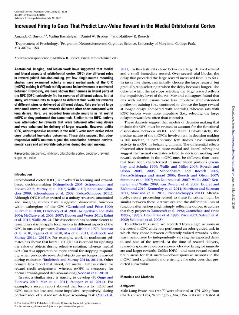
As illustrated by firing of the single-cell examples inFigure 2a,b, and for the population (n = 56) in Figure 2c,d,activity was often higher when the reward was large (Fig. 2a,d;dark gray) or delivered after a short delay (Fig. 2a–c; darkgray), compared with when the reward was small or delayedby several seconds (dark gray vs. gray; reward epoch; singleunit: t-test, P < 0.05; population: Wilcoxon; P < 0.05). To quan-tify these effects, for each reward-responsive neuron, weplotted difference scores between firing during short and long,
and large and small rewards, and asked in how many neuronswas there significant differential firing within each valuemanipulation on forced-choice trials (ANOVA; regression;P < 0.05; reward epoch). The distributions of value indices areplotted in Figure 2e. For both delay (Fig. 2ei) and size(Fig. 2eiii), the distributions were significantly shifted (Wilcox-on; P’ < 0.05) in the positive direction, indicating that themajority of mOFC neurons fired more strongly for high- com-pared with low-value reward (i.e., short and large over longand small, respectively). The 2 effects were not correlated(Fig. 2eii; P = 0.37; r2 = 0.01), suggesting that neurons that in-creased firing for one value manipulation did not show thesame change for the other value manipulation as one wouldexpect if activity in the mOFC reflected some sort of common-currency encoding (Roesch et al. 2006).
Finally, the counts of reward-responsive neurons that firedsignificantly more strongly for high- compared to low-valueoutcomes (ANOVA; P < 0.025; Bonferroni) were in the signifi-cant majority [Fig. 2e; black bars; 20 (49%) vs. 9 (22%); χ2;P < 0.05]. To further illustrate the significance of this result atthe single-unit level, we performed a multiple regression analy-sis with delay and size as factors (Roesch et al. 2006). Duringthe reward epoch, 29 (52%) and 14 (25%) of reward-responsiveneurons showed a positive and negative correlation with avalue in the multiple regression, respectively (P < 0.05).
Firing to Delayed RewardsReward-related activity in the mOFC appeared to reflect theanticipation of reward. During trials in which the delay wasonly 500 ms (i.e., short, big, and small), activity started to riseprior to reward onset. For long-delayed rewards, significant in-creases in firing did not occur until after reward delivery. Thisis evident in the single-cell example shown in Figure 2a andacross the population (Fig. 2c). In Fig. 2a,c, activity forrewards delivered after short delay increased firing during the500 ms preceding reward delivery (dark gray), whereas activitydid not show a change in firing during long-delay trials untilreward was actually delivered at time zero (light gray). Thislikely reflects the difficulty that animals have in timing rewardsthat are delayed by several seconds as measured by anticipat-ory licking (Fiorillo et al. 2008; Kobayashi and Schultz 2008;Takahashi et al. 2009).
Indeed, rats in our study licked more in anticipation ofreward delivery on short- compared with long-delay trials. Theaverage lick rate during the 250 ms before reward delivery wassignificantly higher for short-delay trials (vs. long-delay; t-test;t55 = 8.0; P < 0.05), suggesting that they could better anticipatethe more immediate reward. The strength of this differencewas correlated with the difference in firing in the mOFC duringshort- and long-delay trials during the reward epoch (Fig. 2f;P = 0.05; r2 = 0.07), suggesting that when rats could betteranticipate reward delivery, activity was stronger in the mOFC.
This correlation might suggest that activity in the mOFCsimply reflected motor commands that are coupled to value,such as licking, orofacial movements, and swallowing (Gutier-rez et al. 2006). Although it is difficult to rule this out, we donot think this is the case because the average licking rateduring the reward epoch was not correlated with the averagefiring rate during the same period (reward epoch; P = 0.63;r2 = 0.004) during performance of size blocks. In addition, aswe will describe below, activity in the mOFC represented the
Cerebral Cortex December 2014, V 24 N 12 3313
by guest on March 22, 2016
http://cercor.oxfordjournals.org/D
ownloaded from
spatial direction of the movement, whereas the licking rate wasnot significantly modulated by the spatial location (rewardepoch; t-test; t55 = 0.06 P = 0.95). To further address this issue,we examined activity and licking 2–5 s after reward delivery,during which time activity in the mOFC might have reflectedprolonged licking associated with consumption of the largereward. Even during this extended period, the correlationbetween licking and firing rates was not significant (P = 0.18;r2 = 0.04).
Although reward-related activity was attenuated on long- rela-tive to short-delay trials as in Figure 2a, other neurons did main-tain firing during anticipation of rewards delayed by severalseconds (long-delay trials). This is best illustrated by the single-cell example in Figure 2b (long). Activity immediately precedingreward delivery (500 ms) was significantly higher comparedwith baseline when rats were waiting for the delayed reward(light gray). Of the 56 reward-responsive neurons, 27 (48%;t-test; P < 0.05) exhibited significantly higher firing during the
500 ms before reward delivery compared with baseline,whereas only 7 fired significantly less (χ2; P < 0.05), demonstrat-ing that many single neurons in the mOFC fired in anticipationof the delayed reward.
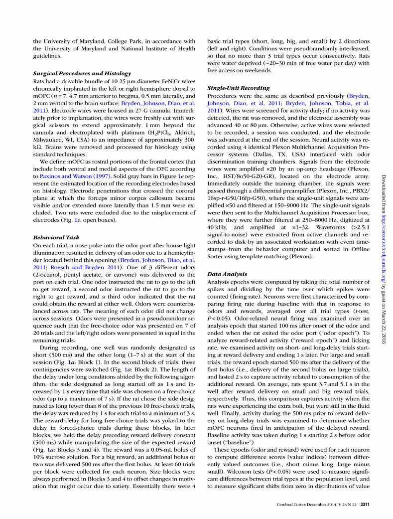
Odor-Evoked Activity in the OFC Was Stronger for CuesThat Predict Long Delay and Small RewardThen, we examined activity during odor sampling. Of the 251total mOFC neurons, 41 [16%; n’ = 1 (11%), 1 (3%), 4 (11%), 17(21%), and 18 (20%)] fired significantly more strongly duringodor sampling compared with baseline (odor epoch; t-test;P < 0.05; χ2; P < 0.05). Surprisingly, neurons in the mOFC firedsignificantly more strongly for odor cues that predicted low-value outcomes. This is illustrated in the single-cell examplesand across the entire population of odor-responsive mOFCneurons in Figure 3a–d. Immediately after odor onset, beforeinitiation of the behavioral response (port exit), populationactivity was significantly higher when small versus large
Figure 2. Reward-related activity in the OFC was stronger for an immediate and large reward. (a–d) Histograms representing the activity of single cells (a–b) and across thepopulation (c–d) of reward-responsive neurons (n= 56; 22%) in the mOFC during task performance of delay (dark gray = short; light gray = long) and size (dark gray = big; lightgray = small) blocks. Activity is aligned to reward delivery (time zero). For short, big, and small trials, well entry occurred 500 ms before reward delivery. On long-delay trials, wellentry was 1–7 s before reward delivery. Neurons were selected by comparing activity during the reward epoch when compared with baseline (see text; t-test; P< 0.05). Activity isnormalized by subtracting the mean and dividing by the standard deviation (z-score). Bins are 100 ms. Thickness of line reflects standard error of the mean (SEM). Note that activitythat precedes the 500 ms before reward delivery for rewards that were delivered after a short delay (short, big, and small) cannot be directly compared with activity that precedes500 ms before reward delivery on long-delay trials, because task events (well entry, port exit, etc.) occur at different time points across these trial types. (e) Correlation (ii) betweendifference scores for size and delay blocks (i.e., short minus long (i) and large minus small (iii)). Neural activity was taken during the reward epoch. Black bars in distributionhistogram represent neurons that showed a significant difference between differently valued outcomes (P<0.05; main or interaction effect of value in a 2-factor ANOVA; rewardepoch). ( f ) Correlation between value indices for the licking rate in anticipation of reward (250 ms before reward delivery) and for the firing rate during the reward epoch (1 s afterreward) on short- and long-delay trials (short-long). All data are taken from forced-choice trials.
3314 mOFC Neurons Fire to Cues That Predict Low Value • Burton et al.
by guest on March 22, 2016
http://cercor.oxfordjournals.org/D
ownloaded from

reward was predicted (Fig. 3d; dark vs. light gray; odor epoch;Wilcoxon; P < 0.05) and when long versus short delay was pre-dicted (Fig. 3c; dark vs. light gray; odor epoch; Wilcoxon;P < 0.05).
To further quantify this effect, we computed the differencescores between high- and low-value rewards and asked in howmany odor-responsive units did forced-choice odors that pre-dicted low-value reward elicit significantly stronger firing(Fig. 3e; ANOVA; odor epoch; black bars). The number ofneurons that fired more strongly for odors that predicted a lowvalue (ANOVA; P < 0.025; Bonferroni) were in the significantmajority [11 (26%) vs. 2 (5%); χ2 = 3.8; P < 0.05]. To furtherillustrate the significance of this result at the single-unit level,we performed a multiple regression analysis with delay andsize as factors during the odor epoch (Roesch et al. 2006). Asexpected from the ANOVA, 1 (2.4%) and 10 (24%) showed apositive and negative correlation with the value, respectively(P < 0.05).
At the population level, both delay (Fig. 3ei) and size(Fig. 3eiii) distributions were significantly shifted in the nega-tive direction (Wilcoxon; P’ < 0.05). Although delay effects ap-peared weaker than size effects, the 2 distributions were notsignificantly different (Wilcoxon; P = 0.75). As we will describebelow, stronger differences emerge when trials are brokendown by the direction.
Although the 2 effects appeared to be correlated—most ofthe cells fell in the bottom left quadrant—the correlationbetween size and delay indices was not significant (Fig. 3eii;P = 0.20; r2 = 0.04). This indicates that those neurons that firedmore strongly for cues that predicted longer delays were notnecessarily those that fired more strongly when the same cuepredicted small reward, and vice versa, even though the overalleffect was one of higher firing for longer delay and smallerreward predicting cues. As in the lOFC, this suggests that encod-ing in the mOFC does not reflect some sort of common-currencyencoding for expected rewards (Roesch et al. 2006).
Figure 3. Odor-evoked activity in the OFC was stronger for cues that predict long delay and small reward. (a–d) Histograms representing the activity of single cell (a–b) and acrossthe population (c–d) of odor-responsive neurons (n=41; 16%) in the mOFC during task performance of delay (dark gray = short; light gray = long) and size (dark gray = big; lightgray = small) blocks aligned to odor onset. For short, big, and small trials reward occurred several seconds later. This included the 500 ms of odor delivery and prefluid delay plusthe intervening time taken to respond to the odor and move to the odor port. On long-delay trials, reward occurred an additional 0.5–6.5 s later, thus cannot be examined in thisfigure. (e) Correlation (ii) between difference scores for size and delay blocks [i.e., short minus long (i) and large minus small (iii)]. Activity was taken during the odor epoch (100 msafter odor onset to odor port exit). Black bars in distribution histogram represent neurons that showed a significant difference between differently valued outcomes (P<0.05; mainor interaction effect of value in a 2-factor ANOVA; odor epoch). (f ) Activity is aligned to odor port exit to show that differences in activity between high- and low-value outcomeswas not a product of different reaction times. Neurons were selected by comparing activity during the odor epoch with baseline (1 s before nosepoke; t-test; P<0.05). Activity isnormalized by subtracting the mean and dividing by the standard deviation (z-score). Bins are 100 ms. Thickness of line reflects standard error of the mean (SEM). All data are takenfrom forced-choice trials.
Cerebral Cortex December 2014, V 24 N 12 3315
by guest on March 22, 2016
http://cercor.oxfordjournals.org/D
ownloaded from
Increased activity during odors that predict a low-valuereward does not simply reflect the fact that rats spent moretime in the odor port. Activity in the mOFC does not continueto fire until port exit. This is illustrated in Figure 3f, whichaligns activity on odor port exit for high- and low-value trialsaveraged over size and delay manipulations. Note that activityon low- and high-value trials peak and come back togetherbefore port exit. Further, if we repeat the analysis that exam-ines difference scores described in the previous paragraphwith an analysis epoch that is cut off at 100 ms after odoroffset, instead of being variable to port exit, the results remainthe same; both delay and size distributions are significantlyshifted in the negative direction (Wilcoxon; P’ < 0.05).
Thus, activity during reward delivery and odor sampling inthe mOFC carry different signs in relation to rewarding out-comes, with odor- and reward-related activity being strongerand weaker for low-value outcomes, respectively. To deter-mine whether the 2 effects were correlated, we performed aregression analysis on value indices during the odor andreward epoch for all odor- and reward-responsive neurons(n = 97). The correlation between the 2 was not significant(r2 = 0.03; P = 0.10), suggesting that neurons that tended to firemore strongly for cues that predicted a low-value reward didnot tend to fire more strongly during delivery of high-valueoutcomes.
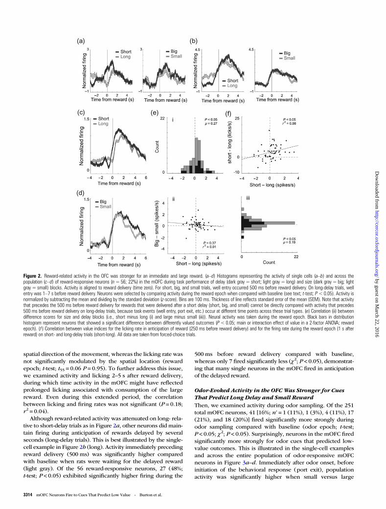
Encoding of Response Direction in the mOFCPrevious results have shown that activity in rat lOFC respondsdifferently depending on the direction of the behavioral
response (Feierstein et al. 2006; Roesch et al. 2006, 2007).Here, we asked if activity in the mOFC was also modulated byresponse direction. Of the 41 odor-responsive neurons, 9(22%) showed a significant main or interaction effect withresponse direction in a 2-factor ANOVA (P < 0.025; χ2; P < 0.05)as illustrated by the single-cell example in Figure 4a; activitywas highest when odor cues predicted a small reward in theleft well. To further qualify this, we broke down the populationactivity into each cell’s preferred and nonpreferred direction(Fig. 4b), as defined by the direction that elicited the strongestresponse (e.g., left in the example). Here, “preferred” refers tothe direction that elicited the highest activity, not the outcomefavored by the animal. Across the population of odor-responsive cells, the difference between high- and low-valueoutcomes appeared to be stronger for responses made in thepreferred direction (filled dark vs. light gray) compared withthe nonpreferred direction (open dark vs. light gray). In thepreferred direction, the distribution of value indices wasshifted in the negative direction, indicating stronger firing for alower-value reward (Fig. 4c; black; Wilcoxon; P < 0.05). In thenonpreferred direction, the distribution of value indices wasnot significantly shifted (Fig. 4c; gray; Wilcoxon; P = 0.88) andwas significantly different than that of indices obtained frompreferred direction trials (Fig. 4c; Wilcoxon; P < 0.05).
Of the 56 reward-responsive neurons, 21 (38%) showed asignificant main or interaction effect with response direction inthe ANOVA (P < 0.025; χ2; P < 0.05). In addition, the differencebetween high- and low-value outcomes appeared to be stron-ger for responses made in the preferred direction (Fig. 5b;
Figure 4. Odor-responsive neurons in the mOFC were directionally selective. (a) Activity of a single cell during size blocks demonstrating higher firing when odor cues predicted asmall reward on the left. (b) Average firing rate over all 41 odor-responsive neurons broken down by preferred and nonpreferred response direction. Preferred direction was defined foreach cell by determining which trial type elicited the strongest firing. Filled = preferred direction; open = nonpreferred direction; dark gray = high value (short and big); lightgray = low value (long and small). Activity is normalized by subtracting the mean and dividing by the standard deviation (z-score). Bins are 100 ms. Thickness of line reflectsstandard error of the mean (SEM). (c) Distribution of value indices taken during the odor epoch (see Methods) independently for preferred (black) and nonpreferred responsedirections (light gray). Light gray distributions are transparent, and dark gray thus indicates where black (preferred) and light gray (nonpreferred) overlap. The Wilcoxon test wereused to determine whether the 2 distributions were significantly different from zero and from each other (P<0.05).
3316 mOFC Neurons Fire to Cues That Predict Low Value • Burton et al.
by guest on March 22, 2016
http://cercor.oxfordjournals.org/D
ownloaded from

filled dark gray vs. light gray). In the preferred direction,values were shifted in the positive direction indicating strongerfiring for higher-value reward (Fig. 5c; black; Wilcoxon;P < 0.05). The distribution of value indices in the nonpreferreddirection was not significantly shifted (Fig. 5c; gray; Wilcoxon;P = 0.28) and was significantly different from that of valueindices obtained on preferred direction trials (Wilcoxon;P < 0.05). Thus, we conclude that odor- and reward-responsivemOFC neurons showed enhanced value encoding in the cell’spreferred response direction.
Emergence of Outcome Selectivity During Odor Samplingand its Relation to BehaviorIn a final analysis, we examined how selectivity for cues thatpredicted low-value outcomes emerged during learning andwhether activity in the mOFC was correlated with reactiontime. Figure 6a,b plots the average firing rate over delay andsize blocks for movements made in the preferred directionduring the first and last 5 trials for each trial type. Consistentwith the previous sections, activity after learning was strongerfor low-value outcomes (Fig. 6a,b; solid gray vs. black). Inter-estingly, this selectivity developed as a result of decreasedfiring on high-value trials that occurred with learning (Fig. 6a,b; black dashed vs. black solid). That is, activity was signifi-cantly lower for short-delay and large-reward trials at the endof the trial block relative to the beginning (odor epoch; Wilcox-on; P’s < 0.05). This relationship did not exist for cues that pre-dicted low-value rewards (Fig. 6a,b; gray dashed vs. graysolid). Thus, selectivity emerged through a reduction in firingfor cues that predicted a high-value reward.
This is further quantified in Figure 6c, which plots the nor-malized difference between high- and low-value outcomesduring the first 5 (black) and last 5 (gray) trials. The
distribution is significantly shifted below zero only after learn-ing (Wilcoxon; P < 0.05) and significantly different than duringearly trials (Wilcoxon; P < 0.05). Differences in firing reflectedthe rats’ behavior in that value-induced differences in reactiontime (faster for high-value reward) were present during late(Fig. 6d; gray; P < 0.05), but not early, trials (Fig. 6d; black; Wil-coxon; P = 0.81). Furthermore, the change in firing that oc-curred over the course of the trial block was significantlycorrelated with the strength of learning that occurred duringthe session as measured by changes in reaction time (Fig. 6e;r2 = 0.09; P < 0.05).
These results suggest that mOFC may serve to alter behaviorwhen low-value rewards are predicted by forced-choice cues.If true, then neural selectivity observed after learning, duringodor sampling, might be correlated with reaction time differ-ences observed between cues that predict high- and low-valueoutcomes. Consistent with this hypothesis, strong enhance-ment of the firing rate during odor sampling was correlatedwith slower behavioral responses. This is illustrated inFigure 7, which plots the value index (high− low/high + low)computed on average firing rates during the odor epochagainst the value index computed for reaction times duringthose trials. As expected from the analysis above, both indiceswere negative, indicating slower reaction times and higherfiring on low-value trials. Furthermore, both were correlated,demonstrating that when rats showed stronger reaction timedifferences, neural selectivity in the mOFC was enhanced(P < 0.05; r2 = 0.21).
Discussion
Consistent with imaging and anatomical studies, recent lesionwork in rats and primates has shown that subregions of the
Figure 5. Reward-responsive neurons in the mOFC were directionally selective. (a) Activity of a single cell during delay blocks demonstrating higher firing during short- versuslong-delay trials at the time of reward delivery. (b and c) Same as b–c in Figure 4, except for the 56 reward-responsive neurons (reward epoch).
Cerebral Cortex December 2014, V 24 N 12 3317
by guest on March 22, 2016
http://cercor.oxfordjournals.org/D
ownloaded from

OFC perform different functions (Noonan et al. 2010; Mar et al.2011; Rudebeck and Murray 2011a, 2011b). However, few
studies have examined activity in the mOFC making it difficultto understand the exact nature that mOFC plays in reward-guided decision-making. Here, we demonstrate that rewardand cue-evoked responses in the mOFC are modulated by thesize of and delay to reward, 2 value manipulations that clearlyimpact decision making. At the time of reward delivery, activitywas higher when outcomes were of higher value. During odorsampling, the opposite effect was observed, that is, firing washigher for odor cues that predicted low-value outcomes inodor-responsive neurons. Below we discuss these results, com-paring mOFC to activity in other areas, including our ownwork in lOFC, with the caveat that these comparisons aremade across studies, in different rats, and that neurons mighthave been sampled from different layers considering the struc-ture of these 2 areas.
In previous reports, we characterized firing in the lOFC asrats performed the same task (Roesch et al. 2006; Takahashiet al. 2009; Roesch et al. 2012). Activity related to rewardexpectancy and delivery was similar across mOFC and lOFC inthat overall activity was reduced when rewards were delayed.The major difference between these 2 subregions emergedduring the sampling of odors that predicted different out-comes. Although neurons that show increased firing for cues
Figure 6. Emergence of cue selectivity during learning. (a and b) Population activity for the 41 odor-responsive neurons for responses made in the preferred direction, averaged overfree- and forced-choice trials, for delay (a) and size (b) blocks. For each trial type, the average of the first (dashed) and last (solid) 5 trials in a block are shown. Black = short orlarge; gray = long or small. (c) Distribution of value indices (high− low/high + low) reflecting the firing rate (odor epoch) difference between high- and low-value outcomes, early(first 5 trials; black) and late (last 5 trials; late) during learning. (d) Distribution of value indices (high− low/high + low) reflecting the reaction time difference between high- andlow-value outcomes, early (first 5 trials; black) and late (last 5 trials; late) during learning. (e) Scatter plot represents the correlation between changes in firing and in reaction timethat occur during learning (early− late/early + late) on high-value reward trials. FR: firing rate; RT: reaction time. The Wilcoxon test were used to determine whether the 2distributions were significantly different from zero and from each other (P<0.05).
Figure 7. Correlation between reaction time (RT) and firing rate (FR) collapsed acrossboth value manipulations. Scatter plot represents the correlation between high- andlow-value trial-type differences for reaction time (odor offset to odor port exit) andneural firing (odor epoch) averaged across value manipulation and direction. Valueindex = high− low/high + low.
3318 mOFC Neurons Fire to Cues That Predict Low Value • Burton et al.
by guest on March 22, 2016
http://cercor.oxfordjournals.org/D
ownloaded from
that predict a low-value reward have been described pre-viously in the lOFC, the proportion of neurons in the mOFCshowing this effect were in the majority, and the populationresponse was stronger over all neurons when cues signaled alow value. This makes mOFC unique among brain areasthought to be critical in reward-guided decision-making; mostreward-related regions in the brain fire more strongly for cuesthat predict a more valuable reward, including lOFC (Tremblayand Schultz 1999; Roesch and Olson 2004, 2005; Schoenbaumand Roesch 2005; Padoa-Schioppa and Assad 2006; Roeschand Olson 2007; Simmons et al. 2007; van Duuren et al. 2007;Wallis 2007; Kennerley and Wallis 2009; van Duuren et al.2009; Bouret and Richmond 2010; Kennerley et al. 2011;Padoa-Schioppa 2011).
Such a signal might be important for inhibitory control and/or complement more common response bias signals that areelevated when animals expect better rewards. Consistent withthis idea, reports in humans and rats have shown that mOFCdysfunction causes subjects to make more risky decisions,possibly due to a disruption in inhibitory control over biases toselect riskier rewards (Clark et al. 2008; St Onge and Floresco2010; Zeeb et al. 2010; Stopper et al. 2014). Furthermore, ourdata indicate that increased firing in the mOFC during thesampling of odors that predicted low-value outcomes was posi-tively correlated with differences in reaction time, indicatingthat when activity was high, reaction times were slow.However, we must exercise some caution here because in-creased firing on low-value trials might also be interpreted assignals that allow for a behavioral response to occur, albeitaway from the more valued outcome.
In none of our studies, have we found a single brain areathat increased population firing to cues that predicted a low-value reward (Roesch and Bryden 2011). This includes brainareas that are in relatively close proximity to our recordingsites, such as medial prefrontal cortex (mPFC) and lOFC(Roesch et al. 2006, 2012; Gruber et al. 2010). Unfortunately,in this study, we cannot dissociate between medial and ventralOFC because our sample size was too low. With that said, weobserved no obvious differences between the 2 regions, butfuture work is necessary to determine if they carry differentsignals. Although connections of the medial and ventral OFCdo overlap, recent work based on connectivity has suggestedthat ventral and medial OFC might play different roles compar-able with lOFC and mPFC, respectively, and that both ventraland medial aspects of OFC might serve as a link between lOFCand mPFC (Hoover and Vertes 2011; Kahnt et al. 2012; Wallis2012). Findings such as these make it difficult to draw a hardline between mOFC and mPFC. Regardless of whether youconsider this region part of the OFC or PFC, we show that pre-dicted outcome encoding is considerably different relative tothe lOFC (Roesch et al. 2006, 2012) and PFC (Gruber et al.2010), consistent with recent lesion work targeting this specificregion (Mar et al. 2011; Stopper et al. 2014).
To the best of our knowledge, there is only one other single-unit study that has shown elevated firing in the majority ofcue-responsive neurons when animals anticipate a smallreward. In that paper, monkeys performed a go/no-go task fora predicted large or small reward (Minamimoto et al. 2005).They found increased activity in the centromedian nucleus ofthe thalamus (CM) when monkeys made actions (go or no-go)
for a small compared with a large reward. Further, theyshowed that stimulation of CM caused typically speeded reac-tions to be slow, demonstrating a role for CM in a mechanismcomplementary to more common signals that are thought tobias animals toward better reward. Although connectivitybetween mOFC and CM is relatively light, it is possible thatinteractions between them are critical for reward-guided beha-viors (Hoover and Vertes 2011; Vertes et al. 2012).
For decades, it was thought that OFC was critical forresponse inhibition because damage to OFC made animals andhumans lose aspects of inhibitory control and become moreimpulsive in their actions (Damasio et al. 1994; Bechara et al.2000; Berlin et al. 2004; Torregrossa et al. 2008; Schoenbaumet al. 2009). Here, we show that activity was high during situ-ations in which the animal had to inhibit responding at the be-ginning of trial blocks and when forced-choice trials instructedthe rat to respond away from the desired reward toward thelow value well. Loss of signaling of unfavorable outcomesduring decision making and learning could account for manyof the deficits thought to reflect deficits in inhibition. Interest-ingly, if the role of this signal is to inhibit behavioral output, itappears to do so in an outcome-specific manner, because thecorrelation between selective firing during size and delayblocks was not significant (Fig. 3e), suggesting that OFC doesnot output a simple general/global inhibition signal.
One common way to assess response inhibition and impul-sivity is to conduct delay-discounting procedures in whichanimals choose between small immediate rewards and largerewards delivered after long delays (Cardinal et al. 2004; Ka-lenscher and Pennartz 2008; Zeeb et al. 2010). Although the in-volvement of OFC in impulsive choice is indisputable, theexact role it plays is still unclear due to conflicting findingsfrom several different labs (Kheramin et al. 2002; Mobini et al.2002; Winstanley et al. 2004; Rudebeck et al. 2006; Winstanley2007; Clark et al. 2008; Churchwell et al. 2009; Sellitto et al.2010; Zeeb et al. 2010; Mar et al. 2011).
To add to the complexity of this story, more recent work hasshown that different regions of the OFC serve opposing func-tions during delay discounting (Mar et al. 2011). In this study,Mar and colleagues showed that lesions of the mOFC maderats discount less, encouraging responding to the delayedreward after extended postlesion training (i.e., less impulsive),whereas lOFC lesions make rats discount more, decreasingpreference for the delayed reward (i.e., more impulsive). Still,others have reported no impact of mOFC inactivation on delaydiscounting (Stopper et al. 2014).
Here, we show that, like lOFC, activity in mOFC reward-responsive neurons was attenuated for delayed reward. Theseresults suggest that distinctive deficits observed after focallesions to mOFC and lOFC cannot be explained by differencesin firing during reward delivery. However, unlike lOFC, odor-responsive neurons in the mOFC signal low-value outcomes atthe time of the decision. We suggest that mOFC’s role in classicdelay-discounting tasks is to signal low-value outcomes duringdecision making. Although these effects were significant, theywere not dramatic, suggesting that other tasks are necessary tofully uncover the roles that mOFC plays in behavior, such astasks that require more inhibitory-related functions (e.g., stop-signal) and those that require decisions made under risk (e.g.,probability and uncertainty).
Cerebral Cortex December 2014, V 24 N 12 3319
by guest on March 22, 2016
http://cercor.oxfordjournals.org/D
ownloaded from
Funding
This work was supported by grants from the National Instituteon Drug Abuse (R01DA031695, M.R.).
NotesConflict of Interest: None declared.
ReferencesBechara A, Damasio H, Damasio AR. 2000. Emotion, decision making
and the orbitofrontal cortex. Cereb Cortex. 10:295–307.Berlin HA, Rolls ET, Kischka U. 2004. Impulsivity, time perception,
emotion and reinforcement sensitivity in patients with orbitofrontalcortex lesions. Brain. 127:1108–1126.
Bouret S, Richmond BJ. 2010. Ventromedial and orbital prefrontalneurons differentially encode internally and externally driven moti-vational values in monkeys. J Neurosci. 30:8591–8601.
Bryden DW, Johnson EE, Diao X, Roesch MR. 2011. Impact of expectedvalue on neural activity in rat substantia nigra pars reticulata. Eur JNeurosci. 33:2308–2317.
Bryden DW, Johnson EE, Tobia SC, Kashtelyan V, Roesch MR. 2011. At-tention for learning signals in anterior cingulate cortex. J Neurosci.31:18266–18274.
Cardinal RN, Winstanley CA, Robbins TW, Everitt BJ. 2004. Limbic cor-ticostriatal systems and delayed reinforcement. Ann N Y Acad Sci.1021:33–50.
Carmichael ST, Price JL. 1996. Connectional networks within theorbital and medial prefrontal cortex of macaque monkeys. J CompNeurol. 371:179–207.
Carmichael ST, Price JL. 1995a. Limbic connections of the orbital andmedial prefrontal cortex in macaque monkeys. J Comp Neurol.363:615–641.
Carmichael ST, Price JL. 1995b. Sensory and premotor connections ofthe orbital and medial prefrontal cortex of macaque monkeys. JComp Neurol. 363:642–664.
Churchwell JC, Morris AM, Heurtelou NM, Kesner RP. 2009. Inter-actions between the prefrontal cortex and amygdala during delaydiscounting and reversal. Behav Neurosci. 123:1185–1196.
Clark L, Bechara A, Damasio H, Aitken MR, Sahakian BJ, Robbins TW.2008. Differential effects of insular and ventromedial prefrontalcortex lesions on risky decision-making. Brain. 131:1311–1322.
Damasio H, Grabowski T, Frank R, Galaburda AM, Damasio AR. 1994.The return of Phineas Gage: clues about the brain from the skull ofa famous patient. Science. 264:1102–1105.
Elliott R, Dolan RJ, Frith CD. 2000. Dissociable functions in the medialand lateral orbitofrontal cortex: evidence from human neuroima-ging studies. Cereb Cortex. 10:308–317.
Feierstein CE, Quirk MC, Uchida N, Sosulski DL, Mainen ZF. 2006. Rep-resentation of spatial goals in rat orbitofrontal cortex. Neuron.51:495–507.
Fiorillo CD, Newsome WT, Schultz W. 2008. The temporal precision ofreward prediction in dopamine neurons. Nat Neurosci. 11:966–973.
Gruber AJ, Calhoon GG, Shusterman I, Schoenbaum G, Roesch MR,O’Donnell P. 2010. More is less: a disinhibited prefrontal corteximpairs cognitive flexibility. J Neurosci. 30:17102–17110.
Gutierrez R, Carmena JM, Nicolelis MA, Simon SA. 2006. Orbitofrontalensemble activity monitors licking and distinguishes among naturalrewards. J Neurophysiol. 95:119–133.
Hoover WB, Vertes RP. 2011. Projections of the medial orbital andventral orbital cortex in the rat. J Comp Neurol. 519:3766–3801.
Iversen SD, Mishkin M. 1970. Perseverative interference in monkeysfollowing selective lesions of the inferior prefrontal convexity. ExpBrain Res. 11:376–386.
Kable JW, Glimcher PW. 2009. The neurobiology of decision: consen-sus and controversy. Neuron. 63:733–745.
Kahnt T, Chang LJ, Park SQ, Heinzle J, Haynes JD. 2012. Connectivity-based parcellation of the human orbitofrontal cortex. J Neurosci.32:6240–6250.
Kalenscher T, Pennartz CM. 2008. Is a bird in the hand worth two inthe future? The neuroeconomics of intertemporal decision-making.Prog Neurobiol. 84:284–315.
Kennerley SW, Behrens TE, Wallis JD. 2011. Double dissociation ofvalue computations in orbitofrontal and anterior cingulate neurons.Nat Neurosci. 14:1581–1589.
Kennerley SW, Wallis JD. 2009. Encoding of reward and space during aworking memory task in the orbitofrontal cortex and anterior cin-gulate sulcus. J Neurophysiol. 102:3352–3364.
Kheramin S, Body S, Mobini S, Ho MY, Velazquez-Martinez DN, Brad-shaw CM, Szabadi E, Deakin JF, Anderson IM. 2002. Effects of qui-nolinic acid-induced lesions of the orbital prefrontal cortex oninter-temporal choice: a quantitative analysis. Psychopharmacology(Berl). 165:9–17.
Kobayashi S, Schultz W. 2008. Influence of reward delays on responsesof dopamine neurons. J Neurosci. 28:7837–7846.
Kringelbach ML. 2005. The human orbitofrontal cortex: linking rewardto hedonic experience. Nat Rev Neurosci. 6:691–702.
Kringelbach ML, Rolls ET. 2004. The functional neuroanatomy of thehuman orbitofrontal cortex: evidence from neuroimaging and neu-ropsychology. Prog Neurobiol. 72:341–372.
Mar AC, Walker AL, Theobald DE, Eagle DM, Robbins TW. 2011. Disso-ciable effects of lesions to orbitofrontal cortex subregions on impul-sive choice in the rat. J Neurosci. 31:6398–6404.
McClure SM, Ericson KM, Laibson DI, Loewenstein G, Cohen JD.2007. Time discounting for primary rewards. J Neurosci. 27:5796–5804.
McClure SM, Laibson DI, Loewenstein G, Cohen JD. 2004. Separateneural systems value immediate and delayed monetary rewards.Science. 306:503–507.
Minamimoto T, Hori Y, Kimura M. 2005. Complementary process toresponse bias in the centromedian nucleus of the thalamus.Science. 308:1798–1801.
Mobini S, Body S, Ho MY, Bradshaw CM, Szabadi E, Deakin JF, Ander-son IM. 2002. Effects of lesions of the orbitofrontal cortex onsensitivity to delayed and probabilistic reinforcement. Psychophar-ma-cology (Berl). 160:290–298.
Morecraft RJ, Geula C, Mesulam MM. 1992. Cytoarchitecture and neuralafferents of orbitofrontal cortex in the brain of the monkey. J CompNeurol. 323:341–358.
Morrison SE, Saez A, Lau B, Salzman CD. 2011. Different time coursesfor learning-related changes in amygdala and orbitofrontal cortex.Neuron. 71:1127–1140.
Morrison SE, Salzman CD. 2011. Representations of appetitive andaversive information in the primate orbitofrontal cortex. Ann N YAcad Sci. 1239:59–70.
Murray EA, O’Doherty JP, Schoenbaum G. 2007. What we know anddo not know about the functions of the orbitofrontal cortex after 20years of cross-species studies. J Neurosci. 27:8166–8169.
Noonan MP, Walton ME, Behrens TE, Sallet J, Buckley MJ, RushworthMF. 2010. Separate value comparison and learning mechanisms inmacaque medial and lateral orbitofrontal cortex. Proc Natl Acad SciUSA. 107:20547–20552.
O’Doherty J, Kringelbach ML, Rolls ET, Hornak J, Andrews C. 2001. Ab-stract reward and punishment representations in the human orbito-frontal cortex. Nat Neurosci. 4:95–102.
Padoa-Schioppa C. 2011. Neurobiology of economic choice: a good-based model. Annu Rev Neurosci. 34:333–359.
Padoa-Schioppa C, Assad JA. 2006. Neurons in the orbitofrontal cortexencode economic value. Nature. 441:223–226.
Paxinos G, Watson C. 1997. The Rat Brain in Stereotaxic Coordinates,Compact 3rd ed. London: Academic Press. p 11–15.
Price JL. 2007. Definition of the orbital cortex in relation to specificconnections with limbic and visceral structures and other corticalregions. Ann N Y Acad Sci. 1121:54–71.
Price JL, Carmichael ST, Drevets WC. 1996. Networks related to theorbital and medial prefrontal cortex: a substrate for emotional be-havior? Prog Brain Res. 107:523–536.
Roesch MR, Bryden DW. 2011. Impact of size and delay on neuralactivity in the rat limbic corticostriatal system. Front Neurosci.5:130.
3320 mOFC Neurons Fire to Cues That Predict Low Value • Burton et al.
by guest on March 22, 2016
http://cercor.oxfordjournals.org/D
ownloaded from

Roesch MR, Bryden DW, Cerri DH, Haney ZR, Schoenbaum G. 2012.Willingness to wait and altered encoding of time-discounted rewardin the orbitofrontal cortex with normal aging. J Neurosci.32:5525–5533.
Roesch MR, Calu DJ, Burke KA, Schoenbaum G. 2007. Should I stay orshould I go? Transformation of time-discounted rewards in orbito-frontal cortex and associated brain circuits. Ann N Y Acad Sci.1104:21–34.
Roesch MR, Olson CR. 2005. Neuronal activity in primate orbitofrontalcortex reflects the value of time. J Neurophysiol. 94:2457–2471.
Roesch MR, Olson CR. 2007. Neuronal activity related to anticipatedreward in frontal cortex: does it represent value or reflect motiv-ation? Ann N Y Acad Sci. 1121:431–446.
Roesch MR, Olson CR. 2004. Neuronal activity related to reward valueand motivation in primate frontal cortex. Science. 304:307–310.
Roesch MR, Taylor AR, Schoenbaum G. 2006. Encoding of time-discounted rewards in orbitofrontal cortex is independent of valuerepresentation. Neuron. 51:509–520.
Rudebeck PH, Murray EA. 2011a. Balkanizing the primate orbitofron-tal cortex: distinct subregions for comparing and contrastingvalues. Ann N Y Acad Sci. 1239:1–13.
Rudebeck PH, Murray EA. 2011b. Dissociable effects of subtotallesions within the macaque orbital prefrontal cortex on reward-guided behavior. J Neurosci. 31:10569–10578.
Rudebeck PH, Walton ME, Smyth AN, Bannerman DM, Rushworth MF.2006. Separate neural pathways process different decision costs.Nat Neurosci. 9:1161–1168.
Rygula R, Walker SC, Clarke HF, Robbins TW, Roberts AC. 2010. Differ-ential contributions of the primate ventrolateral prefrontal and orbi-tofrontal cortex to serial reversal learning. J Neurosci. 30:14552–14559.
Saleem KS, Kondo H, Price JL. 2008. Complementary circuits connect-ing the orbital and medial prefrontal networks with the temporal,insular, and opercular cortex in the macaque monkey. J CompNeurol. 506:659–693.
Schilman EA, Uylings HB, Galis-de Graaf Y, Joel D, Groenewegen HJ.2008. The orbital cortex in rats topographically projects to centralparts of the caudate-putamen complex. Neurosci Lett. 432:40–45.
Schoenbaum G, Roesch M. 2005. Orbitofrontal cortex, associativelearning, and expectancies. Neuron. 47:633–636.
Schoenbaum G, Roesch MR, Stalnaker TA, Takahashi YK. 2009. A newperspective on the role of the orbitofrontal cortex in adaptive be-haviour. Nat Rev Neurosci. 10:885–892.
Sellitto M, Ciaramelli E, di Pellegrino G. 2010. Myopic discounting offuture rewards after medial orbitofrontal damage in humans. JNeurosci. 30:16429–16436.
Simmons JM, Ravel S, Shidara M, Richmond BJ. 2007. A comparison ofreward-contingent neuronal activity in monkey orbitofrontal cortexand ventral striatum: guiding actions toward rewards. Ann N YAcad Sci. 1121:376–394.
St Onge JR, Floresco SB. 2010. Prefrontal cortical contribution to risk-based decision making. Cereb Cortex. 20:1816–1828.
Stopper CM, Green EB, Floresco SB. 2014. Selective involvement bythe medial orbitofrontal cortex in biasing risky, but not impulsive,choice. Cereb Cortex. 24:154–162.
Takahashi YK, Roesch MR, Stalnaker TA, Haney RZ, Calu DJ, TaylorAR, Burke KA, Schoenbaum G. 2009. The orbitofrontal cortex andventral tegmental area are necessary for learning from unexpectedoutcomes. Neuron. 62:269–280.
Torregrossa MM, Quinn JJ, Taylor JR. 2008. Impulsivity, compulsivity,and habit: the role of orbitofrontal cortex revisited. Biol Psychiatry.63:253–255.
Tremblay L, Schultz W. 1999. Relative reward preference in primate or-bitofrontal cortex. Nature. 398:704–708.
van Duuren E, Escamez FA, Joosten RN, Visser R, Mulder AB, PennartzCM. 2007. Neural coding of reward magnitude in the orbitofrontalcortex of the rat during a five-odor olfactory discrimination task.Learn Mem. 14:446–456.
van Duuren E, van der Plasse G, Lankelma J, Joosten RN, Feenstra MG,Pennartz CM. 2009. Single-cell and population coding of expectedreward probability in the orbitofrontal cortex of the rat. J Neurosci.29:8965–8976.
Vertes RP, Hoover WB, Rodriguez JJ. 2012. Projections of the centralmedial nucleus of the thalamus in the rat: node in cortical, striataland limbic forebrain circuitry. Neuroscience. 219:120–136.
Wallis JD. 2012. Cross-species studies of orbitofrontal cortex and value-based decision-making. Nat Neurosci. 15:13–19.
Wallis JD. 2007. Orbitofrontal cortex and its contribution to decision-making. Annu Rev Neurosci. 30:31–56.
Wallis JD, Miller EK. 2003. Neuronal activity in primate dorsolateraland orbital prefrontal cortex during performance of a reward pre-ference task. Eur J Neurosci. 18:2069–2081.
Winstanley CA. 2007. The orbitofrontal cortex, impulsivity, and addic-tion: probing orbitofrontal dysfunction at the neural, neurochemi-cal, and molecular level. Ann N Y Acad Sci. 1121:639–655.
Winstanley CA, Theobald DE, Cardinal RN, Robbins TW. 2004. Con-trasting roles of basolateral amygdala and orbitofrontal cortex inimpulsive choice. J Neurosci. 24:4718–4722.
Zeeb FD, Floresco SB, Winstanley CA. 2010. Contributions of the orbi-tofrontal cortex to impulsive choice: interactions with basal levelsof impulsivity, dopamine signalling, and reward-related cues.Psychopharmacology (Berl). 211:87–98.
Cerebral Cortex December 2014, V 24 N 12 3321
by guest on March 22, 2016
http://cercor.oxfordjournals.org/D
ownloaded from
Related Documents