RESEARCH ARTICLE Open Access Increased expression of RXRa in dementia: an early harbinger for the cholesterol dyshomeostasis? Afia Akram 1,2* , James Schmeidler 2 , Pavel Katsel 2 , Patrick R Hof 1 , Vahram Haroutunian 2,3 Abstract Background: Cholesterol content of cerebral membranes is tightly regulated by elaborate mechanisms that balance the level of cholesterol synthesis, uptake and efflux. Among the conventional regulatory elements, a recent research focus has been nuclear receptors, a superfamily of ligand-activated transcription factors providing an indispensable regulatory framework in controlling cholesterol metabolism pathway genes. The mechanism of transcriptional regulation by nuclear receptors such as LXRs involves formation of heterodimers with RXRs. LXR/RXR functions as a sensor of cellular cholesterol concentration and mediates cholesterol efflux by inducing the transcription of key cholesterol shuffling vehicles namely, ATP-binding cassette transporter A1 (ABCA1) and ApoE. Results: In the absence of quantitative data from humans, the relevance of expression of nuclear receptors and their involvement in cerebral cholesterol homeostasis has remained elusive. In this work, new evidence is provided from direct analysis of human postmortem brain gene and protein expression suggesting that RXRa, a key regulator of cholesterol metabolism is differentially expressed in individuals with dementia. Importantly, RXRa expression showed strong association with ABCA1 and ApoE gene expression, particularly in AD vulnerable regions. Conclusions: These findings suggest that LXR/RXR-induced upregulation of ABCA1 and ApoE levels may be the molecular determinants of cholesterol dyshomeostasis and of the accompanying dementia observed in AD. Introduction Differential control of gene expression is an important means by which cells respond to physiological and environmental stimuli. Nuclear receptors comprise a superfamily of ligand regulated, DNA-binding transcrip- tion factors that can both activate and repress gene expression [1]. The liver X receptors (LXRs) are type II nuclear receptors, initially identified as orphan nuclear receptors, because their natural ligands were not known [2,3]. LXRs have been deorphanized or adopted follow- ing the discovery of oxysterols (hydroxylated derivatives of cholesterol) as their endogenous ligands [3,4]. Two LXR isoforms are known, namely LXRa and LXRb with distinct tissue distributions [5]. LXRa expression is rela- tively restricted to tissues involved in lipid metabolism, such as liver and intestine [6,7], whereas LXRb is ubiquitously expressed. Both LXR isoforms are expressed in the brain [5]. LXRb expression, in particu- lar is 2-5 fold higher in the brain than in liver [8]. The mechanisms of transcriptional regulation by LXRs involve formation of heterodimers with retinoid X receptor (RXR). RXR is another deorphanized nuclear receptor, activated by 9-cis-retinoic acid (a vitamin A derivative) [9]. Upon heterodimerization, LXR/RXR bind to specific DNA sequences called LXR-responsive ele- ments (LXREs) in the target genes [10]. In the absence of a ligand, LXRs bound to cognate LXREs, are in com- plex with corepressors such as silencing mediator of retinoic acid and thyroid hormone receptor (SMRT) [11] and nuclear receptor corepressor (N-CoR) [12], consequently, the transcription of target genes is repressed. Receptor ligation induces conformational changes in the ligand binding domain which mechanisti- cally facilitates the release of corepressors and the recruitment of coactivators and histone acetyltransferase (HAT), the enzyme that acetylates lysine amino acids on * Correspondence: [email protected] 1 Department of Neuroscience Mount Sinai School of Medicine, New York, NY 10029, USA Full list of author information is available at the end of the article Akram et al. Molecular Neurodegeneration 2010, 5:36 http://www.molecularneurodegeneration.com/content/5/1/36 © 2010 Akram et al; licensee BioMed Central Ltd. This is an Open Access article distributed under the terms of the Creative Commons Attribution License (http://creativecommons.org/licenses/by/2.0), which permits unrestricted use, distribution, and reproduction in any medium, provided the original work is properly cited.

Welcome message from author
This document is posted to help you gain knowledge. Please leave a comment to let me know what you think about it! Share it to your friends and learn new things together.
Transcript

RESEARCH ARTICLE Open Access
Increased expression of RXRa in dementia: anearly harbinger for the cholesteroldyshomeostasis?Afia Akram1,2*, James Schmeidler2, Pavel Katsel2, Patrick R Hof1, Vahram Haroutunian2,3
Abstract
Background: Cholesterol content of cerebral membranes is tightly regulated by elaborate mechanisms thatbalance the level of cholesterol synthesis, uptake and efflux. Among the conventional regulatory elements, a recentresearch focus has been nuclear receptors, a superfamily of ligand-activated transcription factors providing anindispensable regulatory framework in controlling cholesterol metabolism pathway genes. The mechanism oftranscriptional regulation by nuclear receptors such as LXRs involves formation of heterodimers with RXRs. LXR/RXRfunctions as a sensor of cellular cholesterol concentration and mediates cholesterol efflux by inducing thetranscription of key cholesterol shuffling vehicles namely, ATP-binding cassette transporter A1 (ABCA1) and ApoE.
Results: In the absence of quantitative data from humans, the relevance of expression of nuclear receptors andtheir involvement in cerebral cholesterol homeostasis has remained elusive. In this work, new evidence is providedfrom direct analysis of human postmortem brain gene and protein expression suggesting that RXRa, a keyregulator of cholesterol metabolism is differentially expressed in individuals with dementia. Importantly, RXRaexpression showed strong association with ABCA1 and ApoE gene expression, particularly in AD vulnerable regions.
Conclusions: These findings suggest that LXR/RXR-induced upregulation of ABCA1 and ApoE levels may be themolecular determinants of cholesterol dyshomeostasis and of the accompanying dementia observed in AD.
IntroductionDifferential control of gene expression is an importantmeans by which cells respond to physiological andenvironmental stimuli. Nuclear receptors comprise asuperfamily of ligand regulated, DNA-binding transcrip-tion factors that can both activate and repress geneexpression [1]. The liver X receptors (LXRs) are type IInuclear receptors, initially identified as orphan nuclearreceptors, because their natural ligands were not known[2,3]. LXRs have been deorphanized or adopted follow-ing the discovery of oxysterols (hydroxylated derivativesof cholesterol) as their endogenous ligands [3,4]. TwoLXR isoforms are known, namely LXRa and LXRb withdistinct tissue distributions [5]. LXRa expression is rela-tively restricted to tissues involved in lipid metabolism,such as liver and intestine [6,7], whereas LXRb is
ubiquitously expressed. Both LXR isoforms areexpressed in the brain [5]. LXRb expression, in particu-lar is 2-5 fold higher in the brain than in liver [8].The mechanisms of transcriptional regulation by LXRs
involve formation of heterodimers with retinoid Xreceptor (RXR). RXR is another deorphanized nuclearreceptor, activated by 9-cis-retinoic acid (a vitamin Aderivative) [9]. Upon heterodimerization, LXR/RXR bindto specific DNA sequences called LXR-responsive ele-ments (LXREs) in the target genes [10]. In the absenceof a ligand, LXRs bound to cognate LXREs, are in com-plex with corepressors such as silencing mediator ofretinoic acid and thyroid hormone receptor (SMRT)[11] and nuclear receptor corepressor (N-CoR) [12],consequently, the transcription of target genes isrepressed. Receptor ligation induces conformationalchanges in the ligand binding domain which mechanisti-cally facilitates the release of corepressors and therecruitment of coactivators and histone acetyltransferase(HAT), the enzyme that acetylates lysine amino acids on
* Correspondence: [email protected] of Neuroscience Mount Sinai School of Medicine, New York,NY 10029, USAFull list of author information is available at the end of the article
Akram et al. Molecular Neurodegeneration 2010, 5:36http://www.molecularneurodegeneration.com/content/5/1/36
© 2010 Akram et al; licensee BioMed Central Ltd. This is an Open Access article distributed under the terms of the Creative CommonsAttribution License (http://creativecommons.org/licenses/by/2.0), which permits unrestricted use, distribution, and reproduction inany medium, provided the original work is properly cited.

histone proteins by transferring an acetyl group fromacetyl CoA and is generally associated with transcrip-tional activation [13,14] (Figure 1). Interestingly, LXR/RXR receptors exhibit the “phantom ligand effect,” theability of ligand-induced allosteric signal transmission bynuclear receptors to activate the unliganded heterodi-meric partners [15]. These heterodimers are alsoreferred to as permissive because the complex can beactivated by ligands of either partner. These unique fea-tures allow multiple ligand-mediated pathways to beintegrated into a transcriptional response.Studies over the last decade have established LXRs as
master regulators of lipid metabolism. LXR mediates activa-tion of target genes such as sterol responsive element bind-ing protein 1c (SREBP1c), a master transcription factor thatcontrols the entire fatty acid biosynthetic pathway [16],lipid transporters including members of the superfamily ofATP-binding cassette (ABC) transporters such as ABCA1[17-19], apolipoproteins (ApoE, ApoD) [20,21] and lipopro-tein modifying enzymes (cholesteryl ester transfer protein(CETP) and phospholipid transfer protein (PLTP) [22,23].In addition, primary astrocyte cultures treated with syn-thetic LXR ligands exhibit increased cholesterol efflux andelevated expression of LXR target genes including ABCA1and ApoE [8,24,25]. LXRa/b knockout mice show a varietyof CNS defects including lipid accumulation, astrocyte pro-liferation and disorganized myelin sheaths [26,27].The expression of RXRs has been observed by immu-
nohistochemistry and in situ hybridization in mousebrain [28-30]. There are three isoforms of RXRs: RXRa,RXRb, RXRg. RXRa and b are most prevalent in theneocortex and hippocampus while RXRg expression isrestricted to the neocortex [31].Analysis of data from a large-scale microarray study of
postmortem brain specimens obtained from multiplebrain regions of elderly patients with varying severity ofdementia [32] indicated significant changes in RXR gene
expression [33,34]. Observed changes in gene expressionwere isoform-specific with more robust Alzheimer’s dis-ease (AD)-associated changes observed in RXRa levels. Inparticular, the dysregulated expression was most obviousin AD vulnerable regions such as inferior temporal gyrus(area 20) and superior temporal gyrus (area 22) and in thehippocampus, but not in primary visual cortex (area 17)which is relatively spared from age related or AD-asso-ciated neurodegeneration [35-40]. In addition, changes inLXR/RXR target genes, ABCA1 and ApoE in AD brains asa function of the increasing severity of dementia and neu-rofibrillary pathology were also observed [33,34,41]. Thesignificance of these results is twofold. First, expressionlevels of nuclear receptors and their target genes have notbeen previously reported in a single cohort of clinically,neuropsychologically and neuropathologically well-charac-terized AD and control individuals with minimal mRNAvariability, known medical history and absence of pro-tracted agonal state. Second, ABCA1 and ApoE are notonly LXR/RXR target genes but also the major determi-nants of net cholesterol flux. These novel findings pro-vided an impetus to study LXR/RXR gene and proteinexpression using more quantitative technique. To this end,we analyzed LXRb and RXRa mRNA expression in thehippocampus and inferior temporal gyrus (area 20) of alarge series of cases at different stages of dementia and ADassociated neuropathology by quantitative PCR. BecausemRNA and protein levels can diverge significantly throughpost-transcriptional regulation, Western blotting was usedto quantify protein levels in the hippocampus of a subsetof the postmortem brain specimens used in PCR analyses.
ResultsqPCR analysis of RXRa expression in area 20In order to perform post-assay analyses based on a clini-cal index of disease severity, the subjects were classifiedwith respect to the clinical dementia rating (CDR) score
Figure 1 Schematic diagram of LXR/RXR activation mechanism (adapted from the references cited above). In the absence of a ligand,LXR/RXR heterodimers are bound to the LXREs in the promoter region of target genes and in complex with corepressors (SMRT or NCoR).Ligand binding (e.g., oxysterols) induces a dissociation of corepressors and recruitment of coactivators and the target gene expression isinduced.
Akram et al. Molecular Neurodegeneration 2010, 5:36http://www.molecularneurodegeneration.com/content/5/1/36
Page 2 of 14

at the time of death (Table 1). Comparison of indivi-duals with and without dementia {i.e., CDR 0 vs. CDR0.5-5) showed higher levels of RXRa gene expression inindividuals with dementia (F1,84 = 15.48, p < 0.0005; Fig-ure 2). Comparisons of individuals with and withoutAD-associated neuropathology also showed more RXRagene expression as a function of increasing neuritic pla-que (NP) density (F1,84 = 3.36, p = 0.035) but not Braakneuropathological stages (F1,84 = 1.42, p = 0.237; Figure2). These results suggest that RXRa expression is dysre-gulated in the earliest quantified stage of dementia andAD-associated neuropathology. The partial correlationsof RXRa mRNA expression demonstrated significantassociations with CDR (r = 0.301, df = 84, p = 0.005).However, the linear association of RXRa mRNA levelswith either Braak neuropathological stages (r = 0.193, df= 84, p = 0.076) or NP density (r = 0.132, df = 84, p =0.225) was not significant, indicating that RXRa levelsare elevated at the earliest stages of dementia andremain near maximally elevated throughout the courseof dementia. In ANCOVAs controlling for age and RNAintegrity number (RIN), CDR (F5,80 = 3.62, p = 0.005)and NP density (F4,81 = 3.55, p = 0.010) showed
significant associations with RXRa mRNA expression.Whereas, the association of Braak neuropathologicalstages with RXRa gene expression was not significant(F6,79 = 0.85, p = 0.536). Figure 3 presents the estimatedmeans and SEM from the ANCOVAs, adjusting for thecovariates.
qPCR analysis of LXRb expression in area 20Gene expression analysis in controls and individualswith dementia (CDR ≥ 0.5) or AD-associated neuro-pathology showed higher levels of LXRb gene expression(F1,84 = 3.14, p = 0.040) only in individuals with varyingdementia severity (Figure 2). The partial correlation ana-lysis showed that after controlling for age and PMI,LXRb mRNA expression was not significantly associatedwith CDR (r = 0.045, df = 84, p = 0.684), Braak neuro-pathological stages (r = 0.007, df = 84, p = 0.951) or NPdensity criteria (r = 0.032, df = 84, p = 0.771). Therewas a significant increase in LXRb gene expression withthe earliest signs of dementia (CDR 0.5), however, inANCOVAs the association of CDR (F5,80 = 1.30, p =0.274) with LXRb gene expression was not significant.Similarly, the association of Braak neuropathologicalscore (F6,79 = 0.30, p = 0.938) or NP density (F4,81 =1.83, p = 0.132) with LXRb gene expression was not sig-nificant. Figure 3 presents the estimated means andSEM from the ANCOVAs, adjusting for the covariates.
qPCR analysis of RXRa expression in hippocampusComparisons of controls and cases presenting with vary-ing degree of dementia or AD-associated neuropathol-ogy showed higher levels of RXRa gene expressionassociated with increases in CDR (F1,69 = 6.14, p =0.008) but not with Braak neuropathological staging(F1,69 = 0.15, p = 0.703) or NP density (F1,69 = 0.52, p =0.474; Figure 4). RXRa mRNA expression demonstratedsignificant linear associations with CDR (r = 0.251, df =69, p = 0.035) but not with Braak neuropathologicalstages (r = 0.127, df = 69, p = 0.290) or NP density (r =0.021, df = 69, p = 0.861). In ANCOVAs controlling forage and pH, when CDR (F5,65 = 1.83, p = 0.119), Braakneuropathological stages (F6,64 = 0.59, p = 0.740) andNP density (F3,67 = 0.53, p = 0.667) were treated as cate-gories, the associations were not significant. Figure 5presents the estimated means and SEM from theANCOVAs, adjusting for the covariates. Overall,changes in RXRa gene expression aligned significantlywith clinical measure of the disease compared to theneuropathological parameters.
qPCR analysis of LXRb expression in the hippocampusGene expression analysis in controls and cases withdementia or AD-associated neuropathology did notreveal significant difference in LXRb gene expression
Table 1 Group classifications for gene expressionanalyses
Gene Expression Analysis
Number ofindividuals
CDR Groups Dementia Severity Hippocampus Area20
0 No dementia 18 18
0.5 Questionable dementia 13 13
1 Mild dementia 9 8
2 Moderate dementia 9 13
3 Severe dementia 12 18
4-5 Very severe/terminaldementia
12 18
Braak Groups Braak stages
0 None 7 9
I Mild transentorhinal 8 9
II Severe transentorhinal 15 16
III Limbic 10 12
IV Limbic/Hippocampal CA1 8 8
V Isocortical 12 15
VI Isocortical/Primary sensoryareas
13 19
NP DensityGroups
Plaques (number/mm2)
0 0 24 26
1 1-5 10 13
2 6-10 20 23
3 11-20 14* 14
4 21 and more 5* 12
Akram et al. Molecular Neurodegeneration 2010, 5:36http://www.molecularneurodegeneration.com/content/5/1/36
Page 3 of 14

(Figure 4). Similarly, the partial correlations of LXRbmRNA expression did not show association with CDR (r= 0.025, df = 70, p = 0.835), Braak neuropathologicalstages (r = 0.029, df = 70, p = 0.809) or NP density (r =0.116, df = 70, p = 0.334). In ANCOVAs controlling forage, when CDR (F5,66 = 0.48 p = 0.794), Braak neuro-pathological stages (F6,65 = 0.94, p = 0.472) and NP den-sity (F3,68 = 1.27, p = 0.290) were analyzed as categories,
the lack of associations were similarly evident. Figure 5presents the estimated means and SEM from theANCOVAs, adjusting for the covariates.
RXRa protein expressionWestern blot analysis revealed robust RXRa proteinexpression in tissue homogenates from the hippocampus(Figure 6A). RXRa protein expression was highly
Figure 2 RXRa (Black bars) and LXRb mRNA (white bars) expression in individuals with and without dementia or AD-associatedneuropathology in area 20. Mean values ± SEM are shown. *, p < 0.05; ****, p < 0.0001. Number within the parentheses indicates theindividuals within each group.
Figure 3 Normalized RXRa (Black bars) and LXRb mRNA (white bars) expression in area 20 plotted against CDR scores, Braakneuropathological stages and NP density. ANCOVA was used to compare gene expression in individuals with varying degree of dementia(CDR 0.5-5) and AD associated neuropathology (Braak stage I-VI, NP density 2-5) relative to the control group. Mean values ± SEM are shown. *,p < 0.05; **, p < 0.01. Number within the parentheses indicates the individuals within each disease severity group.
Akram et al. Molecular Neurodegeneration 2010, 5:36http://www.molecularneurodegeneration.com/content/5/1/36
Page 4 of 14

correlated (r = 0.616, df = 29, p < 0.0005) with mRNAlevels of RXRa. These findings suggest coordinated tran-scriptional and translational modulation of RXRa duringthe course of AD. Partial correlation analysis of RXRaprotein expression showed strong associations with CDR(r = 0.525, df = 29, p = 0.002) but not with Braak scores
(r = 0.266, df = 29, p = 0.147) or NP density (r = 0.077,df = 29, p = 0.681). Even after controlling for Braakscore and NP density, RXRa protein expression was sig-nificantly correlated with CDR (r = 0.500, df = 27, p =0.006). As was the case for gene expression, in ANCO-VAs for protein expression, there was significant
Figure 4 RXRa (Black bars) and LXRb mRNA (white bars) expression in individuals with and without dementia or AD-associatedneuropathology in hippocampus. Mean values ± SEM are shown. **, p < 0.01. Number within the parentheses indicates the individuals withineach group.
Figure 5 Normalized RXRa (Black bars) and LXRb mRNA (white bars) expression in hippocampus plotted against CDR scores, Braakneuropathological stages and NP density groups. ANCOVA was used to compare gene expression in individuals with varying degree ofdementia (CDR 0.5-5) and AD-associated neuropathology (Braak stage I-VI, NP density 2-5) relative to the control group. Mean values ± SEM areshown. *, p < 0.05; **, p < 0.01. Number within the parentheses indicates the individuals within control groups and each of the disease severitygroup.
Akram et al. Molecular Neurodegeneration 2010, 5:36http://www.molecularneurodegeneration.com/content/5/1/36
Page 5 of 14

association with CDR (F3,27 = 3.65, p = 0.025; Figure6B), but not with Braak scores (F3,27 = 0.874, p = 0.467)or NP density (F1,29 = 0.172, p = 0.681). Significantlymore RXRa protein expression was observed in caseswith dementia (CDR ≥ 0.5) relative to controls (CDR =0) (F1,29 = 8.27, p = 0.007).
Association with ABCA1, ApoE, and LRP gene expressionThe association of ABCA1, ApoE and LRP, the majordeterminants of net cholesterol flux, with RXRa mRNAexpression was determined using partial correlation ana-lysis. RXRa mRNA expression indicated strong associa-tion with mRNA levels of ABCA1 (r = 0.531, df = 83, p< 0.0001; Figure 7A), ApoE (r = 0.622, df = 84, p <0.0001; Figure 7B) and LRP in area 20 (r = 0.888, df =
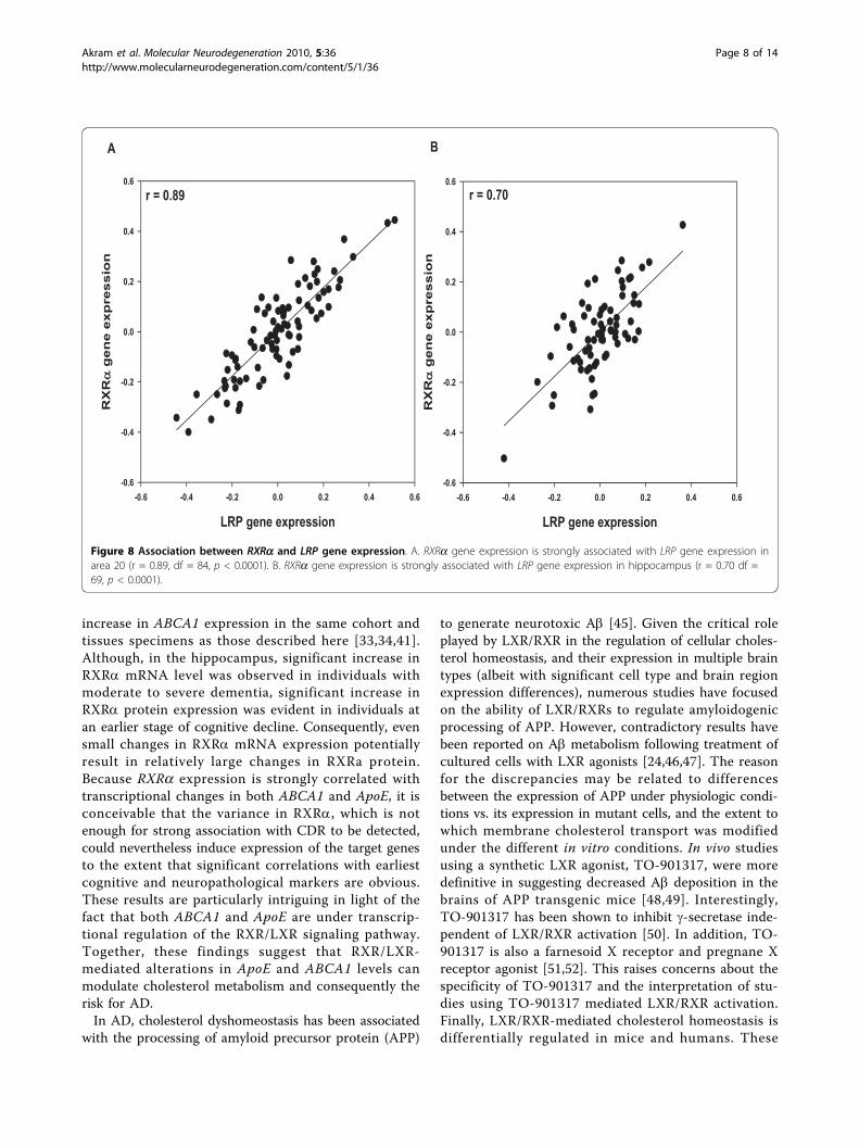
84, p < 0.0001; Figure 8A). RXRa gene expression in thehippocampus also showed strong associations withABCA1 (r = 0.436, df = 69 p < 0.0005), ApoE (r =0.446, df = 69 p < 0.0001) and LRP gene expression (r =0.697, df = 69 p < 0.0001; Figure 8B).
DiscussionThis study explored the expression profile of the masterregulators of lipid metabolism in two of the most vul-nerable regions of the AD brain. The primary findingwas that RXRa levels increased at the very earliest stageof dementia and remained elevated, in general, through-out the course of the disease. In addition, these eleva-tions in gene and protein expression were more stronglyassociated with the development of AD-associated
Figure 6 Western blot analysis of RXRa in the hippocampus of cognitively intact controls and subjects with varying severity ofdementia. A, Representative immunoblots of RXRa protein expression are shown. Total tissue homogenates were separated by reducing SDS-PAGE and probed with rabbit anti-RXRa and mouse anti-VCP antibodies. Tissue lysate from each subjects were loaded in triplicate and pooledtissue lysate (first 4 lanes) were run in quadruplicates. B, Protein quantification was done by assessing the ratio of RXRa and VCP signal. Meanvalues ± SEM are shown. *, p < 0.05; **, p < 0.01. Number within the parentheses indicates the individuals within each group.
Akram et al. Molecular Neurodegeneration 2010, 5:36http://www.molecularneurodegeneration.com/content/5/1/36
Page 6 of 14

dementia than with the measures of mature neuropatho-logic lesions of AD. In order to identify earliest tran-scriptional changes during the course of ADprogression, study individuals were grouped based ondementia severity (CDR score) at the time of death, pro-gression of NFT pathology and the severity of NPpathology. This strategy allowed us to correlate expres-sion of nuclear receptors to cognitive and pathologicalindices of AD, with an emphasis on individuals present-ing earliest signs of AD. These approaches revealed apreviously unrecognized transcriptional dysregulation ofRXRa in AD. Specifically, alteration in gene expressionin both regions was strongly correlated with cognitiveimpairment. Additionally, we observed highly coordi-nated upregulation of RXRa protein in AD hippocam-pus. Along with the identification of altered RXRaexpression, this study highlights closely correlatedexpression of RXRa with the downstream target genesthat have been previously implicated in AD pathogenesisincluding ApoE and ABCA1. One parsimonious inter-pretation of these findings is that changes in the expres-sion of RXR become evident before the advent of theneuropathological hallmarks of AD and raise the possi-bility that the upregulation of RXR may be responsiblefor the changes in subsequent progressive cholesterol
dyshomeostasis and AD neuropathology. However,because of the postmortem nature of the current study,the results of this study do not address directly whetherthe elevated levels of LXR/RXR in dementia are causal,secondary or bystander. It is also important to note thatalthough we interpret transcriptional changes as a func-tion of the severity of dementia or neuropathology torepresent disease progression, the cross-sectional natureof this postmortem study, like all postmortem studies,permits only the inference of progression rather than itsdirect measurement. In addition, the reported resultsare based on assays of brain tissue homogenates, andtherefore cannot inform on the cellular localization ofthe dysregulated gene expression or the laminar identityof the affected cells.Although the pathogenesis of AD is not fully under-
stood much direct and circumstantial evidence suggeststhat many of the genes and pathways involved in choles-terol and/or lipoprotein metabolism in brain are alsointimately involved in the pathogenesis of AD. Mostnotable are ApoE, the principal cholesterol carrier pro-tein in the CNS, and ABCA1, a protein that modulatesthe efflux of cellular cholesterol and phospholipids tolipid-deficient apolipoprotein acceptors such as ApoE[42-44]. Interestingly, we have observed significant
Figure 7 Association between RXRa and target gene expression in area 20. A. RXRa gene expression is strongly associated with ABCA1gene expression (r = 0.531, df = 83, p < 0.0001). B. RXRa gene expression is strongly associated with ApoE gene expression (r = 0.622, df = 84, p< 0.0001).
Akram et al. Molecular Neurodegeneration 2010, 5:36http://www.molecularneurodegeneration.com/content/5/1/36
Page 7 of 14
increase in ABCA1 expression in the same cohort andtissues specimens as those described here [33,34,41].Although, in the hippocampus, significant increase inRXRa mRNA level was observed in individuals withmoderate to severe dementia, significant increase inRXRa protein expression was evident in individuals atan earlier stage of cognitive decline. Consequently, evensmall changes in RXRa mRNA expression potentiallyresult in relatively large changes in RXRa protein.Because RXRa expression is strongly correlated withtranscriptional changes in both ABCA1 and ApoE, it isconceivable that the variance in RXRa, which is notenough for strong association with CDR to be detected,could nevertheless induce expression of the target genesto the extent that significant correlations with earliestcognitive and neuropathological markers are obvious.These results are particularly intriguing in light of thefact that both ABCA1 and ApoE are under transcrip-tional regulation of the RXR/LXR signaling pathway.Together, these findings suggest that RXR/LXR-mediated alterations in ApoE and ABCA1 levels canmodulate cholesterol metabolism and consequently therisk for AD.In AD, cholesterol dyshomeostasis has been associated
with the processing of amyloid precursor protein (APP)
to generate neurotoxic Ab [45]. Given the critical roleplayed by LXR/RXR in the regulation of cellular choles-terol homeostasis, and their expression in multiple braintypes (albeit with significant cell type and brain regionexpression differences), numerous studies have focusedon the ability of LXR/RXRs to regulate amyloidogenicprocessing of APP. However, contradictory results havebeen reported on Ab metabolism following treatment ofcultured cells with LXR agonists [24,46,47]. The reasonfor the discrepancies may be related to differencesbetween the expression of APP under physiologic condi-tions vs. its expression in mutant cells, and the extent towhich membrane cholesterol transport was modifiedunder the different in vitro conditions. In vivo studiesusing a synthetic LXR agonist, TO-901317, were moredefinitive in suggesting decreased Ab deposition in thebrains of APP transgenic mice [48,49]. Interestingly,TO-901317 has been shown to inhibit g-secretase inde-pendent of LXR/RXR activation [50]. In addition, TO-901317 is also a farnesoid X receptor and pregnane Xreceptor agonist [51,52]. This raises concerns about thespecificity of TO-901317 and the interpretation of stu-dies using TO-901317 mediated LXR/RXR activation.Finally, LXR/RXR-mediated cholesterol homeostasis isdifferentially regulated in mice and humans. These
Figure 8 Association between RXRa and LRP gene expression. A. RXRa gene expression is strongly associated with LRP gene expression inarea 20 (r = 0.89, df = 84, p < 0.0001). B. RXRa gene expression is strongly associated with LRP gene expression in hippocampus (r = 0.70 df =69, p < 0.0001).
Akram et al. Molecular Neurodegeneration 2010, 5:36http://www.molecularneurodegeneration.com/content/5/1/36
Page 8 of 14

differences are accentuated in studies attributing benefi-cial outcomes to LXR/RXR-mediated transcriptionalregulation. In mice but not in primates, hepatic choles-terol 7a-hydroxylase is upregulated by LXR [4,53,54].Mice do not express CETP. Direct induction of CETP inhuman cell lines and two CETP-containing animal mod-els, Syrian hamsters and long-tailed macaque monkeys,is accompanied by a significant increase in LDL choles-terol levels that was not previously observed in mice[23,55,56]. These findings emphasize the need to exer-cise caution when extrapolating results from animal stu-dies to humans, especially when significant species-specific differences have been identified in the underly-ing biological processes. It is noteworthy that in the cur-rent systematic analysis, LXR/RXR expression levelswere not associated with elevations in NP counts. Itmay be plausible that change in RXRa expression mayinfluence soluble toxic forms or fractions of Ab, yet itdoes not correlate with NP density because RXRa has amonotonic pattern of expression, whereas NPs continueto increase in density.LXRs are activated by endogenous oxysterols, the
most potent of which include 24, 25-epoxycholesterol,24-OH, and 27-hydroxycholesterol. The role of 24-OH(also known as cerbrosterol) is particularly intriguingbecause it is primarily produced in the brain by neuronspecific CYP46 [57] and its level is increased in plasmaand CSF during early stages of AD [58,59]. 24-OH hasbeen shown to activate LXRs and dramatically elevateABCA1 expression in both neurons and astrocytes[8,25,60]. Therefore increased expression of ABCA1 andApoE in incipient AD might be a reflection of 24-OHmediated increased LXR/RXR activation. Paradoxically,24-OH level is reduced in late stages of AD [61], sug-gesting that sustained increases in the expression ofLXR/RXR target genes in relatively late stages is modu-lated by other factors.LXRb heterodimer with RXRa is the only nuclear
receptor complex known to date that can be activatedin the absence of a ligand, via a mechanism termed“dimerization-induced activation” [62-64]. In this modelof LXR transactivation, the interaction of RXR with LXRcan allosterically activate LXR by inducing a conforma-tional change in its ligand-binding domain. The relativeexpression levels of both receptors are therefore likelyto regulate signaling via LXRb and RXRa in a highlycomplex fashion. Indeed, in transient transfection stu-dies LXR/RXR activation is only observed upon RXRacotransfection [3,65,66], which results in a higher num-ber of LXRb/RXRa heterodimers for which coregulatorswould have to compete. Because the activated LXRb/RXRa heterodimer also exhibits dual ligand permissive-ness and synergism [62,67], its net transcriptional poten-tial depends on the occurrence of dimerization-induced
activation and ligand availability. Consistent with thisidea, the overexpression of RXRa reported here couldallosterically activate LXRb and consequently increasetarget gene expression, in addition to that induced by24-OH. Recently, LRP has been shown to participate inABCA1 expression by relieving LXR/RXR repression(via cPLA2 activation) [68]. Strong association of LRPgene expression with that of LXR and RXR suggeststhat LRP modulates not only LXR/RXR activation butalso their transcription. Alternatively, LRP may be anLXR/RXR target gene.
ConclusionsBased on previous studies where a drastic reduction incholesterol level decreased Ab production [69-71] andowing to their ability to induce expression of genesmediating cholesterol efflux, LXR/RXR heterodimershave emerged as potential targets for AD therapeutics[72]. However, there are obvious contradicting reportsof increased Ab generation upon lowering brain choles-terol level [73,74]. More importantly, the hippocampusof AD cases presents a moderate, yet significant, reduc-tion in membrane cholesterol [75]. These latter findingsare consistent with increased expression of RXRareported here. Taken together, increased expression ofRXRa and concomitant activation of LXR/RXR canmodulate ABCA1 and ApoE gene expression. Increasedlevels of ABCA1 and ApoE may be the molecular deter-minants of cholesterol dyshomeostasis and accompany-ing dementia observed in AD.
Materials and methodsStudy CohortThe cohort included in this study was part of a largerclinical and neuropsychological investigation of earlyAD. These individuals were extensively evaluated fortheir cognitive function. Their cognitive status duringthe 6 months proximal to death was used to define theabsence, presence and extent of dementia at the time ofdeath, as previously described [76-78]. Cases wereselected from a pool of over 600 donors with either nodiscernable neuropathology or only those neuropatholo-gical lesions associated with AD alone (e.g., exclusion ofcases with vascular lesions, Lewy body inclusions, nor-mal pressure hydrocephalus). Because postmortem inter-vals (PMI) [79,80] and tissue pH (a proxy measure foragonal state) [81,82] are important issues for consistencyand reproducibility of quantitative gene and proteinexpression studies, brain samples were included fromcases who met the following criteria only: postmortemdelay of less than 24 hours, brain tissue pH of 6.3 orgreater no perimortem coma longer than 6 hrs, no evi-dence of seizures in the 3 months preceding death. Con-trols were derived from persons who, on extensive
Akram et al. Molecular Neurodegeneration 2010, 5:36http://www.molecularneurodegeneration.com/content/5/1/36
Page 9 of 14

medical record review and/or neuropsychological exami-nation and caregiver interview, showed no evidence ofneurological or neuropsychiatric diseases, died of naturalcauses (myocardial infarction, various non-brain non-hepatic cancers, and congestive heart failure) and hadno discernable neuropathology [83]. None of the sub-jects had a history of licit or illicit drug abuse (tobaccouse excepted). All diagnostic and cognitive assessmentprocedures were approved by the Mount Sinai MedicalCenter (New York, NY)/J. J. Peters Veterans Administra-tion Medical Center (Bronx, NY) Institutional ReviewBoards, and postmortem consent for autopsy andresearch use of tissue was obtained from the next of kinor a legally authorized official.
Classification of Subjects into Dementia Severity GroupsIn order to perform post-assay analyses based on aclinical index of disease and dementia everity, the sub-jects were classified with respect to the CDR score atthe time of death [84-87] (Table 1). CDR is a scalethat objectively stages dementia severity from 0-5 with0 representing no dementia, 0.5 representing question-able dementia or mild cognitive impairment and 1-5representing gradations of dementia severity from mildto terminal. The assessments, on which these classifi-cations were based, were performed blind to clinical orneuropathological disease diagnosis. Table 2 describesthe sample size, sex, age at the time of death, pH andPMI of the study cohort when grouped on the basis ofCDR.
Neuropathological AssessmentThe neuropathological assessment procedures used havebeen previously described in detail [76,78]. Neuropatho-logical assessments were performed on the right hemi-sphere and consisted of microscopic assessment ofparaffin embedded blocks from multiple brain regions
using hematoxylin and eosin, modified Bielschowski,modified thioflavin S, and anti-amyloid, anti-tau tauwhen necessary. All neuropathology data regarding theextent and distribution of neuropathologic lesions werecollected blind to the subject’s dementia status. Speci-mens for this study were dissected from the frozen,never-thawed, left hemisphere, using previouslydescribed procedures [88].For pathologic staging of AD neurofibrillary tangle
density was assessed using the Consortium to Establisha Registry for Alzheimer’s Disease (CERAD) [89,90]criteria, NFTs were evaluated using the criteria byBraak and Braak [91] (Table 1). Neuritic plaques (NPs)were identified as the dystrophic neurites arrangedradially and forming a discrete spherical lesion about30 mm in diameter with amyloid cores. NP groups inTable 1 reflect a composite score of NPs counts in 5cortical regions. The composite measure of cortical NPdensity was used to reflect better the general level ofdisease severity and to match more closely to the glo-bal assessment of cognitive function measured by theCDR.
RNA IsolationTotal RNA was isolated from 50 mg of microdissectedpulverized frozen brain samples from inferior temporalgyrus and the hippocampus with the guanidinium iso-thiocyanate method [92] using ToTALLY RNA kits(Ambion, Austin, TX) according to the manufacturer’sprotocol as described previously [93]. The quality of theisolated total RNA for each case was assessed using acombination of 260 nm/280 nm ratio obtained spectro-photometrically (Beckman Instruments, Fullerton, CA)and by Bioanalyzer 2100 (Agilent Technologies, PaloAlto, CA) before proceeding with cDNA synthesis. Onlyspecimens with an RIN ≥ 5.5 were included in theanalyses.
Table 2 Demographic details of study cohort stratified with respect to CDR (Clinical Dementia Rating) groups
Characteristics Area CDR 0 CDR 0.5 CDR 1 CDR 2 CDR 3 CDR4-5
Total subjects* Hipp 18 13 9 9 12 12
Area 20 18 13 8 13 18 18
Gender (men/women) Hipp 7/11 6/7 3/6 0/9 3/9 3/9
Area 20 6/12 7/6 3/5 1/12 7/11 8/10
Age (years) Hipp 75.2 ± 3.5 85.4 ± 2.7 83.4 ± 3.4 87.9 ± 2.0 88.8 ± 1.7 85.0 ± 1.9
Area 20 77.0 ± 3.9 85.5 ± 2.8 85.6 ± 3.8 87.6 ± 2.0 86.2 ± 8.5 84.2 ± 2.5
Brain pH Hipp 6.43 ± 0.04 6.43 ± 0.07 6.31 ± 0.1 6.38 ± 0.09 6.34 ± 0.05 6.39 ± 0.07
Area 20 6.42 ± 0.05 6.42 ± 0.07 6.35 ± 0.11 6.39 ± 0.08 6.43 ± 0.05 6.36 ± 0.05
RNA integrity number (RIN) Hipp 6.6 ± 0.1 6.2 ± 0.1 6.2 ± 0.2 6.2 ± 0.2 6.1 ± 0.1 6.2 ± 0.1
Area 20 6.9 ± 0.1 6.9 ± 0.1 6.7 ± 0.2 6.3 ± 0.2 6.9 ± 0.2 6.3 ± 0.1
Postmortem interval (minutes) Hipp 713 ± 137 393 ± 85 264 ± 39 336 ± 66 276 ± 40 332 ± 80
Area 20 574 ± 109 381 ± 91 325 ± 52 358 ± 64 244 ± 29 310 ± 73
Akram et al. Molecular Neurodegeneration 2010, 5:36http://www.molecularneurodegeneration.com/content/5/1/36
Page 10 of 14
Reverse Transcriptase ReactioncDNA synthesis was performed with iScript cDNASynthesis kit (BioRad Laboratories, Hercules, CA) whichuses both random and poly-dT priming for the reversetranscription (RT) reaction. Total RNA (1 μg) wasemployed for each 20 μl reaction. The resulting cDNAwas diluted 25 times for qPCR.
qPCRLXRb and RXRa mRNA expression was measured byquantitative polymerase chain reaction (qPCR) using anABI Prism 7700 Sequence Detector (Applied Biosystems,Foster City, CA) and gene-specific fluorogenic TaqMan®probes (Applied Biosystems). Each 20 μl PCR reactioncontained 5 μl of the relevant cDNA, 20X TaqMan®assay (used at a final concentration of 0.5X), and 10 μlof TaqMan® Universal PCR Reaction Mix which con-tains ROX as a passive internal reference (Applied Bio-systems). The thermal cycling program consisted of 2min at 50°C, 10 min at 95°C, followed by 40 cycles of 15s at 95°C and 1 min at 60°C. The reactions were quanti-fied by selecting the amplification cycle when the PCRproduct of interest was first detected (threshold cycle,Ct). Tests of primers and probes sensitivity and assaylinearity were conducted for all real-time PCR assays byamplification of mRNA in 10-fold serial dilutions ofpooled as previously described [94]. Each reaction wasperformed in triplicate and the average Ct value wasused in all analyses.The relative gene expression level was calculated using
the Relative Standard Curve Method (see Guide to Per-forming Relative Quantitation of Gene Expression UsingReal-time Quantitative PCR, Applied Biosystems). Stan-dard curves were generated for target assay and for eachendogenous control assay by the association betweenthe Ct values and different quantities (5 serial dilutionsteps) of a “calibrator” cDNA. The “calibrator” was pre-pared by mixing small quantities of all experimentalsamples. Expression values of the target and the controlgenes were extrapolated from their respective standardcurves. Relative expression of target genes was com-puted as the ratio of the target mRNA levels to the geo-metric mean of the four endogenous controls: b-glucuronidase (GUSB), cyclophilin A (PP1A), b2-micro-globulin (b2M), and ribosomal protein, large, P0(RPLP0) which were picked for their stability using geN-orm [95,96]. Samples with Ct values > 33 were consid-ered outside the range of sensitivity of the assay andwere not included in the analyses.
Protein QuantitationProtein expression studies were carried out to determinewhether different levels of RXRa gene expression werereflected in the expression level of RXRa protein.
Because of the inherently lower reproducibility andhigher variability characteristic of Westerns in postmor-tem tissue relative to qPCR, we restricted RXRa proteinanalyses to cases with most robust changes in geneexpression. Therefore, a subset of the hippocampal sam-ples (N = 30) studied for mRNA expression was ana-lyzed by Western blotting to reflect broad variations ingene expression. As fewer protein analyses were per-formed than gene analyses, adjacent categories for allthree indices of disease severity (see below) for geneexpression analyses were combined to achieve suffi-ciently large sample sizes for comparisons (Table 3).
Tissue Lysate PreparationTotal tissue lysates were prepared from frozen hippo-campal specimens from sister aliquots of the same brainsamples as those used for qPCR analysis as describedpreviously [41]. Total protein concentration of the lysatewas determined using a CBQCA Quantitation Kit(Molecular Probes, Eugene, OR) with fluorescence mea-sured on a SpectraMAX Gemini XS spectrofluorometer(Molecular Devices, Sunnyvale, CA).
Western Blot AnalysisFor gel electrophoresis, 10 μg of total protein was mixedwith loading buffer and loaded onto pre-cast 10-20%Tris-glycine gels (Bio-Rad Laboratories, Hercules, CA),and run at 150 V for 1 hr. Each gel was loaded withthree experimental samples in triplicate and “standardtissue homogenate” (the mix of small aliquots of tissuefrom all samples), run in quadruplicates. Separated pro-teins were transferred to polyvinylidene difluoride
Table 3 Group classifications for protein expressionanalysis
Protein Expression Analysis in the hippocampus
CDR Groups Dementia Severity Number ofindividuals
0 No dementia 8
0.5 Questionable dementia 6
1-2 Mild/moderate dementia 6
3-5 Severe dementia/terminaldementia
10
Braak Groups Braak stages
0-I None/Mild transentorhinal 8
II Severe transentorhinal 8
III-1V Limbic/Hippocampal CA1 7
V-VI Isocortical/Primary sensoryareas
7
NP DensityGroups
Plaques (number/mm2)
1 0 12
2-5 1 and more 18
Akram et al. Molecular Neurodegeneration 2010, 5:36http://www.molecularneurodegeneration.com/content/5/1/36
Page 11 of 14
membranes at 100 V for 1 hour and probed with anti-RXRa antibody (Abcam, Cambridge, MA) diluted1:5,000 in 3% non-fat dry milk in TBS overnight at 4°Cwith gentle shaking. To ensure equal protein loadingbetween individual samples, membranes were also incu-bated with an anti-valosin containing protein (VCP)antibody. VCP, a 97 kDa protein, has been previouslyvalidated as reliable internal standard [97]. Following 1hour incubation with the fluorescently-labeled secondaryantibodies, blots were scanned and quantified using theOdyssey IR imaging system (LI-COR Biosciences, Lin-coln, NE). RXRa signal was first normalized to the cor-responding average signal for the standard tissuehomogenate and then for the VCP band from the samesample. The linearity of the dose responses for the anti-bodies used was established in preliminary experiments.
Statistical AnalysesWe performed a logarithmic transformation of LXRband RXRa gene expression to eliminate heterogeneity,and used the transformed gene expression values for allsubsequent statistical analyses. A preliminary analysisassessed linear associations with gender, pH, PMI andRIN to evaluate their use as covariates. In addition, age,the most significant risk factor for dementia and a criti-cal determinant of the extent of AD associated neuro-pathology, was used as a covariate in all analysesregardless of its association with the dependent variable.We determined the linear association LXRb and RXRa
gene expression with CDR, Braak stages and NP densityby partial correlation analyses, controlling for potentialcovariates if preliminary analyses showed significant cor-relation with the expression level of the gene under ana-lysis. Because the associations of each of theseinterrelated scales with gene expression is at least partlymediated through the associations with the other twoscales, additional partial correlation analyses assessedeach scale controlling also for the other two scales.In order to determine non-linear association of CDR
Braak stages, and NP density with LXRb and RXRa geneexpression, each of these disease severity indices wasclassified as a categorical variable. ANCOVA was per-formed for each categorical variable controlling for ageand any other potential covariates. Another ANCOVAfor each categorical variable controlled also for othertwo variables as scales, similar to the partial correlationanalyses.Analyses for protein expression were the same as for
gene expression. All analyses were performed with SPSS17.0 (SPSS, Chicago, IL).
AcknowledgementsThis research was supported by NIH grants P01 AG02219 and AG05138.
Author details1Department of Neuroscience Mount Sinai School of Medicine, New York,NY 10029, USA. 2Department of Psychiatry, Mount Sinai School of Medicine,New York, NY 10029, USA. 3J.J. Peters Veterans Affairs Medical Center, Bronx,NY 10468, USA.
Authors’ contributionsAA carried out the qPCR and Western blot studies and drafted themanuscript. JS performed the statistical analysis. PK, PRH and VH conceivedof the study, and participated in its design and coordination and helped todraft the manuscript. All authors read and approved the final manuscript.
Competing interestsThe authors declare that they have no competing interests.
Received: 3 May 2010 Accepted: 15 September 2010Published: 15 September 2010
References1. Mangelsdorf DJ, Evans RM: The RXR heterodimers and orphan receptors.
Cell 1995, 83(6):841-850.2. Apfel R, Benbrook D, Lernhardt E, Ortiz MA, Salbert G, Pfahl M: A novel
orphan receptor specific for a subset of thyroid hormone-responsiveelements and its interaction with the retinoid/thyroid hormone receptorsubfamily. Mol Cell Biol 1994, 14(10):7025-7035.
3. Willy PJ, Umesono K, Ong ES, Evans RM, Heyman RA, Mangelsdorf DJ: LXR,a nuclear receptor that defines a distinct retinoid response pathway.Genes Dev 1995, 9(9):1033-1045.
4. Lehmann JM, Kliewer SA, Moore LB, Smith-Oliver TA, Oliver BB, Su JL,Sundseth SS, Winegar DA, Blanchard DE, Spencer TA, Willson TM:Activation of the nuclear receptor LXR by oxysterols defines a newhormone response pathway. J Biol Chem 1997, 272(6):3137-3140.
5. Lu TT, Repa JJ, Mangelsdorf DJ: Orphan nuclear receptors as eLiXiRs andFiXeRs of sterol metabolism. J Biol Chem 2001, 276(41):37735-37738.
6. Peet DJ, Janowski BA, Mangelsdorf DJ: The LXRs: a new class of oxysterolreceptors. Curr Opin Genet Dev 1998, 8(5):571-575.
7. Repa JJ, Mangelsdorf DJ: The role of orphan nuclear receptors in theregulation of cholesterol homeostasis. Annu Rev Cell Dev Biol 2000,16:459-481.
8. Whitney KD, Watson MA, Collins JL, Benson WG, Stone TM, Numerick MJ,Tippin TK, Wilson JG, Winegar DA, Kliewer SA: Regulation of cholesterolhomeostasis by the liver X receptors in the central nervous system. MolEndocrinol 2002, 16(6):1378-1385.
9. Szanto A, Narkar V, Shen Q, Uray IP, Davies PJ, Nagy L: Retinoid Xreceptors: X-ploring their (patho)physiological functions. Cell Death Differ2004, 11(Suppl 2):S126-S143.
10. Chawla A, Repa JJ, Evans RM, Mangelsdorf DJ: Nuclear receptors and lipidphysiology: opening the X-files. Science 2001, 294(5548):1866-1870.
11. Chen JD, Evans RM: A transcriptional co-repressor that interacts withnuclear hormone receptors. Nature 1995, 377(6548):454-457.
12. Horlein AJ, Naar AM, Heinzel T, Torchia J, Gloss B, Kurokawa R, Ryan A,Kamei Y, Soderstrom M, Glass CK: Ligand-independent repression by thethyroid hormone receptor mediated by a nuclear receptor co-repressor.Nature 1995, 377(6548):397-404.
13. Glass CK, Rosenfeld MG: The coregulator exchange in transcriptionalfunctions of nuclear receptors. Genes Dev 2000, 14(2):121-141.
14. Nagy L, Schwabe JW: Mechanism of the nuclear receptor molecularswitch. Trends Biochem Sci 2004, 29(6):317-324.
15. Schulman IG, Li C, Schwabe JW, Evans RM: The phantom ligand effect:allosteric control of transcription by the retinoid X receptor. Genes Dev1997, 11(3):299-308.
16. Schultz JR, Tu H, Luk A, Repa JJ, Medina JC, Li L, Schwendner S, Wang S,Thoolen M, Mangelsdorf DJ, Lustig KD, Shan B: Role of LXRs in control oflipogenesis. Genes Dev 2000, 14(22):2831-2838.
17. Costet P, Luo Y, Wang N, Tall AR: Sterol-dependent transactivation of theABC1 promoter by the liver X receptor/retinoid X receptor. J Biol Chem2000, 275(36):28240-28245.
18. Repa JJ, Berge KE, Pomajzl C, Richardson JA, Hobbs H, Mangelsdorf DJ:Regulation of ATP-binding cassette sterol transporters ABCG5 andABCG8 by the liver X receptors alpha and beta. J Biol Chem 2002,277(21):18793-18800.
Akram et al. Molecular Neurodegeneration 2010, 5:36http://www.molecularneurodegeneration.com/content/5/1/36
Page 12 of 14

19. Kennedy MA, Bard M: Positive and negative regulation of squalenesynthase (ERG9), an ergosterol biosynthetic gene, in Saccharomycescerevisiae. Biochim Biophys Acta 2001, 1517(2):177-189.
20. Laffitte BA, Repa JJ, Joseph SB, Wilpitz DC, Kast HR, Mangelsdorf DJ,Tontonoz P: LXRs control lipid-inducible expression of the apolipoproteinE gene in macrophages and adipocytes. Proc Natl Acad Sci USA 2001,98(2):507-512.
21. Hummasti S, Laffitte BA, Watson MA, Galardi C, Chao LC, Ramamurthy L,Moore JT, Tontonoz P: Liver X receptors are regulators of adipocyte geneexpression but not differentiation: identification of apoD as a directtarget. J Lipid Res 2004, 45(4):616-625.
22. Cao G, Beyer TP, Yang XP, Schmidt RJ, Zhang Y, Bensch WR, Kauffman RF,Gao H, Ryan TP, Liang Y, Eacho PI, Jiang XC: Phospholipid transfer proteinis regulated by liver X receptors in vivo. J Biol Chem 2002,277(42):39561-39565.
23. Luo Y, Tall AR: Sterol upregulation of human CETP expression in vitroand in transgenic mice by an LXR element. J Clin Invest 2000,105(4):513-520.
24. Fukumoto H, Deng A, Irizarry MC, Fitzgerald ML, Rebeck GW: Induction ofthe cholesterol transporter ABCA1 in central nervous system cells byliver X receptor agonists increases secreted Abeta levels. J Biol Chem2002, 277(50):48508-48513.
25. Liang Y, Lin S, Beyer TP, Zhang Y, Wu X, Bales KR, DeMattos RB, May PC,Li SD, Jiang XC, Eacho PI, Cao G, Paul SM: A liver X receptor and retinoidX receptor heterodimer mediates apolipoprotein E expression, secretionand cholesterol homeostasis in astrocytes. J Neurochem 2004,88(3):623-634.
26. Joseph SB, Tontonoz P: LXRs: new therapeutic targets in atherosclerosis?Curr Opin Pharmacol 2003, 3(2):192-197.
27. Wang L, Schuster GU, Hultenby K, Zhang Q, Andersson S, Gustafsson JA:Liver X receptors in the central nervous system: from lipid homeostasisto neuronal degeneration. Proc Natl Acad Sci USA 2002,99(21):13878-13883.
28. Mangelsdorf DJ, Borgmeyer U, Heyman RA, Zhou JY, Ong ES, Oro AE,Kakizuka A, Evans RM: Characterization of three RXR genes that mediatethe action of 9-cis retinoic acid. Genes Dev 1992, 6(3):329-344.
29. Krezel W, Kastner P, Chambon P: Differential expression of retinoidreceptors in the adult mouse central nervous system. Neuroscience 1999,89(4):1291-1300.
30. Zetterstrom RH, Lindqvist E, Mata de UA, Tomac A, Eriksson U, Perlmann T,Olson L: Role of retinoids in the CNS: differential expression of retinoidbinding proteins and receptors and evidence for presence of retinoicacid. Eur J Neurosci 1999, 11(2):407-416.
31. Moreno S, Farioli-Vecchioli S, Ceru MP: Immunolocalization of peroxisomeproliferator-activated receptors and retinoid X receptors in the adult ratCNS. Neuroscience 2004, 123(1):131-145.
32. Haroutunian V, Katsel P, Schmeidler J: Transcriptional vulnerability of brainregions in Alzheimer’s disease and dementia. Neurobiol Aging 2009,30(4):561-73.
33. Akram A, Katsel P, Hof PR, Haroutunian V: Changes in the expression ofgenes involved in cholesterol trafficking with the progression ofAlzheimer’s disease [abstract]. Society for Neuroscience Abstracts 2007,795.9/N1.
34. Akram A, Katsel P, Hof PR, Haroutunian V: Coordinated transcriptional andtranslational changes in cholesterol transporters correlate with cognitivedecline in Alzheimer’s disease [abstract]. Society for Neuroscience Abstracts2008, 45.7/U31.
35. Lewis DA, Campbell MJ, Terry RD, Morrison JH: Laminar and regionaldistributions of neurofibrillary tangles and neuritic plaques inAlzheimer’s disease: a quantitative study of visual and auditory cortices.J Neurosci 1987, 7(6):1799-1808.
36. Kergoat H, Kergoat MJ, Justino L, Chertkow H, Robillard A, Bergman H:Visual retinocortical function in dementia of the Alzheimer type.Gerontology 2002, 48(4):197-203.
37. Lee AG, Martin CO: Neuro-ophthalmic findings in the visual variant ofAlzheimer’s disease. Ophthalmology 2004, 111(2):376-380.
38. Hao J, Li K, Li K, Zhang D, Wang W, Yang Y, Yan B, Shan B, Zhou X: Visualattention deficits in Alzheimer’s disease: an fMRI study. Neurosci Lett2005, 385(1):18-23.
39. Bair W: Visual receptive field organization. Curr Opin Neurobiol 2005,15(4):459-464.
40. Metsaars WP, Hauw JJ, van Welsem ME, Duyckaerts C: A grading system ofAlzheimer disease lesions in neocortical areas. Neurobiol Aging 2003,24(4):563-572.
41. Akram A, Schmeidler J, Katsel P, Hof PR, Haroutunian V: Increasedexpression of cholesterol transporter ABCA1 is highly correlated withseverity of dementia in AD hippocampus. Brain Research 2010,1318:167-177.
42. Bodzioch M, Orso E, Klucken J, Langmann T, Bottcher A, Diederich W,Drobnik W, Barlage S, Buchler C, Porsch-Ozcurumez M, Kaminski WE,Hahmann HW, Oette K, Rothe G, Aslanidis C, Lackner KJ, Schmitz G: Thegene encoding ATP-binding cassette transporter 1 is mutated in Tangierdisease. Nat Genet 1999, 22(4):347-351.
43. Brooks-Wilson A, Marcil M, Clee SM, Zhang LH, Roomp K, van Dam M, Yu L,Brewer C, Collins JA, Molhuizen HO, Loubser O, Ouelette BF, Fichter K,Ashbourne-Excoffon KJ, Sensen CW, Scherer S, Mott S, Denis M,Martindale D, Frohlich J, Morgan K, Koop B, Pimstone S, Kastelein JJ,Hayden MR: Mutations in ABC1 in Tangier disease and familial high-density lipoprotein deficiency. Nat Genet 1999, 22(4):336-345.
44. Repa JJ, Mangelsdorf DJ: The liver X receptor gene team: potential newplayers in atherosclerosis. Nat Med 2002, 8(11):1243-1248.
45. Shobab LA, Hsiung GY, Feldman HH: Cholesterol in Alzheimer’s disease.Lancet Neurol 2005, 4(12):841-852.
46. Koldamova RP, Lefterov IM, Ikonomovic MD, Skoko J, Lefterov PI, Isanski BA,DeKosky ST, Lazo JS: 22R-hydroxycholesterol and 9-cis-retinoic acidinduce ATP-binding cassette transporter A1 expression and cholesterolefflux in brain cells and decrease amyloid beta secretion. J Biol Chem2003, 278(15):13244-13256.
47. Sun Y, Yao J, Kim TW, Tall AR: Expression of liver X receptor target genesdecreases cellular amyloid beta peptide secretion. J Biol Chem 2003,278(30):27688-27694.
48. Koldamova RP, Lefterov IM, Staufenbiel M, Wolfe D, Huang S, Glorioso JC,Walter M, Roth MG, Lazo JS: The liver X receptor ligand T0901317decreases amyloid beta production in vitro and in a mouse model ofAlzheimer’s disease. J Biol Chem 2005, 280(6):4079-4088.
49. Riddell DR, Zhou H, Comery TA, Kouranova E, Lo CF, Warwick HK, Ring RH,Kirksey Y, Aschmies S, Xu J, Kubek K, Hirst WD, Gonzales C, Chen Y,Murphy E, Leonard S, Vasylyev D, Oganesian A, Martone RL, Pangalos MN,Reinhart PH, Jacobsen JS: The LXR agonist TO901317 selectively lowershippocampal Abeta42 and improves memory in the Tg2576 mousemodel of Alzheimer’s disease. Mol Cell Neurosci 2007, 34(4):621-628.
50. Czech C, Burns MP, Vardanian L, Augustin A, Jacobsen H, Baumann K,Rebeck GW: Cholesterol independent effect of LXR agonist TO-901317on gamma-secretase. J Neurochem 2007, 101(4):929-936.
51. Mitro N, Vargas L, Romeo R, Koder A, Saez E: T0901317 is a potent PXRligand: implications for the biology ascribed to LXR. FEBS Lett 2007,581(9):1721-1726.
52. Houck KA, Borchert KM, Hepler CD, Thomas JS, Bramlett KS, Michael LF,Burris TP: T0901317 is a dual LXR/FXR agonist. Mol Genet Metab 2004,83(1-2):184-187.
53. Peet DJ, Turley SD, Ma W, Janowski BA, Lobaccaro JM, Hammer RE,Mangelsdorf DJ: Cholesterol and bile acid metabolism are impaired inmice lacking the nuclear oxysterol receptor LXR alpha. Cell 1998,93(5):693-704.
54. Menke JG, Macnaul KL, Hayes NS, Baffic J, Chao YS, Elbrecht A, Kelly LJ,Lam MH, Schmidt A, Sahoo S, Wang J, Wright SD, Xin P, Zhou G, Moller DE,Sparrow CP: A novel liver X receptor agonist establishes speciesdifferences in the regulation of cholesterol 7alpha-hydroxylase (CYP7a).Endocrin 2002, 143(7):2548-2558.
55. Groot PH, Pearce NJ, Yates JW, Stocker C, Sauermelch C, Doe CP,Willette RN, Olzinski A, Peters T, d’Epagnier D, Morasco KO, Krawiec JA,Webb CL, Aravindhan K, Jucker B, Burgert M, Ma C, Marino JP, Collins JL,Macphee CH, Thompson SK, Jaye M: Synthetic LXR agonists increase LDLin CETP species. J Lipid Res 2005, 46(10):2182-2191.
56. Masson D, Staels B, Gautier T, Desrumaux C, Athias A, Le GN, Schneider M,Zak Z, Dumont L, Deckert V, Tall A, Jiang XC, Lagrost L: Cholesteryl estertransfer protein modulates the effect of liver X receptor agonists oncholesterol transport and excretion in the mouse. J Lipid Res 2004,45(3):543-550.
57. Bjorkhem I: Rediscovery of cerebrosterol. Lipids 2007, 42(1):5-14.58. Lutjohann D, Papassotiropoulos A, Bjorkhem I, Locatelli S, Bagli M,
Oehring RD, Schlegel U, Jessen F, Rao ML, von Bergmann K, Heun R:
Akram et al. Molecular Neurodegeneration 2010, 5:36http://www.molecularneurodegeneration.com/content/5/1/36
Page 13 of 14
Plasma 24S-hydroxycholesterol (cerebrosterol) is increased in Alzheimerand vascular demented patients. J Lipid Res 2000, 41(2):195-198.
59. Papassotiropoulos A, Lutjohann D, Bagli M, Locatelli S, Jessen F, Buschfort R,Ptok U, Bjorkhem I, von Bergmann K, Heun R: 24S-hydroxycholesterol incerebrospinal fluid is elevated in early stages of dementia. J Psychiatr Res2002, 36(1):27-32.
60. Abildayeva K, Jansen PJ, Hirsch-Reinshagen V, Bloks VW, Bakker AH,Ramaekers FC, De Vente J, Groen AK, Wellington CL, Kuipers F, Mulder M:24(S)-Hydroxycholesterol participates in a Liver X Receptor-controlledpathway in astrocytes that regulates Apolipoprotein E-mediatedcholesterol efflux. J Biol Chem 2006, 281(18):12799-12808.
61. Papassotiropoulos A, Lutjohann D, Bagli M, Locatelli S, Jessen F, Rao ML,Maier W, Bjorkhem I, von BK, Heun R: Plasma 24S-hydroxycholesterol: aperipheral indicator of neuronal degeneration and potential statemarker for Alzheimer’s disease. NeuroReport 2000, 11(9):1959-1962.
62. Wiebel FF, Steffensen KR, Treuter E, Feltkamp D, Gustafsson JA: Ligand-independent coregulator recruitment by the triply activatable OR1/retinoid X receptor-alpha nuclear receptor heterodimer. Mol Endocrinol1999, 13(7):1105-1118.
63. Son YL, Park OG, Kim GS, Lee JW, Lee YC: RXR heterodimerizationallosterically activates LXR binding to the second NR box of activatingsignal co-integrator-2. Biochem J 2008, 410(2):319-330.
64. Wiebel FF, Gustafsson JA: Heterodimeric interaction between retinoid Xreceptor alpha and orphan nuclear receptor OR1 reveals dimerization-induced activation as a novel mechanism of nuclear receptor activation.Mol Cell Biol 1997, 17(7):3977-3986.
65. Janowski BA, Willy PJ, Devi TR, Falck JR, Mangelsdorf DJ: An oxysterolsignalling pathway mediated by the nuclear receptor LXR alpha. Nature1996, 383(6602):728-731.
66. Willy PJ, Mangelsdorf DJ: Unique requirements for retinoid-dependenttranscriptional activation by the orphan receptor LXR. Genes Dev 1997,11(3):289-298.
67. Shulman AI, Larson C, Mangelsdorf DJ, Ranganathan R: Structuraldeterminants of allosteric ligand activation in RXR heterodimers. Cell2004, 116(3):417-429.
68. Zhou L, Choi HY, Li WP, Xu F, Herz J: LRP1 controls cPLA2phosphorylation, ABCA1 expression and cellular cholesterol export. PLoSONE 2009, 4(8):e6853.
69. Simons M, Keller P, De Strooper B, Beyreuther K, Dotti CG, Simons K:Cholesterol depletion inhibits the generation of beta-amyloid inhippocampal neurons. Proc Natl Acad Sci USA 1998, 95(11):6460-6464.
70. Fassbender K, Simons M, Bergmann C, Stroick M, Lutjohann D, Keller P,Runz H, Kuhl S, Bertsch T, von Bergmann K, Hennerici M, Beyreuther K,Hartmann T: Simvastatin strongly reduces levels of Alzheimer’s diseasebeta -amyloid peptides Abeta 42 and Abeta 40 in vitro and in vivo. ProcNatl Acad Sci USA 2001, 98(10):5856-5861.
71. Ehehalt R, Keller P, Haass C, Thiele C, Simons K: Amyloidogenic processingof the Alzheimer beta-amyloid precursor protein depends on lipid rafts.J Cell Biol 2003, 160(1):113-123.
72. Zelcer N, Khanlou N, Clare R, Jiang Q, Reed-Geaghan EG, Landreth GE,Vinters HV, Tontonoz P: Attenuation of neuroinflammation andAlzheimer’s disease pathology by liver x receptors. Proc Natl Acad Sci USA2007, 104(25):10601-10606.
73. Abad-Rodriguez J, Ledesma MD, Craessaerts K, Perga S, Medina M,Delacourte A, Dingwall C, De Strooper B, Dotti CG: Neuronal membranecholesterol loss enhances amyloid peptide generation. J Cell Biol 2004,167(5):953-960.
74. Park IH, Hwang EM, Hong HS, Boo JH, Oh SS, Lee J, Jung MW, Bang OY,Kim SU, Mook-Jung I: Lovastatin enhances Abeta production and senileplaque deposition in female Tg2576 mice. Neurobiol Aging 2003,24(5):637-643.
75. Ledesma MD, Abad-Rodriguez J, Galvan C, Biondi E, Navarro P,Delacourte A, Dingwall C, Dotti CG: Raft disorganization leads to reducedplasmin activity in Alzheimer’s disease brains. EMBO Rep 2003,4(12):1190-1196.
76. Haroutunian V, Purohit DP, Perl DP, Marin D, Khan K, Lantz M, Davis KL,Mohs RC: Neurofibrillary tangles in nondemented elderly subjects andmild Alzheimer disease. Arch Neurol 1999, 56(6):713-718.
77. Davis KL, Mohs RC, Marin D, Purohit DP, Perl DP, Lantz M, Austin G,Haroutunian V: Cholinergic markers in elderly patients with early signs ofAlzheimer disease. JAMA 1999, 281(15):1401-1406.
78. Haroutunian V, Perl DP, Purohit DP, Marin D, Khan K, Lantz M, Davis KL,Mohs RC: Regional distribution of neuritic plaques in the nondementedelderly and subjects with very mild Alzheimer disease. Arch Neurol 1998,55(9):1185-1191.
79. Johnson SA, Morgan DG, Finch CE: Extensive postmortem stability of RNAfrom rat and human brain. J Neurosci Res 1986, 16(1):267-280.
80. Barton AJ, Pearson RC, Najlerahim A, Harrison PJ: Pre- and postmorteminfluences on brain RNA. J Neurochem 1993, 61(1):1-11.
81. Vawter MP, Tomita H, Meng F, Bolstad B, Li J, Evans S, Choudary P, Atz M,Shao L, Neal C, Walsh DM, Burmeister M, Speed T, Myers R, Jones EG,Watson SJ, Akil H, Bunney WB: Mitochondrial-related gene expressionchanges are sensitive to agonal-pH state: implications for braindisorders. Molecular Psychaitry 2006, 11:663-679.
82. Lipska BK, Deep-Soboslay A, Weickert CS, Hyde TM, Martin CE, Herman MM,Kleinman JE: Critical factors in gene expression in postmortem humanbrain: Focus on studies in schizophrenia. Biol Psychiatry 2006,60(6):650-658.
83. Purohit DP, Perl DP, Haroutunian V, Powchik P, Davidson M, Davis KL:Alzheimer disease and related neurodegenerative diseases in elderlypatients with schizophrenia: a postmortem neuropathologic study of100 cases. Arch Gen Psychiatry 1998, 55(3):205-211.
84. Hughes CP, Berg L, Danziger WL, Coben LA, Martin RL: A new clinical scalefor the staging of dementia. British Journal of Psychiatry 1982, 140:566-572.
85. Burke WJ, Miller JP, Rubin EH, Morris JC, Coben LA, Duchek J, Wittels IG,Berg L: Reliability of the Washington University Clinical Dementia Rating.Arch Neurol 1988, 45(1):31-32.
86. Morris JC: The clinical dementia rating (CDR): current version and scoringrules. Neurology 1993, 43:2412-2414.
87. Dooneief G, Marder K, Tang MX, Stern Y: The Clinical Dementia Ratingscale: community-based validation of “profound’ and “terminal’ stages.Neurology 1996, 46(6):1746-9.
88. Haroutunian V: Tau protein abnormalities associated with theprogression of alzheimer disease type dementia. Neurobiol Aging 2007,28(1):1-7.
89. Mirra SS, Vogel FS, Heyman A: Guide to the CERAD protocol for theneuropathological assessment of Alzheimer’s disease. CERAD 1987.
90. Mirra SS, Heyman A, McKeel D, Sumi SM, Crain BJ, Brownlee LM, Vogel FS,Hughes JP, van Belle G, Berg L: The Consortium to Establish a Registry forAlzheimer’s Disease (CERAD). Part II. Standardization of theneuropathologic assessment of Alzheimer’s disease. Neurology 1991,41:479-486.
91. Braak H, Braak E: Neuropathological stageing of Alzheimer-relatedchanges. Acta Neuropathol (Berl) 1991, 82:239-259.
92. Chomczynski P, Sacchi N: Single-step method of RNA isolation by acidguanidinium thiocyanate-phenol-chloroform extraction. Analyt Biochem1987, 162:156-159.
93. Katsel P, Tan W, Haroutunian V: Gain in Brain Immunity in the Oldest-OldDifferentiates Cognitively Normal from Demented Individuals. PLoS One2009, 4(10):e7642.
94. Dracheva S, Elhakem SL, Gluck MR, Siever LJ, Davis KL, Haroutunian V:Glutamate synthesis and NMDA receptor and PSD-95 expression inDLPFC and occipital cortices of schizophrenics. Society for NeuroscienceAbstracts 2001.
95. Vandesompele J, De Preter K, Pattyn F, Poppe B, Van Roy N, De Paepe A,Speleman F: Accurate normalization of real-time quantitative RT-PCRdata by geometric averaging of multiple internal control genes. GenomeBiol 2002, 3(7):RESEARCH0034.
96. Byne W, Dracheva S, Chin B, Schmeidler JM, Davis KL, Haroutunian V:Schizophrenia and Sex Associated Differences in the Expression ofNeuronal and Oligodendrocyte Specific Genes in Individual ThalamicNuclei. Schizo Res 2008, 98(1-3):118-128.
97. Bauer DE, Haroutunian V, McCullumsmith RE, Meador-Woodruff JH:Expression of four housekeeping proteins in elderly patients withschizophrenia. J Neural Transm 2009, 116(4):487-491.
doi:10.1186/1750-1326-5-36Cite this article as: Akram et al.: Increased expression of RXRa indementia: an early harbinger for the cholesterol dyshomeostasis?.Molecular Neurodegeneration 2010 5:36.
Akram et al. Molecular Neurodegeneration 2010, 5:36http://www.molecularneurodegeneration.com/content/5/1/36
Page 14 of 14
Related Documents


