Tumorigenesis and Neoplastic Progression Increase in Viral Load, Viral Integration, and Gain of Telomerase Genes during Uterine Cervical Carcinogenesis can be Simultaneously Assessed by the HPV 16/18 MLPA-Assay Wendy Theelen,* Ernst-Jan M. Speel,* Michael Herfs, † Martin Reijans, ‡ Guus Simons, ‡ Els V. Meulemans, § Marcella M. Baldewijns, § Frans C.S. Ramaekers,* Joan Somja, † Philippe Delvenne, † and Anton H.N. Hopman* From the Departments of Molecular Cell Biology,* and Pathology, § GROW - School for Oncology and Developmental Biology, Maastricht University Medical Center, Maastricht, The Netherlands; the Laboratory of Experimental Pathology, † GIGA-Research, Centre for Experimental Cancer Research, University of Lie `ge, Lie `ge, Belgium; and PathoFinder BV, ‡ Maastricht, The Netherlands Oncogenic human papillomavirus (HPV) infection is the most important risk factor in cervical carcinogen- esis cases; high viral loads , viral integration into the host genome , and gain of the telomerase-related genes, TERT and TERC, are all factors associated with progression to cancer. A recently developed multipa- rameter HPV 16/18 multiplex ligation-dependent probe amplification (MLPA) assay , which allows the simultaneous assessment of these factors , was ap- plied to a series of 67 normal and (pre)malignant frozen uterine cervical samples, as well as to 91 cyto- logical preparations, to test the ability of the MLPA assay to identify high-risk lesions on the basis of these factors. Validation was performed using quan- titative PCR , the PapilloCheck and fluorescence in situ hybridization. Only 5 out of 37 normal tissue samples or low-grade cervical lesions (ie , CIN1 and condyloma) showed either an HPV16 viral load higher than 25 copies per cell, viral integration, and/or gain of one of the telomerase-related genes , whereas for the high-grade cervical lesions , one or more of these risk factors was found in 25 of 30 cases. The HPV MLPA assay showed a sensitivity of 83% and a specificity of 86% in frozen cervical specimens. Fur- thermore, the feasibility of the MLPA assay was shown for cytological samples , where in 57% of high- grade squamous intraepithelial lesion cases , the high- risk factors were detected using this assay. (Am J Pathol 2010, 177:2022–2033; DOI: 10.2353/ajpath.2010.090901) Currently preventive screening for (pre)malignant lesions of the uterine cervix is based on the analysis of Papanicolaou stained cytological samples, combined with identifica- tion of high risk human papillomavirus (HPV) types. 1,2 Although the specificity of cytology in the detection of high grade lesions is high, its sensitivity can be as low as 50%. 3,4 On the contrary, the sensitivity for HPV testing is very high, while its specificity is low. 5 Obviously, there is a need for a screening approach that is both highly specific and highly sensitive. We recently developed an HPV multiplex ligation-dependent probe amplification (MLPA)-assay 6 which not only identifies HPV16 and 18, but simultaneously assesses viral load and viral integration for these HPV types, as well as gain of the telomerase genes, since these markers were described to be associated with progression to cancer (see below). In our previous study the MLPA-assay was used to determine these parameters in cervical cancer cell lines and a very small subset of clini- cally derived tissue. 6 Here, the study has been expanded to 67 fresh frozen samples of normal cervix and different stages of CIN, as well as, 91 cytological samples. MLPA is a molecular technique initially developed by Schouten et al 7 for quantification of up to 40 genomic targets. For each target a pair of probes is designed. Each probe contains a universal PCR primer sequence Supported by the Transnational University Limburg (Maastricht, The Nether- lands), a grant provided by LIOF (Limburg Development and Investment Company, Maastricht, The Netherlands) and a grant from OP-Zuid (Opera- tioneel Programma Zuid; 31R263, The Netherlands) through PathoFinder BV. Accepted for publication June 2, 2010. P.D., M.H., and J.S. are Research Associates of the Belgian National Fund for Scientific Research. Address reprint requests to Anton H.N. Hopman, Ph.D., Department of Molecular Cell Biology (UNS50-box 17), Maastricht University Medical Center, P.O. Box 616, 6200 MD Maastricht, The Netherlands. E-mail: [email protected]. The American Journal of Pathology, Vol. 177, No. 4, October 2010 Copyright © American Society for Investigative Pathology DOI: 10.2353/ajpath.2010.090901 2022

Welcome message from author
This document is posted to help you gain knowledge. Please leave a comment to let me know what you think about it! Share it to your friends and learn new things together.
Transcript

Tumorigenesis and Neoplastic Progression
Increase in Viral Load, Viral Integration, and Gainof Telomerase Genes during Uterine CervicalCarcinogenesis can be Simultaneously Assessedby the HPV 16/18 MLPA-Assay
Wendy Theelen,* Ernst-Jan M. Speel,*Michael Herfs,† Martin Reijans,‡ Guus Simons,‡
Els V. Meulemans,§ Marcella M. Baldewijns,§
Frans C.S. Ramaekers,* Joan Somja,†
Philippe Delvenne,† and Anton H.N. Hopman*From the Departments of Molecular Cell Biology,* and Pathology,§
GROW - School for Oncology and Developmental Biology,
Maastricht University Medical Center, Maastricht, The Netherlands;
the Laboratory of Experimental Pathology,† GIGA-Research, Centre
for Experimental Cancer Research, University of Liege, Liege,
Belgium; and PathoFinder BV,‡ Maastricht, The Netherlands
Oncogenic human papillomavirus (HPV) infection isthe most important risk factor in cervical carcinogen-esis cases; high viral loads, viral integration into thehost genome, and gain of the telomerase-relatedgenes, TERT and TERC, are all factors associated withprogression to cancer. A recently developed multipa-rameter HPV 16/18 multiplex ligation-dependentprobe amplification (MLPA) assay, which allows thesimultaneous assessment of these factors, was ap-plied to a series of 67 normal and (pre)malignantfrozen uterine cervical samples, as well as to 91 cyto-logical preparations, to test the ability of the MLPAassay to identify high-risk lesions on the basis ofthese factors. Validation was performed using quan-titative PCR, the PapilloCheck and fluorescence insitu hybridization. Only 5 out of 37 normal tissuesamples or low-grade cervical lesions (ie, CIN1 andcondyloma) showed either an HPV16 viral loadhigher than 25 copies per cell , viral integration,and/or gain of one of the telomerase-related genes,whereas for the high-grade cervical lesions, one ormore of these risk factors was found in 25 of 30 cases.The HPV MLPA assay showed a sensitivity of 83% anda specificity of 86% in frozen cervical specimens. Fur-thermore, the feasibility of the MLPA assay wasshown for cytological samples, where in 57% of high-grade squamous intraepithelial lesion cases, the high-
risk factors were detected using this assay. (Am J
Pathol 2010, 177:2022–2033; DOI: 10.2353/ajpath.2010.090901)
Currently preventive screening for (pre)malignant lesions ofthe uterine cervix is based on the analysis of Papanicolaoustained cytological samples, combined with identifica-tion of high risk human papillomavirus (HPV) types.1,2
Although the specificity of cytology in the detection ofhigh grade lesions is high, its sensitivity can be as low as50%.3,4 On the contrary, the sensitivity for HPV testing isvery high, while its specificity is low.5 Obviously, there isa need for a screening approach that is both highlyspecific and highly sensitive. We recently developed anHPV multiplex ligation-dependent probe amplification(MLPA)-assay6 which not only identifies HPV16 and 18, butsimultaneously assesses viral load and viral integration forthese HPV types, as well as gain of the telomerase genes,since these markers were described to be associated withprogression to cancer (see below). In our previous study theMLPA-assay was used to determine these parameters incervical cancer cell lines and a very small subset of clini-cally derived tissue.6 Here, the study has been expanded to67 fresh frozen samples of normal cervix and differentstages of CIN, as well as, 91 cytological samples.
MLPA is a molecular technique initially developed bySchouten et al7 for quantification of up to 40 genomictargets. For each target a pair of probes is designed.Each probe contains a universal PCR primer sequence
Supported by the Transnational University Limburg (Maastricht, The Nether-lands), a grant provided by LIOF (Limburg Development and InvestmentCompany, Maastricht, The Netherlands) and a grant from OP-Zuid (Opera-tioneel Programma Zuid; 31R263, The Netherlands) through PathoFinder BV.
Accepted for publication June 2, 2010.
P.D., M.H., and J.S. are Research Associates of the Belgian NationalFund for Scientific Research.
Address reprint requests to Anton H.N. Hopman, Ph.D., Department ofMolecular Cell Biology (UNS50-box 17), Maastricht University MedicalCenter, P.O. Box 616, 6200 MD Maastricht, The Netherlands. E-mail:[email protected].
The American Journal of Pathology, Vol. 177, No. 4, October 2010
Copyright © American Society for Investigative Pathology
DOI: 10.2353/ajpath.2010.090901
2022
and a sequence complementary to the target. When theprobes hybridize immediately adjacent to each other theycan be ligated and subsequently amplified using univer-sal primers. Because one of the primers is labeled with afluorescent dye the amplified products can be visualizedusing capillary electrophoresis. Furthermore, the prod-ucts can be discerned based on length because of thevariable stuffer sequences.8 We modified the assay assuch that a simultaneous quantification of both humanand viral targets is possible.
Of the 15 to 18 oncogenic HPV-types described9
HPV16 and 18 account for 70% of the cervical cancercases.10 In addition, it has been described that the viralload for HPV16 increases during cervical carcinogene-sis,11 while also viral integration into the host genome isdescribed to be associated with progression to cancer.12
For HPV18 a high viral load is not related to cervicalmalignancy,13–15 but integration into the host genome isseen more often for this HPV type.16,17
The telomerase genes are included in our MLPA-assaybecause gain of chromosome 3q, with telomerase RNAcomponent (TERC) as one of the suggested candidategenes18,19 is frequently identified during carcinogenesis ofthe cervix. Furthermore, gain of chromosome 5p, containingthe telomerase reverse transcriptase (TERT) gene, is alsoreported to be associated with progression to cancer.20,21
Although all these viral and genomic markers are pre-dominantly detected in high grade lesions they do not iden-tify all these lesions, when assessed separately.11,19,20 As aresult the specificity of these individual markers is very highbut their sensitivity is low. Through the combination of thesemarkers in the HPV 16/18 MLPA-assay we show that thesensitivity is considerably increased, while the specificity ofthe assay remains high when applied to frozen cervicalspecimens. Also we demonstrate the feasibility of the testfor cytological preparations to obtain a risk classificationbased on these MLPA parameters with increasing cytolog-ical grading.
Materials and Methods
Uterine Cervical and Cytological Tissue Samples
Sixty-seven frozen cervical specimens including 7 normalectocervical tissues, 20 normal ectocervical epithelia ad-jacent to (pre)neoplastic lesions, 10 CIN1 lesions/condy-lomas, 6 CIN2 lesions, 7 CIN3 lesions, and 17 squamouscell carcinomas were obtained from the Tissue Bank ofthe University of Liege (Belgium). The samples containedbetween 30% and 95% (pre)malignant cells. DNA wasextracted from each sample by using the NucleoSpinTissue kit (Macherey-Nagel, Duren, Germany) accordingto the manufacturer’s instructions. The project protocolwas approved by the Medical Ethics Committee of theUniversity Hospital of Liege.
DNA was also isolated from a series of 84 liquid basedcytological samples (Surepath, BD, Franklin Lakes, HJ)from the Department of Pathology (Maastricht UniversityMedical Centre, The Netherlands) which were prese-lected based on aberrations detected on microscopic
inspection. These were also analyzed for HPV triage byGP5�/6� testing.22 This was also the case for 7 normalsamples. DNA was previously isolated using the QIAampDNA Mini Kit (Qiagen, Hilden, Germany) according to themanufacturer’s instructions.
MLPA-Assay
The MLPA-assay was performed as previously de-scribed6 and illustrated in Figure 1. A multiplex pre-amplification PCR was performed using the Qiagen Mul-tiplex PCR kit. A 20 �l reaction mixture contained QiagenMaster Mix (3 mmol/L MgCl2, dNTPs, and HotstarTaqDNA polymerase), the previously described multiplexprimer mix targeting the viral E2, E6, and E7 genes forHPV16 and 18, as well as the human �-globin, MSH2,TERT, and TERC genes6 (final concentration 20 nmol/Lfor each forward primer and 200 nmol/L for each reverseprimer), and 10 ng of sample DNA. Amplification wasperformed in a Biometra T1 Thermocycler (Biometra, Got-tingen, Germany) as follows: 15 minutes at 95°C, followedby 20 cycles of each 30 seconds at 94°C, 90 seconds at55°C, and 90 seconds at 72°C, and a final extendedelongation step for 10 minutes at 72°C.
The pre-amplified product was diluted five times usingsterile water, after which 2 �l was mixed with 1.5 �l MLPA-buffer (1.5 M KCl, 300 mmol/L Tris-HCl pH 8.5, 1 mmol/LEDTA; MRC-Holland, Amsterdam, the Netherlands), 1.5 �lof the previously described probe mix containing probesagainst the described viral and human targets6 (3 fmol ofeach synthetic probe oligonucleotide and 1.5 fmol of eachM13-derived oligonucleotide in TE buffer) and 3 �l sterilewater. After a 5 minutes denaturation step at 98°C in aBiometra T1 Thermocycler with a heated lid, the mixturewas incubated for 16 hours at 60°C. For ligation this mixturewas diluted to 40 �l with ligation buffer (2.6 mmol/L MgCl2,5 mmol/L Tris-HCl pH 8.5, 0.013% non-ionic detergents, 0.2mmol/L nicotinamide adenine dinucleotide) containing 1Uheat-stable Ligase-65 enzyme (MRC-Holland, Amsterdam,the Netherlands) and incubated at 54°C for 15 minutes,followed by ligase inactivation at 98°C for 5 minutes. Four �lof thismixture was added to 16 �l of PCRmixture containingdNTPs (2 mmol/L each, Fermentas, St. Leon-Rot, Ger-many), 1 U Taq-polymerase (MRC-Holland, Amsterdam,the Netherlands), 1� PCR buffer (50 mmol/L KCl, 10mmol/L Tris-HCl pH 8.5, 1.6 mmol/L MgCl2) and 4 pmol ofthe two MLPA-PCR primers each, with the forward primer5�-GTGGCAGGGCGCTACGAACAA-3� labeled with car-boxyfluorescein, and the reverse primer 5�-GGACGCGC-CAGCAAGATCCAATCTAGA-3�. Amplification was per-formed on a Biometra T1 Thermocycler as follows: an initialcycle of 2 minutes at 95°C, followed by 33 cycles of 30seconds at 94°C, 30 seconds at 60°C, and 1 minute at72°C, and a final extended elongation step for 10 minutes at72°C. MLPA buffers and enzymes were obtained fromMRC-Holland (Amsterdam, the Netherlands).
Analysis of MLPA Products
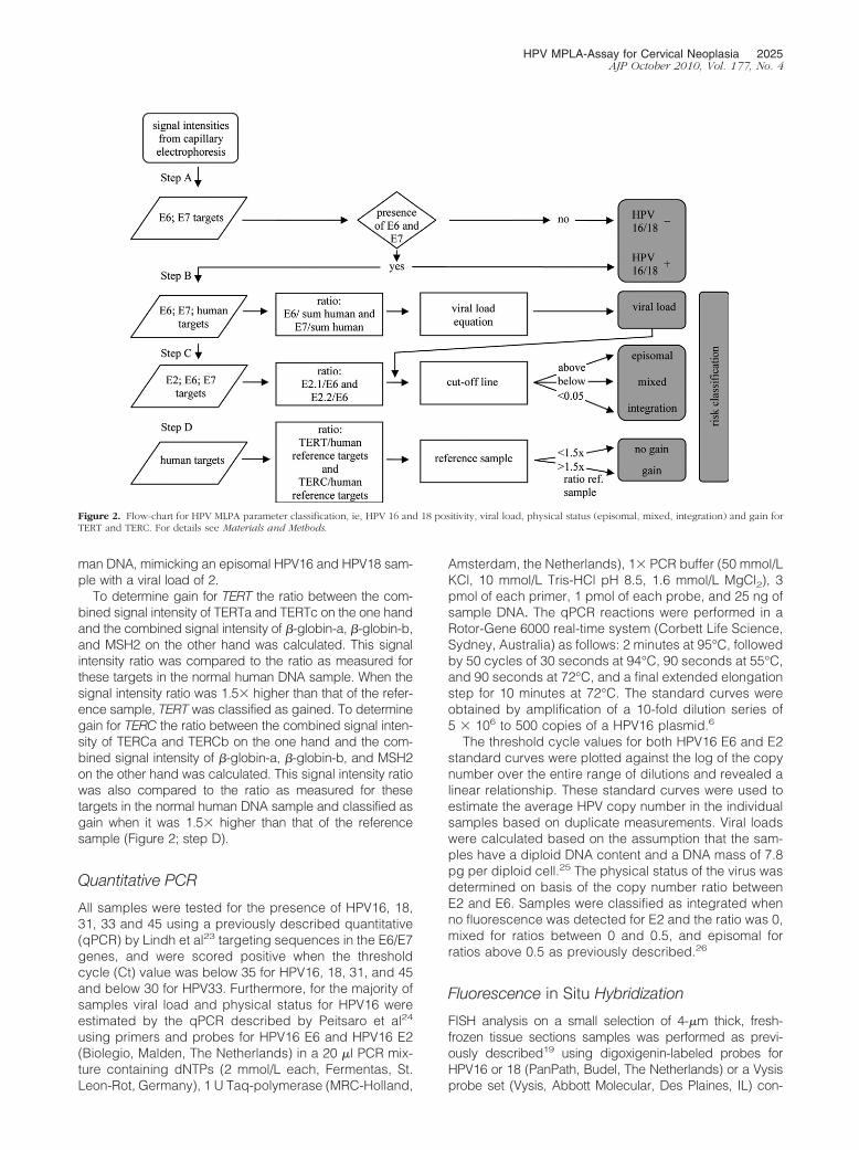
Amplified carboxyfluorescein-labeled MLPA productswere analyzed by electrophoresis on an ABI3730 genetic
HPV MPLA-Assay for Cervical Neoplasia 2023AJP October 2010, Vol. 177, No. 4
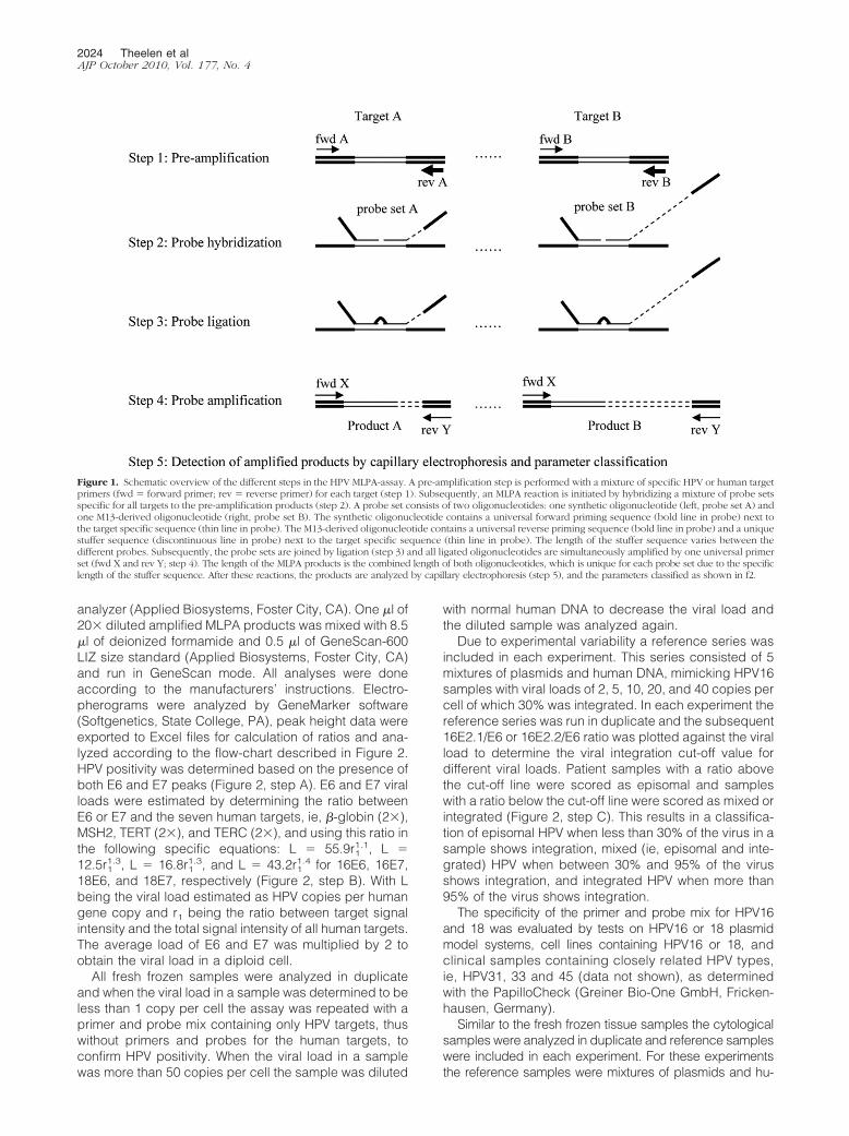
analyzer (Applied Biosystems, Foster City, CA). One �l of20� diluted amplified MLPA products was mixed with 8.5�l of deionized formamide and 0.5 �l of GeneScan-600LIZ size standard (Applied Biosystems, Foster City, CA)and run in GeneScan mode. All analyses were doneaccording to the manufacturers’ instructions. Electro-pherograms were analyzed by GeneMarker software(Softgenetics, State College, PA), peak height data wereexported to Excel files for calculation of ratios and ana-lyzed according to the flow-chart described in Figure 2.HPV positivity was determined based on the presence ofboth E6 and E7 peaks (Figure 2, step A). E6 and E7 viralloads were estimated by determining the ratio betweenE6 or E7 and the seven human targets, ie, �-globin (2�),MSH2, TERT (2�), and TERC (2�), and using this ratio inthe following specific equations: L � 55.9r1
1.1, L �12.5r1
1.3, L � 16.8r11.3, and L � 43.2r1
1.4 for 16E6, 16E7,18E6, and 18E7, respectively (Figure 2, step B). With Lbeing the viral load estimated as HPV copies per humangene copy and r1 being the ratio between target signalintensity and the total signal intensity of all human targets.The average load of E6 and E7 was multiplied by 2 toobtain the viral load in a diploid cell.
All fresh frozen samples were analyzed in duplicateand when the viral load in a sample was determined to beless than 1 copy per cell the assay was repeated with aprimer and probe mix containing only HPV targets, thuswithout primers and probes for the human targets, toconfirm HPV positivity. When the viral load in a samplewas more than 50 copies per cell the sample was diluted
with normal human DNA to decrease the viral load andthe diluted sample was analyzed again.
Due to experimental variability a reference series wasincluded in each experiment. This series consisted of 5mixtures of plasmids and human DNA, mimicking HPV16samples with viral loads of 2, 5, 10, 20, and 40 copies percell of which 30% was integrated. In each experiment thereference series was run in duplicate and the subsequent16E2.1/E6 or 16E2.2/E6 ratio was plotted against the viralload to determine the viral integration cut-off value fordifferent viral loads. Patient samples with a ratio abovethe cut-off line were scored as episomal and sampleswith a ratio below the cut-off line were scored as mixed orintegrated (Figure 2, step C). This results in a classifica-tion of episomal HPV when less than 30% of the virus in asample shows integration, mixed (ie, episomal and inte-grated) HPV when between 30% and 95% of the virusshows integration, and integrated HPV when more than95% of the virus shows integration.
The specificity of the primer and probe mix for HPV16and 18 was evaluated by tests on HPV16 or 18 plasmidmodel systems, cell lines containing HPV16 or 18, andclinical samples containing closely related HPV types,ie, HPV31, 33 and 45 (data not shown), as determinedwith the PapilloCheck (Greiner Bio-One GmbH, Fricken-hausen, Germany).
Similar to the fresh frozen tissue samples the cytologicalsamples were analyzed in duplicate and reference sampleswere included in each experiment. For these experimentsthe reference samples were mixtures of plasmids and hu-
Figure 1. Schematic overview of the different steps in the HPV MLPA-assay. A pre-amplification step is performed with a mixture of specific HPV or human targetprimers (fwd � forward primer; rev � reverse primer) for each target (step 1). Subsequently, an MLPA reaction is initiated by hybridizing a mixture of probe setsspecific for all targets to the pre-amplification products (step 2). A probe set consists of two oligonucleotides: one synthetic oligonucleotide (left, probe set A) andone M13-derived oligonucleotide (right, probe set B). The synthetic oligonucleotide contains a universal forward priming sequence (bold line in probe) next tothe target specific sequence (thin line in probe). The M13-derived oligonucleotide contains a universal reverse priming sequence (bold line in probe) and a uniquestuffer sequence (discontinuous line in probe) next to the target specific sequence (thin line in probe). The length of the stuffer sequence varies between thedifferent probes. Subsequently, the probe sets are joined by ligation (step 3) and all ligated oligonucleotides are simultaneously amplified by one universal primerset (fwd X and rev Y; step 4). The length of the MLPA products is the combined length of both oligonucleotides, which is unique for each probe set due to the specificlength of the stuffer sequence. After these reactions, the products are analyzed by capillary electrophoresis (step 5), and the parameters classified as shown in f2.
2024 Theelen et alAJP October 2010, Vol. 177, No. 4
man DNA, mimicking an episomal HPV16 and HPV18 sam-ple with a viral load of 2.
To determine gain for TERT the ratio between the com-bined signal intensity of TERTa and TERTc on the one handand the combined signal intensity of �-globin-a, �-globin-b,and MSH2 on the other hand was calculated. This signalintensity ratio was compared to the ratio as measured forthese targets in the normal human DNA sample. When thesignal intensity ratio was 1.5� higher than that of the refer-ence sample, TERT was classified as gained. To determinegain for TERC the ratio between the combined signal inten-sity of TERCa and TERCb on the one hand and the com-bined signal intensity of �-globin-a, �-globin-b, and MSH2on the other hand was calculated. This signal intensity ratiowas also compared to the ratio as measured for thesetargets in the normal human DNA sample and classified asgain when it was 1.5� higher than that of the referencesample (Figure 2; step D).
Quantitative PCR
All samples were tested for the presence of HPV16, 18,31, 33 and 45 using a previously described quantitative(qPCR) by Lindh et al23 targeting sequences in the E6/E7genes, and were scored positive when the thresholdcycle (Ct) value was below 35 for HPV16, 18, 31, and 45and below 30 for HPV33. Furthermore, for the majority ofsamples viral load and physical status for HPV16 wereestimated by the qPCR described by Peitsaro et al24
using primers and probes for HPV16 E6 and HPV16 E2(Biolegio, Malden, The Netherlands) in a 20 �l PCR mix-ture containing dNTPs (2 mmol/L each, Fermentas, St.Leon-Rot, Germany), 1 U Taq-polymerase (MRC-Holland,
Amsterdam, the Netherlands), 1� PCR buffer (50 mmol/LKCl, 10 mmol/L Tris-HCl pH 8.5, 1.6 mmol/L MgCl2), 3pmol of each primer, 1 pmol of each probe, and 25 ng ofsample DNA. The qPCR reactions were performed in aRotor-Gene 6000 real-time system (Corbett Life Science,Sydney, Australia) as follows: 2 minutes at 95°C, followedby 50 cycles of 30 seconds at 94°C, 90 seconds at 55°C,and 90 seconds at 72°C, and a final extended elongationstep for 10 minutes at 72°C. The standard curves wereobtained by amplification of a 10-fold dilution series of5 � 106 to 500 copies of a HPV16 plasmid.6
The threshold cycle values for both HPV16 E6 and E2standard curves were plotted against the log of the copynumber over the entire range of dilutions and revealed alinear relationship. These standard curves were used toestimate the average HPV copy number in the individualsamples based on duplicate measurements. Viral loadswere calculated based on the assumption that the sam-ples have a diploid DNA content and a DNA mass of 7.8pg per diploid cell.25 The physical status of the virus wasdetermined on basis of the copy number ratio betweenE2 and E6. Samples were classified as integrated whenno fluorescence was detected for E2 and the ratio was 0,mixed for ratios between 0 and 0.5, and episomal forratios above 0.5 as previously described.26
Fluorescence in Situ Hybridization
FISH analysis on a small selection of 4-�m thick, fresh-frozen tissue sections samples was performed as previ-ously described19 using digoxigenin-labeled probes forHPV16 or 18 (PanPath, Budel, The Netherlands) or a Vysisprobe set (Vysis, Abbott Molecular, Des Plaines, IL) con-
Figure 2. Flow-chart for HPV MLPA parameter classification, ie, HPV 16 and 18 positivity, viral load, physical status (episomal, mixed, integration) and gain forTERT and TERC. For details see Materials and Methods.
HPV MPLA-Assay for Cervical Neoplasia 2025AJP October 2010, Vol. 177, No. 4
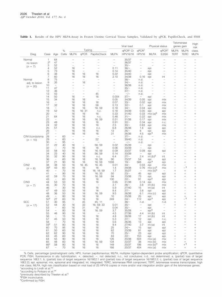
Table 1. Results of the HPV MLPA-Assay in Frozen Uterine Cervical Tissue Samples, Validated by qPCR, PapilloCheck, and FISH
TypingViral load Physical status
Telomerasegenes gain
Highrisk
class.MLPADiag. Case Age
%Cells MLPA qPCR PapilloCheck MLPA
qPCR* CtHPV16/18
qPCR†
HPV16 MLPAqPCR†
E2/E6MLPATERT
MLPATERC
Normalno lesion(n � 7)
1 64 � � � � 35/37 � � � � � �2‡ 47 � � � � 36/37 � � � � � �3 52 � � � � 35/� � � � � � �4 52 16 � � 0.11 36/� � epi � � � �5 73 16 � � 0.14 35/40 � epi � � � �6 38 16 16 16 0.22 34/40 � epi � � � �7 60 16 16 16 2.10 34/38 0.16 epi int � � �
Normaladj. to lesion(n � 20)
8 75 � � � � 38/� n.d. � � � � �9 35 � 16 � � 34/� n.d. � � � � �
10 � � � � � 38/38 n.d. � � � � �11 47 � � � � 35/� n.d. � � � � �12 36 � � � � 37/� n.d. � � � � �13 43 � � 45 � �/� n.d. � � � � �14 50 16 � 16 0.004 37/� � epi � � � �15 34 16 16 � 0.05 34/39 0.05 epi mix � � �16 � 16 16 16 0.07 33/� 0.02 epi mix � � �17 32 16 16 58 0.13 32/� 0.1 epi mix � � �18 � 16 16 16; 59 0.18 34/� 0.04 epi mix � � �19 52 16 16; 31 n.c. 0.18 34/39 0.05 epi epi � � �20 � 16 16 16 0.33 31/40 0.33 mix (a)§ mix � � �21 64 16 16 n.c. 0.48 31/� 0.22 epi mix � � �22 � 16 16 16; 59 0.51 31/38 0.17 epi mix � � �23 49 16 16 16 0.91 32/� 0.34 epi n.c. � � �24 32 16 16 16 2.5 30/� 0.83 epi mix � � �25 51 16 16 n.c. 6.8 26/38 1.6 epi epi � � �26 � 16 16 16 13 26/� 8 epi epi � � �27 � 16 16 16 31 26/36 4.5 epi¶ mix � � �
CIN1/condyloma(n � 10)
28 � 60 � � � � 37/� n.d. � � � � �29 � 60 � � 52 � 39/40 n.d. � � � � �30 � 95 � � � � �/� n.d. � � � � �31 22 40 16 � 56; 59 0.02 35/39 � epi � � � �32 � 70 16 16 16 0.06 33/39 � epi � � � �33 58 50 16 16 16; 59 0.09 33/37 0.09 epi epi � � �34 23 40 16 16 56; 59 0.14 33/39 � epi � � � �35 32 90 16 16 n.c. 6.4 27/� 2.4 int (b)§ int � � �36 � 60 16 16 16; 59 90 23/37 54 epi epi � � �37 21 90 16 16 16; 59 1008 19/� 568 epi¶ epi � � �
CIN2(n � 6)
38 38 50 16 16; 45 16; 45 0.01 34/� 0.03 epi mix � � �39‡ 43 60 16 16 16 1.4 29/38 0.76 mix (c)¶ mix � �¶ �40 � 70 18 18 16; 18; 59 1.7 35/27 � mix (b)§ � � � �41 � 90 16 16 16; 33 50 23/� 45 epi epi � � �42 59 70 16 16 16; 59 94 23/38 73 epi epi � � �43 � 60 16 16 16 130 22/� 91 epi§ epi � � �
CIN3(n � 7)
44 53 50 16 16 16 0.65 31/38 0.14 epi mix � � �45 30 70 16 16 � 3.1 28/� 3.8 int (b) mix � � �46 � 30 16 16 16 5.9 27/40 15 int (a) int � � �47 49 50 16 16 16 8.4 27/� 3.8 epi epi � � �48 36 50 16 16 16; 59 9.5 26/36 6.1 mix (c) epi � � �49 � 80 16 16 16 10 25/38 25 epi epi � � �50‡ 27 60 16 16 16 249 24/� 112 epi¶ epi � �¶ �
SCC(n � 17)
51 30 95 � 45 45; 11 � 38/� n.d. � n.d. � � �52 44 30 16 33 16; 59; 33 0.01 35/� � epi � � � �53 � 60 16 31 16 0.04 35/� � epi � � � �54 � 80 18 18 16; 59 3.7 35/28 � epi§ � � � �55 46 90 16 16 � 4.5 27/36 4.4 int (b) int � � �56 � 15 16 16 16 4.8 26/38 17 int (b) int � � �57 45 50 16 16 16 5.3 27/� 12 epi n.c. � � �58 � 70 16 16 16 5.6 26/36 13 epi epi � � �59‡ 51 80 16 16 16 9.4 27/40 3.7 int (a) int � � �60 70 95 16 16 16 25 24/� 15 epi epi � � �61 50 60 16 16 16 62 23/39 81 epi epi � � �62 62 90 16 16 16 70 25/� 23 epi epi � � �63 45 60 16 16 16 83 22/� 153 epi epi � � �64 55 90 16 16 16 126 20/38 504 mix (a) mix � � �65 48 60 16 16 16; 59 134 22/37 38 mix (b) mix � � �66‡ 58 80 16 16 16 149 20/37 198 mix (b)¶ mix � �¶ �67 � 70 16 16 16 166 20/37 404 mix (a) mix � � �
% Cells, percentage (pre)malignant cells; HPV, human papillomavirus; MLPA, multiplex ligation-dependent probe amplification; qPCR, quantitativePCR; FISH, fluorescence in situ hybridization; �, detected; �, not detected; n.c., not conclusive; n.d., not determined; a, (partial) loss of targetsequence 16E2.1; b, (partial) loss of target sequence 16/18E2.1 and (partial) loss of target sequence 16/18E2.2; c, (partial) loss of target sequence16E2.2); epi, episomal; mix, episomal and integrated; int, integrated; TERC, telomerase RNA component; TERT, telomerase reverse transcriptase; highrisk class; MLPA, high risk classification based on viral load of 25 HPV16 copies or more and/or viral integration and/or gain of the telomerase genes.*according to Lindh et al.23†according to Peitsaro et al.24‡previously described by Theelen et al.6§FISH inconclusive.¶Confirmed by FISH.
2026 Theelen et alAJP October 2010, Vol. 177, No. 4
sisting of: a DNA probe for chromosome 3 centromerelabeled with the fluorescent dye Spectrum Green (SG), a3q26-specific BAC clone containing the TERC gene labeledwith Spectrum Orange (SO), and a probe for chromosome7 centromere labeled with Spectrum Aqua (SA).
Images were acquired using a Leica DMRXA micro-scope (Leica, Wetzlar, Germany) equipped with customoptical filters for 4,6-diamidino-2-phenylindole, SA, SO,and SG (Chroma Technologies, Brattleboro, VT) with a�40 Plan Apo (NA 1.20) objective. The microscope wasconnected to a digital black and white CCD camera(Metasystems Image Pro System, Sandhausen, Ger-many). To determine gain for the TERC target the copynumber for chromosome 7 was used as a control for theploidy of cells. For chromosome 3 and target 3q26(TERC) the maximum number of signals per nucleus wasdetermined and used as an indicator of copy number, asdescribed previously.19
PapilloCheck Assay
The PapilloCheck HPV-Screen DNA-chip (Greiner, Frick-enhausen, Germany) was used for the qualitative detec-tion and differentiation between 24 types of genital HPV(18 high-risk and 6 low-risk). HPV genotyping was per-formed as previously described by Jones et al27 Briefly,for each test at least 40 ng of DNA was used and a 350
bp fragment of the HPV E1 gene was amplified using amultiplex PCR with type-specific primers. An internal PCRcontrol targeting a fragment of the human housekeepinggene ADAT1 was included in each run to avoid falsenegative results. PCR fragments were fluorescence-la-beled with Cy5 and hybridized to specific probes on thePapilloCheck DNA chip. The amplification level was de-termined by the binding of PCR products to five controlspots and their subsequent signal intensity on the chip.Following hybridization and subsequent washing steps,the chip was scanned at excitation wavelengths of 532and 635 nm.
Results
Fresh Frozen Tissue Samples
HPV-Typing
In order for the MLPA-assay to be useful in cervicalscreening the sensitivity in HPV-typing has to be high. Assummarized in Table 1, 52 of the 67 patient samples werefound to be positive for HPV16, including 4 of the 7 (57%)normal samples, 14 of the 20 (70%) normal samplesadjacent to a lesion, 7 of the 10 (70%) CIN1/condylomasamples, 12 of the 13 (92%) CIN2/3 samples and 15 ofthe 17 (88%) carcinoma samples. Furthermore, one
Figure 3. A: Reproducibility of viral load esti-mations (HPV copies per cell) when comparingduplicate MLPA measurements. B–D: Compari-son of the viral load estimations by MLPA andqPCR for fresh frozen tissue samples. B: Com-parison of viral load as determined by MLPA andqPCR as applied by Peitsaro et al.24 C: Correla-tion between the Ct values determined by qPCRusing the method of Lindh et al23 and the viralloads as determined by the MLPA. D: Correlationbetween the Ct values as determined by qPCRusing the method of Lindh et al23 and qPCRusing the method of Peitsaro et al.24
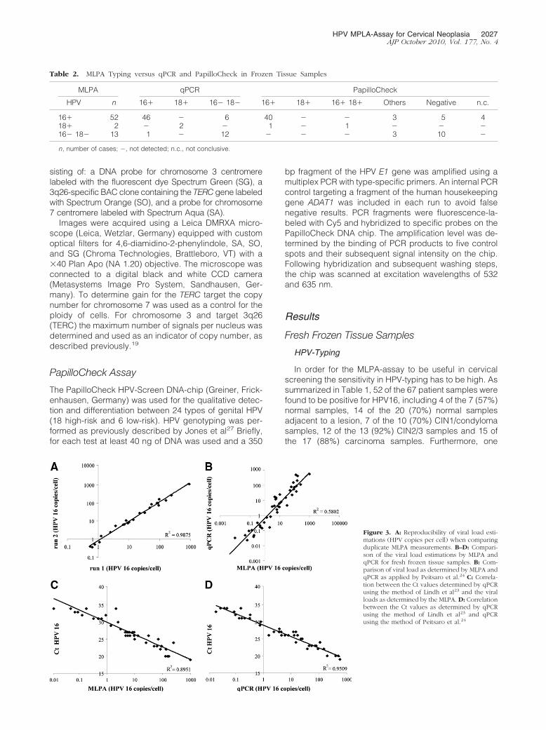
Table 2. MLPA Typing versus qPCR and PapilloCheck in Frozen Tissue Samples
MLPA qPCR PapilloCheck
HPV n 16� 18� 16� 18� 16� 18� 16� 18� Others Negative n.c.
16� 52 46 � 6 40 � � 3 5 418� 2 � 2 � 1 � 1 � � �16� 18� 13 1 � 12 � � � 3 10 �
n, number of cases; �, not detected; n.c., not conclusive.
HPV MPLA-Assay for Cervical Neoplasia 2027AJP October 2010, Vol. 177, No. 4

CIN2/3 and one carcinoma sample were found to bepositive for HPV18.
To determine the validity of the MLPA results all sam-ples were analyzed by two independent qPCR protocols
for HPV16, a qPCR for HPV18, 31, 33, and 45, and thePapilloCheck. The MLPA results were confirmed byqPCR in 48 of the 54 HPV16/18 positive cases and 12of the 13 HPV16/18 negative cases based on a thresh-old cycle (Ct) value below 35. Furthermore, using thePapilloCheck HPV16 was detected in 40 of the 52 casesthat were determined to be HPV16 positive by the MLPA-assay. In 4 samples the PapilloCheck was inconclusive,while 6 of the 8 samples in which no HPV16 was detectedwith the PapilloCheck, were determined to have a viralload of less than 1 copy per 5 cells, and the remaining 2samples had a viral load of 3 and 5 copies per cell. In twoof these HPV16 negative samples a double infection withHPV56 and 59 was detected, one sample had an infec-tion with HPV58 and the other 5 samples were negativefor all HPV types detected by the PapilloCheck (Table 2).
All 13 samples that were determined to be HPV16/18negative by the MLPA-assay were validated using thePapilloCheck, which was also used to identify infectionswith other HPV types. There were 3 samples that werepositive for one or more of the other HPV types. Thisconcerned an HPV45 infection in a normal sample whichwas adjacent to a lesion, an HPV52 infection in a CIN1/condyloma sample and a co-infection with HPV11 and 45in a carcinoma sample. This latter HPV45 infection as wellas a co-infection of HPV16 and 45 was also confirmedby the qPCR by Lindh et al23 The 10 remaining sam-ples were negative for all HPV types detected by thePapilloCheck.
Figure 4. A–C: Examples of the different techniques for fresh frozen tissuesamples: an MLPA capillary electrophoresis peak profile (A; case 59); qPCRcurves for E6 (B) and E2 (C) for a sample with exclusively episomal HPV in blue(case 61); a sample with both episomal and integrated HPV in green (case 64);and a sample with fully integrated HPV in red (case 43). D–G: FISH patterns forepisomal HPV (D; case 37), integrated HPV (E; case 66), and the absence (F;case 50) or presence (G; case 66) of gain for TERC (red) as compared to copynumbers of chromosome 7 (blue) and chromosome 3 (green).
Figure 5. Reproducibility of detection of viralintegration using E2/E6 ratios for fresh frozentissue samples. Comparison of the duplicateMLPA measurements for the E2.1/E6 (A) andE2.2/E6 (B) signal intensity ratios.
Table 3. Comparison between MLPA and qPCR for PhysicalStatus Analysis in Frozen Tissue Samples
MLPAPhysical status
qPCR
n epi mix int n.c. �
Epi 39 17 10 1 2 9mix 2.1 3 � 3 � � �mix 2.2 2 1 1 � � �mix 2.1 and 2.2 2 � 2 � � �int 2.1 2 � � 2 � �int 2.1 and 2.2 4 � 1 3 � �
n, number of cases; epi, episomal; mix, episomal and integrated; int,integrated; 2.1, (partial) loss of target sequence 16E2.1; 2.2, (partial)loss of target sequence 16E2.2; n.c., not conclusive; �, not detected.
2028 Theelen et alAJP October 2010, Vol. 177, No. 4

Viral Load
For HPV16 a high viral load is described to be associatedwith progression to cancer. Overall the viral load de-tected by the MLPA-assay ranged from less than 1 copyper 10 cells to approximately 1000 copies per cell. Asdepicted in Figure 3A the reproducibility of the MLPAprocedure for viral load detection between the duplicateanalyses, starting from the same DNA sample, is veryhigh. A median viral load of 1 copy per 5 cells wasdetected for the normal samples, 2 copies per 5 cells forthe normal samples adjacent to a lesion, 1 copy per 7cells for the CIN1/condyloma samples, 26 copies percell for the CIN2 samples, 8 copies per cell for theCIN3 samples, and 17 copies per cell for the carci-noma samples.
To validate the viral load as determined by the MLPA-assay, all HPV16 positive samples were analyzed byqPCR according to Peitsaro et al.24 Viral load could bedetermined in 43 of these 52 samples analyzed usingqPCR. For the 9 qPCR negative samples the viral loadwas determined to be less than 1 copy per 4 cells usingthe MLPA-assay. When comparing the viral loads asdetermined by the MLPA-assay or qPCR for the individualcases it became evident that both procedures detect asimilar range of HPV copies per cell (Figure 3B).23–24
Overall, the viral load as detected by the qPCR accordingto Lindh et al23 using Ct values correlated well with thecopy numbers as detected by the MLPA (Figure 3C)23–24
and the qPCR according to Peitsaro et al24 (Figure3D).23–24 The detected HPV copies per cell ranged fromless than 1 copy per 10 cells to approximately 570 copiesper cell, with a viral load of 0.16 copies per cell for thenormal sample, and a median viral load of 1 copy per 5cells for the normal samples adjacent to a lesion, 2 cop-ies per 7 cells for the CIN1/condyloma samples, 11 cop-ies per cell for the CIN2/3 samples, and 23 copies percell for the carcinoma samples.
Viral Integration
Viral integration into the host genome is described to beassociated with progression to cancer. As a conse-quence of integration (part of) the HPV E2 gene is almostalways deleted, while the HPV E6 gene is retained (seeFigure 4, A–C). Quantification of the E2/E6 ratio is there-fore used for the estimation of viral integration. The re-producibility of both the E2.1/E6 and the E2.2/E6 signalintensity ratio is depicted in Figure 5, A and B, and foundto be high. Integrated HPV was detected in 1 of the 16HPV16 positive normal samples adjacent to a lesion, 1 ofthe 7 HPV16 positive CIN1/condyloma samples, 4 of the12 HPV16 positive CIN2/3 samples, as well as the HPV18positive CIN2/3 sample and 7 of the 15 HPV16 positivecarcinoma samples.
For the validation of the physical status as deter-mined by the MLPA-assay we used the E2/E6 duplexqPCR. Thirteen of the 14 samples that were determinedto contain integrated HPV by the MLPA-assay wereconfirmed by qPCR. One of these samples (case 45)
was determined to contain predominantly integratedHPV by the MLPA-assay whereas qPCR determinedthe sample to contain both integrated and episomalHPV. In addition, 17 of the 39 samples that were de-termined to be episomal by MLPA were confirmed byqPCR. Nine of these 39 samples could not be con-firmed because no HPV16 was detected by qPCR andin 2 of these 39 samples qPCR was inconclusive be-cause of inconsistency between duplicates. Strikingly,in the remaining 11 samples in which the MLPA-assaydetected episomal HPV the qPCR indicated the pres-ence of integrated HPV (mixed or integrated; summa-rized in Table 3). These 11 discordant cases com-prised 9 normal samples (of which 8 adjacent to alesion), and 2 CIN2/3.
Of the 10 samples that were analyzed for the phys-ical status of HPV16 and HPV18 by FISH episomalHPV16 was confirmed in three cases (cases 27, 37,and 50 in Table 1; Figure 4D) and integrated HPV intwo cases (cases 40, and 66 in Table 1; Figure 4E). Inthe remaining 3 HPV16 samples (cases 20, 35, and 43)FISH analysis was inconclusive, which was also thecase for the 2 samples that were analyzed for HPV18(cases 40 and 54). All these latter 5 cases had arelatively low viral load.
Telomerase Genes
Gain for the telomerase related genes TERT and TERC isdescribed to be associated with progression to cancer,and was therefore included in our HPV MLPA-assay. Gainfor TERT was detected in 8 samples, ie, 1 CIN1, 3 CIN2/3,and 4 carcinoma samples. Gain for TERC was detectedin 16 samples, ie, 1 CIN1, 6 CIN2/3, and 9 carcinomasamples. Seven of the 8 cases with gain for TERT over-lapped with the TERC gain cases. Only in case 51 TERTgain was seen to be independent of gain for TERC. TheFISH analyses for TERC performed on tissue sectionsfrom three patients (cases 39, 50, and 66 in Table 1;Figure 4, F and G) gave confirmatory results.
Sensitivity and Specificity of the MLPA-Assay
To quantify the diagnostic capacity of the MLPA-assay,sensitivity and specificity were calculated for each of thefour parameters, ie, HPV type 16/18, viral load, viral inte-gration, and gain of the telomerase related genes. Forthis purpose the ability of the assay to distinguish highgrade CIN2/3 and carcinoma lesions on the one hand,from normal samples, low grade CIN1 and condylomalesions on the other hand, was determined. For the pres-ence of HPV type 16 and 18 the sensitivity is calculated tobe 97% but the specificity to be only 32%. For a viral loadof more than 25 copies per cell the sensitivity and spec-ificity are calculated to be 40% and 92%, respectively.For viral integration this is 40% and 95%, for gain of TERT23% and 97%, and for gain of TERC 50% and 97%,respectively (Table 4). By combining 2 parameters thesensitivity can be increased up to 63 to 70%, with a smallreduction in specificity to 86 to 92%. When all parameters
HPV MPLA-Assay for Cervical Neoplasia 2029AJP October 2010, Vol. 177, No. 4

are combined the sensitivity increases up to 83%, whileretaining a specificity of 86%.
Cytological Samples
Cytological samples of 91 preselected cases (Table 5),ranging from normal to HSIL (Pap classification 1 to3A28), for which the presence of HPV was known basedon GP5�/6� testing,22 were analyzed by the MLPA-assay and verified by the two qPCR protocols. TheMLPA-assay determined a total of 32 samples to bepositive for HPV16/18 of which 27 were positive forHPV16 and 5 were positive for HPV18. Among the HPV16samples 11 had a viral load higher than 25 copies per celland 6 samples contained integrated HPV whereasamong the HPV18 samples 2 contained integrated HPV.Of these 32 cases, 31 were confirmed by qPCR. Theremaining sample was determined to have a HPV16 viralload of 1 copy per 10 cells by the MLPA-assay and aCt-value of 28 for HPV31 by qPCR. Additionally, of the 59HPV16/18 negative samples qPCR found 4 samples (2normal and 2 LSIL) to be infected with HPV45, 9 samples(2 ASCUS and 7 LSIL) with HPV31 and 1 LSIL samplewith HPV33. Furthermore, a gain for TERT and/or TERCwas detected in 10 of the 32 HPV 16/18 positive samples,as well as in 5 of the 59 HPV16/18 negative samples. Assummarized in Table 5, 23 of the 91 samples were de-termined to be high risk according to the MLPA-assay.This concerned 1 of the 7 (14%) normal, 4 of the 28 (14%)ASCUS, 14 of the 49 (29%) LSIL, and 4 of the 7 (57%)HSIL samples.
Discussion
HPV-related parameters, such as oncogenic type, viralload, and viral integration are prognosticators in cervical(pre)neoplasia.9,11,19 Since these individual markershave been shown to exhibit either a low sensitivity or alow specificity,5,11 we have developed a multiparameterassay for the simultaneous assessment of these indica-tors in combination with the detection of increased copynumbers of the telomerase related genes. The predictive
value of the individual markers in this HPV MLPA-assaywas similar to that described in the literature,11,19,20 butwhen combining the results of the individual parametersin the MLPA-assay a balance between high specificityand high sensitivity was reached which is more accept-able for a screening test.
For typing of HPV16 and 18 all of the fresh frozen tissuesamples that were determined to be negative by theMLPA-assay were confirmed by the PapilloCheck and/orqPCR. This was also the case for 48 of the 52 fresh frozentissue samples that were found to be HPV16 positive. Thediscrepant samples were characterized by a low viralload, which implies that the MLPA-assay is at least assensitive as qPCR and the PapilloCheck in the detectionof these HPV types. This is particularly evident since forthe two latter assays higher amounts of DNA were usedas compared to the MLPA-assay. Also the number ofcycles used for the qPCR as described by Peitsaro et al24
was 50 instead of 40.The prevalence for HPV16 among the CIN2/3 and car-
cinoma cases in this study is as high as 92% and 88%,respectively, while the literature reports a prevalence ofabout 60% in carcinoma cases.9 Based on the abnor-mally high prevalence for HPV16 in this study it is clearthat the sensitivity of this assay is overestimated. Theprevalence for HPV16 is also high in the histologicallynormal samples, particularly when combined with a (pre)neoplastic lesion. The risk of false positive results is,however, unlikely in the MLPA-assay because of the useof four probes per HPV type. HPV infection is only scoredas being positive when a product is detected for both theE6 and E7 probe, while in a majority of cases also prod-ucts will be detected for one or both of the E2 probes,providing additional proof for the reliability of the out-come. Furthermore, the samples in our study were ran-domly selected without prior knowledge of HPV status,and as described above mostly confirmed by qPCR orthe PapilloCheck providing additional arguments againstfalse positive MLPA-results. The presence of HPV in thenormal samples, either without a lesion or adjacent to apremalignancy, indicates that the sole detection of HPV is
Table 4. Sensitivity and Specificity of the HPV MLPA-Assay when Applied to Frozen Tissue Samples
Histology
MLPA single parameter
Type 16and/or 18 (%) Viral load �25 (%) Integration (%) TERT gain (%) TERC gain (%) Telom gain (%)
Normal 4/7 (57) 0/7 (0) 0/7 (0) 0/7 (0) 0/7 (0) 0/7 (0)Normal
adjacentto lesion
14/20 (70) 1/20 (5) 1/20 (5) 0/20 (0) 0/20 (0) 0/20 (0)
CIN1/condyloma
7/10 (70) 2/10 (20) 1/10 (10) 1/10 (10) 1/10 (10) 1/10 (10)
CIN2/3 13/13 (100) 4/13 (31) 5/13 (38) 3/13 (23) 6/13 (46) 6/13 (46)SCC 16/17 (94) 8/17 (47) 7/17 (41) 4/17 (24) 9/17 (53) 10/17 (59)Sensitivity
(95% CI)97%
(80.9–99.8%)40%
(23.2–59.2%)40%
(23.2–59.2%)23%
(10.6–42.7%)50%
(31.7–68.3%)53%
(34.6–71.2%)Specificity
(95% CI)32%
(18.6–49.9%)92%
(77.0–97.9%)95%
(80.5–99.1%)97%
(84.2–99.9%)97%
(84.2–99.9%)97%
(84.2–99.9%)
CI, confidence interval; CIN, cervical interaepithelial neoplasia; SCC, squamous cell carcinoma; telom, telomerase gene; TERC, telomerase RNAcomponent; TERT, telomerase reverse transcriptase; �25, more than 25 copies per cell.
2030 Theelen et alAJP October 2010, Vol. 177, No. 4

not a very specific prognosticator for the presence of ahigh grade lesion.
In the literature, the cut-off value for HPV16 viral loadused to identify with high specificity women with preva-lent high grade lesions is described to be between 22and 35 copies per cell.11,29 Therefore, the cut-off valueused in this study was set at 25 viral copies per cell.Based on this value 40 of the 43 samples were identicallyclassified using either the MLPA-assay or qPCR. How-ever, in this study, the viral load as detected using theMLPA-assay was generally somewhat higher than theviral load as detected using qPCR. As a result, 2 freshfrozen tissue samples, ie, 1 normal sample adjacent to alesion and 1 carcinoma sample, were classified as highgrade based on the viral load determined by the MLPA-assay and as low grade based on qPCR, which might bean indication for the use of a higher cut-off value for theMLPA-assay. On the other hand, one CIN2/3 sample wasclassified as low-grade based on the viral load as deter-mined by the MLPA-assay and as high grade based onthe viral load as determined by qPCR, arguing againstsuch an increase of the cut-off value.
The incidence for viral integration based on qPCR wasvery high in the samples that were classified as normal.Although this observation is in agreement with previousqPCR studies on viral integration24 various other studiesusing APOT, FISH, or 2D-agarose gel electrophoresistechniques showed that integration occurs late in thecarcinogenic process.12,30,31 To explain this apparentdiscrepancy it should be noted that 10 of our samples
had a viral load of less than five copies per cell. The errorin qPCR threshold cycle values and E2/E6 ratios is pos-sibly higher in samples with a low viral load than insamples with a high viral load.32,33 Furthermore, therewas one case that was determined to contain integratedHPV using the MLPA-assay, but exclusively episomalHPV using qPCR. In this case the HPV16E2.2 targetsequence was deleted while the HPV16E2.1 target se-quence was retained. Integration was therefore missedby qPCR because the probe used recognizes a se-quence close to HPV16E2.1. Finally, in two cases theqPCR results were inconclusive.
With respect to the discriminating power of the MLPA-assay in the frozen tissue samples a sensitivity and spec-ificity of 63% and 86%, respectively, were obtained bycombining the three HPV parameters. Inclusion of theresults for the telomerase related genes in these analysesleads to a significant increase of the sensitivity to 83%,while retaining the specificity of 86%. In addition to fourhigh-grade cases with an episomal HPV16 infection anda low viral load, the carcinoma case that was not infectedwith HPV16 or 18 was also classified as high gradebased on a gain for one of the telomerase related genes.Furthermore, the absence or presence of gain for TERCwas confirmed by FISH in selected cases, while the inci-dence was in agreement with our previous findings.19
Similarly, Heselmeyer et al,18 using comparative genomichybridization in cases of severe cervical dysplasia andinvasive carcinoma, demonstrated an increasing inci-dence for gain of TERC with increasing severity of the
Table 4. Continued
MLPA multiparameter
Viral load �25 and/orintegration (%)
Viral load �25 and/ortelom gain (%)
Integration and/ortelom gain (%)
Viral load �25 and/or integrationand/or telom gain (%)
0/7 (0) 0/7 (0) 0/7 (0) 0/7 (0)2/20 (10) 1/20 (5) 1/20 (5) 2/20 (10)
3/10 (30) 2/10 (20) 2/10 (20) 3/10 (30)8/13 (62) 8/13 (62) 8/13 (62) 10/13 (77)
11/17 (65) 13/17 (76) 12/17 (71) 15/17 (88)63%
(43.9–79.5%)70%
(50.4–84.6%)67%
(47.1–82.1%)83%
(64.5–93.6%)86%
(70.4–94.9%)92%
(77.0–97.9%)92%
(77.0–97.9%)86%
(70.4–94.9%)
Table 5. Summary of Results of the MLPA Assay in Cytological Samples Compared with qPCR and GP5�/6�
Cytology n HR HPV (%)
GP5�/6�HPV
16/18 (%)
qPCRHPV
16/18 (%)
MLPA High-riskclass.
MLPA (%)High load (%) Integration (%)Telom.
gain (%)
Normal 7 3 (43) 1 (14) 1 (14) 0 0 1 (14) 1 (14)ASCUS 28 13 (46) 9 (32) 9 (32) 0 1 (4) 3 (11) 4 (14)LSIL 49 38 (78) 14 (29) 15 (31) 8 (16) 6 (12) 8 (16) 14 (29)HSIL 7 7 (100) 7 (100) 7 (100) 3 (43) 1 (14) 3 (43) 4 (57)Total 91 61 31 32 11 8 15 23
n, number of cases; HPV, human papillomavirus; MLPA, multiplex ligation-dependent probe amplification; qPCR, quantitative PCR; Telom.,telomerase genes; high-risk class; MLPA, high-risk classification based on viral load of 25 HPV16 copies or more and/or viral integration and/or gain ofthe telomerase genes.
HPV MPLA-Assay for Cervical Neoplasia 2031AJP October 2010, Vol. 177, No. 4

lesion. We conclude that the assessment of gain of thetelomerase genes contributes significantly to the properclassification of high risk patients.
In 10 of the 67 frozen tissue samples discordancebetween the morphological and molecular characteris-tics was observed. Four of the five cases among the CIN1and normal samples with either integration or a high viralload were confirmed by qPCR. These cases thereforedeserve closer monitoring than the other low gradecases. In five CIN2/3 and carcinoma cases the MLPA-assay resulted in an underestimation of the severity ofthe lesion. This can be partially explained by the pres-ence of other oncogenic HPV types, indicating thenecessity to include additional HPV types in the assay.An expansion of the number of probes targeting the E2gene could also lead to an increased detection ofintegration and subsequently to a higher sensitivity ofthe assay.
Application of the HPV MLPA-assay to a series ofcytological samples revealed an evident increase of highrisk classification with the increase of cytological grading.Overall, 25% of these preselected samples were classi-fied as high risk cases using our MLPA assay. Again,inclusion of additional HPV types in this test will increaseits sensitivity when applied to cytological samples. OurMLPA analysis of cytological samples showed that highrisk HPV16 and 18 is more often associated with highgrade cytological outcomes as compared to any otherhigh risk HPV type. This confirms the results as describedearlier by Munoz et al9 Furthermore, a high frequency ofintegration was seen in the LSIL cases in our series,which is in agreement with a previous study by Gallo etal34 who detected that viral integration was particularlyevident in LSIL of a younger population of women.Finally, gain for TERT and TERC was detected in 8 ofthe 49 LSIL samples, which is in agreement with astudy by Heselmeyer et al35 where gain for TERC wasdemonstrated in 7% of these low-grade samples. Ourresults thus show that the MLPA-assay is able to detectgain for TERT and TERC in cytological samples, despitethe fact that these preparations may contain a highproportion of normal cells. Apparently, in these sam-ples a sufficient fraction of (pre)malignant cells ispresent to reach and exceed the detection threshold forgain of the telomerase associated genes.
In conclusion, we showed that the HPV 16/18 MLPA-assay is capable of distinguishing high grade from lowgrade cervical lesions, both with a high sensitivity andhigh specificity. In biopsy material the combination ofthree parameters results in risk classification which canbe applied as a potential indicator for cervical (pre)neo-plasia. Furthermore, the potential of the MLPA assay as acomplementary technique for cytological samples hasbeen illustrated.
Acknowledgment
We thank Nathalie Krusy (Laboratory of ExperimentalPathology, University of Liege, Belgium) for the DNAisolation and PapilloCheck analysis.
References
1. Jordan J, Arbyn M, Martin-Hirsch P, Schenck U, Baldauf JJ, Da SilvaD, Anttila A, Nieminen P, Prendiville W: European guidelines forquality assurance in cervical cancer screening: recommendations forclinical management of abnormal cervical cytology, part 1. Cytopa-thology 2008, 19:342–354
2. Spitzer M, Apgar BS, Brotzman GL: Management of histologic abnor-malities of the cervix. Am Fam Physician 2006, 73:105–112
3. ACOG Practice Bulletin No. 99: management of abnormal cervicalcytology and histology. Obstet Gynecol 2008, 112:1419–1444
4. Cuzick J, Clavel C, Petry KU, Meijer CJ, Hoyer H, Ratnam S, SzarewskiA, Birembaut P, Kulasingam S, Sasieni P, Iftner T: Overview of theEuropean and North American studies on HPV testing in primary cervicalcancer screening. Int J Cancer 2006, 119:1095–1101
5. Rao A, Pather S, Dalrymple C, Mackie A, Deans R, Carter J: The roleof HPV testing in patients with possible high-grade cervical cytology.J Obstet Gynaecol Res 2009, 35:503–506
6. Theelen W, Reijans M, Simons G, Ramaekers FCS, Speel EJM,Hopman AHN: A new multiparameter assay to assess HPV 16/18 viralload and physical status together with gain of telomerase genes inHPV-related cancers. Int J Cancer 2010 126:959–975
7. Schouten JP, McElgunn CJ, Waaijer R, Zwijnenburg D, Diepvens F,Pals G: Relative quantification of 40 nucleic acid sequences bymultiplex ligation-dependent probe amplification. Nucleic Acids Res2002, 30:e57
8. Shen Y, Wu BL: Designing a simple multiplex ligation-dependentprobe amplification (MLPA) assay for rapid detection of copy numbervariants in the genome. J Genet Genomics 2009, 36:257–265
9. Munoz N, Bosch FX, de Sanjose S, Herrero R, Castellsague X, ShahKV, Snijders PJ, Meijer CJ: Epidemiologic classification of humanpapillomavirus types associated with cervical cancer. N Engl J Med2003, 348:518–527
10. Munoz N, Castellsague X, de Gonzalez AB, Gissmann L: Chapter 1:HPV in the etiology of human cancer. Vaccine 2006, 24 (Suppl3):1–10
11. Saunier M, Monnier-Benoit S, Mauny F, Dalstein V, Briolat J, RiethmullerD, Kantelip B, Schwarz E, Mougin C, Pretet JL: Analysis of humanpapillomavirus type 16 (HPV16) DNA load and physical state for identi-fication of HPV16-infected women with high-grade lesions or cervicalcarcinoma. J Clin Microbiol 2008, 46:3678–3685
12. Klaes R, Woerner SM, Ridder R, Wentzensen N, Duerst M, SchneiderA, Lotz B, Melsheimer P, von Knebel Doeberitz M: Detection ofhigh-risk cervical intraepithelial neoplasia and cervical cancer byamplification of transcripts derived from integrated papillomavirusoncogenes. Cancer Res 1999, 59:6132–6136
13. Cheung JL, Cheung TH, Ng CW, Yu MY, Wong MC, Siu SS, Yim SF,Chan PK: Analysis of human papillomavirus type 18 load and inte-gration status from low-grade cervical lesion to invasive cervicalcancer. J Clin Microbiol 2009, 47:287–293
14. Hesselink AT, Berkhof J, Heideman DA, Bulkmans NW, van TellingenJE, Meijer CJ, Snijders PJ: High-risk human papillomavirus DNA loadin a population-based cervical screening cohort in relation to thedetection of high-grade cervical intraepithelial neoplasia and cervicalcancer. Int J Cancer 2009, 124:381–386
15. Xi LF, Koutsky LA, Castle PE, Wheeler CM, Galloway DA, Mao C, HoJ, Kiviat NB: Human papillomavirus type 18 DNA load and 2-yearcumulative diagnoses of cervical intraepithelial neoplasia grades2–3. J Natl Cancer Inst 2009, 101:153–161
16. Pirami L, Giache V, Becciolini A: Analysis of HPV16, 18, 31, and 35DNA in pre-invasive and invasive lesions of the uterine cervix. J ClinPathol 1997, 50:600–604
17. Vinokurova S,Wentzensen N, Kraus I, Klaes R, Driesch C, Melsheimer P,Kisseljov F, Durst M, Schneider A, von Knebel Doeberitz M: Type-dependent integration frequency of human papillomavirus genomes incervical lesions. Cancer Res 2008, 68:307–313
18. Heselmeyer K, Schrock E, du Manoir S, Blegen H, Shah K, SteinbeckR, Auer G, Ried T: Gain of chromosome 3q defines the transition fromsevere dysplasia to invasive carcinoma of the uterine cervix. ProcNatl Acad Sci USA 1996, 93:479–484
19. Hopman AHN, Theelen W, Hommelberg PP, Kamps MA, Herrington CS,Morrison LE, Speel EJM, Smedts F, Ramaekers FCS: Genomic integra-tion of oncogenic HPV and gain of the human telomerase gene TERC at
2032 Theelen et alAJP October 2010, Vol. 177, No. 4
3q26 are strongly associated events in the progression of uterine cervi-cal dysplasia to invasive cancer. J Pathol 2006, 210:412–419
20. Huang FY, Kwok YK, Lau ET, Tang MH, Ng TY, Ngan HY: Geneticabnormalities and HPV status in cervical and vulvar squamous cellcarcinomas. Cancer Genet Cytogenet 2005, 157:42–48
21. Matthews CP, Shera KA, McDougall JK: Genomic changes and HPVtype in cervical carcinoma. Proc Soc Exp Biol Med 2000, 223:316–321
22. van den Brule AJ, Pol R, Fransen-Daalmeijer N, Schouls LM, MeijerCJ, Snijders PJ: GP5�/6� PCR followed by reverse line blot analysisenables rapid and high-throughput identification of human papillo-mavirus genotypes. J Clin Microbiol 2002, 40:779–787
23. Lindh M, Gorander S, Andersson E, Horal P, Mattsby-Balzer I, Ryd W:Real-time taqman PCR targeting 14 human papilloma virus types.J Clin Virol 2007, 40:321–324
24. Peitsaro P, Johansson B, Syrjanen S: Integrated human papillomavi-rus type 16 is frequently found in cervical cancer precursors asdemonstrated by a novel quantitative real-time PCR technique. J ClinMicrobiol 2002, 40:886–891
25. Lee GM, Thornthwaite JT, Rasch EM: Picogram per cell determinationof DNA by flow cytofluorometry. Anal Biochem 1984, 137:221–226
26. Kulmala SM, Shabalova IP, Petrovitchev N, Syrjanen KJ, GyllenstenUB, Johansson BC, Syrjanen SM: Type-specific persistence of high-risk human papillomavirus infections in the New Independent Statesof the former Soviet Union Cohort Study. Cancer Epidemiol Biomar-kers Prev 2007, 16:17–22
27. Jones J, Powell NG, Tristram A, Fiander AN, Hibbitts S: Comparisonof the PapilloCheck DNA micro-array Human Papillomavirus detec-tion assay with Hybrid Capture II and PCR-enzyme immunoassayusing the GP5/6� primer set. J Clin Virol 2009, 45:100–104
28. Rebolj M, van Ballegooijen M, Berkers LM, Habbema D: Monitoring a
national cancer prevention program: successful changes in cervicalcancer screening in the Netherlands. Int J Cancer 2007, 120:806–812
29. Cricca M, Morselli-Labate AM, Venturoli S, Ambretti S, Gentilomi GA,Gallinella G, Costa S, Musiani M, Zerbini M: Viral DNA load, physicalstatus and E2/E6 ratio as markers to grade HPV16 positive women forhigh-grade cervical lesions. Gynecol Oncol 2007, 106:549–557
30. Hudelist G, Manavi M, Pischinger KI, Watkins-Riedel T, Singer CF,Kubista E, Czerwenka KF: Physical state and expression of HPV DNAin benign and dysplastic cervical tissue: different levels of viral inte-gration are correlated with lesion grade. Gynecol Oncol 2004, 92:873–880
31. Hopman AHN, Smedts F, Dignef W, Ummelen M, Sonke G, MravunacM, Vooijs GP, Speel EJM, Ramaekers FCS: Transition of high-gradecervical intraepithelial neoplasia to micro-invasive carcinoma is char-acterized by integration of HPV 16/18 and numerical chromosomeabnormalities. J Pathol 2004, 202:23–33
32. Roberts I, Ng G, Foster N, Stanley M, Herdman MT, Pett MR,Teschendorff A, Coleman N: Critical evaluation of HPV16 gene copynumber quantification by SYBR green PCR. BMC Biotechnol 2008,8:57
33. Ruutu MP, Kulmala SM, Peitsaro P, Syrjanen SM: The performance ofthe HPV16 real-time PCR integration assay. Clin Biochem 2008,41:423–428
34. Gallo G, Bibbo M, Bagella L, Zamparelli A, Sanseverino F, GiovagnoliMR, Vecchione A, Giordano A: Study of viral integration of HPV-16 inyoung patients with LSIL. J Clin Pathol 2003, 56:532–536
35. Heselmeyer-Haddad K, Janz V, Castle PE, Chaudhri N, White N,Wilber K, Morrison LE, Auer G, Burroughs FH, Sherman ME, Ried T:Detection of genomic amplification of the human telomerase gene(TERC) in cytologic specimens as a genetic test for the diagnosis ofcervical dysplasia. Am J Pathol 2003, 163:1405–1416
HPV MPLA-Assay for Cervical Neoplasia 2033AJP October 2010, Vol. 177, No. 4
Related Documents