Incorporation of an Internal Ribosome Entry Site–Dependent Mechanism in Arsenic-Induced GADD45A Expression Qingshan Chang, 1,3 Deepak Bhatia, 1,2 Yadong Zhang, 1,3 Terry Meighan, 1 Vince Castranova, 1 Xianglin Shi, 1,3 and Fei Chen 1,2 1 The Health Effects Laboratory Division, National Institute for Occupational Safety and Health; 2 Department of Basic Pharmaceutical Sciences, West Virginia University, Morgantown, West Virginia; and 3 Institute for Nutritional Sciences, Shanghai Institute for Biological Sciences, Chinese Academy of Sciences, Shanghai, China Abstract We have previously shown that trivalent arsenic (arsenite, As 3+ ) is able to induce GADD45A expression in human bronchial epithelial cells through activation of c-Jun NH 2 - terminal kinase and nucleolin-dependent mRNA stabilization. In the present report, we show that As 3+ is capable of inducing translation of the GADD45A protein through a cap-indepen- dent, or rather, an internal ribosome entry site (IRES)– dependent mechanism. In growth-arrested cells, As 3+ elevated the GADD45A protein level in a dose- and time-dependent manner which did not correlate with the GADD45A mRNA expression. Pretreatment of the cells with rapamycin, an inhibitor for the cap-dependent translation machinery through the suppression of mTOR and p70S6 kinase, failed to affect the induction of the GADD45A protein induced by As 3+ . Sequence analysis revealed a potential IRES element in the 5¶-untranslated region of the GADD45A mRNA. This IRES element in the 5¶-untranslated region of the GADD45A mRNA is functional in mediating As 3+ -induced translation of the GADD45A protein in a dicistronic reporter gene activity assay. Immunoprecipitation and proteomic studies suggest that As 3+ impairs the assembly of the cap-dependent initiating complex for general protein translation but increases the association of human elongation factor 2 and human heterogeneous nuclear ribonucleoprotin with this complex. Thus, these results suggest that in growth-arrested cells, As 3+ is still capable of inducing GADD45A expression through an IRES-dependent translational regulation. [Cancer Res 2007;67(13):6146–54] Introduction Growth arrest– and DNA damage–induced gene 45a (GADD45a) encodes a small acidic protein that is capable of interacting with a number of important intracellular signaling molecules involved in cell cycle regulation, apoptosis, and immune responses (1). A wide variety of stress inducers, including DNA-damaging reagents, UV radiation, oxidative or osmotic stress, and nutrient deprivation stimulate the expression of GADD45a mRNA and protein in a manner which may be either p53-dependent or p53-independent (2). The fact that GADD45a is preferentially localized in the nuclei suggests that the key function of this protein might be the regulation of DNA replication, DNA repair, and cell division. Indeed, the majority of the known GADD45a-interacting molecules are nuclear functional proteins, such as proliferating cell nuclear antigen (3), cyclin B/CDC2 complex (4), p21 cip1 (5), histone (6), aurora-A kinase (7), xeroderma pigmentosum complementation group G (8), and TAFII70 (9). Interaction of GADD45a with these proteins causes cell cycle arrest, repression of DNA replication or cell apoptosis, thereby providing the cells with time to repair the damaged DNA and prevent the segregation of damaged chromo- somes. Extensive efforts have been made to understand how the transcription of GADD45a is regulated in cellular responses to extracellular signals. The transcriptional induction of GADD45a by DNA damage is thought to be dependent on p53 or FoxO3a (10), which antagonizes transcriptional repressors of the GADD45a gene, such as c-myc (11) and ZBRK (12). Activation of c-Jun NH 2 - terminal kinase (JNK) may play a key role in mediating p53- independent transcription of GADD45a in response to other stress signals (13). The influence of posttranscriptional events, mainly the modulation of GADD45a mRNA stability, has been recognized in recent years. In humans, the bronchial epithelial cell line, BEAS-2B, stimulation of the cells with trivalent arsenic (arsenite, As 3+ ), a general stress inducer, stabilizes GADD45a mRNA through the inducible association of nucleolin and less potently, HuR, with the GADD45a mRNA (14). Removal of the RNA destabilization protein AUF1 from the 3-untranslated region (3¶-UTR) of the GADD45a mRNA was considered to be an important mechanism for the enhanced stability of the GADD45a mRNA in cells treated with genotoxin methyl methanesulfonate (15). Intriguingly, neither nucleolin nor HuR were associated with GADD45a mRNA in UV- or methyl methanesulfonate–treated HeLa cells (16). It is unknown whether the expression of GADD45a is subjected to protein translation regulation under certain circumstances. Environmental or occupational exposure to As 3+ is associated with a number of human diseases including nerve degeneration, skin hyperkeratinization, birth defects, hepatic cirrhosis, cardio- vascular disorders, diabetes, and cancer (17). By binding to sulfhydryl groups, As 3+ impairs the function of many proteins, especially glutathione/thioredoxin reductases and protein tyrosine phosphatases which possess vicinal thiols (18). The cumulative effects of this impairment are oxidative stress and kinase activation, including receptor type and non–receptor type tyrosine kinases, JNK, p38, p70S6 kinase (p70S6K), checkpoint kinases, and Akt. Both sustained oxidative stress and aberrant kinase activation have been linked to cell growth and malignant transformation. As 3+ is also able to induce ubiquitination or sumoylation and proteasomal degradation of some regulatory proteins, such as CDC25C (19), AML1/MDS1/EVI1 (20), and PML (21). This property Note: Q. Chang, D. Bhatia, and Y. Zhang made equal contributions to this report. Disclaimer: The opinions expressed in this article are those of the authors and do not necessarily represent the views of the National Institute for Occupational Safety and Health, Centers for Disease Control and Prevention. Requests for reprints: Fei Chen, Pathology and Physiology Research Branch, National Institute for Occupational Safety and Health, 1095 Willowdale Road, Morgantown, WV 26505. Phone: 304-285-6021; E-mail: [email protected]. I2007 American Association for Cancer Research. doi:10.1158/0008-5472.CAN-07-0867 Cancer Res 2007; 67: (13). July 1, 2007 6146 www.aacrjournals.org Research Article Research. on March 4, 2016. © 2007 American Association for Cancer cancerres.aacrjournals.org Downloaded from

Welcome message from author
This document is posted to help you gain knowledge. Please leave a comment to let me know what you think about it! Share it to your friends and learn new things together.
Transcript

Incorporation of an Internal Ribosome Entry Site–Dependent
Mechanism in Arsenic-Induced GADD45A Expression
Qingshan Chang,1,3Deepak Bhatia,
1,2Yadong Zhang,
1,3Terry Meighan,
1
Vince Castranova,1Xianglin Shi,
1,3and Fei Chen
1,2
1The Health Effects Laboratory Division, National Institute for Occupational Safety and Health; 2Department of Basic PharmaceuticalSciences, West Virginia University, Morgantown, West Virginia; and 3Institute for Nutritional Sciences, Shanghai Institute forBiological Sciences, Chinese Academy of Sciences, Shanghai, China
Abstract
We have previously shown that trivalent arsenic (arsenite,As3+) is able to induce GADD45A expression in humanbronchial epithelial cells through activation of c-Jun NH2-terminal kinase and nucleolin-dependent mRNA stabilization.In the present report, we show that As3+ is capable of inducingtranslation of the GADD45A protein through a cap-indepen-dent, or rather, an internal ribosome entry site (IRES)–dependent mechanism. In growth-arrested cells, As3+ elevatedthe GADD45A protein level in a dose- and time-dependentmanner which did not correlate with the GADD45A mRNAexpression. Pretreatment of the cells with rapamycin, aninhibitor for the cap-dependent translation machinerythrough the suppression of mTOR and p70S6 kinase, failedto affect the induction of the GADD45A protein induced byAs3+. Sequence analysis revealed a potential IRES element inthe 5¶-untranslated region of the GADD45A mRNA. This IRESelement in the 5¶-untranslated region of the GADD45A mRNAis functional in mediating As3+-induced translation of theGADD45A protein in a dicistronic reporter gene activity assay.Immunoprecipitation and proteomic studies suggest that As3+
impairs the assembly of the cap-dependent initiating complexfor general protein translation but increases the association ofhuman elongation factor 2 and human heterogeneous nuclearribonucleoprotin with this complex. Thus, these resultssuggest that in growth-arrested cells, As3+ is still capable ofinducing GADD45A expression through an IRES-dependenttranslational regulation. [Cancer Res 2007;67(13):6146–54]
Introduction
Growth arrest– and DNA damage–induced gene 45a (GADD45a)encodes a small acidic protein that is capable of interacting with anumber of important intracellular signaling molecules involved incell cycle regulation, apoptosis, and immune responses (1). A widevariety of stress inducers, including DNA-damaging reagents, UVradiation, oxidative or osmotic stress, and nutrient deprivationstimulate the expression of GADD45a mRNA and protein in amanner which may be either p53-dependent or p53-independent(2). The fact that GADD45a is preferentially localized in the nuclei
suggests that the key function of this protein might be theregulation of DNA replication, DNA repair, and cell division.Indeed, the majority of the known GADD45a-interacting moleculesare nuclear functional proteins, such as proliferating cell nuclearantigen (3), cyclin B/CDC2 complex (4), p21cip1 (5), histone (6),aurora-A kinase (7), xeroderma pigmentosum complementationgroup G (8), and TAFII70 (9). Interaction of GADD45a with theseproteins causes cell cycle arrest, repression of DNA replication orcell apoptosis, thereby providing the cells with time to repair thedamaged DNA and prevent the segregation of damaged chromo-somes.Extensive efforts have been made to understand how the
transcription of GADD45a is regulated in cellular responses toextracellular signals. The transcriptional induction of GADD45a byDNA damage is thought to be dependent on p53 or FoxO3a (10),which antagonizes transcriptional repressors of the GADD45agene, such as c-myc (11) and ZBRK (12). Activation of c-Jun NH2-terminal kinase (JNK) may play a key role in mediating p53-independent transcription of GADD45a in response to other stresssignals (13). The influence of posttranscriptional events, mainly themodulation of GADD45a mRNA stability, has been recognized inrecent years. In humans, the bronchial epithelial cell line, BEAS-2B,stimulation of the cells with trivalent arsenic (arsenite, As3+), ageneral stress inducer, stabilizes GADD45a mRNA through theinducible association of nucleolin and less potently, HuR, with theGADD45a mRNA (14). Removal of the RNA destabilization proteinAUF1 from the 3-untranslated region (3¶-UTR) of the GADD45amRNA was considered to be an important mechanism for theenhanced stability of the GADD45a mRNA in cells treated withgenotoxin methyl methanesulfonate (15). Intriguingly, neithernucleolin nor HuR were associated with GADD45a mRNA in UV-or methyl methanesulfonate–treated HeLa cells (16). It is unknownwhether the expression of GADD45a is subjected to proteintranslation regulation under certain circumstances.Environmental or occupational exposure to As3+ is associated
with a number of human diseases including nerve degeneration,skin hyperkeratinization, birth defects, hepatic cirrhosis, cardio-vascular disorders, diabetes, and cancer (17). By binding tosulfhydryl groups, As3+ impairs the function of many proteins,especially glutathione/thioredoxin reductases and protein tyrosinephosphatases which possess vicinal thiols (18). The cumulativeeffects of this impairment are oxidative stress and kinaseactivation, including receptor type and non–receptor type tyrosinekinases, JNK, p38, p70S6 kinase (p70S6K), checkpoint kinases, andAkt. Both sustained oxidative stress and aberrant kinase activationhave been linked to cell growth and malignant transformation.As3+ is also able to induce ubiquitination or sumoylation andproteasomal degradation of some regulatory proteins, such asCDC25C (19), AML1/MDS1/EVI1 (20), and PML (21). This property
Note: Q. Chang, D. Bhatia, and Y. Zhang made equal contributions to this report.Disclaimer: The opinions expressed in this article are those of the authors and do
not necessarily represent the views of the National Institute for Occupational Safetyand Health, Centers for Disease Control and Prevention.
Requests for reprints: Fei Chen, Pathology and Physiology Research Branch,National Institute for Occupational Safety and Health, 1095 Willowdale Road,Morgantown, WV 26505. Phone: 304-285-6021; E-mail: [email protected].
I2007 American Association for Cancer Research.doi:10.1158/0008-5472.CAN-07-0867
Cancer Res 2007; 67: (13). July 1, 2007 6146 www.aacrjournals.org
Research Article
Research. on March 4, 2016. © 2007 American Association for Cancercancerres.aacrjournals.org Downloaded from

of As3+, paradoxically, makes it an effective therapeutic agent forsome types of cancers, mainly acute promyelocytic leukemia andlymphomatic malignancy (21). We previously showed that As3+ is apotent inducer for the expression of GADD45a through activationof JNK (13). We have also provided evidence indicating that As3+
could stabilize GADD45a mRNA by inducing the association ofnucleolin and HuR, two mRNA stabilizing proteins, with GADD45amRNA (14). In the present report, we investigated the regulatoryrole of As3+ on GADD45a protein translation in growth-arrestedcells and showed that As3+ enhanced GADD45a translation in acap-independent but internal ribosome entry site (IRES)–depen-dent manner.
Materials and Methods
Cell culture and Western blotting. The human bronchial epithelial cellline, BEAS-2B, was purchased from American Tissue Culture Collection.Arsenic(III) chloride (As3+) and other chemicals were purchased from
Sigma-Aldrich. Antibodies used in Western blotting were purchased from
Santa Cruz Biotechnologies. The cell culture, treatment, and Western
blotting were done as previously described (14). In some experiments, thecell lysates from the untreated or As3+-treated cells were made using a
TRIZOL reagent (Invitrogen) for sequential extraction of total RNA and
protein according to the manufacturer’s instructions.
Reverse transcription-PCR. The expression levels of GADD45a andGAPDH mRNAs in the cells were monitored by RT-PCR (reverse
transcription-PCR) using the AccessQuick RT-PCR system (Promega) with
a temperature scale of 45jC for 50 min for reverse transcription, and 35cycles of 94jC for 1 min, 56jC for 1 min, and 72jC for 1 min. The RT-PCRwas carried out using the primers: GADD45a (5¶-GGAGAGCAGAAGACC-GAAA-3¶, 5¶-TCACTGGAACCCATTGATC-3¶); GAPDH (5¶-CTGAACGGGAA-GCTCACTGGCATGGCCTTC-3¶, 5¶-CATGAGGTCCACCACCCTGTTGC-TGTAGCC-3¶).
Dicistronic reporter gene activity assay. The dicistronic reporterconstruct pRL-HCV-FL was a kind gift from Dr. Richard E. Lloyd at Baylor
College of Medicine. This construct contains a Renilla luciferase reportergene at the upstream 5¶-UTR of the hepatitis C virus (HCV ) gene, and aFirefly luciferase reporter gene (FL) downstream (22). The 5¶-UTR of theHCV in the vector was replaced with the fragments derived from the 5¶-UTRof the GADD45a mRNA. These 5¶-UTR fragments were amplified by RT-PCRwith the following primers: UTR1-328 (5¶-TGATGCGGCCGCTC-CAGTGGCTGGTAGGCAGT-3¶, 5¶-TCGATCCGCGGCTGCTCTCCAGCCGA-GAAT-3¶); UTR2-223 (5¶-TGATGCGGCCGCGTGGCTGGTAGGCAGTGG-3¶,5¶-TCGATCCGCGGGCCTGCTTTCTGCACTCACT-3¶); UTR200-318 (5¶-TG-ATGCGGCCGCTGTGAGTGAGTGCAGAAAGCA-3¶, 5¶-TCGATCCGCGG-GCCGAGAATTCCTCCAAAGT-3¶). The NotI and SacII sites that were
artificially added to the primers for cloning purposes were underlinedand double-underlined, respectively. The promoterless dicistronic reporter
vector, pRL-FL(-P), and the GADD45a promoter and intron 3 reportervectors were gifts from Dr. Jian-Ting Zhang at the Indiana University Cancer
Center and Dr. Daniel Haber at Massachusetts General Hospital CancerCenter, respectively. Each plasmid DNA was transfected into the cells with
LipofectAMINE 2000 (Invitrogen) in the serum-free medium for 24 h. The
transfection efficiency for each plasmid was determined by cotransfectionof the pcDNA-GFP followed by flow cytometry analysis that showed a
similar transfection rate of these vectors. The cells were then incubated
with the regular cell culture medium containing 5% fetal bovine serum for
an additional 24 h followed by dual-luciferase reporter gene activity assay.Protein peptide analysis. Proteins recovered from immunoprecipita-
tion using anti-eIF4G antibody were separated by SDS-PAGE. The protein
bands were visualized by silver staining. The excised protein gel bands
were subjected to in-gel digestion by adding 50 AL of 50 mmol/Lammonium bicarbonate containing 10 ng/AL of modified trypsin, andthen incubated at 37jC overnight. Tryptic peptides were extracted with100 AL of extraction buffer (0.1% trifluoroacetic acid and 50% acetonitrile;pH 2.5), desalted, and analyzed by online reversed phase nano-LC-MS/MS
on a Finnigan LTQ mass spectrometer using a 1-h gradient data-dependent MS/MS collection.
Results
As3+ induces GADD45A translation in arrested cells.We havepreviously reported that exposure of the BEAS-2B cells to As3+
resulted in a dose- and time-dependent increase in both GADD45amRNA and/or protein when the cells were cultured in alogarithmically growing phase (13, 14, 23, 24). We also frequentlyobserved a variable degree of the GADD45a mRNA and proteininduction by As3+ among the cells under different growingconditions. To determine the source of such variations, wedeliberately cultured the cells at two well-defined conditions:logarithmically growing condition and the arrested condition inwhich the cells reached confluence with a high density (Fig. 1A).To minimize variations in sampling of total RNA and protein fromthe cells with or without As3+ treatment, we used the same cells toisolate total RNA and protein sequentially by TRIZOL reagent.The levels of GADD45a mRNA and protein were determined byRT-PCR and Western blotting, respectively. As previously reported,exposure of the BEAS-2B cells in a growing condition to 20 Amol/Lof As3+ resulted in a time-dependent increase in GADD45a mRNAand protein (Fig. 1B, lanes 1–6). Both mRNA and protein of theGADD45a were undetectable in the growing cells without As3+
treatment (Fig. 1, lane 1). The peak induction of GADD45a proteinlagged behind the induction of GADD45a mRNA by As3+ in thesecells (compare RT-PCR results with the Western blots in Fig. 1B). Toour surprise, a considerable level of GADD45amRNA was present inthe arrested cells without As3+ treatment (Fig. 1B, lane 7). Despitethe expression of the GADD45a mRNA in these arrested cellswithout As3+ treatment, the GADD45a protein was undetectable.Treatment of these arrested cells with As3+ did not increase butrather decreased GADD45a mRNA expression. In contrast, a time-dependent induction of GADD45a protein was observed in thesecells (Fig. 1B, lanes 9–12) similar to that of growing cells(Fig. 1B, lanes 3–6). Therefore, these data suggest that expressionof the GADD45a mRNA itself is not sufficient for the expression ofthe GADD45a protein, and As3+ up-regulates protein translation,but not mRNA transcription of the GADD45a in arrested cells. It hasbeen generally viewed that the activity of the classic proteintranslational machinery, cap-dependent translation, is repressed inthe cells under growth arrest or stress conditions (25). The longerthe exposure of the cells to As3+, the more pronounced is the cellularstress induced. Thus, the time-dependent decrease of GADD45amRNA and the increase of GADD45a protein in the arrested cells inresponse to As3+ is possibly a result of cellular switch from cap-dependent to IRES-dependent protein translation (discussed later).Induction of GADD45A protein by As3+ is through a cap-
independent mechanism. More than 90% of the cellular proteinsare translated in eukaryotes in a manner that depends on them7GpppN 5¶-cap structure of mRNA, which recruits cap-bindingprotein eIF4E along with the ribosomal preinitiation complex (26).A successful assembly of the translational initiation complexrequires the activation of mTOR kinase, which phosphorylates 4E-BP and p70S6 kinase (p70S6K). Thus, functional interruption ofmTOR will inhibit cap-dependent protein translation. To deter-mine whether As3+ affects the cap-dependent translationalmachinery for the expression of GADD45a protein in arrestedcells, we pretreated the cells with 60 or 300 nmol/L of rapamycin(a specific inhibitor of mTOR kinase) overnight, followed byexposure of the cells to 20 Amol/L of As3+ for an additional 6 h.
IRES and GADD45a
www.aacrjournals.org 6147 Cancer Res 2007; 67: (13). July 1, 2007
Research. on March 4, 2016. © 2007 American Association for Cancercancerres.aacrjournals.org Downloaded from

Both concentrations of rapamycin exhibited potent inhibition ofthe activation of p70S6K as evidenced by the disappearance of thephosphorylated form of the p70S6K in a Western blottingexperiment using an antibody specific for the phosphorylatedp70S6K on serine389 (Fig. 2A, lanes 7–10, middle). However,rapamycin showed no inhibition on the expression of the GADD45aprotein induced by As3+ (Fig. 2A, lanes 9 and 10, top). To determinethe effect of the newly synthesized mRNA on As3+-inducedGADD45a protein expression, we also preincubated the cells with4 or 8 Amol/L of actinomycin D overnight to diminish mRNA syn-thesis. As expected, both concentrations of actinomycin D showedno effect on the activation of p70S6K, but abolished induction ofthe GADD45a protein by As3+, indicating that the newly synthesizedGADD45a mRNA is required for protein synthesis (Fig. 2A, lanes 5and 6). Based on these results, we concluded that As3+ inducesGADD45a protein translation through a cap-independent mecha-nism requiring GADD45a mRNA synthesis.To investigate whether this cap-independent mechanism is only
specific for cellular response to As3+ or is a general response forother forms of stress signal–induced GADD45a expression, we nextstimulated the cells with taxol or UV radiation. The induction ofGADD45a by taxol, a microtubule-stabilizing drug, and UV hadbeen previously shown to be dependent on p53- or JNK-mediatedtranscription of the GADD45a gene in some cancer cell lines(27–29). Both taxol (2 or 10 Amol/L) and UV (2 or 4 kJ/m2) inducedan appreciable elevation of the GADD45a protein in a roughlydose-dependent manner (Fig. 2B). The dosages of taxol and UVwere predetermined based on the minimal cytotoxic response ofthe tested cells. Pretreatment of the cells with 100 nmol/L ofrapamycin showed no remarkable effect on taxol- or UV-inducedGADD45a protein, despite the fact that this concentration ofrapamycin abrogated p70S6K activation completely (Fig. 2B). Thus,these data suggest that a cap-independent translational mecha-nism of the GADD45a protein is involved not only in cellularresponse to As3+ but to other stress signals from microtubule
interference (taxol) and DNA damage (UV) as well. Blockage of thecap-dependent translational machinery by rapamycin has no effecton the induction of the GADD45a protein using these inducers.GADD45A mRNA contains an IRES. Less than 10% of cellular
proteins were translated in a cap-independent but IRES-dependentmechanism (30). Because rapamycin was unable to inhibit As3+- aswell as UV- or taxol-induced GADD45a protein translation in thearrested cells, we believe that an IRES-dependent mechanism mustbe involved in this case. The IRES-dependent pathway translatesproteins when the cells are in growth-arrested, apoptotic, mitotic,or extreme stress conditions in which the cap-dependenttranslational machinery is stalled. In apoptotic and mitotic cells,this translational mechanism is considered to be important for theexpression of some functional proteins for the completion ofapoptotic and mitotic processes, despite a substantial reduction inthe rate of general protein synthesis (31). The mRNAs subjected tocap-independent but IRES-dependent translation usually containIRES element(s) in the 5¶-UTR. To determine whether the humanGADD45amRNA contains a potential IRES element responsible forthe As3+-induced GADD45a translation, we first analyzed the5¶-UTR region of the GADD45a mRNA using an online version ofUTRScan program.4 After inputting the 295nt 5¶-UTR along with anadditional 25nt of the open reading frame, a potential IRESelement from 209 to 300 nt was identified (Fig. 3A). The sequencewas derived from Genbank NM_001924.To verify the IRES activity of the 5¶-UTR from the GADD45a
mRNA, we next inserted the 5¶-UTR and its derivatives into thepRL-FL dicistronic vector that contains the Renilla luciferase(RL) reporter gene and the Firefly luciferase reporter gene(Fig. 3B). The expression of the RL is cap-dependent, whereasthe expression of the FL is dependent on the presence of the
Figure 1. As3+ induces GADD45a mRNA andprotein to different degrees in growing cells andarrested cells. A, cell morphology of the BEAS-2Bcells in a logarithmic growth phase (left ) and inarrested phase (right ). B, GADD45a mRNA andprotein in the growing cells and arrested cells treatedwith 20 Amol/L of As3+ for the indicated timeswere determined by RT-PCR (top ) and Westernblotting (bottom ), respectively. Left, sizes of DNAmarkers (M ).
4 http://www.ba.itb.cnr.it/BIG/UTRScan/
Cancer Research
Cancer Res 2007; 67: (13). July 1, 2007 6148 www.aacrjournals.org
Research. on March 4, 2016. © 2007 American Association for Cancercancerres.aacrjournals.org Downloaded from
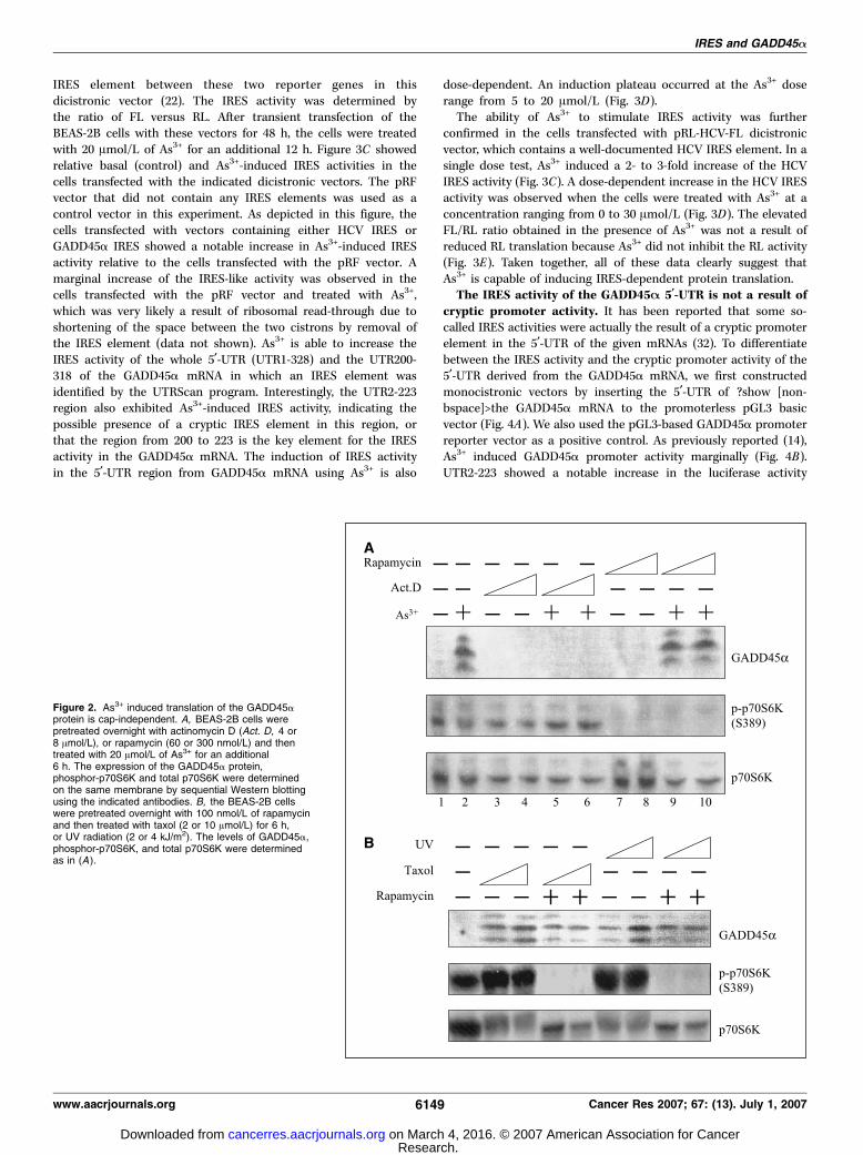
IRES element between these two reporter genes in thisdicistronic vector (22). The IRES activity was determined bythe ratio of FL versus RL. After transient transfection of theBEAS-2B cells with these vectors for 48 h, the cells were treatedwith 20 Amol/L of As3+ for an additional 12 h. Figure 3C showedrelative basal (control) and As3+-induced IRES activities in thecells transfected with the indicated dicistronic vectors. The pRFvector that did not contain any IRES elements was used as acontrol vector in this experiment. As depicted in this figure, thecells transfected with vectors containing either HCV IRES orGADD45a IRES showed a notable increase in As3+-induced IRESactivity relative to the cells transfected with the pRF vector. Amarginal increase of the IRES-like activity was observed in thecells transfected with the pRF vector and treated with As3+,which was very likely a result of ribosomal read-through due toshortening of the space between the two cistrons by removal ofthe IRES element (data not shown). As3+ is able to increase theIRES activity of the whole 5¶-UTR (UTR1-328) and the UTR200-318 of the GADD45a mRNA in which an IRES element wasidentified by the UTRScan program. Interestingly, the UTR2-223region also exhibited As3+-induced IRES activity, indicating thepossible presence of a cryptic IRES element in this region, orthat the region from 200 to 223 is the key element for the IRESactivity in the GADD45a mRNA. The induction of IRES activityin the 5¶-UTR region from GADD45a mRNA using As3+ is also
dose-dependent. An induction plateau occurred at the As3+ doserange from 5 to 20 Amol/L (Fig. 3D).The ability of As3+ to stimulate IRES activity was further
confirmed in the cells transfected with pRL-HCV-FL dicistronicvector, which contains a well-documented HCV IRES element. In asingle dose test, As3+ induced a 2- to 3-fold increase of the HCVIRES activity (Fig. 3C). A dose-dependent increase in the HCV IRESactivity was observed when the cells were treated with As3+ at aconcentration ranging from 0 to 30 Amol/L (Fig. 3D). The elevatedFL/RL ratio obtained in the presence of As3+ was not a result ofreduced RL translation because As3+ did not inhibit the RL activity(Fig. 3E). Taken together, all of these data clearly suggest thatAs3+ is capable of inducing IRES-dependent protein translation.The IRES activity of the GADD45A 5¶-UTR is not a result of
cryptic promoter activity. It has been reported that some so-called IRES activities were actually the result of a cryptic promoterelement in the 5¶-UTR of the given mRNAs (32). To differentiatebetween the IRES activity and the cryptic promoter activity of the5¶-UTR derived from the GADD45a mRNA, we first constructedmonocistronic vectors by inserting the 5¶-UTR of ?show [non-bspace]>the GADD45a mRNA to the promoterless pGL3 basicvector (Fig. 4A). We also used the pGL3-based GADD45a promoterreporter vector as a positive control. As previously reported (14),As3+ induced GADD45a promoter activity marginally (Fig. 4B).UTR2-223 showed a notable increase in the luciferase activity
Figure 2. As3+ induced translation of the GADD45aprotein is cap-independent. A, BEAS-2B cells werepretreated overnight with actinomycin D (Act. D, 4 or8 Amol/L), or rapamycin (60 or 300 nmol/L) and thentreated with 20 Amol/L of As3+ for an additional6 h. The expression of the GADD45a protein,phosphor-p70S6K and total p70S6K were determinedon the same membrane by sequential Western blottingusing the indicated antibodies. B, the BEAS-2B cellswere pretreated overnight with 100 nmol/L of rapamycinand then treated with taxol (2 or 10 Amol/L) for 6 h,or UV radiation (2 or 4 kJ/m2). The levels of GADD45a,phosphor-p70S6K, and total p70S6K were determinedas in (A ).
IRES and GADD45a
www.aacrjournals.org 6149 Cancer Res 2007; 67: (13). July 1, 2007
Research. on March 4, 2016. © 2007 American Association for Cancercancerres.aacrjournals.org Downloaded from
relative to other 5¶-UTR constructs in the cells without anytreatment, indicative of the cryptic promoter activity in this region.However, this cryptic promoter activity was not induced, but wasinstead inhibited by As3+. There was no detectable crypticpromoter activity in UTR200-318, which showed the highest IRESactivity induced by As3+ as indicated in Fig. 3C , in either the controlcells or in the As3+-treated cells (Fig. 4B). We also tested thepotential cryptic promoter activity of the 5¶-UTR derived fromGADD45a mRNA in a promoterless pRL-FL dicistronic vector.Again, although UTR1-328 and UTR2-223 showed cryptic promoteractivity in the control cells, this activity was not induced, butrather, was inhibited by As3+ (data not shown). Similar to themonocistronic reporter vector, the UTR200-318 showed no basal orinducible cryptic promoter activity in this promoterless dicistronicreporter system (data not shown).As3+ impairs the assembly of the cap-dependent preinitia-
tion complex. Accumulating evidence suggests that the rate ofprotein synthesis is reduced in cells under growth arrest or stressconditions (33). To obtain further insight into the translationalregulation of As3+ on protein synthesis, coimmunoprecipitationwas then done using antibodies against eIF4G, a generalcomponent of the cap-binding preinitiation complex for proteintranslation. Although the As3+-treated cells showed a similar levelof total eIF4E to the control cells (Fig. 5A, input), an appreciabledecrease in the interaction between eIF4G and eIF4E was notedin the cells treated overnight with 20 Amol/L of As3+ (Fig. 5A, top).
It is well-known that the association of eIF4E with eIF4G isessential for the further assembly of the translational initiationcomplex (26). Perturbation in the interaction between eIF4G andeIF4E by As3+, therefore, must influence the general translation ofthe cellular proteins. Indeed, analysis of the total proteinsynthesis by running the protein samples in SDS-PAGE followedby Coomassie blue staining showed a reduction of proteinsynthesis in As3+-treated cells (Fig. 5B, filled bars, right). As3+
induced a mild increase of the proteins with a molecular weight>200 kDa and <30 kDa (Fig. 5B, open bars, right), which possiblyresulted from cap-independent, but rather, IRES-dependenttranslation.To determine whether As3+ affects the association of other
proteins with the translational initiation complex, we analyzed theprotein complexes from eIF4G immunoprecipitation in SDS-PAGEand silver staining. Although the majority of protein bands areequally presented between the immunoprecipitated complexesfrom the control cells and the cells treated with 20 Amol/L of As3+
overnight, changes in a few bands in the As3+-treated cells didoccur (Fig. 5C). Bands 1 and 2 were increased, whereas bands 3and 4 were decreased by As3+ treatment. To identify thesechanged bands, the gel slices were excised (Fig. 5C, asterisk) andsubjected to in-gel digestion by trypsin to generate peptides withdifferent lengths based on the distribution of tryptic cleavage sites(arginine and lysine). The digested products were analyzed byreversed phase nano-LC-MS/MS on a Finnigan LTQ mass
Figure 3. The 5¶-UTR of the GADD45amRNA contains an IRES element.A, the 5¶-UTR of the GADD45a mRNAwas analyzed by the UTRScan program.http://www.ba.itb.cnr.it/BIG/UTRScan/Underlined sequence, the computer-identified IRES element. The ATGtranslation start code was highlighted.B, schematic illustration of the dicistronicreporter vectors used in IRES activityassay. C, IRES activity was determinedin the BEAS-2B cells transfected with theindicated dicistronic vectors for 48 h andthen cultured for an additional 12 h inthe absence or presence of 20 Amol/L ofAs3+. D, dose-dependent induction of theGADD45a IRES activity by As3+ in the cellstransfected with the indicated dicistronicvectors harboring the indicated 5¶-UTRregion from the GADD45a mRNA.E, dose-dependent induction of theHCV IRES activity by As3+ in the cellstransfected with the dicistronic vectorcontaining an established IRES elementfrom HCV promoter. F, the increased ratioof FL versus RL is not due to inhibition ofthe Renilla luciferase translation by As3+.Points, means of four repeats; bars, SD.
Cancer Research
Cancer Res 2007; 67: (13). July 1, 2007 6150 www.aacrjournals.org
Research. on March 4, 2016. © 2007 American Association for Cancercancerres.aacrjournals.org Downloaded from

spectrometer. This analysis identified 8 peptides from variousregions of the human elongation factor 2 (eEF-2) for band 1, 6peptides of human heterogeneous nuclear ribonucleoprotin M(hnRNP M) for band 2, 10 peptides of protein disulfide-isomeraseA3 (PDIA3) along with 46 peptides of vimentin for band 3, and 7peptides of eIF4A-I for band 4. Figure 5D shows the representativefragmentation spectrum for bands 1 to 4 from Fig. 5C . The eEF-2is a key factor for efficient peptide elongation during proteintranslation (34). The increased association of eEF-2 with eIF4G inthe As3+-treated cells might be a reflection of reciprocal functionalantagonism or coordination between cap-dependent and IRES-dependent protein translations through sharing or competingcommon cofactors. Some hnRNP family members had beenpreviously implicated in direct binding to the IRES elements(31, 35). Thus, the increased association of hnRNP M with trans-lational initiation complex by As3+ is suggestive of As3+ regulationof IRES-dependent translation. The role of PDIA3 might be mainlyto order proper folding of the newly translated proteins. Adecrease in PDIA3 association with the initiation complex by As3+,thus, would likely result in improper protein folding. The mostimportant finding in this analysis is the nearly complete depletionof eIF4A from the translational initiation complex by As3+. eIF4Ais an essential subunit of the eIF4F complex for cap-dependentinitiation, which possesses RNA helicase activity to unwindsecondary structures within the 5¶-UTR of mRNA (36). Taken
together, these data show that As3+ treatment favors IRES-dependent translation by impairing the assembly of the cap-dependent translational complex.
Discussion
The findings presented here provide the first evidence that As3+
is able to induce translation of the GADD45a protein in an IRES-dependent manner in the cells under growth-arrested conditions.The 5¶-UTR of the GADD45a mRNA contains an IRES element, theactivity of which can be induced by As3+ as evidenced in adicistronic reporter gene activity. Although the 5¶-terminal 200 nt5¶-UTR of the GADD45a mRNA possibly possesses a crypticpromoter activity under basal conditions, As3+ was unable toinduce this cryptic promoter activity in an analysis using either amonocistronic vector or a promoterless dicistronic vector. TheIRES-dependent induction of the GADD45a protein was furthersupported by the fact that As3+ impairs the assembly of the cap-dependent translational initiation complex by decreasing theassociation of eIF4G with both eIF4E and eIF4A. This impairmentcauses a reduction of the global protein translation. As3+ haspreviously been shown to be capable of regulating transcriptionand stabilization of the GADD45a mRNA. The effect of As3+ onIRES-dependent translation of the GADD45a protein suggested anadditional layer of regulation for the expression and function of theGADD45a protein in cellular responses to As3+.A wide spectrum of extracellular signals could induce rapid
elevation of GADD45a (1). Remarkable progress has been madeduring the past decade on the regulation and functional aspects ofGADD45a protein in cellular responses to DNA damage and avariety of stress inducers. However, the answer to how theexpression of the GADD45a is regulated when the cells are underextremely stressed conditions in which the major transcriptionaland translational machineries are severely impaired, is still elusive.It was assumed that cells can use an alternative approach tosynthesize proteins with the remaining trace amount mRNAs whenthe canonical cap-dependent protein translation process iscompromised either by dephosphorylation of 4E-BP1 or cleavageof eIF4G (37, 38). When the cells are in a state of mitosis,quiescence, differentiation, or hypoxia, the phosphorylated free4E-BP1 is dephosphorylated, possibly by protein phosphatase 2A,leading to association with and inhibition of eIF4E. Consequently,cap-dependent protein translation is drastically reduced (25, 39). Ahallmark of cell apoptosis is the activation of initiating andexecuting caspases. It has been shown that caspases are highlycapable of cleaving cap-dependent translational initiation factorssuch as eIF4B, eIF3, eIF2a, eIF4GI, and eIF4GII, leading to globalrepression of protein translation (33). However, a subset of mRNAs,<10% of total mRNAs, manages to escape this translationaldepression and maintains translation through a cap-independent,or rather, IRES-dependent translation. Considering the fact thatGADD45a was induced mostly by the signals leading to cell cyclearrest or apoptosis, it is very likely that the expression of GADD45aprotein is achieved via IRES-mediated translation.The IRES-dependent translation was initially discovered in the
viral gene translation of picornaviruses in host cells (40). ThemRNAs of these viruses are naturally uncapped at their 5¶-end.More importantly, many of these viruses produce proteases toproteolytically deplete cap-binding complex to shut off proteintranslation of the host cells completely during their infection.However, the viral proteins are still efficiently translated in the
Figure 4. As3+-induced IRES activity does not result from a cryptic promoter inthe 5¶-UTR of the GADD45a mRNA. A, schematic illustration of the monocistronicluciferase reporter vectors. The indicated regions of the GADD45a 5¶-UTRwere inserted into the promoterless pGL3 basic vector. The GADD45a promoterwas used as a positive control. B, luciferase activity was determined in thecells transfected with these vectors in (A) and cultured in the absence orpresence of 20 Amol/L of As3+ for 12 h (right ). Columns, means of fourrepeats; bars, SD.
IRES and GADD45a
www.aacrjournals.org 6151 Cancer Res 2007; 67: (13). July 1, 2007
Research. on March 4, 2016. © 2007 American Association for Cancercancerres.aacrjournals.org Downloaded from

infected cells. Further studies led to the discovery of the viral IRESelement in the 5¶-UTR of viral mRNA which mediates viral proteintranslation by recruiting IRES binding and/or recognition ribo-somal proteins and other RNA-binding proteins from the infectedhost cells (41). Since then, a growing list of mammalian cell mRNAshave been found to possess an IRES element, especially for thosecontaining a relatively long 5¶-UTR and function as importantregulators for cell growth and stress responses (30). These mRNAsinclude c-myc (42), p53 (43), vascular endothelial growth factor(44), fibroblast growth factor 2 (45), platelet-derived growth factorB/c-sis (46), hypoxia-inducible factor-1 (47), death-associatedprotein 5 (33), apoptotic protease activating factor (48), X-linkedinhibitor of apoptosis (31), and others. The IRES-dependenttranslation of the growth factors confers a survival advantage ofthe cells under hypoxia or stressed conditions, such as solid tumorcells undergoing chemotherapy. On the other hand, cells use theIRES-dependent translational machinery to express proteins
functioning in apoptosis or cell cycle arrest to eliminate geneticallydamaged cells, or prevent accumulation of genomic damage.GADD45a has been implicated in cell cycle regulation and DNAdamage repair. Thus, the IRES-dependent translation of theGADD45a protein might be theoretically beneficial for protectingnormal cells from malignant transformation under extreme stressconditions. The final outcome of the IRES-dependent expression ofthe GADD45a protein in either normal cells or tumor cells,however, might be largely dependent on the integrity of otherapoptotic and DNA repair machineries which act in concert withGADD45a. In addition, the fate of a cell following GADD45ainduction is also dependent on whether the expressed GADD45acan be sufficient to override the tumorigenic signals that areinduced simultaneously or asynchronously by the treatments.Because IRES-dependent protein translation occurred mostly in
the cells under circumstances of genotoxic stress, apoptosis,nutrient starvation, mitosis, or growth arrest, it is conceivable
Figure 5. As3+ impairs the assembly of thecap-dependent translational initiation complex.A, BEAS-2B cells cultured in the absence or presenceof 20 Amol/L of As3+ for 12 h and then subjected toimmunoprecipitation using antibody against eIF4G. Theimmunoprecipitated complexes were separated on 14%SDS-PAGE gel and were sequentially immunoblottedwith antibodies for eIF4G and eIF4E. Total cell lysateswere used as input to determine the level of eIF4E.B, total cellular proteins were separated on 14%SDS-PAGE gel and stained by Coomassie blue. Left,molecular weights (M , kDa). Right, open and filledbars indicate increased and decreased expression,respectively, of proteins induced by 20 Amol/L of As3+ for12 h. C, the complexes from eIF4G immunoprecipitationof the control and As3+-treated cells were separated on14% SDS-PAGE gel and monitored by silver staining.Left, molecular weights (M , kDa). Right, number ofprotein bands that were affected by As3+ treatment.*, positions of the protein bands that were excisedfor in-gel digestion followed by reversed phasenano-LC-MS/MS analysis. D, representative peptidefragmentation spectrum for bands 1 to 4 from (C).
Cancer Research
Cancer Res 2007; 67: (13). July 1, 2007 6152 www.aacrjournals.org
Research. on March 4, 2016. © 2007 American Association for Cancercancerres.aacrjournals.org Downloaded from

that manipulating the IRES pathway would alter the growth orsurvival status of the cell. Evidence supporting this notion ismainly from studies that show mutations of IRES elements orfunctional impairment of IRES binding proteins, the so-calledIRES trans-acting factors in cancers or diseases associated withcancer susceptibility (49, 50). In cell lines derived from patientswith neoplasia multiple myeloma, a single C to U transition in theIRES region of c-myc mRNA was found to be responsible for theenhanced IRES activity and the consequent increased expressionof c-myc protein (49). The increase of c-myc protein obviouslyprovides the cells with a growth advantage and elevatedoncogenic potentials. On the other hand, a defect in the IRESmachinery due to a genetic deficiency in a gene encodingpseudouridine synthase, dyskerin, has been linked to cancerpredisposition, a result of decreased IRES-mediated expression ofantiapoptotic protein, p27 (50). Currently, IRES-mediated proteintranslation has been implicated in the expression of bothprosurvival and antisurvival factors. An interesting question tobe asked is, therefore, whether different IRES machineries presentin given cells mediate the expressions of proteins with uniquefunctions in context with the type and duration of extracellularsignals.In summary, our data support the model that GADD45a protein
translation is up-regulated by As3+, as well as UV radiation or taxoltreatment, through the IRES element identified in the 5¶-UTR of
the GADD45a mRNA. Treatment of the cells with As3+, andpossibly other stress inducers, compromises the function of thecap-dependent translational machinery, and thereafter favorsIRES-dependent protein translation. The IRES-dependent transla-tion of the GADD45a protein may not be limited to the cells insevere stress conditions, such as growth arrest in this study, butalso in the cells with mild environmental insults. The regulation ofthe GADD45a protein on transcriptional, mRNA stability, andtranslational levels might be important to ensure that cells producesufficient amounts of the GADD45a protein to reduce thepossibility of improper chromosomal segregation and cell division.
Acknowledgments
Received 3/5/2007; revised 4/19/2007; accepted 4/25/2007.Grant support: Intramural research grant from the National Institute for
Occupational Safety and Health/Centers for Disease Control and Prevention (F.Chen), partial support from the National Science Foundation of China (30471980,Q. Chang and Y. Zhang), and NIH 7RO1CA119028-02 (X. Shi).The costs of publication of this article were defrayed in part by the payment of page
charges. This article must therefore be hereby marked advertisement in accordancewith 18 U.S.C. Section 1734 solely to indicate this fact.We thank Drs. Richard E. Lloyd at Baylor College of Medicine (Houston, TX), Jian-
Ting Zhang at the Indiana University Cancer Center (Indianapolis, IN), and DanielHaber at Massachusetts General Hospital Cancer Center (Boston, MA) for thedicistronic vectors and GADD45a promoter reporter constructs, respectively. We alsothank Dr. Kristy J. Brown at CTL Bio Services (Rockville, MD) for her technicalassistance in peptide analyses using reversed phase nano-LC-MS/MS.
References
1. Hollander MC, Fornace AJ, Jr. Genomic instability,centrosome amplification, cell cycle checkpoints andGadd45a. Oncogene 2002;21:6228–33.
2. Chen F, Zhang Z, Leonard SS, Shi X. Contrastingroles of NF-nB and JNK in arsenite-induced p53-independent expression of GADD45a. Oncogene 2001;20:3585–9.
3. Smith ML, Chen IT, Zhan Q, et al. Interaction of thep53-regulated protein Gadd45 with proliferating cellnuclear antigen. Science 1994;266:1376–80.
4. Wang XW, Zhan Q, Coursen JD, et al. GADD45induction of a G2/M cell cycle checkpoint. Proc NatlAcad Sci U S A 1999;96:3706–11.
5. Kearsey JM, Coates PJ, Prescott AR, Warbrick E,Hall PA. Gadd45 is a nuclear cell cycle regulatedprotein which interacts with p21Cip1. Oncogene 1995;11:1675–83.
6. Carrier F, Georgel PT, Pourquier P, et al. Gadd45, ap53-responsive stress protein, modifies DNA accessi-bility on damaged chromatin. Mol Cell Biol 1999;19:1673–85.
7. Jin S, Antinore MJ, Lung FD, et al. The GADD45inhibition of Cdc2 kinase correlates with GADD45-mediated growth suppression. J Biol Chem 2000;275:16602–8.
8. Barreto G, Schafer A, Marhold J, et al. Gadd45apromotes epigenetic gene activation by repair-mediatedDNA demethylation. Nature 2007;445:671–5.
9. Wang W, Nahta R, Huper G, Marks JR. TAFII70isoform-specific growth suppression correlates with itsability to complex with the GADD45a protein. MolCancer Res 2004;2:442–52.
10. Brunet A, Sweeney LB, Sturgill JF, et al. Stress-dependent regulation of FOXO transcription factors bythe SIRT1 deacetylase. Science 2004;303:2011–5.
11. Marhin WW, Chen S, Facchini LM, Fornace AJ, Jr.,Penn LZ. Myc represses the growth arrest gene gadd45.Oncogene 1997;14:2825–34.
12. Zheng L, Pan H, Li S, et al. Sequence-specifictranscriptional corepressor function for BRCA1 througha novel zinc finger protein, ZBRK1. Mol Cell 2000;6:757–68.
13. Chen F, Lu Y, Zhang Z, et al. Opposite effect of NF-nB
and c-Jun N-terminal kinase on p53-independentGADD45 induction by arsenite. J Biol Chem 2001;276:11414–9.
14. Zhang Y, Bhatia D, Xia H, Castranova V, Shi X,Chen F. Nucleolin links to arsenic-induced stabiliza-tion of GADD45a mRNA. Nucleic Acids Res 2006;34:485–95.
15. Lal A, Abdelmohsen K, Pullmann R, et al. Posttran-scriptional derepression of GADD45a by genotoxicstress. Mol Cell 2006;22:117–28.
16. Lal A, Gorospe M. Egad, more forms of generegulation: the gadd45a story. Cell Cycle 2006;5:1422–5.
17. Kitchin KT. Recent advances in arsenic carcinogen-esis: modes of action, animal model systems, andmethylated arsenic metabolites. Toxicol Appl Pharmacol2001;172:249–61.
18. Scott N, Hatlelid KM, MacKenzie NE, Carter DE.Reactions of arsenic(III) and arsenic(V) species withglutathione. Chem Res Toxicol 1993;6:102–6.
19. Chen F, Zhang Z, Bower J, et al. Arsenite-inducedCdc25C degradation is through the KEN-box andubiquitin-proteasome pathway. Proc Natl Acad SciU S A 2002;99:1990–5.
20. Shackelford D, Kenific C, Blusztajn A, Waxman S, RenR. Targeted degradation of the AML1/MDS1/EVI1oncoprotein by arsenic trioxide. Cancer Res 2006;66:11360–9.
21. Chen Z, Zhao WL, Shen ZX, et al. Arsenic trioxideand acute promyelocytic leukemia: clinical and biolog-ical. Curr Top Microbiol Immunol 2007;313:129–44.
22. Sherrill KW, Byrd MP, Van Eden ME, Lloyd RE. BCL-2translation is mediated via internal ribosome entryduring cell stress. J Biol Chem 2004;279:29066–74.
23. Zhang Y, Lu Y, Ding M, Castranova V, Shi X, Chen F.Deficiency in Ikkh gene enhances arsenic-inducedgadd45a expression. Mol Cell Biochem 2005;279:163–8.
24. Zheng X, Zhang Y, Chen YQ, Castranova V, Shi X,Chen F. Inhibition of NF-nB stabilizes gadd45a mRNA.Biochem Biophys Res Commun 2005;329:95–9.
25. Gingras AC, Raught B, Gygi SP, et al. Hierarchicalphosphorylation of the translation inhibitor 4E-BP1.Genes Dev 2001;15:2852–64.
26. Richter JD, Sonenberg N. Regulation of cap-depen-dent translation by eIF4E inhibitory proteins. Nature2005;433:477–80.
27. Debernardis D, Sire EG, De Feudis P, et al. p53 statusdoes not affect sensitivity of human ovarian cancer celllines to paclitaxel. Cancer Res 1997;57:870–4.
28. An W, Kim J, Roeder RG. Ordered cooperativefunctions of PRMT1, p300, and CARM1 in transcrip-tional activation by p53. Cell 2004;117:735–48.
29. Tong T, Fan W, Zhao H, et al. Involvement of the MAPkinase pathways in induction of GADD45 following UVradiation. Exp Cell Res 2001;269:64–72.
30. Hellen CU, Sarnow P. Internal ribosome entry sitesin eukaryotic mRNA molecules. Genes Dev 2001;15:1593–612.
31. Holcik M, Gordon BW, Korneluk RG. The internalribosome entry site-mediated translation of antiapop-totic protein XIAP is modulated by the heterogeneousnuclear ribonucleoproteins C1 and C2. Mol Cell Biol2003;23:280–8.
32. Han B, Zhang JT. Regulation of gene expression byinternal ribosome entry sites or cryptic promoters: theeIF4G story. Mol Cell Biol 2002;22:7372–84.
33. Marash L, Kimchi A. DAP5 and IRES-mediatedtranslation during programmed cell death. Cell DeathDiffer 2005;12:554–62.
34. Jorgensen R, Merrill AR, Andersen GR. The life anddeath of translation elongation factor 2. Biochem SocTrans 2006;34:1–6.
35. Lu H, Li W, Noble WS, Payan D, Anderson DC.Riboproteomics of the hepatitis C virus internalribosomal entry site. J Proteome Res 2004;3:949–57.
36. Sonenberg N, Dever TE. Eukaryotic translationinitiation factors and regulators. Curr Opin Struct Biol2003;13:56–63.
37. Vagner S, Galy B, Pyronnet S. Irresistible IRES.Attracting the translation machinery to internal ribo-some entry sites. EMBO Rep 2001;2:893–8.
38. Komar AA, Hatzoglou M. Internal ribosome entrysites in cellular mRNAs: mystery of their existence. J BiolChem 2005;280:23425–8.
39. Pham FH, Sugden PH, Clerk A. Regulation of proteinkinase B and 4E-BP1 by oxidative stress in cardiacmyocytes. Circ Res 2000;86:1252–8.
40. Jang SK, Pestova TV, Hellen CU, Witherell GW,Wimmer E. Cap-independent translation of picornavirusRNAs: structure and function of the internal ribosomalentry site. Enzyme 1990;44:292–309.
IRES and GADD45a
www.aacrjournals.org 6153 Cancer Res 2007; 67: (13). July 1, 2007
Research. on March 4, 2016. © 2007 American Association for Cancercancerres.aacrjournals.org Downloaded from

41. Jang SK. Internal initiation: IRES elements ofpicornaviruses and hepatitis c virus. Virus Res 2006;119:2–15.
42. Thoma C, Bergamini G, Galy B, Hundsdoerfer P,Hentze MW. Enhancement of IRES-mediated translationof the c-myc and BiP mRNAs by the poly(A) tail isindependent of intact eIF4G and PABP. Mol Cell 2004;15:925–35.
43. Ray PS, Grover R, Das S. Two internal ribosome entrysites mediate the translation of p53 isoforms. EMBO Rep2006;7:404–10.
44. Stein I, Itin A, Einat P, Skaliter R, Grossman Z, KeshetE. Translation of vascular endothelial growth factor
mRNA by internal ribosome entry: implications fortranslation under hypoxia. Mol Cell Biol 1998;18:3112–9.
45. Creancier L, Morello D, Mercier P, Prats AC.Fibroblast growth factor 2 internal ribosome entry site(IRES) activity ex vivo and in transgenic mice reveals astringent tissue-specific regulation. J Cell Biol 2000;150:275–81.
46. Bernstein J, Sella O, Le SY, Elroy-Stein O. PDGF2/c-sismRNA leader contains a differentiation-linked internalribosomal entry site (D-IRES). J Biol Chem 1997;272:9356–62.
47. Lang KJ, Kappel A, Goodall GJ. Hypoxia-induciblefactor-1a mRNA contains an internal ribosome entry
site that allows efficient translation during normoxiaand hypoxia. Mol Biol Cell 2002;13:1792–801.
48. Coldwell MJ, Mitchell SA, Stoneley M, MacFarlane M,Willis AE. Initiation of Apaf-1 translation by internalribosome entry. Oncogene 2000;19:899–905.
49. Chappell SA, LeQuesne JP, Paulin FE, et al. Amutation in the c-myc-IRES leads to enhanced internalribosome entry in multiple myeloma: a novel mecha-nism of oncogene de-regulation. Oncogene 2000;19:4437–40.
50. Yoon A, Peng G, Brandenburger Y, et al. Impairedcontrol of IRES-mediated translation in X-linked dysker-atosis congenita. Science 2006;312:902–6.
Cancer Research
Cancer Res 2007; 67: (13). July 1, 2007 6154 www.aacrjournals.org
Research. on March 4, 2016. © 2007 American Association for Cancercancerres.aacrjournals.org Downloaded from

2007;67:6146-6154. Cancer Res Qingshan Chang, Deepak Bhatia, Yadong Zhang, et al.
ExpressionαMechanism in Arsenic-Induced GADD45Dependent−Incorporation of an Internal Ribosome Entry Site
Updated version
http://cancerres.aacrjournals.org/content/67/13/6146
Access the most recent version of this article at:
Cited articles
http://cancerres.aacrjournals.org/content/67/13/6146.full.html#ref-list-1
This article cites 50 articles, 24 of which you can access for free at:
Citing articles
http://cancerres.aacrjournals.org/content/67/13/6146.full.html#related-urls
This article has been cited by 7 HighWire-hosted articles. Access the articles at:
E-mail alerts related to this article or journal.Sign up to receive free email-alerts
Subscriptions
Reprints and
To order reprints of this article or to subscribe to the journal, contact the AACR Publications
Permissions
To request permission to re-use all or part of this article, contact the AACR Publications
Research. on March 4, 2016. © 2007 American Association for Cancercancerres.aacrjournals.org Downloaded from
Related Documents

![Ribosome Stoichiometry: From Form to Function · Ribosome abundance: A major model, also termed the ribosome concentration hypothesis [3], that explains how ribosomes could exert](https://static.cupdf.com/doc/110x72/60de31e56d30fc4fb30719b8/ribosome-stoichiometry-from-form-to-function-ribosome-abundance-a-major-model.jpg)