Annals of Oncology 21: 1794–1803, 2010 doi:10.1093/annonc/mdq022 Published online 5 February 2010 original article Incidence, patterns of care and prognostic factors for outcome of gastroenteropancreatic neuroendocrine tumors (GEP-NETs): results from the National Cancer Registry of Spain (RGETNE) R. Garcia-Carbonero 1 *, J. Capdevila 2 , G. Crespo-Herrero 3 , J. A. Dı´az-Pe ´ rez 4 , M. P. Martı´nez del Prado 5 , V. Alonso Ordun ˜a 6 , I. Sevilla-Garcı´a 7 , C. Villabona-Artero 8 , A. Beguiristain-Go ´ mez 9 , M. Llanos-Mun ˜ oz 10 , M. Marazuela 11 , C. Alvarez-Escola 12 , D. Castellano 13 , E. Vilar 14 , P. Jime ´ nez- Fonseca 3 , A. Teule ´ 15 , J. Sastre-Valera 16 , M. Benavent-Vin ˜ uelas 1 , A. Monleon 17 & R. Salazar 15 1 Department of Medical Oncology, Instituto de Biomedicina de Sevilla, Hospital Universitario Virgen del Rocı´o, Sevilla; 2 Department of Medical Oncology, Vall d#Hebron University Hospital, Barcelona; 3 Department of Medical Oncology, Hospital Central de Asturias, Oviedo; 4 Department of Endocrinology, Hospital Clı´nico Universitario San Carlos, Madrid; 5 Department of Medical Oncology, Hospital de Basurto, Vizcaya; 6 Department of Medical Oncology, Hospital Miguel Servet, Zaragoza; 7 Department of Medical Oncology, Hospital Virgen de la Victoria, Ma ´laga; 8 Department of Endocrinology, Hospital de Bellvitge, L#Hospitalet, Barcelona; 9 Department of Medical Oncology, Hospital de Donostia, San Sebastia ´n; 10 Department of Medical Oncology, Hospital Universitario de Canarias, Santa Cruz de Tenerife; 11 Department of Endocrinology, Hospital de la Princesa; 12 Department of Endocrinology, Hospital La Paz; 13 Department of Medical Oncology, Hospital 12 de Octubre, Madrid; 14 Department of Internal Medicine, University of Michigan, MI, USA; 15 Department of Medical Oncology, Hospital Duran i Reynals, Institut Catala ´ d’Oncologia, Barcelona; 16 Department of Medical Oncology, Hospital Clı´nico Universitario San Carlos, Madrid and 17 Department of Statistics, Faculty of Biology, Barcelona University, Barcelona, Spain Received 29 October 2009; revised 28 December 2009; accepted 29 December 2009 Background: Neuroendocrine tumors (NETs) are an unusual family of neoplasms with a wide and complex spectrum of clinical behavior. Here, we present the first report of a National Cancer Registry of gastroenteropancreatic neuroendocrine tumors from a Southern European country. Patients and methods: Data was provided online at www.retegep.net by participating centers and assessed for internal consistency by external independent reviewers. Results: The study cohort comprised 907 tumors. The most common tumor types were carcinoids (55%), pancreatic nonfunctional tumors (20%), metastatic NETs of unknown primary (9%), insulinomas (8%) and gastrinomas (4%). Forty-four percent presented with distant disease at diagnosis, most often those from small intestine (65%), colon (48%), rectum (40%) and pancreas (38%), being most unusual in appendix primaries (1.3%). Stage at diagnosis varied significantly according to sex, localization of primary tumor, tumor type and grade. Overall 5-year survival was 75.4% (95% confidence interval 71.3% to 79.5%) and was significantly greater in women, younger patients and patients with hormonal syndrome and early stage or lower grade tumors. Prognosis also differed according to tumor type and primary tumor site. However, stage and Ki-67 index were the only independent predictors for survival. Conclusion: This national database reveals relevant information regarding epidemiology, current clinical practices and prognosis of NETs in Spain, providing valuable insights that may contribute to understand regional disparities in the incidence, patterns of care and survival of this heterogeneous disease across different continents and countries. Key words: gastrointestinal, neuroendocrine tumors, pancreatic, registry, survival, treatment introduction Neuroendocrine tumors (NETs) comprise a heterogeneous family of neoplasms with a wide and complex spectrum of clinical behavior [1, 2]. They originate in a great diversity of tissues and are characterized by their ability to produce different peptides that cause distinct hormonal syndromes. However, many are clinically silent until late advanced disease. Although they are generally more indolent than carcinomas, they often have unpredictable biological behavior and are on occasions associated with a very aggressive clinical course. Recent international efforts are helping to improve the prognostic classifications of this type of tumors and to better tailor therapeutic strategies in these patients [3–6]. original article *Correspondence to: Dr R. Garcı ´a-Carbonero, Department of Medical Oncology, Instituto de Biomedicina de Sevilla, Hospital Universitario Virgen del Rocı´o, Avenida Manuel Siurot s/n, 41013 Sevilla, Spain. Tel: +34-955-013068; Fax: +34-954-232992; E-mail: [email protected] ª The Author 2010. Published by Oxford University Press on behalf of the European Society for Medical Oncology. All rights reserved. For permissions, please email: [email protected] by guest on May 26, 2012 http://annonc.oxfordjournals.org/ Downloaded from

Welcome message from author
This document is posted to help you gain knowledge. Please leave a comment to let me know what you think about it! Share it to your friends and learn new things together.
Transcript
Annals of Oncology 21: 1794–1803, 2010
doi:10.1093/annonc/mdq022
Published online 5 February 2010original article
Incidence, patterns of care and prognostic factors foroutcome of gastroenteropancreatic neuroendocrinetumors (GEP-NETs): results from the National CancerRegistry of Spain (RGETNE)
R. Garcia-Carbonero1*, J. Capdevila2, G. Crespo-Herrero3, J. A. Dıaz-Perez4, M. P. Martınez delPrado5, V. Alonso Orduna6, I. Sevilla-Garcıa7, C. Villabona-Artero8, A. Beguiristain-Gomez9,M. Llanos-Munoz10, M. Marazuela11, C. Alvarez-Escola12, D. Castellano13, E. Vilar14, P. Jimenez-Fonseca3, A. Teule15, J. Sastre-Valera16, M. Benavent-Vinuelas1, A. Monleon17 & R. Salazar15
1Department of Medical Oncology, Instituto de Biomedicina de Sevilla, Hospital Universitario Virgen del Rocıo, Sevilla; 2Department of Medical Oncology, Vall d#Hebron
University Hospital, Barcelona; 3Department of Medical Oncology, Hospital Central de Asturias, Oviedo; 4Department of Endocrinology, Hospital Clınico Universitario
San Carlos, Madrid; 5Department of Medical Oncology, Hospital de Basurto, Vizcaya; 6Department of Medical Oncology, Hospital Miguel Servet, Zaragoza;7Department of Medical Oncology, Hospital Virgen de la Victoria, Malaga; 8Department of Endocrinology, Hospital de Bellvitge, L#Hospitalet, Barcelona; 9Department of
Medical Oncology, Hospital de Donostia, San Sebastian; 10Department of Medical Oncology, Hospital Universitario de Canarias, Santa Cruz de Tenerife; 11Department
of Endocrinology, Hospital de la Princesa; 12Department of Endocrinology, Hospital La Paz; 13Department of Medical Oncology, Hospital 12 de Octubre, Madrid;14Department of Internal Medicine, University of Michigan, MI, USA; 15Department of Medical Oncology, Hospital Duran i Reynals, Institut Catala d’Oncologia,
Barcelona; 16Department of Medical Oncology, Hospital Clınico Universitario San Carlos, Madrid and 17Department of Statistics, Faculty of Biology, Barcelona
University, Barcelona, Spain
Received 29 October 2009; revised 28 December 2009; accepted 29 December 2009
Background: Neuroendocrine tumors (NETs) are an unusual family of neoplasms with a wide and complex spectrum
of clinical behavior. Here, we present the first report of a National Cancer Registry of gastroenteropancreatic
neuroendocrine tumors from a Southern European country.
Patients and methods: Data was provided online at www.retegep.net by participating centers and assessed for
internal consistency by external independent reviewers.
Results: The study cohort comprised 907 tumors. The most common tumor types were carcinoids (55%), pancreatic
nonfunctional tumors (20%), metastatic NETs of unknown primary (9%), insulinomas (8%) and gastrinomas (4%).
Forty-four percent presented with distant disease at diagnosis, most often those from small intestine (65%), colon
(48%), rectum (40%) and pancreas (38%), being most unusual in appendix primaries (1.3%). Stage at diagnosis varied
significantly according to sex, localization of primary tumor, tumor type and grade. Overall 5-year survival was 75.4%
(95% confidence interval 71.3% to 79.5%) and was significantly greater in women, younger patients and patients with
hormonal syndrome and early stage or lower grade tumors. Prognosis also differed according to tumor type and
primary tumor site. However, stage and Ki-67 index were the only independent predictors for survival.
Conclusion: This national database reveals relevant information regarding epidemiology, current clinical practices
and prognosis of NETs in Spain, providing valuable insights that may contribute to understand regional disparities in
the incidence, patterns of care and survival of this heterogeneous disease across different continents and countries.
Key words: gastrointestinal, neuroendocrine tumors, pancreatic, registry, survival, treatment
introduction
Neuroendocrine tumors (NETs) comprise a heterogeneousfamily of neoplasms with a wide and complex spectrum ofclinical behavior [1, 2]. They originate in a great diversity of
tissues and are characterized by their ability to producedifferent peptides that cause distinct hormonal syndromes.However, many are clinically silent until late advanced disease.Although they are generally more indolent than carcinomas,they often have unpredictable biological behavior and are onoccasions associated with a very aggressive clinical course.Recent international efforts are helping to improve theprognostic classifications of this type of tumors and to bettertailor therapeutic strategies in these patients [3–6].
ori
gin
al
art
icle
*Correspondence to: Dr R. Garcıa-Carbonero, Department of Medical Oncology,
Instituto de Biomedicina de Sevilla, Hospital Universitario Virgen del Rocıo, Avenida
Manuel Siurot s/n, 41013 Sevilla, Spain. Tel: +34-955-013068; Fax: +34-954-232992;
E-mail: [email protected]
ª The Author 2010. Published by Oxford University Press on behalf of the European Society for Medical Oncology.
All rights reserved. For permissions, please email: [email protected]
by guest on May 26, 2012
http://annonc.oxfordjournals.org/D
ownloaded from
The incidence of NETs ranges from 2.5 to 5 cases per 100 000in Caucasian population [7–10]. The reported incidence hassubstantially increased over the last decades, partially due toimproved diagnostic techniques and clinical awareness.However, incidence rates overall and per individual anatomicsite are widely variable in the literature. Many issues mayaccount for these discrepancies, including differences in patientselection, specific institutional or registration biases, racialdisparities and other as yet unknown genetic andenvironmental factors. These issues along with patterns of caremay greatly differ across countries and may ultimatelyinfluence outcome in a significant way.To provide information regarding demographic
characteristics, diagnostic procedures, tumor features,therapeutic interventions and survival of patients withgastroenteropancreatic neuroendocrine tumors (GEP-NETs),a national tumor registry was launched in 2001 by GETNE, theSpanish Scientific Society of Neuroendocrine Tumors. Wepresent here the results of this broad-based multi-institutionalregistry that comprises 46 academic and community sitesrepresenting all regions of Spain. To date, published datamostly refer to United States, UK and Northern Europeanpopulations [7–15]. This is to our knowledge the first studyproviding information on this type of tumors from a SouthernEuropean country.
patients and methods
The study population was obtained from the National Cancer Registry for
Gastroenteropancreatic Neuroendocrine Tumors (RGETNE). RGETNE was
launched by the scientific society GETNE, which is composed by specialists
from multiple disciplines (oncologists, 77%; endocrinologists, 18%;
surgeons, 4%; pathologists; biologists; .) and leads at the national level
multiple educational and research projects on NETs. This broad-based
multi-institutional registry comprises 46 academic and community sites
representing all regions of Spain (Appendix 1). Data collection was
provided online at www.retegep.net by investigators or study nurses from
participating institutions and assessed for internal consistency by external
independent reviewers. The registry database and standard operating
procedures were approved by a National Scientific and Ethics Committee.
From June 2001 through December 2008, 907 tumors from 887 patients
were prospectively registered. Only patients with survival data have been
included in this report (855 tumors and 837 patients). The medical records
were systematically reviewed to collect the following data: age, gender,
functional syndrome, familial hereditary syndrome, diagnostic procedures,
localization of the primary tumor, histopathological features (World Health
Organization classification [16], immunohistochemical staining, grade and
vascular invasion), tumor stage at diagnosis, therapeutic interventions and
outcomes (surgery, local–regional therapies, hormone therapy,
chemotherapy, radiotherapy and radionuclide therapy), date of diagnosis,
date of relapse or disease progression, date of last visit or death and cause of
death. Tumor stage was classified as localized (confined to the organ of
origin), regional (invasion of surrounding organs or tissues or regional
lymph nodes) or distant (spread to distant organs). Number (i.e. single or
multiple) and size of primary tumors and specific sites of distant metastasis
were also recorded.
Descriptive statistics were used to characterize the most relevant clinical
parameters. The association of categorical variables was assessed by the chi-
square test or Fisher’s exact test when appropriate. One-way analysis of
variance was used for comparison of continuous variables between groups.
Overall survival was defined as the time elapsed from the date of diagnosis
to the date of death from any cause or last follow-up in alive patients.
Survival was estimated according to the Kaplan–Meier product limit
method, and differences observed among patient subgroups were assessed
by the log-rank test. Multivariate analyses using the Cox proportional
hazards model were carried out to identify factors independently associated
with prognosis. Gender, age, hormonal syndrome, stage, Ki-67, tumor type
and localization of primary tumor were included as covariates in the model.
Two-sided P values were computed; P <0.05 was considered statistically
significant. All analyses were carried out using the SPSS statistical package
(SPSS version 16.0 for Windows; SPSS Inc., Chicago, IL).
results
patient population
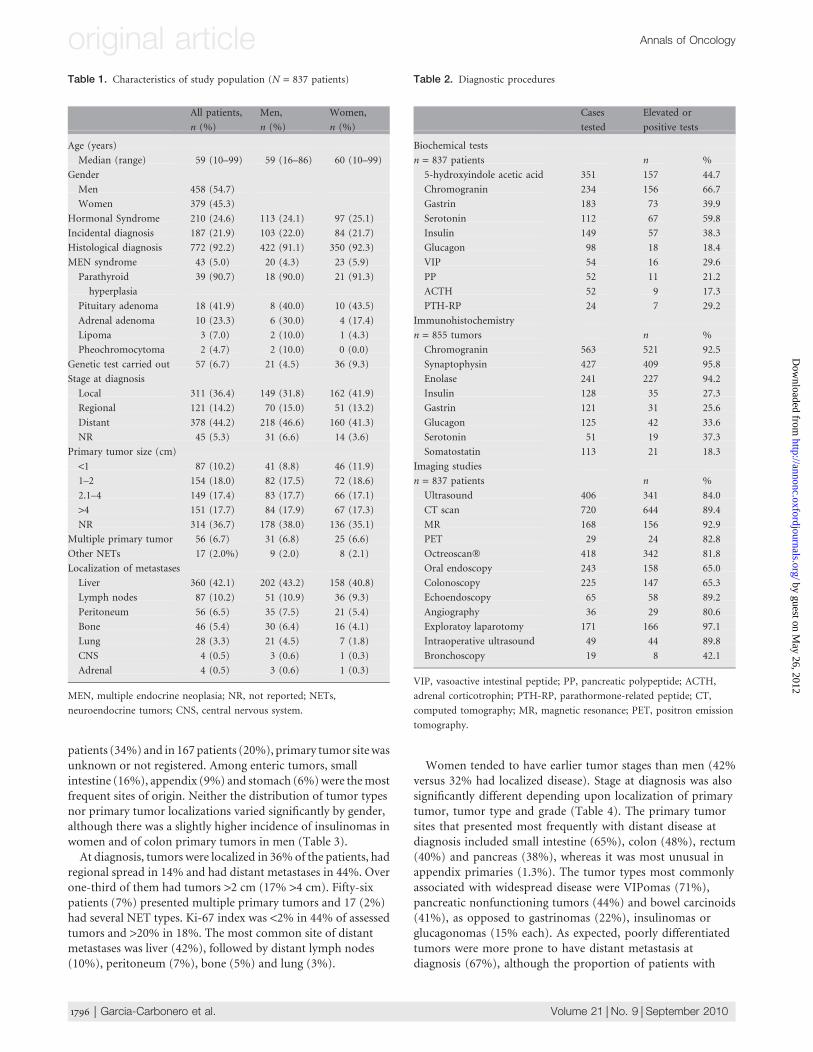
Of 837 assessable patients with GEP-NETs, 458 (55%) weremen and 379 (45%) were women. The median age at diagnosiswas 59 years (range 10–99 years), and 25% presented withhormone hypersecretion symptoms, with no significantdifferences according to gender (Table 1). Appendix primariesand functional tumors were diagnosed at younger ages (medianage at diagnosis: appendix primary, 42 years; insulinoma,48 years and gastrinoma, 52 years). Multiple endocrineneoplasia was diagnosed in 43 patients (5%). Of them, 91% hadhyperparathyroidism, 42% pituitary adenomas, 23% adrenaladenomas and 5% pheochromocytomas. Both adrenaladenomas (30% versus 17%) and pheochromocytomas(10% versus 0%) were more commonly observed in men ascompared with women.
diagnostic procedures
Incidental diagnosis occurred in 22% of cases. The mostcommonly carried out imaging studies included computedtomography (CT) scan, ultrasound and somatostatin receptorscintigraphy (octreotide scintigraphy). CT scan was theprocedure with the highest yield of tumor detection (75%).Octreoscan� was done in 49% of patients and 81% of themwere positive. Only about one-third of the registered patientsunderwent endoscopic procedures. Biochemical tests such asserum chromogranin A or urinary 5-hydroxyindole acetic acid(5-HIAA) levels were only done in 41% and 27% of thepopulation and were increased in 67% and 45% of testedpatients, respectively. Immunohistochemical staining forchromogranin and synaptophysin was done in 66% and 50% oftumors, being positive in 93% and 96% of reported cases,respectively. Ki-67 index was carried out in only 36% of tumors.Other diagnostic procedures are summarized in Table 2.
tumor characteristics
The most common tumor types were gastrointestinal carcinoids(55%), followed by pancreatic nonfunctional tumors (20%)and metastatic NETs of unknown primary (9%) (Table 3).Among functional tumors, enteric carcinoids (10%),insulinomas (8%) and gastrinomas (4%) were the mostcommonly encountered. Glucagonomas, vasoactive intestinalpeptidomas (VIPomas) or somatostatinomas were foundin <2% of the population. The gastrointestinal tract was theprimary tumor site in 400 patients (47%), the pancreas in 288
Annals of Oncology original article
Volume 21 |No. 9 | September 2010 doi:10.1093/annonc/mdq022 | 1795
by guest on May 26, 2012
http://annonc.oxfordjournals.org/D
ownloaded from
patients (34%) and in 167patients (20%), primary tumor sitewasunknown or not registered. Among enteric tumors, smallintestine (16%), appendix (9%) and stomach (6%)were themostfrequent sites of origin. Neither the distribution of tumor typesnor primary tumor localizations varied significantly by gender,although there was a slightly higher incidence of insulinomas inwomen and of colon primary tumors in men (Table 3).At diagnosis, tumors were localized in 36% of the patients, had
regional spread in 14% and had distant metastases in 44%. Overone-third of them had tumors >2 cm (17% >4 cm). Fifty-sixpatients (7%) presented multiple primary tumors and 17 (2%)had several NET types. Ki-67 index was <2% in 44% of assessedtumors and >20% in 18%. The most common site of distantmetastases was liver (42%), followed by distant lymph nodes(10%), peritoneum (7%), bone (5%) and lung (3%).
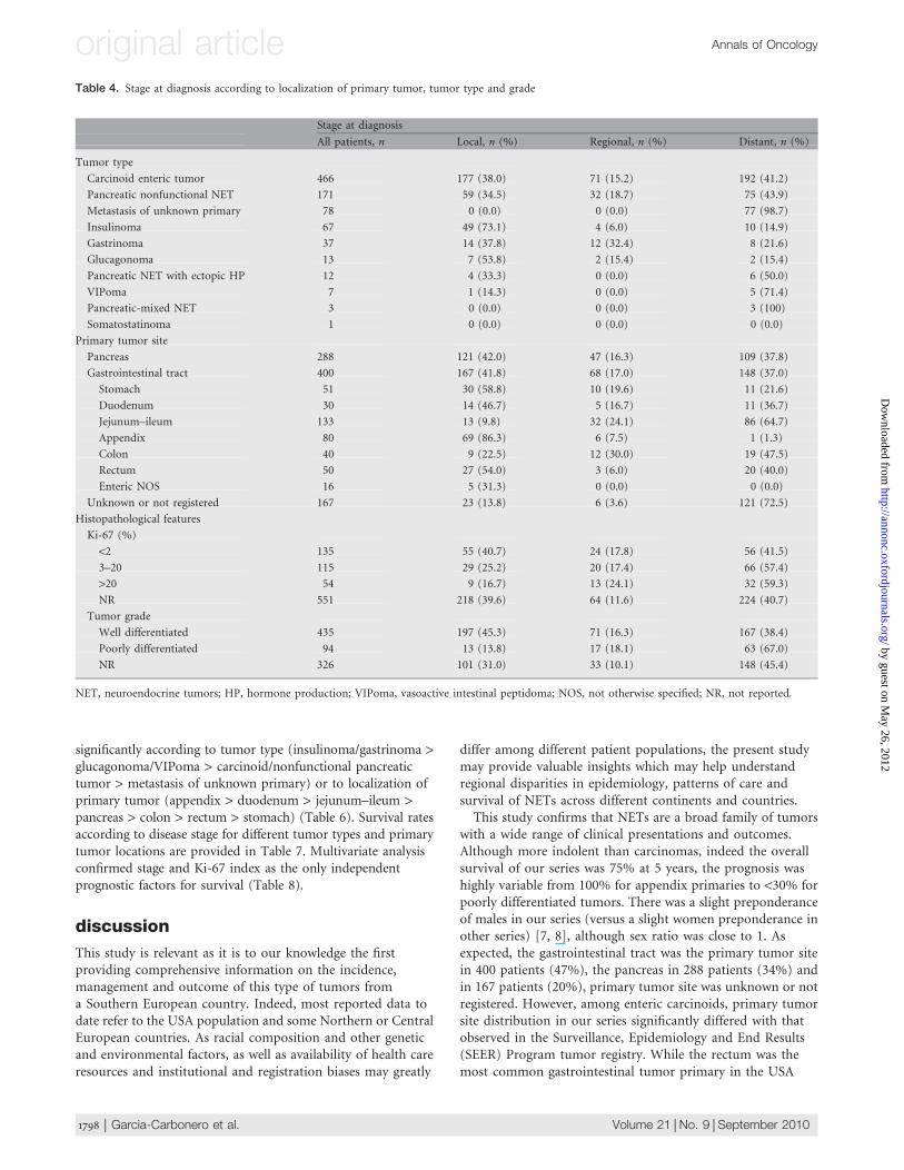
Women tended to have earlier tumor stages than men (42%versus 32% had localized disease). Stage at diagnosis was alsosignificantly different depending upon localization of primarytumor, tumor type and grade (Table 4). The primary tumorsites that presented most frequently with distant disease atdiagnosis included small intestine (65%), colon (48%), rectum(40%) and pancreas (38%), whereas it was most unusual inappendix primaries (1.3%). The tumor types most commonlyassociated with widespread disease were VIPomas (71%),pancreatic nonfunctioning tumors (44%) and bowel carcinoids(41%), as opposed to gastrinomas (22%), insulinomas orglucagonomas (15% each). As expected, poorly differentiatedtumors were more prone to have distant metastasis atdiagnosis (67%), although the proportion of patients with
Table 1. Characteristics of study population (N = 837 patients)
All patients,
n (%)
Men,
n (%)
Women,
n (%)
Age (years)
Median (range) 59 (10–99) 59 (16–86) 60 (10–99)
Gender
Men 458 (54.7)
Women 379 (45.3)
Hormonal Syndrome 210 (24.6) 113 (24.1) 97 (25.1)
Incidental diagnosis 187 (21.9) 103 (22.0) 84 (21.7)
Histological diagnosis 772 (92.2) 422 (91.1) 350 (92.3)
MEN syndrome 43 (5.0) 20 (4.3) 23 (5.9)
Parathyroid
hyperplasia
39 (90.7) 18 (90.0) 21 (91.3)
Pituitary adenoma 18 (41.9) 8 (40.0) 10 (43.5)
Adrenal adenoma 10 (23.3) 6 (30.0) 4 (17.4)
Lipoma 3 (7.0) 2 (10.0) 1 (4.3)
Pheochromocytoma 2 (4.7) 2 (10.0) 0 (0.0)
Genetic test carried out 57 (6.7) 21 (4.5) 36 (9.3)
Stage at diagnosis
Local 311 (36.4) 149 (31.8) 162 (41.9)
Regional 121 (14.2) 70 (15.0) 51 (13.2)
Distant 378 (44.2) 218 (46.6) 160 (41.3)
NR 45 (5.3) 31 (6.6) 14 (3.6)
Primary tumor size (cm)
<1 87 (10.2) 41 (8.8) 46 (11.9)
1–2 154 (18.0) 82 (17.5) 72 (18.6)
2.1–4 149 (17.4) 83 (17.7) 66 (17.1)
>4 151 (17.7) 84 (17.9) 67 (17.3)
NR 314 (36.7) 178 (38.0) 136 (35.1)
Multiple primary tumor 56 (6.7) 31 (6.8) 25 (6.6)
Other NETs 17 (2.0%) 9 (2.0) 8 (2.1)
Localization of metastases
Liver 360 (42.1) 202 (43.2) 158 (40.8)
Lymph nodes 87 (10.2) 51 (10.9) 36 (9.3)
Peritoneum 56 (6.5) 35 (7.5) 21 (5.4)
Bone 46 (5.4) 30 (6.4) 16 (4.1)
Lung 28 (3.3) 21 (4.5) 7 (1.8)
CNS 4 (0.5) 3 (0.6) 1 (0.3)
Adrenal 4 (0.5) 3 (0.6) 1 (0.3)
MEN, multiple endocrine neoplasia; NR, not reported; NETs,
neuroendocrine tumors; CNS, central nervous system.
Table 2. Diagnostic procedures
Cases
tested
Elevated or
positive tests
Biochemical tests
n = 837 patients n %
5-hydroxyindole acetic acid 351 157 44.7
Chromogranin 234 156 66.7
Gastrin 183 73 39.9
Serotonin 112 67 59.8
Insulin 149 57 38.3
Glucagon 98 18 18.4
VIP 54 16 29.6
PP 52 11 21.2
ACTH 52 9 17.3
PTH-RP 24 7 29.2
Immunohistochemistry
n = 855 tumors n %
Chromogranin 563 521 92.5
Synaptophysin 427 409 95.8
Enolase 241 227 94.2
Insulin 128 35 27.3
Gastrin 121 31 25.6
Glucagon 125 42 33.6
Serotonin 51 19 37.3
Somatostatin 113 21 18.3
Imaging studies
n = 837 patients n %
Ultrasound 406 341 84.0
CT scan 720 644 89.4
MR 168 156 92.9
PET 29 24 82.8
Octreoscan� 418 342 81.8
Oral endoscopy 243 158 65.0
Colonoscopy 225 147 65.3
Echoendoscopy 65 58 89.2
Angiography 36 29 80.6
Exploratoy laparotomy 171 166 97.1
Intraoperative ultrasound 49 44 89.8
Bronchoscopy 19 8 42.1
VIP, vasoactive intestinal peptide; PP, pancreatic polypeptide; ACTH,
adrenal corticotrophin; PTH-RP, parathormone-related peptide; CT,
computed tomography; MR, magnetic resonance; PET, positron emission
tomography.
original article Annals of Oncology
1796 | Garcia-Carbonero et al. Volume 21 |No. 9 | September 2010
by guest on May 26, 2012
http://annonc.oxfordjournals.org/D
ownloaded from
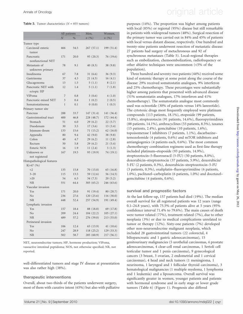
well-differentiated tumors and stage IV disease at presentationwas also rather high (38%).
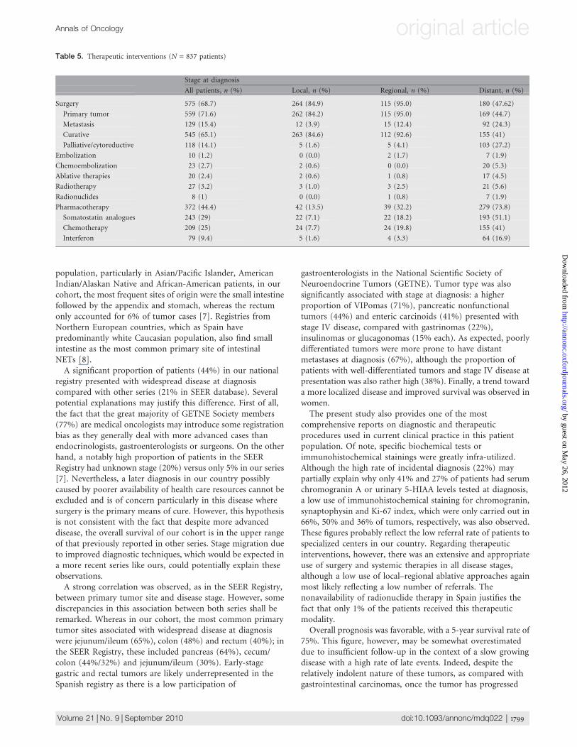
therapeutic interventions
Overall, about two-thirds of the patients underwent surgery,most of them with curative intent (65%) but also with palliative
purposes (14%). The proportion was higher among patientswith local (85%) or regional (95%) disease but still remarkablein patients with widespread tumors (48%). Surgical resection ofthe primary tumor was carried out in 84% and 45% of patientswith local versus distant disease, respectively. One hundred andtwenty-nine patients underwent resection of metastatic disease:27 patients had surgery of metachronous and 92 ofsynchronous metastases (Table 5). Local–regional therapiessuch as embolization, chemoembolization, radiofrequency orother ablative techniques were uncommon (<5% of thepopulation).Three hundred and seventy-two patients (44%) received some
kind of systemic therapy at some point along the course of thedisease: 29% received somatostatin analogues, 9% interferonand 25% chemotherapy. These percentages were substantiallyhigher among patients that presented with advanced disease(51% somatostatin analogues, 17% interferon and 41%chemotherapy). The somatostatin analogue most commonlyused was octreotide (30% of patients versus 14% lanreotide).The cytotoxic drugs most frequently employed were platinumcompounds (113 patients, 18.1%), etoposide (99 patients,15.8%), streptozotocin (91 patients, 14.6%), fluoropyrimidines(88 patients, 14.1%), anthracyclines (53 patients, 8.5%), taxanes(15 patients, 2.4%), gemcitabine (10 patients, 1.6%),topoisomerase I inhibitors (7 patients, 1.1%), dacarbazine–temozolomide (4 patients, 0.6%) and mTOR inhibitors andantiangiogenics (4 patients each, 0.6%). The most commonchemotherapy combination regimens used as first-line therapyincluded platinum–etoposide (93 patients, 14.9%),streptozotocin–5-fluorouracil (5-FU) (50 patients, 8.0%),doxorubicin–streptozotocin (37 patients, 5.9%), doxorubicin/5-FU (2 patients, 0.3%), doxorubicin–streptozotocin–5-FU(3 patients, 0.5%), oxaliplatin–fluoropyrimidine (6 patients,1.0%), paclitaxel–carboplatin (6 patients, 1.0%) and docetaxel–gemcitabine (4 patients, 0.6%).
survival and prognostic factors
At the last follow-up, 157 patients had died (19%). The medianoverall survival for all registered patients was 12 years (range0.1–24.8 years), with 75.5% of patients alive at 5 years (95%confidence interval 71.4% to 79.6%). The main causes of deathwere tumor related (77%), treatment related (7%), due to otherneoplasia (3%) or due to medical complications unrelated totumor or therapy (12%). Sixty-one patients (7%) developedother non-neuroendocrine malignant neoplasia, whichincluded 28 gastrointestinal tumors (22 colorectal, 4biliopancreatic and 1 gastric adenocarcinomas), 15genitourinary malignancies (5 urothelial carcinomas, 4 prostateadenocarcinomas, 4 clear-cell renal carcinomas, 1 Sertoli celltesticular tumor and 1 penis carcinoma), 9 gynecologicalcancers (3 breast, 3 ovarian, 2 endometrial and 1 cervicalcarcinomas), 4 head and neck tumors (1 meningioma, 1neurinoma, 1 laryngeal and 1 follicular thyroid carcinoma), 3hematological malignancies (1 multiple myeloma, 1 lymphomaand 1 leukemia) and a liposarcoma. Overall survival wassignificantly greater in women, younger patients and patientswith hormonal syndrome and in early stage or lower gradetumors (Table 6) (Figure 1). Prognosis also differed
Table 3. Tumor characteristics (N = 855 tumors)
All patients Men,
n (%)
Women,
n (%)N %
Tumor type
Carcinoid enteric
tumor
466 54.5 267 (57.1) 199 (51.4)
Pancreatic
nonfunctional NET
171 20.0 95 (20.3) 76 (19.6)
Metastasis of
unknown primary
78 9.1 40 (8.5) 38 (9.8)
Insulinoma 67 7.8 31 (6.6) 36 (9.3)
Gastrinoma 37 4.3 21 (4.5) 16 (4.1)
Glucagonoma 13 1.5 5 (1.1) 8 (2.1)
Pancreatic NET with
ectopic HP
12 1.4 5 (1.1) 7 (1.8)
VIPoma 7 0.8 3 (0.6) 4 (1.0)
Pancreatic-mixed NET 3 0.4 1 (0.2) 2 (0.5)
Somatostatinoma 1 0.1 0 (0.0) 1 (0.3)
Primary tumor site
Pancreas 288 33.7 147 (31.4) 141 (36.4)
Gastrointestinal tract 400 46.8 228 (48.7) 172 (44.4)
Stomach 51 6.0 29 (6.2) 22 (5.7)
Duodenum 30 3.5 17 (3.6) 13 (3.4)
Jejunum–ileum 133 15.6 71 (15.2) 62 (16.0)
Appendix 80 9.4 42 (9.0) 38 (9.8)
Colon 40 4.7 29 (6.2) 11 (2.8)
Rectum 50 5.8 29 (6.2) 21 (5.4)
Enteric NOS 16 1.9 11 (2.4) 5 (1.3)
Unknown or
not registered
167 19.5 93 (19.9) 74 (19.1)
Histopathological features
Ki-67 (%)
<2 135 15.8 70 (15.0) 65 (16.8)
3–20 115 13.5 59 (12.6) 56 (14.5)
>20 54 6.3 34 (7.3) 20 (5.2)
NR 551 64.4 305 (65.2) 246 (63.6)
Vascular invasion
Yes 171 20.0 91 (19.4) 80 (20.7)
No 236 27.6 120 (25.6) 116 (30.0)
NR 448 52.4 257 (54.9) 191 (49.4)
Lymphatic invasion
Yes 157 18.4 88 (18.8) 69 (17.8)
No 209 24.4 104 (22.2) 105 (27.1)
NR 489 57.2 276 (59.0) 213 (55.0)
Perineural invasion
Yes 106 12.4 65 (13.9) 41 (10.6)
No 247 28.9 118 (25.2) 129 (33.3)
NR 502 58.7 285 (60.9) 217 (56.1)
NET, neuroendocrine tumors; HP, hormone production; VIPoma,
vasoactive intestinal peptidoma; NOS, not otherwise specified; NR, not
reported.
Annals of Oncology original article
Volume 21 |No. 9 | September 2010 doi:10.1093/annonc/mdq022 | 1797
by guest on May 26, 2012
http://annonc.oxfordjournals.org/D
ownloaded from
significantly according to tumor type (insulinoma/gastrinoma >glucagonoma/VIPoma > carcinoid/nonfunctional pancreatictumor > metastasis of unknown primary) or to localization ofprimary tumor (appendix > duodenum > jejunum–ileum >pancreas > colon > rectum > stomach) (Table 6). Survival ratesaccording to disease stage for different tumor types and primarytumor locations are provided in Table 7. Multivariate analysisconfirmed stage and Ki-67 index as the only independentprognostic factors for survival (Table 8).
discussion
This study is relevant as it is to our knowledge the firstproviding comprehensive information on the incidence,management and outcome of this type of tumors froma Southern European country. Indeed, most reported data todate refer to the USA population and some Northern or CentralEuropean countries. As racial composition and other geneticand environmental factors, as well as availability of health careresources and institutional and registration biases may greatly
differ among different patient populations, the present studymay provide valuable insights which may help understandregional disparities in epidemiology, patterns of care andsurvival of NETs across different continents and countries.This study confirms that NETs are a broad family of tumors
with a wide range of clinical presentations and outcomes.Although more indolent than carcinomas, indeed the overallsurvival of our series was 75% at 5 years, the prognosis washighly variable from 100% for appendix primaries to <30% forpoorly differentiated tumors. There was a slight preponderanceof males in our series (versus a slight women preponderance inother series) [7, 8], although sex ratio was close to 1. Asexpected, the gastrointestinal tract was the primary tumor sitein 400 patients (47%), the pancreas in 288 patients (34%) andin 167 patients (20%), primary tumor site was unknown or notregistered. However, among enteric carcinoids, primary tumorsite distribution in our series significantly differed with thatobserved in the Surveillance, Epidemiology and End Results(SEER) Program tumor registry. While the rectum was themost common gastrointestinal tumor primary in the USA
Table 4. Stage at diagnosis according to localization of primary tumor, tumor type and grade
Stage at diagnosis
All patients, n Local, n (%) Regional, n (%) Distant, n (%)
Tumor type
Carcinoid enteric tumor 466 177 (38.0) 71 (15.2) 192 (41.2)
Pancreatic nonfunctional NET 171 59 (34.5) 32 (18.7) 75 (43.9)
Metastasis of unknown primary 78 0 (0.0) 0 (0.0) 77 (98.7)
Insulinoma 67 49 (73.1) 4 (6.0) 10 (14.9)
Gastrinoma 37 14 (37.8) 12 (32.4) 8 (21.6)
Glucagonoma 13 7 (53.8) 2 (15.4) 2 (15.4)
Pancreatic NET with ectopic HP 12 4 (33.3) 0 (0.0) 6 (50.0)
VIPoma 7 1 (14.3) 0 (0.0) 5 (71.4)
Pancreatic-mixed NET 3 0 (0.0) 0 (0.0) 3 (100)
Somatostatinoma 1 0 (0.0) 0 (0.0) 0 (0.0)
Primary tumor site
Pancreas 288 121 (42.0) 47 (16.3) 109 (37.8)
Gastrointestinal tract 400 167 (41.8) 68 (17.0) 148 (37.0)
Stomach 51 30 (58.8) 10 (19.6) 11 (21.6)
Duodenum 30 14 (46.7) 5 (16.7) 11 (36.7)
Jejunum–ileum 133 13 (9.8) 32 (24.1) 86 (64.7)
Appendix 80 69 (86.3) 6 (7.5) 1 (1.3)
Colon 40 9 (22.5) 12 (30.0) 19 (47.5)
Rectum 50 27 (54.0) 3 (6.0) 20 (40.0)
Enteric NOS 16 5 (31.3) 0 (0.0) 0 (0.0)
Unknown or not registered 167 23 (13.8) 6 (3.6) 121 (72.5)
Histopathological features
Ki-67 (%)
<2 135 55 (40.7) 24 (17.8) 56 (41.5)
3–20 115 29 (25.2) 20 (17.4) 66 (57.4)
>20 54 9 (16.7) 13 (24.1) 32 (59.3)
NR 551 218 (39.6) 64 (11.6) 224 (40.7)
Tumor grade
Well differentiated 435 197 (45.3) 71 (16.3) 167 (38.4)
Poorly differentiated 94 13 (13.8) 17 (18.1) 63 (67.0)
NR 326 101 (31.0) 33 (10.1) 148 (45.4)
NET, neuroendocrine tumors; HP, hormone production; VIPoma, vasoactive intestinal peptidoma; NOS, not otherwise specified; NR, not reported.
original article Annals of Oncology
1798 | Garcia-Carbonero et al. Volume 21 |No. 9 | September 2010
by guest on May 26, 2012
http://annonc.oxfordjournals.org/D
ownloaded from
population, particularly in Asian/Pacific Islander, AmericanIndian/Alaskan Native and African-American patients, in ourcohort, the most frequent sites of origin were the small intestinefollowed by the appendix and stomach, whereas the rectumonly accounted for 6% of tumor cases [7]. Registries fromNorthern European countries, which as Spain havepredominantly white Caucasian population, also find smallintestine as the most common primary site of intestinalNETs [8].A significant proportion of patients (44%) in our national
registry presented with widespread disease at diagnosiscompared with other series (21% in SEER database). Severalpotential explanations may justify this difference. First of all,the fact that the great majority of GETNE Society members(77%) are medical oncologists may introduce some registrationbias as they generally deal with more advanced cases thanendocrinologists, gastroenterologists or surgeons. On the otherhand, a notably high proportion of patients in the SEERRegistry had unknown stage (20%) versus only 5% in our series[7]. Nevertheless, a later diagnosis in our country possiblycaused by poorer availability of health care resources cannot beexcluded and is of concern particularly in this disease wheresurgery is the primary means of cure. However, this hypothesisis not consistent with the fact that despite more advanceddisease, the overall survival of our cohort is in the upper rangeof that previously reported in other series. Stage migration dueto improved diagnostic techniques, which would be expected ina more recent series like ours, could potentially explain theseobservations.A strong correlation was observed, as in the SEER Registry,
between primary tumor site and disease stage. However, somediscrepancies in this association between both series shall beremarked. Whereas in our cohort, the most common primarytumor sites associated with widespread disease at diagnosiswere jejunum/ileum (65%), colon (48%) and rectum (40%); inthe SEER Registry, these included pancreas (64%), cecum/colon (44%/32%) and jejunum/ileum (30%). Early-stagegastric and rectal tumors are likely underrepresented in theSpanish registry as there is a low participation of
gastroenterologists in the National Scientific Society ofNeuroendocrine Tumors (GETNE). Tumor type was alsosignificantly associated with stage at diagnosis: a higherproportion of VIPomas (71%), pancreatic nonfunctionaltumors (44%) and enteric carcinoids (41%) presented withstage IV disease, compared with gastrinomas (22%),insulinomas or glucagonomas (15% each). As expected, poorlydifferentiated tumors were more prone to have distantmetastases at diagnosis (67%), although the proportion ofpatients with well-differentiated tumors and stage IV disease atpresentation was also rather high (38%). Finally, a trend towarda more localized disease and improved survival was observed inwomen.The present study also provides one of the most
comprehensive reports on diagnostic and therapeuticprocedures used in current clinical practice in this patientpopulation. Of note, specific biochemical tests orimmunohistochemical stainings were greatly infra-utilized.Although the high rate of incidental diagnosis (22%) maypartially explain why only 41% and 27% of patients had serumchromogranin A or urinary 5-HIAA levels tested at diagnosis,a low use of immunohistochemical staining for chromogranin,synaptophysin and Ki-67 index, which were only carried out in66%, 50% and 36% of tumors, respectively, was also observed.These figures probably reflect the low referral rate of patients tospecialized centers in our country. Regarding therapeuticinterventions, however, there was an extensive and appropriateuse of surgery and systemic therapies in all disease stages,although a low use of local–regional ablative approaches againmost likely reflecting a low number of referrals. Thenonavailability of radionuclide therapy in Spain justifies thefact that only 1% of the patients received this therapeuticmodality.Overall prognosis was favorable, with a 5-year survival rate of
75%. This figure, however, may be somewhat overestimateddue to insufficient follow-up in the context of a slow growingdisease with a high rate of late events. Indeed, despite therelatively indolent nature of these tumors, as compared withgastrointestinal carcinomas, once the tumor has progressed
Table 5. Therapeutic interventions (N = 837 patients)
Stage at diagnosis
All patients, n (%) Local, n (%) Regional, n (%) Distant, n (%)
Surgery 575 (68.7) 264 (84.9) 115 (95.0) 180 (47.62)
Primary tumor 559 (71.6) 262 (84.2) 115 (95.0) 169 (44.7)
Metastasis 129 (15.4) 12 (3.9) 15 (12.4) 92 (24.3)
Curative 545 (65.1) 263 (84.6) 112 (92.6) 155 (41)
Palliative/cytoreductive 118 (14.1) 5 (1.6) 5 (4.1) 103 (27.2)
Embolization 10 (1.2) 0 (0.0) 2 (1.7) 7 (1.9)
Chemoembolization 23 (2.7) 2 (0.6) 0 (0.0) 20 (5.3)
Ablative therapies 20 (2.4) 2 (0.6) 1 (0.8) 17 (4.5)
Radiotherapy 27 (3.2) 3 (1.0) 3 (2.5) 21 (5.6)
Radionuclides 8 (1) 0 (0.0) 1 (0.8) 7 (1.9)
Pharmacotherapy 372 (44.4) 42 (13.5) 39 (32.2) 279 (73.8)
Somatostatin analogues 243 (29) 22 (7.1) 22 (18.2) 193 (51.1)
Chemotherapy 209 (25) 24 (7.7) 24 (19.8) 155 (41)
Interferon 79 (9.4) 5 (1.6) 4 (3.3) 64 (16.9)
Annals of Oncology original article
Volume 21 |No. 9 | September 2010 doi:10.1093/annonc/mdq022 | 1799
by guest on May 26, 2012
http://annonc.oxfordjournals.org/D
ownloaded from
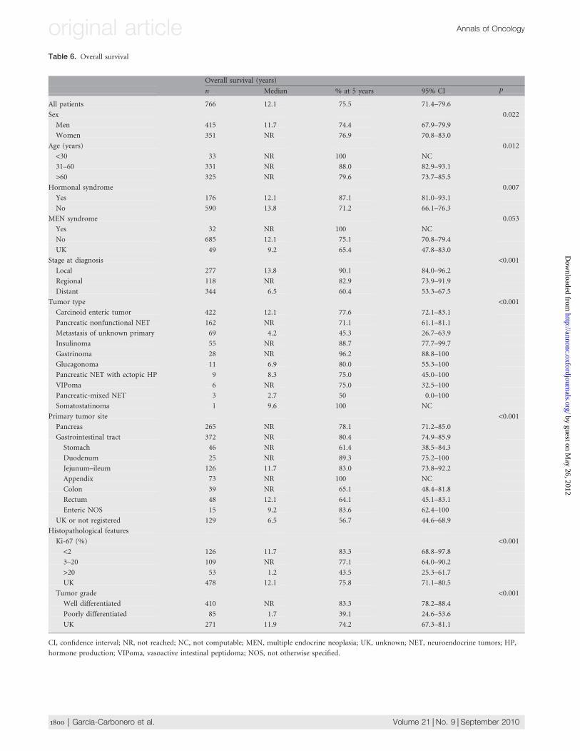
Table 6. Overall survival
Overall survival (years)
n Median % at 5 years 95% CI P
All patients 766 12.1 75.5 71.4–79.6
Sex 0.022
Men 415 11.7 74.4 67.9–79.9
Women 351 NR 76.9 70.8–83.0
Age (years) 0.012
<30 33 NR 100 NC
31–60 331 NR 88.0 82.9–93.1
>60 325 NR 79.6 73.7–85.5
Hormonal syndrome 0.007
Yes 176 12.1 87.1 81.0–93.1
No 590 13.8 71.2 66.1–76.3
MEN syndrome 0.053
Yes 32 NR 100 NC
No 685 12.1 75.1 70.8–79.4
UK 49 9.2 65.4 47.8–83.0
Stage at diagnosis <0.001Local 277 13.8 90.1 84.0–96.2
Regional 118 NR 82.9 73.9–91.9
Distant 344 6.5 60.4 53.3–67.5
Tumor type <0.001Carcinoid enteric tumor 422 12.1 77.6 72.1–83.1
Pancreatic nonfunctional NET 162 NR 71.1 61.1–81.1
Metastasis of unknown primary 69 4.2 45.3 26.7–63.9
Insulinoma 55 NR 88.7 77.7–99.7
Gastrinoma 28 NR 96.2 88.8–100
Glucagonoma 11 6.9 80.0 55.3–100
Pancreatic NET with ectopic HP 9 8.3 75.0 45.0–100
VIPoma 6 NR 75.0 32.5–100
Pancreatic-mixed NET 3 2.7 50 0.0–100
Somatostatinoma 1 9.6 100 NC
Primary tumor site <0.001Pancreas 265 NR 78.1 71.2–85.0
Gastrointestinal tract 372 NR 80.4 74.9–85.9
Stomach 46 NR 61.4 38.5–84.3
Duodenum 25 NR 89.3 75.2–100
Jejunum–ileum 126 11.7 83.0 73.8–92.2
Appendix 73 NR 100 NC
Colon 39 NR 65.1 48.4–81.8
Rectum 48 12.1 64.1 45.1–83.1
Enteric NOS 15 9.2 83.6 62.4–100
UK or not registered 129 6.5 56.7 44.6–68.9
Histopathological features
Ki-67 (%) <0.001<2 126 11.7 83.3 68.8–97.8
3–20 109 NR 77.1 64.0–90.2
>20 53 1.2 43.5 25.3–61.7
UK 478 12.1 75.8 71.1–80.5
Tumor grade <0.001Well differentiated 410 NR 83.3 78.2–88.4
Poorly differentiated 85 1.7 39.1 24.6–53.6
UK 271 11.9 74.2 67.3–81.1
CI, confidence interval; NR, not reached; NC, not computable; MEN, multiple endocrine neoplasia; UK, unknown; NET, neuroendocrine tumors; HP,
hormone production; VIPoma, vasoactive intestinal peptidoma; NOS, not otherwise specified.
original article Annals of Oncology
1800 | Garcia-Carbonero et al. Volume 21 |No. 9 | September 2010
by guest on May 26, 2012
http://annonc.oxfordjournals.org/D
ownloaded from
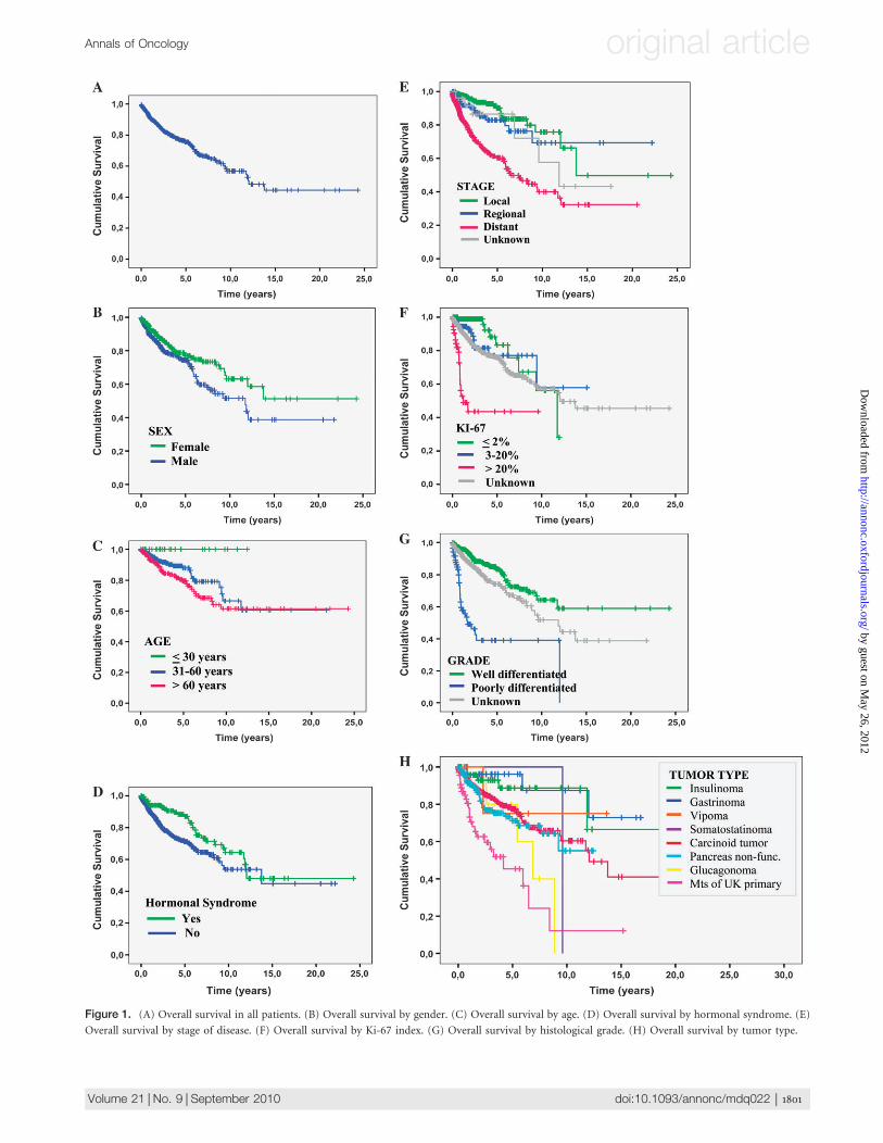
Figure 1. (A) Overall survival in all patients. (B) Overall survival by gender. (C) Overall survival by age. (D) Overall survival by hormonal syndrome. (E)
Overall survival by stage of disease. (F) Overall survival by Ki-67 index. (G) Overall survival by histological grade. (H) Overall survival by tumor type.
Annals of Oncology original article
Volume 21 |No. 9 | September 2010 doi:10.1093/annonc/mdq022 | 1801
by guest on May 26, 2012
http://annonc.oxfordjournals.org/D
ownloaded from

beyond surgical resectability, the disease is eminently incurable.In this regard, it is remarkable that 77% of deaths in our cohortwere due to tumor progression and an additional 7% were dueto treatment-related issues (drug toxicity, surgicalcomplications). Survival was significantly greater in women,younger patients and patients with hormonal syndrome and inearly stage or lower grade tumors. As observed by others,prognosis also differed significantly according to tumor type orto primary tumor site, although with some striking differencescompared with other geographical regions (i.e. poorer survivalfor gastric or rectal primaries in our country). However, stageand grade remained the only independent predictors foroutcome in multivariate analysis, which underscores the needfor earlier diagnosis and for improved systemic therapies foradvanced disease.This national database reveals relevant information regarding
current clinical practices and provides valuable insights into theepidemiology and outcome of this heterogeneous and not souncommon disease. Indeed, GEP-NETs are more prevalent andlethal than previously thought. Despite some recent progress
[17–22], patient survival has not significantly changed over thelast 30 years. Improving our understanding of the molecularbasis of this disease, as well as the mechanisms involved inresponse and resistance to therapy, will be essential tools thatwill help us develop early diagnosis tools and newer morerationally designed treatment strategies that will potentiallychange the natural history of malignant NETs. Finally,encouraging physicians to refer these patients to specializedcenters and patients to participate in clinical trials is of utmostimportance.
funding
Novartis Oncology (Spain); Ipsen Pharma (Spain).
acknowledgements
We are most grateful to all participating physicians thatgenerously contributed with their patients to this registry(Appendix 1).
disclosure
The authors state no conflict of interest.
references
1. Obendorfer S. Karzinoide tumoren des dunndarms. Frankf Z Pathol 1907; 1:
425–429.
2. Modlin IM, Oberg K, Chung DC et al. Gastroenteropancreatic neuroendocrine
tumours. Lancet Oncol 2008; 9(1): 61–72.
3. Kloppel G, Couvelard A, Perren A et al. ENETS Consensus Guidelines for the
Standards of Care in Neuroendocrine Tumors: towards a standardized approach
to the diagnosis of gastroenteropancreatic neuroendocrine tumors and their
prognostic stratification. Neuroendocrinology 2009; 90(2): 162–166.
4. Eriksson B, Annibale B, Bajetta E et al. ENETS Consensus Guidelines for the
Standards of Care in Neuroendocrine Tumors: chemotherapy in patients with
neuroendocrine tumors. Neuroendocrinology 2009; 90(2): 214–219.
5. Oberg K, Ferone D, Kaltsas G et al. ENETS Consensus Guidelines for the
Standards of Care in Neuroendocrine Tumors: biotherapy. Neuroendocrinology
2009; 90(2): 209–213.
Table 7. Survival according to primary tumor site, tumor type and stage
Survival (% at 5 years)
All Local Regional Distant
n % n % n % n %
Tumor type
Carcinoid enteric
tumor
422 77.6 166 91.1 69 79.7 174 64.3
Pancreatic
nonfunctional
NET
162 71.1 54 85.3 31 83.7 72 56.4
Metastasis of
unknown
primary
69 45.3 0 – 0 – 69 45.3
Insulinoma 55 88.7 39 88.4 4 100 9 75.0
Gastrinoma 28 96.2 8 100 12 100 7 83.3
Glucagonoma 11 80.0 6 100 2 50.0 2 50.0
Pancreatic NET
with ectopic HP
9 75.0 3 – 0 – 4 27.2
VIPoma 6 75.0 1 100 0 – 4 27.2
Pancreatic-mixed
NET
3 50.0 0 – 0 – 3 35.4
Somatostatinoma 1 100 0 – 0 – 0 –
Primary tumor site
Pancreas 265 78.1 108 89.7 46 87.0 102 60.9
Gastrointestinal
tract
372 80.4 155 91.5 66 78.9 137 68.1
Stomach 46 61.4 27 87.4 10 35.6 9 0.0
Duodenum 25 89.3 10 100 5 100 10 72.9
Jejunum–ileum 126 83.0 13 100 31 74.0 80 82.4
Appendix 73 100 65 100 5 100 1 100.0
Colon 39 65.1 9 71.1 12 91.7 18 42.9
Rectum 48 64.1 26 75.0 3 66.7 19 29.8
Enteric NOS 15 83.6 5 66.7 0 – 0 –
Unknown or not
registered
129 56.7 14 83.1 6 100 105 50.0
NET, neuroendocrine tumors; HP, hormone production; VIPoma,
vasoactive intestinal peptidoma; NOS, not otherwise specified.
Table 8. Cox multivariate analysis for overall survival (N = 659 patients)
Variable Hazard risk 95% CI P
Stage at diagnosis 3.96 1.97–7.96 0.0001
Ki-67 6.69 1.96–22.88 0.008
Variables included in the regression model: gender (men versus women),
age (<30 versus 31–60 versus >60 years), hormonal syndrome (yes versus
no), MEN syndrome (yes versus no versus unknown), stage at diagnosis
(distant versus localized), tumor type (enteric carcinoids, pancreatic
nonfunctional NET, metastasis of unknown primary, insulinomas,
gastrinomas, glucagonoma, pancreatic NET with ectopic HP, VIPoma,
pancreatic-mixed NET and somatostatinoma), primary tumor site
(pancreas versus stomach versus duodenum versus jejunum–ileum versus
appendix versus colon versus rectum versus enteric NOS), tumor grade
(well versus poorly differentiated) and Ki-67 (>20% versus £2%). Only
significant variables are given in the table.
CI, confidence interval; MEN, multiple endocrine neoplasia; NET,
neuroendocrine tumors; HP, hormone production; VIPoma, vasoactive
intestinal polypeptidoma; NOS, not otherwise specified.
original article Annals of Oncology
1802 | Garcia-Carbonero et al. Volume 21 |No. 9 | September 2010
by guest on May 26, 2012
http://annonc.oxfordjournals.org/D
ownloaded from
6. Arnold R, Chen YJ, Costa F et al. ENETS Consensus Guidelines for the Standards
of Care in Neuroendocrine Tumors: follow-up and documentation.
Neuroendocrinology 2009; 90(2): 227–233.
7. Yao JC, Hassan M, Phan A et al. One hundred years after ‘‘carcinoid’’:
epidemiology of and prognostic factors for neuroendocrine tumors in 35,825
cases in the United States. J Clin Oncol 2008; 26(18): 3063–3072.
8. Hauso O, Gustafsson BI, Kidd M et al. Neuroendocrine tumor epidemiology:
contrasting Norway and North America. Cancer 2008; 113(10):
2655–2664.
9. Hemminki K, Li X. Incidence trends and risk factors of carcinoid tumors:
a nationwide epidemiologic study from Sweden. Cancer 2001; 92(8):
2204–2210.
10. Lepage C, Rachet B, Coleman MP. Survival from malignant digestive endocrine
tumors in England and Wales: a population-based study. Gastroenterology 2007;
132(3): 899–904.
11. Fischer L, Kleeff J, Esposito I et al. Clinical outcome and long-term survival in
118 consecutive patients with neuroendocrine tumours of the pancreas.
Br J Surg 2008; 95(5): 627–635.
12. Yao JC, Eisner MP, Leary C et al. Population-based study of islet cell carcinoma.
Ann Surg Oncol 2007; 14(12): 3492–3500.
13. Landry CS, Woodall C, Scoggins CR et al. Analysis of 900 appendiceal carcinoid
tumors for a proposed predictive staging system. Arch Surg 2008; 143(7):
664–670.
14. Modlin IM, Champaneria MC, Chan AK, Kidd M. A three-decade analysis of
3,911 small intestinal neuroendocrine tumors: the rapid pace of no progress. Am
J Gastroenterol 2007; 102(7): 1464–1473.
15. Halfdanarson TR, Rabe KG, Rubin J, Petersen GM. Pancreatic neuroendocrine
tumors (PNETs): incidence, prognosis and recent trend toward improved survival.
Ann Oncol 2008; 19(10): 1727–1733.
16. Kloppel G, Perren A, Heitz PU. The gastroenteropancreatic neuroendocrine cell
system and its tumors: the WHO classification. Ann N Y Acad Sci 2004; 1014:
13–27.
17. Duran I, Salazar R, Casanovas O et al. New drug development in digestive
neuroendocrine tumors. Ann Oncol 2007; 18(8): 1307–1313.
18. Vilar E, Salazar R, Perez-Garcıa J et al. Chemotherapy and role of the
proliferation marker Ki-67 in digestive neuroendocrine tumors. Endocr Relat
Cancer 2007; 14(2): 221–232.
19. Modlin IM, Kidd M, Drozdov I et al. Pharmacotherapy of neuroendocrine cancers.
Expert Opin Pharmacother 2008; 9(15): 2617–2626.
20. Rinke A, Muller HH, Schade-Brittinger C et al. Placebo-controlled, double-blind,
prospective, randomized study on the effect of octreotide LAR in the control
of tumor growth in patients with metastatic neuroendocrine midgut tumors:
a report from the PROMID Study Group. J Clin Oncol 2009; 27(28):
4656–4663.
21. Yao JC, Lombard-Bohas C, Baudin E et al. Daily oral everolimus activity in
patients with metastatic pancreatic neuroendocrine tumors after failure of
cytotoxic chemotherapy: a phase II trial. J Clin Oncol 2010; 28(1): 69–76.
22. Raymond E, Faivre S, Hammel P et al. Sunitinib paves the way for targeted
therapies in neuroendocrine tumors. Target Oncol 2009; 4(4): 253–254.
appendix 1: list of participating centersand physicians in order of contribution(after authors of this paper)
Marrupe Gonzalez D. (Hospital General de Mostoles, Madrid);Fraile Lopez-Amor M. (Hospital Ramon y Cajal, Madrid);Fuster Salva J. A. (Hospital Son Dureta, Mallorca); GimenezPerez G. (Hospital de Sabadell, Corporacion Parc Taulı,Barcelona); La Casta Munoa A. (Hospital Donostia, SanSebastian); Biarnes Costa J. (Hospital Dr Josep Trueta,Gerona); Abad Esteve A. (Hospital Universitario Germans Triasi Pujol, Barcelona); Aller J. (Hospital Puerta de Hierro,Madrid); Alarco Hernandez A. (Hospital Universitario deCanarias); Blanco Carrera C. (Hospital Central Universitario deAsturias); Cano J. M. (Complejo Hospitalario Torrecardenas,Almerıa); Molina Garrido M. J. (Hospital General Universitariode Elche); Tome Martinez de Rituerto M. A. (Hospital deConxo, Complejo Hospitalario Universitario de Santiago(CHUS), Santiago de Compostela); Lomas Garrido M. (HospitalUniversitario Infanta Cristina, Badajoz); Serrano Blanch R.(Hospital Reina Sofia, Cordoba); Cordido Carballido F.(Hospital Juan Canalejo, La Coruna); Del Pozo Pico C. (HospitalMutua de Terrassa, Barcelona); Etxeberria Larrea A. (InstitutoOncologico, Guipuzcoa); Moneva Arce A. (Hospital NuestraSenora de la Candelaria, Santa Cruz de Tenerife); Afonso GomezR. (Hospital Insular de las Palmas de Gran Canaria); ArribasPalomar L. (Hospital Marina Alta, Valencia); Carabantes OconF. J. (Hospital Regional Carlos Haya; Malaga); Catot Tort S.(Hospital Althaia Manresa, Barcelona); Garcıa Fernandez H.(Hospital Son Dureta, Mallorca); Leon Carbonero A.(Fundacion Jimenez Dıaz, Madrid); Marco Martinez A.(Hospital Virgen de la Salud, Toledo); Martınez Olmos A.(Complejo Hospitalario Universitario de Santiago); Reina ZoiloJ. J. (Hospital SAS Juan Ramon Jimenez, Huelva); Segura HuertaA. (Hospital Universitario La Fe, Valencia); Vicente Delgado A.(Hospital Virgen de la Salud, Toledo); Aguilar Bujanda D.(Hospital Doctor Negrin, Gran Canaria); Baena Canada J. M.(Hospital Universitario Puerta del Mar, Barcelona); CarmonaBayonas A. (Hospital Morales Messeguer, Murcia); JimenezOrozco E. (Hospital de Jerez, Jerez); Losa Gaspa F. (HospitalGeneral de L’Hospitalet, Barcelona); Manzano Monzo J. L.(Hospital Universitario Germans Trias i Pujol, Barcelona); TofePovedano S. (Hospital Reina Sofia, Cordoba); Wagner Fahlin A.M. (Hospital San Pau, Barcelona, Spain).
Annals of Oncology original article
Volume 21 |No. 9 | September 2010 doi:10.1093/annonc/mdq022 | 1803
by guest on May 26, 2012
http://annonc.oxfordjournals.org/D
ownloaded from
Related Documents