Case Control Study: Incidence And Risk Factors Of Postpartum Haemorrhage In Primigravida Juliana Yusof ( [email protected] ) Research article Keywords: Postpartum Haemorrhage, primigravida, Caesarean Posted Date: June 25th, 2020 DOI: https://doi.org/10.21203/rs.3.rs-36771/v1 License: This work is licensed under a Creative Commons Attribution 4.0 International License. Read Full License

Welcome message from author
This document is posted to help you gain knowledge. Please leave a comment to let me know what you think about it! Share it to your friends and learn new things together.
Transcript
Case Control Study: Incidence And Risk Factors OfPostpartum Haemorrhage In PrimigravidaJuliana Yusof ( [email protected] )
Research article
Keywords: Postpartum Haemorrhage, primigravida, Caesarean
Posted Date: June 25th, 2020
DOI: https://doi.org/10.21203/rs.3.rs-36771/v1
License: This work is licensed under a Creative Commons Attribution 4.0 International License. Read Full License
CASE CONTROL STUDY:
INCIDENCE AND RISK FACTORS OF POSTPARTUM
HAEMORRHAGE IN PRIMIGRAVIDA
Dr Juliana Yusof

22
Case Control Study: Incidence and Risk Factors of Postpartum Haemorrhage in
Primigravida
Juliana Yusof
Masters of Obstetric & Gynecology, Faculty of Medicine, University
Technology of MARA , Sungai Buloh : [email protected]
ABSTRACT
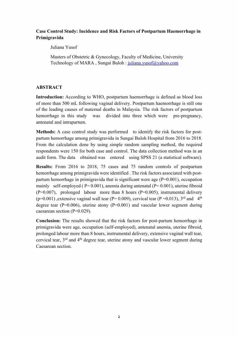
Introduction: According to WHO, postpartum haemorrhage is defined as blood loss
of more than 500 mL following vaginal delivery. Postpartum haemorrhage is still one
of the leading causes of maternal deaths in Malaysia. The risk factors of postpartum
hemorrhage in this study was divided into three which were pre-pregnancy,
antenatal and intrapartum.
Methods: A case control study was performed to identify the risk factors for post-
partum hemorrhage among primigravida in Sungai Buloh Hospital from 2016 to 2018.
From the calculation done by using simple random sampling method, the required
respondents were 150 for both case and control. The data collection method was in an
audit form. The data obtained was entered using SPSS 21 (a statistical software).
Results: From 2016 to 2018, 75 cases and 75 random controls of postpartum
hemorrhage among primigravida were identified . The risk factors associated with post-
partum hemorrhage in primigravida that is significant were age (P=0.001), occupation
mainly self-employed ( P= 0.001), anemia during antenatal (P= 0.001), uterine fibroid
(P=0.007), prolonged labour more than 8 hours (P=0.005), instrumental delivery
(p=0.001) ,extensive vaginal wall tear (P= 0.009), cervical tear (P =0.013), 3rd and 4th
degree tear (P=0.006), uterine atony (P=0.001) and vascular lower segment during
caesarean section (P=0.029).
Conclusion: The results showed that the risk factors for post-partum hemorrhage in
primigravida were age, occupation (self-employed), antenatal anemia, uterine fibroid,
prolonged labour more than 8 hours, instrumental delivery, extensive vaginal wall tear,
cervical tear, 3rd and 4th degree tear, uterine atony and vascular lower segment during
Caesarean section.
33
BACKGROUND
For centuries, the biggest concerns in pregnancy would be the mother and the
baby, where it will be either one to survive the whole pregnancy or both of them. In
United Kingdom, the most recent Confidential Enquiry into Maternal Deaths (CEMD)
reports indicates that from 2009 till 2012, 357 women died during or within 6 weeks of
the end of their pregnancy. This represents a decrease in the maternal mortality ratio
(MMR) where in 2006 to 2008 it was 11, while in 2009 to 2012 it was 10.12 per 100,000
live births, mainly due to direct obstetric causes. (3) In Malaysia however since
embarking on CEMD in 1991, the maternal mortality ratio has declined from 44 in 1991
to 27 per 100,000 live births in 2011. (4)
The causes of maternal deaths can be divided into direct causes or indirect
causes. Direct causes from complications during pregnancy are the prime suspects for
maternal deaths which includes obstetric embolism, postpartum haemorrhage and
hypertensive disorders in pregnancy. Indirect causes resulted in about 17% of death
whereby heart diseases is the most common cause. Out of all these causes,
postpartum haemorrhage is still one of the leading causes of maternal deaths in
Malaysia. There were 27 deaths in 2001, 16 deaths in 2003, 17 deaths in 2005, 24 deaths
in 2006, 23 deaths in 2007 and 26 deaths in 2008. (2)
According to WHO, postpartum haemorrhage ( PPH) is defined as blood loss
of more than 500 mL . A loss of these amounts within 24 hours of delivery is termed
early or primary PPH, whereas such losses are termed late or secondary PPH if they
occur 24 hours after delivery until 12 weeks postpartum. The amount of blood loss
required to cause hemodynamic compromise will depend on the pre-existing condition
of the woman. Hemodynamic compromise is more likely to occur in conditions such as
anemia or volume contracted states. All women who carry a pregnancy beyond 20
weeks’ gestation are at risk for PPH and its sequelae (1).
Risk factors of postpartum hemorrhage can be divided into three which are pre-
pregnancy, antenatal and intrapartum. There are quite a lot of risk factors for
development of postpartum hemorrhage that need to be elicited from history taking.
Example are anemia, hemophilia A, hemophilia B and Von Willebrands
disease. During antenatal, the example of risks that were identified were
severe preeclampsia with thrombocytopenia, maternal obesity and
thromboprophylaxis drugs . The risk factors that were studied intrapartum were
delivery by ceasarean section, retained placenta, genital tract trauma, prolonged labor
for more than 8 hours, delivery of a macrosomic baby of more than 4 kg, operative
vaginal delivery and pyrexia in the intrapartum period. (1)
44
OBJECTIVE
1. To determine the risk factors for postpartum haemorrhage in primigravida at
Sungai Buloh Hospital ( HSgB) from 2016-2018 during antenatal and
intrapartum period.
METHODOLOGY
This was a retrospective case control study conducted at Hospital Sungai Buloh
( HSgB), Sungai Buloh, Selangor conducted from 1st January 2016 to 15th July 2018.
We identified 150 patients for cases and controls. The definition of cases is
primigravida deliver at HSgB from 1st January 2016 to 15th July 2018 with PPH. PPH
is defined as more than 500ml blood loss during delivery in normal delivery and more
than 1000ml in caesarean section. Meanwhile, the definition of control is
primigravida that delivered at HSgB from 1st January 2016 to 15th July 2018
without PPH.
We calculate our sample size by using formula !"($%!") ( !) ($%!))
(!"%!))) 𝑋 (𝑍, + 𝑍.)/.
By taking α = 0.05, 80% power of study and proportion of group 1 was 41.7% from
study by Chandrika et al., (2014) and the proportion of group 2 was 19.3% from study
by Tan et al., (2013), the sample size needed will be 62 in each group. After adjusting
20% attrition rate, the minimum sample required in this study will be 75 in each group.
We selected cases and control from delivery book of HSgB. The first 75 patient
that had been chosen in the delivery book will be taken in this study for both cases and
controls. The patient’s demographic, antenatal and intrapartum data were
retrieved from hospital computer system based on their registration number. In our
audit form, the antenatal risk factors were anemia, placenta previa, polyhydramnios,
multiple pregnancy, booking body mass index ( BMI) >30kg/m2, excessive weight
gain, uterine fibroid, magnesium sulphate MgSO4 infusion,
thrombocytopenia/bleeding disorder, induction of labour (IOL) with prostin,
prophylaxis clexane and aspirin. Risk factors for intrapartum identified were method of
delivery, abruptio placenta, precipitated labour less than 3 hours, poor progress of
labour more than 8 hours , prolonged second stage more than 1 hour, instrumental
delivery, extensive vaginal wall tear, cervical tear, 3rd and 4th degree tear, retained
placenta, macrosomic baby >4kg and uterine atony. For intraoperative, the risk
factors identified were disseminated intravascular coagulopathy ( DIVC),
vascular lower segment during caesarean section, classical caesarean section and
extended tear of incision during caesarean section.
The data was then entered into SPSS version 21 for analysis. In describing the
socio-demographic and first pregnancy details, measure central tendencies (means)
were used. For categorical data, frequencies were computed. Chi square or Fisher’s
test was used to compare proportions to determine strength of association between
PPH and its risk factors during antenatal, intrapartum and intraoperative. A p -value
55
less than 0.05 used to determine association between variables was considered statically
significant.
RESULTS
Total number of patients in this study were 150 of which 75 of them were cases
and another 75 patients were control. The patient’s data who delivered at Sungai Buloh
Hospital from 1st January 2016 till 15th July 2018 were retrieved. Cases and control
were selected by using simple random sampling using random number computer
generated.
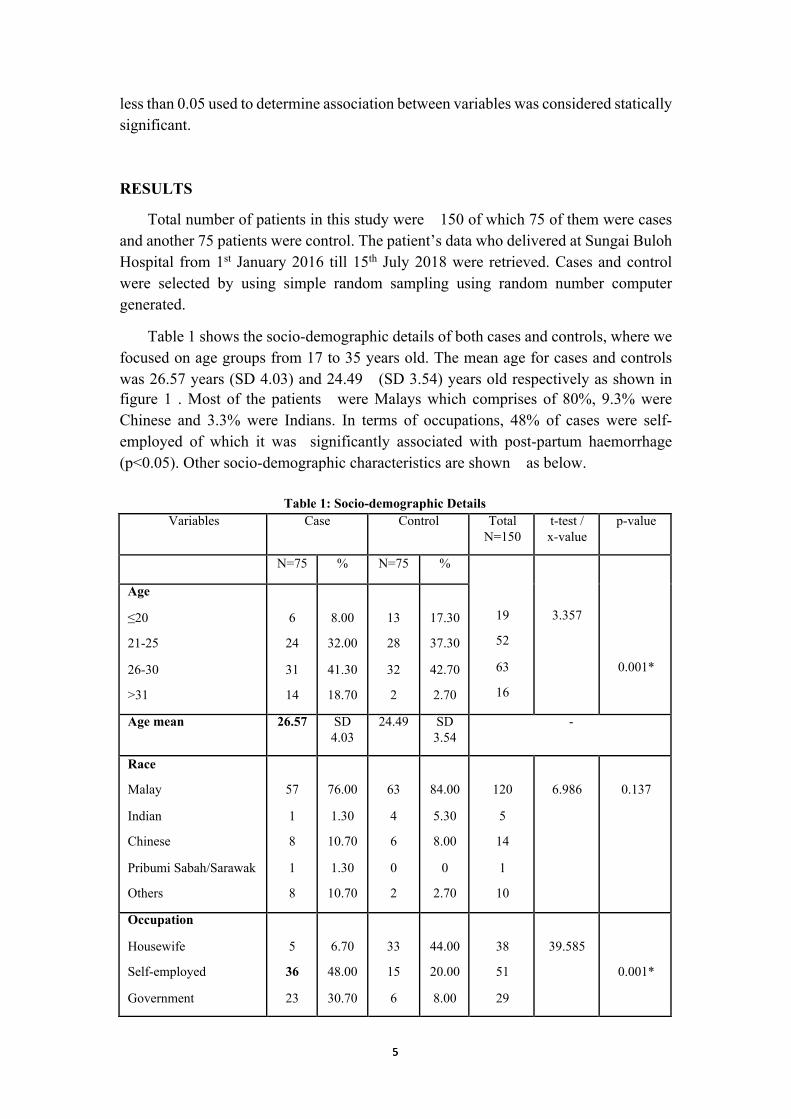
Table 1 shows the socio-demographic details of both cases and controls, where we
focused on age groups from 17 to 35 years old. The mean age for cases and controls
was 26.57 years (SD 4.03) and 24.49 (SD 3.54) years old respectively as shown in
figure 1 . Most of the patients were Malays which comprises of 80%, 9.3% were
Chinese and 3.3% were Indians. In terms of occupations, 48% of cases were self-
employed of which it was significantly associated with post-partum haemorrhage
(p<0.05). Other socio-demographic characteristics are shown as below.
Table 1: Socio-demographic Details
Variables Case Control Total
N=150
t-test /
x-value
p-value
N=75 % N=75 %
19
52
63
16
3.357
0.001*
Age
≤20
21-25
26-30
>31
6
24
31
14
8.00
32.00
41.30
18.70
13
28
32
2
17.30
37.30
42.70
2.70
Age mean 26.57 SD
4.03
24.49 SD
3.54
-
Race
Malay
Indian
Chinese
Pribumi Sabah/Sarawak
Others
57
1
8
1
8
76.00
1.30
10.70
1.30
10.70
63
4
6
0
2
84.00
5.30
8.00
0
2.70
120
5
14
1
10
6.986
0.137
Occupation
Housewife
Self-employed
Government
5
36
23
6.70
48.00
30.70
33
15
6
44.00
20.00
8.00
38
51
29
39.585
0.001*
66
Private
No record
6
5
8.00
6.70
9
12
12.00
16.00
15
17
Marital Status
Single
Married
No Record
2
72
1
2.70
96.00
1.30
4
64
7
5.30
85.30
9.30
6
136
8
-
0.301
Figure 1:
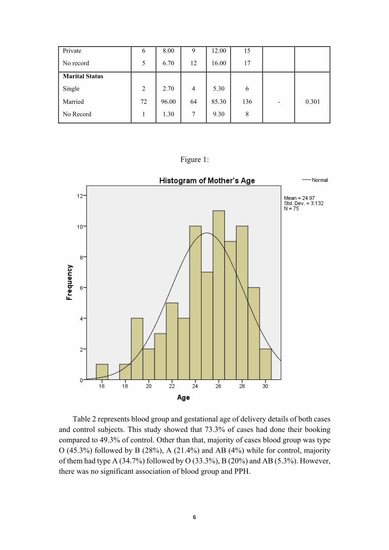
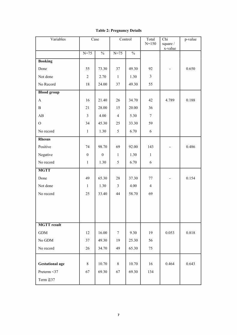
Table 2 represents blood group and gestational age of delivery details of both cases
and control subjects. This study showed that 73.3% of cases had done their booking
compared to 49.3% of control. Other than that, majority of cases blood group was type
O (45.3%) followed by B (28%), A (21.4%) and AB (4%) while for control, majority
of them had type A (34.7%) followed by O (33.3%), B (20%) and AB (5.3%). However,
there was no significant association of blood group and PPH.
77
Table 2: Pregnancy Details
Variables Case Control Total N=150
Chi square /
x-value
p-value
N=75 % N=75 %
92
3
55
-
0.650
Booking
Done
Not done
No Record
55
2
18
73.30
2.70
24.00
37
1
37
49.30
1.30
49.30
Blood group
A
B
AB
O
No record
16
21
3
34
1
21.40
28.00
4.00
45.30
1.30
26
15
4
25
5
34.70
20.00
5.30
33.30
6.70
42
36
7
59
6
4.789
0.188
Rhesus
Positive
Negative
No record
74
0
1
98.70
0
1.30
69
1
5
92.00
1.30
6.70
143
1
6
-
0.486
MGTT
Done
Not done
No record
49
1
25
65.30
1.30
33.40
28
3
44
37.30
4.00
58.70
77
4
69
-
0.154
MGTT result
GDM
No GDM
No record
12
37
26
16.00
49.30
34.70
7
19
49
9.30
25.30
65.30
19
56
75
0.053
0.818
Gestational age
Preterm <37
Term ≧37
8
67
10.70
69.30
8
67
10.70
69.30
16
134
0.464
0.643
88
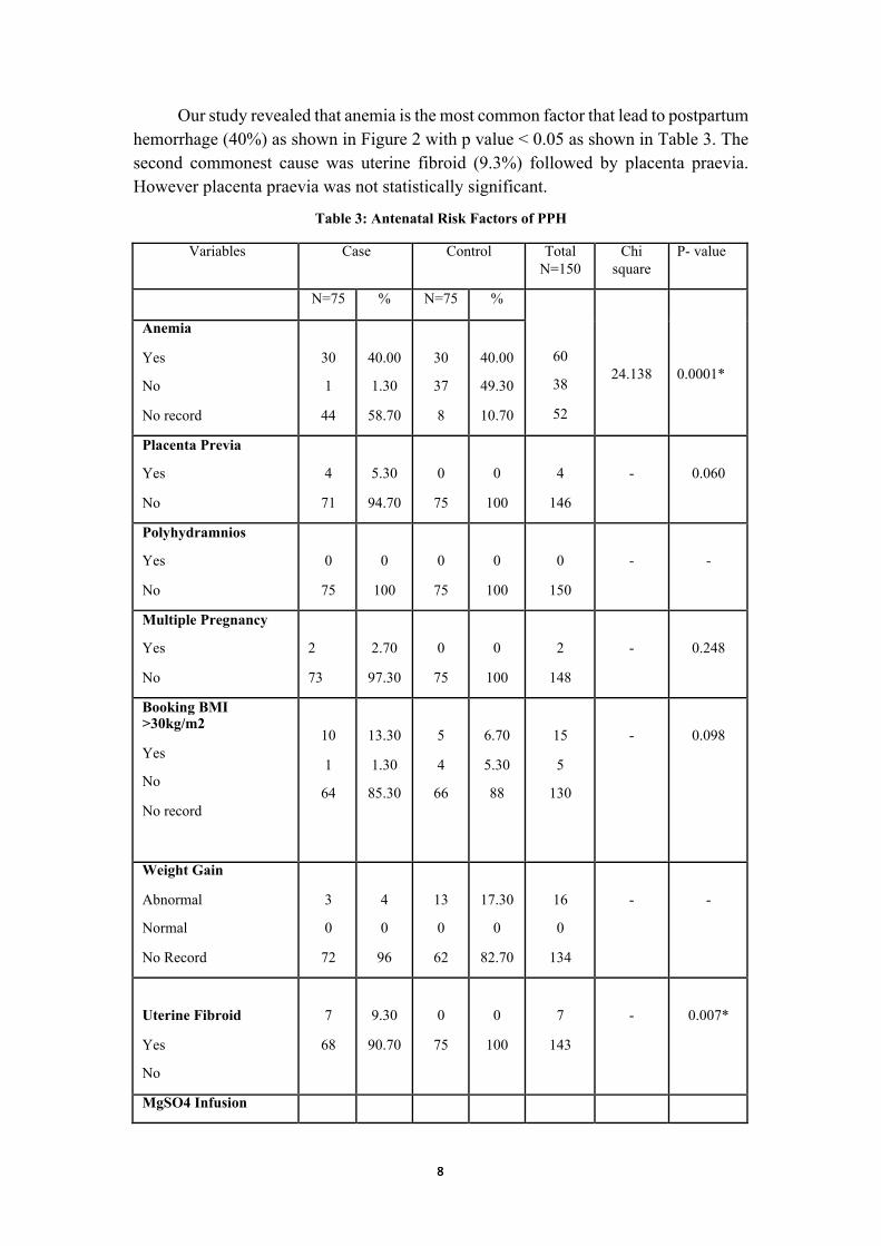
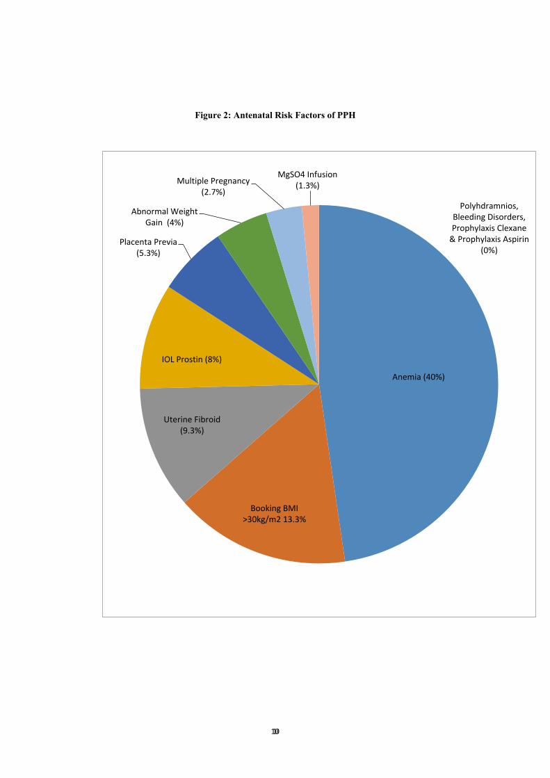
Our study revealed that anemia is the most common factor that lead to postpartum
hemorrhage (40%) as shown in Figure 2 with p value < 0.05 as shown in Table 3. The
second commonest cause was uterine fibroid (9.3%) followed by placenta praevia.
However placenta praevia was not statistically significant.
Table 3: Antenatal Risk Factors of PPH
Variables Case Control Total
N=150
Chi
square
P- value
N=75 % N=75 %
60
38
52
24.138
0.0001*
Anemia
Yes
No
No record
30
1
44
40.00
1.30
58.70
30
37
8
40.00
49.30
10.70
Placenta Previa
Yes
No
4
71
5.30
94.70
0
75
0
100
4
146
-
0.060
Polyhydramnios
Yes
No
0
75
0
100
0
75
0
100
0
150
-
-
Multiple Pregnancy
Yes
No
2
73
2.70
97.30
0
75
0
100
2
148
-
0.248
Booking BMI >30kg/m2
Yes
No
No record
10
1
64
13.30
1.30
85.30
5
4
66
6.70
5.30
88
15
5
130
-
0.098
Weight Gain
Abnormal
Normal
No Record
3
0
72
4
0
96
13
0
62
17.30
0
82.70
16
0
134
-
-
Uterine Fibroid
Yes
No
7
68
9.30
90.70
0
75
0
100
7
143
-
0.007*
MgSO4 Infusion
99
Yes
No
1
74
1.30
98.70
2
73
2.70
97.30
3
147
- 0.500
Thrombocytopenia/ bleeding disorders
Yes
No
0
75
0
100
0
75
0
100
0
150
-
-
Prophylaxis Clexane
Yes
No
0
75
0
100
0
75
0
100
0
150
-
-
Prophylaxis Aspirin
Yes
No
0
75
0
100
1
74
1.30
98.70
1
149
-
0.500
IOL with Prostin
Yes
No
6
69
8.00
92.00
11
64
14.70
85.30
17
133
0.1659
0.198
1010
Figure 2: Antenatal Risk Factors of PPH
Anemia(40%)
BookingBMI
>30kg/m213.3%
UterineFibroid
(9.3%)
IOLProstin(8%)
PlacentaPrevia
(5.3%)
AbnormalWeight
Gain(4%)
MultiplePregnancy
(2.7%)
MgSO4Infusion
(1.3%)
Polyhdramnios,
BleedingDisorders,
ProphylaxisClexane
&ProphylaxisAspirin
(0%)
1111
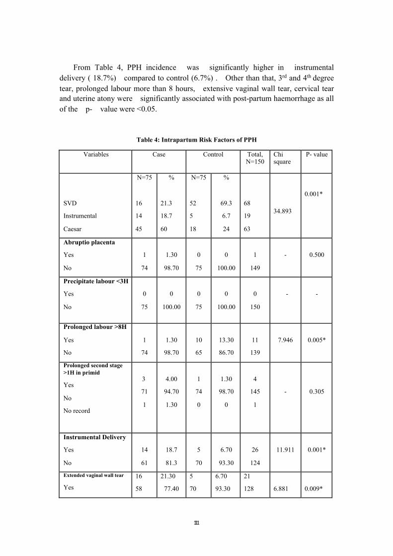
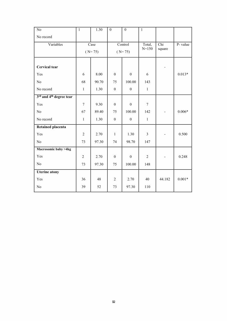
From Table 4, PPH incidence was significantly higher in instrumental
delivery ( 18.7%) compared to control (6.7%) . Other than that, 3rd and 4th degree
tear, prolonged labour more than 8 hours, extensive vaginal wall tear, cervical tear
and uterine atony were significantly associated with post-partum haemorrhage as all
of the p- value were <0.05.
Table 4: Intrapartum Risk Factors of PPH
Variables Case Control Total, N=150
Chi square
P- value
SVD
Instrumental
Caesar
N=75
16
14
45
%
21.3
18.7
60
N=75
52
5
18
%
69.3
6.7
24
68
19
63
34.893
0.001*
Abruptio placenta
Yes
No
1
74
1.30
98.70
0
75
0
100.00
1
149
-
0.500
Precipitate labour <3H
Yes
No
0
75
0
100.00
0
75
0
100.00
0
150
-
-
Prolonged labour >8H
Yes
No
1
74
1.30
98.70
10
65
13.30
86.70
11
139
7.946
0.005*
Prolonged second stage
>1H in primid
Yes
No
No record
3
71
1
4.00
94.70
1.30
1
74
0
1.30
98.70
0
4
145
1
-
0.305
Instrumental Delivery
Yes
No
14
61
18.7
81.3
5
70
6.70
93.30
26
124
11.911
0.001*
Extended vaginal wall tear
Yes
16
58
21.30
77.40
5
70
6.70
93.30
21
128
6.881
0.009*
1212
No
No record
1 1.30 0 0 1
Variables Case
( N= 75)
Control
( N= 75)
Total, N=150
Chi square
P- value
Cervical tear
Yes
No
No record
6
68
1
8.00
90.70
1.30
0
75
0
0
100.00
0
6
143
1
-
0.013*
3rd and 4th degree tear
Yes
No
No record
7
67
1
9.30
89.40
1.30
0
75
0
0
100.00
0
7
142
1
-
0.006*
Retained placenta
Yes
No
2
73
2.70
97.30
1
74
1.30
98.70
3
147
-
0.500
Macrosomic baby >4kg
Yes
No
2
73
2.70
97.30
0
75
0
100.00
2
148
-
0.248
Uterine atony
Yes
No
36
39
48
52
2
73
2.70
97.30
40
110
44.182
0.001*
1313
Table 4a : Uterine atony and anemia
Anemia
Total Yes No
No
Record
Uterine
Atony
Yes 14 1 21 36
No 12 1 26 39
Total 26 2 47 75
Crosstab
Count
Anemia
Total Case Control No Record
Uterine
atony
Yes 14 1 0 15
No 12 29 0 41
Total 26 30 0 56
P= 0.00021 ( P< 0.05)
Table 4a (i) : Uterine atony and mode of delivery
Crosstab
Count (case)
Uterine Atony
Total Yes No
Method of
Delivery
SVD 5 11 16
Forceps 1 2 3
Vacuum 3 8 11
ELLSCS 0 1 1
EMLSCS 27 17 44
Total 36 39 75
1414
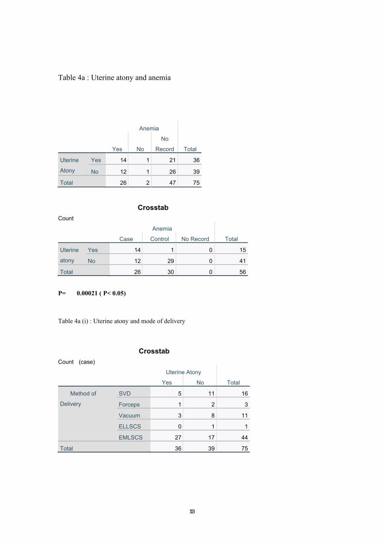
Table 4a revealed that anemia was profound most in uterine atony, and if compared
with control, anemia is statistically significant to cause uterine atony in primigravida .
In Table 4a (i) , uterine atony most commonly occurred in patient delivered by
Caesarean section .
Table 4b : Risk factors of uterine atony and their P- value
Risk factors of uterine atony Frequency of PPH P - value
Macrosomic baby 1 0.480
Uterine fibroid 2/3 0.457
MgSO4 ½ ( 50%) 0.733
Polyhydramnios 0 -
Multiple pregnancy 0 -
Prolonged labour 7/11 0.261
Prolonged second stage 2/4 ( 50.0%) 0.574
Induction of labour with 1
prostin
4/8 ( 50.0%) 0.598
Induction of labour with 2
prostins
1/5 ( 20.0%) 0.195
Induction of labour with 3
prostins
0 -
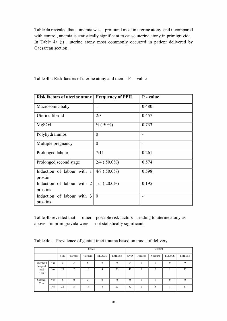
Table 4b revealed that other possible risk factors leading to uterine atony as
above in primigravida were not statistically significant.
Table 4c: Prevalence of genital tract trauma based on mode of delivery
Cases Control
SVD Forceps Vacuum ELLSCS EMLSCS SVD Forceps Vacuum ELLSCS EMLSCS
Extended
Vaginal
wall
Tear
Yes 7 3 6 0 0 5 0 0 0 0
No 19 2 10 4 23 47 0 5 1 17
Cervical
Tear
Yes 4 0 2 0 0 0 0 0 0 0
No 22 5 14 4 23 52 0 5 1 17
1515
3rd/4rd
Degree
Tear
Yes 5 0 2 0 0 0 0 0 0 0
No 21 5 14 4 23 52 0 5 1 17
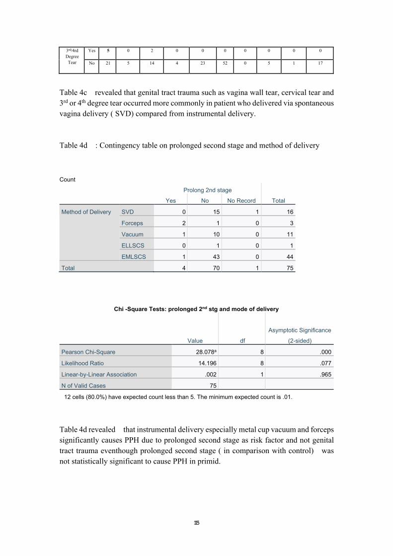
Table 4c revealed that genital tract trauma such as vagina wall tear, cervical tear and
3rd or 4th degree tear occurred more commonly in patient who delivered via spontaneous
vagina delivery ( SVD) compared from instrumental delivery.
Table 4d : Contingency table on prolonged second stage and method of delivery
Count
Prolong 2nd stage
Total Yes No No Record
Method of Delivery SVD 0 15 1 16
Forceps 2 1 0 3
Vacuum 1 10 0 11
ELLSCS 0 1 0 1
EMLSCS 1 43 0 44
Total 4 70 1 75
Chi -Square Tests: prolonged 2nd stg and mode of delivery
Value df
Asymptotic Significance
(2-sided)
Pearson Chi-Square 28.078a 8 .000
Likelihood Ratio 14.196 8 .077
Linear-by-Linear Association .002 1 .965
N of Valid Cases 75
12 cells (80.0%) have expected count less than 5. The minimum expected count is .01.
Table 4d revealed that instrumental delivery especially metal cup vacuum and forceps
significantly causes PPH due to prolonged second stage as risk factor and not genital
tract trauma eventhough prolonged second stage ( in comparison with control) was
not statistically significant to cause PPH in primid.
1616
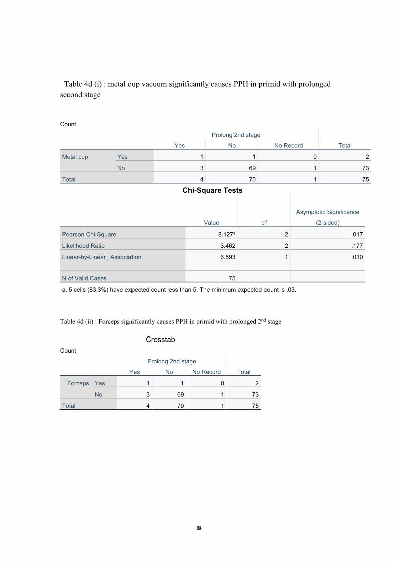
Table 4d (i) : metal cup vacuum significantly causes PPH in primid with prolonged
second stage
Count
Prolong 2nd stage
Total Yes No No Record
Metal cup Yes 1 1 0 2
No 3 69 1 73
Total 4 70 1 75
Chi-Square Tests
Value df
Asymptotic Significance
(2-sided)
Pearson Chi-Square 8.127a 2 .017
Likelihood Ratio 3.462 2 .177
Linear-by-Linear j Association 6.593 1 .010
N of Valid Cases 75
a. 5 cells (83.3%) have expected count less than 5. The minimum expected count is .03.
Table 4d (ii) : Forceps significantly causes PPH in primid with prolonged 2nd stage
Crosstab
Count
Prolong 2nd stage
Total Yes No No Record
Forceps Yes 1 1 0 2
No 3 69 1 73
Total 4 70 1 75
1717
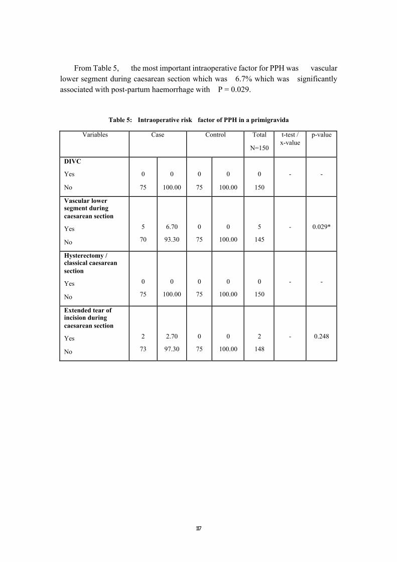
From Table 5, the most important intraoperative factor for PPH was vascular
lower segment during caesarean section which was 6.7% which was significantly
associated with post-partum haemorrhage with P = 0.029.
Table 5: Intraoperative risk factor of PPH in a primigravida
Variables Case Control Total
N=150
t-test /
x-value
p-value
DIVC
Yes
No
0
75
0
100.00
0
75
0
100.00
0
150
-
-
Vascular lower segment during
caesarean section
Yes
No
5
70
6.70
93.30
0
75
0
100.00
5
145
-
0.029*
Hysterectomy / classical caesarean
section
Yes
No
0
75
0
100.00
0
75
0
100.00
0
150
-
-
Extended tear of incision during
caesarean section
Yes
No
2
73
2.70
97.30
0
75
0
100.00
2
148
-
0.248
1818
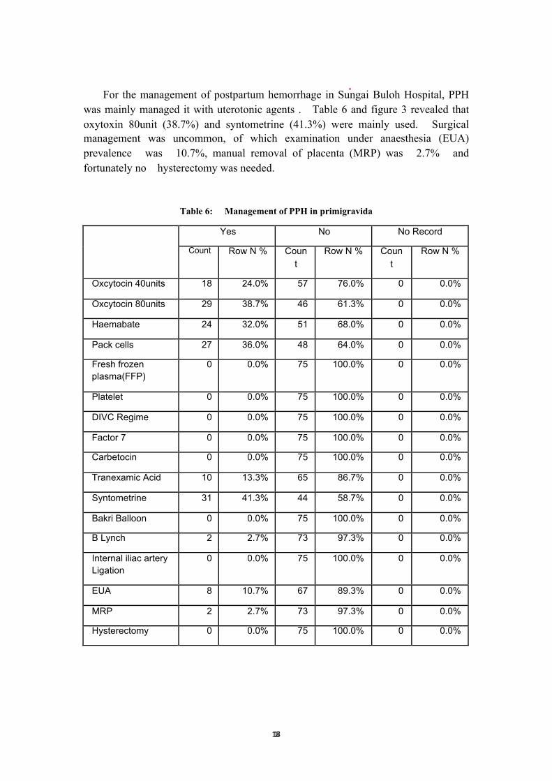
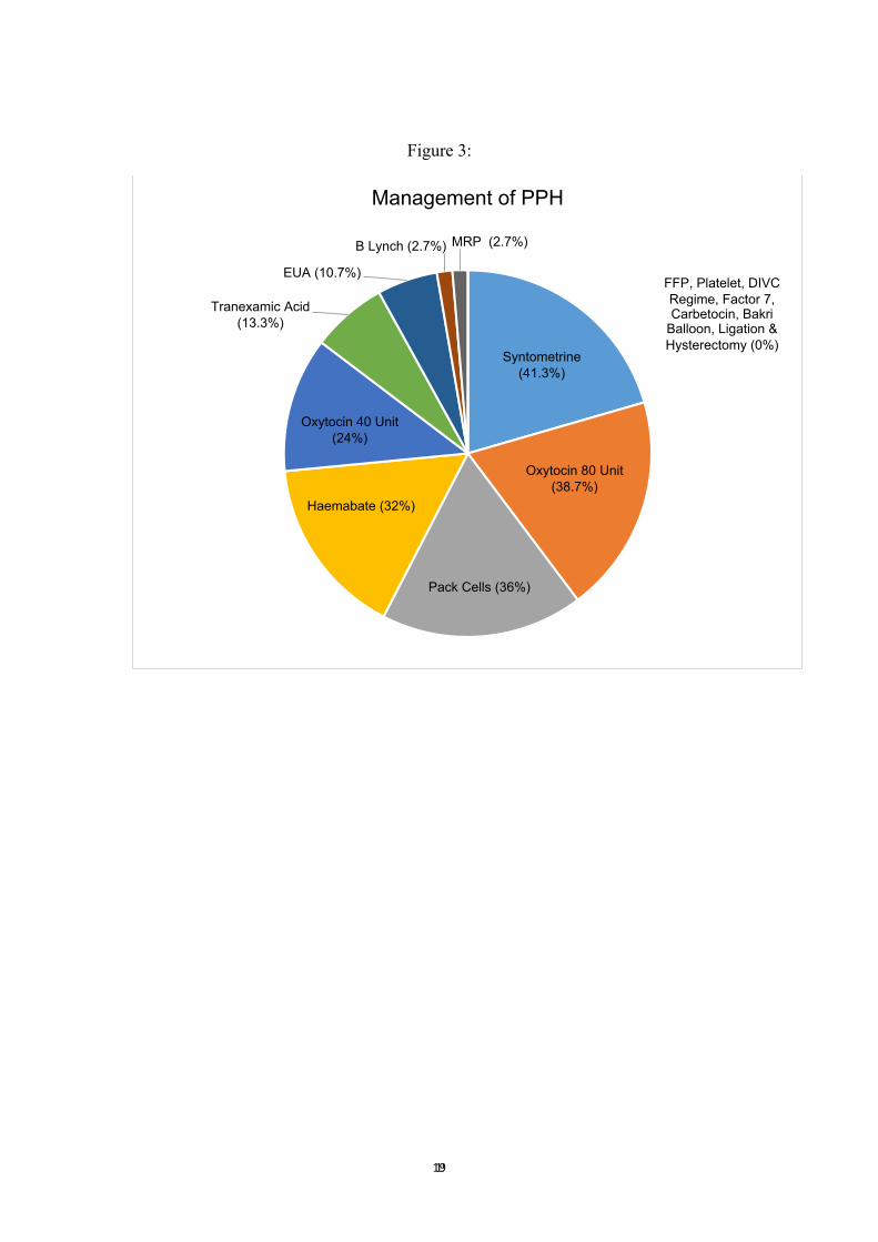
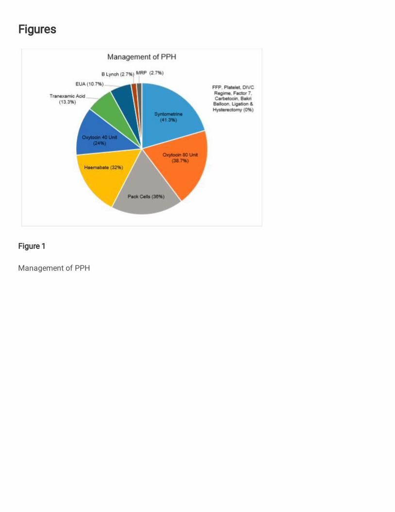
For the management of postpartum hemorrhage in Sungai Buloh Hospital, PPH
was mainly managed it with uterotonic agents . Table 6 and figure 3 revealed that
oxytoxin 80unit (38.7%) and syntometrine (41.3%) were mainly used. Surgical
management was uncommon, of which examination under anaesthesia (EUA)
prevalence was 10.7%, manual removal of placenta (MRP) was 2.7% and
fortunately no hysterectomy was needed.
Table 6: Management of PPH in primigravida
Yes No No Record
Count Row N % Coun
t
Row N % Coun
t
Row N %
Oxcytocin 40units 18 24.0% 57 76.0% 0 0.0%
Oxcytocin 80units 29 38.7% 46 61.3% 0 0.0%
Haemabate 24 32.0% 51 68.0% 0 0.0%
Pack cells 27 36.0% 48 64.0% 0 0.0%
Fresh frozen
plasma(FFP)
0 0.0% 75 100.0% 0 0.0%
Platelet 0 0.0% 75 100.0% 0 0.0%
DIVC Regime 0 0.0% 75 100.0% 0 0.0%
Factor 7 0 0.0% 75 100.0% 0 0.0%
Carbetocin 0 0.0% 75 100.0% 0 0.0%
Tranexamic Acid 10 13.3% 65 86.7% 0 0.0%
Syntometrine 31 41.3% 44 58.7% 0 0.0%
Bakri Balloon 0 0.0% 75 100.0% 0 0.0%
B Lynch 2 2.7% 73 97.3% 0 0.0%
Internal iliac artery
Ligation
0 0.0% 75 100.0% 0 0.0%
EUA 8 10.7% 67 89.3% 0 0.0%
MRP 2 2.7% 73 97.3% 0 0.0%
Hysterectomy 0 0.0% 75 100.0% 0 0.0%
1919
Figure 3:
Syntometrine
(41.3%)
Oxytocin 80 Unit
(38.7%)
Pack Cells (36%)
Haemabate (32%)
Oxytocin 40 Unit
(24%)
Tranexamic Acid
(13.3%)
EUA (10.7%)
B Lynch (2.7%) MRP (2.7%)
FFP, Platelet, DIVC
Regime, Factor 7, Carbetocin, Bakri
Balloon, Ligation &
Hysterectomy (0%)
Management of PPH
2020
DISCUSSION
In this case-control study, we evaluated risk factors for PPH in a primigravida.
Assessing risk factors in retrospective manner is a limitation in this study. In order to
minimize selection bias, we selected a number of cases of PPH according to our sample
size and a random sample of controls from the same source population. There is a
possibility that some cases were misclassified. Secondly, our sample size may not be
sufficient to evaluate potential risk factors that are less frequent. Another limitation was
limited by the missing data. Measures should be taken to ensure data entry is complete
for a comprehensive analysis.
In this study, we limited the age group from 17-35 years old which differs to
other study as we are focusing on primigravida who are mostly young. From our
analysis, the commonest age group for PPH with primigravida were from age group
of 26-30 years (41.3%) followed by age group of 21-25 years old. From the socio-
demographic details, patient’s occupation contributed to risk factors for PPH. A
definition of woman’s health as defined by Koblinsky et al l[10] stated “A woman’s
health is her total wellbeing, not determined solely by biological factors and
reproduction, but also the effects of work load, nutrition, stress, war and migration,
among others” [11]. Therefore, from the results, self-employed was the highest factor to
contribute to PPH which was similar to study done by Gregory Hella Ekane et al [12]
followed by occupation in government sector, private and least housewife. This might
be due to financial instability among self employed primigravida which could lead to
stress.
The strongest risk factor during antenatal in our study was anemia during
pregnancy which is same as the finding in Chandrika et al and Tan et al[6,7]. An
explanation for the association between anemia and post-partum hemorrhage is uterine
muscle hypoxia. Hemoglobin is the oxygen-carrying protein in blood. Anemia is the
state of a reduction in hemoglobin levels, and therefore, in the blood’s oxygen-carrying
capacity. After hours of maximal exertion, due to insufficient oxygen, the uterus will
have inefficient uterine contractility hence leading to haemorrhage . Early detection
of anaemia and timely correction is important to reduce morbidity associated with PPH.
As shown in Table 4a , anemia was significantly associated with uterine atony.
Another risk factor that was significantly related to PPH in primigravida was
presence of uterine fibroid. Based on study by Hee Joong Lee [17], the association
between fibroid and postpartum hemorrhage was due to the fibroid will distort the
uterine cavity and interfere with myometrial contractions leading to uterine atony and
postpartum hemorrhage. Uterine dysfunction before the onset of active labor, such as
uterine fibroid, overdistended uterus, scarred uterus, and infection, may result in delay
of all stages of labor and thereby cause PPH [17]. To prevent PPH in patient with uterine
fibroid , myomectomy can be considered prior to pregnancy. Several studies have
2121
reported that antepartum myomectomy can be safely performed in the first and second
trimester of pregnancy [18].
According to Chandrika et al [6] delivery by emergency caesarean section carries
the highest risk for severe post-partum hemorrhage which is consistent with this study,
of which 45 out of 75 or 60% primigravida who delivered by caesarean section
had PPH. Uterine atony contributed to 60% of blood loss in patient who delivered by
Caesarean section as shown in Table 4a. Another study done by Waheed et al [18]
also highlighted that Caesarean section and instrumental deliveries are more likely to
be complicated by PPH. In this study, instrumental delivery carried higher prevalence
of PPH ( p < 0.05) especially metal cup and forceps in primigravida who had prolonged
second stage as shown in Table 4d . Hashim N et al revealed that primigravida has
higher risk of prolonged second stage. [5]
In this study, we found that the duration of active labor of more than 8 hours was
associated with an increased risk of PPH ( p < 0.05) Nyflot et. al [16] stated that
long lasting labor, including a prolonged first stage, may increase the risk of PPH by
causing uterine atony due to prolonged regular contractions over several hours of
labor will exhaust the uterine muscles and thereby reduce their contractility over time,
causing uterine dysfunction [17].
Genital tract trauma that occurred during childbirth could lead to postpartum
hemorrhage and in this study, we had identified that extensive vaginal wall tear,
cervical tear , 3rd as well as 4th degree tear ( p= 0.006) were the risk factors to
postpartum hemorrhage in primigravida. A study from Smith et.al , showed that these
tears were closely related to shoulder dystocia, episiotomy, prolonged second stage
of labour, macrosomic baby and instrumental delivery [13]. In nulliparous women at
term, external cervical os remains stationary during the cervical shortening, but it
quickly evolves once the cervical effacement is completed [14].
The next risk factor of postpartum hemorrhage in primigravida that was identified
in this study was 3rd and 4th degree tear. Third and 4th degree tear occurs in about
3 in 100 women having a vaginal birth. It is slightly more common with a first vaginal
birth, occurring in 6 in 100 women, compared with 2 in 100 women who have had a
vaginal birth previously [11]. This is because the age related changes in the connective
tissue decrease the ability of cervical, vaginal and perineal muscles to stretch as needed
during delivery resulting in greater trauma to tissues, prolonged labor and diminished
uterine contractility after delivery [13]
However surprisingly in this study, genital tract trauma majority occurred in patient
who delivered by spontaneous vaginal delivery, not instrumental delivery as shown in
Table 4c.
2222
During intrapartum, uterine atony (48%) was the commonest cause of post-partum
hemorrhage, this is the same as the finding in previous studies done [8-10]. After the
placenta is delivered, these contractions help compress the bleeding vessels in the area
where the placenta was attached. If the uterus has inefficient uterine contraction,
bleeding from blood vessels tends to occur that can lead to post-partum
hemorrhage. Uterine atony is treated initially by uterine massage, followed by
uterotonic agents such as oxyctocin, syntometrine or hemabate that promote uterine
contraction. Hence continuous training should be provided to all doctors and nursing
staff on how to managing uterine atony as the uterus can be salvage easily.
The last risk factors that we had identified which has p value of < 0.05 was
vascular lower segment during caesarean section ; 5 cases (p=0.029). A thick lower
segment is usually encountered in elective cesarean sections performed before the onset
of labor. Proportionate to the thickness, the incision will have to be larger and hence
blood loss will be greater. Trying to arrest bleeding at that point with cautery or ligation
is not advisable. One has to proceed to deliver the fetus and then tackle the bleeding. A
thick lower segment may demand use of forceps or ventouse to facilitate delivery of the
fetus [15].
Syntometrine administration is most commonly used to manage post-partum
hemorrhage in this study in which there were 31 cases with 41.3% followed by
oxcytocin 80 unit with 29 cases (38.7%). There are significant variations in
practice but systematic review of all the available uterotonics for prevention of PPH
found oxytocin to be the first choice and a recent Cochrane review supported this
recommendation [20]. Syntometrine which is combination of ergometrine and oxytocin
is more effective than oxytocin alone in preventing blood loss, however their side
effects are more common, with increased levels of hypertension, nausea, and vomiting
[19]. According to WHO [4], the use of oxytocin for active management of the third stage
of labour is strongly recommended, because it reduces post-partum hemorrhage by
more than 60%.
In this study, there was no maternal death in primigravida with PPH as Sungai
Buloh Hospital has well-trained personnel and good facilities of managing
postpartum haemorrhage .
2323
CONCLUSION
In this study, the significant risk factors for post-partum hemorrhage in
primigravida were age, self-employed, uterine fibroid, prolonged labor for more
than 8 hours, antenatal anemia, instrumental delivery with prolonged second stage,
extensive vaginal wall tear, cervical tear, 3rd and 4th degree tear, uterine atony and
vascular lower segment . Anemia significantly causes uterine atony and hence should
be corrected antenatally. In terms of instrumental delivery, silicone vacuum less
commonly associated with PPH as compared from forceps and metal cup vacuum.
Further studies are needed for explanation of higher incidence of genital tract
trauma in spontaneous vaginal delivery compared to instrumental delivery . Risk
factors identified in our study could be considered in future studies such as examining
risk prediction models for post-partum hemorrhage.
ABBREVIATIONS
BMI Body mass index
CEMD Confidential Enquiry into Maternal Deaths (CEMD)
DIVC Disseminated Intravascular Coagulopathy
EUA Examination under Anaesthesia
HSgB Sungai Buloh Hospital
IOL Induction of labour
MMR Maternal Mortality Ratio
MRP Manual Removal of Placenta
PPH Postpartum Haemorrhage
2424
Ethics approval and consent to participate
This research has ethics of approval from Ethics Committee of University Technology
of MARA ( UiTM) .
Consent for publication
Not applicable
Availability of data and materials
The datasets used and/or analyzed during the current study are available from the author
on reasonable request
Disclosure
This research study received no specific funding
Conflict of interest
Authors have no conflict of interest to declare.
Funding
This research received no funding
Acknowledgement
I would like to thank my UiTM medical students who helped us in collecting data,
entering and analysing data in SPSS. They are Suhairy Osman, Nor Syuhada
Roszianti, Aida Sofi Azizan, and Zatul Syalihah Mohamed. I would also like to thank
Dr Mariam Mohamad Public Health Physician for calculating the sample size and Dr
Norashikin Abdul Fuad, the Head of Department of Obstetric & Gynaecology for her
moral support and my family for their endless support for me to complete this research.
2525
REFERENCES
1. Saxena R., Bedside Obstetrics and Gynaecology 1st Edition; 2010
2. Report on the Confidential Enquiries into Maternal Deaths in Malaysia; 2006-2008
3. World Health Organization, Maternal and Perinatal Death Reviews in UK, Retrieved
January 2 2018, from
http://www.who.int/maternal_child_adolescent/epidemiology/maternal-death-
surveillance/case-studies/united-kingdom
4. World Health Organization, Malaysia’s Experience with Maternal Deaths,
Retrieved January 24, 2018, from
http://www.who.int/maternal_child_adolescent/epidemiology/maternal-death-
surveillance/case-studies/malaysia/en/
5. Hashim N, Naqvi S, Khanam M, Jafry HF; Primiparity as an intrapartum obstetric risk
factor, J Pak Med Association, vol 62, 2012.
https://www.ncbi.nlm.nih.gov/m/pubmed/23866518/
6. Chandrika S ; A study of prevalence, causes, risk factors and outcome of severe
obstetrics haemorrhage , BioMedical Journal. 2008. ISSN 2320-4818
7. Tan A.C., Leong E. W., Chua A. C., Moy F. M; Racial variations in booking
haemoglobin of primigravidae in Malaysia: A prospective study, BMC Research
Notes, 2013. http://www.biomedcentral.com/1756-0500/6/173
8. Tsu V. D. , Postpartum haemorrhage in Zimbabwe: A risk factor analysis. , Obstet
Gynaecol Journal, 1993. DOI: 10.1111/j.1471-0528.1993.tb12974.x
9. Carlos Montufar Rueda , Laritza Rodriguez, Jose Douglas Jarquin , Alejandra Barboza
Flor Marin, Severe Postpartum Hemorrhage from Uterine Atony: A Multicentric Study.
Journal of Pregnancy, 2013. http://dx.doi.org/10.1155/2013/525914
10. Koblinsky M., Campbell O. , Harlow S ,The Health of Women: A Global
Perspective, Journal of Public Health Policy, 1994.
https://doi.org/10.1177%2F027046769401400152
11. Third- or fourth-degree tear during childbirth, Green top guideline 29, 2015.
https://www.rcog.org.uk
12. Halle-Ekane G., Emade, F., Bechem, N., Palle, J., Fongaing, D., Essome, H., &
Fomulu, N. Prevalence and Risk Factors of Primary Postpartum Hemorrhage after
2626
Vaginal Deliveries in the Bonassama District Hospital, Cameroon. International
Journal of Tropical Disease& Health, 13(2), 1-12, 2015. DOI:
10.9734/IJTDH/2016/23078
13. Smith, L. A., Price, N., Simonite, V., Burns E. E , Incidence of and risk factors for
perineal trauma: A prospective observational study ; BMC Pregnancy and
Childbirth,13(1), 2013. doi: 10.1186/1471-2393-13-59
14. Djokovic, D., Costa, C., Martins, A., Abushad, S, Spontaneous delivery
through a cervical tear without cervical os dilatation. Clinical Case Reports,3(1),
3-6, 2014
15. Sapkal, R. Chapter-03 Internal Iliac Artery: A Study on Cadaver. Surgical Skills
on Internal Iliac Artery Ligation for Controlling Postpartum & Pelvic
Hemorrhage,19-38, 2015
16. Nyfløt, L. T., Stray-Pedersen, B., Forsén, L., & Vangen, S. , Duration of labor and
the risk of severe postpartum hemorrhage: A case-control study. Plos One,12(4),
2017.
17. Joong, L. H., Norwitz, E. R., & Shaw J , Contemporary Management of Fibroids
in Pregnancy. Rev Obstet Gynecol,3(1), 20-27, 2010.
18. Waheed, G., Toheed R., Mansha, M, Comparison of Causes of Postpartum
Haemorrhage Following Vaginal Deliveries and Caesarean Sections in a Tertiary
Care Hospital of Pakistan, Pakistan Journal of Medical and Health, 2013.
19. Weeks A, The prevention and treatment of postpartum haemorrhage: What do
we know, and where do we go to next? BJOG: An International Journal of
Obstetrics & Gynaecology,122(2), 2014.
20. Pavord, S., Maybury, H, How I treat postpartum hemorrhage?, American Society
of Haematology, 2015

2727

2828
Figures
Figure 1
Management of PPH
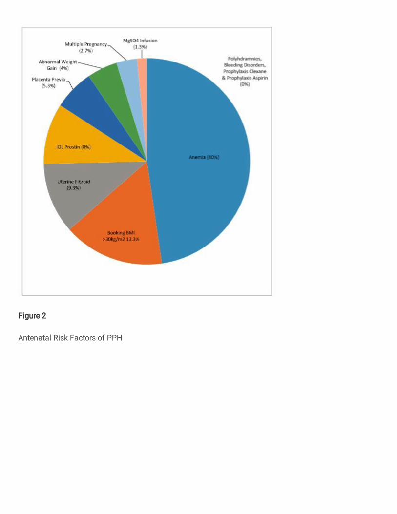
Figure 2
Antenatal Risk Factors of PPH
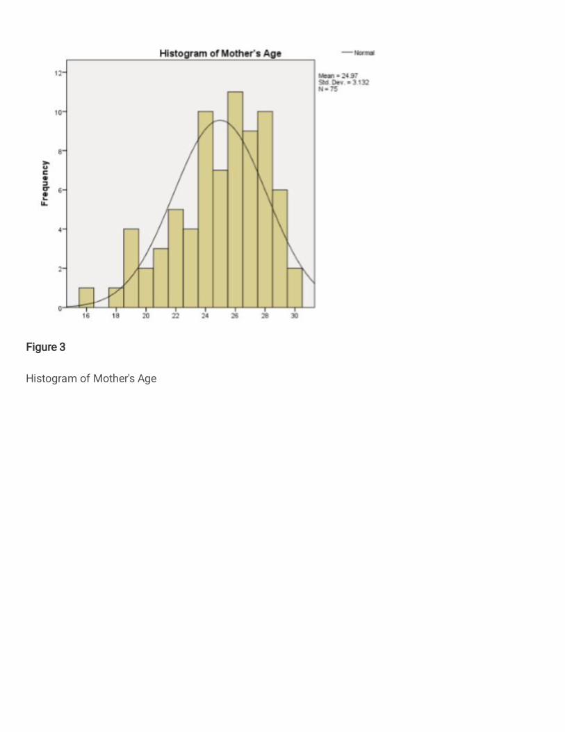
Figure 3
Histogram of Mother's Age
Related Documents