APPLIED AND ENVIRONMENTAL MICROBIOLOGY, Aug. 2009, p. 5273–5283 Vol. 75, No. 16 0099-2240/09/$08.000 doi:10.1128/AEM.00774-09 Copyright © 2009, American Society for Microbiology. All Rights Reserved. Inactivation of PadR, the Repressor of the Phenolic Acid Stress Response, by Molecular Interaction with Usp1, a Universal Stress Protein from Lactobacillus plantarum, in Escherichia coli Je ´ro ˆme Gury,†‡ He ´le `ne Seraut,† Ngoc Phuong Tran,§ Lise Barthelmebs,¶ Ste ´phanie Weidmann, Patrick Gervais, and Jean-Franc ¸ois Cavin* Laboratoire GPMA EA4181 IFR92, AgroSup Dijon ENSBANA, 1 Esplanade Erasme, Universite ´ de Bourgogne, F-21000 Dijon, France Received 6 April 2009/Accepted 12 June 2009 The phenolic acid decarboxylase gene padA is involved in the phenolic acid stress response (PASR) in gram-positive bacteria. In Lactobacillus plantarum, the padR gene encodes the negative transcriptional regu- lator of padA and is cotranscribed with a downstream gene, usp1, which encodes a putative universal stress protein (USP), Usp1, of unknown function. The usp1 gene is overexpressed during the PASR. However, the role and the mechanism of action of the USPs are unknown in gram-positive bacteria. Therefore, to gain insights into the role of USPs in the PASR; (i) a usp1 deletion mutant was constructed; (ii) the two genes padR and usp1 were coexpressed with padA under its own promoter as a reporter gene in Escherichia coli; and (iii) molecular in vitro interactions between the PadR, Usp1, and the padA promoter were studied. Although the usp1 mutant strain retained phenolic acid-dependent PAD activity, it displayed a greater sensitivity to strong acidic conditions compared to that of the wild-type strain. PadR cannot be inactivated directly by phenolic acid in E. coli recombinant cultures but is inactivated by Usp1 when the two proteins are coexpressed in E. coli. The PadR inactivation observed in recombinant E. coli cells was supported by electrophoretic mobility shift assays. Although Usp1 seems not to be absolutely required for the PASR, its capacity to inactivate PadR indicates that it could serve as an important mediator in acid stress response mechanisms through its capacity to interact with transcriptional regulators. Phenolic acids are essential plant compounds involved in the molecular bonds between cellulose, hemicellulose, and lignin in the cell wall. They are significant in the mammalian diet (32). Their specific structure confers important biological ac- tivities. They have free radical scavenging properties that can trigger antimutagenetic effects (14) but that can also lead to pro-oxidant DNA degradation mediated by divalent cations (37). Recently, it was shown that some phenolic acids that induce the phenolic acid stress response (PASR) in gram- positive bacteria regulate the expression of genes of the type III secretion system required for virulence by the plant patho- gen bacterium Dickeya dadantii (23). Phenolic acids such as p-coumaric, ferulic, and caffeic acids are toxic for numerous gram-positive bacteria such as Bacillus subtilis (33), Pediococ- cus pentosaceus (4), and Lactobacillus plantarum (16) under acidic condition. Phenolic acid decarboxylase (PAD) activity in these bacteria is a detoxifying system specifically and strongly induced by these chemicals. Two genes involved in the PASR have been characterized: padA and padR. The padA gene (named padC in B. subtilis) encodes the PadA enzyme and padR encodes the PadR transcriptional repressor. Deletion of padA leads to growth inhibition in the presence of phe- nolic acids, especially at low pH (3), while deletion of padR leads to constitutive overexpression of padA (16) and, con- sequently, to high resistance to phenolic acids. Although the PadR-like proteins constitute a regulatory family of more than 1350 putative members in the bacterial genome data- base (accession no. Pfam PF03551), only a few of them are currently under investigation. Among them, the best char- acterized is AphA, the repressor of the penicillin amidase gene (21) and the transcriptional activator of the virulence cascade of Vibrio cholerae, which acts in consortium with the coactivator AphB (20). Another member of this family, LmrR, acts as a repressor of the multidrug recognition operon lmrCD in Lactococcus lactis, and its molecular struc- ture in the presence or absence of H33342 or daunomycin drugs has been analyzed (26). As well as for the other PadR like proteins family, the way by which PadR is inactivated remains also poorly elucidated. Concerning the PASR, we have previously demonstrated that in growing cultures of a recombinant Escherichia coli strain expressing PadR from P. pentosaceus and in L. plantarum, PadR could not be inacti- * Corresponding author. Mailing address: Laboratoire GPMA EA4181 IFR92, AgroSup Dijon ENSBANA, 1 Esplanade Erasme, Universite ´ de Bourgogne, F-21000 Dijon, France. Phone: (33) 3 80 39 66 72. Fax: (33) 3 80 39 66 40. E-mail: [email protected]. ‡ Present address: UMR CNRS 5557, Universite ´ Lyon I, 69622 Villeurbanne, France. § Present address: IGM Ba ˆt. 409 Universite ´ Paris-Sud 11, F-91405 Orsay Cedex, France. ¶ Present address: IMAGES EA 4218, Centre de Phytopharmacie, Universite ´ de Perpignan Via Domitia, 52 Ave. Paul Alduy, 66860 Perpignan Cedex, France. Present address: Laboratoire REVV, IFR92, Institut Jules Guyot, Universite ´ de Bourgogne, 21000 Dijon, France. † J.G. and H.S. contributed equally to this study. Published ahead of print on 19 June 2009. 5273

Welcome message from author
This document is posted to help you gain knowledge. Please leave a comment to let me know what you think about it! Share it to your friends and learn new things together.
Transcript

APPLIED AND ENVIRONMENTAL MICROBIOLOGY, Aug. 2009, p. 5273–5283 Vol. 75, No. 160099-2240/09/$08.00�0 doi:10.1128/AEM.00774-09Copyright © 2009, American Society for Microbiology. All Rights Reserved.
Inactivation of PadR, the Repressor of the Phenolic Acid StressResponse, by Molecular Interaction with Usp1, a Universal
Stress Protein from Lactobacillus plantarum, inEscherichia coli�
Jerome Gury,†‡ Helene Seraut,† Ngoc Phuong Tran,§ Lise Barthelmebs,¶ Stephanie Weidmann,�Patrick Gervais, and Jean-Francois Cavin*
Laboratoire GPMA EA4181 IFR92, AgroSup Dijon ENSBANA, 1 Esplanade Erasme,Universite de Bourgogne, F-21000 Dijon, France
Received 6 April 2009/Accepted 12 June 2009
The phenolic acid decarboxylase gene padA is involved in the phenolic acid stress response (PASR) ingram-positive bacteria. In Lactobacillus plantarum, the padR gene encodes the negative transcriptional regu-lator of padA and is cotranscribed with a downstream gene, usp1, which encodes a putative universal stressprotein (USP), Usp1, of unknown function. The usp1 gene is overexpressed during the PASR. However, the roleand the mechanism of action of the USPs are unknown in gram-positive bacteria. Therefore, to gain insightsinto the role of USPs in the PASR; (i) a usp1 deletion mutant was constructed; (ii) the two genes padR and usp1were coexpressed with padA under its own promoter as a reporter gene in Escherichia coli; and (iii) molecularin vitro interactions between the PadR, Usp1, and the padA promoter were studied. Although the usp1 mutantstrain retained phenolic acid-dependent PAD activity, it displayed a greater sensitivity to strong acidicconditions compared to that of the wild-type strain. PadR cannot be inactivated directly by phenolic acid in E.coli recombinant cultures but is inactivated by Usp1 when the two proteins are coexpressed in E. coli. The PadRinactivation observed in recombinant E. coli cells was supported by electrophoretic mobility shift assays.Although Usp1 seems not to be absolutely required for the PASR, its capacity to inactivate PadR indicates thatit could serve as an important mediator in acid stress response mechanisms through its capacity to interactwith transcriptional regulators.
Phenolic acids are essential plant compounds involved in themolecular bonds between cellulose, hemicellulose, and ligninin the cell wall. They are significant in the mammalian diet(32). Their specific structure confers important biological ac-tivities. They have free radical scavenging properties that cantrigger antimutagenetic effects (14) but that can also lead topro-oxidant DNA degradation mediated by divalent cations(37). Recently, it was shown that some phenolic acids thatinduce the phenolic acid stress response (PASR) in gram-positive bacteria regulate the expression of genes of the typeIII secretion system required for virulence by the plant patho-gen bacterium Dickeya dadantii (23). Phenolic acids such asp-coumaric, ferulic, and caffeic acids are toxic for numerousgram-positive bacteria such as Bacillus subtilis (33), Pediococ-
cus pentosaceus (4), and Lactobacillus plantarum (16) underacidic condition. Phenolic acid decarboxylase (PAD) activity inthese bacteria is a detoxifying system specifically and stronglyinduced by these chemicals. Two genes involved in the PASRhave been characterized: padA and padR. The padA gene(named padC in B. subtilis) encodes the PadA enzyme andpadR encodes the PadR transcriptional repressor. Deletionof padA leads to growth inhibition in the presence of phe-nolic acids, especially at low pH (3), while deletion of padRleads to constitutive overexpression of padA (16) and, con-sequently, to high resistance to phenolic acids. Although thePadR-like proteins constitute a regulatory family of morethan 1350 putative members in the bacterial genome data-base (accession no. Pfam PF03551), only a few of them arecurrently under investigation. Among them, the best char-acterized is AphA, the repressor of the penicillin amidasegene (21) and the transcriptional activator of the virulencecascade of Vibrio cholerae, which acts in consortium with thecoactivator AphB (20). Another member of this family,LmrR, acts as a repressor of the multidrug recognitionoperon lmrCD in Lactococcus lactis, and its molecular struc-ture in the presence or absence of H33342 or daunomycindrugs has been analyzed (26). As well as for the other PadRlike proteins family, the way by which PadR is inactivatedremains also poorly elucidated. Concerning the PASR, wehave previously demonstrated that in growing cultures of arecombinant Escherichia coli strain expressing PadR from P.pentosaceus and in L. plantarum, PadR could not be inacti-
* Corresponding author. Mailing address: Laboratoire GPMAEA4181 IFR92, AgroSup Dijon ENSBANA, 1 Esplanade Erasme,Universite de Bourgogne, F-21000 Dijon, France. Phone: (33) 3 80 3966 72. Fax: (33) 3 80 39 66 40. E-mail: [email protected].
‡ Present address: UMR CNRS 5557, Universite Lyon I, 69622Villeurbanne, France.
§ Present address: IGM Bat. 409 Universite Paris-Sud 11, F-91405Orsay Cedex, France.
¶ Present address: IMAGES EA 4218, Centre de Phytopharmacie,Universite de Perpignan Via Domitia, 52 Ave. Paul Alduy, 66860Perpignan Cedex, France.
� Present address: Laboratoire REVV, IFR92, Institut Jules Guyot,Universite de Bourgogne, 21000 Dijon, France.
† J.G. and H.S. contributed equally to this study.� Published ahead of print on 19 June 2009.
5273

vated by exogenously added phenolic acids, even when theexpression of padR was low (4, 16). We recently showed thatpartial inactivation of PadR was possible by the addition ofphenolic acid at the onset of growth of a recombinant E. colistrain expressing the padC and padR genes from B. subtilis(33). However, this inactivation was very limited compared tothat observed in phenolic acid-induced B. subtilis cultures. InL. plantarum, padA and padR form a divergon, and padR waspredicted to be coexpressed with usp1, whose product displayssignificant identity with proteins belonging to the universalstress protein (USP) family (16). Expression of the putativebicistronic operon padR-usp1 is induced by the addition ofphenolic acids to the culture medium (24). Taken together,these data suggest that Usp1 might be a mediator of PASR byinactivating PadR, or a putative transcriptional activator ofpadA, which could enter in competition with PadR for itspromoter. In order to study the role of usp1 in PadR inactiva-tion, we deleted the usp1 gene from the wild-type L. plantarumstrain or coexpressed it with padR and padA in E. coli, a speciesdevoid of the PASR. Although the usp1 deletion mutant stillretained inducible PAD activity, we demonstrate that Usp1inactivates PadR in recombinant E. coli strains. These resultswere confirmed by electrophoretic mobility shift assays (EM-SAs) with a padA promoter probe and PadR. We also dem-onstrate that Usp1 is involved in the acid stress response incells entering into stationary growth phase.
MATERIALS AND METHODS
Bacterial strains, plasmids, and culture conditions. The bacterial strains andplasmids used in the present study are listed in Table 1. L. plantarum LPNC8strain, kindly given by Lars Axelsson (Matforsk, Osloveien, Norway), was rou-tinely grown in MRS agar or liquid medium (9) at 37°C without shaking, and E.coli was grown aerobically in Luria-Bertani (LB) medium at 37°C. The growth ofbacteria was evaluated by monitoring the absorbance of culture at 600 nm withthe appropriate dilution. Antibiotics were used in the following concentrations inthe corresponding media: ampicillin, 200 �g/ml; erythromycin, 100 �g/ml; andkanamycin, 50 �g/ml for E. coli and 5 �g of erythromycin/ml for L. plantarum. Totest bacterial resistance to acid shock, 100-�l samples of bacteria grown in MRSmedium at an initial pH of 6.5 at 37°C were diluted 100-fold in a 10-ml volumeof MRS medium with a pH 2.5. Cells were incubated for 1 h at 37°C. Viabilitywas determined by CFU counts of culture dilutions plated onto MRS agar plates.
DNA manipulation, PCR amplification, and transformation procedures. DNAmanipulation, purification, ligation, restriction analysis, and gel electrophoresiswere carried out as described by Sambrook et al. (31). L. plantarum chromo-somal DNA was prepared by the method described by Posno et al. (30). PCR wasperformed with 0.1 U of Taq DNA polymerase (MP Medicals), according to themanufacturer’s recommendations, in an automatic thermocycler (Bio-Rad). Cus-tom primers (Eurogentec) are listed in Table 2. PCR, and restriction productswere purified by using a QIAquick PCR purification kit or a QIAgel agarose gelextraction kit (Qiagen). E. coli and L. plantarum strains were transformed byelectroporation as described by Dower et al. (12) and Aukrust and Nes (1),respectively. E. coli BL21(DE3) Star was transformed by the CaCl2 proce-dure (7).
Whole-RNA extraction. Cells from a 20-ml culture of L. plantarum wereharvested and washed in cold ultrapure water by centrifugation and immediatelyresuspended in 1 ml of Tri-Reagent (Sigma) containing 100 mg of 50- to 70-�mglass beads. Total RNA was extracted immediately by disrupting the cells usinga FastPrep System (MP Medicals). After this step, the procedure for total RNA
TABLE 1. Bacterial strains and plasmids
Strain or plasmid Relevant characteristicsa Source or reference
StrainsE. coli
TG1 supE hsd�5thi �(lac-proAB)F� �traD36 proAB� lacIq lacZ�M15� 15BL21(DE3) Star F� ompT hsdSB (rB
� mB�) gal dcm (DE3) Invitrogen
TOP 10 F� mcrA �(mrr-hsdRMS-mcrBC) �80lacZ�M15 �lacX74 recA1 deoR araD139�(ara-leu)7697 galU galK rpsL (Strr) endA1 nupG
Invitrogen
L. plantarumNC8 (wild type) Wild type, gram-positive ubiquitous homolactic acid bacterium, plasmid-free strain 2LPNC8�USP1 (�usp1) L. plantarum NC8 strain with usp1 disrupted by double homologous recombination This study (Fig. 3)LPNC8�PADR (�padR) L. plantarum NC8 strain with padR disrupted by double homologous recombination 16; Fig. 3
PlasmidspTZ19R Ampr; �lacZ NovagenpET28a� Kanr; vector for overexpression of His-tagged proteins using the T7 bacteriophage
promoterNovagen
TOPO PCR 2.1 Ampr Kanr; �lacZ, vector for TA cloning InvitrogenpGID023 Shuttle vector for E. coli and L. plantarum; derivative of pJDC9 containing the
pE194 replication functions; used as an unstable integration vector; Ermr18
pJPDC1 Emr; pJDC9 containing the 2.3-kbp Sau3A fragment of L. plantarum correspondingto the locus with padA-padR genes
6; Fig. 4
pLOCPAD Ampr Kanr; TOPO vector containing the 2.212-kbp LOCPAD fragment with padA,padR, and usp1 PCR amplified with primers LPLOC1 and LPLOC2
This study (Fig. 4)
pTD14 Ampr; pTZ19R containing the 311- and 385-bp DNA fragments PCR amplifiedwith the primers LPDELU1-LPDELU2 and LPDELU3-LPDELU4, respectively
This study
pG�USP1 Emr; pGID023 containing the 694-bp D14 fragment PCR amplified with theprimers LPDELU1 and LPDELU4
This study
pED pET28a� with padA under its own promoter cloned into SphI restriction site This study (Fig. 4)pER pET28a� containing padR between NcoI and XhoI sites to produce PadR 16; Fig. 4pEU pET28a� containing usp1 between NcoI and XhoI sites to produce Usp1 This study (Fig. 4)pEDR pER with padA under its own promoter cloned into SphI restriction site This study (Fig. 4)pERU pET28a� with padR and usp1 cloned between NcoI and XhoI restriction sites This study (Fig. 4)pEDRU pERU with padA under its own promoter cloned into SphI restriction site This study (Fig. 4)
a Emr, erythromycin resistance; Ampr, ampicillin resistance; Kanr, kanamycin resistance.
5274 GURY ET AL. APPL. ENVIRON. MICROBIOL.

purification was carried out according to the guidelines of the Tri-Reagentmanufacturer. DNA traces were eliminated by incubating samples at 37°C for 40min with 3 U of DNase I (MBI Fermentas). This enzyme was then inactivated for10 min at 65°C. The effectiveness of the DNase treatment was checked by theabsence of amplicons in PCR amplification of samples.
Southern and Northern blot analyses. The Southern blot was carried out withthe usp1 DNA probe on the L. plantarum genomic DNA digested with EcoRI.For Northern blotting, total RNA were resolved by denaturing formaldehydeagarose gel electrophoresis and transferred to nylon membranes (Nytran; Schlei-cher & Schuell) by using the Pharmacia vacuum system. padR- and usp1-specificprobes were PCR amplified with the primer pairs LPD1/LPD2 and LPU1/LPU2,respectively, designed from L. plantarum NC8 chromosomal DNA. After puri-fication, the DNA probe was labeled with [-32P]dATP (3,000 Ci/mmol; Perkin-Elmer) by random priming (Invitrogen). Transcript size was determined bycomparison with an RNA ladder (0.24 to 9.5 kb; Invitrogen).
Plasmid constructs. All of the constructs used in the present study are pre-sented in Fig. 3. Plasmid pLOCPAD was obtained by cloning into the vectorTOPO TA (Invitrogen) the whole PCR-amplified (with the primers LPDLOC1and LPDLOC2) 2.212-kbp padA DNA locus, including padA, padR, and usp1under their own promoters and transcriptional terminators. The pEDR plasmidwas constructed by inserting the 979-bp padA fragment into the SphI restrictionsite of plasmid pER (16). The padA DNA fragment encompassing the first 60-bpof padR gene (16), the padA coding sequence, and the 267-bp downstream theTAA stop codon containing the transcriptional terminator was PCR amplifiedwith the primers LPD6 and LPD7. Plasmids pEU and pERU were obtained byinserting the NcoI-XhoI DNA fragments corresponding, respectively, to the usp1coding sequence and the padR and the usp1 coding sequences between the NcoIand XhoI sites of pET28a�. The DNA fragments were PCR amplified with theprimers LPU4 and LPU5 for usp1 and the primers LPR1 and LPR2 for theoperon padR-usp1. The TAA stop codon of usp1 was conserved to produce anative Usp1 protein without His tag. The plasmid pEDRU was derived frompERU by inserting the padA DNA fragment into the SphI restriction site (see thedetails above). The padR gene or the padR-usp1 operon was cloned under thecontrol of the T7 promoter of vector pET28a�, and resulting plasmids weretransformed into E. coli BL21(DE3) Star (Invitrogen).
Knockout of usp1. The plasmid pTD14, which carries usp1 with a deletion ofa 352-bp fragment between 37 bp downstream of the start codon and 73 bpupstream of the stop codon, was constructed by simultaneously cloning twoseparate DNA fragments between HindIII and BamHI sites of pTZ19R. PrimersLPDELU1 and LPDELU2, which include an HindIII and a PstI site, respec-tively, were used to amplify the 311-bp region encompassing the last 73 bp ofusp1, and the 238-bp region downstream of usp1, which contains the transcrip-
tional terminator of the putative padR-usp1 operon (Fig. 3A) (16). The 385-bpDNA fragment encompassing the 157-bp upstream of the stop codon of padR,the 190-bp region between the stop codon of padR and the ATG codon of usp1,and the first 37 bp of the 5� end of usp1 were PCR amplified with the primersLPDELU3 and LPDELU4, which include PstI and BamHI restriction sites,respectively. The 575-bp fragment encompassing the two 311- and 385-bp frag-ments were PCR amplified from plasmid pTD14 with primers LPDELU1 andLPDELU4 and cloned between the HindIII and BamHI sites of the vectorpGID023 to obtain plasmid pG�USP1 (Table 1). This plasmid was transformedinto L. plantarum NC8, and transformants were selected on MRS mediumcontaining 5 �g of erythromycin and 12 �g of lincomycin/ml. A few colonies weregrown individually without antibiotics to produce 50 generations, and a dilutedsample of each culture was poured onto MRS solid medium also without anti-biotics. About 20 colonies were tested for simultaneous resistance to erythromy-cin and lincomycin. Five sensitive colonies were then screened by PCR amplifi-cation, with appropriate primers, to select clones giving the expected usp1 genedeletion (Fig. 3A and B). Sequencing (Cogenics, France) of the amplicon ob-tained with the primers LPDELU1 and LPDELU4 was performed to confirm thedeletion.
Cell extracts, assays for PAD activity, protein purification, and electrophore-sis. L. plantarum and recombinant E. coli strain cultures, as well as cell disrup-tions, were performed as previously described (3) by using a Z Plus series celldisrupter (Constant System) (two passages at 108 Pa). The PAD activity in theremaining whole cells and in cell extracts was measured as previously described(3), which consists of monitoring the disappearance of absorption peaks of thephenolic acid substrates and the simultaneous appearance of new peaks corre-sponding to the vinyl derivatives by UV spectrophotometry. The protein con-centration in cell extracts was determined by using a protein assay kit (Bio-Rad)with bovine serum albumin as a standard. For overexpression of PadR and Usp1proteins, 1 mM IPTG (isopropyl--D-thiogalactopyranoside) was added to theappropriate cultures of E. coli at the mid-exponential growth phase, and incu-bation was continued overnight at 37°C. Cells were harvested by centrifugation(4,000 � g), washed with saline, suspended in 25 mM phosphate buffer (pH 8) to2% of the initial culture volume, and disrupted with the Z Plus series celldisrupter. His-tagged PadR protein was purified by overexpression and purifica-tion over a 0.5-ml nickel-nitrilotriacetate (Ni-NTA) agarose column (Qiagen).The PadR protein was eluted with an imidazole gradient of 20 to 100 mM.Protein extracts were resolved by denaturing sodium dodecyl sulfate-polyacryl-amide gel electrophoresis (SDS-PAGE) with a 12.5% polyacrylamide resolvinggel, as previously described (16). To study the interaction between PadR andUsp1, extracts from recombinant E. coli expressing separately or simultaneouslythe two proteins were mixed together and resolved in 10% (wt/vol) PAGE (60 V,10 h) without SDS denaturation or heating.
EMSAs. The 189-bp DNA probe corresponding to the promoter region ofpadA was PCR amplified with primers LPD3 and LPD4 and labeled with T4polynucleotide kinase (Invitrogen) in the presence of [�-32P]ATP (Perkin-Elmer). Standard EMSAs were performed as follows. Purified PadR (from 2 to20 nM) was incubated for 20 min at 28°C in 15 �l of binding buffer containing 10mM Tris-HCl (pH 7.8), 5% (vol/vol) glycerol, 0.2 mM EDTA, 50 mM KCl, 2 mMMgCl2, 2 mM dithiothreitol, 2.5 �g of bovine serum albumin/ml as unspecificprotein competitor, and 2.5 �g of salmon sperm DNA/ml as an unspecific DNAcompetitor. To verify the specificity of the binding of PadR to the padA pro-moter, 50- and 200-fold excesses of specific unlabeled padA promoter fragmentor a 1,000-fold excess of unspecific competing poly(dI-dC) was tested as previ-ously described (33). The samples were resolved onto 5% (wt/vol) PAGE gels,which were dried and analyzed by autoradiography. For testing the effect ofphenolic acids on the binding, PadR was first incubated with different concen-trations of p-coumaric acid in 15 �l of binding buffer at room temperature for 15min. The probe was then added in the above mixture and incubated for 20 minat 28°C before loading in a 5% polyacrylamide gel. For testing the effect of Usp1on PadR, the protein extract from the Usp1 overexpressing E. coli pEU (U)strain, where Usp1 represents ca. 90% of the total proteins, was used. An extractof the E. coli strain with the vector pET28a� without usp1 was used as a negativecontrol.
Glutaraldehyde cross-linking of PadR and Usp1. A modified procedure ofDerre et al. (10), described by Gury et al. (16), was used for the cross-linkingof PadR and Usp1. Glutaraldehyde was used at concentrations ranging from0.25 to 1% (vol/vol). The samples were heated with loading buffer andanalyzed by SDS-PAGE on a 12% (wt/vol) gel. The gel was stained withCoomassie blue.
TABLE 2. Primers
Function andprimer Sequence (5�33�) Site
createda
Probe synthesisLPD1 ACCGACACTGATCCACTCATLPD2 CACCGATCTCGTCATCAAACGLPD3 CACCGATCTCGTCATCAAACGLPD4 GTCTAATATGTCGTTTTAATCLPD5 GACTGCAGGGCGACCGTTTTCCCGC
AAGCPstI
LPU1 AATGACGTCATCCACATCACLPU2 TCAGAACGTGCGTTTCGATALPU3 GTCGAAGATCCAAGCTAAAC
Deletion of usp1LPDELU1 GATAAGCTTTATAGCCATCCTTGCT HindIIILPDELU2 GTTCTGCAGTTCTAAGATTGCTGG PstILPDELU3 TTGCTGCAGGATAGACAAGTGGCT PstILPDELU4 AACGGATCCAGTTAGCACACTTAC BamHI
Plasmidconstruction
LPDLOC1 AATGAACAATAGCAGTCAAAACAALPDLOC2 ACGTTTGTCCTACCACCACATTTLPD6 GGAGCATGCTAATGGTTGCTGGTTTA SphILPD7 GGAGCATGCACGTTTGTCCTACCAC SphILPU4 AAGCCATGGAAAATCAAAAAATGC NcoILPU5 AATCTCGAGTTACCGAACAACGAT XhoILPR1 CCCATGGCGCAAAAAAATAAGTTACAA NcoI
a The sites are underlined in column 2.
VOL. 75, 2009 INACTIVATION OF PadR BY Usp1 FROM L. PLANTARUM 5275

RESULTS
Nucleotide and protein sequence analysis of the padA/padR-usp1 locus. To clone the complete LPNC8 sequence down-stream of usp1, an inverse PCR amplification strategy was used(data not shown). This sequence was unknown at the time thepresent study was performed because the genome sequence ofthe L. plantarum WCFS1 strain had not yet been published.Analysis of the 462-bp sequence of usp1 (AJ289188) revealedthat the deduced product of 154 amino acid residues displayed100% identity with the putative LP_3663 protein in the subse-quent available genome of L. plantarum WCFS1 (http://cmr.tigr.org/CMR/Search.shtml) (19). LP_3663 is 1 of the 10 pu-tative USPs of unknown function that are found in the WCFS1strain. The paralogs that displayed the more significant identitywith Usp1 in the WCFS1 strain are LP_2877 (36%), LP_2993(33%), and LP_1163 (24%) (Fig. 1). Identity with the sevenother L. plantarum putative USPs is below 20%. Multiplealignment of the USP amino acid sequences revealed thatUsp1 displays higher identity with putative USPs of lactic acidbacteria belonging to the genus Lactobacillus (L. brevis, L.sakei, L. casei, and L. salivarius) but also with Pedicoccus spp.and Oenococcus oeni (Fig. 1). The alignment revealed thatthese proteins can effectively be classified as members of theUSP family (Pfam PF00582 or COG0589). They possess the
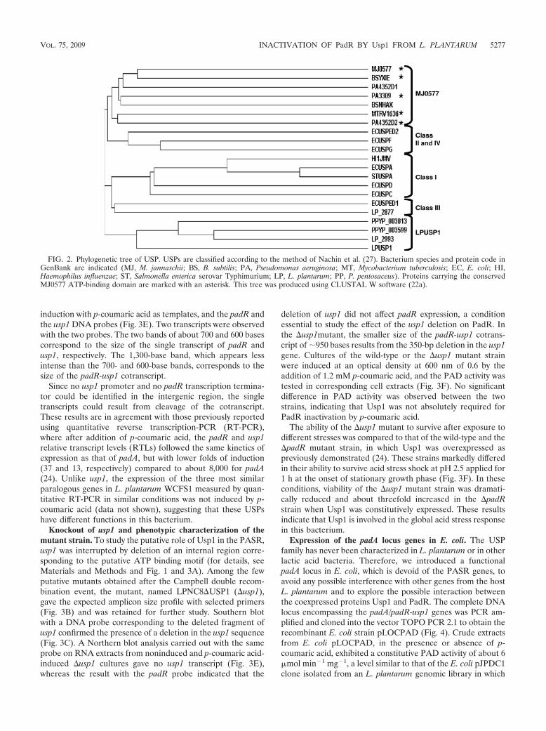
ATP binding domain (G-2X-G-9X-G-S/T) carried by the Meth-anococcus jannaschii MJ0577 Usp (36), but in a weakly conservedform. It is interesting that all of these species share many of thenatural habitats of L. plantarum and that P. pentosaceus, the otherlactic acid bacterium in which the PASR was studied, possessestwo high similar Usp1 homologues (PPYP_803599 and PPYP_803813). Phylogenetic analysis of USPs primary sequences showsthat PPYP_803599, PPYP_803813, LP2993, and Usp1 form adistinct subfamily (27) (Fig. 2).
Transcriptional analysis of padA, padR, and usp1 in L. plan-tarum. Sequence analysis (http://www.softberry.com) of thepadR-usp1 intergenic region (Fig. 3A) did not detect the pres-ence of a typical transcription terminator for padR. Moreover,our attempts to identify a putative usp1 initiation transcriptionsite by primer extension were unsuccessful, both with RNAextract from noninduced and p-coumaric-acid induced bacteria(data not shown). For Usp1, a strong transcription terminator(�G �27 kcal mol�1) was found 46 nucleotides downstreamof the usp1 stop codon. Downstream of this terminator wefound the promoter of the adhE gene, which encodes a puta-tive bifunctional alcohol and acetaldehyde dehydrogenase, agene also found in the same locus in the strain WCFS1(lp_3662). Northern blot hybridization was performed usingRNA samples from cultures of L. plantarum, with or without
FIG. 1. Amino acid sequence alignment of Usp1 with homologous proteins from GenBank and MJ0577. Alignment was achieved by usingonline software (http://bioinfo.genopole-toulouse.prd.fr/multalin/multalin.html; version 5.4.1) (8). The protein origin and the GenBank codes areindicated (LP, L. plantarum; LVIS, L. brevis; LSEI, L. casei; LSA L. sakei; PPYP, P. pentosaceus; OENOO, O. oeni; MJ, M. jannaschii). Theconsensus sequence of USP is also shown (capital letters correspond to conserved residues). Symbol comparison table: blosum62. Consensussymbols: !, anyone of IV; %, anyone of FY; #, anyone of NDQEBZ. The sizes and the global identity percentages between these sequences andUsp1 are indicated on the right. Residues of the ATP binding domain are boxed, and regions predicted to contact the adenosine (A), ribose (R),or triphosphate (P) are marked.
5276 GURY ET AL. APPL. ENVIRON. MICROBIOL.
induction with p-coumaric acid as templates, and the padR andthe usp1 DNA probes (Fig. 3E). Two transcripts were observedwith the two probes. The two bands of about 700 and 600 basescorrespond to the size of the single transcript of padR andusp1, respectively. The 1,300-base band, which appears lessintense than the 700- and 600-base bands, corresponds to thesize of the padR-usp1 cotranscript.
Since no usp1 promoter and no padR transcription termina-tor could be identified in the intergenic region, the singletranscripts could result from cleavage of the cotranscript.These results are in agreement with those previously reportedusing quantitative reverse transcription-PCR (RT-PCR),where after addition of p-coumaric acid, the padR and usp1relative transcript levels (RTLs) followed the same kinetics ofexpression as that of padA, but with lower folds of induction(37 and 13, respectively) compared to about 8,000 for padA(24). Unlike usp1, the expression of the three most similarparalogous genes in L. plantarum WCFS1 measured by quan-titative RT-PCR in similar conditions was not induced by p-coumaric acid (data not shown), suggesting that these USPshave different functions in this bacterium.
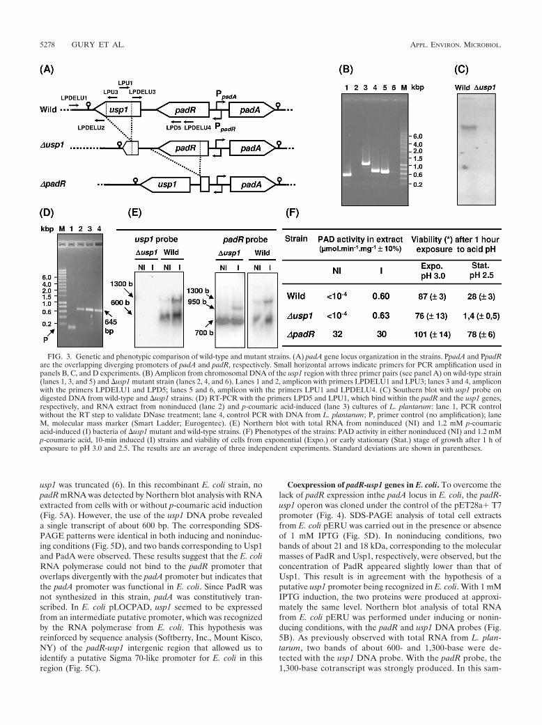
Knockout of usp1 and phenotypic characterization of themutant strain. To study the putative role of Usp1 in the PASR,usp1 was interrupted by deletion of an internal region corre-sponding to the putative ATP binding motif (for details, seeMaterials and Methods and Fig. 1 and 3A). Among the fewputative mutants obtained after the Campbell double recom-bination event, the mutant, named LPNC8�USP1 (�usp1),gave the expected amplicon size profile with selected primers(Fig. 3B) and was retained for further study. Southern blotwith a DNA probe corresponding to the deleted fragment ofusp1 confirmed the presence of a deletion in the usp1 sequence(Fig. 3C). A Northern blot analysis carried out with the sameprobe on RNA extracts from noninduced and p-coumaric acid-induced �usp1 cultures gave no usp1 transcript (Fig. 3E),whereas the result with the padR probe indicated that the
deletion of usp1 did not affect padR expression, a conditionessential to study the effect of the usp1 deletion on PadR. Inthe �usp1mutant, the smaller size of the padR-usp1 cotrans-cript of �950 bases results from the 350-bp deletion in the usp1gene. Cultures of the wild-type or the �usp1 mutant strainwere induced at an optical density at 600 nm of 0.6 by theaddition of 1.2 mM p-coumaric acid, and the PAD activity wastested in corresponding cell extracts (Fig. 3F). No significantdifference in PAD activity was observed between the twostrains, indicating that Usp1 was not absolutely required forPadR inactivation by p-coumaric acid.
The ability of the �usp1 mutant to survive after exposure todifferent stresses was compared to that of the wild-type and the�padR mutant strain, in which Usp1 was overexpressed aspreviously demonstrated (24). These strains markedly differedin their ability to survive acid stress shock at pH 2.5 applied for1 h at the onset of stationary growth phase (Fig. 3F). In theseconditions, viability of the �usp1 mutant strain was dramati-cally reduced and about threefold increased in the �padRstrain when Usp1 was constitutively expressed. These resultsindicate that Usp1 is involved in the global acid stress responsein this bacterium.
Expression of the padA locus genes in E. coli. The USPfamily has never been characterized in L. plantarum or in otherlactic acid bacteria. Therefore, we introduced a functionalpadA locus in E. coli, which is devoid of the PASR genes, toavoid any possible interference with other genes from the hostL. plantarum and to explore the possible interaction betweenthe coexpressed proteins Usp1 and PadR. The complete DNAlocus encompassing the padA/padR-usp1 genes was PCR am-plified and cloned into the vector TOPO PCR 2.1 to obtain therecombinant E. coli strain pLOCPAD (Fig. 4). Crude extractsfrom E. coli pLOCPAD, in the presence or absence of p-coumaric acid, exhibited a constitutive PAD activity of about 6�mol min�1 mg�1, a level similar to that of the E. coli pJPDC1clone isolated from an L. plantarum genomic library in which
FIG. 2. Phylogenetic tree of USP. USPs are classified according to the method of Nachin et al. (27). Bacterium species and protein code inGenBank are indicated (MJ, M. jannaschii; BS, B. subtilis; PA, Pseudomonas aeruginosa; MT, Mycobacterium tuberculosis; EC, E. coli; HI,Haemophilus influenzae; ST, Salmonella enterica serovar Typhimurium; LP, L. plantarum; PP, P. pentosaceus). Proteins carrying the conservedMJ0577 ATP-binding domain are marked with an asterisk. This tree was produced using CLUSTAL W software (22a).
VOL. 75, 2009 INACTIVATION OF PadR BY Usp1 FROM L. PLANTARUM 5277
usp1 was truncated (6). In this recombinant E. coli strain, nopadR mRNA was detected by Northern blot analysis with RNAextracted from cells with or without p-coumaric acid induction(Fig. 5A). However, the use of the usp1 DNA probe revealeda single transcript of about 600 bp. The corresponding SDS-PAGE patterns were identical in both inducing and noninduc-ing conditions (Fig. 5D), and two bands corresponding to Usp1and PadA were observed. These results suggest that the E. coliRNA polymerase could not bind to the padR promoter thatoverlaps divergently with the padA promoter but indicates thatthe padA promoter was functional in E. coli. Since PadR wasnot synthesized in this strain, padA was constitutively tran-scribed. In E. coli pLOCPAD, usp1 seemed to be expressedfrom an intermediate putative promoter, which was recognizedby the RNA polymerase from E. coli. This hypothesis wasreinforced by sequence analysis (Softberry, Inc., Mount Kisco,NY) of the padR-usp1 intergenic region that allowed us toidentify a putative Sigma 70-like promoter for E. coli in thisregion (Fig. 5C).
Coexpression of padR-usp1 genes in E. coli. To overcome thelack of padR expression inthe padA locus in E. coli, the padR-usp1 operon was cloned under the control of the pET28a� T7promoter (Fig. 4). SDS-PAGE analysis of total cell extractsfrom E. coli pERU was carried out in the presence or absenceof 1 mM IPTG (Fig. 5D). In noninducing conditions, twobands of about 21 and 18 kDa, corresponding to the molecularmasses of PadR and Usp1, respectively, were observed, but theconcentration of PadR appeared slightly lower than that ofUsp1. This result is in agreement with the hypothesis of aputative usp1 promoter being recognized in E. coli. With 1 mMIPTG induction, the two proteins were produced at approxi-mately the same level. Northern blot analysis of total RNAfrom E. coli pERU was performed under inducing or nonin-ducing conditions, with the padR and usp1 DNA probes (Fig.5B). As previously observed with total RNA from L. plan-tarum, two bands of about 600- and 1,300-base were de-tected with the usp1 DNA probe. With the padR probe, the1,300-base cotranscript was strongly produced. In this sam-
FIG. 3. Genetic and phenotypic comparison of wild-type and mutant strains. (A) padA gene locus organization in the strains. PpadA and PpadRare the overlapping diverging promoters of padA and padR, respectively. Small horizontal arrows indicate primers for PCR amplification used inpanels B, C, and D experiments. (B) Amplicon from chromosomal DNA of the usp1 region with three primer pairs (see panel A) on wild-type strain(lanes 1, 3, and 5) and �usp1 mutant strain (lanes 2, 4, and 6). Lanes 1 and 2, amplicon with primers LPDELU1 and LPU3; lanes 3 and 4, ampliconwith the primers LPDELU1 and LPD5; lanes 5 and 6, amplicon with the primers LPU1 and LPDELU4. (C) Southern blot with usp1 probe ondigested DNA from wild-type and �usp1 strains. (D) RT-PCR with the primers LPD5 and LPU1, which bind within the padR and the usp1 genes,respectively, and RNA extract from noninduced (lane 2) and p-coumaric acid-induced (lane 3) cultures of L. plantarum: lane 1, PCR controlwithout the RT step to validate DNase treatment; lane 4, control PCR with DNA from L. plantarum; P, primer control (no amplification); laneM, molecular mass marker (Smart Ladder; Eurogentec). (E) Northern blot with total RNA from noninduced (NI) and 1.2 mM p-coumaricacid-induced (I) bacteria of �usp1 mutant and wild-type strains. (F) Phenotypes of the strains: PAD activity in either noninduced (NI) and 1.2 mMp-coumaric acid, 10-min induced (I) strains and viability of cells from exponential (Expo.) or early stationary (Stat.) stage of growth after 1 h ofexposure to pH 3.0 and 2.5. The results are an average of three independent experiments. Standard deviations are shown in parentheses.
5278 GURY ET AL. APPL. ENVIRON. MICROBIOL.

ple, a smear of mRNA of lower molecular mass may repre-sent degradative products of the 1,300-base transcript. Thecotranscription of padR and usp1 in E. coli was confirmed byRT-PCR amplification experiments with mRNA preparedfrom E. coli pERU cells under inducing or noninducingconditions (1 mM IPTG) and the primers LPD5 and LPDU1(Fig. 3A and D).
Expression of usp1 in E. coli inactivates the PadR repressor.To investigate the involvement of usp1 in the PASR, padR andusp1 genes were cloned individually or together in thepET28a� vector under the control of the T7 promoter. Inaddition, padA was cloned under its own promoter into theSphI site of the pERU plasmid to serve as a reporter gene (Fig.4). Control E. coli BL21 pER and E. coli BL21 pERU strainswithout padA did not display PAD activity. In E. coli BL21, theT7 promoter was operational to express the operon padR-usp1. SDS-PAGE analysis of crude extracts from E. coliBL21 pEDR and E. coli BL21 pEDRU showed that PadRwas either produced at a low level without induction (datanot shown) or overproduced with IPTG induction (Fig. 5D)and that Usp1 was well produced even without IPTG induc-tion (Fig. 5D). This is in agreement with bioinformatics
analysis, which revealed that the padR-usp1 intergenic re-gion could serve as promoter in E. coli. No PAD activity wasdetected in either noninduced or p-coumaric acid-inducedextracts from E. coli pEDR cells (Fig. 4). In contrast, crudeextracts of E. coli BL21 pEDRU, in which PadR and Usp1were produced at nearly the same concentrations (Fig. 5D),exhibited a constitutive PAD activity of �7 �mol min�1
mg�1 either with or without induction with p-coumaric acid(Fig. 4). This heterologous expression of the three genes ofthe L. plantarum padA locus in E. coli demonstrates thatUsp1 is able to inactivate the PadR repressor when it isproduced at near the same level in the bacteria.
Usp1 abolishes the binding of PadR with the padA promoter.Mobility shift assays were carried out to study the influenceof Usp1 on the ability of PadR to bind to the padA pro-moter. The PadR-His6 and Usp1 proteins were overpro-duced in pER and pEU recombinant E. coli strains, respec-tively (Fig. 4 and Fig. 6A) and used in different EMSAsinvolving different combinations or treatments and with theappropriate controls (Fig. 6). Before carrying out EMSAexperiments, the capacity of these two proteins to dimerizewas tested by glutaraldehyde cross-linking experiments (Fig.
FIG. 4. Physical map of the different plasmid constructs of padA, padR, and usp1 and the resulting PAD activity obtained from the host strains.Plasmids pJPDC1 and pLOCPAD were constructed with the pJDC9 and the TOPO PCR 2.1 vectors, respectively. Plasmids pED, pER, pEU1,pEDR, pERU, and pEDRU were constructed by using the pET28a� vector. Closed triangles represent His tag fusions. Promoters are indicatedby small arrows at the �35 position. Restriction sites used for construction are indicated. For PAD activity, I and NI correspond to protein extractfrom cells either induced (I) or not induced (NI) with 1.2 mM p-coumaric acid. (�), Induction with IPTG.
VOL. 75, 2009 INACTIVATION OF PadR BY Usp1 FROM L. PLANTARUM 5279

6B). Although PadR forms dimers and oligomers at very lowconcentrations of glutardhedyde, Usp1 did not, despite itsamino acid sequence showing a putative pattern of dimer-ization (Fig. 1) and despite the high concentrations of glu-taraldehyde tested. EMSA results indicate that PadR wasable to bind specifically to the padA promoter and, depend-ing on its concentration, to form one or two complexes withthe padA promoter probe (Fig. 6C). This binding was spe-cific, as demonstrated by the disappearance of the shiftwhen PadR was preincubated in excess of unlabeled specificpadA promoter and by the conservation of the shift in thepresence of high concentrations of the unspecific competitorpoly(dI-dC) (Fig. 6D). Preincubation of PadR with 10 mMconcentration of p-coumaric, ferulic, and o-coumaric acidwas unable to abolish the probe shift (Fig. 6E).
Taking into account the results of our recent work on thePASR in B. subtilis (33), which suggested a possible interfer-ence of MgCl2 in EMSA, the EMSA was performed in thesame conditions as those shown in Fig. 6E but without MgCl2in the binding buffer (Fig. 6F). In contrast to the results ob-
tained with B. subtilis, the addition of phenolic acid did notabolish the binding of PadR to the padA promoter. To inves-tigate whether Usp1 binds to the padA promoter, serving as acompetitor for this promoter or modifying the affinity of PadRto the padA promoter, different EMSAs with the padA pro-moter were carried out (Fig. 6G and H). A concentrate ofcrude protein extract from E. coli carrying the vector pET28a�without the usp1 gene was unable to abolish the binding of thepadA promoter with PadR (Fig. 6G). Usp1 was also unable tobind to the padA promoter (Fig. 6H), demonstrating that Usp1had no affinity for the padA promoter and is not a competitorof padR for the padA promoter.
Since Usp1 inactivates PadR (Fig. 4), EMSAs were carried outwith PadR preincubated or not with Usp1 extract, in the samerange of concentrations (Fig. 6I). The results indicate that Usp1abolishes the binding of PadR to the padA promoter, and theysupport the inactivation of PadR by Usp1 in E. coli pEDRU (Fig.4), despite high levels of PadR in the bacteria (Fig. 5D). SincePadR and Usp1 might form a coprecipitate, a nonspecific inter-action that could account for the inability of PadR to bind to the
FIG. 5. Transcriptional analysis of padR and usp1 in recombinant E. coli strains and corresponding SDS-PAGE of protein extracts. (A) North-ern blot with RNA extract from noninduced (NI) and 1.2 mM p-coumaric acid 10-min-induced (I) cells. (B) Northern blot with RNA extract from1 mM IPTG-induced (�) or not induced (�) cells. The 600- and 1,300-base bands correspond to the usp1 and padR-usp1 transcripts, respectively,while “Smear” probably represents degradative products from the 1,300-base transcript. (C) DNA sequence upstream of usp1 indicating the �35and �10 boxes that could serve as a promoter for usp1 in the recombinant E. coli strains pLOCPAD and pERU. (D) SDS-PAGE analysis of crudeprotein extracts from the recombinant E. coli strains. pET28a�, E. coli TG1 with the vector pET28a�; I, 1.2 mM p-coumaric acid-induced cellextracts; – and �, noninduced (�) and 1 mM IPTG-induced (�) cell extract. M, molecular mass standards.
5280 GURY ET AL. APPL. ENVIRON. MICROBIOL.

padA promoter, individual and mixed protein extracts of the twoproteins were resolved in nondenaturing PAGE (Fig. 6J) withoutheating the sample before loading. Although the two proteinshave similar migration rates in SDS-PAGE (Fig. 6A), they displaydistinct migration rates in their native form in a PAGE (Fig. 6J,lanes R and U). Therefore, mixed extracts from purified PadRand increasing concentrations of Usp1 extract were incubated andresolved by PAGE (Fig. 6J, lanes U2R to U8R). In the mixedextracts, each protein conserved perfectly the same migration rateand no other protein band corresponding to a putative PadR-Usp1 coprecipitate was detected.
DISCUSSION
The aim of this study was to elucidate genetic and biochem-ical mechanisms of the phenolic acid stress response in L.
plantarum, a specific and strong stress response induced bysome of these chemicals. Sequence and transcriptional analysisof the L. plantarum padA locus indicates that padR, whichencodes the negative repressor of padA, is divergently orientedfrom padA. PadR also forms a bicistronic operon with a geneof unknown function named usp1, since its deduced productshares significant identity with proteins belonging to the USPfamily. However, the phylogenetic tree (Fig. 2) clearly indi-cates that Usp1 forms a subfamily with another putative USPof L. plantarum (LP-2993) and two putative USPs from P.pentosaceus. The expression of the padR-usp1 operon specifi-cally induced by phenolic acids is autoregulated by PadR itself(24). Taking together the fact that usp1 is overexpressed duringthe PASR and the fact that expression of USPs is induced bya large variety of stresses such as the entry in the stationarygrowth phase and various situations of starvation of bacteria
FIG. 6. EMSA analyses of PadR and Usp1 with the padA promoter. (A) SDS-PAGE of proteins extracts containing purified PadR andoverexpressed Usp1 used in the EMSA. M, molecular mass standard; R, 2.5 �g of purified PadR-His6; U, 2 �g of protein extract from E. coli pEUin which Usp1 represents ca. 90% of the total protein in the extract. (B) SDS-PAGE of PadR and Usp1 preincubated 10 min with increasingconcentrations of glutaraldehyde. (C) EMSA of increasing concentrations from 2 to 20 nM PadR (R) with the padA promoter probe. P, padApromoter DNA probe without PadR. C1 and C2 indicate putative complexes formed between the DNA probe and PadR. (D) EMSA with probeand 5 nM PadR (R5) with addition of a 50-fold (SC50) or a 200-fold (SC200) excess of unlabeled padA promoter fragment as a specific competitoror a 1,000-fold (dIdC1000) excess of competitor poly(dI-dC). (E) EMSA with probe and 5 nM PadR (R5) preincubated for 10 min with 10 mMp-coumaric (Pc), ferulic (Fe), and o-coumaric (Oc) acid. (F) Same as panel E, but without MgCl2 in the binding buffer. (G) EMSA with probe and5 nM PadR without (R5) and with (R5 pE) total protein extract from E. coli pET28a� at a protein concentration equivalent to 10 nM Usp1. pEcorresponds to the EMSA with probe and the extract of E. coli pET28a� without cloned gene. (H) EMSA with probe and 5 (U5) or 20 (U20)nM Usp1 extract. R5, the EMSA with probe and 5 nM PadR was used as the positive binding control. (I) EMSA with probe and 5 nM (R5) and20 nM (R20) PadR, followed by EMSA with probe and 5 nM (R5) PadR, in which PadR was preincubated before the binding reaction for10 min with increasing concentrations of Usp1 at 1, 2, 5, 10, and 20 nM. (J) Nondenaturing PAGE of PadR and Usp1 produced fromrecombinant E. coli strains pER and pEU, respectively. These extracts were similar to extracts R and U analyzed in SDS-PAGE in the panelA. R, 3 �g of PadR; U, 4 �g of pEU extract. The lanes correspond to mixes of 4 �g of PadR incubated for 10 min with increasing amountsof pEU extract (2, 4, and 8 �g) in 20 �l of binding buffer. Binding reactions for panels C, D, E, G, H, and I were carried out in standardconditions (with 2.5 mM MgCl2).
VOL. 75, 2009 INACTIVATION OF PadR BY Usp1 FROM L. PLANTARUM 5281

(29, 34), we hypothesized that Usp1 may be involved in thePASR. To address this, a �usp1 mutant was constructed andtested for its ability to induce the p-coumaric acid-dependentPAD activity. Although the �usp1 mutant conserved the wild-type phenotype, the usp1 expression was specifically induced byp-coumaric acid, whereas the three usp1 highest similar paralo-gous genes of L. plantarum were not. These results promptedus to study the possible interaction between PadR and Usp1 ina heterologous system devoid of the PASR.
Coexpression of padR and usp1 demonstrated that Usp1produced in E. coli was able to inactivate PadR, while theaddition of p-coumaric acid to a recombinant E. coli pEDRexpressing PadR could not, as it was also observed in recom-binant E. coli pJPADP1 expressing the padAR operon from P.pentosaceus (4). All of these results provide evidence thatPadR is not inactivated directly by p-coumaric acid or by one ofthe six putative USPs found in the E. coli genome (28) andindependently of the stage of growth (data not shown). This isin agreement with the phylogenetic sequence analysis, whichclusters Usp1 in a distinct family from the E. coli USPs (Fig. 2).
The capacity of Usp1 to inactivate PadR was confirmed byEMSA with PadR preincubated or not with Usp1. It is to ourknowledge the first time that the capacity of a USP to inacti-vate a transcriptional regulator is demonstrated, and it is pos-sible that Usp1 modulates other regulators in the same fashion.USPs could act as coordinators of regulator status duringstresses. Although PadR and Usp1 possess conserved andcharacterized dimerization domains (16, 27) (Fig. 1), Usp1does not seem to be able to form homodimers nor het-erodimers with PadR. According to the putative ATP bindingdomains of PadR (16) and Usp1 (Fig. 1) (27), ATP exchangebetween Usp1 and PadR could be suspected to be involved inthe inactivation of PadR. Usp1 should not act directly withPadR in living L. plantarum cells, but could act as an ATPdonor. It was found in E. coli that the addition of ATP toGroEL complexes releases the UspG form, and conversely,that the presence of ATP blocks the interaction of UspG withGroEL (5). Moreover, it was demonstrated in E. coli that theMJ0577 protein, a Usp1 paralogue able to bind ATP, requiresan additional cellular factor for ATP hydrolysis. Phosphoryla-tion studies of PadR and Usp1, and the possible transfer ofphosphate from Usp1 to PadR during the PASR, will be stud-ied soon. Such a mechanism might depend on the environmentof the regulators. Hence, to address this, it is necessary tooverexpress and purify these two proteins from the native hostL. plantarum, as was done for LmrR (26). Functional analysisof the proteins expressed in E. coli versus those expressed in itsnative host L. plantarum will be undertaken.
In mammals, phenolic acids, particularly ferulic and caffeicacids, are diet compounds strongly associated with a reducedrisk of developing chronic diseases (25). This effect is in partattributed to their antioxidant properties. However, studieshave shown that caffeic acid can also trigger DNA degradationin the presence of cupper ions due to its pro-oxidant activity(35, 37). Caffeic acid, a PASR inducer like ferulic and p-coumaric acids, has a strong pro-oxidant effect due to thepresence of an ortho-dihydroxyl group that chelates O2 withCu2� ions and generates reactive oxygen species, which areresponsible for the DNA damage. It is important to note thatin E. coli, UspA is required for resistance to DNA-damaging
agents (11, 17). Thus, we could hypothesize that Usp1, a UspAfamily protein, might also be involved in resistance to DNA-damaging agents in L. plantarum, and it may explain the ad-vantage for this bacterium to have its expression induced byphenolic acids, potentially DNA-damaging agents. Neverthe-less, this hypothesis does not exclude that Usp1 might be di-rectly involved in the PASR. The lack of an evident phenotypeof the usp1 mutant in the PASR could be explained by thepresence of another protein with functional redundancy ex-pressed when usp1 is knocked out. We provided evidence forthis kind of relay in a recent analysis of the B. subtilis responseto salicylic acid, another phenolic acid (13). We showed thatthe absence of sensitivity to salicylic acid of a mutant in thebsdBCD operon defective in the BsdB, C, and D enzymesinvolved in the decarboxylation of phenylacrylic and hydroxy-benzoic acids was taken over by the expression of the padCgene involved in the PASR. In contrast to the simple mutantbsdBCD, the double mutant bsdBCD-padC was sensitive to thisphenolic acid. Recent analyses of USPs function in severalgroups of microorganisms highlight the complexity of themechanism of action of proteins suspected to be componentsof the stress response network in cells, but their role remainsmost often enigmatic (22). Although we provide evidence thatusp1 is involved in the global acid stress response of cellsentering into the stationary phase, a comparison of transcrip-tomic and proteomic data from the usp1 mutant and padRmutants versus the wild-type strain will be undertaken to ob-tain an overview of the Usp1 and PadR roles in the behavior ofL. plantarum.
ACKNOWLEDGMENTS
J.G. was supported by a Ph.D. grant from INRA and the ConseilRegional de Bourgogne. H.S. and N.P.T. were supported, respectively,by grants from the French Ministere de l’Education Nationale, de laRecherche et de la Technologie and from the Ambassade de France auVietnam.
We thank Christine Rojas for her laboratory work. We are verygrateful to Philippe Sansonetti and Wilmara Salgado, Institut Pasteur,Paris, France, for helpful discussions and revision of the manuscript.
REFERENCES
1. Aukrust, T., and I. F. Nes. 1988. Transformation of Lactobacillus plantarumwith the plasmid pTV1 by electroporation. FEMS Microbiol. Lett. 52:127–132.
2. Axelsson, L. 1996. Proceedings of the Fifth Symposium on Lactic AcidBacteria: genetics, metabolism, and applications, Veldhoven, The Nether-lands. Antonie van Leeuwenhoek 70(2–4):97–358.
3. Barthelmebs, L., C. Divies, and J.-F. Cavin. 2000. Knockout of the p-cou-marate decarboxylase gene from Lactobacillus plantarum reveals the exis-tence of two other inducible enzymatic activities involved in phenolic acidmetabolism. Appl. Environ. Microbiol. 66:3368–3375.
4. Barthelmebs, L., B. Lecomte, C. Divies, and J.-F. Cavin. 2000. Induciblemetabolism of phenolic acids in Pediococcus pentosaceus is encoded by anautoregulated operon which involves a new class of negative transcriptionalregulator. J. Bacteriol. 182:6724–6731.
5. Bochkareva, E. S., A. S. Girshovich, and E. Bibi. 2002. Identification andcharacterization of the Escherichia coli stress protein UP12, a putative in vivosubstrate of GroEL. Eur. J. Biochem. 269:3032–3040.
6. Cavin, J.-F., L. Barthelmebs, and C. Divies. 1997. Molecular characteriza-tion of an inducible p-coumaric acid decarboxylase from Lactobacillus plan-tarum: gene cloning, transcriptional analysis, overexpression in Escherichiacoli, purification, and characterization. Appl. Environ. Microbiol. 63:1939–1944.
7. Cohen, S. N., A. C. Chang, and L. Hsu. 1972. Nonchromosomal antibioticresistance in bacteria: genetic transformation of Escherichia coli by R-factorDNA. Proc. Natl. Acad. Sci. USA 69:2110–2114.
8. Corpet, F. 1988. Multiple sequence alignment with hierarchical clustering.Nucleic Acids Res. 16:10881–10890.
9. De Man, P. J., M. Rogosa, and M. Sharpe. 1960. A medium for the cultiva-tion of lactobacilli. J. Appl. Bacteriol. 23:130–135.
5282 GURY ET AL. APPL. ENVIRON. MICROBIOL.

10. Derre, I., G. Rapoport, and T. Msadek. 1999. CtsR, a novel regulator ofstress and heat shock response, controls clp and molecular chaperone geneexpression in gram-positive bacteria. Mol. Microbiol. 31:117–131.
11. Diez, A., N. Gustavsson, and T. Nystrom. 2000. The universal stress proteinA of Escherichia coli is required for resistance to DNA damaging agents andis regulated by a RecA/FtsK-dependent regulatory pathway. Mol. Microbiol.36:1494–1503.
12. Dower, W. J., J. F. Miller, and C. W. Ragsdale. 1988. High-efficiency trans-formation of Escherichia coli by high-voltage electroporation. Nucleic AcidsRes. 16:6127–6145.
13. Duy, N. V., U. Mader, N. P. Tran, J.-F. Cavin, T. Tam le, D. Albrecht, M.Hecker, and H. Antelmann. 2007. The proteome and transcriptome analysisof Bacillus subtilis in response to salicylic acid. Proteomics 7:698–710.
14. Ferguson, L. R., I. F. Lim, A. E. Pearson, J. Ralph, and P. J. Harris. 2003.Bacterial antimutagenesis by hydroxycinnamic acids from plant cell walls.Mutat. Res. 542:49–58.
15. Gibson, T. J. 1984. Studies on the Epstein-Barr virus genome. Ph.D. thesis.Cambridge University, Cambridge, United Kingdom.
16. Gury, J., L. Barthelmebs, N. P. Tran, C. Divies, and J.-F. Cavin. 2004.Cloning, deletion, and characterization of PadR, the transcriptional repres-sor of the phenolic acid decarboxylase-encoding padA gene of Lactobacillusplantarum. Appl. Environ. Microbiol. 70:2146–2153.
17. Gustavsson, N., A. Diez, and T. Nystrom. 2002. The universal stress proteinparalogues of Escherichia coli are co-ordinately regulated and co-operate inthe defense against DNA damage. Mol. Microbiol. 43:107–117.
18. Hols, P., T. Ferain, D. Garmyn, N. Bernard, and J. Delcour. 1994. Use ofhomologous expression-secretion signals and vector-free stable chromo-somal integration in engineering of Lactobacillus plantarum for alpha-amy-lase and levanase expression. Appl. Environ. Microbiol. 60:1401–1413.
19. Kleerebezem, M., J. Boekhorst, R. van Kranenburg, D. Molenaar, O. P.Kuipers, R. Leer, R. Tarchini, S. A. Peters, H. M. Sandbrink, M. W. Fiers,W. Stiekema, R. M. Lankhorst, P. A. Bron, S. M. Hoffer, M. N. Groot, R.Kerkhoven, M. de Vries, B. Ursing, W. M. de Vos, and R. J. Siezen. 2003.Complete genome sequence of Lactobacillus plantarum WCFS1. Proc. Natl.Acad. Sci. USA 100:1990–1995.
20. Kovacikova, G., W. Lin, and K. Skorupski. 2005. Dual regulation of genesinvolved in acetoin biosynthesis and motility/biofilm formation by the viru-lence activator AphA and the acetate-responsive LysR-type regulator AlsRin Vibrio cholerae. Mol. Microbiol. 57:420–433.
21. Kovacikova, G., W. Lin, and K. Skorupski. 2003. The virulence activatorAphA links quorum sensing to pathogenesis and physiology in Vibrio chol-erae by repressing the expression of a penicillin amidase gene on the smallchromosome. J. Bacteriol. 185:4825–4836.
22. Kvint, K., L. Nachin, A. Diez, and T. Nystrom. 2003. The bacterial universalstress protein: function and regulation. Curr. Opin. Microbiol. 6:140–145.
22a.Larkin, M. A., G. Blackshields, N. P. Brown, R. Chenna, P. A. McGettigan,H. McWilliam, F. Valentin, I. M. Wallace, A. Wilm, R. Lopez, J. D. Thomp-son, T. J. Gibson, and D. G. Higgins. 2007. Clustal W and Clustal X version2.0. Bioinformatics 23:2947–2948.
23. Li, Y., Q. Peng, D. Selimi, Q. Wang, A. O. Charkowski, X. Chen, and C. H.Yang. 2009. The plant phenolic compound p-coumaric acid represses geneexpression in the Dickeya dadantii type III secretion system. Appl. Environ.Microbiol. 75:1223–1228.
24. Licandro-Seraut, H., J. Gury, N. P. Tran, L. Barthelmebs, and J.-F. Cavin.2008. Kinetics and intensity of the expression of genes involved in the stressresponse tightly induced by phenolic acids in Lactobacillus plantarum. J. Mol.Microbiol. Biotechnol. 14:41–47.
25. Liu, R. H. 2004. Potential synergy of phytochemicals in cancer prevention:mechanism of action. J. Nutr. 134:3479S–3485S.
26. Madoori, P. K., H. Agustiandari, A. J. Driessen, and A. M. Thunnissen.2009. Structure of the transcriptional regulator LmrR and its mechanism ofmultidrug recognition. EMBO J. 28:156–166.
27. Nachin, L., L. Brive, K. C. Persson, P. Svensson, and T. Nystrom. 2008.Heterodimer formation within universal stress protein classes revealed by anin silico and experimental approach. J. Mol. Biol. 380:340–350.
28. Nachin, L., U. Nannmark, and T. Nystrom. 2005. Differential roles of theuniversal stress proteins of Escherichia coli in oxidative stress resistance,adhesion, and mobility. J. Bacteriol. 187:6265–6272.
29. Nystrom, T., and F. C. Neidhardt. 1992. Cloning, mapping, and nucleotidesequencing of a gene encoding a universal stress protein in Escherichia coli.Mol. Microbiol. 6:3187–3198.
30. Posno, M., R. J. Leer, N. Van Luik, M. J. F. Van Giezen, P. T. H. M.Heuvelmans, B. C. Lockman, and P. H. Pouwels. 1991. Incompatibility ofLactobacillus vectors with replicons derived from small cryptic Lactobacillusplasmids and segregational instability of the introduced vectors. Appl.Environ. Microbiol. 57:1822–1828.
31. Sambrook, J., E. F. Fritsch, and T. Maniatis. 1989. Molecular cloning: alaboratory manual, 2nd ed. Cold Spring Harbor Laboratory, Cold SpringHarbor, NY.
32. Scalbert, A., and G. Williamson. 2000. Dietary intake and bioavailability ofpolyphenols. J. Nutr. 130:2073S–2085S.
33. Tran, N. P., J. Gury, V. Dartois, T. K. Nguyen, H. Seraut, L. Barthelmebs, P.Gervais, and J.-F. Cavin. 2008. Phenolic acid-mediated regulation of thepadC gene, encoding the phenolic acid decarboxylase of Bacillus subtilis. J.Bacteriol. 190:3213–3224.
34. VanBogelen, R. A., M. E. Hutton, and F. C. Neidhardt. 1990. Gene-proteindatabase of Escherichia coli K-12, edition 3. Electrophoresis 11:1131–1166.
35. Yamanaka, N., O. Oda, and S. Nagao. 1997. Prooxidant activity of caffeicacid, dietary non-flavonoid phenolic acid, on Cu2�-induced low density li-poprotein oxidation. FEBS Lett. 405:186–190.
36. Zarembinski, T. I., L. W. Hung, H. J. Mueller-Dieckmann, K. K. Kim, H.Yokota, R. Kim, and S. H. Kim. 1998. Structure-based assignment of thebiochemical function of a hypothetical protein: a test case of structuralgenomics. Proc. Natl. Acad. Sci. USA 95:15189–15193.
37. Zheng, L. F., F. Dai, B. Zhou, L. Yang, and Z. L. Liu. 2008. Prooxidantactivity of hydroxycinnamic acids on DNA damage in the presence of Cu(II)ions: mechanism and structure-activity relationship. Food Chem. Toxicol.46:149–156.
VOL. 75, 2009 INACTIVATION OF PadR BY Usp1 FROM L. PLANTARUM 5283
Related Documents