In vivo optical coherence tomography detection of differences in regional large airway smoke inhalation induced injury in a rabbit model Matthew Brenner, University of California, Irvine, Beckman Laser Institute, Irvine, California 92612 and University of California, Irvine Medical Center, Pulmonary and Critical Care Division, Orange, California 92868, E-mail: [email protected] Kelly Kreuter, University of California, Irvine, Beckman Laser Institute, Irvine, California 92612 Johnny Ju, University of California, Irvine Medical Center, Pulmonary and Critical Care Division, Orange, California 92868 Sari Mahon, University of California, Irvine, Beckman Laser Institute, Irvine, California 92612 Lillian Tseng, University of California, Irvine Medical Center, Pulmonary and Critical Care Division, Orange, California 92868 David Mukai, University of California, Irvine, Beckman Laser Institute, Irvine, California 92612 Tanya Burney, University of California, Irvine, Beckman Laser Institute, Irvine, California 92612 Shuguang Guo, University of California, Irvine, Beckman Laser Institute, Irvine, California 92612 Jianping Su, University of California, Irvine, Beckman Laser Institute, Irvine, California 92612 Andrew Tran, University of California, Irvine Medical Center, Pulmonary and Critical Care Division, Orange, California 92868 Andriy Batchinsky, United States Army Institute of Surgical Research, Fort Sam Houston, Texas Leopoldo C. Cancio, United States Army Institute of Surgical Research, Fort Sam Houston, Texas Navneet Narula, and University of California, Irvine Medical Center, Pathology Division, Orange, California 92868 Zhongping Chen © 2008 Society of Photo-Optical Instrumentation Engineers. Address all correspondence to Matthew Brenner, Pulmonary and Critical Care Division, Univ. of California/Irvine, 101 City Dr South, Building 53, Rm 119 Orange, CA 92868; Tel: 714 456 5150; Fax: 714 456 8349; E-mail: [email protected]. NIH Public Access Author Manuscript J Biomed Opt. Author manuscript; available in PMC 2009 November 17. Published in final edited form as: J Biomed Opt. 2008 ; 13(3): 034001. doi:10.1117/1.2939400. NIH-PA Author Manuscript NIH-PA Author Manuscript NIH-PA Author Manuscript

Welcome message from author
This document is posted to help you gain knowledge. Please leave a comment to let me know what you think about it! Share it to your friends and learn new things together.
Transcript
In vivo optical coherence tomography detection of differences inregional large airway smoke inhalation induced injury in a rabbitmodel
Matthew Brenner,University of California, Irvine, Beckman Laser Institute, Irvine, California 92612 and University ofCalifornia, Irvine Medical Center, Pulmonary and Critical Care Division, Orange, California 92868,E-mail: [email protected]
Kelly Kreuter,University of California, Irvine, Beckman Laser Institute, Irvine, California 92612
Johnny Ju,University of California, Irvine Medical Center, Pulmonary and Critical Care Division, Orange,California 92868
Sari Mahon,University of California, Irvine, Beckman Laser Institute, Irvine, California 92612
Lillian Tseng,University of California, Irvine Medical Center, Pulmonary and Critical Care Division, Orange,California 92868
David Mukai,University of California, Irvine, Beckman Laser Institute, Irvine, California 92612
Tanya Burney,University of California, Irvine, Beckman Laser Institute, Irvine, California 92612
Shuguang Guo,University of California, Irvine, Beckman Laser Institute, Irvine, California 92612
Jianping Su,University of California, Irvine, Beckman Laser Institute, Irvine, California 92612
Andrew Tran,University of California, Irvine Medical Center, Pulmonary and Critical Care Division, Orange,California 92868
Andriy Batchinsky,United States Army Institute of Surgical Research, Fort Sam Houston, Texas
Leopoldo C. Cancio,United States Army Institute of Surgical Research, Fort Sam Houston, Texas
Navneet Narula, andUniversity of California, Irvine Medical Center, Pathology Division, Orange, California 92868
Zhongping Chen
© 2008 Society of Photo-Optical Instrumentation Engineers.Address all correspondence to Matthew Brenner, Pulmonary and Critical Care Division, Univ. of California/Irvine, 101 City Dr South,Building 53, Rm 119 Orange, CA 92868; Tel: 714 456 5150; Fax: 714 456 8349; E-mail: [email protected].
NIH Public AccessAuthor ManuscriptJ Biomed Opt. Author manuscript; available in PMC 2009 November 17.
Published in final edited form as:J Biomed Opt. 2008 ; 13(3): 034001. doi:10.1117/1.2939400.
NIH
-PA Author Manuscript
NIH
-PA Author Manuscript
NIH
-PA Author Manuscript
University of California, Irvine, Beckman Laser Institute, Irvine, California 92612
AbstractSmoke inhalation injury causes acute airway injury that may result in airway compromise withsignificant morbidity and mortality. We investigate the ability of high resolution endobronchialoptical coherence tomography (OCT) to obtain real-time images for quantitatively assessing regionaldifferences between upper tracheal versus lower tracheal and bronchial airway injury responses tosmoke inhalation in vivo using a prototype spectral domain (SLD)-OCT system we constructed, andflexible fiber optic probes. 33 New Zealand White rabbits are intubated and mechanically ventilated.The treatment groups are exposed to inhaled smoke. The OCT probe is introduced through theendotracheal tube and maintained in place for 5 to 6 h. Images of airway mucosa and submucosa areobtained at baseline and at specified intervals postexpo-sure. Starting within less than 15 min aftersmoke inhalation, there is significant airway thickening in the smoke-exposed animals. This ismaintained over 5 h of imaging studies. The lower tracheal airway changes, correlating closely withcarboxyhemoglobin levels, are much greater than upper tracheal changes. Significant differences areseen in lower trachea and bronchi after acute smoke inhalation compared to upper trachea as measuredin vivo by minimally invasive OCT. OCT is capable of quantitatively detecting regional changes inairway swelling following inhalation injury.
Keywordsoptical coherence tomography; smoke inhalation injury; bronchial; tracheal; fiber optic probe
1 IntroductionSmoke inhalation injury is a major cause of morbidity and mortality among victims of fires.Pathophysiologic changes include hyperemia, edema, sloughing, and necrosis, which can causeairway compromise and acute lung injury.1–9 Currently, there are no highly reliable diagnostictechniques in the clinical setting to predict or assess the degree of airway compromise followingsmoke inhalation. High-resolution flexible fiber optic optical coherence tomography (OCT)provides the potential to observe real-time changes in the airway mucosa and epithelium usingminimally invasive imaging techniques.10–25 Most importantly, some of the early pathologicchanges following smoke inhalation that may be indicative of the extent of injury such as edemaand hyperemia2,26–29 may not be apparent clinically, endoscopically, or following histologicpreparations of excised specimens, but can be readily detected on OCT.30
Previous OCT airway injury studies30 in rabbit smoke inhalation models suggested to us thatthe upper trachea airway may react very differently to acute smoke inhalation compared todistal trachea and proximal bronchi. The purpose of this study was to demonstrate the potentialrole of OCT in quantitatively detecting early smoke inhalation injury and assessing differencesin response of more proximal versus distal large airways to smoke inhalation injury.
2 Materials and MethodsThis protocol was approved by the University of California, Irvine (UCI) Academic ResearchCommittee (2002–2397) and complied with all federal and state regulations for animal welfareassurance. General model and preparation methods have been described previously30 and arebriefly summarized here.
Brenner et al. Page 2
J Biomed Opt. Author manuscript; available in PMC 2009 November 17.
NIH
-PA Author Manuscript
NIH
-PA Author Manuscript
NIH
-PA Author Manuscript
2.1 General Preparation33 male New Zealand White rabbits, weighing 3 to 5 kg (Western Oregon Rabbit Company,Philomath, Oregon) were anesthetized with a 2:1 ratio of ketamine HCl (100 mg/ml) (Ketaject,Phoenix Pharmaceutical Incorporated, Saint Joseph, Michigan): xylazine (20 mg/ml) (Anased,Lloyd Laboratories, Shenandoa, Iowa), 0.75-cc/kg IM using a 23 gauge 5/8 in. needle. Afterthe IM injection, a 23 gauge 1-in. catheter was placed in the marginal ear vein to administerIV maintenance anesthetic of a 1:1:3 mixture of ketamine:xylazine:saline (Ketamine 100 mg/ml: xylazine 20 mg/ml) as a continuous infusion, at a rate of 0.17 ml/min. A dose of analgesic,Torbutrol 0.1 to 0.5 mg/kg SQ, was administered prior to intubation. The animals were orallyintubated with a 3.5-mm cuffed endotracheal tube and mechanically ventilated (dual phasecontrol respirator, model 613, Harvard Apparatus, Chicago, Illinois) at a respiratory rate of 32/min and a tidal volume of 60 cc and FiO2 of 100%. A humidifier (Humid-Vent mini Ref. 10011,Hudson RCI, Temecula, California) was positioned between the ventilator and endotrachealtube to prevent drying out of the mucosa, which could result in airway changes due to prolongedexposure to ventilated O2. On completion of the experiment, the animals were euthanized withan intravenous injection of Eutha-6 (1.0–2.0 cc) administered through the marginal ear vein.30
2.2 Systemic Arterial Blood Pressure, Blood Gas Analysis, and Co-OximetryFemoral arterial and venous cutdowns were performed to collect blood samples and recordsystemic blood pressure. An 18 gauge catheter (C-PMA-400-FA, Cook Incorporated,Bloomington, Indiana) was inserted into the vein and artery, and a three-way stop-cock wasplaced on the ends. To measure systemic arterial pressure, a calibrated pressure transducer(TSD104A Transducer and MP100 WSW System, Biopac Systems, Incorporated, SantaBarbara, California) was connected to a pulmonary artery extension set, which was thenattached to the end of the stop-cock. Blood was drawn from both the arterial and venous linesand measured by a blood gas analyzer (IRMA SL Series 2000 Blood Analysis System,Diametrics Medical Incorporated, Saint Paul, Minnesota) to obtain arterial and venous bloodgas analysis, respectively. On-site co-oximetry measurements (AVOXimeter 4000, AVOXSystems, San Antonio, Texas) were conducted to measure oxyhemoglobin,carboxyhemoglobin, methemoglobin fractions, and total hemoglobin. The carboxy Hgb levelswere analyzed to assess the degree of smoke exposure. The co-oximeter was calibrated withrabbit blood by the manufacturer for the research purposes in these studies.
2.3 Administration of SmokeSmoke was administered according to the previously published protocol.30 70 g of unbleachedcotton was burned in a modified bee smoker (Smoke Stack Smoker 644, Brushy Mountain BeeFarm, Moravian Falls, North California) for approximately 20 min. The bee smoker was thenconnected to the inlet port of the mechanical ventilator with the tidal volume and ventilationrate set at 700 ml and 25 breaths per minute, respectively. A Mylar Douglas bag (model 6060,Hans Rudolph, Kansas City, Missouri) was connected to a second “smoke exposure” ventilator(ventilator 2) (also a dual phase control respirator, model 613, Harvard Apparatus, Chicago,Illinois) via the multiport valve to the output port to actively fill the bag with smoke from thebee smoker, and over 25 L of smoke was collected. The Mylar bag was then connected to theinlet port of the smoke exposure ventilator and set to a tidal volume of 60 ml and a ventilationrate of 18 breaths per minute to deliver the (now cool) smoke-filled contents of the Douglasbag to the rabbit in a controlled manner. The rabbit was disconnected from the regular ventilator(1) circuit and connected to the smoke ventilator (2) for exposure. Exposure was achieved bygiving the animal repetitive alternate increments of 18 breaths of smoke, followed by 100%oxygen (accomplished by switching between the traditional ventilator setup and the smokeventilator). For smoke exposure, rabbits were ventilated with 0 breaths of smoke (controls) or
Brenner et al. Page 3
J Biomed Opt. Author manuscript; available in PMC 2009 November 17.
NIH
-PA Author Manuscript
NIH
-PA Author Manuscript
NIH
-PA Author Manuscript
a variable number of breaths of smoke (smoke group). The range of smoke breaths administeredwas between 54 and 216 breaths (mean 127 breaths) that were administered by alternating aminimum of three cycles of 18 breaths of cooled smoke with nine breaths of ventilated O2(i.e., a minimum of 54 breaths in the treatment groups). Co-oximetry measurements were takenat the end of smoke exposure. To obtain a broad range of carbon monoxide levels, additionalbreaths were given to some animals to raise the carboxyhemoglobin levels throughout thedesired dosage range. Carboxyhemoglobin measurements of between 0.2 and 48% wereobtained in the exposure group animals.
2.4 Optical Coherence Tomography System and ProbesThe OCT system used in this study contains a superlumines-cent diode source that delivers anoutput power of 10 mW at a central wavelength of 1310 nm with a full width at half maximum(FWHM) of 80 nm, resulting in approximately 10-µm axial resolution.30
In the reference arm, a rapid-scanning optical delay line is used that employs a grating to controlthe phase and group delays separately, so that no phase modulation is generated when the groupdelay is scanned. The phase modulation is generated through an electro-optic phase modulatorthat produces a carrier frequency. The axial line scanning rate is 500 Hz, and the modulationfrequency of the phase modulator is 500 kHz. Reflected beams from the two arms arerecombined in the interferometer and detected on a photodetector. The detected opticalinterference fringe intensity signals are bandpass filtered at the carrier frequency. Resultantsignals are then digitized with an analog-digital converter, which performs 12 bit at 5-MHzsignal conversion and is transferred to a computer where the structural image is generated.12,19,31
Flexible fiber optic OCT probes were constructed from a single mode fiber (ThorLabs, Newton,New Jersey). The bare-ended fiber was attached to a 0.7-mm-diam gradient index (GRIN) lens(NSG America, Irvine, California), using optical adhesive (Dymax Company, Torrington,Connecticut) under a microscope. A right angle-light path was achieved using a 0.7-mm prism.The probe was placed in fluorinated ethylene propylene tubing (17 gauge thin wall, Zeus,Orangeburg, South Carolina) for added fiber support. The outer diameter of the probe isapproximately 2 mm.
A linear motor (Newport Instruments, Irvine, California) was used to drive the coated flexiblefiber optic distally and proximally along the length of the probe within the sheath, moving theGRIN lens and prism imaging components within the sheath to obtain linear images along thelong axis of the trachea and bronchi. Axial scans were obtained every 10 um along the lengthof the probe during 16-mm-long scan sweeps. A 16-mm scan is obtained in 3.2 sec.Translational imaging has a number of distinct theoretical advantages over rotational scanningmethods for initial investigations in relatively large lumen sites such as trachea and proximalbronchi. These advantages include that the translational probe can be placed in the trachea atany location, as long as it is not touching the tissue itself; whereas the rotational probe wouldhave to be centered in the trachea, the translational probe can be fixed in a location, and doingthis with a rotational probe would be difficult (a fraction of a movement with the rotationalprobe would yield false results); and finally, the rotational probe can only capture a fractionof the image.12,19,31
2.5 Airway Thickness MeasurementsAirway thickness was measured as the distance between the epithelial surface and the surfaceof landmark submucosal cartilaginous rings in the trachea and bronchi, and includes epithelial,subepithelial, and glandular tissues above the cartilage.30 Measurements were obtained by twoindependent researchers (Ju and Tseng) who were blinded to all clinical information. Airway
Brenner et al. Page 4
J Biomed Opt. Author manuscript; available in PMC 2009 November 17.
NIH
-PA Author Manuscript
NIH
-PA Author Manuscript
NIH
-PA Author Manuscript
thickness measurements were taken at as many landmark cartilaginous rings as could be seenin an individual animal (generally 3 to 5 cartilage ring sites per animal). Measurements of theairway thickness at the landmark rings were obtained at baseline, and at each specified timeperiod following inhalation injury. Approximately nine measurements were taken per timepoint. The percent change from baseline was calculated for each airway thickness measurementat every time point. Values were then averaged for all airway thickness measurements withina given animal at each specified point.
2.6 Upper Trachea Versus Lower Trachea/Bronchi PlacementUpper trachea was defined in this study to be within 5 cm below the larynx, and lower trachea/bronchi to be below that level. For lower trachea/bronchi studies, the standard 3.5-mmendotracheal tube (16 cm length) was advanced fully down the trachea and secured in position.The OCT probe was then placed through the endotracheal tube just beyond the tip of the tube.For upper tracheal measurements, the 3.5-mm-diam endotracheal tube was cut to a length of14 cm prior to insertion, advanced to approximately 2 cm from maximal insertion distance,and secured in place. With this approach, the tip of the endotracheal tube usually resided in theupper trachea. Due to variability in positioning and tracheal length, the actual position of theOCT probe, and therefore the animal group, was definitively determined with certainty onlyat the time of sacrifice.
2.7 Statistical AnalysisDifferences between controls, and upper and lower trachea/bronchi were determined byanalysis of variance. A two-tailed p value less than 0.05 was considered statistically significant.For descriptive statistics, mean +/− standard error is reported. Correlations were assessed usingstandard linear regression. All statistical analysis was performed using commercial statisticalsoftware (Systat 10, SPSS, Incorporated, 2002).
3 Results30 animals total were exposed to inhaled room-temperature smoke for this study (mean 127 ±9 breaths, range from 54 to 216 breaths total). 14 animals were in the lower trachea/bronchiexposure group, 16 in the upper tracheal exposure group, and 3 controls (no smoke exposure).As seen in our previous studies, the smoke-treated animal study groups had significantincreases in airway thickness seen by in vivo OCT. Changes were evident in the thickness ofthe airway mucosa at the time of the first follow-up measurement 15 min following exposurewith an average increase in airway thickness of 27 ± 5% (SEM) for the overall smoke-treatedanimals (p <0.001).21 No change was seen in the airway thickness of the control group animals(three upper and ten lower airway) that were not exposed to smoke during the first 60 min ofanesthesia, intubation, and ventilator support administered in an analogous manner to thesmoke-treatment animals. A slight increase in the mucosal thickness of the control group wasobserved during the experiments, although this increase was not significant. This increase wasmost likely due to the positive pressure of the ventilator or the anesthesia, as these are bothknown to cause effects similar to the ones we observed. Despite the marked changes in thesubepithelial layers seen on OCT, only epithelial cell layer changes were seen on histologicexamination (Fig. 1).
Carboxyhemoglobin levels were obtained immediately postexposure in 26 of the 33 animalsstudied (7 lower trachea, 19 upper trachea). The range of carboxyhemoglobin levels attainedin the smoke-treated animals was 21.7 ± 2.7% overall (range 0.2 to 46.7%), while the controlgroup averaged 1.0 ± 0.4% percent (range 0.2 to 1.7%), (p<0.02). There was no significantdifference in the average carboxyhemoglobin levels in the upper trachea group (mean 22.7±2.9%, range 8.6 to 42.5%) versus lower trachea/bronchi (mean 19.8 ± 2.9%, range 0.2 to
Brenner et al. Page 5
J Biomed Opt. Author manuscript; available in PMC 2009 November 17.
NIH
-PA Author Manuscript
NIH
-PA Author Manuscript
NIH
-PA Author Manuscript
46.7%) (p=0.68). There was no difference in the total number of smoke breaths administeredto the upper tracheal group (mean 141 ± 11 breaths, range 90 to 198) verus the lower trachea/bronchi (mean 110 ± 13 breaths, range 54 to 216) (p=ns).
Regional differences were seen in the degree of airway thickness changes following smokeexposure in the lower trachea/bronchi in comparison to the upper tracheal exposure groupswhen the OCT images at baseline were compared to those images taken 360 min after smokeexposure (Fig. 2). Statistically significant increases from baseline were seen in the lowertrachea/bronchi treated animal group at all time periods postexposure (Fig. 3). At 15-minpostexposure, the lower trachea/bronchi treated group showed a 29 ± 7% increase in airwaythickness compared to baseline (p<0.001). Airway thickness peaked in at 240 to 300-minpostexposure with a mean increase of 85 ± 22% increase (p<0.003). The upper trachea smoke-exposed animals also showed early increases in mucosal thickness at 15-min postexposure (25.± 8% increase, p<.007). However, no significant further increases occurred after 15 to 30 min(at 30 min r =0.36, r2 =0.13, p=0.23, and at 240 min r =0.14, r2=0.02, and p =0.66), with airwayswelling peaking at 30-min postexposure at a 29 ± 7% increase from baseline (p<0.001). Thedegree of airway swelling from baseline was statistically significantly higher in the lowertrachea/bronchi groups compared to the upper tracheal group beginning at 180-minpostexposure; 21 ± 7% for the upper group versus 76 ± 14% increase for the lower trachea/bronchi (p<0.002) (Fig. 4). These differences continue to increase out to 300-min postexposure(25 ± 8% increase for the upper group versus 85±22% increase for the lower trachea/bronchigroup, p<0.02 at 300 min).
To further explain the individual animal and regional variability in airway response to smokeexposure, the degree of airway thickness changes was correlated with carboxyhemoglobinlevels attained for the upper trachea group in comparison to the lower trachea/bronchi group.In the lower trachea/bronchi group, there was a very close correlation between the degree ofairway thickening and the carboxyhemoglobin level beginning at the first follow-upmeasurement period, 15-min postexposure (r=0.83, r2 =0.70, p<0.01, n=7), and this trendcontinued to be observed at 30-min postexposure (r =0.86, r2 =0.74, p=0.01, n=7), and 240-min postexposure (r =0.75, r2 =0.56, p=0.05, n=7) (Fig. 5). This close correlation wasmaintained throughout the postexposure measurement period as the airway swellingprogressed. In contrast, in the upper trachea group, no correlation was seen at any time periodpostexposure between the degree of upper tracheal swelling and carboxyhemoglobin levels.
4 DiscussionThis study demonstrated the ability of optical coherence tomography minimally invasiveimaging to detect acute changes in airway thickening following inhalation smoke exposure atvarious levels within the trachea and proximal bronchi. Optical coherence tomography imagesused in this study were obtained in real time at resolutions of 10 µm using flexible fiber opticGRIN lens-based probes and were capable of providing a mean to quantitatively assess thedegree of airway thickness through measurements of the images. The changes could bemonitored continuously, as in this study, for more than five hours following smoke exposure.Previous studies using this model30 have shown that substantial airway changes occur veryearly after room temperature smoke exposure. The current study was designed to use OCT toassess regional response differences in the degree of airway changes seen in the upper tracheacompared to lower trachea/bronchi following smoke exposure. The ability to quantitativelymeasure such differences with OCT is important for understanding potential regionalvariability mechanisms in inhalation injury in vivo, and will be important in future studiesdesigned to correlate acute injury with subsequent pathophysiologic events.
Brenner et al. Page 6
J Biomed Opt. Author manuscript; available in PMC 2009 November 17.
NIH
-PA Author Manuscript
NIH
-PA Author Manuscript
NIH
-PA Author Manuscript
This study demonstrated substantial statistically and clinically significant differences in thedegree of airway changes occurring in the upper trachea compared to proximal bronchi anddistal trachea. Variable but dramatic increases were seen in the thickness of the lower trachea/bronchial airways, while only modest changes developed in the higher trachea. These changesbegan very early following smoke exposure, frequently being observed within five minutesfollowing exposure.
We are uncertain why the upper trachea and lower large airway regions may have respondedso differently. There may be inherent differences in sensitivity to smoke in upper trachea versusdistal trachea and bronchi. It is possible that the differences result from differences in depositionof smoke particles in the distal trachea and bronchi following smoke exposure. The smokeexposure was performed with room-temperature smoke administered down the endotrachealtube of intubated animals. For the upper tracheal assessments, the endotracheal tube was cutshort (14 cm length). For the lower trachea/bronchi assessments, the endotracheal tube waskept at full length. Thus, the tip of the endotracheal tube (where the smoke exits and exposurecan begin) was equidistant from the OCT probe in both cases. It is possible that the diameterand curvature of the lower trachea and bronchi result in toxic particle impaction with resultantincrease in airway injury. Prior studies have shown that tracheobronchial injury in smokeinhalation is primarily due to toxic chemicals contained in the smoke.2 Future studies will beneeded to further clarify the causes for these regional differences, and may be very importantto understanding the nature of inhalation injury and improving treatment/outcomes.
There are a number of limitations with these studies. It was not until the time of sacrifice thatthe position of the endotracheal tube and optical coherence tomography probe was determinedwith certainty. It was not possible to precisely place the OCT probe in the right main stembronchus at specific locations prior to smoke treatment with this study design. Furthermore,the imaging direction of the OCT probe was generally pointed anteriorly (as evidenced by asthe presence of cartilaginous rings and continuous airway wall). However, the exact angle ofimaging was not clearly defined, and precise delineation of distal trachea from proximalbronchi, or right versus left mainstem bronchi, could not be determined until after sacrifice.
The exact pathophysiology of the airway injury seen on OCT is also uncertain. The very earlyappearance of the changes suggests that edema and/or hyperemia may be predominantcomponents of the injury process. The findings are consistently present on OCT evaluation.However, a proportion of the airway thickening changes seen on OCT disappears immediatelyfollowing sacrifice, six hours postexposure. This would suggest that previously describedhyperemia1,2,26,27 in the initial stages following smoke exposure may be a significantcomponent of this process. Furthermore, histology of the tracheal specimens followingsacrifice does not demonstrate significant changes from controls, even in animals that haddramatic changes seen on OCT; this is consistent with published literature and suggests edemaor hyperemia as major components of the early injury process that are lost during histologicpreparation.
Imaging with small diameter flexible fiber optic white-light bronchoscopy showed only mildevidence of early hyperemia following smoke exposure. Regional deposition of smokeparticles could not be differentiated visibly, and as expected, the degree of airway swellingcould not be determined by fiber optic bronchoscopy, despite significant changes seen on OCT.
Thermal effects are unlikely a significant component of these findings, because room-temperature smoke was used and thermal injury would be expected to be more severe closerto the tip of the endotracheal tube.2
Carboxyhemoglobin levels were obtained in the animals as a marker of the degree of exposure.A range of carbon monoxide levels was obtained in animals through variation in numbers of
Brenner et al. Page 7
J Biomed Opt. Author manuscript; available in PMC 2009 November 17.
NIH
-PA Author Manuscript
NIH
-PA Author Manuscript
NIH
-PA Author Manuscript
administered smoke-containing breaths, as well as intrinsic variability in the level ofcarboxyhemoglobin achieved for a given number of breaths. The average carboxyhemoglobinlevels achieved and the total number of smoke breaths administered were not statisticallysignificantly different between the upper trachea and distal trachea exposure groups, and wereactually slightly higher in the upper trachea group where less effect was seen. Therefore, greaterdegrees of airway swelling in the lower trachea/bronchial injury group could not be explainedby greater degrees of total smoke exposure.
Interestingly, the degree of airway swelling correlated very closely with the level ofcarboxyhemoglobin attained in the animals during the postexposure period in the lower trachea/bronchi group. This is evidence of a dose-response relationship between total smoke exposureand early airway injury effects in this lower tracheal and proximal bronchial region.32 Incontrast, more upper trachea showed significantly less injury, and no correlation withcarboxyhemoglobin levels at any time following exposure.
Future studies to further elucidate the causes behind the regional differences in response tosmoke injury, association between airway changes and various pathologic mediators of tissueinjury and repair, as well as correlation between early changes and later effects are needed.
Flexible fiber optic optical coherence tomography is minimally invasive, and can be performedwith very narrow diameter flexible probes that can be left in place to image intermittently orcontinuously. This can potentially facilitate follow up evaluations of smoke inhalation injury,which progresses over time,1,2,26,27,33 and that may not be evident during bronchoscopicevaluation. This study was able to demonstrate that OCT has the capability of not only detectingchanges in thickness in the layers of the mucosa, but also quantifying differences in themagnitude of changes in the upper and lower trachea. The ability of OCT to distinguish suchchanges in real time may someday assist clinically in determining the extent of injury,prognosis, and need for intervention. Quantitative determination of acute and longer termchanges in the airway epithelium, mucosa, and submucosa with OCT may also provide a moresensitive tool for investigation of the effectiveness of various therapeutic interventions insmoke inhalation and other airway injuries.
AcknowledgmentsThis work was supported by Department of Defense: AFOS-2004-0011A and 0012A, AF-9550–04–1–0101, LAMMP445474-30136, NIH CA-91717, and EB-000293.
References1. Cox RA, Burke AS, Soejima K, Murakami K, Katahira J, Traber LD, Herndon DN, Schmalstieg FC,
Traber DL, Hawkins HK. Airway obstruction in sheep with burn and smoke inhalation injuries. Am.J. Respir. Cell Mol. Biol 2003;29(3 Pt 1):295–302. [PubMed: 12936906]
2. Traber DL, Linares HA, Herndon DN, Prien T. The pathophysiology of inhalation injury—a review.Burns Incl Therm Inj 1988;14(5):357–364. [PubMed: 3067821]
3. Cecil, RL.; Goldman, L.; Bennett, JC. Cecil Textbook of Medicine. Philadelphia: W. B. Saunders;2000.
4. Thorning DR, Howard ML, Hudson LD, Schumacher RL. Pulmonary responses to smoke inhalation:morphologic changes in rabbits exposed to pine wood smoke. Hum. Pathol 1982;13(4):355–364.[PubMed: 7076218]
5. Sheridan RL. Airway management and respiratory care of the burn patient. Int. Anesthesiol. Clin2000;38(3):129–145. [PubMed: 10984850]
6. Muehlberger T, Kunar D, Munster A, Couch M. Efficacy of fiber optic laryngoscopy in the diagnosisof inhalation injuries. Arch. Otolaryngol. Head Neck Surg 1998;124(9):1003–1007. [PubMed:9738810]
Brenner et al. Page 8
J Biomed Opt. Author manuscript; available in PMC 2009 November 17.
NIH
-PA Author Manuscript
NIH
-PA Author Manuscript
NIH
-PA Author Manuscript
7. Masanes MJ, Legendre C, Lioret N, Saizy R, Lebeau B. Using bronchoscopy and biopsy to diagnoseearly inhalation injury. Macroscopic and histologic findings. Chest 1995;107(5):1365–1369.[PubMed: 7750332]
8. Masanes MJ, Legendre C, Lioret N, Maillard D, Saizy R, Lebeau B. Fiber optic bronchoscopy for theearly diagnosis of subglottal inhalation injury: comparative value in the assessment of prognosis. J.Trauma 1994;36(1):59–67. [PubMed: 8295250]
9. Palmieri TL. Inhalation injury: research progress and needs. J. Burn Care Res 2007;28(4):549–554.[PubMed: 17502839]
10. Yang Y, Whiteman S, van Pittius DG, He Y, Wang RK, Spiteri MA. Use of optical coherencetomography in delineating airways microstructure: comparison of OCT images to histopathologicalsections. Phys. Med. Biol 2004;49(7):1247–1255. [PubMed: 15128202]
11. Brenner M, Poggemeyer H, Jung W, Krutzik M, Lee J, Tran P, Chen Z. High resolution non-invasiveoptical coherence tomography imaging of airways. Chest 2002;122 S(4):31S.
12. Han S, El-Abbadi NH, Hanna N, Mahmood U, Mina-Araghi R, Jung WG, Chen Z, Colt H, BrennerM. Evaluation of tracheal imaging by optical coherence tomography. Respiration 2005;72(5):537–541. [PubMed: 16210894]
13. Xie T, Guo S, Che Z, Mukai D, Brenner M. GRIN lens rod based probe for endoscopic spectraldomain optical coherence tomography with fast dynamic focus tracking. Opt. Express 2006;14(8):3238–3245. [PubMed: 19516465]
14. Mahmood U, Hanna NM, Han S, Jung WG, Chen Z, Jordan B, Yershov A, Walton R, Brenner M.Evaluation of rabbit tracheal inflammation using optical coherence tomography. Chest 2006;130(3):863–868. [PubMed: 16963687]
15. Fujimoto JG, Drexler W, Morgner U, Kartner F, Ippen E. Optical coherence tomography: highresolution imaging using echoes of light. Opt. Photonics News 2000;11(1):24–31.
16. Fujimoto, JG.; Bouma, BE.; Tearney, GJ.; Boppart, SA.; Pitris, C.; Herrmann, J.; Swanson, EA.;Southern, JF.; Brezinski, ME. Optical coherence tomography for biomedical imaging anddiagnostics; 12th International Conference on Optical Fiber Sensors. Technical Digest; 1997. p. 2-6.
17. Tearney, GJ.; Bouma, BE.; Boppart, SA.; Brezinski, ME.; Southern, JF.; Swanson, EA.; Fujimoto,JG. Endoscopic optical coherence tomography; Proceedings of the Institute of Electrical andElectronics Engineers, Lasers and Electro-Optics Society, Annual Meeting; 1996. p. 328-329.
18. Sutedja G. New techniques for early detection of lung cancer. Eur. Respir. J. Suppl 2003;39:57s–66s.[PubMed: 12572703]
19. Woonggyu J, Jun Z, Wilder-Smith PBB, Reza M, Brenner M, YongJin S, Nelson JS, Zhongping C.Optical coherence tomography in pulmonary imaging: feasibility study. Proc. SPIE 2004;5316:44–55.
20. Whiteman SC, Yang Y, van Pittius D Gey, Stephens M, Parmer J, Spiteri MA. Optical coherencetomography: real-time imaging of bronchial airways microstructure and detection of inflammatory/neoplastic morphologic changes. Clin. Cancer Res 2006;12(3 Pt 1):813–818. [PubMed: 16467093]
21. Brenner M, Kreuter K, Mukai D, Burney T, Guo S, Su J, Mahon S, Tran A, Tseng L, Ju J, Chen Z.Detection of acute smoke-induced airway injury in a New Zealand white rabbit model using opticalcoherence tomography. J. Biomed. Opt 2007;12(5)051701
22. Tearney GJ, Jang IK, Bouma BE. Optical coherence tomography for imaging the vulnerable plaque.J. Biomed. Opt 2006;11(2)021002
23. Chen Y, Aguirre AD, Hsiung PL, Desai S, Herz PR, Pedrosa M, Huang Q, Figueiredo M, Huang SW,Koski A, Schmitt JM, Fujimoto JG, Mashimo H. Ultrahigh resolution optical coherence tomographyof Barrett’s esophagus: preliminary descriptive clinical study correlating images with histology.Endoscopy 2007;39(7):599–605. [PubMed: 17611914]
24. Hariri LP, Tumlinson AR, Besselsen DG, Utzinger U, Gerner EW, Barton JK. Endoscopic opticalcoherence tomography and laser-induced fluorescence spectroscopy in a murine colon cancer model.Lasers Surg. Med 2006;38(4):305–313. [PubMed: 16596657]
25. Li XD, Boppart SA, Van Dam J, Mashimo H, Mutinga M, Drexler W, Klein M, Pitris C, KrinskyML, Brezinski ME, Fujimoto JG. Optical coherence tomography: advanced technology for theendoscopic imaging of Barrett’s esophagus. Endoscopy 2000;32(12):921–930. [PubMed: 11147939]
Brenner et al. Page 9
J Biomed Opt. Author manuscript; available in PMC 2009 November 17.
NIH
-PA Author Manuscript
NIH
-PA Author Manuscript
NIH
-PA Author Manuscript
26. Herndon DN, Barrow RE, Linares HA, Rutan RL, Prien T, Traber LD, Traber DL. Inhalation injuryin burned patients: effects and treatment. Burns Incl Therm Inj 1988;14(5):349–356. [PubMed:3228693]
27. Murakami K, Traber DL. Pathophysiological basis of smoke inhalation injury. News Physiol. Sci2003;18:125–129. [PubMed: 12750450]
28. Bidani A, Hawkins HK, Wang CZ, Heming TA. Dose dependence and time course of smoke inhalationinjury in a rabbit model. Lung 1999;177(2):111–122. [PubMed: 9929408]
29. Cancio LC, Batchinsky AI, Dubick MA, Park MS, Black IH, Gomez R, Faulkner JA, PfannenstielTJ, Wolf SE. Inhalation injury: pathophysiology and clinical care proceedings of a symposiumconducted at the Trauma Institute of San Antonio, San Antonio, TX, USA on 28 March 2006. Burns2007;33:681–692. [PubMed: 17532146]
30. Brenner M, Kreuter K, Mukai D, Burney T, Guo S, Su J, Mahon S, Tran A, Tseng L, Ju J, Chen Z.Detection of acute smoke induced airway injury in a new zealand white rabbit model using opticalcoherence tomography. J. Biomed. Opt 2007;12051701
31. Hanna N, Saltzman D, Mukai D, Chen Z, Sasse S, Milliken J, Guo S, Jung W, Colt H, Brenner M.Two-dimensional and 3-dimensional optical coherence tomographic imaging of the airway, lung,and pleura. J. Thorac. Cardiovasc. Surg 2005;129(3):615–622. [PubMed: 15746746]
32. Park MS, Cancio LC, Batchinsky AI, McCarthy MJ, Jordan BS, Brinkley WW, Dubick MA, GoodwinCW. Assessment of severity of ovine smoke inhalation injury by analysis of computed tomographicscans. J. Trauma 2003;55(3):417–427. [PubMed: 14501881]discussion 427-419
33. Loick HM, Traber LD, Stothert JC, Herndon DN, Traber DL. Smoke inhalation causes a delayedincrease in airway blood flow to primarily uninjured lung areas. Intensive Care Med 1995;21(4):326–333. [PubMed: 7650255]
Brenner et al. Page 10
J Biomed Opt. Author manuscript; available in PMC 2009 November 17.
NIH
-PA Author Manuscript
NIH
-PA Author Manuscript
NIH
-PA Author Manuscript
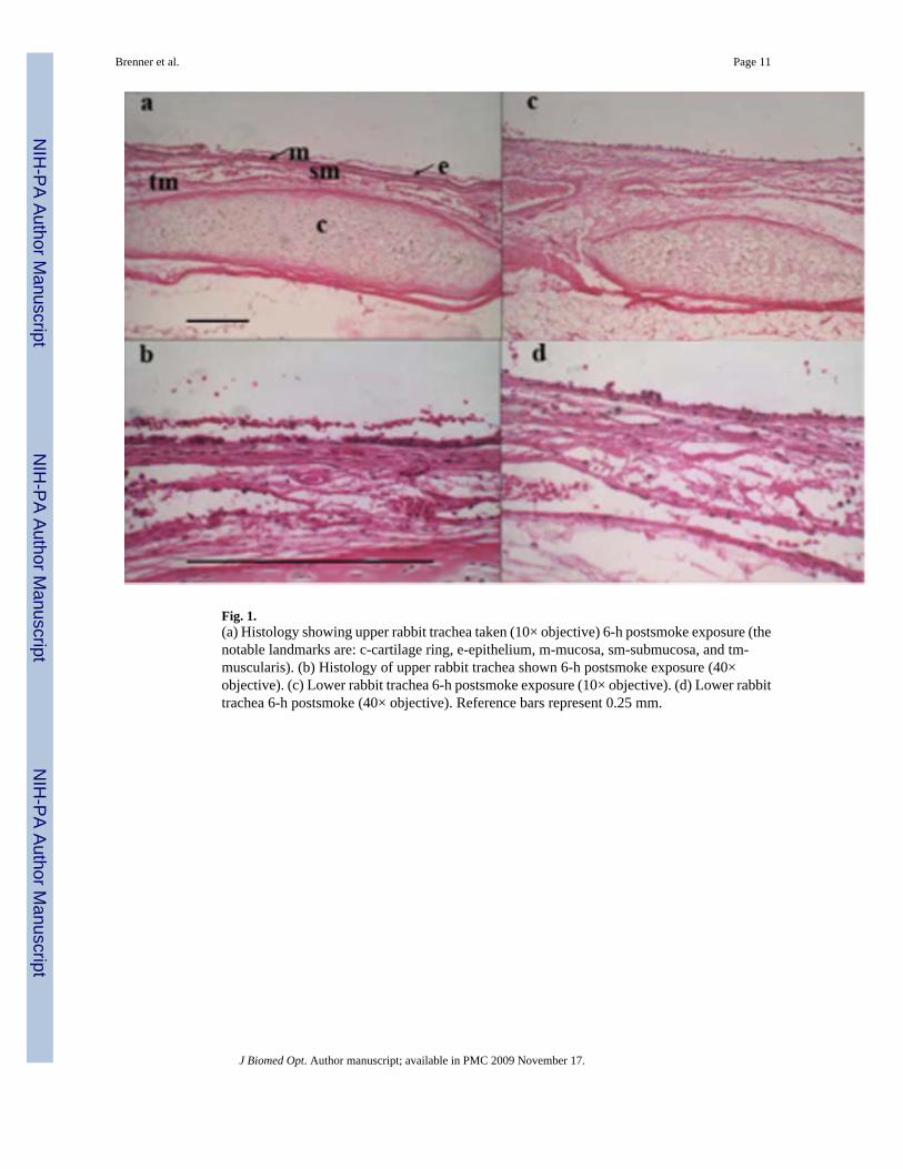
Fig. 1.(a) Histology showing upper rabbit trachea taken (10× objective) 6-h postsmoke exposure (thenotable landmarks are: c-cartilage ring, e-epithelium, m-mucosa, sm-submucosa, and tm-muscularis). (b) Histology of upper rabbit trachea shown 6-h postsmoke exposure (40×objective). (c) Lower rabbit trachea 6-h postsmoke exposure (10× objective). (d) Lower rabbittrachea 6-h postsmoke (40× objective). Reference bars represent 0.25 mm.
Brenner et al. Page 11
J Biomed Opt. Author manuscript; available in PMC 2009 November 17.
NIH
-PA Author Manuscript
NIH
-PA Author Manuscript
NIH
-PA Author Manuscript
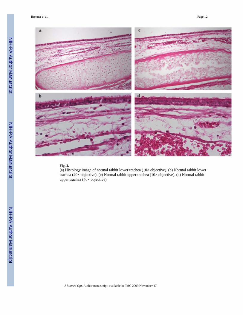
Fig. 2.(a) Histology image of normal rabbit lower trachea (10× objective). (b) Normal rabbit lowertrachea (40× objective). (c) Normal rabbit upper trachea (10× objective). (d) Normal rabbitupper trachea (40× objective).
Brenner et al. Page 12
J Biomed Opt. Author manuscript; available in PMC 2009 November 17.
NIH
-PA Author Manuscript
NIH
-PA Author Manuscript
NIH
-PA Author Manuscript
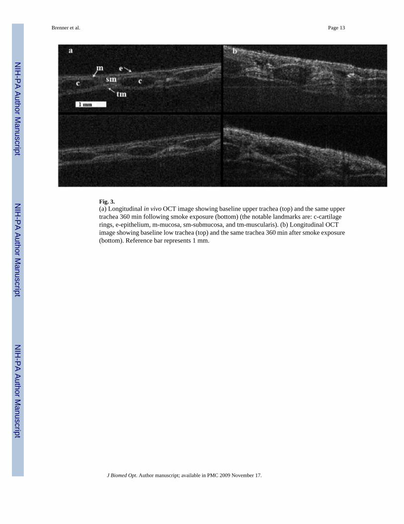
Fig. 3.(a) Longitudinal in vivo OCT image showing baseline upper trachea (top) and the same uppertrachea 360 min following smoke exposure (bottom) (the notable landmarks are: c-cartilagerings, e-epithelium, m-mucosa, sm-submucosa, and tm-muscularis). (b) Longitudinal OCTimage showing baseline low trachea (top) and the same trachea 360 min after smoke exposure(bottom). Reference bar represents 1 mm.
Brenner et al. Page 13
J Biomed Opt. Author manuscript; available in PMC 2009 November 17.
NIH
-PA Author Manuscript
NIH
-PA Author Manuscript
NIH
-PA Author Manuscript
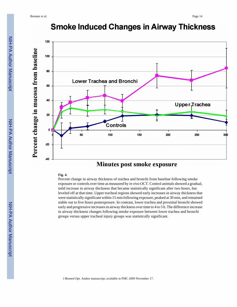
Fig. 4.Percent change in airway thickness of trachea and bronchi from baseline following smokeexposure or controls over time as measured by in vivo OCT. Control animals showed a gradual,mild increase in airway thickness that became statistically significant after two hours, butleveled off at that time. Upper tracheal regions showed early increases in airway thickness thatwere statistically significant within 15 min following exposure, peaked at 30 min, and remainedstable out to five hours postexposure. In contrast, lower trachea and proximal bronchi showedearly and progressive increases in airway thickness over time to 4 to 5 h. The difference increasein airway thickness changes following smoke exposure between lower trachea and bronchigroups versus upper tracheal injury groups was statistically significant.
Brenner et al. Page 14
J Biomed Opt. Author manuscript; available in PMC 2009 November 17.
NIH
-PA Author Manuscript
NIH
-PA Author Manuscript
NIH
-PA Author Manuscript
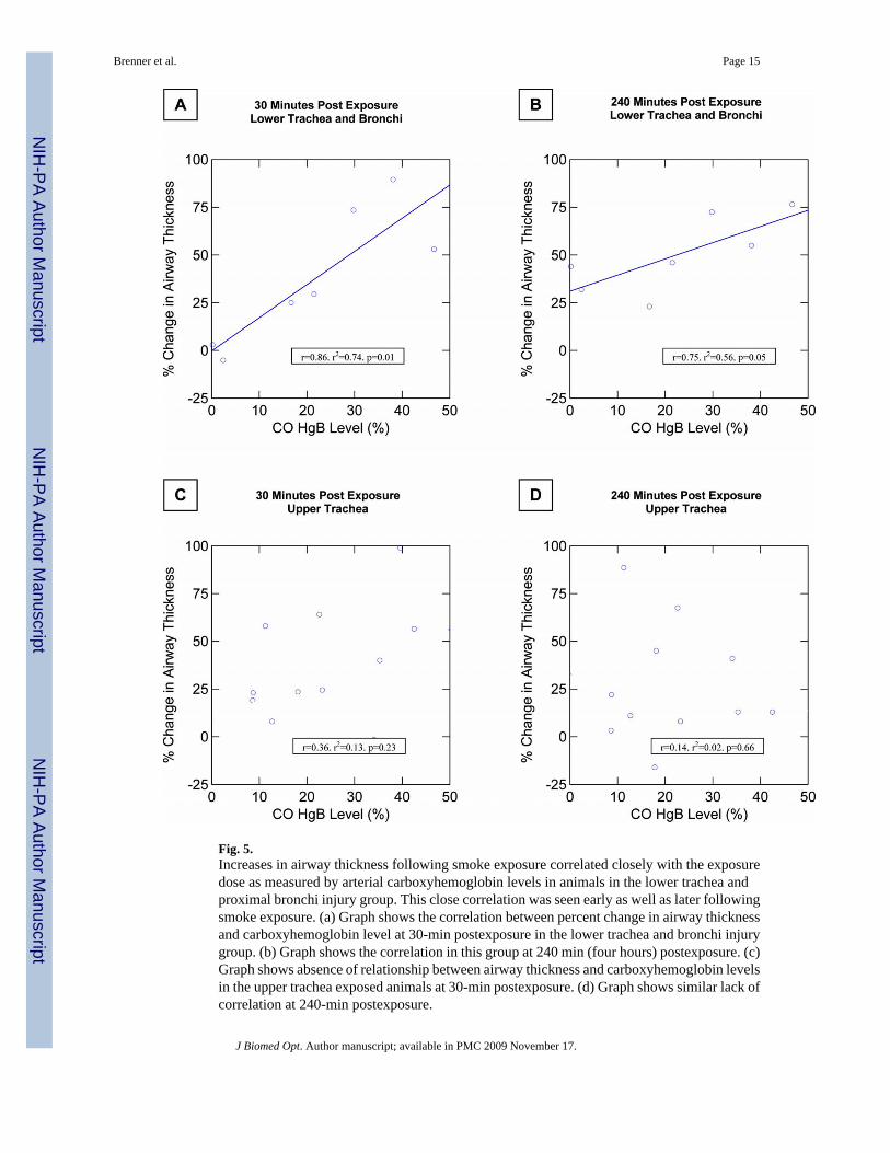
Fig. 5.Increases in airway thickness following smoke exposure correlated closely with the exposuredose as measured by arterial carboxyhemoglobin levels in animals in the lower trachea andproximal bronchi injury group. This close correlation was seen early as well as later followingsmoke exposure. (a) Graph shows the correlation between percent change in airway thicknessand carboxyhemoglobin level at 30-min postexposure in the lower trachea and bronchi injurygroup. (b) Graph shows the correlation in this group at 240 min (four hours) postexposure. (c)Graph shows absence of relationship between airway thickness and carboxyhemoglobin levelsin the upper trachea exposed animals at 30-min postexposure. (d) Graph shows similar lack ofcorrelation at 240-min postexposure.
Brenner et al. Page 15
J Biomed Opt. Author manuscript; available in PMC 2009 November 17.
NIH
-PA Author Manuscript
NIH
-PA Author Manuscript
NIH
-PA Author Manuscript
Related Documents