In Vivo Biotinylation of the Major Histocompatibility Complex (MHC) Class II/Peptide Complex by Coexpression of BirA Enzyme for the Generation of MHC Class II/Tetramers Junbao Yang, Andre ´s Jaramillo, Ruili Shi, William W. Kwok, and T. Mohanakumar ABSTRACT: Success in generation of major histocom- patibility complex (MHC) tetramer relies on application of a key technique, biotinylation of MHC molecule spe- cifically on a single lysine residue using the BirA enzyme. However, in vitro biotinylation of MHC-BSP (BirA en- zyme substrate peptide) fusion protein using BirA enzyme is laborious and is prone to losses of target proteins to unacceptable levels. To circumvent this problem, an in vivo biotinylation strategy was developed where the BirA enzyme was coexpressed with target protein, HLA- DR2BSP/MBP, in an insect cell expression system. Bac- terial BirA enzyme expressed in Drosophila melanogaster 2 (D. Mel-2) cell lines was biologically functional and was able to biotinylate secretary target protein (on specific lysine residue present on the BSP tag). Biotinylation efficiency was maximized by providing exogenous d-bi- otin in the culture medium and optimization of the expression vector ratios for cotransfection. By limiting dilution cloning, a clone was identified where the ex- pressed DR2BSP/MBP protein was completely biotinyl- ated. DR2BSP/MBP protein expressed and purified from such a clone was ready to be tetramerized with streptavi- din to be used for staining antigen-specific T cells. Human Immunology 65, 692– 699 (2004). © American Society for Histocompatibility and Immunoge- netics, 2004. Published by Elsevier Inc. KEYWORDS: in vivo biotinylation; BirA enzyme; MHC class II tetramer; expression of MHC class II/peptide complex ABBREVIATIONS BSP BirA enzyme substrate peptide HLA human leukocyte antigen MBP myelin basic protein MHC major histocompatibility complex TCR T cell receptor INTRODUCTION The introduction of peptide–major histocompatibility complex (MHC) tetramer technology has revolutionized identification and enumeration of antigen specific T cells [1]. The success of these techniques rests in the increas- ing avidity of soluble MHC/peptide complex (monomer) and T cell receptor (TCR) by tetramerization of biotin- ylated MHC/peptide monomer with avidin or streptavi- din. The key technique in production of MHC/peptide tetramer relies on the biotinylation of MHC/peptide complex monomer on a single lysine residue. This is normally accomplished by using bacterial BirA enzyme to biotinylate the lysine residue on an enzyme-specific recognition tag called BirA substrate peptide (BSP) [2] fused at the C-terminus of one of the MHC polypeptides. Biotinylation with BirA enzyme at single lysine residue solves several problems associated with traditional chem- ical-mediated biotinylation method, including random and heterogeneous modification, inactivation of protein biological function, cross-linking, and aggregation after mixing with streptavidin or avidin [3]. This also enables From the Departments of Surgery (J.Y., A.J., R.S., T.M.) and Pathol- ogy and Immunology (T.M.), Washington University School of Medicine, St. Louis, Missouri; and Benaroya Research Institute (W.W.K.), Seattle, Wash- ington. Address reprint requests to: Dr. T. Mohanakumar, Department of Sur- gery, Washington University School of Medicine, Box 8109-3328.CSRB, 660 South Euclid Avenue, St. Louis, MO 63110-1093; Tel: (314) 362- 8463; Fax: (314) 747-1560; E-mail: [email protected]. Human Immunology 65, 692– 699 (2004) © American Society for Histocompatibility and Immunogenetics, 2004 0198-8859/04/$–see front matter Published by Elsevier Inc. doi:10.1016/j.humimm.2004.04.001

Welcome message from author
This document is posted to help you gain knowledge. Please leave a comment to let me know what you think about it! Share it to your friends and learn new things together.
Transcript
IHIBC
JW
ITci[ia
oLi
g68
H©P
n Vivo Biotinylation of the Majoristocompatibility Complex (MHC) Class
I/Peptide Complex by Coexpression ofirA Enzyme for the Generation of MHClass II/Tetramers
unbao Yang, Andres Jaramillo, Ruili Shi,illiam W. Kwok, and T. Mohanakumar
eoedpasdcAn
Kc
ABSTRACT: Success in generation of major histocom-patibility complex (MHC) tetramer relies on applicationof a key technique, biotinylation of MHC molecule spe-cifically on a single lysine residue using the BirA enzyme.However, in vitro biotinylation of MHC-BSP (BirA en-zyme substrate peptide) fusion protein using BirA enzymeis laborious and is prone to losses of target proteins tounacceptable levels. To circumvent this problem, an invivo biotinylation strategy was developed where the BirAenzyme was coexpressed with target protein, HLA-DR2BSP/MBP, in an insect cell expression system. Bac-terial BirA enzyme expressed in Drosophila melanogaster 2(D. Mel-2) cell lines was biologically functional and wasable to biotinylate secretary target protein (on specific
lysine residue present on the BSP tag). Biotinylation cABBREVIATIONSMT
NTRODUCTIONydtcntrfBsiabm463; Fax: (314) 747-1560; E-mail: [email protected].
uman Immunology 65, 692–699 (2004)American Society for Histocompatibility and Immunogenetics, 2004
ublished by Elsevier Inc.
fficiency was maximized by providing exogenous d-bi-tin in the culture medium and optimization of thexpression vector ratios for cotransfection. By limitingilution cloning, a clone was identified where the ex-ressed DR2BSP/MBP protein was completely biotinyl-ted. DR2BSP/MBP protein expressed and purified fromuch a clone was ready to be tetramerized with streptavi-in to be used for staining antigen-specific Tells. Human Immunology 65, 692–699 (2004). ©merican Society for Histocompatibility and Immunoge-etics, 2004. Published by Elsevier Inc.
EYWORDS: in vivo biotinylation; BirA enzyme; MHClass II tetramer; expression of MHC class II/peptide
omplexBSP BirA enzyme substrate peptideHLA human leukocyte antigen
HC major histocompatibility complexCR T cell receptor
MBP myelin basic protein
he introduction of peptide–major histocompatibilityomplex (MHC) tetramer technology has revolutionizeddentification and enumeration of antigen specific T cells1]. The success of these techniques rests in the increas-ng avidity of soluble MHC/peptide complex (monomer)nd T cell receptor (TCR) by tetramerization of biotin-
From the Departments of Surgery (J.Y., A.J., R.S., T.M.) and Pathol-gy and Immunology (T.M.), Washington University School of Medicine, St.ouis, Missouri; and Benaroya Research Institute (W.W.K.), Seattle, Wash-
ngton.Address reprint requests to: Dr. T. Mohanakumar, Department of Sur-
ery, Washington University School of Medicine, Box 8109-3328.CSRB,60 South Euclid Avenue, St. Louis, MO 63110-1093; Tel: (314) 362-
lated MHC/peptide monomer with avidin or streptavi-in. The key technique in production of MHC/peptideetramer relies on the biotinylation of MHC/peptideomplex monomer on a single lysine residue. This isormally accomplished by using bacterial BirA enzymeo biotinylate the lysine residue on an enzyme-specificecognition tag called BirA substrate peptide (BSP) [2]used at the C-terminus of one of the MHC polypeptides.iotinylation with BirA enzyme at single lysine residue
olves several problems associated with traditional chem-cal-mediated biotinylation method, including randomnd heterogeneous modification, inactivation of proteiniological function, cross-linking, and aggregation after
ixing with streptavidin or avidin [3]. This also enables0198-8859/04/$–see front matterdoi:10.1016/j.humimm.2004.04.001
ttbHvbabPToptIb[Mofesmede
lomoapOpemsattSk[wpwpFidoipb
Mtb
MGEBDuGAGCvcwCiedBpwspDwFDDpa(g
TETLia(t�imtotSs
693In Vivo Biotinylation of MHC Class II/Peptide Complex
he proper orientation of these complexes for binding tohe TCRs. Technically, this enzyme-mediated in vitroiotinylation process is effective and efficient [1, 4].owever, it is generally cumbersome, expensive, and
ery often results in severe loss of proteins during theuffer exchange and protein concentration steps, whichre required for enzyme reaction and final removal of freeiotin from the reaction (NIAID Tetramer Core Facilityrotocols available at: http://www.emory.edu/WHSC/ETRAMER/pdf/Protocols.pdf). Therefore, eliminationf this in vitro biotinylation process could greatly sim-lify the production of MHC tetramers. Unlike produc-ion of MHC class I/peptide monomers, MHC classI/peptide complex monomers are usually expressed asiological functional proteins in insect expression system4–6]. Therefore, it provides opportunity to biotinylateHC class II/peptide complex monomers in vivo. Previ-
usly, a few studies have demonstrated that proteinsused with biotinylation site could be biotinylated in vivoither by endogenous BirA enzyme in the Escherichia coliystem or by cotransfection of the BirA enzyme in mam-alian cell system [7, 8]. Although the biotinylation
fficiency of the expressed fusion protein is low [7] or notetermined [8], these studies indicated that our hypoth-sis is feasible.
BirA enzyme is a component of biotin holoenzymeocalized in the E. coli cytoplasm [9]. Part of the functionf this enzyme is to mediate the attachment of biotinolecule to lysine residue that resulted in the formation
f an amide linkage between the carboxyl group of biotinnd the �-amino group of the lysine residue [10]. Thisosttranslational modification has exquisite specificity.nly one lysine residue in the biotin carboxyl carrierrotein of acetyl-CoA carboxylase is biotinylated by BirAnzyme in all proteins expressed in E. coli [11]. Theinimum peptide lengths of BirA enzyme recognition
ite in the biotin carboxyl carrier protein were identifieds about 75–105 aa [12]. By use of the library-screeningechnique, a series of short artificial BirA substrate pep-ides now known as BSP has also been created [2, 12].ome of these BSPs even have a superior biotinylationinetic than the biotin carboxyl carrier protein in vitro12]. Because most MHC class II/peptide complexesere expressed in insect cell system [4, 13–16], weostulated that MHC class II/peptide complex attachedith BSP tag could be biotinylated in vivo with coex-ression of BirA enzyme. By using an HLA-DRA1*0101-os-BSP/MBP-HLA-DRB1*1501-Jun (DR2BSP/MBPn text) fusion protein whose TCR specificity had beenetermined previously [17] as model protein, we dem-nstrate here that DR2BSP/MBP could be biotinylatedn vivoduring the expression. Clones that expressed com-letely biotinylated HLA-DR2BSP/MBP proteins could
e isolated by limiting dilution protocol. HLA-DR2BSP/ wBP protein isolated by this procedure was ready to beetramerized with streptavidin without further in vitroiotinylation treatment.
ATERIALS AND METHODSene Cloning and Construction ofxpression Vectorsacterial BirA gene was amplified from the genomicNA extracted from E. coli DH5� strain by PCR thatsed forward primer 5�-AAA GAA TTC ATG AAGAT AAC ACC GTG CCA-3� and reverse primer 5�-AA GGA TCC TTA TTT TTC TGC ACT TACCA-3� and inserted into the EcoRI and BamHI sites ofu-inducible pRmHa3 insect cell expression vector. Thisector was designated as p18BirA vector and expressed aytosolic form of BirA enzyme. A modified BirA geneith HLA-DR � chain leader sequence (MV-LKLPGGSCMTALTVTLMVLSSPLALSGDTG) encod-
ng region was fused in frame at the N-terminus of BirAnzyme and cloned into pRmHa3 vector. This vector wasesignated as p21BirA vector expressed secretion form ofirA enzyme in insect cells. An HLA-DR2/mylin basicrotein peptide (amino acid 85–99, MBP85–99) complexas used as a model molecule for the in vivo biotinylation
tudy. The HLA-DR2/MBP85–99 expression vectors,RmHa-DRA1*0101-Fos-Fc and pRmHa3-RB1*1501-Jun (designated as pDRB/MBP for short),ere obtained from Dr. Kai W. Wucherpfennig (Dana-arber Cancer Institute, Boston, MA). The pRmHa-RA1*0101-Fos-Fc vector was modified as pRmHa3-RA1*0101-Fos-BSP expression vector (designated asDRA-BSP) by replacing Fc encoding region with a 15a, GLNDIFEAQKIEWHE, BirA substrate peptideBSP) [2, 12] encoding gene. The sequences of fusionenes were confirmed by DNA sequencing.
ransfection of Cells With Expression Vectors andstablishing Stable Expression Cell Lineswo million Drosophila melanogaster 2 cells (D. Mel-2;ife Technologies, Grand Island, New York) were seededn a 12-well plate. Cells were cultured at 28°C overnightnd washed with 2 ml Drosophila serum-free mediumSFM; Invitrogen, Carlsbad, CA ). The transfection mix-ure was prepared by mixing 250 �l SFM containing 2g DNA of expression vectors and 250 �l SFM contain-
ng 8 �l of CELLFectin reagent (Life Technologies). Theixture was allowed to react for 20 minutes at room
emperature, then mixed with 0.8 ml SFM and overlaidn washed cells. After incubation at 28°C for 3–4 hours,he transfection mixture was replaced with 2 ml of freshFM and incubation was continued. For transient expres-ion, 24 hours after transfection, the cells were induced
ith 1 �M CuSO4 in culture medium for additional 72
ht2�mdr
wl9wict
ESc1oti4btfbnavHpHdnwicC
WAtLbhr(2asps
gmmr(a1iaTBccm
TFPbfTa�oFMs
DTFawtL52�3tt
RBBIBctDpz
694 J. Yang et al.
ours. Supernatants were tested for the expression ofarget protein. To establish stable expression cell lines,4 hours after transfection the cells were treated with 25g/ml of blasticidin (Invitrogen). The blasticidin treat-ent was continued for �2 weeks with the change of
rug selection medium every 4–5 days until stable drug-esistant cell lines were established.
To isolate individual expression clones, stable cell lineas diluted with irradiated (20,000 rads) parental cell
ine as feeder cells (1 � 106 cells/ml) and seeded in6-well plates at 10 cells/well, 3 cells/well, and 1 cell/ell, respectively. The cells were grown in SFM contain-
ng 25 �g/ml of blasticidin for 1–2 weeks until clonesould be spotted in the plates. Individual clones wereransferred to a new plate and expanded.
xpression and Purification of HLA-DR2BSP/MBPtable transfectants were expanded in Drosophila-SFMontaining 25 �g/ml blasticidin in conical flasks at 27°C40 rpm in a rotary shaker. The cells were split at ratiof 1 to 5 when the density reached 2 � 107 cells/ml. Aotal of 800 ml of cell culture at density of 107/ml werenduced with 1 mM CuSO4 and 2 �g/ml of d-biotin for
days in a 2-l flask. The cells and debris were removedy centrifugation at 10,000 g for 20 minutes. Superna-ant was further clarified by filtration through 0.2-�milter. The clarified medium was passed through a 2.5-mled volume of LB3.1 monoclonal antibody (ATCC, Ma-assas, VA) affinity column at a flow rate of 0.5 ml/mint 4°C. After the column was washed with 10 bedolumes of phosphate-buffered saline (PBS), pH 7.4, theLA-DR2BSP/MBP was eluted out with 0.1 M glycine,
H 11.5, and immediately neutralized with 1 M Tris-Cl, pH 6.8. The fractions containing proteins were
etermined by reading absorbance at optical density 280m. The fractions with higher absorbance at 280 nmere pooled and buffer-exchanged with PBS by dialyz-
ng. The protein was concentrated to 1–2 mg/ml byentrifugation with a 10-kDa molecular weight cutoffentricon (Millipore, Billerica, MA).
estern Blot Analysisfter induction, 0.2 ml of culture supernatants from each
ransfection was immunoprecipitated with 20 �l ofB3.1 monoclonal antibody conjugated Sepharose-4Beads (Amersham Biosciences, Piscataway, NJ) for 4ours. The beads were washed with PBS three times andesuspended in 25 �l of 1� NuPAGE LDS sample buffer10% glycerol, 141 mM Tris base, 106 mM Tris HCl,% LDS, 0.51 mM EDTA, 0.22 mM SERVA Blue G250nd 0.175 mM phenol red, pH 8.5; Invitrogen). Theamples were incubated at 70°C for 10 minutes andelleted by brief centrifugation. Twenty milliliters of
upernatant was loaded on a 4%–12% NuPAGE Bis-Tris Hel and separated in NuPAGE MES running buffer (50M MES, pH8.5, 50 mM Tris base, 0.1% SDS, and 1M EDTA) (Invitrogen) by electrophoresis. The sepa-
ated proteins were transferred onto a PVDF membraneMillipore) and blocked with 5% skim milk for 2 hourst room temperature. The membrane was incubated in0 ml 0.2 �g/ml streptavidin conjugated with horserad-sh peroxidase (SA-HRP; Molecular Probes, Eugene, OR)nd washed three times with PBS with 0.05% (v/v)ween-20. The biotinylation of HLA-DR2BSP/MBP byirA enzyme was detected by enhanced chemilumines-ence (Amersham Biosciences). Expression of HLA-DR �hain was detected with HRP-conjugated anti-� chainonoclonal antibody TDR31.1 (Ancell, Bayport, MN).
etramerization of HLA-DR2BSP/MBP and Geliltration Analysisurified biotinylated HLA-DR2BSP/MBP was incu-ated with one-tenth of its molar amount of streptavidinor 30 minutes. This procedure was repeated 10 times.he reaction mixture was buffer-exchanged with PBSnd passed through gel filtration column (Superose 6, 1.6
30 cm; Amersham Pharmacia) equilibrated with PBSn GP-250 FPLC instrument (Amersham Biosciences).or T cell staining, purified biotinylated HLA-DR2BSP/BP was tetramerized with phycoerythrin-conjugated
treptavidin (Molecular Probes).
evelopment of T Cell Line andetramer Stainingive million human peripheral blood lymphocytes fromhealthy donor (HLA-DRA1*0103, HLA-DRB1*1501)ere stimulated with 20 �g/ml of influenza hemoagglu-
inin peptide306–318(HA306–318; Sigma-Aldrich, St.ouis, MO) in RPMI 1640 with 10% human serum and0 units/ml of human interleukin-2 for 16 days in a4-well plate. One million T cells was stained with 20g of HLA-DRB1*0103/HA306–318-PE tetramer at7°C for 90 min and with anti–CD4-FITC at roomemperature for 20 minutes, and analyzed by flow cy-ometry.
ESULTSiotinylation of HLA-DR2BSP/MBP Monomer onSP Tag In Vivo
n order to biotinylate HLA-DR2BSP/MBP in vivo, theirA gene from E. coli K12 strain was cloned into insectell expression vector under the control of the metallo-hionein promoter. In vivo biotinylation of HLA-R2BSP/MBP was conducted by cotransfection ofDRA-BSP vector, pDRB/MBP vector, and BirA en-yme expression vector p18BirA into D. Mel-2 cells.
LA-DR2BSP/MBP was biotinylated by cytosolic BirA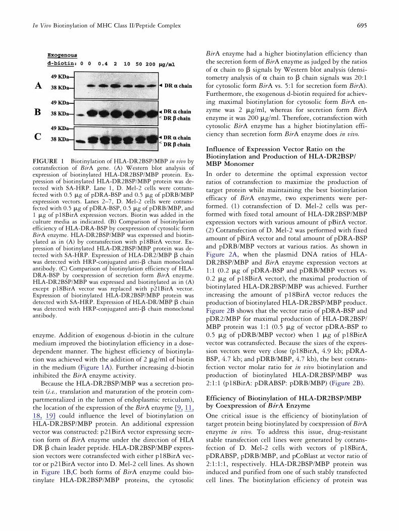
emdtii
tpt1HvtDstit
BtotfFizecc
IBMIrteffe(aaFD10bipFpM0vsBfp2
EbOtesfp2i
Fceptfef1ceByptwaDHeEdwa
695In Vivo Biotinylation of MHC Class II/Peptide Complex
nzyme. Addition of exogenous d-biotin in the cultureedium improved the biotinylation efficiency in a dose-
ependent manner. The highest efficiency of biotinyla-ion was achieved with the addition of 2 �g/ml of biotinn the medium (Figure 1A). Further increasing d-biotinnhibited the BirA enzyme activity.
Because the HLA-DR2BSP/MBP was a secretion pro-ein (i.e., translation and maturation of the protein com-artmentalized in the lumen of endoplasmic reticulum),he location of the expression of the BirA enzyme [9, 11,8, 19] could influence the level of biotinylation onLA-DR2BSP/MBP protein. An additional expression
ector was constructed: p21BirA vector expressing secre-ion form of BirA enzyme under the direction of HLAR � chain leader peptide. HLA-DR2BSP/MBP expres-
ion vectors were cotransfected with either p18BirA vec-or or p21BirA vector into D. Mel-2 cell lines. As shownn Figure 1B,C both forms of BirA enzyme could bio-
IGURE 1 Biotinylation of HLA-DR2BSP/MBP in vivo byotransfection of BirA gene. (A) Western blot analysis ofxpression of biotinylated HLA-DR2BSP/MBP protein. Ex-ression of biotinylated HLA-DR2BSP/MBP protein was de-ected with SA-HRP. Lane 1, D. Mel-2 cells were cotrans-ected with 0.5 �g of pDRA-BSP and 0.5 �g of pDRB/MBPxpression vectors. Lanes 2–7, D. Mel-2 cells were cotrans-ected with 0.5 �g of pDRA-BSP, 0.5 �g of pDRB/MBP, and�g of p18BirA expression vectors. Biotin was added in the
ulture media as indicated. (B) Comparison of biotinylationfficiency of HLA-DRA-BSP by coexpression of cytosolic formirA enzyme. HLA-DR2BSP/MBP was expressed and biotin-lated as in (A) by cotransfection with p18BirA vector. Ex-ression of biotinylated HLA-DR2BSP/MBP protein was de-ected with SA-HRP. Expression of HLA-DR2/MBP � chainas detected with HRP-conjugated anti-� chain monoclonal
ntibody. (C) Comparison of biotinylation efficiency of HLA-RA-BSP by coexpression of secretion form BirA enzyme.LA-DR2BSP/MBP was expressed and biotinylated as in (A)
xcept p18BirA vector was replaced with p21BirA vector.xpression of biotinylated HLA-DR2BSP/MBP protein wasetected with SA-HRP. Expression of HLA-DR/MBP � chainas detected with HRP-conjugated anti-� chain monoclonal
ntibody.
inylate HLA-DR2BSP/MBP proteins, the cytosolic c
irA enzyme had a higher biotinylation efficiency thanhe secretion form of BirA enzyme as judged by the ratiosf � chain to � signals by Western blot analysis (densi-ometry analysis of � chain to � chain signals was 20:1or cytosolic form BirA vs. 5:1 for secretion form BirA).urthermore, the exogenous d-biotin required for achiev-ng maximal biotinylation for cytosolic form BirA en-yme was 2 �g/ml, whereas for secretion form BirAnzyme it was 200 �g/ml. Therefore, cotransfection withytosolic BirA enzyme has a higher biotinylation effi-iency than secretion form BirA enzyme does in vivo.
nfluence of Expression Vector Ratio on theiotinylation and Production of HLA-DR2BSP/BP Monomer
n order to determine the optimal expression vectoratios of cotransfection to maximize the production ofarget protein while maintaining the best biotinylationfficacy of BirA enzyme, two experiments were per-ormed. (1) cotransfection of D. Mel-2 cells was per-ormed with fixed total amount of HLA-DR2BSP/MBPxpression vectors with various amount of pBirA vector.2) Cotransfection of D. Mel-2 was performed with fixedmount of pBirA vector and total amount of pDRA-BSPnd pDRB/MBP vectors at various ratios. As shown inigure 2A, when the plasmid DNA ratios of HLA-R2BSP/MBP and BirA enzyme expression vectors at:1 (0.2 �g of pDRA-BSP and pDRB/MBP vectors vs..2 �g of p18BirA vector), the maximal production ofiotinylated HLA-DR2BSP/MBP was achieved. Furtherncreasing the amount of p18BirA vector reduces theroduction of biotinylated HLA-DR2BSP/MBP product.igure 2B shows that the vector ratio of pDRA-BSP andDR2/MBP for maximal production of HLA-DR2BSP/BP protein was 1:1 (0.5 �g of vector pDRA-BSP to
.5 �g of pDRB/MBP vector) when 1 �g of p18BirAector was cotransfected. Because the sizes of the expres-ion vectors were very close (p18BirA, 4.9 kb; pDRA-SP, 4.7 kb; and pDRB/MBP, 4.7 kb), the best cotrans-
ection vector molar ratio for in vivo biotinylation androduction of biotinylated HLA-DR2BSP/MBP was:1:1 (p18BirA: pDRABSP: pDRB/MBP) (Figure 2B).
fficiency of Biotinylation of HLA-DR2BSP/MBPy Coexpression of BirA Enzymene critical issue is the efficiency of biotinylation of
arget protein being biotinylated by coexpression of BirAnzyme in vivo. To address this issue, drug-resistanttable transfection cell lines were generated by cotrans-ection of D. Mel-2 cells with vectors of p18BirA,DRABSP, pDRB/MBP, and pCoBlast at vector ratio of:1:1:1, respectively. HLA-DR2BSP/MBP protein wasnduced and purified from one of such stably transfected
ell lines. The biotinylation efficiency of protein was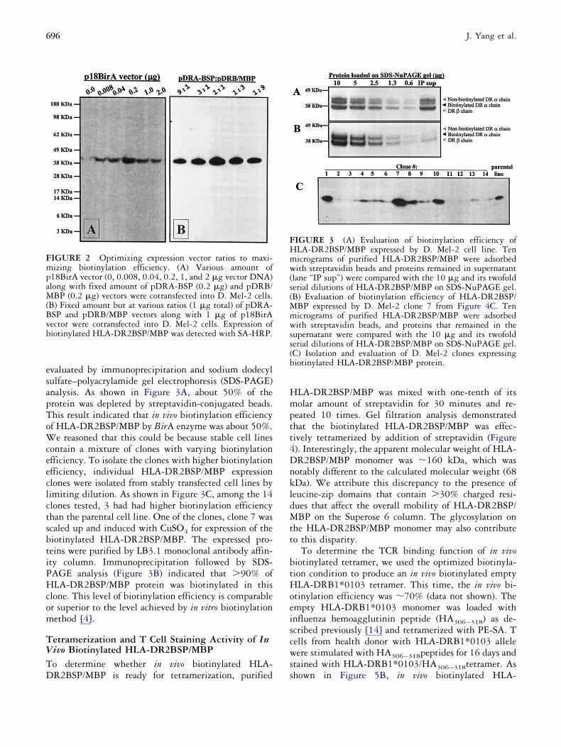
esapToWceeclctsbtiPHcom
TVTD
Hmptt4DnkldMtt
btHoeiscws
FmpaM(Bvb
FHmw(s(Mmwss(b
696 J. Yang et al.
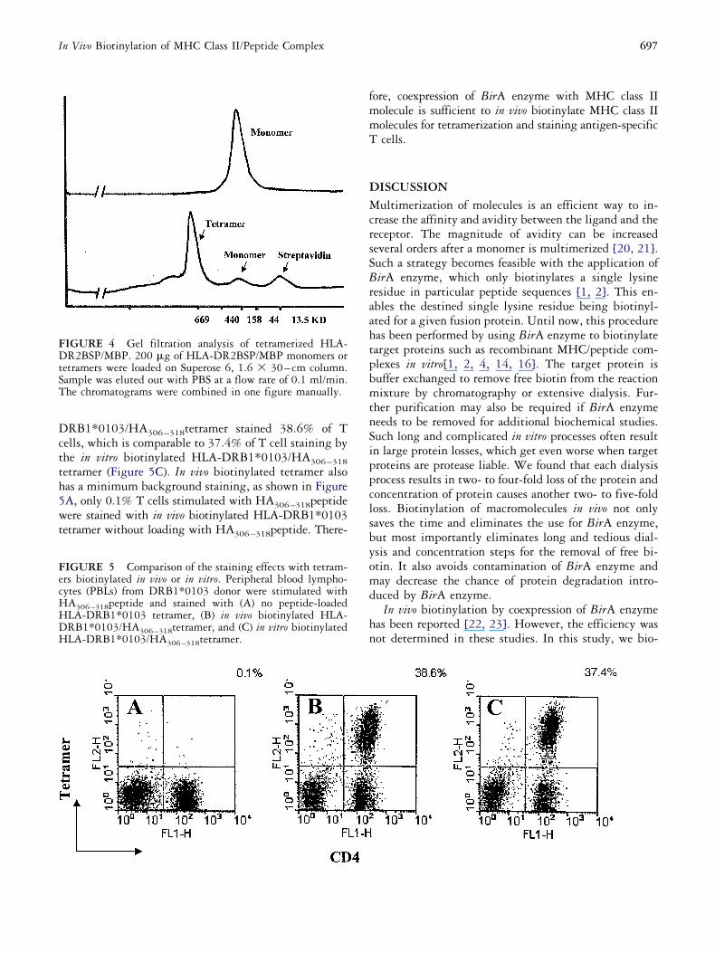
valuated by immunoprecipitation and sodium dodecylulfate–polyacrylamide gel electrophoresis (SDS-PAGE)nalysis. As shown in Figure 3A, about 50% of therotein was depleted by streptavidin-conjugated beads.his result indicated that in vivo biotinylation efficiencyf HLA-DR2BSP/MBP by BirA enzyme was about 50%.
e reasoned that this could be because stable cell linesontain a mixture of clones with varying biotinylationfficiency. To isolate the clones with higher biotinylationfficiency, individual HLA-DR2BSP/MBP expressionlones were isolated from stably transfected cell lines byimiting dilution. As shown in Figure 3C, among the 14lones tested, 3 had had higher biotinylation efficiencyhan the parental cell line. One of the clones, clone 7 wascaled up and induced with CuSO4 for expression of theiotinylated HLA-DR2BSP/MBP. The expressed pro-eins were purified by LB3.1 monoclonal antibody affin-ty column. Immunoprecipitation followed by SDS-AGE analysis (Figure 3B) indicated that �90% ofLA-DR2BSP/MBP protein was biotinylated in this
lone. This level of biotinylation efficiency is comparabler superior to the level achieved by in vitro biotinylationethod [4].
etramerization and T Cell Staining Activity of Inivo Biotinylated HLA-DR2BSP/MBPo determine whether in vivo biotinylated HLA-
IGURE 2 Optimizing expression vector ratios to maxi-izing biotinylation efficiency. (A) Various amount of
18BirA vector (0, 0.008, 0.04, 0.2, 1, and 2 �g vector DNA)long with fixed amount of pDRA-BSP (0.2 �g) and pDRB/BP (0.2 �g) vectors were cotransfected into D. Mel-2 cells.
B) Fixed amount but at various ratios (1 �g total) of pDRA-SP and pDRB/MBP vectors along with 1 �g of p18BirAector were cotransfected into D. Mel-2 cells. Expression ofiotinylated HLA-DR2BSP/MBP was detected with SA-HRP.
R2BSP/MBP is ready for tetramerization, purified s
LA-DR2BSP/MBP was mixed with one-tenth of itsolar amount of streptavidin for 30 minutes and re-
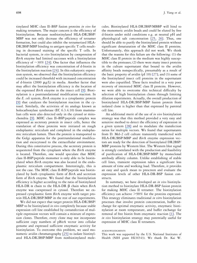
eated 10 times. Gel filtration analysis demonstratedhat the biotinylated HLA-DR2BSP/MBP was effec-ively tetramerized by addition of streptavidin (Figure). Interestingly, the apparent molecular weight of HLA-R2BSP/MBP monomer was �160 kDa, which wasotably different to the calculated molecular weight (68Da). We attribute this discrepancy to the presence ofeucine-zip domains that contain �30% charged resi-ues that affect the overall mobility of HLA-DR2BSP/BP on the Superose 6 column. The glycosylation on
he HLA-DR2BSP/MBP monomer may also contributeo this disparity.
To determine the TCR binding function of in vivoiotinylated tetramer, we used the optimized biotinyla-ion condition to produce an in vivo biotinylated emptyLA-DRB1*0103 tetramer. This time, the in vivo bi-
tinylation efficiency was �70% (data not shown). Thempty HLA-DRB1*0103 monomer was loaded withnfluenza hemoagglutinin peptide (HA306–318) as de-cribed previously [14] and tetramerized with PE-SA. Tells from health donor with HLA-DRB1*0103 alleleere stimulated with HA306–318peptides for 16 days and
tained with HLA-DRB1*0103/HA306–318tetramer. As
IGURE 3 (A) Evaluation of biotinylation efficiency ofLA-DR2BSP/MBP expressed by D. Mel-2 cell line. Tenicrograms of purified HLA-DR2BSP/MBP were adsorbedith streptavidin beads and proteins remained in supernatant
lane “IP sup”) were compared with the 10 �g and its twofolderial dilutions of HLA-DR2BSP/MBP on SDS-NuPAGE gel.B) Evaluation of biotinylation efficiency of HLA-DR2BSP/
BP expressed by D. Mel-2 clone 7 from Figure 4C. Tenicrograms of purified HLA-DR2BSP/MBP were adsorbedith streptavidin beads, and proteins that remained in the
upernatant were compared with the 10 �g and its twofolderial dilutions of HLA-DR2BSP/MBP on SDS-NuPAGE gel.C) Isolation and evaluation of D. Mel-2 clones expressingiotinylated HLA-DR2BSP/MBP protein.
hown in Figure 5B, in vivo biotinylated HLA-
Dctth5wt
fmmT
DMcrsSBraahtpbmtnSippclsbyomd
hn
FecHHDH
FDtST
697In Vivo Biotinylation of MHC Class II/Peptide Complex
RB1*0103/HA306–318tetramer stained 38.6% of Tells, which is comparable to 37.4% of T cell staining byhe in vitro biotinylated HLA-DRB1*0103/HA306–318etramer (Figure 5C). In vivo biotinylated tetramer alsoas a minimum background staining, as shown in FigureA, only 0.1% T cells stimulated with HA306–318peptideere stained with in vivo biotinylated HLA-DRB1*0103
etramer without loading with HA306–318peptide. There-
IGURE 5 Comparison of the staining effects with tetram-rs biotinylated in vivo or in vitro. Peripheral blood lympho-ytes (PBLs) from DRB1*0103 donor were stimulated withA306–318peptide and stained with (A) no peptide-loadedLA-DRB1*0103 tetramer, (B) in vivo biotinylated HLA-RB1*0103/HA306–318tetramer, and (C) in vitro biotinylatedLA-DRB1*0103/HA306–318tetramer.
IGURE 4 Gel filtration analysis of tetramerized HLA-R2BSP/MBP. 200 �g of HLA-DR2BSP/MBP monomers or
etramers were loaded on Superose 6, 1.6 � 30– cm column.ample was eluted out with PBS at a flow rate of 0.1 ml/min.he chromatograms were combined in one figure manually.
ore, coexpression of BirA enzyme with MHC class IIolecule is sufficient to in vivo biotinylate MHC class IIolecules for tetramerization and staining antigen-specificcells.
ISCUSSIONultimerization of molecules is an efficient way to in-
rease the affinity and avidity between the ligand and theeceptor. The magnitude of avidity can be increasedeveral orders after a monomer is multimerized [20, 21].uch a strategy becomes feasible with the application ofirA enzyme, which only biotinylates a single lysine
esidue in particular peptide sequences [1, 2]. This en-bles the destined single lysine residue being biotinyl-ted for a given fusion protein. Until now, this procedureas been performed by using BirA enzyme to biotinylatearget proteins such as recombinant MHC/peptide com-lexes in vitro[1, 2, 4, 14, 16]. The target protein isuffer exchanged to remove free biotin from the reactionixture by chromatography or extensive dialysis. Fur-
her purification may also be required if BirA enzymeeeds to be removed for additional biochemical studies.uch long and complicated in vitro processes often resultn large protein losses, which get even worse when targetroteins are protease liable. We found that each dialysisrocess results in two- to four-fold loss of the protein andoncentration of protein causes another two- to five-foldoss. Biotinylation of macromolecules in vivo not onlyaves the time and eliminates the use for BirA enzyme,ut most importantly eliminates long and tedious dial-sis and concentration steps for the removal of free bi-tin. It also avoids contamination of BirA enzyme anday decrease the chance of protein degradation intro-
uced by BirA enzyme.In vivo biotinylation by coexpression of BirA enzyme
as been reported [22, 23]. However, the efficiency wasot determined in these studies. In this study, we bio-
tmbMfDibBebtscomtnt[thlcetemttDsicypnyfeHept
Metssgbna
ctdpssUtMtiattwrwsdbic
ssarfHtMioacaaes
tfeTpcnrie
A
T
698 J. Yang et al.
inylated MHC class II–BSP fusion proteins in vivo foraking tetramers. The major concern is the efficiency of
iotinylation. Because nonbiotinylated HLA-DR2BSP/BP was not only decrease the efficiency of tetramer
ormation, but could also inhibit the tetramerized HLA-R2BSP/MBP binding to antigen specific T cells result-
ng in decreased staining of the specific T cells. Inacterial system, in vivo biotinylation by coexpression ofirA enzyme had limited successes with a biotinylationfficiency of �30% [24]. One factor that influences theiotinylation efficiency was concentration of d-biotin inhe biotinylation reaction [24]. In this eukaryotic expres-ion system, we observed that the biotinylation efficiencyould be increased threefold with increased concentrationf d-biotin (2000 �g/L) in media. Another factor thatay affect the biotinylation efficiency is the location of
he expressed BirA enzyme in the insect cell [9]. Bioti-ylation is a posttranslational modification reaction. Inhe E. coli system, BirA enzyme is a cytoplasmic protein9] that catalyzes the biotinylation reaction in the cy-osol. Similarly, the activities of its analogs known asolocarboxylase synthetase (EC 6.3.4.10) from mamma-ian cells were also detected only in the cytosol or mito-hondria [9]. MHC class II–BSP/peptide complex wasxpressed as secretory protein in insect cells, hence theranslation of these proteins were initiated on roughndoplasmic reticulum and completed in the endoplas-ic reticulum lumen. Then the protein is transported to
he Golgi apparatus for the posttranslational modifica-ion and excocytosed to the extracellular environment.uring this constitutive process, the secretory protein is
equestered from the cytoplasm where the BirA enzymes located. Therefore, one would speculate that MHClass II-BSP/peptide monomer is only able to be biotin-lated when BirA enzyme was also located in the endo-lasmic reticulum compartment. Interestingly, this isot the case. The MHC class II-BSP/peptide was biotin-lated by both cytoplasmic form of BirA and secretionorm of BirA enzyme. We found that the biotinylationfficiency is higher according to the ratio of biotinylatedLA-DR � chain to the HLA-DR � chain when BirA
nzyme was coexpressed in cytosol. Therefore we ex-ressed cytoplasmic form BirA enzyme with target pro-ein HLA-DR2BSP/MBP in the rest of our experiments.
We did not expect that target protein HLA-DR2BSP/BP to be biotinylated in vivo completely because stable
xpression cell line established by cotransfection of mul-iple expression vectors will contain a mixture of expres-ion clones. Therefore, every clone may not incorporateufficient copy numbers of pBirA vector into cellularenome and expressed sufficient enzymatic activity foriotinylation. To overcome this problem, we used mo-omeric avidin chromatography [25] to isolate biotinyl-
ted HLA-DR2BSP/MBP from nonbiotinylated mole- Hules. Biotinylated HLA-DR2BSP/MBBP will bind tohe monomeric avidin beads and could be eluted by free-biotin under mild conditions e.g. at neutral pH andhysiological salt concentration [25, 26]. Thus, onehould be able to purify the biotinylated protein withoutignificant denaturation of the MHC class II proteins.nfortunately, this approach did not work. We think
hat the reasons for this failure are the following: (1) theHC class II protein in the medium was highly suscep-
ible to the proteases; (2) there were many insect proteinsn the culture supernatant that bound to the avidinffinity beads nonspecifically, presumably as a result ofhe basic property of avidin (pI 10) [27]; and (3) some ofhe biotinylated insect cell proteins in the supernatantere also copurified. These facts resulted in a very poor
ecovery of interested MHC class II proteins. However,e were able to overcome this technical difficulty by
election of high biotinylation clones through limitingilution experiments. Actually, the overall production ofiotinylated HLA-DR2BSP/MBP fusion protein fromsolated clone is higher than that expressed by parentalell line.
An additional merit of the use of in vivo biotinylationtrategy was that this method provided a very easy andensitive method to detect the efficiency of expression ingiven system [28] and to modulate the transfection
atios for multiple vectors. We found that supernatantsrom D. Mel-2 cell culture transiently transfected withLA-DR2BSP/MBP and BirA enzyme expression vec-
ors are ready for the detection of biotinylated DR2BSP/BP proteins by Western blot. The Western blot signal
s strongly correlated with the production and feasibilityf purification of HLA-DR2BSP/MBP by monoclonalntibody affinity column. Unlike establishing of stableell lines, transient expression takes a significant lessmount of time and working load. Therefore, it providesn easy and quick mean to prescreen and evaluate thexpression levels of other HLA-DR-BSP fusion con-tructs.
In summary, we have developed an in vivo biotinyla-ion method to biotinylate HLA-DR-BSP fusion proteinor making MHC class II tetramer. The biotinylationfficiency can achieve �90% for the proteins expressed.his strategy eliminates traditional in vitro biotinylationrocesses that involve protein concentration, buffer ex-hange for optimal enzymatic activity, enzymatic bioti-ylation at room temperature, and buffer exchange foremoval of free biotin from enzymatic reaction [1]. Then vivo biotinylation strategy may potentially useful forxpression of MHC class II tetramers.
CKNOWLEDGMENTS
his work was supported by the U.S. National Institutes of
ealth (NIH grant HL56543). We thank Dr. Kai W.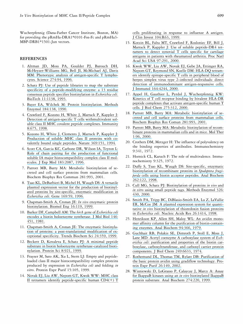
WfM
R
1
1
1
1
1
1
1
1
1
1
2
2
2
2
2
2
2
2
2
699In Vivo Biotinylation of MHC Class II/Peptide Complex
ucherpfennig (Dana-Farber Cancer Institute, Boston, MA)or providing the pRmHa-DRA1*0101-Fos-Fc and pRmHa3-
BP-DRB1*1501-Jun vectors.
EFERENCES
1. Altman JD, Moss PA, Goulder PJ, Barouch DH,McHeyzer-Williams MG, Bell JI, McMichael AJ, DavisMM: Phenotypic analysis of antigen-specific T lympho-cytes. Science 274:94, 1996.
2. Schatz PJ: Use of peptide libraries to map the substratespecificity of a peptide-modifying enzyme: a 13 residueconsensus peptide specifies biotinylation in Escherichia coli.BioTech 11:1138, 1993.
3. Bayer EA, Wilchek M: Protein biotinylation. MethodsEnzymol 184:138, 1990.
4. Crawford F, Kozono H, White J, Marrack P, Kappler J:Detection of antigen-specific T cells withmultivalent sol-uble class II MHC covalent peptide complexes. Immunity8:675, 1998.
5. Kozono H, White J, Clements J, Marrack P, Kappler J:Production of soluble MHC class II proteins with co-valently bound single peptides. Nature 369:151, 1994.
6. Scott CA, Garcia KC, Carbone DR, Wilson IA, Teyton L:Role of chain pairing for the production of functionalsoluble IA major histocompatibility complex class II mol-ecules. J Exp Med 183:2087, 1996.
7. Parrott MB, Barry MA: Metabolic biotinylation of se-creted and cell surface proteins from mammalian cells.Biochem Biophys Res Commun 281:993, 2001.
8. Tsao KL, DeBarbieri B, Michel H, Waugh DS: A versatileplasmid expression vector for the production of biotinyl-ated proteins by site-specific, enzymatic modification inEscherichia coli. Gene 169:59, 1996.
9. Chapman-Smith A, Cronan JE: In vivo enzymatic proteinbiotinylation. Biomol Eng 16:119, 1999.
0. Barker DF, Campbell AM: The birA gene of Escherichia coliencodes a biotin holoenzyme synthetase. J Mol Biol 146:451, 1981.
1. Chapman-Smith A, Cronan JE: The enzymatic biotinyla-tion of proteins: a post-translational modification of ex-ceptional specificity. Trends Biochem Sci 24:359, 1999.
2. Beckett D, Kovaleva E, Schatz PJ: A minimal peptidesubstrate in biotin holoenzyme synthetase-catalyzed bioti-nylation. Protein Sci 8:921, 1999.
3. Frayser M, Sato AK, Xu L, Stern LJ: Empty and peptide-loaded class II major histocompatibility complex proteinsproduced by expression in Escherichia coli and folding invitro. Protein Expr Purif 15:105, 1999.
4. Novak EJ, Liu AW, Nepom GT, Kwok WW: MHC classII tetramers identify peptide-specific human CD4() T
cells proliferating in response to influenze A antigen.J Clin Invest 104:R63, 1999.
5. Kotzin BL, Falta MT, Crawford F, Rosloniec EF, Bill J,Marrack P, Kappler J: Use of soluble peptide-DR4 tet-ramers to detect synovial T cells specific for cartilageantigens in patients with rheumatoid arthritis. Proc NatlAcad Sci USA 97:291, 2000.
6. Kwok WW, Liu AW, Novak EJ, Gebe JA, Ettinger RA,Nepom GT, Reymond SN, Koelle DM: HLA-DQ tetram-ers identify epitope-specific T cells in peripheral blood ofherpes simplex virus type 2–infected individuals: directdetection of immunodominant antigen-responsive cells.J Immunol 164:4244, 2000.
7. Appel H, Gauthier L, Pyrdol J, Wucherpfennig KW:Kinetics of T cell receptor binding by bivalent HLA-DRpeptide complexes that activate antigen-specific human Tcells. J Biol Chem 275:312, 2000.
8. Parrott MB, Barry MA: Metabolic biotinylation of se-creted and cell surface proteins from mammalian cells.Biochem Biophys Res Comun 281:993, 2001.
9. Parrott MB, Barry MA: Metabolic biotinylation of recom-binant proteins in mammalian cells and in mice. Mol Ther1:96, 2000.
0. Crothers DM, Metzger H: The influence of polyvalency onthe binding eoperties of antibodies. Immunochemistry9:341, 1972.
1. Hornick CL, Karuch F: The role of multivalence. Immu-nochemistry 9:325, 1972.
2. Duffy S, Tsao KL, Waugh DS: Site-specific, enzymaticbiotinylation of recombinant proteins in Spodoptera frugi-perda cells using biotin acceptor peptides. Anal Biochem262:122, 1998.
3. Cull MG, Schatz PJ: Biotinylation of proteins in vivo andin vitro using small peptide tags. Methods Enzymol 326:430, 2000.
4. Smith PA, Tripp BC, DiBlasio-Smith EA, Lu Z, LaVallieER, McCoy JM: A plasmid expression system for quanti-tative in vivo biotinylation of thioredoxin fusion proteinsin Escherichia coli. Nucleic Acids Res 26:1414, 1998.
5. Henrikson KP, Allen SH, Maloy WL: An avidin mono-mer affinity column for the purification of biotin-contain-ing enzymes. Anal Biochem 94:366, 1979.
6. Guchhait RB, Polakis SE, Dimroth P, Stoll E, Moss J,Lane MD: Acetyl coenzyme A carboxylase system of Esch-erichia coli: purification and properties of the biotin car-boxylase, carboxyltransferase, and carboxyl carrier proteincomponents. J Biol Chem 249:6633, 1974.
7. Rothemund DL, Thomas TM, Rylatt DB: Purification ofthe basic protein avidin using gradiflow technology. Pro-tein Expr Purif 26:149, 2002.
8. Wisniewski D, LoGrasso P, Calaycay J, Marcy A: Assayfor IkappaB kinases using an in vivo biotinylated IkappaBprotein substrate. Anal Biochem 274:220, 1999.
Related Documents


