ORIGINAL RESEARCH In vitro trans-differentiation of human umbilical cord derived hematopoietic stem cells into hepatocyte like cells using combination of growth factors for cell based therapy S. Sellamuthu • R. Manikandan • R. Thiagarajan • G. Babu • D. Dinesh • D. Prabhu • C. Arulvasu Received: 1 November 2010 / Accepted: 11 January 2011 / Published online: 17 February 2011 Ó Springer Science+Business Media B.V. 2011 Abstract The aim of the study was to develop a new strategy for the differentiation of hematopoietic stem cell (HSC) derived from UCB into hepatocyte like cells and also to estimate the effects of combi- nation of fibroblast growth factor 4 (FGF 4) and hepatocyte growth factor (HGF) on hematopoietic stem cell differentiation. HSCs were isolated and purified by magnetic activated cell sorting. HSCs were induced to hepatocyte like cells under a 2-step protocol with combination of growth factors. Reverse transcription polymerase chain reaction was per- formed to detect multiple genes related to hepatocyte like cells development and function. Hepatocyte like morphology was illustrated by inverted repeat micro- scope and the secretion of albumin and a- fetoprotein by these cells was confirmed by enzyme linked immunosorbent assay. Hepatocyte like cells was observed at the end of the protocol (days 14). These differentiated cells were observed to show high expression of genes related to hepatocytes (trypto- phan 2, 3-dioxygenase [TO], glucose 6-phosphate [G6P], cytokeratin 18 [CK 18], albumin and a- fetoprotein [AFP]). The quantities of albumin and AFP at day 0 were low and upon differentiation the cells were able to produce albumin and AFP at high levels. Our results show a new strategy for differen- tiation in a short duration, using a combination of growth factors for the differentiation of umbilical cord blood derived HSC into hepatocyte like cells under certain in vitro conditions. After further studies this approach has the potency, for widespread cell replace- ment therapy for liver diseases. Keywords Growth factors Hematopoietic stem cell Hepatocyte Trans-differentiation Umbilical cord blood Introduction Most liver diseases lead to hepatocyte dysfunction with the possibility of eventual organ failure. In India, mortality rate due to liver disease is high, 60% of patients admitted to the gastroenterology depart- ments are with liver diseases. The most common liver diseases are fibrosis, cirrhosis, hepatitis, cancer, etc., occurring as a result of viral infection or alcohol abuse (Trey and Davidson 1970). The liver is a S. Sellamuthu G. Babu D. Dinesh D. Prabhu C. Arulvasu (&) Department of Zoology, University of Madras, Guindy Campus, Chennai 600 025, Tamilnadu, India e-mail: [email protected] R. Manikandan Department of Animal Health and Management, Alagappa University, Karaikudi 03, Tamilnadu, India R. Thiagarajan Department of Biotechnology, School of Chemical and Biotechnology, Sastra University , Thanjavur 613 401, Tamilnadu, India 123 Cytotechnology (2011) 63:259–268 DOI 10.1007/s10616-011-9337-x

Welcome message from author
This document is posted to help you gain knowledge. Please leave a comment to let me know what you think about it! Share it to your friends and learn new things together.
Transcript
ORIGINAL RESEARCH
In vitro trans-differentiation of human umbilical cordderived hematopoietic stem cells into hepatocyte like cellsusing combination of growth factors for cell based therapy
S. Sellamuthu • R. Manikandan •
R. Thiagarajan • G. Babu • D. Dinesh •
D. Prabhu • C. Arulvasu
Received: 1 November 2010 / Accepted: 11 January 2011 / Published online: 17 February 2011
� Springer Science+Business Media B.V. 2011
Abstract The aim of the study was to develop a
new strategy for the differentiation of hematopoietic
stem cell (HSC) derived from UCB into hepatocyte
like cells and also to estimate the effects of combi-
nation of fibroblast growth factor 4 (FGF 4) and
hepatocyte growth factor (HGF) on hematopoietic
stem cell differentiation. HSCs were isolated and
purified by magnetic activated cell sorting. HSCs
were induced to hepatocyte like cells under a 2-step
protocol with combination of growth factors. Reverse
transcription polymerase chain reaction was per-
formed to detect multiple genes related to hepatocyte
like cells development and function. Hepatocyte like
morphology was illustrated by inverted repeat micro-
scope and the secretion of albumin and a- fetoprotein
by these cells was confirmed by enzyme linked
immunosorbent assay. Hepatocyte like cells was
observed at the end of the protocol (days 14). These
differentiated cells were observed to show high
expression of genes related to hepatocytes (trypto-
phan 2, 3-dioxygenase [TO], glucose 6-phosphate
[G6P], cytokeratin 18 [CK 18], albumin and
a- fetoprotein [AFP]). The quantities of albumin and
AFP at day 0 were low and upon differentiation the
cells were able to produce albumin and AFP at high
levels. Our results show a new strategy for differen-
tiation in a short duration, using a combination of
growth factors for the differentiation of umbilical cord
blood derived HSC into hepatocyte like cells under
certain in vitro conditions. After further studies this
approach has the potency, for widespread cell replace-
ment therapy for liver diseases.
Keywords Growth factors � Hematopoietic stem
cell � Hepatocyte � Trans-differentiation � Umbilical
cord blood
Introduction
Most liver diseases lead to hepatocyte dysfunction
with the possibility of eventual organ failure. In
India, mortality rate due to liver disease is high, 60%
of patients admitted to the gastroenterology depart-
ments are with liver diseases. The most common liver
diseases are fibrosis, cirrhosis, hepatitis, cancer, etc.,
occurring as a result of viral infection or alcohol
abuse (Trey and Davidson 1970). The liver is a
S. Sellamuthu � G. Babu � D. Dinesh �D. Prabhu � C. Arulvasu (&)
Department of Zoology, University of Madras,
Guindy Campus, Chennai 600 025, Tamilnadu, India
e-mail: [email protected]
R. Manikandan
Department of Animal Health and Management,
Alagappa University, Karaikudi 03, Tamilnadu, India
R. Thiagarajan
Department of Biotechnology,
School of Chemical and Biotechnology,
Sastra University , Thanjavur 613 401, Tamilnadu, India
123
Cytotechnology (2011) 63:259–268
DOI 10.1007/s10616-011-9337-x
quiescent organ and the adult liver can regenerate by
hepatocytes reentering cell cycle after surgical resec-
tion or injury. In several cases of liver injury, the
proliferative capacity of liver cells is not sufficient to
successfully restore organ function. In such situa-
tions, hepatocyte progenitor cells and stem cell of
intrahepatic and/or extrahepatic organ may come into
play in organ regeneration (Qin et al. 2004).
Orthotropic liver transplantation has proven to be
effective in the treatment of a variety of life-threatening
liver diseases; however, significant morbidity and
mortality remains. In addition, the growing disparity
between the number of donated organs and the dispro-
portionately large number of patients awaiting trans-
plantation has provided an impetus for developing
alternative therapies for the treatment of liver failure.
Novel strategies designed to increase the number of
organs transplanted, such as the use of adult living
donors, are not without significant risk to both the donor
and recipient (Fox and Chowdhury 2004). The replace-
ment of diseased hepatocyte and the stimulation of
endogenous or exogenous regeneration by stem cells are
the main aims of liver-directed cell therapy. There is
growing evidence to suggest that reservoirs of stem cells
may reside in several types of adult tissue (Visconti et al.
2006). These cells may retain the potential to trans-
differentiate from one phenotype to another one,
presenting exciting possibilities for cell therapy.
Within an adult tissue, locally resident stem cells
were formally considered to be capable of only
giving rise to the cell lineage(s) it is normally present
in. However, adult hematopoietic stem cells (HSCs)
in particular appear to be much more flexible;
removed from their usual niche, they are capable of
differentiating into all types of tissues including
skeletal, cardiac, muscle, endothelia and a variety of
epithelia including neuronal cells, pneumocytes and
hepatocytes. Some hepatocytes were first revealed to
be derived from circulating bone morrow cells in the
rat (Forbes et al. 2002). There are three sources of
HSC that are routinely used for medical treatments,
and they are: the bone marrow of an adult person, the
peripheral blood of an adult person and the umbilical
cord blood of a newborn baby. As a source of HSC
for regenerative medicine, cord blood (normally
discarded) has certain advantages over bone marrow
and peripheral blood. Umbilical cord blood (UCB)
contains a high concentration of highly proliferative
HSC and there are no ethical problems for basic
studies and clinical application (McAdams et al.
1996). UCB cells can be collected without any harm
to the newborn infant and it is immediately available
for transplantation (Bromeyer 1995), having a lower
rate of infection with cytomegalovirus. Stem cells in
UCB are less mature than those in bone marrow and
peripheral blood cells and they carry much lower
incidence of graft verses host disease (GVHD).
Based on these pervious findings, the aim of this
study was to demonstrate that UCB derived HSC can
be differentiated into hepatocyte like cells in vitro
using a 2-step protocol with combination of growth
factors. Step 1—conditioning step: L-DMEM ?
EGF ? bFGF for 2 days) and step 2—differentiation
and maturation step: H-DMEM ? HGF ? FGF 4 ?
dexamethasone for 14 days. It is important to note here
that our study presents a short protocol for hepatic cell
differentiation of about 14 days as compared to
21 days mentioned in earlier studies. This study
provides support for continuing efforts utilizing UCB
stem cells as a steady and renewable source of
hepatocytes for cell based therapy. Moreover, the
response to inductive extracellular signals and the role
of growth factor in the differentiation process in vitro
have been revealed.
Materials and methods
Collection of umbilical cord blood
Human UCB was obtained from local government
hospital in Tamilnadu, India. Blood was collected
from the umbilical cord vein with informed consent
of the mother. A bag system containing 17 mL of
anticoagulant (citrate, phosphate and dextrose) was
used. All UCB units were processed within 3 h after
delivery (Feng et al. 2008).
Isolation of mononuclear cells
UCB mononuclear cells (MNCs) were prepared from 40
to 50 mL UCB by density gradient separation using
lymphocyte separation medium (1.077 g/mL). Cells
were centrifuged at 400 9 g for 30 min at room
temperature (RT). The MNCs at the interface were
washed with phosphate-buffered saline (PBS) and
resuspended in PBS containing 2 mM EDTA (Yu
et al. 2007).
260 Cytotechnology (2011) 63:259–268
123
Cell labeling and magnetic cell sorting
The portion of MNCs was further purified using
magnetic activated cell sorting (MACS). Briefly,
1 9 106 cells were suspended in a final volume of
80 lL MACS (Miltenyi Biotech) buffer and labeled
with 20 lL of microbeads with FITC (fluorescein
isothiocyanate) conjugated mouse anti-human CD34
antibodies (QBEND/10). The cells were mixed well
and incubated at 4 �C for 15 min in dark. After
incubation the cells were washed thrice with 500 lL of
MACS buffer by spinning at 300 9 g for 10 min. The
cells were resuspended in 500 lL of buffer and used
for magnetic sorting. The column was washed with
500 lL of MACS buffer. The magnetically labeled
cells were passed through the column. The cells with
magnetic microbeads are retained within the column
and those that are unlabelled will elute out. The eluted
fraction was collected as negative fraction. The column
was washed thrice with 500 lL of MACS buffer. Then
the column was removed from the magnetic field. The
retained cells in the column were firmly flushed out by
applying pressure on the matrix of the column by a
plunger supplied with the kit. These were the positive
fractions which were washed twice with MACS buffer
by spinning at 300 9 g for 5 min and resuspended in
500 lL of MACS buffer.
Flow cytometric analysis of hematopoietic stem
cells
Flow cytometric analysis was performed with a
FACS (fluorescent activated cell sorting) caliber flow
cytometer. Cells were resuspended in 1 9 106 in
200 lL PBS and incubated with respective
conjugated antibodies, using isotype-matched con-
trols (BD Biosciences). The ratio of fluorescence
signals versus scatter signals were calculated by the
EPICS XL/MCL flow cytometer (Beckman Coulter)
this analysis was carried out after the initial samples
were obtained from MACS (Feng et al. 2008).
Cell culture and hepatocyte differentiation
CD34? cells (HSC) were suspended in DMEM
(Sigma, St. Louis, MO, USA) supplemented with
100 mL/L FCS, 100 U/mL penicillin and 100 lg/mL
streptomycin. The cells were plated at a final concen-
tration of 1 9 106 cells/mL. The culture at 85%
confluency was used for differentiation assays. The
cells were serum deprived for 24 h and pre-cultured in
DMEM supplemented with 2 ng/mL EGF (Sigma, St.
Louis, MO, USA) and 10 ng/mL bFGF (Sigma, St.
Louis, MO, USA) (conditioning step), to stop the
proliferation prior to induction of differentiation
toward a hepatic phenotype. Then a second step
differentiation and maturation of hepatocyte was
performed by culturing in H-DMEM supplemented
with 10 mL/L FBS, 20 ng/mL HGF (Sigma, St. Louis,
MO, USA), 10 ng/mL FGF-4, 1 lmol/L dexametha-
sone to achieve cell differentiation and maturation for
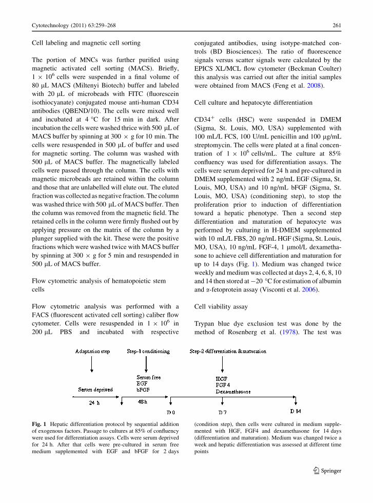
up to 14 days (Fig. 1). Medium was changed twice
weekly and medium was collected at days 2, 4, 6, 8, 10
and 14 then stored at -20 �C for estimation of albumin
and a-fetoprotein assay (Visconti et al. 2006).
Cell viability assay
Trypan blue dye exclusion test was done by the
method of Rosenberg et al. (1978). The test was
Fig. 1 Hepatic differentiation protocol by sequential addition
of exogenous factors. Passage to cultures at 85% of confluency
were used for differentiation assays. Cells were serum deprived
for 24 h. After that cells were pre-cultured in serum free
medium supplemented with EGF and bFGF for 2 days
(condition step), then cells were cultured in medium supple-
mented with HGF, FGF4 and dexamethasone for 14 days
(differentiation and maturation). Medium was changed twice a
week and hepatic differentiation was assessed at different time
points
Cytotechnology (2011) 63:259–268 261
123
based on the exclusion of trypan blue by viable cells,
whereas dead cells are stained by this dye. Trypan
blue solution and cell suspension were mixed in the
ratio of 1:1. Then the cells were observed under
microscope and counted. The percentage of viable
cells was calculated as the number of viable cells
divided by total number of cells (viable ? dead cells)
9100.
Cytotoxicity assay
MTT assay was done as described by Mossman
(1983). The reaction involves the conversion of
tetrazolium salt (3- [4,5-dimethylthiozol-2yl]-2,5-
dephenyl tetrazolium bromide), a pale yellow sub-
strate to formazan which is a dark blue product, by
the active mitochondria in living cells only. This was
dissolved in isopropanol and the absorbance was
measured spectrophotometrically at 540 nm. Cells
were taken at a concentration of 1 9 106 cells/mL,
and centrifuged at 1,000 rpm. The supernatant was
removed and the cell pellet was incubated with 3 mL
of MTT reagent at 37 �C for 2 h. The cells were
again centrifuged and 1 mL of isopropanol was added
to the cell pellet and incubated at room temperature
for 20 min. They were again centrifuged and the
purple color supernatant was transferred to a cuvette
and read at 540 nm. The amount of formazan formed
was expressed as lM/106 cells.
Functional assessment of differentiated cells
Cell morphology changes were investigated under
microscope. Hepatic gene marker (TO, ALB, AFP,
G6P and CK-18) expression was detected with
reverse transcription polymerase chain reaction
(RT–PCR), and albumin and a-fetoprotein were
detected with enzyme linked immuno-sorbant assay
(ELISA).
Enzyme-linked immunosorbent assay (ELISA)
The amounts of Human albumin and a-fetoprotein
(AFP) secretion into the medium were measured by
enzyme-linked immunosorbent assay. Standard
human albumin and AFP, goat anti-human antibody,
mouse anti-human antibody and peroxidase-conju-
gated goat immunoglobulin G fraction to human
albumin, horseradish peroxidase-conjugated mouse
monoclonal anti-AFP antibody were purchased from
Omega Diagnostics Limited. Microplates were pre-
coated with anti-human-albumin and AFP antibody
(4 lg/mL) in PBS by incubating overnight at 4 �C.
The buffer containing the unbound antibodies was
drained from the plate and the wells were washed
four times with PBS containing 0.05% Tween-20.
The unbound sites on the wells were blocked by
incubation of 200 lL of a block solution (2% (w/v)
milk powder in PBS) in each well for 2 h at room
temperature. The wells were then washed as
described above. Human albumin and AFP standard
was diluted with PBS buffer containing 0.05%
Tween-20 and 0.1% milk. An aliquot (100 lL) of
these human albumin standards was added to each
well in duplicate and samples of media 100 lL) were
added to test wells. The plates were incubated for 1 h
at room temperature. The wells were the washed as
described above. PBS containing polyclonal anti-
human-albumin (3 lg/mL) and AFP antibody (3 lg/
mL) conjugated with horseradish peroxidase were
added to each well and incubated for 1 h, and the
wells were washed again. Colour development was
started by adding 100 lL of substrate solution
prepared freshly [3, 30, 5, 50- tetramethylbenzidine
(0.2 mg/mL) and H2O2 (0.3 mg/mL) in 0.1 M
Na2HPO4 and citric acid buffer, pH 4.3]. The reaction
was stopped by adding 100 lL of 1 M H2SO4, after
which the absorbance was determined at 450 nm with
a 96 well plate ELISA reader (Bio Rad).
RNA extraction and RT–PCR analysis
Total RNA was prepared from the undifferentiated
and differentiated HSC by TRIzol (Acid guanidinium
thiocyanate-phenol–chloroform) method followed by
DNase treatment (Chomaczynski and Sacchi 1987).
One entire plate of cells was used for each isolation
of total RNA. Gene expression level of AFP, ALB,
TO, CK18 and G6P were determined by RT–PCR.
Reverse transcription was carried out using 1 lg total
RNA, 20 mmol/L dNTP and 100 unit reverse trans-
criptase in a total volume of 20 lL. PCR was carried
out using 2 lL cDNA in a total volume of 20 lL.
PCR products were analyzed in 2% agarose gel. The
name and sequences of the primers, cycling condi-
tions and annealing temperature for each pair are
listed in Table 1.
262 Cytotechnology (2011) 63:259–268
123
Statistical analysis
The results were expressed as mean ± standard
deviation (SD). The statistical significance of differ-
ence was assessed by the student’s t test. A value of
p \ 0.05 was considered statistically significant.
Results
Purification of hematopoietic stem cells
Hematopoietic and hepatic stem cells share charac-
teristic markers such as CD34, c-kit, and thy1. CD45
antigen also expressed on the HSCs except for some
mature cell types. Cells expressing CD45 and CD34
are well documented in HSCs and CD45?, CD34-
cells are probably less mature HSCs. Based on the
recent observations that hepatocytes may originate
from hematopoietic stem cells, we investigated the
potential of CD34? umbilical cord hematopoietic
cells in vitro. CD34? cells were isolated from UCB
MNC fractions by incubation with CD34 microbeads,
followed by sequential passages through two Mini-
MACS columns. Fluorescence-activated cell sorting
analysis with anti-CD34 antibody was performed to
determine the percentage purity of the positive
fraction. The result showed that CD34? cells were
enriched after magnetic cell sorting (36.5 ± 3 cells;
Fig. 2).
In vitro hepatic differentiation of cord blood-
derived hematopoietic stem cells
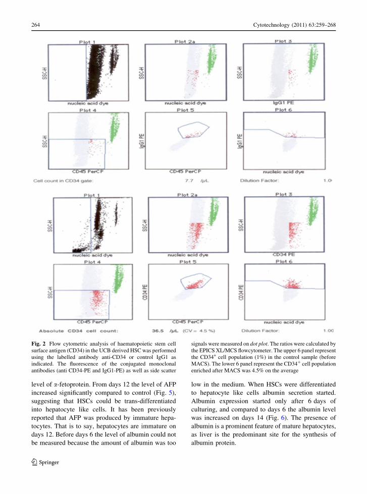
We analysed the morphological changes of UCB
HSC during differentiation protocol stages in order to
evaluate the effect of combining growth factors and
hormone. When cells were precultured for 48 h in
serum-free medium supplemented with EGF and
bFGF, cell proliferation stopped. Cells before differ-
entiation (day 0) exhibited an oval-like morphology
(Fig. 3a). Cell morphology of UCB HSC did not
change significantly during conditioning step-1, when
cultures were treated with HGF, although the mor-
phology was lost and cells developed a broadened
flattened shape. However, a polygonal shape devel-
oped during differentiation step-2 when cells were
exposed to medium containing FGF4 and dexameth-
asone (Fig. 3b–l). The protocol used includes the
sequential addition of exogenous factors that have
been reported to be implicated in liver development
and proved to be effective to induce the hepatic
differentiation of human HSC from UCB.
Cytotoxicity assay
The viability of cells was estimated by using the TBE
assay. The result thus obtained revealed that the
viability of the cells using TBE was significantly
high. Further, attempts to ascertain the functional
viability of the cells was studied by MTT assay, and
the results showed that the mitochondrial activity
increased in the 14 days culture compared to previous
day’s culture (Fig. 4).
Expression of hepatic marker protein analysis
by ELISA
We examined the expression of hepatic protein
markers such as albumin and a-fetoprotein by
ELISA. Albumin and a-fetoprotein are the functional
markers characteristic of liver cells and used to
determine the population of hepatic cells. We found
that AFP could be detected throughout the differen-
tiation process because the medium contained a low
Table 1 Primers for RT–PCR
Genes Sense Antisense
ALB GCTTTGCCGAGGAGGGTAA GGTAGGCTGAGATGCTTTTAAATG
CK 18 TGGTACTCTCCTCAATCTGCTG CTCTGGATTGACTGTGGAAGT
TO ATACAGAGACTTCAGGGAGC TGGTTTGGGTTCATCTTCGGTATC
G6P GCTGGAGTCCTGTCAGGCATTGC TAGAGCTGAGGCGGAATCGGAG
AFP TGCAGCCAAAGTGAAGAGGGAAGA CATAGCGAGCAGCCCAAAGAAGAA
Conditions Initial denaturation at 95 �C for 4 min. followed by 40 cycles of 94 �C, 1 min; 56 �C, 30 s: 72 �C, 1 min; A final
extension at 72 �C for 10 min
Cytotechnology (2011) 63:259–268 263
123
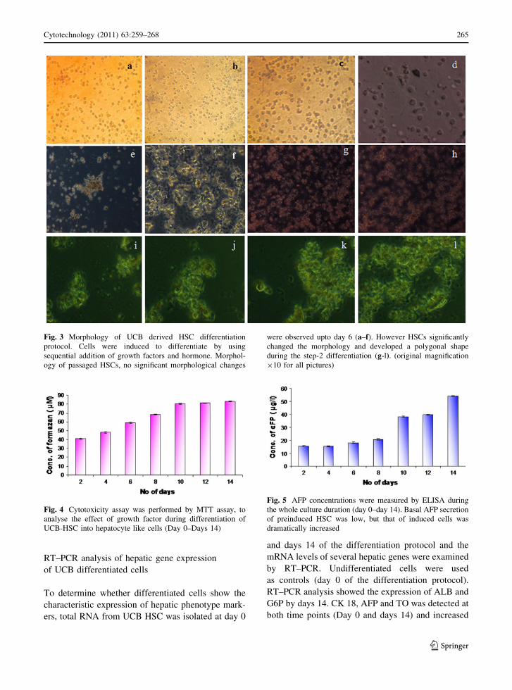
level of a-fetoprotein. From days 12 the level of AFP
increased significantly compared to control (Fig. 5),
suggesting that HSCs could be trans-differentiated
into hepatocyte like cells. It has been previously
reported that AFP was produced by immature hepa-
tocytes. That is to say, hepatocytes are immature on
days 12. Before days 6 the level of albumin could not
be measured because the amount of albumin was too
low in the medium. When HSCs were differentiated
to hepatocyte like cells albumin secretion started.
Albumin expression started only after 6 days of
culturing, and compared to days 6 the albumin level
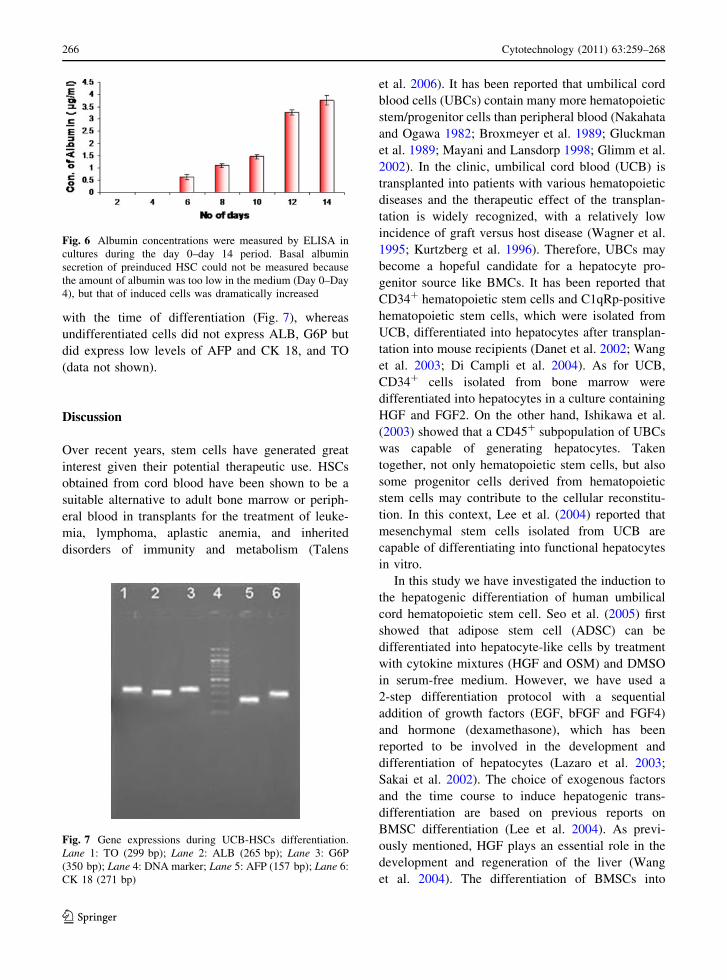
was increased on days 14 (Fig. 6). The presence of
albumin is a prominent feature of mature hepatocytes,
as liver is the predominant site for the synthesis of
albumin protein.
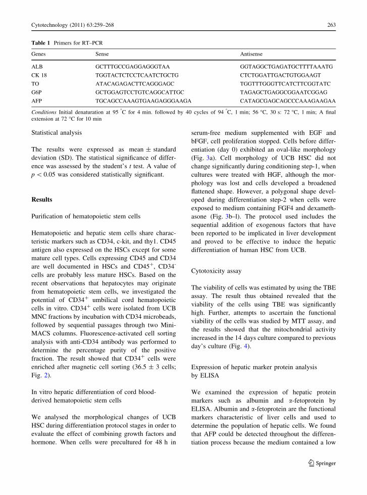
Fig. 2 Flow cytometric analysis of haematopoietic stem cell
surface antigen (CD34) in the UCB derived HSC was performed
using the labelled antibody anti-CD34 or control IgG1 as
indicated. The fluorescence of the conjugated monoclonal
antibodies (anti CD34-PE and IgG1-PE) as well as side scatter
signals were measured on dot plot. The ratios were calculated by
the EPICS XL/MCS flowcytometer. The upper 6 panel represent
the CD34+ cell population (1%) in the control sample (before
MACS). The lower 6 panel represent the CD34? cell population
enriched after MACS was 4.5% on the average
264 Cytotechnology (2011) 63:259–268
123
RT–PCR analysis of hepatic gene expression
of UCB differentiated cells
To determine whether differentiated cells show the
characteristic expression of hepatic phenotype mark-
ers, total RNA from UCB HSC was isolated at day 0
and days 14 of the differentiation protocol and the
mRNA levels of several hepatic genes were examined
by RT–PCR. Undifferentiated cells were used
as controls (day 0 of the differentiation protocol).
RT–PCR analysis showed the expression of ALB and
G6P by days 14. CK 18, AFP and TO was detected at
both time points (Day 0 and days 14) and increased
Fig. 3 Morphology of UCB derived HSC differentiation
protocol. Cells were induced to differentiate by using
sequential addition of growth factors and hormone. Morphol-
ogy of passaged HSCs, no significant morphological changes
were observed upto day 6 (a–f). However HSCs significantly
changed the morphology and developed a polygonal shape
during the step-2 differentiation (g-l). (original magnification
910 for all pictures)
Fig. 4 Cytotoxicity assay was performed by MTT assay, to
analyse the effect of growth factor during differentiation of
UCB-HSC into hepatocyte like cells (Day 0–Days 14)
Fig. 5 AFP concentrations were measured by ELISA during
the whole culture duration (day 0–day 14). Basal AFP secretion
of preinduced HSC was low, but that of induced cells was
dramatically increased
Cytotechnology (2011) 63:259–268 265
123
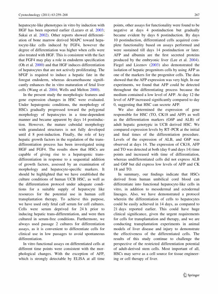
with the time of differentiation (Fig. 7), whereas
undifferentiated cells did not express ALB, G6P but
did express low levels of AFP and CK 18, and TO
(data not shown).
Discussion
Over recent years, stem cells have generated great
interest given their potential therapeutic use. HSCs
obtained from cord blood have been shown to be a
suitable alternative to adult bone marrow or periph-
eral blood in transplants for the treatment of leuke-
mia, lymphoma, aplastic anemia, and inherited
disorders of immunity and metabolism (Talens
et al. 2006). It has been reported that umbilical cord
blood cells (UBCs) contain many more hematopoietic
stem/progenitor cells than peripheral blood (Nakahata
and Ogawa 1982; Broxmeyer et al. 1989; Gluckman
et al. 1989; Mayani and Lansdorp 1998; Glimm et al.
2002). In the clinic, umbilical cord blood (UCB) is
transplanted into patients with various hematopoietic
diseases and the therapeutic effect of the transplan-
tation is widely recognized, with a relatively low
incidence of graft versus host disease (Wagner et al.
1995; Kurtzberg et al. 1996). Therefore, UBCs may
become a hopeful candidate for a hepatocyte pro-
genitor source like BMCs. It has been reported that
CD34? hematopoietic stem cells and C1qRp-positive
hematopoietic stem cells, which were isolated from
UCB, differentiated into hepatocytes after transplan-
tation into mouse recipients (Danet et al. 2002; Wang
et al. 2003; Di Campli et al. 2004). As for UCB,
CD34? cells isolated from bone marrow were
differentiated into hepatocytes in a culture containing
HGF and FGF2. On the other hand, Ishikawa et al.
(2003) showed that a CD45? subpopulation of UBCs
was capable of generating hepatocytes. Taken
together, not only hematopoietic stem cells, but also
some progenitor cells derived from hematopoietic
stem cells may contribute to the cellular reconstitu-
tion. In this context, Lee et al. (2004) reported that
mesenchymal stem cells isolated from UCB are
capable of differentiating into functional hepatocytes
in vitro.
In this study we have investigated the induction to
the hepatogenic differentiation of human umbilical
cord hematopoietic stem cell. Seo et al. (2005) first
showed that adipose stem cell (ADSC) can be
differentiated into hepatocyte-like cells by treatment
with cytokine mixtures (HGF and OSM) and DMSO
in serum-free medium. However, we have used a
2-step differentiation protocol with a sequential
addition of growth factors (EGF, bFGF and FGF4)
and hormone (dexamethasone), which has been
reported to be involved in the development and
differentiation of hepatocytes (Lazaro et al. 2003;
Sakai et al. 2002). The choice of exogenous factors
and the time course to induce hepatogenic trans-
differentiation are based on previous reports on
BMSC differentiation (Lee et al. 2004). As previ-
ously mentioned, HGF plays an essential role in the
development and regeneration of the liver (Wang
et al. 2004). The differentiation of BMSCs into
Fig. 6 Albumin concentrations were measured by ELISA in
cultures during the day 0–day 14 period. Basal albumin
secretion of preinduced HSC could not be measured because
the amount of albumin was too low in the medium (Day 0–Day
4), but that of induced cells was dramatically increased
Fig. 7 Gene expressions during UCB-HSCs differentiation.
Lane 1: TO (299 bp); Lane 2: ALB (265 bp); Lane 3: G6P
(350 bp); Lane 4: DNA marker; Lane 5: AFP (157 bp); Lane 6:
CK 18 (271 bp)
266 Cytotechnology (2011) 63:259–268
123
hepatocyte-like phenotypes in vitro by induction with
HGF has been reported earlier (Lazaro et al. 2003;
Sakai et al. 2002). Other reports showed differenti-
ation of bone marrow derived MAPC toward hepa-
tocyte-like cells induced by FGF4, however the
degree of differentiation was higher when cells were
also treated with HGF. This is consistent with the fact
that FGF4 may play a role in endoderm specification
(Oh et al. 2000) and that HGF induces differentiation
of hepatocytes that are not actively proliferating. The
bFGF is required to induce a hepatic fate in the
foregut endoderm, whereas dexamethasone signifi-
cantly enhances the in vitro maturation of fetal liver
cells (Wang et al. 2004; Wells and Melton 2000).
In the present study the morphologic features and
gene expression changes in HSC were evaluated.
Under hepatogenic conditions, the morphology of
HSCs gradually progressed toward the polygonal
morphology of hepatocytes in a time-dependent
manner and became apparent by days 14 postinduc-
tion. However, the mature cuboidal morphology
with granulated structures is not fully developed
until d 8 post-induction. Finally, the role of key
hepatic growth factors in the regulation of the trans-
differentiation process has been investigated using
HGF and FGF4. The results show that HSCs are
capable of giving rise to a hepatogenic trans-
differentiation in response to a sequential addition
of growth factors, assessed by an examination of
morphology and hepatocyte-specific markers. It
should be highlighted that we have established the
culture conditions of human UCB HSC, as well as
the differentiation protocol under adequate condi-
tions for a suitable supply of hepatocyte like
resources for the potential use in human cell
transplantation therapy. To achieve this purpose,
we have used only fetal calf serum for cell cultures.
Cells were serum deprived for 24 h prior to
inducing hepatic trans-differentiation, and were then
cultured in serum-free conditions. Furthermore, we
always used passage 2 cultures for differentiation
assays, as it is convenient to differentiate cells for
clinical use in low passages to avoid spontaneous
differentiation.
In vitro functional assays on differentiated cells at
different time points were consistent with the mor-
phological changes. With the exception of AFP,
which is strongly detectable by ELISA at all time
points, other assays for functionality were found to be
negative at days 4 postinduction but gradually
became evident by days 6 postinduction. By days
10 postinduction, differentiated cells acquired com-
plete functionality based on assays performed and
were sustained till days 14 postinduction or later.
AFP and albumin are the first secreted proteins
produced by the embryonic liver (Lee et al. 2004).
Fiegel and Lioznov (2003) also demonstrated the
isolation of hepatic progenitors cells by using AFP as
one of the markers for the progenitor cells. The data
showed that the AFP expression was very high. In our
experiments, we found that AFP could be detected
throughout the differentiating process because the
medium contained a low level of AFP. At day 12 the
level of AFP increased significantly compared to day
0, suggesting that HSC can secrete AFP.
We also determined the expression of gene
responsible for HSC (TO, CK18 and AFP) as well
as the differentiation markers (G6P and ALB) of
adult hepatic genotypic in UCB derived HSC. We
compared expression levels by RT–PCR at the initial
and final times of the differentiation procedure.
Levels of the expression of ALB, and G6P was
observed at days 14. The expression of CK18, AFP
and TO was detected at both (day 0 and days 14) time
points and increased with time of differentiation,
whereas undifferentiated cells did not express ALB
and G6P but did express low levels of AFP and CK
18 and TO.
In summary, our findings indicate that HSCs
derived from human umbilical cord blood can
differentiate into functional hepatocyte-like cells in
vitro, in addition to mesodermal and ectodermal
lineages. Also, we have demonstrated a protocol
wherein the differentiation of cells to hepatocytes
could be easily achieved in 14 days, as compared to
21 days reported earlier. This could have huge
clinical significance, given the urgent requirements
for cells for transplantation and therapy, and we are
conducting transplantation experiments in animal
models of liver disease and injury to demonstrate
the effectiveness of the differentiated cells. The
results of this study continue to challenge the
perspective of the restricted differentiation potential
of adult-derived stem cells. Most important of all,
HSCs may serve as a cell source for tissue engineer-
ing or cell therapy of liver.
Cytotechnology (2011) 63:259–268 267
123
References
Bromeyer HE (1995) Questions to be answered regarding
umbilical cord blood hematopoietic stem and progenitor
cells and their use in transplantation. Transfusion 35:
694–702
Broxmeyer HE, Douglas GW, Hangoc G et al (1989) Human
umbilical cord blood as a potential source of transplan-
table hematopoietic stem/progenitor cells. Proc Natl Acad
Sci USA 86:3828–3832
Chomaczynski P, Sacchi N (1987) Single-step method of
RNA isolation by acid guanidinium thiocyanate-phenol-
chloroform extraction. Anal Biochem 162:196–199
Danet GH, Luongo JL, Butler G et al (2002) C1qRp defines a new
human stem cell population with hematopoietic and hepatic
potential. Proc Natl Acad Sci USA 99:10441–10445
Di Campli C, Piscaglia AC, Pierelli L et al (2004) A human
umbilical cord stem cell rescue therapy in a murine model
of toxic liver injury. Dig Liver Dis 36:603–613
Feng G, De-Quan W, Yah-Hua H et al (2008) Extracellular
matrix gel is necessary for in vitro cultivation of insulin
producing cells from human umbilical cord blood derived
mesenchymal stem cells. Chinese Med J 121:811–818
Fiegel HC, Lioznov MV (2003) Cortes-Dericks liver-specific
gene expression in cultured human hematopoietic stem
cells. Stem Cells 21:98–104
Forbes S, Vig P, Poulsom R et al (2002) Hepatic stem cells.
J Pathol 197:510–518
Fox IJ, Chowdhury JR (2004) Hepatocyte transplantation.
Amer J Transplant 4:7–13
Glimm H, Tang P, Clark-Lewis I et al (2002) Ex vivo treatment
of proliferating human cord blood stem cells with stroma-
derived factor-1 enhances their ability to engraft NOD/
SCID mice. Blood 99:3454–3457
Gluckman E, Broxmeyer HE, Auerbach AD et al (1989)
Hematopoietic reconstitution in a patient with Fanconi’s
anemia by means of umbilical-cord blood from an HLA-
identical sibling. N Engl J Med 321:1174–1178
Ishikawa F, Drake CJ, Yang S et al (2003) Transplanted human
cord blood cells give rise to hepatocytes in engrafted
mice. Ann NY Sci 996:174–185
Kurtzberg J, Laughlin M, Graham ML et al (1996) Placental
blood as a source of hematopoietic stem cells for trans-
plantation into unrelated recipients. N Engl J Med 335:
157–166
Lazaro CA, Croager EJ, Mitchell C et al (2003) Establishment,
characterization, and long-term maintenance of cultures of
human fetal hepatocytes. Hepatology 38:1095–1106
Lee KD, Kuo TK, Whang-Peng J et al (2004) In vitro hepatic
differentiation of human mesenchymal stem cells. Hepa-
tology 40:1275–1284
Mayani H, Lansdorp PM (1998) Biology of human umbilical
cord blood-derived hematopoietic stem/progenitor cells.
Stem Cells 16:153–165
McAdams TA, Miller WM, Papoutsakis ET (1996) Hematopoi-
etic cell culture therapies. Trends Biotechnol 14:388–396
Mossman T (1983) Rapid colorimetric assay of cellular growth
and survival: a application to proliferation and cytotox-
icity assays. J Immunol Method 65:55–63
Nakahata T, Ogawa M (1982) Hemopoietic colony-forming
cells in umbilical cord blood with extensive capability to
generate mono- and multipotential hemopoietic progeni-
tors. J Clin Invest 70:1324–1328
Oh SH, Miyazaki M, Kouchi H et al (2000) Hepatocyte growth
factor induces differentiation of adult rat bone marrow
cells into a hepatocyte lineage in vitro. Biochem Biophys
Res Commun 279:500–504
Qin AL, Zhou XQ, Zhang WQ et al (2004) Characteriation an
enrichment of hepatic progenitor cells in adult liver.
World J Gastroenterol 10:1480–1486
Rosenberg IL, Russel CW, Giles GR (1978) Cell viability
studies on the exfoliated colonic cancer cell. British J Sur
65:188–190
Sakai Y, Jiang J, Kojima N et al (2002) Enhanced in vitro matu-
ration of fetal mouse liver cells with oncostatin M, nicotin-
amide, and dimethyl sulfoxide. Cell Transplant 11:435–441
Seo MJ, Suh SY, Bae YC et al (2005) Differentiation of human
adipose stromal cells into hepatic lineage in vitro and in
vivo. Biochem Biophys Res Commun 328:258–264
Talens R, Bonora A, Jover R et al (2006) Hepatogenic dif-
ferentiation of human mesenchymal stem cells from adi-
pose tissue in comparison with bone marrowmesenchymal
stem cells. World J Gastroenterol 12:5834–5845
Trey C, Davidson CS (1970) The management of fulminant
hepatic failure. Prog Liver Dis 3:282–298
Visconti RT, Bonora A, Jover R et al (2006) Hepatogenic
differentiation of human mesenchymal stem cells from
adipose tissue in comparison with bone marrow mesen-
chymal stem cells. World J Gastroenterol 28:5834–5845
Wagner JE, Kernan NA, Steinbuch M et al (1995) Allogeneic
sibling umbilical-cord-blood transplantation in children
with malignant and non-malignant disease. Lancet 346:
214–219
Wang X, Ge S, McNamara G et al (2003) Albumin-expressing
hepatocyte-like cells develop in the livers of immune-
deficient mice that received transplants of highly purified
human hematopoietic stem cells. Blood 101:4201–4208
Wang PP, Wang JH, Yan P et al (2004) Expression of hepa-
tocyte-like phenotypes in bone marrow stromal cells after
HGF induction. Biochem Biophys Res Commun 320:
712–716
Wells JM, Melton DA (2000) Early mouse endoderm is pat-
terned by soluble factors from adjacent germ layers.
Development 127:1563–1572
Yu S, Li C, Xin-Guo H et al (2007) Differentiation of bone
marrow-derived mesenchymal stem cell from diabetic
patients into insulin-producing cells in vitro. Chinese Med
J 120:771–776
268 Cytotechnology (2011) 63:259–268
123
Related Documents