In vitro study on silk fibroin textile structure for Anterior Cruciate Ligament regeneration Silvia Farè a, b, ⁎, Paola Torricelli c , Gianluca Giavaresi c , Serena Bertoldi a, b , Antonio Alessandrino d , Tomaso Villa a , Milena Fini c , Maria Cristina Tanzi a, b , Giuliano Freddi d a Politecnico di Milano, Dept. of Chemistry, Materials and Chemical Engineering “G. Natta”, P.zza Leonardo da Vinci 32, 20133, Milan, Italy b Local Unit Politecnico di Milano, Istituto Nazionale di Scienza e Tecnologia dei Materiali, INSTM, Firenze, Italy c Laboratory of Preclinical and Surgical Studies, Rizzoli Orthopedic Institute, via di Barbiano 1/10, 40136, Bologna, Italy d INNOVHUB—SSI, Div. Stazione Sperimentale per la Seta, via G. Colombo 83, 20133, Milan, Italy abstract article info Article history: Received 27 August 2012 Received in revised form 28 March 2013 Accepted 15 April 2013 Available online 20 April 2013 Keywords: Silk fibroin ACL Hierarchical textile structure Cytocompatibility Micro-CT Tensile mechanical properties A novel hierarchical textile structure made of silk fibroin from Bombyx mori capable of matching the mechanical performance requirements of anterior cruciate ligament (ACL) and in vitro cell ingrowth is described. This sericin-free, Silk Fibroin Knitted Sheath with Braided Core (SF-KSBC) structure was fabricated using available textile technologies. Micro-CT analysis confirmed that the core was highly porous and had a higher degree of interconnectivity than that observed for the sheath. The in vivo cell colonization of the scaffolds is thus expected to penetrate even the internal parts of the structure. Tensile mechanical tests demonstrated a maximum load of 1212.4 ± 56.4 N (under hydrated conditions), confirming the scaffold's suitability for ACL reconstruction. The absence of cytotoxic substances in the extracts of the SF-KSBC structure in culture medium was verified by in vitro tests with L929 fibroblasts. In terms of extracellular matrix production, Human Periodontal Ligament Fibroblasts (HPdLFs) cultured in direct contact with SF-KSBC, compared to control samples, demonstrated an in- creased secretion of aggrecan (PG) and fibronectin (FBN) at 3 and 7 days of culture, and no change in IL-6 and TNF-α secretion. Altogether, the outcomes of this investigation confirm the significant utility of this novel scaf- fold for ACL tissue regeneration. © 2013 Elsevier B.V. All rights reserved. 1. Introduction Anterior Cruciate Ligament (ACL), a band of dense connective tissue, mainly composed of collagen type I, is required for the articula- tion of the knee and connects the lateral femoral condyle to the tibial plateau. Its function is mainly to limit both horizontal translation and relative rotation between the tibia and femur [1,2]. The mechanical properties of the ACL are influenced by several factors, including age, weight, and activity level [3]. In the U.S.A. alone, approximately 100,000 surgical operations to repair damaged ACLs are performed annually [4], and as such it is a major activity in orthopedic surgery. In the ACL, collagen fibers are organized in a hierarchical structure in order to maximize tensile strength and minimize friction. Due to the elastic recovery of this tissue post injury, in macroscopic lesions the distance between the two parts of the Anterior Cruciate Ligament becomes too large (>5 mm), and sutures are unable to restore the pre-injury properties of ACL. This fact, along with the ACL being a tissue with overall low healing potential and limited vascularization, has driven the development of alternate strategies for ligament repair [5,6]. Currently, autografts are the gold standard in ACL replacement. Bone–patellar tendon–bone grafts and hamstring tendon grafts repre- sent the two most implanted autografts. In particular, hamstring tendon grafts are most often used, due to a flexibility offered by a relatively new surgical technique, called double bundle ACL reconstruction. In this technique, two autologous tendon strips are implanted to substitute for the damaged ACL, allowing for appropriate physiological positioning of the graft [7]. Advantages of autografts include the possibility of vascularization, low risk of immune response and integration with the perimplant tissue, as tenocytes are able to differentiate in ligamentocytes [8]. However, autografts do not guarantee a complete functional return to pre-injury activity levels, with previous studies noting large decreases in flexion and extension movements [9]. Harvest-site morbidity and pain, regrowth of tissues without pre-injury properties, and the possibility of damage to the donor site and adjacent tissues during the harvesting represent some of the main drawbacks of the autografts [5,6,10,11]. To overcome some of these deficiencies and further, the impossibility of replacing previously failed autografts, allografts may be implanted. However, there are still drawbacks associated with this alternate tissue source, including the risk of disease transmission, lack of appropriate donors, immune response and high costs [5,12]. Synthetic materials and devices have also been investigated for ACL replacement or for the in vivo regeneration of the ligament tissue, including for example, Dacron® prostheses, Gore-Tex® prostheses, polypropylene-based Kennedy Ligament-Augmentation Device, carbon Materials Science and Engineering C 33 (2013) 3601–3608 ⁎ Corresponding author at: Politecnico di Milano, Dept. of Chemistry, Materials and Chemical Engineering “G. Natta”, Piazza Leonardo da Vinci, 32-20133 Milan, Italy. Tel.: +39 0223993389; fax: +39 0223993360. E-mail address: [email protected] (S. Farè). 0928-4931/$ – see front matter © 2013 Elsevier B.V. All rights reserved. http://dx.doi.org/10.1016/j.msec.2013.04.027 Contents lists available at ScienceDirect Materials Science and Engineering C journal homepage: www.elsevier.com/locate/msec

Welcome message from author
This document is posted to help you gain knowledge. Please leave a comment to let me know what you think about it! Share it to your friends and learn new things together.
Transcript

Materials Science and Engineering C 33 (2013) 3601–3608
Contents lists available at ScienceDirect
Materials Science and Engineering C
j ourna l homepage: www.e lsev ie r .com/ locate /msec
In vitro study on silkfibroin textile structure for Anterior Cruciate Ligament regeneration
Silvia Farè a,b,⁎, Paola Torricelli c, Gianluca Giavaresi c, Serena Bertoldi a,b, Antonio Alessandrino d,Tomaso Villa a, Milena Fini c, Maria Cristina Tanzi a,b, Giuliano Freddi d
a Politecnico di Milano, Dept. of Chemistry, Materials and Chemical Engineering “G. Natta”, P.zza Leonardo da Vinci 32, 20133, Milan, Italyb Local Unit Politecnico di Milano, Istituto Nazionale di Scienza e Tecnologia dei Materiali, INSTM, Firenze, Italyc Laboratory of Preclinical and Surgical Studies, Rizzoli Orthopedic Institute, via di Barbiano 1/10, 40136, Bologna, Italyd INNOVHUB—SSI, Div. Stazione Sperimentale per la Seta, via G. Colombo 83, 20133, Milan, Italy
⁎ Corresponding author at: Politecnico di Milano, Dand Chemical Engineering “G. Natta”, Piazza LeonardItaly. Tel.: +39 0223993389; fax: +39 0223993360.
E-mail address: [email protected] (S. Farè).
0928-4931/$ – see front matter © 2013 Elsevier B.V. Allhttp://dx.doi.org/10.1016/j.msec.2013.04.027
a b s t r a c t
a r t i c l e i n f oArticle history:Received 27 August 2012Received in revised form 28 March 2013Accepted 15 April 2013Available online 20 April 2013
Keywords:Silk fibroinACLHierarchical textile structureCytocompatibilityMicro-CTTensile mechanical properties
A novel hierarchical textile structure made of silk fibroin from Bombyx mori capable of matching the mechanicalperformance requirements of anterior cruciate ligament (ACL) and in vitro cell ingrowth is described. Thissericin-free, Silk Fibroin Knitted Sheath with Braided Core (SF-KSBC) structure was fabricated using availabletextile technologies. Micro-CT analysis confirmed that the core was highly porous and had a higher degree ofinterconnectivity than that observed for the sheath. The in vivo cell colonization of the scaffolds is thus expectedto penetrate even the internal parts of the structure. Tensile mechanical tests demonstrated a maximum load of1212.4 ± 56.4 N (under hydrated conditions), confirming the scaffold's suitability for ACL reconstruction.The absence of cytotoxic substances in the extracts of the SF-KSBC structure in culture medium was verifiedby in vitro tests with L929 fibroblasts. In terms of extracellular matrix production, Human Periodontal LigamentFibroblasts (HPdLFs) cultured in direct contact with SF-KSBC, compared to control samples, demonstrated an in-creased secretion of aggrecan (PG) and fibronectin (FBN) at 3 and 7 days of culture, and no change in IL-6 andTNF-α secretion. Altogether, the outcomes of this investigation confirm the significant utility of this novel scaf-fold for ACL tissue regeneration.
© 2013 Elsevier B.V. All rights reserved.
1. Introduction
Anterior Cruciate Ligament (ACL), a band of dense connectivetissue, mainly composed of collagen type I, is required for the articula-tion of the knee and connects the lateral femoral condyle to the tibialplateau. Its function is mainly to limit both horizontal translation andrelative rotation between the tibia and femur [1,2]. The mechanicalproperties of the ACL are influenced by several factors, includingage, weight, and activity level [3]. In the U.S.A. alone, approximately100,000 surgical operations to repair damaged ACLs are performedannually [4], and as such it is a major activity in orthopedic surgery.In the ACL, collagen fibers are organized in a hierarchical structurein order to maximize tensile strength and minimize friction. Due tothe elastic recovery of this tissue post injury, in macroscopic lesionsthe distance between the two parts of the Anterior Cruciate Ligamentbecomes too large (>5 mm), and sutures are unable to restore thepre-injury properties of ACL. This fact, alongwith the ACL being a tissuewith overall low healing potential and limited vascularization, hasdriven the development of alternate strategies for ligament repair [5,6].
ept. of Chemistry, Materialso da Vinci, 32-20133 Milan,
rights reserved.
Currently, autografts are the gold standard in ACL replacement.Bone–patellar tendon–bone grafts and hamstring tendon grafts repre-sent the twomost implanted autografts. In particular, hamstring tendongrafts aremost often used, due to a flexibility offered by a relatively newsurgical technique, called double bundle ACL reconstruction. In thistechnique, two autologous tendon strips are implanted to substitutefor the damaged ACL, allowing for appropriate physiological positioningof the graft [7]. Advantages of autografts include the possibility ofvascularization, low risk of immune response and integration with theperimplant tissue, as tenocytes are able to differentiate in ligamentocytes[8]. However, autografts do not guarantee a complete functional return topre-injury activity levels, with previous studies noting large decreases inflexion and extension movements [9]. Harvest-site morbidity and pain,regrowth of tissues without pre-injury properties, and the possibilityof damage to the donor site and adjacent tissues during the harvestingrepresent some of the main drawbacks of the autografts [5,6,10,11].
To overcome some of these deficiencies and further, the impossibilityof replacing previously failed autografts, allografts may be implanted.However, there are still drawbacks associated with this alternate tissuesource, including the risk of disease transmission, lack of appropriatedonors, immune response and high costs [5,12].
Synthetic materials and devices have also been investigated forACL replacement or for the in vivo regeneration of the ligament tissue,including for example, Dacron® prostheses, Gore-Tex® prostheses,polypropylene-based Kennedy Ligament-Augmentation Device, carbon
3602 S. Farè et al. / Materials Science and Engineering C 33 (2013) 3601–3608
fiber device and Leeds-Keio Artificial Ligament [13,14]. Unfortunately,mechanical breakage, plastic deformation, poor abrasion resistance,the presence of debris, lack of tissue regeneration, and stress-shieldinghave been some of the reasons for the failure of such devices [6,15–18].
In the last decade, different biodegradable synthetic and naturalpolymers (e.g. polylactic acid, PLA, polyglicolic acid, PGA, and theircopolymers, collagen fibers) have been investigated and are stillunder investigation as alternative solutions to the use of autografts.Even though these degradable materials show significant potential,improvements are still required to overcome the lack of adequatemechanical properties, rapid degradation rates, degradation producttoxicity and the lack of viable sterilization methods [19–21]. An alterna-tive approach is in vitro and/or in vivo tissue engineering, in which abiodegradable scaffold is fabricated to provide the required mechanicalsupport and encourage the development of neo-tissue in the shortterm, and permit increases, at a controlled rate, in neo-tissuemechanicalproperties, avoiding the risk of stress shielding [5,6,22].
Silk has been used clinically as a suturematerial for decades and hasrecently gained renewed interest as a biomaterial for tissue engineering[23–26]. In this current investigation, silk fibroin (SF) was selected fromthe available array of synthetic and natural fibers for this application, asit is the only protein-based fiber that: (1) can match the requiredmechanical properties of an ACL, (2) is biocompatible when properlyprepared [23], (3) avoids bio-burdens associated with mammalian-derived materials, (4) maintains mechanical tensile integrity in vitroin tissue culture conditions and (5) exhibits slow degradation in vivo[24], allowing adequate time for host tissue infiltration andmaturation.Further, silk fibroin has previously been utilized in the fabrication ofACL substitutes, both in combination with other polymers, such asnanofibrous PLGA [27,28], as a peptide nanofiber mesh [29] and in atextile form (i.e. a hierarchical wire-rope matrix) [22], obtaining somepromising results, but unfortunately still not reaching clinical applica-tion, although clinical trials are currently going on (NCT00775892 andNCT00490594, www.clinicaltrials.gov).
In the present study, we detail the development of a novel hierar-chical structure made of Bombyx mori silk fibroin [30], and providedata describing the resultant morphology, tensile mechanical proper-ties, in vitro cytotoxicity and bioactivity. The hierarchical structure iscomposed of an external sheath and an inner tubular core, aimed atimproving the mechanical properties and encouraging adequate cellcolonization. The different core and sheath morphologies have beendesigned to perform different tasks. The porous core structure, withthe fibers arranged in an oriented manner, has the role of bothguaranteeing the mechanical characteristics necessary for sustainingloads, and providing a preferential direction for the formation ofcollagen fibers in the newly formed ligament. The sheath structure,with a lower porosity, is designed to protect the core against weardue to rushing as a result of the contact with other tissue present inthe joint (e.g. bone tissue) and to allow for cell adhesion, proliferationand migration towards the interior (i.e. the core) structure. The resultspresented suggest that this innovative structure has the requisitemechanical integrity to match ACL performance requirements over asuitable time period, allowing for in vivo ingrowth of new tissue.
2. Materials and methods
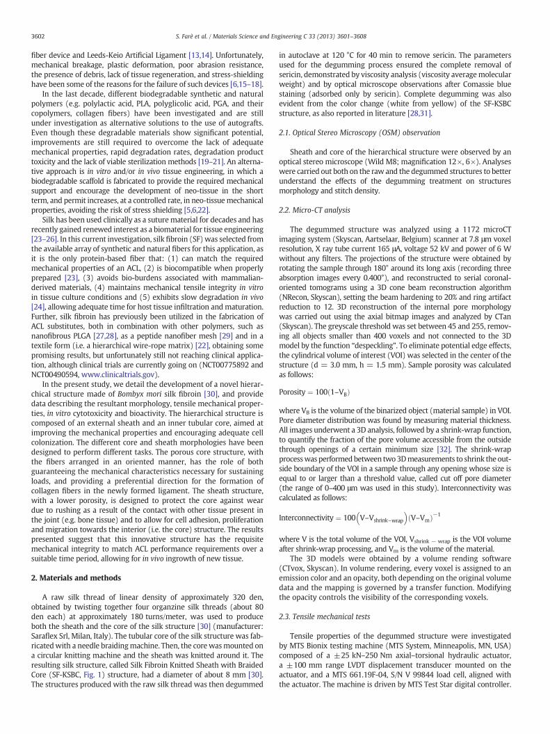
A raw silk thread of linear density of approximately 320 den,obtained by twisting together four organzine silk threads (about 80den each) at approximately 180 turns/meter, was used to produceboth the sheath and the core of the silk structure [30] (manufacturer:Saraflex Srl, Milan, Italy). The tubular core of the silk structure was fab-ricatedwith a needle braidingmachine. Then, the core wasmounted ona circular knitting machine and the sheath was knitted around it. Theresulting silk structure, called Silk Fibroin Knitted Sheath with BraidedCore (SF-KSBC, Fig. 1) structure, had a diameter of about 8 mm [30].The structures produced with the raw silk thread was then degummed
in autoclave at 120 °C for 40 min to remove sericin. The parametersused for the degumming process ensured the complete removal ofsericin, demonstrated by viscosity analysis (viscosity averagemolecularweight) and by optical microscope observations after Comassie bluestaining (adsorbed only by sericin). Complete degumming was alsoevident from the color change (white from yellow) of the SF-KSBCstructure, as also reported in literature [28,31].
2.1. Optical Stereo Microscopy (OSM) observation
Sheath and core of the hierarchical structure were observed by anoptical stereo microscope (Wild M8; magnification 12×, 6×). Analyseswere carried out both on the raw and the degummed structures to betterunderstand the effects of the degumming treatment on structuresmorphology and stitch density.
2.2. Micro-CT analysis
The degummed structure was analyzed using a 1172 microCTimaging system (Skyscan, Aartselaar, Belgium) scanner at 7.8 μm voxelresolution, X ray tube current 165 μA, voltage 52 kV and power of 6 Wwithout any filters. The projections of the structure were obtained byrotating the sample through 180° around its long axis (recording threeabsorption images every 0.400°), and reconstructed to serial coronal-oriented tomograms using a 3D cone beam reconstruction algorithm(NRecon, Skyscan), setting the beam hardening to 20% and ring artifactreduction to 12. 3D reconstruction of the internal pore morphologywas carried out using the axial bitmap images and analyzed by CTan(Skyscan). The greyscale threshold was set between 45 and 255, remov-ing all objects smaller than 400 voxels and not connected to the 3Dmodel by the function “despeckling”. To eliminate potential edge effects,the cylindrical volume of interest (VOI) was selected in the center of thestructure (d = 3.0 mm, h = 1.5 mm). Sample porosity was calculatedas follows:
Porosity ¼ 100 1–VBð Þ
where VB is the volume of the binarized object (material sample) in VOI.Pore diameter distribution was found by measuring material thickness.All images underwent a 3D analysis, followed by a shrink-wrap function,to quantify the fraction of the pore volume accessible from the outsidethrough openings of a certain minimum size [32]. The shrink-wrapprocesswas performedbetween two3Dmeasurements to shrink the out-side boundary of the VOI in a sample through any opening whose size isequal to or larger than a threshold value, called cut off pore diameter(the range of 0–400 μm was used in this study). Interconnectivity wascalculated as follows:
Interconnectivity ¼ 100 V–Vshrink–wrap
� �V–Vmð Þ−1
where V is the total volume of the VOI, Vshrink − wrap is the VOI volumeafter shrink-wrap processing, and Vm is the volume of the material.
The 3D models were obtained by a volume rending software(CTvox, Skyscan). In volume rendering, every voxel is assigned to anemission color and an opacity, both depending on the original volumedata and the mapping is governed by a transfer function. Modifyingthe opacity controls the visibility of the corresponding voxels.
2.3. Tensile mechanical tests
Tensile properties of the degummed structure were investigatedby MTS Bionix testing machine (MTS System, Minneapolis, MN, USA)composed of a ±25 kN–250 Nm axial–torsional hydraulic actuator,a ±100 mm range LVDT displacement transducer mounted on theactuator, and a MTS 661.19F-04, S/N V 99844 load cell, aligned withthe actuator. The machine is driven by MTS Test Star digital controller.
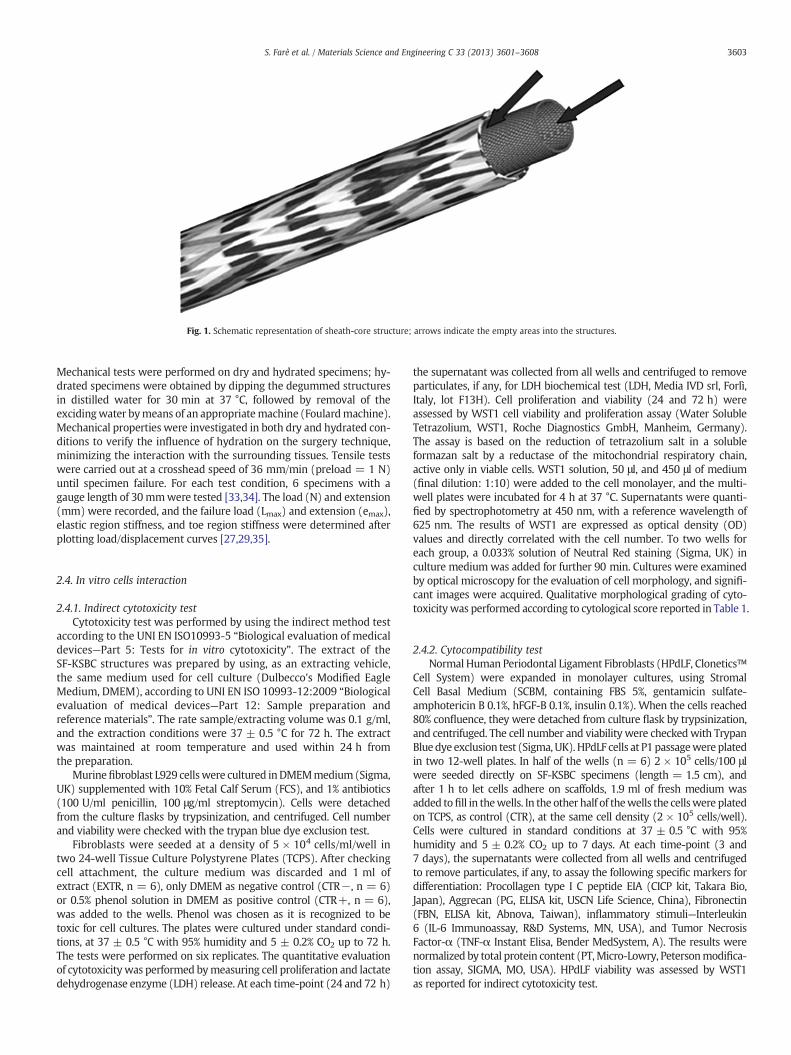
Fig. 1. Schematic representation of sheath-core structure; arrows indicate the empty areas into the structures.
3603S. Farè et al. / Materials Science and Engineering C 33 (2013) 3601–3608
Mechanical tests were performed on dry and hydrated specimens; hy-drated specimens were obtained by dipping the degummed structuresin distilled water for 30 min at 37 °C, followed by removal of theexcidingwater bymeans of an appropriatemachine (Foulardmachine).Mechanical properties were investigated in both dry and hydrated con-ditions to verify the influence of hydration on the surgery technique,minimizing the interaction with the surrounding tissues. Tensile testswere carried out at a crosshead speed of 36 mm/min (preload = 1 N)until specimen failure. For each test condition, 6 specimens with agauge length of 30mmwere tested [33,34]. The load (N) and extension(mm) were recorded, and the failure load (Lmax) and extension (emax),elastic region stiffness, and toe region stiffness were determined afterplotting load/displacement curves [27,29,35].
2.4. In vitro cells interaction
2.4.1. Indirect cytotoxicity testCytotoxicity test was performed by using the indirect method test
according to the UNI EN ISO10993-5 “Biological evaluation of medicaldevices—Part 5: Tests for in vitro cytotoxicity”. The extract of theSF-KSBC structures was prepared by using, as an extracting vehicle,the same medium used for cell culture (Dulbecco's Modified EagleMedium, DMEM), according to UNI EN ISO 10993-12:2009 “Biologicalevaluation of medical devices—Part 12: Sample preparation andreference materials”. The rate sample/extracting volume was 0.1 g/ml,and the extraction conditions were 37 ± 0.5 °C for 72 h. The extractwas maintained at room temperature and used within 24 h fromthe preparation.
Murinefibroblast L929 cellswere cultured inDMEMmedium(Sigma,UK) supplemented with 10% Fetal Calf Serum (FCS), and 1% antibiotics(100 U/ml penicillin, 100 μg/ml streptomycin). Cells were detachedfrom the culture flasks by trypsinization, and centrifuged. Cell numberand viability were checked with the trypan blue dye exclusion test.
Fibroblasts were seeded at a density of 5 × 10⁴ cells/ml/well intwo 24-well Tissue Culture Polystyrene Plates (TCPS). After checkingcell attachment, the culture medium was discarded and 1 ml ofextract (EXTR, n = 6), only DMEM as negative control (CTR−, n = 6)or 0.5% phenol solution in DMEM as positive control (CTR+, n = 6),was added to the wells. Phenol was chosen as it is recognized to betoxic for cell cultures. The plates were cultured under standard condi-tions, at 37 ± 0.5 °C with 95% humidity and 5 ± 0.2% CO2 up to 72 h.The tests were performed on six replicates. The quantitative evaluationof cytotoxicitywas performed bymeasuring cell proliferation and lactatedehydrogenase enzyme (LDH) release. At each time-point (24 and 72 h)
the supernatant was collected from all wells and centrifuged to removeparticulates, if any, for LDH biochemical test (LDH, Media IVD srl, Forlì,Italy, lot F13H). Cell proliferation and viability (24 and 72 h) wereassessed by WST1 cell viability and proliferation assay (Water SolubleTetrazolium, WST1, Roche Diagnostics GmbH, Manheim, Germany).The assay is based on the reduction of tetrazolium salt in a solubleformazan salt by a reductase of the mitochondrial respiratory chain,active only in viable cells. WST1 solution, 50 μl, and 450 μl of medium(final dilution: 1:10) were added to the cell monolayer, and the multi-well plates were incubated for 4 h at 37 °C. Supernatants were quanti-fied by spectrophotometry at 450 nm, with a reference wavelength of625 nm. The results of WST1 are expressed as optical density (OD)values and directly correlated with the cell number. To two wells foreach group, a 0.033% solution of Neutral Red staining (Sigma, UK) inculture medium was added for further 90 min. Cultures were examinedby optical microscopy for the evaluation of cell morphology, and signifi-cant images were acquired. Qualitative morphological grading of cyto-toxicitywas performed according to cytological score reported in Table 1.
2.4.2. Cytocompatibility testNormal Human Periodontal Ligament Fibroblasts (HPdLF, Clonetics™
Cell System) were expanded in monolayer cultures, using StromalCell Basal Medium (SCBM, containing FBS 5%, gentamicin sulfate-amphotericin B 0.1%, hFGF-B 0.1%, insulin 0.1%). When the cells reached80% confluence, they were detached from culture flask by trypsinization,and centrifuged. The cell number and viability were checkedwith TrypanBluedye exclusion test (Sigma, UK). HPdLF cells at P1passagewere platedin two 12-well plates. In half of the wells (n = 6) 2 × 10⁵ cells/100 μlwere seeded directly on SF-KSBC specimens (length = 1.5 cm), andafter 1 h to let cells adhere on scaffolds, 1.9 ml of fresh medium wasadded tofill in thewells. In the other half of thewells the cellswere platedon TCPS, as control (CTR), at the same cell density (2 × 10⁵ cells/well).Cells were cultured in standard conditions at 37 ± 0.5 °C with 95%humidity and 5 ± 0.2% CO2 up to 7 days. At each time-point (3 and7 days), the supernatants were collected from all wells and centrifugedto remove particulates, if any, to assay the following specific markers fordifferentiation: Procollagen type I C peptide EIA (CICP kit, Takara Bio,Japan), Aggrecan (PG, ELISA kit, USCN Life Science, China), Fibronectin(FBN, ELISA kit, Abnova, Taiwan), inflammatory stimuli—Interleukin6 (IL-6 Immunoassay, R&D Systems, MN, USA), and Tumor NecrosisFactor-α (TNF-α Instant Elisa, Bender MedSystem, A). The results werenormalized by total protein content (PT,Micro-Lowry, Petersonmodifica-tion assay, SIGMA, MO, USA). HPdLF viability was assessed by WST1as reported for indirect cytotoxicity test.

Table 1Qualitative morphological grading of the extract cytotoxicity.
Grade Reaction Condition of cultures
0 None No cell lysis, no reduction of cell growth,discrete intercytoplasmatic granules
1 Slight No more than 20% of the cells are round,loosely attached and without intercytoplasmatic granules,or show changes in morphology; occasional lysed cells,slight growth inhibition
2 Mild No more than 50% of the cells are round,devoid of intercytoplasmatic granules,cell lysis and not more than 50% growth inhibition
3 Moderate No more than 70% of the cells are round and lysed,more than 50% growth inhibition
4 Severe Nearly complete or complete destruction of the cell layer
3604 S. Farè et al. / Materials Science and Engineering C 33 (2013) 3601–3608
2.4.3. Statistical analysisStatistical evaluation of data was performed using the software
package SPSS/PC+ Statistics™ 10.1 (SPSS Inc., Chicago, IL, USA). Dataare reported as mean ± standard deviation (SD) of six replicates ata significance level of p b 0.05. After having verified the normal distri-bution and homogeneity of variance, ANalysis Of VAriance (ANOVA)followed by Scheffé post hoc multiple comparison test was done tocompare data between groups.
3. Results
3.1. Optical Stereo Microscopy observations
The OSM images reported in Fig. 2 show the morphology ofthe raw and degummed SF-KSBC structures. The textile pattern ofthe core visibly demonstrates to be woven as a tubular braid, whilethe sheath displays the typical interlaced knit structure. After thedegumming process, low morphological changes are detectable inboth the core and the sheath, except for a looser aspect on the textureand the change in color (from dull yellow to brilliant white).
3.2. Micro-CT analysis
Micro-CT 3D reconstructions of the core and the complex SFtextile structure are reported in Fig. 3. The core (Fig. 3a) shows adistribution of SF yarns different from the external sheath (Fig. 3b),due to the different textile methods used in the fabrication ofthe two parts of the structure. In particular, as roughly observed byOSM, the core exhibits a tubular woven braided structure, showingan open porosity (64 ± 6%, Fig. 3c) that should permit the in vivo
Fig. 2. Optical Stereo Microscopy images of raw and degummed sheath and core inthe complex textile SF-KSBC structures (scale bar: 1 mm).
Fig. 3.Micro-CT reconstruction of (a) the core and (b) the hierarchical complex structure ofthe SF-KSBC structure (external sheath and internal core); (c) open porosity; (d) accessiblevoid volume; (e) pore size distribution.
cell colonization even in the internal part of the SF-KSBC structure.The different arrangement of the threads forming the sheath andthe core, evidenced by optical microscopy and micro-CT 3D recon-structions, leads to different open porosity values for the sheath andcore, as shown in Fig. 3c. In addition, the core exhibits pore intercon-nectivity, expressed as accessible void volume percentage at different
Table 2Mechanical properties of SF-KSBC textile structures (d = dry; h = hydrated). Mean ±SD, n = 6.
SF-KSBC-d SF-KSBC-h
Toe region stiffness [N/mm] 18.2 ± 2.3a 38.2 ± 8.3Linear region stiffness [N/mm] 128.2 ± 14.9 116.8 ± 10.0Lmax [N] 1219.1 ± 141.5 1212.4 ± 56.4emax [%] 44.4 ± 3.3 45.1 ± 3.6
Toe region stiffness: a, SF-KSBC-d versus SF-KSBC-h (p b 0.05).
3605S. Farè et al. / Materials Science and Engineering C 33 (2013) 3601–3608
connection size (Fig. 3d), higher than the one detected for the sheath,that shows a more compact thread structure. As an example fromFig. 3d at a chosen lengthscale of 200 μm, core and sheath show apore interconnectivity of 34 and 18%, respectively.
As expected, as awhole, the SF-KSBC hierarchical structure shows anopen porosity (47 ± 1%) and a volume of accessible voids (26%at 200 μm connection size) intermediate between the values foundfor the two components (Fig. 3c–d). In addition, the core and sheath,and hence also the complete structure, show a high percentage ofpores in a very narrow range of size (b100 μm), and a low percentageof pore sizes above 500 μm (Fig. 3e).
3.3. Tensile mechanical properties
The SF-KSBC structures produced using 320D yarns displayed themechanical properties reported in Table 2. The hydrated conditioninfluenced the behavior of the structure in the toe region (38.2vs 18.2 N/mm in dry condition), causing a higher stiffness value(p b 0.05). This result could be explained by the effect of waterabsorption that caused an increase of the force value (i.e. higherslope in the toe region, Fig. 4) necessary to stretch out (i.e. un-crimping) the crimp of the silk fibroin filaments. For the other consid-ered parameters no significant difference (p > 0.05) was observedbetween the dry (Lmax = 1219.1 ± 141.5 N) and the hydrated(Lmax = 1212.4 ± 56.4 N) condition, thus indicating a low contri-bution to the mechanical behavior of the SF-KSBC structure.
3.4. In vitro cells interaction
3.4.1. Indirect cytotoxicity testThe results of L929 viability (WST-1 assay) and LDH release,
cultured in the presence of SF-KSBC extracts at 24 and 72 h afterseeding are summarized in Table 3. According to the WST1 results, the
Fig. 4. Representative load-displacement curves for SF-KSBC structures tested in dry and hythe crimp in the silk fibroin yarns. Since it is easier to stretch out the crimp of the SF yarnuncrimped, the structure itself is being stretched, giving rise to a stiffer material (linear rbegin to fail, damage accumulates, stiffness is reduced and the structure begins to fail (failu
cells cultured in presence of SF-KSBC extracts (EXTR) did not showany decrease in viability or cell damage. In particular, the WST1 resultsof L929 fibroblasts cultured in the presence of SF-KSBC extracts (EXTR,2.635 OD at 72 h) were significantly higher than those relating tothe negative control (CRT−, 1.728 OD at 72 h), at both experimentaltime-points. The measure of LDH release in culture medium didnot show any differences between the (EXTR, 7.9 U/L at 72 h) and(CTR−, 10.2 U/L at 72 h) groups, whereas the values for the positivecontrol (CTR+, 184.3 U/L at 72 h) were significantly higher at allexperimental time-points. Images of Neutral Red staining and qualita-tive grading of cytotoxicity of SF-KSBC extracts (EXTR), and controls(CTR−, and CTR+) at 72 h, are reported in Fig. 5. The layer of theL929 fibroblasts of the (CTR+) group was completely destroyed andthe cells did not take the stain, as they were not viable, as also revealedby the rounded shape of these cells. On the contrary, fibroblastsseeded in the (EXTR) and (CTR−) groups displayed normal growth.These cells presented a normal morphology, with a fusiform shapeand well defined margins of their membranes.
3.4.2. Cytocompatibility testViability, differentiation and synthetic activity of HPdLFs cultured in
direct contact with the SF-KSBC were evaluated 3 and 7 days afterseeding (Table 4). HPdLFs cultured on SF-KSBC showed significantlyhigher WST1 values when compared to the control culture (well withno specimens) at 7 days (3.247 vs 3.165 OD, respectively, p b 0.005).HPdLF cultures were positively stimulated by the direct contact withSF-KSBC, showing significantly higher values of aggrecan (PG, p b 0.05) and fibronectin (FBN, p b 0.0005) secretion at 3 days and 7 daysof culture (PG and FBN, p b 0.05), while no differences were found forCICP production. The direct contact of SF-KSBC with HPdLF cells didnot result in levels of IL-6 and TNF-α secretion being different fromthe control cultures (p > 0.05) at both experimental time-points,indicating a lack of any increased pro-inflammatory effects.
4. Discussion
A hierarchical porous structure with a knitted sheath and a tubularbraided core (SF-KSBC) was fabricated using silk fibroin threads bymeans of existing textile manufacturing technology. The resultingdiameter of the degummed structure (Ø = 8.0 ±0.7 mm) was inthe physiological range of the natural ACL tissues (i.e. between 8and 13 mm).
drated condition. The toe region (0–5 mm displacement) represents “un-crimping” ofs, this part of the curve shows a relatively low slope. As the silk fibroin yarns becomeegion, 5–12 mm displacement). When individual fibrils within the SF-KSBC structurere region).
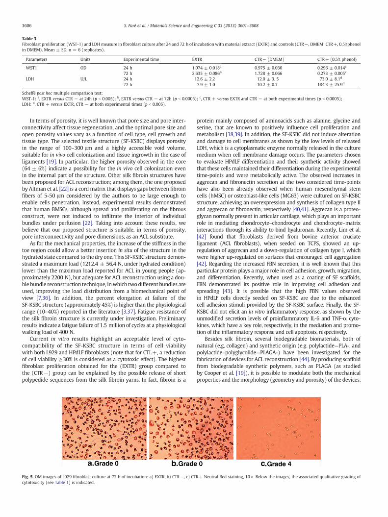
Table 3Fibroblast proliferation (WST-1) and LDHmeasure in fibroblast culture after 24 and 72 h of incubation with material extract (EXTR) and controls (CTR−, DMEM; CTR+, 0.5%phenolin DMEM). Mean ± SD, n = 6 (replicates).
Parameters Units Experimental time EXTR CTR− (DMEM) CTR+ (0.5% phenol)
WST1 OD 24 h 1.074 ± 0.018a 0.975 ± 0.030 0.296 ± 0.014c
72 h 2.635 ± 0.086b 1.728 ± 0.066 0.273 ± 0.005c
LDH U/L 24 h 12.6 ± 2.2 12.0 ± 3. 5 73.0 ± 8.1d
72 h 7.9 ± 1.0 10.2 ± 0.7 184.3 ± 25.9d
Scheffé post hoc multiple comparison test:WST-1: a, EXTR versus CTR − at 24h (p b 0.005); b, EXTR versus CTR − at 72h (p b 0.0005); c, CTR + versus EXTR and CTR − at both experimental times (p b 0.0005);LDH: d, CTR + versus EXTR, CTR − at both experimental times (p b 0.005).
3606 S. Farè et al. / Materials Science and Engineering C 33 (2013) 3601–3608
In terms of porosity, it is well known that pore size and pore inter-connectivity affect tissue regeneration, and the optimal pore size andopen porosity values vary as a function of cell type, cell growth andtissue type. The selected textile structure (SF-KSBC) displays porosityin the range of 100–300 μm and a highly accessible void volume,suitable for in vivo cell colonization and tissue ingrowth in the case ofligaments [19]. In particular, the higher porosity observed in the core(64 ± 6%) indicate a possibility for the in vivo cell colonization evenin the internal part of the structure. Other silk fibroin structures havebeen proposed for ACL reconstruction; among them, the one proposedby Altman et al. [22] is a cord matrix that displays gaps between fibroinfibers of 5-50 μm considered by the authors to be large enough toenable cells penetration. Instead, experimental results demonstratedthat human BMSCs, although spread and proliferating on the fibrousconstruct, were not induced to infiltrate the interior of individualbundles under perfusion [22]. Taking into account these results, webelieve that our proposed structure is suitable, in terms of porosity,pore interconnectivity and pore dimensions, as an ACL substitute.
As for the mechanical properties, the increase of the stiffness in thetoe region could allow a better insertion in situ of the structure in thehydrated state compared to the dry one. This SF-KSBC structure demon-strated a maximum load (1212.4 ± 56.4 N, under hydrated condition)lower than the maximun load reported for ACL in young people (ap-proximately 2200 N), but adequate for ACL reconstruction using a dou-ble bundle reconstruction technique, inwhich two different bundles areused, improving the load distribution from a biomechanical point ofview [7,36]. In addition, the percent elongation at failure of theSF-KSBC structure (approximately 45%) is higher than the physiologicalrange (10–40%) reported in the literature [3,37]. Fatigue resistance ofthe silk fibroin structure is currently under investigation. Preliminaryresults indicate a fatigue failure of 1.5 million of cycles at a physiologicalwalking load of 400 N.
Current in vitro results highlight an acceptable level of cyto-compatibility of the SF-KSBC structure in terms of cell viabilitywith both L929 and HPdLF fibroblasts (note that for CTL+, a reductionof cell viability ≥30% is considered as a cytotoxic effect). The highestfibroblast proliferation obtained for the (EXTR) group compared tothe (CTR−) group can be explained by the possible release of shortpolypedide sequences from the silk fibroin yarns. In fact, fibroin is a
Fig. 5. OM images of L929 fibroblast culture at 72 h of incubation: a) EXTR, b) CTR−, c) CTcytotoxicity (see Table 1) is indicated.
protein mainly composed of aminoacids such as alanine, glycine andserine, that are known to positively influence cell proliferation andmetabolism [38,39]. In addition, the SF-KSBC did not induce alterationand damage to cell membranes as shown by the low levels of releasedLDH, which is a cytoplasmatic enzyme normally released in the culturemedium when cell membrane damage occurs. The parameters chosento evaluate HPdLF differentiation and their synthetic activity showedthat these cells maintained their differentiation during the experimentaltime-points and were metabolically active. The observed increases inaggrecan and fibronectin secretion at the two considered time-pointshave also been already observed when human mesenchymal stemcells (hMSC) or osteoblast-like cells (MG63) were cultured on SF-KSBCstructure, achieving an overexpression and synthesis of collagen type IIand aggrecan or fibronectin, respectively [40,41]. Aggrecan is a proteo-glycan normally present in articular cartilage, which plays an importantrole in mediating chondrocyte–chondrocyte and chondrocyte–matrixinteractions through its ability to bind hyaluronan. Recently, Lim et al.[42] found that fibroblasts derived from bovine anterior cruciateligament (ACL fibroblasts), when seeded on TCPS, showed an up-regulation of aggrecan and a down-regulation of collagen type I, whichwere higher up-regulated on surfaces that encouraged cell aggregation[42]. Regarding the increased FBN secretion, it is well known that thisparticular protein plays a major role in cell adhesion, growth, migration,and differentiation. Recently, when used as a coating of SF scaffolds,FBN demonstrated its positive role in improving cell adhesion andspreading [43]. It is possible that the high FBN values observedin HPdLF cells directly seeded on SF-KSBC are due to the enhancedcell adhesion stimuli provided by the SF-KSBC surface. Finally, the SF-KSBC did not elicit an in vitro inflammatory response, as shown by theunmodified secretion levels of proinflammatory IL-6 and TNF-α cyto-kines, which have a key role, respectively, in the mediation and promo-tion of the inflammatory response and cell apoptosis, respectively.
Besides silk fibroin, several biodegradable biomaterials, both ofnatural (e.g. collagen) and synthetic origin (e.g. polylactide—PLA-, andpolylactide–polyglycolide—PLAGA-) have been investigated for thefabrication of devices for ACL reconstruction [44]. By producing scaffoldfrom biodegradable synthetic polymers, such as PLAGA (as studiedby Cooper et al. [19]), it is possible to modulate both the mechanicalproperties and themorphology (geometry and porosity) of the devices.
R+ Neutral Red staining, 10×. Below the images, the associated qualitative grading of
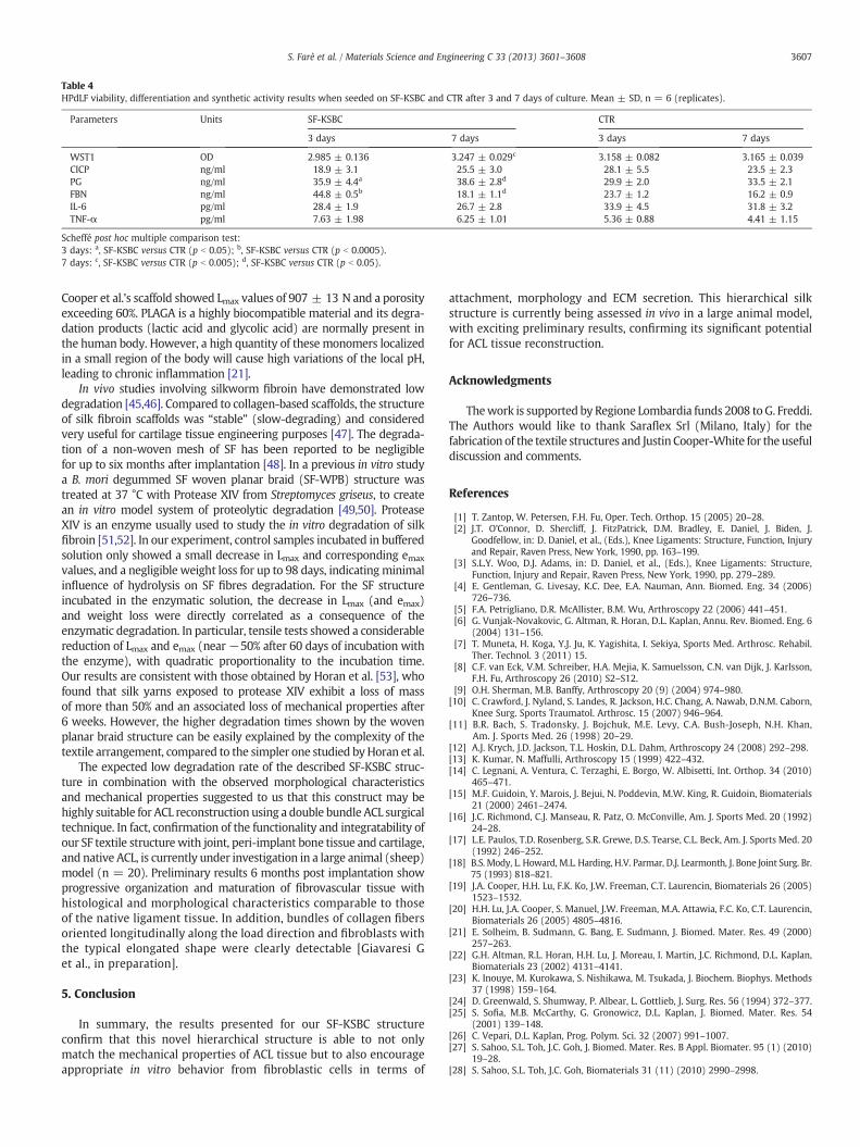
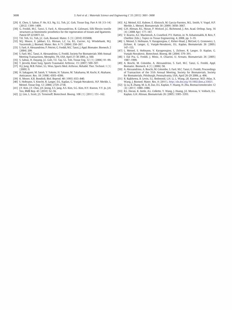
Table 4HPdLF viability, differentiation and synthetic activity results when seeded on SF-KSBC and CTR after 3 and 7 days of culture. Mean ± SD, n = 6 (replicates).
Parameters Units SF-KSBC CTR
3 days 7 days 3 days 7 days
WST1 OD 2.985 ± 0.136 3.247 ± 0.029c 3.158 ± 0.082 3.165 ± 0.039CICP ng/ml 18.9 ± 3.1 25.5 ± 3.0 28.1 ± 5.5 23.5 ± 2.3PG ng/ml 35.9 ± 4.4a 38.6 ± 2.8d 29.9 ± 2.0 33.5 ± 2.1FBN ng/ml 44.8 ± 0.5b 18.1 ± 1.1d 23.7 ± 1.2 16.2 ± 0.9IL-6 pg/ml 28.4 ± 1.9 26.7 ± 2.8 33.9 ± 4.5 31.8 ± 3.2TNF-α pg/ml 7.63 ± 1.98 6.25 ± 1.01 5.36 ± 0.88 4.41 ± 1.15
Scheffé post hoc multiple comparison test:3 days: a, SF-KSBC versus CTR (p b 0.05); b, SF-KSBC versus CTR (p b 0.0005).7 days: c, SF-KSBC versus CTR (p b 0.005); d, SF-KSBC versus CTR (p b 0.05).
3607S. Farè et al. / Materials Science and Engineering C 33 (2013) 3601–3608
Cooper et al.'s scaffold showed Lmax values of 907 ± 13 N and a porosityexceeding 60%. PLAGA is a highly biocompatible material and its degra-dation products (lactic acid and glycolic acid) are normally present inthe human body. However, a high quantity of these monomers localizedin a small region of the body will cause high variations of the local pH,leading to chronic inflammation [21].
In vivo studies involving silkworm fibroin have demonstrated lowdegradation [45,46]. Compared to collagen-based scaffolds, the structureof silk fibroin scaffolds was “stable” (slow-degrading) and consideredvery useful for cartilage tissue engineering purposes [47]. The degrada-tion of a non-woven mesh of SF has been reported to be negligiblefor up to six months after implantation [48]. In a previous in vitro studya B. mori degummed SF woven planar braid (SF-WPB) structure wastreated at 37 °C with Protease XIV from Streptomyces griseus, to createan in vitro model system of proteolytic degradation [49,50]. ProteaseXIV is an enzyme usually used to study the in vitro degradation of silkfibroin [51,52]. In our experiment, control samples incubated in bufferedsolution only showed a small decrease in Lmax and corresponding emax
values, and a negligible weight loss for up to 98 days, indicatingminimalinfluence of hydrolysis on SF fibres degradation. For the SF structureincubated in the enzymatic solution, the decrease in Lmax (and emax)and weight loss were directly correlated as a consequence of theenzymatic degradation. In particular, tensile tests showed a considerablereduction of Lmax and emax (near−50% after 60 days of incubation withthe enzyme), with quadratic proportionality to the incubation time.Our results are consistent with those obtained by Horan et al. [53], whofound that silk yarns exposed to protease XIV exhibit a loss of massof more than 50% and an associated loss of mechanical properties after6 weeks. However, the higher degradation times shown by the wovenplanar braid structure can be easily explained by the complexity of thetextile arrangement, compared to the simpler one studied byHoran et al.
The expected low degradation rate of the described SF-KSBC struc-ture in combination with the observed morphological characteristicsand mechanical properties suggested to us that this construct may behighly suitable for ACL reconstruction using a double bundle ACL surgicaltechnique. In fact, confirmation of the functionality and integratability ofour SF textile structurewith joint, peri-implant bone tissue and cartilage,and native ACL, is currently under investigation in a large animal (sheep)model (n = 20). Preliminary results 6 months post implantation showprogressive organization and maturation of fibrovascular tissue withhistological and morphological characteristics comparable to thoseof the native ligament tissue. In addition, bundles of collagen fibersoriented longitudinally along the load direction and fibroblasts withthe typical elongated shape were clearly detectable [Giavaresi Get al., in preparation].
5. Conclusion
In summary, the results presented for our SF-KSBC structureconfirm that this novel hierarchical structure is able to not onlymatch the mechanical properties of ACL tissue but to also encourageappropriate in vitro behavior from fibroblastic cells in terms of
attachment, morphology and ECM secretion. This hierarchical silkstructure is currently being assessed in vivo in a large animal model,with exciting preliminary results, confirming its significant potentialfor ACL tissue reconstruction.
Acknowledgments
Thework is supported by Regione Lombardia funds 2008 toG. Freddi.The Authors would like to thank Saraflex Srl (Milano, Italy) for thefabrication of the textile structures and Justin Cooper-White for the usefuldiscussion and comments.
References
[1] T. Zantop, W. Petersen, F.H. Fu, Oper. Tech. Orthop. 15 (2005) 20–28.[2] J.T. O'Connor, D. Shercliff, J. FitzPatrick, D.M. Bradley, E. Daniel, J. Biden, J.
Goodfellow, in: D. Daniel, et al., (Eds.), Knee Ligaments: Structure, Function, Injuryand Repair, Raven Press, New York, 1990, pp. 163–199.
[3] S.L.Y. Woo, D.J. Adams, in: D. Daniel, et al., (Eds.), Knee Ligaments: Structure,Function, Injury and Repair, Raven Press, New York, 1990, pp. 279–289.
[4] E. Gentleman, G. Livesay, K.C. Dee, E.A. Nauman, Ann. Biomed. Eng. 34 (2006)726–736.
[5] F.A. Petrigliano, D.R. McAllister, B.M. Wu, Arthroscopy 22 (2006) 441–451.[6] G. Vunjak-Novakovic, G. Altman, R. Horan, D.L. Kaplan, Annu. Rev. Biomed. Eng. 6
(2004) 131–156.[7] T. Muneta, H. Koga, Y.J. Ju, K. Yagishita, I. Sekiya, Sports Med. Arthrosc. Rehabil.
Ther. Technol. 3 (2011) 15.[8] C.F. van Eck, V.M. Schreiber, H.A. Mejia, K. Samuelsson, C.N. van Dijk, J. Karlsson,
F.H. Fu, Arthroscopy 26 (2010) S2–S12.[9] O.H. Sherman, M.B. Banffy, Arthroscopy 20 (9) (2004) 974–980.
[10] C. Crawford, J. Nyland, S. Landes, R. Jackson, H.C. Chang, A. Nawab, D.N.M. Caborn,Knee Surg. Sports Traumatol. Arthrosc. 15 (2007) 946–964.
[11] B.R. Bach, S. Tradonsky, J. Bojchuk, M.E. Levy, C.A. Bush-Joseph, N.H. Khan,Am. J. Sports Med. 26 (1998) 20–29.
[12] A.J. Krych, J.D. Jackson, T.L. Hoskin, D.L. Dahm, Arthroscopy 24 (2008) 292–298.[13] K. Kumar, N. Maffulli, Arthroscopy 15 (1999) 422–432.[14] C. Legnani, A. Ventura, C. Terzaghi, E. Borgo, W. Albisetti, Int. Orthop. 34 (2010)
465–471.[15] M.F. Guidoin, Y. Marois, J. Bejui, N. Poddevin, M.W. King, R. Guidoin, Biomaterials
21 (2000) 2461–2474.[16] J.C. Richmond, C.J. Manseau, R. Patz, O. McConville, Am. J. Sports Med. 20 (1992)
24–28.[17] L.E. Paulos, T.D. Rosenberg, S.R. Grewe, D.S. Tearse, C.L. Beck, Am. J. Sports Med. 20
(1992) 246–252.[18] B.S. Mody, L. Howard, M.L. Harding, H.V. Parmar, D.J. Learmonth, J. Bone Joint Surg. Br.
75 (1993) 818–821.[19] J.A. Cooper, H.H. Lu, F.K. Ko, J.W. Freeman, C.T. Laurencin, Biomaterials 26 (2005)
1523–1532.[20] H.H. Lu, J.A. Cooper, S. Manuel, J.W. Freeman, M.A. Attawia, F.C. Ko, C.T. Laurencin,
Biomaterials 26 (2005) 4805–4816.[21] E. Solheim, B. Sudmann, G. Bang, E. Sudmann, J. Biomed. Mater. Res. 49 (2000)
257–263.[22] G.H. Altman, R.L. Horan, H.H. Lu, J. Moreau, I. Martin, J.C. Richmond, D.L. Kaplan,
Biomaterials 23 (2002) 4131–4141.[23] K. Inouye, M. Kurokawa, S. Nishikawa, M. Tsukada, J. Biochem. Biophys. Methods
37 (1998) 159–164.[24] D. Greenwald, S. Shumway, P. Albear, L. Gottlieb, J. Surg. Res. 56 (1994) 372–377.[25] S. Sofia, M.B. McCarthy, G. Gronowicz, D.L. Kaplan, J. Biomed. Mater. Res. 54
(2001) 139–148.[26] C. Vepari, D.L. Kaplan, Prog. Polym. Sci. 32 (2007) 991–1007.[27] S. Sahoo, S.L. Toh, J.C. Goh, J. Biomed. Mater. Res. B Appl. Biomater. 95 (1) (2010)
19–28.[28] S. Sahoo, S.L. Toh, J.C. Goh, Biomaterials 31 (11) (2010) 2990–2998.
3608 S. Farè et al. / Materials Science and Engineering C 33 (2013) 3601–3608
[29] K. Chen, S. Sahoo, P. He, K.S. Ng, S.L. Toh, J.C. Goh, Tissue Eng. Part A 18 (13–14)(2012) 1399–1409.
[30] G. Freddi, M.C. Tanzi, S. Farè, A. Alessandrino, R. Calimani, Silk fibroin textilestructures as biomimetic prosthetics for the regeneration of tissues and ligaments.Patent EP 2210971 A1.
[31] T.K. Teh, S.L. Toh, J.C. Goh, Biomed. Mater. 5 (3) (2010) 035008.[32] M.J. Moore, E. Jabbari, E.L. Ritman, L.C. Lu, B.L. Currier, A.J. Windebank, M.J.
Yaszemski, J. Biomed. Mater. Res. A 71 (2004) 258–267.[33] S. Farè, A. Alessandrino, P. Petrini, G. Freddi, M.C. Tanzi, J. Appl. Biomater. Biomech. 2
(2004) 209.[34] S. Farè, M.C. Tanzi, A. Alessandrino, G. Freddi, Society For Biomaterials 30th Annual
Meeting Transactions, Memphis, TN, USA, April 27-30 2005, p. 560.[35] S. Sahoo, H. Ouyang, J.C. Goh, T.E. Tay, S.L. Toh, Tissue Eng. 12 (1) (2006) 91–99.[36] T. Järvelä, Knee Surg. Sports Traumatol. Arthrosc. 15 (2007) 500–507.[37] H.J. Jung, M.B. Fisher, S.L. Woo, Sports Med. Arthrosc. Rehabil. Ther. Technol. 1 (1)
(2009) 9.[38] H. Sakagami, M. Satoh, Y. Yokote, H. Takano, M. Takahama, M. Kochi, K. Akahane,
Anticancer. Res. 18 (1998) 4303–4306.[39] K. Moore, K.R. Bondioli, Biol. Reprod. 48 (1993) 833–840.[40] S. Hofmann, S. Knecht, R. Langer, D.L. Kaplan, G. Vunjak-Novakovic, H.P. Merkle, L.
Meinel, Tissue Eng. 12 (2006) 2729–2738.[41] J.Y. Kim, J.Y. Choi, J.H. Jeong, E.S. Jang, A.S. Kim, S.G. Kim, H.Y. Kweon, Y.Y. Jo, J.H.
Yeo, BMB Rep. 43 (2010) 52–56.[42] J.J. Lim, L. Scott, J.S. Temenoff, Biotechnol. Bioeng. 108 (1) (2011) 151–162.
[43] A.J. Meinel, K.E. Kubow, E. Klotzsch, M. Garcia-Fuentes, M.L. Smith, V. Vogel, H.P.Merkle, L. Meinel, Biomaterials 30 (2009) 3058–3067.
[44] G.H. Altman, R.L. Horan, P. Weitzel, J.C. Richmond, J. Am. Acad. Orthop. Surg. 16(4) (2008 Apr) 177–187.
[45] V. Kearns, A.C. MacIntosh, A. Crawford, P.V. Hatton, in: N. Ashammakhi, R. Reis, F.Chiellini (Eds.), Topics in Tissue Engineering, 4, 2008, pp. 3–19.
[46] L. Meinel, S. Hofmann, V. Karageorgiou, C. Kirker-Head, J. McCool, G. Gronowicz, L.Zichner, R. Langer, G. Vunjak-Novakovic, D.L. Kaplan, Biomaterials 26 (2005)147–155.
[47] L. Meinel, S. Hofmann, V. Karageorgiou, L. Zichner, R. Langer, D. Kaplan, G.Vunjak-Novakovic, Biotechnol. Bioeng. 88 (2004) 379–391.
[48] I. Dal Pra, G. Freddi, J. Minic, A. Chiarini, U. Armato, Biomaterials 26 (2005)1987–1999.
[49] A. Boschi, M. Colombo, A. Alessandrino, S. Farè, M.C. Tanzi, G. Freddi, Appl.Biomater. Biomech. 4 (1) (2006) 58.
[50] A. Alessandrino, A. Boschi, M. Colombo, S. Farè, M.C. Tanzi, G. Freddi, Proceedingsof Transaction of the 31th Annual Meeting, Society for Biomaterials, Societyfor Biomaterials, Pittsburgh, Pennsylvania, USA, April 26-29 2006, p. 496.
[51] R. Rajkhowa, B. Levin, S.L. Redmond, L.H. Li, L. Wang, J.R. Kanwar, M.D. Atlas, X.Wang, J. Biomed. Mater. Res. A (2011), http://dx.doi.org/10.1002/jbm.a.33021.
[52] Q. Lu, B. Zhang, M. Li, B. Zuo, D.L. Kaplan, Y. Huang, H. Zhu, Biomacromolecules 12(4) (2011) 1080–1086.
[53] R.L. Horan, K. Antle, A.L. Collette, Y. Wang, J. Huang, J.E. Moreau, V. Volloch, D.L.Kaplan, G.H. Altman, Biomaterials 26 (2005) 3385–3393.
Related Documents