Welcome message from author
This document is posted to help you gain knowledge. Please leave a comment to let me know what you think about it! Share it to your friends and learn new things together.
Transcript

In Vitro Neurotoxicology

In Vitro Neurotoxicology: Principles and Challengesedited by Evelyn Tiffany-Castiglioni, 2004
Cardiac Drug Development Guideedited by Michael K. Pugsley, 2003
Methods in Biological Oxidative Stressedited by Kenneth Hensley and Robert A. Floyd, 2003
Apoptosis Methods in Pharmacology and Toxicology:Approaches to Measurement and Quantificationedited by Myrtle A. Davis, 2002
Ion Channel Localization: Methods and Protocolsedited by Anatoli N. Lopatin and Colin G. Nichols, 2001
METHODS IN PHARMACOLOGY AND TOXICOLOGY
MANNFRED A. HOLLINGER, PhD, SERIES EDITOR

In VitroNeurotoxicology
Principles and Challenges
Edited by
Evelyn Tiffany-CastiglioniDepartment of Veterinary Anatomy and Public HealthTexas A&M University College of Veterinary Medicine
and Center for Environmental and Rural Health,College Station, TX
Foreword by
Mannfred A. HollingerUniversity of California
Davis, CA
METHODS IN PHARMACOLOGY AND TOXICOLOGY
Humana Press Totowa, New Jersey

© 2004 Humana Press Inc.999 Riverview Drive, Suite 208Totowa, NJ 07512
www.humanapress.com
All rights reserved. No part of this book may be reproduced, stored in a retrieval system, or transmittedin any form or by any means, electronic, mechanical, photocopying, microfilming, recording, or other-wise without written permission from the Publisher.
The content and opinions expressed in this book are the sole work of the authors and editors, who havewarranted due diligence in the creation and issuance of their work. The publisher, editors, and authorsare not responsible for errors or omissions or for any consequences arising from the information oropinions presented in this book and make no warranty, express or implied, with respect to its contents.
Production Editor: Tracy Catanese
Cover Illustration: Figure 2 from Chapter 7, “Cell-Type-Specific Responses of the Nervous System toLead” by Evelyn Tiffany-Castiglioni.
Cover design by Patricia F. Cleary.
For additional copies, pricing for bulk purchases, and/or information about other Humana titles, contactHumana at the above address or at any of the following numbers: Tel.: 973-256-1699; Fax: 973-256-8341; E-mail: [email protected] or visit our website: http://humanapress.com
This publication is printed on acid-free paper.ANSI Z39.48-1984 (American National Standards Institute) Permanence of Paper for Printed LibraryMaterials.
Photocopy Authorization Policy:Authorization to photocopy items for internal or personal use, or the internal or personal use of specificclients, is granted by Humana Press Inc., provided that the base fee of US $25.00 per copy is paid directlyto the Copyright Clearance Center at 222 Rosewood Drive, Danvers, MA 01923. For those organizationsthat have been granted a photocopy license from the CCC, a separate system of payment has beenarranged and is acceptable to Humana Press Inc. The fee code for users of the Transactional ReportingService is: [1-58829-047-6/04 $25.00].
Printed in the United States of America. 10 9 8 7 6 5 4 3 2 1
1-59259-651-7 (e-book)
Library of Congress Cataloging-in-Publication Data
Tiffany-Castiglioni, Evelyn. In vitro neurotoxicology : principles and challenges / edited by Evelyn Tiffany-Castiglioni. p. cm. -- (Methods in pharmacology and toxicology) Includes bibliographical references and index. ISBN 1-58829-047-6 (alk. paper) 1. Neurotoxicology. 2. Toxicity testing--In vitro. I. Title. II. Series.
RC347.5.T54 2003 616.8’047--dc21
2003049933

Dedication
To Robert S. Tiffany, Jr. and Frances James Tiffany
In Memoriam
v
Foreword
Researchers in pharmacology and toxicology are constantly searching forrelevant in vitro methods in order to obtain valid data without the use ofwhole animals with their attendant costs and ethical questions. This is par-ticularly true for workers interested in neurotoxicology, where we continueto discover new neurotoxic effects of drugs and other xenobiotics. Over theyears, a number of creative and useful methods have emerged. For someoneentering the field of neurotoxicology, the decision regarding type of methodmost appropriate for his or her work can be a daunting one. For example, thetime and effort required to search the literature and evaluate candidate sys-tems can require weeks, if not months.
In Vitro Neurotoxicology: Principles and Challenges, edited by Dr.Evelyn Tiffany-Castiglioni, is a masterful contribution to the field ofneurotoxicology. With each passing year the need for new and improved invitro methods to help further our understanding of neurotoxicology willincrease. This volume brings us up to date.
Mannfred A. HollingerProfessor Emeritus
University of CaliforniaDavis, CA
vii
Preface
Neurotoxicity assessment with in vitro systems is the focus of bothincreasing expectations and heightened challenges. Such systems prospec-tively offer a means to improve screening efficiency for potential neuro-toxicants, a method for better understanding mechanisms of toxicant action,a decreasing use of animals, and a means to obtain data from human samples.On the other hand, in vitro systems have not yet been used in consistent,broadly applied formats that would validate and exploit their value for neu-rotoxicity testing. Inherent problems, such as test chemical concentration anddelivery, lack of heterogeneous cell–cell interactions, immaturity of cell typesavailable, phenotypic variations induced by culture techniques, and insensi-tivity of endpoints tested, significantly impede the use and interpretation ofin vitro assays. In addition, standardized metrics and methods for comparingresults across studies and laboratories, as well as benchmark criteria for link-ing in vitro to in vivo studies, are often lacking.
The purpose of In Vitro Neurotoxicology: Principles and Challenges is tosynthesize principles and concepts of in vitro neurotoxicology that willfacilitate the development of significantly improved methods and systemsfor in vitro neurotoxicity testing, with emphasis on their relevance to in vivosystems. An outstanding list of contributors has been assembled, includingwell-respected leaders in the field and new investigators who are exploringemerging frontiers in the area of genomic toxicology. Contributors havetaken a fresh look at their own and others’ work, critically and compara-tively analyzed it across experimental systems and toxicants, and formal-ized essential principles for in vitro neurotoxicity testing. In most cases,chapters are arranged around major themes or central ideas, rather thanaround individual toxicants or specific in vitro models. Most chapters arecollaborative efforts that address a theme and employ examples comprisedof multiple experimental systems and endpoints. The chapters emphasizeseveral neurotoxicants that are of prominent human health concern and aboutwhich metabolism and dose–responses are best understood, both in vivo andin vitro: lead, mercury, organophosphorus insecticides, polychlorinatedbiphenyls and dioxin, ethanol, and endogenous proteins.
There are already several excellent articles and monographs that describematerials and techniques applicable to in vitro neurotoxicology, such as celllines, methods of primary cell culture, brain slice preparations, and in vitro
ix

assays for viability and function. Rather than repeating the contents of theseprevious works, In Vitro Neurotoxicology: Principles and Challenges pro-vides an Appendix containing a critically reviewed list of related works.The list, carefully selected and annotated by the contributors, includesimportant review articles, books on in vitro toxicology, neurotoxicology,and in vitro neurotoxicology, and chapters from methods manuals. TheAppendix collects in one place references to most of the major reviews andseminal work related to in vitro neurotoxicology that have appeared in thepast ten years.
Evelyn Tiffany-Castiglioni
x Preface

Contents
Dedication .......................................................................................... vForeword .......................................................................................... viiPreface ................................................................................................ ixContributors ................................................................................... xiii
1 In Vitro Neurotoxicology: Introduction to ConceptsEvelyn Tiffany-Castiglioni ............................................................. 1
2 Predictive Value of In Vitro Systemsfor Neurotoxicity Risk Assessment
Marion Ehrich and David C. Dorman ......................................... 293 Exposure–Dose–Response Paradigm
as It Relates to ToxicogenomicsWilliam H. Hanneman, Melvin E. Andersen,
Marie E. Legare, Christine T. French,Tami S. McMullin, Carolyn Broccardo,and Ruth E. Billings ................................................................... 41
4 In Vitro Studies of Neurotoxicant Effectson Cellular Homeostasis
Gerald J. Audesirk and Ronald B. Tjalkens ............................... 595 Role of Apoptosis in Neurotoxicology
Lori D. White, Sid Hunter, Michael W. Miller,Marion Ehrich, and Stanley Barone, Jr. ................................. 95
6 Impairment of Neurotransmitter Metabolismand Function by Neurotoxicants:Enzyme Pathways in Neurons and Astroglia
Michael Aschner and Ursula Sonnewald ................................. 1337 Cell-Type-Specific Responses
of the Nervous System to LeadEvelyn Tiffany-Castiglioni and Yongchang Qian .................. 151
8 Effects of Toxicants on Neural DifferentiationStanley Barone, Jr., Prasada R. S. Kodavanti,
and William R. Mundy ............................................................ 187
xi
xii Contents
9 Impairment of Synaptic Function by Exposure to LeadStephen M. Lasley and Mary E. Gilbert ................................... 217
10 Aggregating Brain Cell Culturesfor Neurotoxicological Studies
Marie-Gabrielle Zurich, Florianne Monnet-Tschudi,Lucio G. Costa, Benoît Schilter, and Paul Honegger ......... 243
11 Use of Complimentary In Vitro and In Vivo Methodsfor Assessing Neuroendocrine Disruptors
W. Les Dees, Jill K. Hiney, Robert K. Dearth,and Vinod K. Srivastava ......................................................... 267
12 Establishing In Vitro Models to Study EndogenousNeurotoxicants
Heather D. Durham ....................................................................... 291
Appendix: Annotated Reading ListEvelyn Tiffany-Castiglioni, Lucio G. Costa,Marion Ehrich, William R. Mundy, Gerald J. Audesirk,Michael Aschner, Prasada R. S. Kodavanti,and Stephen M. Lasley ................................................................. 315
Index ........................................................................................................ 325

Contributors
MELVIN E. ANDERSEN • Associate Professor, Department of Environmentaland Radiological Sciences, College of Veterinary Medicine and BiomedicalSciences, Colorado State University, Ft. Collins, CO
MICHAEL ASCHNER • Professor, Department of Physiology andPharmacology, Wake Forest University School of Medicine, Winston-Salem, NC
GERALD J. AUDESIRK • Professor, Department of Biology, Universityof Colorado at Denver, Denver, CO
STANLEY BARONE, JR. • Research Biologist, Cellular and MolecularToxicology Branch, Neurotoxicology Division/NHEERL/ORD USEnvironmental Protection Agency, Research Triangle Park, NC
RUTH E. BILLINGS • Research Associate, Department of Environmentaland Radiological Sciences, College of Veterinary Medicine andBiomedical Sciences, Colorado State University, Ft. Collins, CO
CAROLYN BROCCARDO • Research Assistant, Department of Environmentaland Radiological Sciences, College of Veterinary Medicine and BiomedicalSciences, Colorado State University, Ft. Collins, CO
LUCIO G. COSTA • Professor, Department of Environmental andOccupational Health Sciences, University of Washington, Seattle, WA
ROBERT K. DEARTH • Technician I, Department of Veterinary Anatomyand Public Health, College of Veterinary Medicine, Texas A&MUniversity, College Station, TX
W. LES DEES • Professor, Department of Veterinary Anatomy PublicHealth, College of Veterinary Medicine, Texas A&M University,College Station, TX
DAVID C. DORMAN • Director of the Division of Biological Sciences, CIITCenters for Health Research, Research Triangle Park, NC
HEATHER D. DURHAM • Professor, Department of Neurology andNeurosurgery, McGill University, Montreal Neurological Institute,Montreal, Quebec, Canada
MARION EHRICH • Professor, Virginia-Maryland Regional College ofVeterinary Medicine, Blacksburg, VA
xiii

xiv Contributors
CHRISTINE T. FRENCH • Research Assistant, Department of Environmentaland Radiological Sciences, College of Veterinary Medicine and BiomedicalSciences, Colorado State University, Ft. Collins, CO
MARY E. GILBERT • Assistant Professor, Neurotoxicology Division, USEnvironmental Protection Agency, Research Triangle Park, NC
WILLIAM H. HANNEMAN • Assistant Professor, Head of ToxicologySection, Department of Environmental and Radiological Sciences,College of Veterinary Medicine and Biomedical Sciences, ColoradoState University, Ft. Collins, CO
JILL K. HINEY • Research Assistant Professor, Department of VeterinaryAnatomy and Public Health, College of Veterinary Medicine, TexasA&M University, College Station, TX
PAUL HONEGGER • Professeur Associe, Institute of Physiology,University of Lausanne, Lausanne, Switzerland
SID HUNTER • Toxicologist, Reproductive Toxicology Division, USEnvironmental Protection Agency, Research Triangle Park, NC
PRASADA R. S. KODAVANTI • Research Toxicologist, NeurotoxicologyDivision, US Environmental Protection Agency, Research TrianglePark, NC
STEPHEN M. LASLEY • Professor, Department of Biomedical andTherapeutic Sciences, University of Illinois College of Medicine,Peoria, IL
MARIE E. LEGARE • Assistant Professor, Department of Environmental andRadiological Health Science, College of Veterinary Medicine andBiomedical Sciences, Colorado State University, Ft. Collins, CO
TAMI S. MCMULLIN • Research Assistant, Department of Environmentaland Radiological Sciences, College of Veterinary Medicine andBiomedical Sciences, Colorado State University, Ft. Collins, CO
MICHAEL W. MILLER • Professor, Department of Neuroscience andPhysiology, State University of New York-Upstate Medical University,Syracuse, NY
FLORIANNE MONNET-TSCHUDI • Maitre Assistant, Institute of Physiology,University of Lausanne, Lausanne, Switzerland
WILLIAM R. MUNDY • Research Toxicologist, Neurotoxicology Division,US Environmental Protection Agency, Research Triangle Park, NC
YONGCHANG QIAN • Research Assistant Professor, Department ofVeterinary Anatomy and Public Health, College of Veterinary Medicine,Texas A&M University, College Station, TX

BENOÎT SCHILTER • Nestlé Research Centre, Vers-chez-les Blanc,Lausanne, Switzerland
URSULA SONNEWALD • Professor, Department of Clinical Neuroscience,Norwegian University of Science and Technology, Trondheim, Norway
VINOD K. SRIVASTAVA • Research Assistant Professor, Departmentof Veterinary Anatomy and Public Health, College of VeterinaryMedicine, Texas A&M University, College Station, TX
EVELYN TIFFANY-CASTIGLIONI • Professor and Head, Department ofVeterinary Anatomy and Public Health, Associate Dean forUndergraduate Education, College of Veterinary Medicine, TexasA&M University, College Station, TX
RONALD B. TJALKENS • Assistant Professor, Department of VeterinaryAnatomy and Public Health, College of Veterinary Medicine, TexasA&M University, and Center for Environmental and Rural Health,College Station, TX
LORI D. WHITE • Biologist, Cellular and Molecular Toxicology Branch,Neurotoxicology Division, NHEERL/ORD US EnvironmentalProtection Agency, Research Triangle Park, NC
MARIE-GABRIELLE ZURICH • Premiere Assistante, Institute of Physiology,University of Lausanne, Lausanne, Switzerland
xv Contributors

In Vitro Neurotoxicology 1
1
From: Methods in Pharmacology and Toxicology: In Vitro Neurotoxicology: Principles and ChallengesEdited by: E. Tiffany-Castiglioni © Humana Press Inc., Totowa, NJ
1In Vitro Neurotoxicology
Introduction to Concepts
Evelyn Tiffany-Castiglioni
1. UTILITY OF IN VITRO SYSTEMSThe history of neuroscience is punctuated by oracular disclosures from in
vitro systems. In 1907, a pivotal tissue culture study by Harrison proved thatRamón y Cajal’s theory on the developmental origin of nerve fibers wascorrect. Cajal had proposed in 1890, based on microscopic analysis of statichistologic tissue sections, that the immature neuronal cell body sends out anaxon that elongates freely, bearing a motile growth cone at its tip. Compet-ing theories held that free growth of neurites did not occur, but that theneurites formed from the fusion of elements produced by other cells or fromthe stretching of a protoplasmic bridge between central and peripheral cellbodies of a multinucleated cell (1). These theories could not be tested by thehistologic methods of the time, because axonal growth by a living neuroncould not be directly observed. Harrison (2) pioneered a culture system forlong-term microscopic observation of neuronal differentiation in living tad-pole neural tube tissue. His observation that neurites grow out from cellbodies has been hailed as “one of the most revolutionary results in experi-mental biology” (3).
Some 50 yr later, tissue culture provided the means for an advance ofsimilar magnitude by Levi-Montalcini, Hamberger, and Cohen (4,5), thediscovery of nerve growth factor (NGF). NGF became the paradigm for thediscovery of other growth and differentiation factors. These investigatorsused an in vitro chick ganglion bioassay to detect NGF in a variety of sources

2 Tiffany-Castiglioni
suspected to harbor this heretofore undefined polypeptide. Neurons in thechick ganglion explants perceived the subtle presence of NGF in extracts ofthe S-180 mouse sarcoma, snake venom, and the male mouse submaxillarygland and extended neurites toward it. Thus, Levi-Montalcini’s Nobel Prizeaddress was published in the journal of the Tissue Culture Association (6),with the recognition by its editor, Gordon Sato, that the award to Levi-Montalcini and Cohen was “an affirmation of the growing importance ofcell culture in biological research.”
Contemporary neuroscience has also profited extensively from tissue cul-ture models, examples of which are the glial guidance theory for neuronalmigration and the emerging appreciation of glial–neuronal signaling. Theglial guidance theory, whereby radial glial cell processes provide a scaffoldfor the directed migration of postmitotic neurons during development, washypothesized from painstaking morphological studies at both the light andelectron microscopic levels by Rakic (7–9). The theory gained support andmechanistic explication from the in vitro work of Hatten and colleagues(10–12), who devised a cell culture system for the videomicroscopic exami-nation of the migration of living cerebellar granule cells along the cytoplas-mic “monorails” of radial glia. Among their many discoveries with this invitro system has been the identity of cell–cell adhesion molecules, such asastrotactin (13,14), with which neurons and glia interact to form a complexhistoarchitecture. The emerging story of bidirectional communication be-tween glia and neurons, including the requirement of astrocytes for synapseformation by neurons, is similarly founded upon cell culture work. Astro-cytes apparently integrate and modulate neuronal synaptic transmissionthrough intrinsic signaling properties discovered in cell culture models. As-trocytes exhibit Ca2+ excitability, functional neurotransmitter receptors thatregulate intracellular Ca2+ concentrations, the ability to propagate [Ca2+]oscillations to neighboring cells through gap junctions, and the release ofneuroactive transmitters to neurons (reviewed in refs. 15–17). A major fo-cus of current neurobiology is to confirm these tantalizing properties in in-tact tissues.
The achievements of in vitro neurotoxicology, to date, have been moremodest, but its potential is still untold. With many technical improvementsin imaging and molecular biology, in vitro neurotoxicology has become amajor focus for understanding basic mechanisms of toxicant action. In time,it may form the basis for reliable, high-throughput screening systems for theneurotoxicity of new and untested chemicals. In order to develop in vitroneurotoxicology to a higher level of utility, its strengths must be exploitedand its weaknesses overcome. Like oracles and like the classic experimentsof Harrison, Levi-Montalcini, and Hatten, cell and tissue culture studies of

In Vitro Neurotoxicology 3
neurotoxicity are, by themselves, abstract. They must be critically designedand interpreted in the context of biological complexity. In vitro studies offerthe greatest insights to biology when they are performed in complementwith in vivo experimentation.
The central theme of this book is that neuroscience and neurotoxicologyexhibit a significant degree of alignment in the common ground of in vitromodels. Alignment is visible in two areas. First, both disciplines recognize theneed for valid in vitro models in which the biological significance of the endpoints measured and limitations of the model are well understood. Validitywill be addressed in several chapters of this volume by comparisons betweenobservations made in vivo and in vitro. Furthermore, contributing authorspresent underlying concepts and detailed commentary about the use of comple-mentary in vivo/in vitro strategies. Second, the range of neurological diseaseswith a toxicologic component is expanding. Neurotoxicology may provide pre-liminary road maps for exploring the basis of some neurodevelopmental andneurodegenerative diseases. Evidence that neurotoxicology has advanced ba-sic biomedical knowledge is beginning to emerge. Three selected examples inthis volume are the concept of astroglia as depots for lead and possibly othermetals in the central nervous system, the elucidation of factors involved inonset of puberty in females, and the exploration of endogenous proteins asneurotoxicants.
As an introduction to the ensuing chapters, this chapter will brieflydescribe several background topics: common neurotoxicants and their tar-get cells, acute and accumulated damage from exposure to neurotoxicants,biological concepts in in vitro neurotoxicology and their interrelationships,trends in in vitro neurotoxicology, and general research needs.
2. NEUROTOXICANTS ANDTHEIR CELLULAR TARGETS
Common neurotoxicants selected for the focus of this book are organo-phosphorus pesticides, lead (Pb), methyl mercury, halogenated aromatichydrocarbons (HAHs), and ethanol. Organophosphorus (OP) compoundsrepresent the largest group of chemical insecticides in use throughout theworld today (18). In addition, OP compounds comprise a major portion ofthe US military stockpile of chemical nerve agents that include Tabun, sarin,soman, and VX. OPs cause potent neurotoxicological effects in humans andanimals. Although the immediate, acute neurotoxic action of OPs is the in-hibition of acetylcholinesterase (AChE), some OPs also produce aneurodegenerative disorder known as organophosphate-induced delayedneurotoxicity (OPIDN) with Wallerian-type degeneration of the axon and

4 Tiffany-Castiglioni
myelin (19). Growing experimental and epidemiologic evidence suggeststhat OPs are developmental neurotoxicants as manifested by developmentaldelays and impaired cognitive function (20–25). OPs will be a focus of Chap-ter 2 on risk assessment and Chapter 5 on apoptosis.
Lead ranks second of 275 substances on the ATSDR/EPA Priority List ofHazardous Substances for 2001 (26). Despite an encouraging decline in boththe number and severity of lead poisoning cases over the past 20 yr by thereduction of lead levels in gasoline and paint, lead continues to be a perva-sive contaminant in the environment with significant health risks, causingdevelopmental neurotoxicity in children manifested by cognitive deficits andincreased aggression (27,28). In addition, long-term occupational exposureto lead may be a risk factor in the development of Parkinson’s disease (29–32). The latter studies are suggestive but not conclusive, as they are smallcase studies or population-based case studies. The effects of lead and othertoxicants on cellular homeostasis will be addressed in Chapter 4. In Chap-ters 7–9, the effects of lead on glia, neuritogenesis, and synaptic function,respectively, will be examined.
Mercury is another neurotoxic metal, with methylmercury (MeHg), inor-ganic mercury (Hg+ and Hg+2), and elemental mercury (Hg0) of long-stand-ing concern and ethyl mercury under recent scrutiny for possible health risks.MeHg is produced by bacteria exposed to inorganic mercury and concen-trates in the aquatic food chain in edible fish (33). MeHg is more likely toenter the primate nervous system than is inorganic mercury (34). Toxiceffects are most notable if exposure occurs when the nervous system is stilldeveloping. Cell division and migration are impaired in the prenatal humanbrain (35,36), resulting severe brain damage from high exposure (37,38),and deficits in motor and visuospatial function from lower exposure in chil-dren (39–42). The ethyl mercury-containing preservative thiomersal (thime-rosal) has been in use in the United States since the early 1930s. In 1999, thesafety of this compound when administered to infants was questioned by theAmerican Academy of Pediatrics and the United States Public Health Ser-vice and it is no longer used by manufacturers for vaccines administered inthe United States (43). However, the first reported study that specificallyexamined mercury levels in American infants given vaccinations containingthiomersal suggests that this metal is eliminated rapidly from blood viastools (44). Larger studies that measure end points in addition to mercuryclearance are needed. In Chapters 5, 6, and 8, contributors will address theeffects of methylmercury on apoptosis, neurotransmitter metabolism, andneurite extension, respectively.
Other neurotoxicants to be considered are high-molecular-weight HAHs,such as polychlorinated biphenyls (PCBs) and polychlorinated dibenzo-

In Vitro Neurotoxicology 5
dioxins (PCDDs). These lipophilic compounds persist in the environmentand accumulate in the food chain, with potential risks to human health, in-cluding immunotoxicity and cancer (45,46). Intellectual impairment hasbeen reported in children exposed to PCBs in utero (47). Gestational andlactational exposure also alters neurobehavior and neurodevelopment inmonkeys (48) and rats (49). Although PCBs and PCDDs apparently elicitmost of their toxic and biochemical effects through signal transductioninvolving the aryl hydrocarbon receptor (AhR) (45), alternative mechanismshave been suggested in cell culture (50–53) and brain slice studies (54). Inaddition, HAHs may alter cognitive function by indirect effects upon theendocrine system (55). The example of 2,3,7,8-tetrachlorodioxin-p-dioxin,a paradigmatic planar HAH and strong AhR agonist, is used in Chapter 3 toillustrate emerging ideas in toxicogenomics.
Ethanol is a teratogen and a neuroendocrine disruptor. Ethanol consump-tion during pregnancy is associated with reduced neurogenesis, cell death,decreased neuronal migration, impaired axonal and dendritic arboraization,and abnormal astroglial development in the fetus and neonate (56–58). Fetalalcohol syndrome is characterized by cognitive functional deficits, reducedbrain weight, and other congenital malformations (59). Furthermore, alco-hol use and abuse by human adolescents may disrupt endocrine function.The possibility for alcohol use to alter the secretion of puberty-related hor-mones in human adolescents has not been evaluated, but studies in rats haveshown that ethanol ingestion delays female puberty and alters levels ofpuberty-related hormones (60–62). Ethanol ingestion also suppresses the in-creased secretion of puberty-related hormones in the developing femalerhesus monkey and affects the development of a regular menstrual pattern(63). Contributors will consider the induction of apoptosis by ethanol inChapter 5 and will address the effects of ethanol on cell–cell interactions inaggregating cell cultures in Chapter 10. The complementary use of in vitroand in vivo techniques to provide important insights into the effects of etha-nol (ETOH) on the neuroendocrinology of puberty will be addressed inChapter 11.
Each of the above-mentioned exogenous toxicants has been studiedextensively in vivo and in vitro. These neurotoxicants can therefore serve asmodels for the design and interpretation of future studies with otherneurotoxicants. Endogenous proteins are also implicated in neurodegerativediseases, among them the -amyloid protein in Alzheimer’s disease (64–66)and -synuclein in Parkinson’s disease (67,68). Therefore, the toxicity ofthe endogenous proteins in the brain will be addressed in Chapter 12.
The contributors to this book will examine the molecular, pathological,and functional responses of the major cells of the mammalian nervous sys-

6 Tiffany-Castiglioni
tem to common neurotoxicants, with emphasis on the central nervous system(brain and spinal cord). All types of brain cell can be primary or second-ary targets for damage by neurotoxic substances, particularly when viewedin a temporal context. Neurons, which are of neuroectodermal origin, arethe signaling cells of the nervous system. Neurons are responsible for theperception of sensory stimuli and the coordination of cellular, tissue, andorganismal responses to stimuli from the environment. Among the pos-sible effects of neurotoxicants on neurons are apoptosis or necrosis ofneuronal stem cells in both the developing and mature brain, impairedneuronal migration (a secondary effect of damage to radial glia), andimpaired synaptogenesis or synaptic function. Some manifestations ofneuronal effects are developmental or late-onset cognitive, a sensory ormotor dysfunction. Toxic effects on neurons will be addressed in Chap-ters 5–10.
Neuronal function and nervous tissue structure require the participation ofneuroglia, or glia. The three main types of neuroglia in the central nervoussystem are astroglia, oligodendroglia, and microglia. Astroglia and oligo-dendroglia, like neurons, are of neuroectodermal origin. Astroglia partici-pate in neurotransmitter metabolism and respond to stress and injury. Radialglia and Bergmann glia, two specialized types of astroglia, provide scaffold-ing for neuronal migration during development. Astroglia and radial glia mayrespond to toxicants by disruption of radial glial scaffolding in the develop-ing nervous system, gliosis or glial activation, altered metabolism (e.g.,activation of protective mechanisms against oxidative damage to brain), and,possibly, glial tumor formation. Oligodendroglia myelinate axons in the cen-tral nervous system. Their counterparts in the peripheral nervous system areSchwann cells. Toxic effects on oligodendroglia may include demyelination,apoptosis succeeded by proliferation, and loss of oligodendroglial progenitorcells. Toxic effects on astroglia, oligodendroglia, and Schwann cells will beaddressed in Chapters 6, 7, and 10. Microglia, which are the only glia ofmesenchymal origin, mediate inflammatory responses in the central nervoussystem (69). Microglia have received little attention as primary targets forneurotoxicants but have been viewed as reactive cells. Activated microgliamay have a pathogenic role in neurodegenerative diseases, such as dopamin-ergic cell injury in Parkinson’s disease, based on elevated levels of cytokines,a plausible but largely untested hypothesis (70). Recently, several studieshave appeared that investigate the underappreciated importance of microgliain neurotoxic processes. Two of these studies (71–73) are identified in Table 1,which surveys in vitro systems of increasing complexity and providesselected references of their use for in vitro neurotoxicology in the past few

In Vitro Neurotoxicology 7
years. Also, the use of aggregating cell cultures that contain neurons, oligo-dendroglia, astroglia, and microglia for the comparative analysis of severalneurotoxicants, including OPs, trimethyl tin, and methylmercury, is describedin Chapter 10.
Cells of the central nervous system directly interact with other cell types,notably the endothelial cells that compose the blood–brain barrier. Theblood–brain barrier should be considered in two respects when discussingneurotoxicity: the transport of toxicants across it to the brain parenchymaand the direct effects of toxicants on the integrity of the barrier. Althoughthe dependence of cerebral endothelial cells on astrocytes for differentiationof signals is well established (114–116), the blood–brain barrier itself hasrarely been the direct subject of neurotoxicity studies in vitro. This situationmay be improved by the development in several laboratories of selectivelypermeable blood–brain barrier models in culture (117). One promising buttechnically difficult and costly approach is to culture cells intraluminally orextraluminally on microporous hollow fibers in a perfusion system. Bothastroglia alone (118) and astroglia with endothelial cells have been culturedin these types of vessels (119).
3. PARADIGM OF ACCUMULATED DAMAGEToxic damage to the brain must be considered in a temporal context to
include both acute and cumulative damage. Unless reversible, the processesthat occur after toxic exposure as the cell, tissue, or organism degeneratesfrom a state of health to a state of irreversible damage or death form a chro-nological continuum. The value of thinking about toxic effects in the contextof time is that one can separately consider the effects of several variables onthe outcome: dose (lethal or sublethal; one time or repeated), developmentalage at time(s) of exposure, secondary effects resulting from primary damage,and plasticity and repair in the nervous system. Acute effects occur shortlyafter exposure to a neurotoxicant and are by definition severe enough to beobserved in the organism. In general, acute toxicity refers to cytotoxicity,massive brain damage, and perhaps death from high exposure to the toxicant.Thus, the acute effects of high methylmercury exposure on the developingbrain are encephalopathy, neuronal necrosis, seizures, and death or severebrain damage (37,38). Cumulative damage, on the other hand, reflects incre-mental, sublethal effects of neurotoxicants on target cells and tissues. Thecumulative, often latent, effects of neurotoxicants are more poorly under-stood, and likely more prevalent, than the acute effects.
The temporal context is more important in the immature than the maturenervous system because the former must establish a complex histoarchitecture

8 Tiffany-CastiglioniT
able
1
Exa
mp
les
of I
n V
itro
Neu
roto
xici
ty S
tud
ies
wit
h M
odel
s of
In
crea
sin
g C
omp
lexi
ty
Mod
el T
ype
Exa
mpl
eS
elec
ted
stud
yR
ef.
Cel
l li
nes
Neu
robl
asto
ma
SK
-N-S
H-S
Y5Y
hum
anIn
duct
ion
of a
popt
osis
by
orga
noph
osph
orus
inse
ctic
ides
74ne
urob
last
oma
cell
sS
K-N
-SH
-SY
5Y h
uman
Inhi
biti
on o
f ne
urit
e ou
tgro
wth
by
mip
afox
but
not
by
75ne
urob
last
oma
cell
s di
ffer
enti
ated
para
oxon
; di
srup
tion
of
Ca
hom
eost
asis
by
para
oxon
wit
h ne
rve
grow
th f
acto
r (N
GF
)S
K-N
-SH
-SY
5Y h
uman
Uti
lity
of
this
cel
l lin
e fo
r th
e st
udy
of S
NA
RE
pro
tein
76ne
urob
last
oma
cell
s f
unct
ion
in n
orep
inep
hrin
e re
leas
e an
d in
sens
itiv
ity
of c
ell
line
to
botu
lism
tox
ins
Phe
ochr
omoc
ytom
aP
C-1
2 ra
t phe
ochr
omoc
ytom
a ce
lls
Inhi
biti
on o
f ne
urit
e ou
tgro
wth
by
mip
afox
but
not
by
77di
ffer
enti
ated
wit
h N
GF
chlo
rpyr
ifos
oxo
nP
C-1
2 ra
t phe
ochr
omoc
ytom
a ce
lls
Inhi
biti
on o
f di
ffer
enti
atio
n by
chl
orpy
rifo
s78
diff
eren
tiat
ed w
ith
NG
FP
C-1
2 ra
t phe
ochr
omoc
ytom
a ce
lls
Coc
aine
inhi
biti
on o
f ne
uron
al d
iffe
rent
iati
on in
depe
nden
t79
dif
fere
ntia
ted
wit
h N
GF
of r
as s
igna
ling
PC
-12
rat p
heoc
hrom
ocyt
oma
cell
sA
lter
atio
n of
neu
ral d
iffe
rent
iati
on a
nd S
p1 D
NA
bin
ding
80pr
imed
or
unpr
imed
wit
h N
GF
by l
ead
PC
-12
rat p
heoc
hrom
ocyt
oma
cell
sIn
hibi
tion
of
neur
ite
outg
row
th b
y m
ipaf
ox b
ut n
ot b
y81
prim
ed o
r un
prim
ed w
ith
NG
Fsu
ble t
hal
leve
ls o
f m
e thy
l m
e rc u
ryP
C-1
2 ra
t phe
ochr
omoc
ytom
a c e
lls
Eff
e cts
of
prol
onge
d e x
posu
re to
na n
omol
a r c
onc e
ntra
tion
s82
prim
ed o
r un
prim
ed w
ith
NG
Fof
me t
hylm
e rc u
ry o
n vo
lta g
e -se
nsit
ive
sodi
um a
ndc a
lciu
m c
urre
nts
in P
C-1
2 c e
lls
Ne u
rona
l ce l
l lin
eS
N47
41 n
igra
l dop
amin
e rgi
c c l
ona l
Ne u
ropr
ote c
tion
aga
inst
1-m
e thy
l-4-
phen
ylpy
ridi
nium
,83
c ell
lin
e de
rive
d fr
om t
rans
geni
cgl
utam
a te ,
and
nit
ric
oxid
e -in
duc e
d ne
urot
oxic
ity
bym
ouse
em
bryo
s c o
nta i
ning
the
e xog
enou
s br
a in-
deri
ved
neur
otro
phic
fa c
tor
targ
e te d
exp
ress
ion
for
SV
40T
agS
N47
41 n
igra
l dop
amin
e rgi
c c e
llIn
duc t
ion
of th
e e n
dopl
a sm
ic r
e tic
ulum
-loc
a liz
e d p
rote
in84
line
c ha p
e ron
e gl
ucos
e re
gula
ted
prot
e in
78 k
Da
(GR
P78
) a n
dc a
spa s
e s b
y m
anga
nese
Gli
oma
C6
rat
glio
ma
Indu
c tio
n of
GR
P78
by
lea d
85C
6 ra
t gli
oma
Inhi
biti
on o
f c A
MP
-ind
uce d
dif
fere
ntia
tion
by
2,3,
7,8-
86te
tra c
hlor
odib
enzo
-p-d
ioxi
n (T
CD
D)
8

In Vitro Neurotoxicology 9
9
Ast
rocy
tom
a13
21 N
1 hu
man
ast
roct
yom
aIn
hibi
tion
of
carb
acho
l-in
duce
d pr
olif
erat
ion
via
M3
87m
usca
rini
c ac
etyl
chol
ine
rece
ptor
s by
eth
anol
; sp
ecif
icsi
gnal
ing
path
way
sC
6 ra
t gli
oma,
132
1 N
1 hu
man
Eva
luat
ion
of m
ulti
ple
astr
ocyt
ic s
yste
ms
for
abil
ity
to88
astr
octy
oma,
neo
nata
l ra
t pr
imar
yid
enti
fy s
ubst
ance
s kn
own
to b
e gl
ioto
xic
in v
ivo
astr
ogli
aIm
mor
tali
zed
N9
mur
ine
mic
rogl
ial
cell
line
Pot
enti
atio
n of
nit
ric
oxid
e pr
oduc
tion
by
N9
cell
s by
71m
icro
glia
l ce
lls
m
anga
nese
, but
not
oth
er t
rans
itio
n m
etal
sP
rim
ary
cell
cul
ture
sN
euro
n-en
rich
edD
evel
opin
g cu
ltur
es o
f ra
t pri
mar
yD
emon
stra
tion
that
a to
xica
nt (
PC
Bs)
89cu
ltur
esco
rtic
al n
euro
nsca
n af
fect
mul
tipl
e as
pect
s of
cal
cium
sig
nali
ng f
rom
the
plas
ma
mem
bran
e to
the
nuc
leus
.F
etal
rat
cer
ebel
lar
gran
ule
cell
sT
rans
loca
tion
of
prot
ein
kina
se C
(P
KC
) is
ofor
ms
and
90by
non
plan
ar b
ut n
ot p
lana
r P
CB
Dis
soci
ated
cer
ebel
lar
cell
s of
Enh
ance
men
t of
lea
d en
try
into
cer
ebel
lar
neur
ons
by91
10-d
-old
mic
eli
popo
lysa
ccha
ride
and
int
erle
ukin
-6F
etal
rat
hip
poca
mpa
l ne
uron
sIn
crea
sed
bran
chin
g of
axo
ns a
nd d
endr
ites
by
nano
mol
ar92
conc
entr
atio
ns o
f ni
coti
ne b
ut n
ot i
ts m
etab
olit
e co
tini
neF
etal
rat
hip
poca
mpa
l ne
uron
sD
emon
stra
tion
by
patc
h cl
ampi
ng th
at n
omin
al n
anom
olar
93co
ncen
trat
ions
of
Pb2+
dec
reas
ed t
etro
doto
xin-
sens
itiv
e,C
a2+-d
epen
dent
glu
tam
ate
and
GA
BA
rel
ease
Em
bryo
nic
c hic
k fo
rebr
a in
neur
ons
Inhi
biti
on o
f a x
ona l
mor
phog
ene s
is b
y m
e thy
lme r
c ury
not
94du
e to
ce l
l de
a th
or m
icro
tubu
le d
isa s
sem
bly
Pit
uita
ry c
e lls
Pri
ma r
y ra
t ant
e rio
r pi
tuit
a ry
c ell
sS
tim
ula t
ion
of a
dre n
ocor
tic o
trop
ic h
orm
one
sec r
e tio
n by
95T
CD
D v
ia a
ryl
hydr
oca r
bon
(Ah)
re c
e pto
rA
stro
glia
Ne o
nata
l ra t
pri
ma r
y a s
trog
lia l
Mic
roa r
ray
a na l
ysis
of
diff
e re n
tia l
ge n
e e x
pre s
sion
in le
a d-
96c u
ltur
e se x
pose
d a s
trog
lia
Tra
nsie
ntly
tra n
sfe c
ted
rat p
rim
a ry
Inc r
e ase
d re
sist
a nc e
to
me t
hylm
e rc u
ry-i
nduc
e d97
a str
ogli
a l c
ultu
res
c yto
toxi
c ity
in
neon
a ta l
ra t
pri
ma r
y a s
troc
yte
c ult
ure s
and
a str
ocyt
oma
c ell
s ov
e re x
pre s
sing
me t
a llo
thio
nein
(M
T)-
I
Tra
nsie
ntly
tra n
sfe c
ted
rat p
rim
a ry
Inc r
e ase
d pr
ote c
tion
aga
inst
ac u
te m
e thy
lme r
c ury
98a s
trog
lia l
cul
ture
sc y
toto
xic i
ty i
n M
T-I
and
-II
nul
l a s
troc
yte s
by
tra n
sie n
ttr
a nsf
e cti
on w
ith
fore
ign
MT
-Ic o
ntin
ues

10 Tiffany-Castiglioni
10
Tab
le 1
(Con
tinu
ed)
Exa
mp
les
of I
n V
itro
Neu
roto
xici
ty S
tud
ies
wit
h M
odel
s of
In
crea
sin
g C
omp
lexi
ty
Mod
el T
ype
Exa
mpl
eS
elec
ted
stud
yR
ef.
Oli
gode
ndro
glia
Neo
nata
l ra
t ol
igod
endr
ocyt
eIn
hibi
tion
by
lead
of
prol
ifer
atio
n an
d di
ffer
enti
atio
n of
99pr
ogen
itor
cel
ls
olig
oden
droc
yte
line
age
cell
s in
vit
ro t
hrou
gh a
mec
hani
smre
quir
ing
PK
C a
ctiv
atio
nN
eona
tal r
at p
rim
ary
cult
ures
Cyt
otox
icit
y of
-a
myl
oid
pept
ide
to o
ligo
dend
rogl
ia10
0S
chw
ann
cell
sN
eona
tal r
at s
ciat
ic n
erve
pri
mar
yG
reat
er s
ensi
tivi
ty o
f S
chw
ann
cell
s th
an a
stro
cyte
s to
101
cult
ures
lead
-ind
uced
cyt
otox
icit
yA
dren
al m
edul
lary
Per
mea
bili
zed
chro
maf
fin
cell
sU
se o
f fr
ee-i
on c
once
ntra
tion
s to
sup
port
mul
tipl
e bi
ndin
g10
2ce
lls
site
s fo
r P
b2+ o
n th
e C
PK
C e
nzym
e,in
dica
ting
tha
t P
b2+ i
s a
part
ial
agon
ist
capa
ble
of b
oth
acti
vati
on a
nd i
nhib
itio
n.T
rans
fect
edX
enop
us o
ocyt
es e
xpre
ssin
g va
riou
sA
naly
sis
of s
peci
es-
and
rece
ptor
-typ
e di
vers
ity
in10
3pr
imar
y ce
lls
rat
nico
tini
c ac
eylc
holi
ne r
ecep
tor
neur
otox
ic r
espo
nses
to
the
inse
ctic
ide
WL
145
004
and
subu
nits
lead
Syst
ems
wit
h he
tero
gene
ous
cell
int
erac
tion
sH
eter
olog
ous
Cer
ebel
lar
rat g
ranu
le c
ells
cul
ture
dD
emon
stra
tion
that
con
diti
oned
med
ium
fro
m C
6 ce
lls
104
cond
itio
ned
in c
ondi
tion
ed m
ediu
m f
rom
C6
over
expr
essi
ng b
ut n
ot s
ecre
ting
GM
F p
rote
cted
cer
ebel
lar
med
ium
cell
s tr
ansf
ecte
d w
ith
glia
lgr
anul
e ce
lls
from
eth
anol
tox
icit
ym
atur
atio
n fa
ctor
(G
MF
)C
ultu
res
of p
rim
ary
neon
atal
rat
Sti
mul
atio
n of
intr
acel
lula
r le
ad a
ccum
ulat
ion
by10
5a s
trog
lia
in c
ondi
tion
ed m
ediu
m o
fc o
ndit
ione
d m
ediu
m f
rom
SY
5Y c
e lls
but
not
ce r
e be l
lar
othe
r c e
lls
e ndo
the l
ial
c ell
sB
icam
e ra l
cul
ture
sC
ocul
ture
s of
ra t
ast
rogl
ial
Se l
e cti
ve a
c cum
ula t
ion
of P
b by
ast
rogl
ia c
ompa
red
to10
5pr
ima r
ies
a nd
SH
-SY
5Y h
uman
SY
5Y c
e lls
fro
m s
hare
d m
ediu
m c
onta
inin
g P
bne
urob
last
oma
c ell
s in
a M
illi
pore
®se
mi-
perm
e abl
e m
embr
a ne
syst
emM
ixed
-ce l
l cul
ture
sP
rim
a ry
neon
a ta l
ra t
hip
poc a
mpa
lD
isru
ptio
n of
ga p
junc
tion
a l c
omm
unic
a tio
n fr
om a
stro
glia
53ne
uron
s a n
d a s
trog
lia
to n
e uro
ns b
y 2,
3,7,
8-T
CD
DP
rim
a ry
neon
a ta l
ra t
cot
ica l
Inhi
biti
on o
f up
take
of
c yst
ine
into
ast
roc y
tes
but
not
106
a str
ogli
a l a
nd f
e ta l
hip
poc a
mpa
lne
uron
s by
me t
hylm
e rc u
ryne
uron
a l c
ultu
res
Ra t
pri
ma r
y m
e se n
c eph
a lic
mix
edP
ivot
a l r
ole
of m
icro
glia
in th
e se
lec t
ive
dege
nera
tion
of
72
neur
on/g
lia
c ult
ure s
dopa
min
e rgi
c ne
uron
s in
cul
ture
s tr
e ate
d w
ith
the
herb
icid
e ro
teno
ne

In Vitro Neurotoxicology 11
11
Mix
ed p
ostn
atal
rat
pri
mar
y gl
ial
Rol
e of
pro
infl
amm
ator
y cy
toki
nes
in th
e tr
imet
hyl t
in-
73cu
ltur
esin
duce
d gl
ial
resp
onse
thr
ough
exp
erim
enta
l m
odul
atio
nA
ggre
gati
ngR
eagg
rega
tes
of d
ispe
rsed
cel
lsM
atur
atio
n-de
pend
ent e
ffec
ts o
f ch
lorp
yrif
os a
nd p
arat
hion
107
cult
ures
from
fet
al r
at b
rain
and
thei
r ox
on d
eriv
ativ
es o
n ac
etyl
chol
ines
tera
se a
ctiv
ity
Ner
vous
tiss
ueE
mbr
yoni
c ra
t cer
ebra
l cor
tex
Inhi
biti
on b
y et
hano
l of
neur
onal
mig
rati
on a
nd a
bnor
mal
108
expl
ants
dist
ribu
tion
of
neur
onal
cel
l ad
hesi
on m
olec
ule
Bra
in s
lice
sA
dole
scen
t to
adul
t rat
hip
poca
mpa
lC
onge
ner-
spec
ific
sup
pres
sion
of
CA
1 fi
eld
exci
tato
ry54
slic
espo
stsy
napt
ic p
oten
tial
by
halo
gena
ted
arom
atic
hydr
ocar
bons
Isol
ated
cel
lsA
cute
ly is
olat
edR
at r
etin
al g
angl
ion
cell
sD
emon
stra
tion
that
rod
mit
ocho
ndri
a ar
e th
e ta
rget
sit
e fo
r10
9ne
uron
sC
a2+-
and
Pb2+
-ind
uced
apo
ptos
is a
nd t
hat
thes
e io
ns b
ind
to t
he i
nter
nal
met
al b
indi
ng s
ite
of t
he p
erm
eabi
lity
tran
siti
on p
ore
Acu
tely
isol
ated
Rat
ast
rocy
tes
from
pos
tnat
al a
geC
lose
r re
sem
blan
ce o
f ac
utel
y is
olat
ed c
ells
than
pri
mar
y11
0as
troc
ytes
1–35
dcu
ltur
es t
o in
sit
u pr
epar
atio
ns w
ith
rega
rd t
o m
etab
otro
pic
glut
amat
e re
cept
or e
xpre
ssio
nE
x v i
v o p
repa
rati
ons
from
tox
ican
t-tr
e ate
d an
imal
sR
adia
l gli
aR
a dia
l gli
a l p
rim
a ry
c ult
ure s
fro
mD
e la y
of
e xpr
e ssi
on o
f gl
ial f
ibri
lla r
y a c
idic
pro
tein
by
111
e t
hano
l tre
a te d
and
con
trol
pre n
a ta l
eth
a nol
exp
osur
e (i
n vi
vo/e
x vi
vo c
orre
lati
on)
13
-d-o
ld r
a t f
e tus
e sB
rain
sli
c es
Hip
poc a
mpa
l sli
c es
from
ra t
sE
nhan
c em
ent o
f ph
orbo
l est
e r-s
tim
ula t
e d P
KC
112
e xpo
sed
duri
ng g
e sta
tion
or
tra n
sloc
a tio
nla
c ta t
ion
to l
e ad
Hip
poc a
mpa
l sli
c es
from
adu
lt r
a ts
Inc r
e ase
d in
hibi
tory
ac t
ions
of
a cut
e e t
hano
l exp
osur
e in
113
e xpo
sed
duri
ng d
eve l
opm
ent
to l
e ad
vitr
o on
sli
c es
from
le a
d-tr
e ate
d ra
ts

12 Tiffany-Castiglioni
with neural connections that are reinforced by synaptic activity. Thehistoarchitecture depends on interactions between neurons and glia for neu-ronal migration. Therefore, the developing nervous system is exquisitely sen-sitive to damage by several well-known neurotoxicants, such as lead (27),methylmercury (43), and ethanol (120), with various types of damage accru-ing in each critical period of development. The temporal context also includesrepair through nervous tissue regeneration and/or plasticity and resistance oradaptation through the induction of cellular protective mechanisms.
Figure 1 illustrates a chronological continuum for radiation-induced tox-icity in the adult brain. Radiation was chosen because its cellular effects aresomewhat more temporally distinct than the patterns seen with many othertoxic exposures. Classically, radiation-induced injury to the central nervoussystem progresses through three phases: acute, early delayed, and latedelayed (reviewed in ref. 121). Acute and early-delayed injury may besevere, but are typically considered reversible in medical radiotherapy pro-tocols. Late-delayed effects are irreversible and are characterized by demy-elination and necrosis of white matter, which implies primary damage tooligodendroglia and the vascular system. Figure 1 shows a progression ofthe nervous system from health to permanent damage or death. Early effectsof exposure to X-rays may include death of immature and proliferating cells,such as oligodendroglia progenitors, astroglia and radial glia, and neuronalprogenitors. These effects are extremely significant in the developing brainin which cells are actively proliferating. Our appreciation of the importanceof loss of progenitor cells in the adult brain may grow as our knowledge ofthe role of these cells in neural regeneration and plasticity increases. It isplausible that progression may be slowed or reversed by the induction ofprotective, adaptative, or repair mechanisms within the brain tissue. A latereffect of toxic damage to the brain may be the activation of astroglia andmicroglia, producing scarring and oxidative damage from cytokines. Tem-porally, the last cumulative effect of radiation damage on the adult brainmay be sublethal, functional damage to terminally differentiated cells, in-cluding mature neurons and mature astroglia.
The speed at which damage begins and progresses through the continuumdepends on interactions of many factors, such as toxicant, dose, develop-mental stage of the nervous system, and relative vulnerability of various celltypes. Various toxic insults would have different patterns of progressionthrough this sequence. For example, oligodendroglia and neurons are moresensitive to lead than are astroglia, and astroglia apparently have the abilityto resist or adapt to high amounts of intracellular lead (122). On the otherhand, ethanol and radiation produce very similar patterns of damage to thefetal brain (111,120,123,124).

In Vitro Neurotoxicology 13
4. INTERACTING FACETSOF IN VITRO NEUROTOXICOLOGY
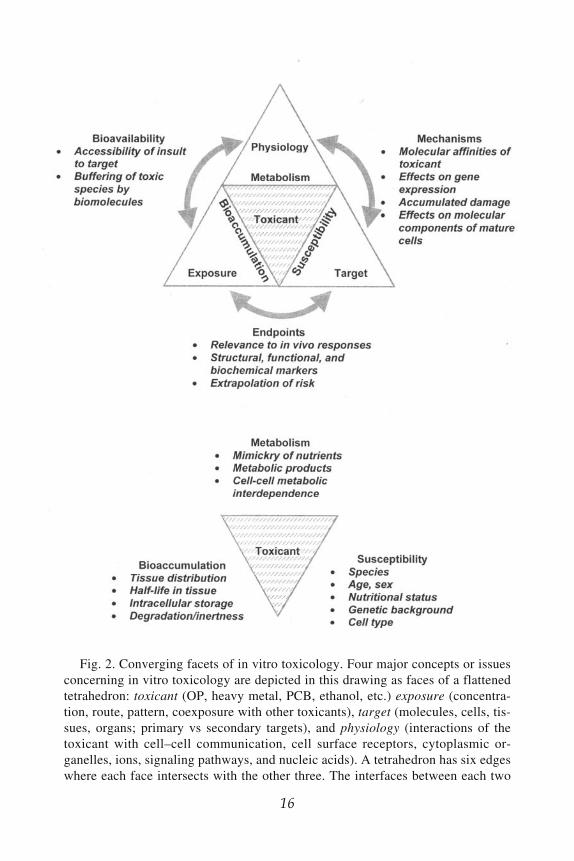
The underlying principles of in vitro neurotoxicology can be expressed asthe interactions of four fundamental factors or facets: exposure (concentra-tion, duration of exposure, and pattern of exposure, as well as coexposure withother toxic insults), target of damage (molecules, cells, tissues, and secondarytargets), physiology (functional significance of target cells, interactions of thetoxic insult with the cell surface, cytoplasm, and DNA), and the toxic insult
Fig. 1. Chronological continuum of neurotoxic events in the mammalian ner-vous system following X-ray exposure. The large arrow represents progressivedamage (accumulation of bomb symbols) from a state of nonexposure or health topermanent injury or death. Driving the arrow forward are time (because of cumula-tive degenerative effects subsequent to the initial damage) and additional exposure.Driving the arrow backward is repair or plasticity. Immediate effects of X-irradia-tion are DNA damage and apoptosis of proliferating cells, such as neuronal andglial progenitor cells, in both the immature and mature brain, as well as dividingglia in the immature brain. A subsequent reactive response to cell death or damageis gliosis and microglial activation. A possible cumulative effect of radiation dam-age to the mature brain is damage of neural networks that exceeds the capacity ofneuronal plasticity and redundancy to confer full restoration of function. This modelcould be adapted for other types of neurotoxic exposure.

14 Tiffany-Castiglioni
itself (e.g., heavy metals or pesticides). These factors are depicted as the facesof a flattened tetrahedron in Fig. 2. Although each of these four factors isextremely important in itself as an underlying concept of neurotoxicology, theconvergence of each facet with the other three is equally important. One mightthink of the six edges of the tetrahedron as illustrative of additional criticalissues: end points, mechanisms, bioavailability, bioaccumulation, susceptibil-ity, and metabolism. Thus, the intersection of exposure and target is end points,and the intersection of target and physiology is mechanisms. The followingbrief discussion of these two intersections will serve to highlight their signifi-cance in the context of in vitro neurotoxicology.
Structural, functional, genetic, and biochemical end points can be mea-sured in vitro in both early and latent phases of neurotoxicity, but only withinthe limitations of the in vitro system used. In vitro systems do not lend them-selves well to long-term studies that would parallel the life-span of the ex-posed organism or half-life of the toxic substance in brain tissue. The usefullife of various in vitro preparations ranges from hours to several weeks (125).For example, viable tissue slices can be maintained for a period of hours andprimary cell cultures for days to weeks. Immortalized cell cultures have beenmaintained for decades, such as C6 rat glioma cells (126,127) and SY5Yhuman neuroblastoma cells (128,129). Belying their name, however, suchcultures do not provide an opportunity for long-term exposures to toxicchemicals. Because of their short population doubling times, immortalizedcell lines require frequent passaging, which temporarily disrupts both cellattachment and cell–cell interactions and adds a confounding factor to long-term studies. Examples of neural cultures with extended longevity areaggregating cell cultures (130), and hollow fiber perfusion cultures (118),which can be maintained functionally intact for 2 or 3 mo.
Clarification of the meaning of the term “mechanism” in culture systemscan illustrate how intricately it is tied to other facets of neurotoxicology.Shown in Fig. 2 is a definition of mechanism as the intersection betweentarget and physiology, which is a contextually rich framework in which toconsider this concept. Mechanism is not merely the molecular or cellularentities acted upon by the toxic substance; it is also the associated perturba-tions in physiology. Examples include the disruption of normal synapticoverproduction and pruning by exposure during development, impairmentof plasticity and repair, and altered synaptic function. Each of these physi-ological processes has molecular components amenable to examination. Inthe case of synaptic function, these include the molecular interactionsinvolved in presynaptic neurotransmitter release, postsynaptic receptor func-tion, and postsynaptic intracellular signaling. Each of these effects could be

In Vitro Neurotoxicology 15
studied in a detailed fashion in vitro with validation in vivo. Therefore,mechanism is a critical link for validation of in vitro results.
The manner in which the above 10 interacting facets (see Fig. 2) of invitro neurotoxicology translate into concepts is a work in progress that willbe considered in depth throughout this book. The basic principle of toxicol-ogy that the dose makes the poison, as formulated by the early 16th-centuryphysician Paracelsus, can be directly applied at the level of molecular andcellular processes in vitro. A preliminary list of essential concepts reflectingthis utility of in vitro toxicology is as follows:
• Each toxicant has unique chemical properties that govern its toxicity, such assolubility in biological environments and affinity for specific biomolecules.
• The toxicity of an agent is modulated by its bioavailability to target cells, aswell as by the inherent phenotypic and genotypic sensitivity of the target cellsto the agent.
• The operative dose is modified chemically by the solubility and binding prop-erties of the toxicant in the extracellular and intracellular milieus.
• The operative dose is modified biologically by the degree of biodegradation,cytosolic buffering, and/or metabolism of the toxicant.
• Target cells interact dynamically to form structural and functional componentsof complex nervous tissues. Therefore, toxic actions upon them are likely toproduce secondary effects on the cells with which they interact.
5. TRENDS IN IN VITRO NEUROTOXICOLOGYMuch current work with in vitro systems for neurotoxicity testing lies in
maximizing their potential for yielding valid mechanistic responses. Experi-mental systems for the mechanistic understanding of toxicant-induced dam-age to the nervous system are often reductionist in nature in order to increasethe specificity and sensitivity of end points measured. In vitro models offermany advantages for neurotoxicity assessment that have been described indetail elsewhere (131,132). Among these advantages are the option to studya single cell type of interest in the absence of other cell types, ease of directobservation and measurement of cellular responses to toxicants, a definedextracellular environment, and direct interactions of the toxicant with testcells. Furthermore, in vitro systems may offer the economic benefit of areduced requirement for test chemicals, although in general this potentialbenefit has not yet been realized.
On the other hand, conceptual weaknesses are inherent in reductionistsystems. In vitro systems lack the capacity to assess behavioral end points,which is the major outcome of concern to neurotoxicologists. Lacking thisability, the value of in vitro systems lies in their potential capacity to respondmechanistically to a toxicant in a manner similar to that occurring from in
16 Tiffany-Castiglioni
16
Fig. 2. Converging facets of in vitro toxicology. Four major concepts or issuesconcerning in vitro toxicology are depicted in this drawing as faces of a flattenedtetrahedron: toxicant (OP, heavy metal, PCB, ethanol, etc.) exposure (concentra-tion, route, pattern, coexposure with other toxicants), target (molecules, cells, tis-sues, organs; primary vs secondary targets), and physiology (interactions of thetoxicant with cell–cell communication, cell surface receptors, cytoplasmic or-ganelles, ions, signaling pathways, and nucleic acids). A tetrahedron has six edgeswhere each face intersects with the other three. The interfaces between each two

In Vitro Neurotoxicology 17
vivo exposure. Furthermore, in vitro systems have limited (although expand-able) capacities for mimicking heterogeneous cell–cell interactions, sys-temic endocrine control, or metabolism of xenobiotics. Additionally,appropriate age and developmental stage of the nervous system at the timeof exposure have been extremely difficult to approximate in culture, to thepoint of disregarding the effects of experience and learning on the develop-ing nervous system. Technical improvement should be possible in most ofthese areas. Improvement can also come from continuous re-evaluation ofthe concept of toxicity testing in vitro. In this regard, well-designed comple-mentary in vivo/in vitro approaches offer the promise to accelerate progresstoward both an understanding of the mechanistic effects of neurotoxicityand the development of in vitro models for extrapolating risk.
Four major trends in in vitro neurotoxicology address these needed im-provements and will be discussed in greater detail by other chapters in thisvolume. The first trend is the refinement of end points. One of the criticaldecisions in the design of in vitro assays is the selection of appropriate endpoints, which must be relevant to in vivo responses. Whereas older studiesfocused on cytotoxicity, newer studies are increasingly mechanism driven,with careful selection of functional end points that are relevant to in vivoeffects of the toxicant. This approach is expected to allow the fine dissectionof biochemical mechanisms of toxicity. A second trend reflects the use ofmore histotypic, tissuelike culture systems for certain types of study. Thistrend counterbalances three decades of work on clonal cell lines that hasdominated much of modern in vitro toxicology. Clonal cell lines have beenthe system of choice for many studies because they are well characterized,easy to culture, and homogeneous in their responses to toxicants. Such celllines still have considerable value for specific applications, as will be de-scribed in several chapters. However, wide morphological and functionalheterogeneities exist in both neurons and glia, so that toxic chemicals do notuniformly affect each member of a class of cells. Researchers are returningto the use of biologically more complex models, such as heterologous cellcultures, explants, and ex vivo tissue slices from toxicant-exposed animals.Their use is supported by improvements in analytical techniques that make
juxtaposed faced are labeled to illustrate six additional concepts of in vitroneurotoxicology: bioavailability of the toxicant, biological end points that changeas a result of toxic exposure, mechanisms of toxic action, bioaccumulation of thetoxicant, susceptibility of the target to toxic damage, and metabolism of the toxicagent. Each of these concepts is mentioned briefly in the text and will be addressedin detail in subsequent chapters.

18 Tiffany-Castiglioni
single cells accessible to measurement, such as interactive laser cytometry(89,133,134). Table 1 provides examples of both simple and complex bio-logical models used for in vitro neurotoxiciology in recent years.
The third and fourth trends are sophisticated extensions of reductionistsystems that will demand interdisciplinary innovation to achieve. The thirdtrend is a renewed emphasis on validation. Two problems have been perva-sive regarding validation of in vitro systems. One is the delivery of toxico-logically relevant concentrations of chemicals to target cells at appropriatetimes. The other is that the developmental relevance of some in vitro sys-tems, such as cell culture, is very limited at this time. Attempts are beingmade through toxicodynamics, improved culture methods, and methodicalcomparisons with in vivo systems to deal with these issues (135,136). Thefourth trend is toward new applications for in vitro neurotoxicity testing.Although in vitro screening of possible or suspected neurotoxicants remainsan important goal of in vitro neurotoxicologists, other applications are alsobecoming apparent, especially the development of mechanism-based thera-pies for toxic exposure. Cell and tissue culture systems may expedite thedevelopment and testing of pharmacologic or molecular therapies to ame-liorate the effects of neurotoxicants on brain cell function.
6. GENERAL RESEARCH NEEDSThe following is a list of essential research objectives for in vitro
neurotoxicology. This list is applicable to organophosphorus compounds,heavy metals, radiation, ethanol, aromatic hydrocarbons, and endogenousneurotoxic proteins in a general sense, although subsequent chapters in thebook will illustrate toxicant-specific contemporary approaches and needs.Major areas are as follows:
• Mechanistic integration of any known behavioral effects of the toxicant withits molecular and cellular substrates
• Molecular, physiologic, and morphologic effects of neurotoxicants onsynaptogenesis, neuronal plasticity, and regeneration
• Complete chronological effects of neurotoxicants on tumorigenesis in brain• Differences in sensitivity between immature and mature cells of all types (neu-
rons, oligodendroglia, astroglia, and microglia) to neurotoxicants• Interactions among neurotoxicants
Each of these areas is quite broad and most of them are still in early stagesof investigation. Furthermore, progress in these areas is heavily dependenton progress in basic and applied neuroscience and will be facilitated by closeinterdisciplinary collaborations. Addressing these and similar issues should

In Vitro Neurotoxicology 19
provide significant advances in identifying, treating, and preventing diseasesand functional impairments associated with neurotoxic exposures.
Neurotoxicology has already made contributions that advance basic bio-medical knowledge, as will be discussed in this book. In the future, in vitrosystems will offer insight into how genetic polymorphisms affect suscepti-bility to diseases induced by environmental contaminants. As a platform forexamining genetic susceptibility, in vitro neurotoxicology may become notonly a central approach for risk assessment but also for understanding com-monalities between neurodegenerative diseases caused by chemicals in theenvironment and those caused by endogenous proteins.
ACKNOWLEDGMENTSThe author’s work is supported by National Institutes of Health grants
P42 ES04917, P30 ES09106, and T32 ES07273 and by ATSDR grant U61/ATU684505. I gratefully acknowledge helpful comments by Dr. MarionEhrich, Dr. Stephen Lasley, Dr. A. Joseph Castiglioni, and Dr. MichaelAschner on earlier drafts of this manuscript. I also thank these investigatorsand Dr. William Mundy, Dr. Stanley Barone, and Dr. Gerald Audesirk forrecommendations on Table 1.
REFERENCES1. Ramón y Cajal, S. (1991) Recollections of My Life (Craig, E. H. and Cano, J.,
transl.), The MIT Press, Cambridge, MA.2. Harrison, R. G. (1907) Observations on the living developing nerve fiber. Anat.
Rec. 1, 116–118.3. Shephard, G. M. (1994) Neurobiology, 3rd ed. Oxford University Press. New
York.4. Levi-Montalcini, R., Meyer, H., and Hamburger, V. (1954) In vitro effects of
mouse sarcomas 180 and 37 on the spinal and sympathetic ganglia of the chickembryo. Cancer Res. 14, 49–57.
5. Cohen, S., Levi-Montalcini, R., and Hamburger V. (1954) A nerve growth-stimulating factor isolated from sarcomas 37 and 180. Proc. Natl. Acad. Sci.USA 40, 1014–1018.
6. Levi-Montalcini, R. (1987) The nerve growth factor thirty-five years later. InVitro Cell. Dev. Biol. 23, 227–238.
7. Rakic, P. (1971) Neuron-glia relationship during granule cell migration in de-veloping cerebellar cortex. A Golgi and electronmicroscopic study in MacacusRhesus. J. Comp. Neurol. 141, 283–312.
8. Rakic, P. (1972) Mode of cell migration to the superficial layers of fetal mon-key neocortex. J. Comp. Neurol. 145, 61–83.
9. Rakic, P. (1978) Neuronal migration and contact guidance in the primate telen-cephalon. Postgrad. Med. J. 54(Suppl. 1), 25–40.

20 Tiffany-Castiglioni
10. Edmondson, J. C. and Hatten, M. E. (1987) Glial-guided granule neuron mi-gration in vitro: a high-resolution time-lapse video microscopic study. J.Neurosci. 7(6), 1928–1934.
11. Hatten, M. E. (1993) The role of migration in central nervous system neuronaldevelopment. Curr. Opin. Neurobiol. 3, 38–44.
12. Hatten, M. E. (1999) Central nervous system neuronal migration. Annu. Rev.Neurosci. 22, 511–539.
13. Edmondson, J. C., Liem, R. K., Kuster, J. E., and Hatten, M. E. (1988)Astrotactin: a novel neuronal cell surface antigen that mediates neuron-astroglial interactions in cerebellar microcultures. J. Cell. Biol. 106, 505–517.
14. Adams, N. C., Tomoda, T., Cooper, M., Dietz, G., and Hatten, M. E. (2002)Mice that lack astrotactin have slowed neuronal migration. Development129(Suppl.), 965–972.
15. Carmignoto, G. (2000) Reciprocal communication systems between astrocytesand neurones. Prog. Neurobiol. 62, 561–581.
16. Bezzi, P. and Volterra, A. (2001) A neuron-glia signalling network in the ac-tive brain. Curr. Opin. Neurobiol. 11, 387–394.
17. Araque, A., Carmignoto, G., and Haydon, P. G. (2001) Dynamic signaling be-tween astrocytes and neurons. Annu. Rev. Physiol. 63, 795–813.
18. National Research Council, Committee on Alternative Chemical Demilitariza-tion Technologies (CACDT). (1993) Alternative Technologies for the Destruc-tion of Chemical Agents and Munitions, Board on Army Science andTechnology, Commission on Engineering and Technical Systems, NationalAcademy of Sciences, Washington, DC.
19. Ecobichon, D. J. (1991) Toxic effects of pesticides, in Casarett and Doull’sToxicology: The Basic Science of Poisons (Amdur, M.O., Doull, J., andKlaassen, C.D., eds.), Pergamon, New York, pp. 565–622.
20. Pope, A. M., Heavner, J. E, Guarnieri, J. A., and Knobloch, C.P. (1986)Trichlorfon-induced congenital cerebellar hypoplasia in neonatal pigs. J. Am.Vet. Med. Assoc. 189, 781–783.
21. Berge, G. N., Fonnum, F., and Brodal, P. (1987) Neurotoxic effects of prenataltrichlorfon administration in pigs. Acta Vet. Scand. 28, 321–332.
22. Czeizel, A. E., Elek, C., Gundy, S., et al. (1993) Environmental trichlorfon andcluster of congenital abnormalities. Lancet 341, 539–542.
23. Chanda, S. M. and Pope, C. N. (1996) Neurochemical and neurobehavioraleffects of repeated gestational exposure to chlorpyrifos in maternal and devel-oping rats. Pharmacol. Biochem. Behav. 53, 771–776.
24. Loewenherz, C., Fenske, R. A., Simcox, N. J., Bellamy, G., and Kalman, D.(1997) Biological monitoring of organophosphorus pesticide exposure amongchildren of agricultural workers in central Washington State. Environ. HealthPerspect. 105, 1344–1353.
25. Hjelde, T., Mehl, A., Schanke, T. M., and Fonnum, F. (1998) Teratogenic ef-fects of tricholorfon (Metrifonate) on the guinea-pig brain. Determination ofthe effective dose and the sensitive period. Neurochem. Int. 32, 469–477.

In Vitro Neurotoxicology 21
26. ATSDR (2001) CERCLA List of Priority Hazardous Substances. ATSDR In-formation Center, Division of Toxicology, Agency for Toxic Substances andDisease Registry, US Department of Health and Human Services, Atlanta, GA.
27. Markowitz, M. (2000) Lead poisoning: a disease for the new millennium. Curr.Probl. Pediatr. 30, 62–70.
28. Tong, S., von Schirnding, Y. E., and Prapamontol, T. (2000) Environmentallead exposure: a public health problem of global dimensions. Bull. WorldHealth Organ. 78, 1068–1077.
29. Duckett, S., Galle., P, and Kradin, R. (1977) The relationship betweenParkinson syndrome and vascular siderosis: an electron microprobe study. Ann.Neurol. 2, 225–229
30. Kuhn, W., Winkel, R., Woitalla, D., Meves, S., Przuntek, H., and Müller T.(1998) High prevalence of parkinsonism after occupational exposure to lead-sulfate batteries. Neurology 50, 885–1886
31. Gorell, J. M., Rybicki, B. A., Johnson, C. C., and Peterson, E. L. (1999) Occu-pational metal exposures and the risk of Parkinson’s disease.Neuroepidemiology 18, 303–308.
32. Gorell, J. M., Johnson, C. C., Rybicki, B. A., et al. (1999) Occupational expo-sure to manganese, copper, lead, iron, mercury and zinc and the risk ofParkinson’s disease. Neurotoxicology 20, 239–248.
33. ATSDR (1999) Toxicological Profile for Mercury, Agency for Toxic Sub-stances and Disease Registry, US Department of Health and Human Services,Atlanta, GA.
34. Charleston, J. S., Body, R. L., Mottet, N. K., Vahter, M. E., and Burbacher T.M. (1995) Autometallographic determination of inorganic mercury distribu-tion in the cortex of the calcarine sulcus of the monkey Macaca fascicularisfollowing long-term subclinical exposure to methylmercury and mercuric chlo-ride. Toxicol. Appl. Pharmacol. 132, 325–333.
35. Miura, K. and Imura N. (1987) Mechanism of methylmercury cytotoxicity.Crit. Rev. Toxicol. 18(3), 161–168.
36. Rodier, P. M., Aschner, M., and Sager, P. R. (1984) Mitotic arrest in the devel-oping CNS after prenatal exposure to methyl mercury. Neurobehav. Toxicol.Teratol. 6, 379–385.
37. Choi, B. H., Lapham, L. W., Amin-Zaki, L., and Saleem T. (1978) Abnormalneuronal migration, deranged cerebral cortical organization, and diffuse whitematter astrocytosis of human fetal brain: a major effect of methylmercury poi-soning in utero. J. Neuropathol. Exp. Neurol. 37, 719–733.
38. Matsumoto, H., Koya, G., and Takeuchi, T. (1965) Fetal Minamata disease: aneuropathological study of two cases of intrauterine intoxication by a meth-ylmercury compound. J. Neuropathol. Exp. Neurol. 24, 563–574.
39. Myers, G. J., Marsh, D. O., Davidson, P. W., et al. (1995) Mainneurodevelopmental study of Seychellois children following in utero exposureto methylmercury from a maternal fish diet: outcome at six months.Neurotoxicology 16, 653–664.

22 Tiffany-Castiglioni
40. Grandjean, P., Weihe, P., White, R. F., and Debes, F. (1998) Cognitive perfor-mance of children prenatally exposed to “safe” levels of methylmercury.Environ. Res. 77, 165–172.
41. Grandjean, P., White, R. F., Nielsen, A., Cleary, D., and de Olivereira SantosE. C. (1999) Methylmercury neurotoxicity in Amazonian children downstreamfrom gold mining. Environ. Health Perspect. 107, 587–591.
42. Dolbec, J., Mergler, D., Sousa-Passos C. J., Sousa, de-M. S., and Lebel, J.(2000) Methylmercury exposure affects motor performance of a riverine popu-lation of the Tapajos river, Brazilian Amazon. Int. Arch. Occup. Environ.Health 73, 195–203.
43. Clarkson, T. W. (2002) The three modern faces of mercury. Environ. HealthPerspect. 110, 11–23.
44. Pichichero, M. E., Cernichiari, E., Lopreiato, J., and Treanor, J. (2002) Mer-cury concentrations and metabolism in infants receiving vaccines containingthiomersal: a descriptive study. Lancet 360, 1737–1741.
45. Safe, S. H. (1990) Polychlorinated biphenyls (PCBs) dibenzo-p-dioxins(PCDDs), dibenzofurans (PCDFs) and related compounds: environmental andmechanistic considerations which support the development of toxic equiva-lency factors (TEFs). CRC Crit. Rev. Toxicol. 21, 51–88.
46. Swanson, G. M., Ratcliffe, H. E., and Fischer, L. J. (1995) Human exposures topolychlorinated biphenyls (PCBs): a critical assessment of the evidence foradverse health effects. Regul. Toxicol. Pharmacol. 21, 136–150.
47. Jacobson, J. L. and and Jacobson, S. W. (1996) Intellectual impairment inchildren exposed to polychlorinated biphenyls in utero. N. Engl. J. Med. 335,783–789.
48. Schantz, S. L., Ferguson, S. A., and Bowman, R. E. (1992) Effects of 2,3,7,8-tetrachlorodibenzo-p-dioxin on behavior of monkeys in peer groups. Neurotox.Teratol. 14, 433–446.
49. Schantz, S. L., Moshtaghian, J., and Ness, D. K. (1995) Spatial learning defi-cits in adult rats exposed to ortho-substituted PCB congeners during gestationand lactation. Fundam. Appl. Toxicol. 26, 117–126.
50. Hanneman, W. H., Legare, M. E., Barhoumi, R., Burghardt, R. C., Safe, S., andTiffany- Castiglioni, E. (1996) Stimulation of calcium uptake in cultured rathippocampal neurons by 2,3,7,8-tetrachlorodibenzo-p-dioxin. Toxicology112(1), 19–28.
51. Barhoumi, R., Mouneimne, Y., Phillips, T. D., Safe, S. H., and Burghardt, R.C. (1996) Alteration of oxytocin-induced calcium oscillations in clone 9 cellsby toxin exposure. Fundam. Appl. Toxicol. 33(2), 220–228.
52. Kodavanti, P. R. S. and Tilson, H. (2000). Neurochemical effects of environ-mental chemicals: in vitro and in vivo correlations on second messenger path-ways. Ann. NY Acad. Sci. 919, 97–105.
53. Legare, M. E., Hanneman, W. H., Barhoumi, R., Burghardt, R. C., and Tif-fany-Castiglioni, E. (2000) 2,3,7,8-Tetrachlorodibenzo-p-dioxin alters hippoc-ampal astroglia-neuronal gap junctional communication. Neurotoxicology 21,1109–1116.

In Vitro Neurotoxicology 23
54. Hong, S. J., Grover, C. A., Safe, S. H., Tiffany-Castiglioni, E., and Frye, G. D.(1998) Halogenated aromatic hydrocarbons suppress CA1 field excitatorypostsynaptic potentials in rat hippocampal slices. Toxicol. Appl. Pharmacol.148, 7–13.
55. Schantz, S. L. and Widholm, J. J. (2001) Cognitive effects of endocrine-dis-rupting chemicals in animals. Environ. Health Perspect. 109, 1197–1206.
56. Clarren, S. K. and Smith, D. W. (1978) The fetal alcohol syndrome. N. Engl. J.Med. 298, 1063–1067.
57. Miller, M. W. (1992) The effects of prenatal exposure to ethanol on cell prolif-eration and neuronal migration, in Development of the Central Nervous Sys-tem: Effects of Alcohol and Opiates (Miller M., ed.), Alan R. Liss, New York,pp. 47–69.
58. Guerri, C., Sáez, R., Portolés, M., and Renau-Piqueras, J. (1993) Derangementof astrogliogenesis as a possible mechanism involved in alcohol-induced alter-ations of central nervous system development. Alcohol Alcohol. 2(Suppl.),203–208.
59. Jones, K. L., Smith, D. W., Ulleland, C. N., and Streissguth, A. P. (1973)Pattern of malformation in offspring of chronic alcoholic mothers. Lancet 1,1267–1271.
60. Bo, W. J., Krueger, W. A., Rudeen, P. K., and Symmes, S. K. (1982) Ethanol-induced alterations in the morphology and function of the rat ovary. Anat. Rec.202, 255–260.
61. Dees, W. L. and Skelley, C. W. (1990) Effects of ethanol during the onset offemale puberty. Neuroendocrinology 51, 64–69.
62. Dees, W. L., Skelley, C. W., Hiney, J. K., and Johnston, C. A. (1990) Actionsof ethanol on hypothalamic and pituitary hormones in prepubertal female rats.Alcohol 7, 21–25.
63. Dees, W. L., Dissen, G. A., Hiney, J. K., Lara, F., and Ojeda, S. R. (2000)Alcohol ingestion inhibits the increased secretion of puberty-related hormonesin the developing female rhesus monkey. Endocrinology 141, 1325–1331.
64. Masters, C. L., Multhaup, G., Simms, G., Pottgiesser, J., Martins, R. N., andBeyreuther, K. (1985) Neuronal origin of a cerebral amyloid: neurofibrillarytangles of Alzheimer’s disease contain the same protein as the amyloid ofplaque cores and blood vessels. EMBO J. 4, 2757–2763.
65. Yankner, B. A., Dawes, L. R., Fisher, S., Villa-Komaroff, L., Oster-Granite,M. L., and Neve, R. L. (1989) Neurotoxicity of a fragment of the amyloidprecursor associated with Alzheimer’s disease. Science 245, 417–420.
66. Behl, C. (1997) Amyloid beta-protein toxicity and oxidative stress inAlzheimer’s disease. Cell Tissue Res. 290, 471–480.
67. Polymeropoulos, M. H., Lavedan, C., Leroy, E., et al. (1997) Mutation in thealpha-synuclein gene identified in families with Parkinson’s disease. Science276, 2045–2047.
68. Spillantini, M. G. and Goedert, M. (2000) The alpha-synucleinopathies:Parkinson’s disease, dementia with Lewy bodies, and multiple system atrophy.Ann. NY Acad. Sci. 920, 16–27.

24 Tiffany-Castiglioni
69. Aschner, M., Allen, J. W., Kimelberg, H. K., LoPachin, R. M., and Streit, W. J.(1999) Glial cells in neurotoxicity development. Annu. Rev. Pharm. Toxicol.39, 151–173.
70. Le, W-D., Rowe, D., Xie, W., Ortiz, I., He, Y., and Appel, S. H. (2001) Micro-glial activation and dopaminergic cell injury: an in vitro model relevant toParkinson’s Disease. J. Neurosci. 2, 8447–8455.
71. Chang, J. Y. and Liu, L-Z. (1999) Manganese potentiates nitric oxide produc-tion by microglia. Mol. Brain. Res. 68, 22–28.
72. Gao, H.-M., Hong, J.-S., Zhang, W., and Liu, B. (2002) Distinct role for micro-glia in rotenone-induced degeneration of dopaminergic neurons. J. Neurosci.22, 782–790.
73. Harry, G. J., Tyler, K., d’Hellencourt, C. L., Tilson, H. A., and Maier, W. E.(2002) Morphological alterations and elevations in tumor necrosis factor- ,interleukin (IL)-1 , and IL-6 in mixed glia cultures following exposure totrimethyltin: modulation by proinflammatory cytokine recombinant proteinsand neutralizing antibodies. Toxicol. Appl. Pharm. 180, 205–218.
74. Carlson, K., Jortner, B. S., and Ehrich, M. (2000) Organophosphus compound-induced apoptosis in SH-SY5Y human neuroblastoma cells. Toxicol. Appl.Pharmacol. 168, 102–113.
75. Hong, M. S., Hong, S. J., Barhoumi, R., et al. (2003). Neurotoxicity induced indifferentiated SK-N-SH-SY5Y human neuroblastoma cells by organophospho-rus compounds. Toxicol. Appl. Pharmacol. 186, 110–118.
76. Purkiss, J. R., Friis, L. M., Doward, S., and Quinn, C. P. (2001) Clostridiumbotulinum neurotoxins act with a wide range of potencies on SH-SY5Y humanneuroblastoma cells. Neurotoxicology 22, 447–453.
77. Li, W. F. and Casida, J. E. (1998) Organophosphorus neuropathy target esteraseinhibitors selectively block outgrowth of neurite-like and cell processes in cul-tured cells. Toxicol. Lett. 98, 139–146.
78. Das, K. D. and Barone, S. J. (1999). Neuronal differentiation in PC12 cells isinhibited by chlorpyrifos and its metabolites: Is acetylcholinesterase inhibitionthe site of action? Toxicol. Appl. Pharmacol. 160, 217–230.
79. Zachor, D. A., Moore, J. F., Brezauzek, C. M., Theibert, A. B., and Percy, A.K. (2000) Cocaine inhibition of neuronal differentiation in NGF-induced PC12cells is independent of ras signaling. Int. J. Dev. Neurosci. 18, 765–772.
80. Crumpton, T. L., Atkins, D., Zawia, N., and Barone, S., Jr. (2001) Lead expo-sure in pheochromocytoma (PC12) cells alters neural differentiation and Sp1DNA-binding. Neurotoxicology 22, 49–62.
81. Parran, D. K., Mundy, W. R., and Barone, S., Jr. (2001) Effects of methylmer-cury and mercuric chloride on differentiation and cell viability in PC12 cells.Toxicol. Sci. 59, 278–290.
82. Shafer, T. J., Meacham, C. A., and Barone, S. (2002). Effects of prolongedexposure to nanomolar concentrations of methylmercury on voltage-sensitivesodium and calcium currents in PC12 cells. Dev. Brain Res. 136, 151–164.
In Vitro Neurotoxicology 25
83. Son, J. H., Chun, H. S., Joh, T. H., Cho, S., Conti, B., and Lee, J.W., (1999)Neuroprotection and neuronal differentiation studies using substantia nigradopaminergic cells derived from transgenic mouse embryos. J. Neurosci. 19,10–20.
84. Chun, H. S., Lee, H., and Son, J. H. (2001). Manganese induces endoplasmicreticulum (ER) stress and activates multiple caspases in nigral dopaminergicneuronal cells, SN4741. Neurosci. Lett. 316, 5–8.
85. Qian, Y, Harris, E. D., Zheng, Y., and Tiffany-Castiglioni, E. (2000) Lead (Pb)targets GRP78, a molecular chaperone, in C6 rat glioma cells. Toxicol. Appl.Pharmacol. 163, 260–266.
86. Takanaga, H., Kunimoto, M., Adachi, T., Tohyama, C., and Aoki, Y. (2001)Inhibitory effect of 2,3,7,8-tetrachlorodibenzo-p-dioxin on cAMP-induced dif-ferentiation of rat C6 glial cell line. J. Neurosci. Res. 64, 402–409.
87. Guizzetti, M. and Costa, L. G. (2002) Effect of ethanol on protein kinase Cand p70S6 kinase activation by carbachol: a possible mechanism for ethanol-induced inhibition of glial cell proliferation. J. Neurochem. 82, 38–46.
88. Mead, C. and Pentreath, V. M. (1998) Evaluation of toxicity indicators in ratprimary astrocytes, C6 glioma and human 1321N1 astrocytoma cells: cangliotoxicity be distinguished from cytotoxicity? Arch. Toxicol. 72, 372–380.
89. Inglefield, J. R., Mundy, W. R., and Shafer, T. J. (2001) inositol 1,4,5-trisphosphate receptor- sensitive Ca2+ release, store-operated Ca2+ entry, andcamp responsive element binding protein phosphorylation in developing corti-cal cells following exposure to polychlorinated biphenyls. J. Pharmacol. Exp.Ther. 297, 762–773.
90. Yang, J.-H. and Kodavanti, R. S. (2001) Possible molecular targets of haloge-nated aromatic hydrocarbons in neuronal cells. Biochem. Biophys. Res.Commun. 280, 1372–1377.
91. Dyatlov, V. A., Dyatlov, O. M., Parsons, P. J., Lawrence, D. A., and Carpenter,D. O. (1998) Lipopolysaccharide and interleukin-6 enhance lead entry into cer-ebellar neurons: application of a new and sensitive flow cytometric techniqueto measure intracellular lead and calcium concentrations. Neurotoxicology 19,293–302.
92. Audesirk, T. and Cabell, L. (1999). Nanomolar concentrations of nicotine andcotinine alter the development of cultured hippocampal neurons via non-ace-tylcholine receptor-mediated mechanisms. Neurotoxicology 20, 639–646.
93. Braga, M. F. M., Pereira, E. F. R., and Albuquerque, E. X. (1999) Nanomolarconcentrations of lead inhibit glutamatergic and GABAergic transmission inhippocampal neurons. Brain Res. 826, 22–34.
94. Heidemann, S. R., Lamoureux, P., and Atchison, W. D. (2001) Inhibition ofaxonal morphogenesis by nonlethal, submicromolar concentrations of meth-ylmercury. Toxicol. Appl. Pharmacol. 174, 49–59.
95. Bestervelt, L. L., Pitt, J. A., and Piper, W. N. (1998) Evidence for Ah receptormediation of increased ACTH concentrations in primary cultures of rat ante-rior pituitary cells exposed to TCDD. Toxicol. Sci. 46, 294–299.

26 Tiffany-Castiglioni
96. Bouton, C. M. L. S., Hossain, M. A., Frelin, L. P., Laterra, J., and Pevsner, J.(2001) Microarray analysis of differential gene expression in lead-exposed as-trocytes. Toxicol. Appl. Pharmacol. 176, 34–53.
97. Yao, C. P., Allen, J. W., Conklin, D. R., and Aschner, M. (1999) Transfectionand overexpression of metallothionein-I in neonatal rat primary astrocyte cul-tures and in astrocytoma cells increases their resistance to methylmercury-in-duced cytotoxicity. Brain Res. 818, 414–420.
98. Yao, C. P., Allen, J. W., Mutkus, L. A., Xu, S. B., Tan, K. H., and Aschner, M.(2000) Foreign metallothionein-I (MT-I) expression by transient transfectionin MT-I and -II null astrocytes confers increased protection against acute me-thylmercury cytotoxicity. Brain Res. 855, 32–38.
99. Deng, W. and Poretz, R. D. (2002) Protein kinase C activation is required forthe lead-induced inhibition of proliferation and differentiation of cultured oli-godendroglial progenitor cells. Brain Res. 929, 87–95.
100. Xu, J., Chen, S., Ahmed, S. H., et al. (2001) Amyloid- peptides are cytotoxicto oligodendrocytes. J. Neurosci. 21, 1–5.
101. Tang, H. W., Yan, H. L., Hu, X. H., Liang, Y. X., and Shen, X.Y. (1996) Leadcytotoxicity in primary cultured rat astrocytes and Schwann cells. J. Appl.Toxicol. 16, 187–196
102. Tomsig, J.L., and Suszkiw, J.B. (1995) Multisite interactions between Pb+2
and protein kinase C and its role in norepinephrine release from bovine adre-nal chromaffin cells. J. Neurochem. 64, 2667–2773.
103. Vijverberg, H. P. M., Zwart, R., Van Den Beukel, I., Oortgiesen, M., and VanKleef, R. G. D. M. (1997) In vitro approaches to species and receptor diversityin cellular neurotoxicology. Toxicol. In Vitro 11, 491–498.
104. Pantazis, N. J., Zaheer, A., Dai, D., Zahee,r S., Green, S. H., and Lim, R.(2000) Transfection of C6 glioma cells with glia maturation factorupregulates brain-derived neurotrophic factor and nerve growth factor:trophic effects and protection against ethanol toxicity in cerebellar granulecells. Brain Res. 865, 59–76.
105. Lindahl, L. S., Bird, L., Legare, M. E., Mikeska, G., Bratton, G. R., and Tif-fany-Castiglioni, E. (1999) Differential ability of astroglia and neuronal cellsto accumulate lead: dependence on cell type and on degree of differentiation.Toxicol. Sci. 50, 236–243.
106. Shanker, G., Allen, J. W., Mutkus, L. A., Aschner, M. (2001) Methylmercuryinhibits cysteine uptake in cultured primary astrocytes, but not in neurons.Brain Res. 914, 159–165.
107. Monnet-Tschudi, F., Zurich, M.-G., Schilter, B., Costa, L. G., and Honegger,P. (2000) Maturation-dependent effects of chlorpyrifos and parathion and theiroxygen analogs on acetylcholinesterase and neuronal and glial markers in ag-gregating brain cell cultures. Toxicol. Appl. Pharmacol. 165, 175–183.
108. Hirai, K., Yoshioka, H., Kihara, M., Hasegawa, K., Sawada, T., and Fushiki,S. (1999) Effects of ethanol on neuronal migration and neural cell adhesionmolecules in the embryonic rat cerebral cortex: a tissue culture study. Dev.Brain Res. 118, 205–210.

In Vitro Neurotoxicology 27
109. He, L., Poblenz, A. T., Medrano, C. J., and Fox, D.A. (2000) Lead and cal-cium produce rod photoreceptor cell apoptosis by opening the mitochondrialpermeability transition pore. J. Biol. Chem. 275, 12,175–12,184.
110. Kimelberg, H. K., Cai, Z., Schools, G., and Zhou, M. (2000) Acutely iso-lated astrocytes as models to probe astrocyte functions. Neurochem. Int.36, 359–367.
111. Vallés, S., Sancho-Tello, M., Miñana, R., Climent, E., Renau-Piqueras, J., andGuerri, C. (1996) Glial fibrillary acidic protein expression in rat brain and inradial glia culture is delayed by prenatal ethanol exposure. J. Neurochem. 67,2425–2433.
112. Chen, H-H., Ma, T., and Ho, I. K. (1999) Protein kinase C in rat brain is al-tered by developmental lead exposure. Neurochem. Res. 24, 415–421.
113. Grover, C. A. and Frye, G. D. (1996) Ethanol effects on synaptic neurotrans-mission and tetanus-induced synaptic plasticity in hippocampal slices ofchronic in vivo lead-exposed adult rats. Brain Res. 734, 61–71.
114. Goldstein, G. W. (1988) Endothelial cell-astrocyte interactions: a cellularmodel of the blood-brain barrier. Ann. NY Acad. Sci. 529, 31–39.
115. Sobue, K., Yamamoto, N., Yoneda, K., et al. (1999) Induction of blood–brainbarrier properties in immortalized bovine brain endothelial cells by astrocyticfactors. Neurosci. Res. 35, 155–164.
116. Wolburg, H., Neuhaus, J., Kniesel, U., et al. (1994) Modulation of tight junc-tion structure in blood–brain barrier endothelial cells: effects of tissue culture,second messengers and cocultured astrocytes. J. Cell. Sci. 107, 1347–1357.
117. Gumbleton, M. and Audus, K. L. (2001) Progress and limitations in the use ofin vitro cell cultures to serve as a permeability screen for the blood–brainbarrier. J. Pharm. Sci. 90, 1681–1698.
118. Tiffany-Castiglioni, E., Neck, K.F., and Caceci, T. (1986) Glial culture onartificial capillaries: electron microscopic comparison of C6 rat glioma cellsand rat astroglia. J. Neurosci. Res. 16, 387–396.
119. Stanness, K. A., Westrum, L. E., Fornaciari, E., et al. (1997) Morphologicaland functional characterization of an in vitro blood-brain barrier model. BrainRes. 771, 329–342.
120. Guerri, C., Pascual, M., and Renau-Piqueras, J. (2001) Glia and fetal alcoholsyndrome. Neurotoxicology 22, 593–599.
121. Tofolin, P. J. and Fike, J. R. (2000) The radioresponse of the central nervoussystem: a dynamic process. Radiat. Res. 153, 357–370.
122. Tiffany-Castiglioni, E. (1993) Cell culture models for lead toxicity in neu-ronal and glial cells. Neurotoxicology 14(4), 513–536.
123. Roper, S. N., Abraham, L. A., and Streit, W. J. (1997) Exposure to inutero irradiation produces disruption of radial glia in rats. Dev. Neurosci.19,521–528.
124. Gressens, P. (2000) Mechanisms and disturbances of neuronal migration.Pediatr. Res. 48, 725–730.
125. Freshney, R. I. (2000) Culture on Animal Cells: A Manual of Basic Tech-nique, 4th ed., Wiley–Liss, New York.

28 Tiffany-Castiglioni
126. Bissell, M. G., Eng, L.F., Herman, M. M., Bensch, K. G., and Miles, L. E.(1975) Quantitative increase of neuroglia-specific GFA protein in rat C-6glioma cells in vitro. Nature 255, 633–634.
127. Lee, K., Kentroti, S., Billie, H., Bruce, C., and Vernadakis, A. (1992) Com-parative biochemical, morphological, and immunocytochemical studies be-tween C-6 glial cells of early and late passages and advanced passages of glialcells derived from aged mouse cerebral hemispheres. Glia 6, 245–257.
128. Biedler, J. L., Helson, L., and Spengler, B. A. (1973) Morphology and growth,tumorigenicity, and cytogenetics of human neuroblastoma cells in continuousculture. Cancer Res. 33, 2643–2652.
129. Perez-Polo, J. R., Werrbach-Perez, K., and Tiffany-Castiglioni, E. (1979)A human clonal cell line model of differentiating neurons. Dev. Biol. 71,341–355.
130. Zurich, M. G., Honegger, P., Schilter, B., Costa, L. G., and Monnet-Tschudi,F. (2000) Use of aggregating brain cell cultures to study developmental ef-fects of organophosphorus insecticides. Neurotoxicology 21, 599–606.
131. Harry, G.J., Billingsley, M., Bruinink, A., et al. (1998) In vitro techniquesfor the assessment of neurotoxicity. Environ. Health Perspect. 106(Suppl.),131–158.
132. Gad, S.C. (2000) Neurotoxicology in vitro, in In Vitro Toxicology, 2nd ed.,(Gad, C. G., ed.), Taylor and Francis, New York, pp. 188–221.
133. Barhoumi, R., Mouneimne, Y., Ramos, K. S., et al. (2000)Analysis of benzo[a]pyrene partitioning and cellular homeostasis in a rat livercell line. Toxicol. Sci. 53, 264–270.134. Barhoumi, R., Mouneimne, Y., Awooda, I., Safe, S.H., Donnelly, K.C., and
Burghardt, R.C. (2002) Characterization of calcium oscillations in normal andbenzo[a]pyrene-treated Clone 9 cells. Toxicol. Sci. 68, 444–450.
135. Chanda, S. M., Mortensen, S. R., Moser, V. C., and Padilla, S. (1997) Tissue-specific effects of chlorpyrifos on carboxylesterase and cholinesterase activ-ity in adult rats: An in-vitro and in vivo comparison. Fund. Appl. Toxicol. 38,148–157.
136. Carpenter, D. O., Hussain, R. J., Berger, D. F., Lombardo, J. P., and Park, H-Y. (2002) Electrophysiologic and behavioral effects of perinatal and acuteexposure of rats to lead and polychlorinated biphenyls. Environ. HealthPerspect. 110(Suppl.), 377–386.

In Vitro Neurotoxicity Risk Assessment 29
29
2Predictive Value of In Vitro Systems
for Neurotoxicity Risk Assessment
Marion Ehrich and David C. Dorman
1. INTRODUCTION:NEUROTOXICITY RISK ASSESSMENT
Risk assessment has been broadly defined as the characterization of theadverse health effects of human exposures to environmental hazards andcan be divided into four major steps: hazard identification, dose–responseassessment, exposure assessment, and risk characterization (1). Hazard iden-tification is defined as determining whether human exposure to an agent cancause an increased incidence of an adverse health effect (e.g., neurotoxic-ity). Dose–response assessment is the process of characterizing the relation-ship between the administered or effective dose of an agent and the incidenceof an adverse health effect in exposed populations, estimating the incidenceof the effect as a function of human exposure to the agent. A dose–responseassessment should account for exposure intensity and duration, developmen-tal age, and other factors that may modify the response (e.g., gender, diet).Exposure assessment is the process of measuring or estimating the intensity,frequency, and duration of human exposure to an agent found in the envi-ronment or an agent that may be released into the environment. Risk charac-terization integrates these preceding steps by estimating the incidence of ahealth effect under various conditions of human exposure.
These four steps form the basis of risk assessment. They are independentof the nature of the adverse health effect (e.g., neurotoxicity vs carcinogen-esis), although underlying assumptions (e.g., threshold vs nonthresholdeffects) may influence the approaches used.
From: Methods in Pharmacology and Toxicology: In Vitro Neurotoxicology: Principles and ChallengesEdited by: E. Tiffany-Castiglioni © Humana Press Inc., Totowa, NJ

30 Ehrich and Dorman
Neurotoxicity is defined as any adverse effect on the chemistry, structureor function of the nervous system during development or at maturity inducedby chemical or physical influences (2). For a chemical to be regarded as aneurotoxicant, effects on the nervous system should be direct rather than indi-rect, adverse rather than adaptive, and toxicological rather than pharmaco-logical. Chemically induced neurotoxic effects are of special concern becauseneurotoxicological syndromes may be delayed and are often progressive orirreversible and prevention is far less costly than treatment (3,4). Only recentlyhave regulatory agencies focused their attention on developing guidelines forthe conduct of neurotoxicity risk assessments (5).
Requirements for animal testing of pesticides and some commercial chemi-cals for neurotoxicity are promulgated worldwide by a number of regulatoryagencies. For example, the US Environmental Protection Agency administersthe Federal Insecticide, Fungicide and Rodenticide Act (FIFRA) and the ToxicSubstances Control Act (TSCA). Testing for specific end points indicative ofneurotoxicity may also be recommended by the Food and Drug Administra-tion for certain food additives that demonstrate positive results in a very ba-sic, initial neurotoxicity screen (6). It is estimated that only a small fraction ofthe 70,000 chemicals currently in commerce have been adequately assessedfor neurotoxicity (7); thus, there is a need to develop cost-effective screens toassess chemicals for potential neurotoxicity.
Bioassays remain the principal method to identify possible human healthrisks posed by exposure to chemicals and other potential neurotoxicants. Theprimary advantage of using animals for hazard identification and risk charac-terization is that all potential targets for injury (e.g., the many types of cell,tissue, neurochemical) are included in the test system (4,8). This is especiallyimportant because neurotoxicants can affect a variety of different organs andtissues and they can induce alterations in chemistry, function, structure, orbehavior (see Fig. 1). End points of interest in bioassays often include histo-pathology to assess morphologic damage and batteries of functional,neurobehavioral, neurochemical, and neurophysiological tests to examine theoperational integrity of the nervous system (9). Because there are physiologi-cal and anatomical similarities among mammals, the finding of a positiveresponse in vivo is taken as evidence that an agent may also pose a risk forexposed humans. This integrated in vivo approach is valuable for a detailedcharacterization of both the effects and possible mechanisms of suspectedneurotoxicants under specific exposure conditions. In vivo methods are rela-tively well developed and the data are used to determine no observable adverseeffect levels (NOAELs), uncertainty factors, and benchmark doses.
The use of animals in toxicity testing is often the subject of intense scrutinyand criticism by the general public. The toxicology community continually

In Vitro Neurotoxicity Risk Assessment 31
strives to replace the use of animals in research, to reduce the numbers ofanimals to the minimum necessary to obtain valid results where replacementwas impossible, and to refine all experimental procedures to minimize adverseeffects on animals. The use of in vitro test systems is a logical alternative tothe use of animals and can often complement and enhance in vivo data. Invitro tests can be sensitive, replicable, valid, and cost-effective. They areamenable to studies done over a wide range of concentrations and over mul-tiple periods of time. Exposure to the test chemical can be tightly controlledand human materials can be used. In vitro data can provide important struc-ture–activity data concerning the relative potency of different chemicals andcontribute to choices for chemicals to study in vivo. In addition, in vitro datacan be used to identify the mode of action of a chemical and identify criticalfactors that determine species- or tissue-specific differences in response. Stud-ies of relationships between in vitro and in vivo data have helped to identifythese and other factors that contribute to and modify toxicity (2–4,7,8,10–14).It is well recognized that the risk assessment process is improved if data con-cerning the chemical’s mechanism(s) of action are included. However, it
Fig. 1. Critical elements in characterizing neurotoxicity risk from exposure to achemical. (Reprinted with permission from ref. 9.)

32 Ehrich and Dorman
remains speculative whether in vitro data can replace any in vivo data usedfor the risk assessment of a neurotoxicant. Indeed, in vitro data are rarelyconsidered in the risk assessment process because current statutory guide-lines do not classify changes observed in vitro as indicative of an adverseresponse (7,10,11,15–17).
As with any technology, in vitro systems have distinct limitations. In vitrotest systems have a reduced cellular complexity, therefore, the responsesobserved in vitro may not be representative of those observed in the substan-tially more complex intact nervous system (see Fig. 2). One important struc-tural difference of most neural cell culture systems is the lack of a functionalequivalent to the so-called blood–brain barrier. This blood–brain barrierexcludes the movement of certain chemicals or their metabolites into the intactnervous system, thereby attenuating the observed neurotoxic response. Addi-tionally, isolated in vitro systems often lack the hepatic and extrahepatic meta-bolic systems that are normally present in the intact animal to activate ordetoxify the agent under investigation. Thus, in vitro systems have only alimited capacity to metabolize selective toxicants. Current choices are oftenbetween simple systems that are significantly divergent to the in vivo situa-tion but are easy to manipulate and complex systems that are technically dif-ficult to establish and use. In spite of these limitations, in vitro models areproving useful for the screening of chemicals for neurotoxic potential. In vitromodel systems may provide an economical first-tier evaluation that will helpto guide more extensive whole-animal studies.
2. SYSTEMS FOR EVALUATIONOF NEUROTOXICITY IN VITRO: MECHANISTICMODELS AND SCREENING TESTS
A number of reports suggest that in vitro tests for neurotoxicity would bemore useful in providing mechanistic information than they would be for gen-eral screening purposes for agents of unknown toxicity. This suggestion isbased on the complexity and multiple targets of the nervous system and thecomparative simplicity of in vitro test systems (2–4,8,18). Although the devel-opment of screening tests may appear formidable to some, the suggestion hasbeen made that end points indicative of cytotoxicity (i.e., cell viability) couldbe more sensitive in cells of the nervous system exposed to neurotoxicantsthan cells of extraneural origin. This assumption, based on the premise of tis-sue selectivity, may have value when appropriate comparisons are made. How-ever, although potent cytotoxins can be neurotoxicants, they are likely to betoxic to other tissues as well (e.g., liver, kidney, lung) (2,12,15,16).

In Vitro Neurotoxicity Risk Assessment 33
33
Fig
. 2.
Typ
es o
f in
vit
ro s
yste
ms
used
in
neur
otox
icol
ogy
rese
arch
. C
ultu
res
com
pose
d of
ind
ivid
ual
cell
lin
es h
ave
the
leas
tst
ruct
ural
com
plex
ity
and
pred
icta
bili
ty to
man
.

34 Ehrich and Dorman
Like their in vivo counterparts, in vitro neurotoxicity screens require theuse of test systems and end points that are sensitive, efficient, and neural-specific. The systems should provide low numbers of false negatives or posi-tives (2). The sole reliance on a single experimental model, whether it be invivo or in vitro, with a limited number of biological markers is not generallyconsidered sufficient for estimation of risk. For this reason, a tiered systemfor in vitro neurotoxicity screening has been proposed (15). The tiered sys-tem was designed to include cytotoxicity and cell-specific effects determinedin simple and more complex in vitro systems in the first and second tiers andmechanistic studies in the third tier. An initial study, using a neuronal cellline and multiple end points (some neural-specific, some not), suggestedthat the number of end points was not sufficient to use a single clonal cellline as a screening system for neurotoxicity (19). This contrasts with resultsof another large study examining cytotoxicity in non-neural cells, whichsuggested that cell viability appeared to be a valid indicator of general tox-icity (20). Regardless of end points, however, concentration–response andtime–response studies need to be included into any in vitro test screen(10,20,21). It has also been suggested that in vitro screens should includehuman neural cells to allow evaluation of interspecies differences inresponse (1,3,14,16,18,22).
Many neurotoxicants have unknown and/or multiple mechanisms ofaction (4,11,23,24). The use of in vitro test systems often yields valuablemechanistic data on chemical-induced neurotoxicities and thus offers anattractive alternative to the use of animals for this type of research. Mecha-nistic studies can be designed to evaluate multiple mechanisms of actionand can be modified to the toxic agent of concern. End points can includegeneral glial or neuronal measures, neurotransmitter systems, and indicatorsof the biochemical and electrical responses of neural cells. Reversibility andirreversibility of effects, protective mechanisms and repair, and ability toaffect cell proliferation and differentiation can be determined. Characteriza-tion of the cellular and molecular substrates and pathways that follow expo-sure to neurotoxicants can be evaluated (2,3,7,10,13,15,24). The generationof useful mechanistic data requires realization that the data obtained will notnecessarily provide explanations for all manifestations of neurotoxicity seenin man and animals, including behavioral, cognitive, sensory, developmen-tal, or age-related effects. Furthermore, standard protocols that include well-defined culture conditions and means to reduce potential for cell instabilityneed to be followed to permit intralaboratory and interlaboratory validationof results (2,3,7,10,13,15,22).

In Vitro Neurotoxicity Risk Assessment 35
3. IN VITRO MODELING OF IN VIVO SYSTEMS:DOSE–RESPONSE CONSIDERATIONS
It is well recognized that risk assessment considers dose–response datafor an adverse effect. Dose (concentration)–response studies can be donewith relative efficiency using in vitro test systems, and test compounds canbe applied directly without concern for the pharmacokinetic factors of ab-sorption, distribution, metabolism, and excretion encountered in animals.Isolated test systems are often exposed to concentrations of chemicals thatfar exceed those achievable in tissues from exposed animals or humans. In-terpretation of in vitro studies is aided dramatically by detailed knowledgeconcerning concentrations of the parent chemical or major metabolites inblood, brain, and other potential target tissues. In some cases (e.g., organo-phosphate insecticides, polychlorinated biphenyls, and ethanol) effects canoccur in vitro at concentrations similar to those observed in vivo (13,25).Effects occurring in vitro at lower concentrations than those achieved invivo may indicate that the test chemical is metabolized in vivo to a less toxicform, the chemical or the active metabolites are excluded from the nervoussystem, or that compensatory or repair mechanisms occur that attenuate thetoxicity of the chemical observed in vivo. Neurotoxic effects that require invitro concentrations higher than blood concentrations of intoxicated animalsmay occur because concentrations in target tissues are higher than concen-trations in blood. Concentration differences may also depend on the in vitrosystem used for testing (e.g., cells of neoplastic origin are notoriously resis-tant to chemical-induced cytotoxic effects). It is, therefore, important to con-sider the context in which the in vitro data are collected (8,13,16,25).
A specific example of dosing considerations can be noted with exposureof neuronal cell lines of neoplastic origin to cholinesterase-inhibiting orga-nophosphorus compounds. Inhibition of acetylcholinesterase, which is re-sponsible for clinical signs seen in people and animals, occurs followingminutes of exposure to physiological (nanomolar to micromolar) concentra-tions of these agents in neuroblastoma cells of mouse and human origin, yetcytotoxic and lethal effects require many hours and concentrations of thesecompounds in millimolar ranges (25–28). Primary cell cultures (e.g., neu-rons isolated from chick dorsal root ganglia) exposed to these same testagents can demonstrate cytotoxic effects to organophosphorus compoundsat micromolar concentrations (Massicotte and Ehrich, unpublished).
As noted in several reports, dose–response data obtained from in vitrostudies do not generally consider pharmacokinetic differences between in

36 Ehrich and Dorman
vitro and in vivo systems, which can limit the potential for in vitro to in vivoextrapolations (2,3,7,29). This, however, does not totally detract from theirusefulness, for another application of in vitro test systems is to examinebiological processes that may affect the pharmacokinetics of a chemical.For example, useful data concerning the transport of chemicals into the ner-vous system can be obtained from in vitro test systems. Studies using iso-lated primary rat neural cultures have demonstrated that the transferrinreceptor plays a critical role in the uptake of aluminum, iron, and other met-als (30,31). Experiments conducted using isolated brain microvessels havedemonstrated the role of amino acid carriers in the transport of mercury tothe central nervous system (32). Other investigators have used brain tissueslices, brain homogenates, and other in vitro test systems to examine themetabolism of m-dinitrobenzene (33), 1-methyl-4-phenyl-1,2,3,6-tetra-hydropyridine (MPTP) (34,35), and other neurotoxicants. Predictive phar-macokinetic models that include in vitro metabolic data extrapolated to thewhole animal are, however, still under development (3,4,16,29).
A significant advantage of in vitro test systems is that concentration–re-sponse curves can be readily created and concentrations responsible for 50%effects (EC50 values) can be determined and compared (4,13,16,19,25). Thesecomparisons are very useful in considering structure–activity relationshipsamong different chemicals. The best comparisons of EC50 values are madewhen concentration–response curves include several data points, when thesecurves are parallel, when the end point of interest is specific, and when all dataare collected under the same conditions. EC50 values can also be used to exam-ine whether tissue or species differences in response to a neurotoxicant occur(3,19,25,36). Care, however, must be taken when EC50 values are used to com-pare sensitivity of different end points, especially when comparisons are be-tween nonspecific end points (e.g., cytotoxicity) and specific targets ofparticular compounds (e.g., esterase inhibition caused by organophosphates),as mechanisms associated with expression of the end points may differ(12,13,25). Considerable care must be taken when comparing in vitro data(e.g., EC50 values) and in vivo data (e.g., LD50 values, blood concentrations inintoxicated subjects, behavior of exposed subjects). The differences betweenin vivo and in vitro test systems are so great that correlation of EC50 values andLD50 values (or blood concentrations) may have little value. To date, the bestcorrelations have occurred with very potent toxicants (2,3,16,20,37).
4. RECOMMENDATIONS AND CONCLUSIONS
Inclusion of data collected during neurotoxicity testing using in vitro sys-tems in risk assessment mandates that end points be relevant and that in vitro

In Vitro Neurotoxicity Risk Assessment 37
testing systems be validated. The validation needs to be at multiple test sitesand include reproducibility, repeatability among various test sites, protocolstandardization, chemical reference standardization, and quality assurance. Invivo methods with reasonably developed in vitro alternatives will be easiestto replace, although it must be recognized that statutory requirements must bemet and acceptance may be slow (2,4,10,11,13,15,16,21,22,25). In vitro sys-tems could help classify test chemicals as to their likely mode of action, selectchemicals from a larger group for further testing, and suggest which chemi-cals and which tests should be done in vivo (8,11). In vitro and in vivo testsrun in parallel may provide the most information about general toxicity andmechanisms of neurotoxicity, especially for new compounds. In this case,both types of data would be more likely to be included in the risk assessmentprocess (10,11,13,15) (see Fig. 3).
REFERENCES
1. National Research Council (1983). Risk Assessment in the Federal Govern-ment: Managing the Process, National Academy Press, Washington, DC.
2. Costa, L. G. (1998a) Neurotoxicity testing: a discussion of in vitro alterna-tives. Environ. Health Perspect. 106S, 505–510.
Fig. 3. Scheme for hazard and risk assessment. Reprinted from ref. 11 with per-mission of ATLA.

38 Ehrich and Dorman
3. Harry, G. J., Billingsley, M., Bruinink, A., et al. (1998) In vitro techniques forthe assessment of neurotoxicity. Environ. Health Perspect. 106S, 131–158.
4. National Research Council Committee on Neurotoxicology and Models forAssessing Risk (1992) Environmental Neurotoxicology, National AcademyPress, Washington, DC.
5. US Environmental Protection Agency (1991) Pesticide Assessment Guidelines,Subdivision E. Hazard Evaluation: Human and Domestic Animals (Addendum10: Neurotoxicity, series 81, 82, and 83), Office of Prevention, Pesticides andToxic Substances, Washington, DC.
6. US Food and Drug Administration Center for Food Safety and Applied Nutri-tion. (1993) Toxicological Principles for the Safety Assessment of Direct FoodAdditives and Color Additives Used in Food, US Food and Drug Administra-tion, Washington, D.C.
7. Ehrich, M. and Veronesi, B. (1999) In vitro neurotoxicology, inNeurotoxicology (Tilson, H. A. and Harry, G. J., eds.), Taylor & Francis, Phila-delphia, pp. 37–51.
8. Flint, O. P. (1999) An introduction to the practical applications of new in vitrotests, in Neurotoxicology in Vitro (Pentreath, V. W., ed.), Taylor & Francis,Philadelphia, pp. 3–16.
9. Dorman, D. C. (2000). An integrative approach to neurotoxicology. Toxicol.Pathol. 28, 37–42.
10. Campbell, I. C., Fletcher, L., Grant, P. A. A., and Abdulla, E. M. (1996) Vali-dation of in vitro tests in neurotoxicology. ATLA 24, 339–347.
11. Purchase, I. F. H. (1996) In vitro toxicology methods in risk assessment. ATLA24, 325–331.
12. Halks-Miller, M., Fedor, V., and Tyson, C. A. (1991) Overview of approachesto in vitro neurotoxicity testing. J. Am. Coll. Toxicol. 10, 727–736.
13. Tiffany-Castiglioni, E., Ehrich, M., Dees, L., et al. (1999) Bridging the gap be-tween in vitro and in vivo models for neurotoxicology. Toxicol. Sci. 51, 178–183.
14. Veronesi, B., Ehrich, M., Blusztain, J. K., Oortgiesen, M., and Durham, H.(1997) Cell culture models of interspecies selectivity to organophosphorus in-secticides. Neurotoxicology 18, 283–298.
15. Atterwill, C. K., Bruinink, A., Drejer, J., et al. (1994) In vitro neurotoxicity tests,the report and recommendations of ECVAM workshop 3. ATLA 22, 350–362.
16. Balls, M. and Walum, E. (1999) Towards the acceptance of in vitro neurotox-icity tests, in Neurotoxicology in Vitro (Pentreath, V. W., ed.), Taylor &Francis, Philadelphia, pp. 269–283.
17. Slikker, W. Jr., Crump, K. S., Anderson, M. E., and Bellinger, D. (1996) Bio-logically based, quantitative risk assessment of neurotoxicants. Fundam. Appl.Toxicol. 229, 18–30.
18. Costa, L. G. (1998) Biochemical and molecular neurotoxicology: relevance tobiomarker development, neurotoxicity testing and risk assessment. Toxicol.Lett. 102–103, 417–421.

In Vitro Neurotoxicity Risk Assessment 39
19. Forsby, A., Pilli, F., Bianchi V., and Walum. E. (1995) Determination of criti-cal cellular neurotoxic concentrations in human neuroblastoma (SH-SY5Y) cellcultures. ATLA 23, 800–811.
20. Clemedson, C., McFarlane-Abdulla, E., Andersson, M., et al. (1996) MEICevaluation of acute systemic toxicity. Part II. In vitro results from 68 toxicityassays used to test the first 30 reference chemicals and a comparative cytotox-icity analysis. ATLA 24, 273–311.
21. Walum, E., Forsby, A., Clemedson, C., and Ekwall, B. (1996) Dynamic quali-ties of validation and the evoluation of new in vitro toxicological tests. ATLA24, 333–338.
22. Ehrich, M. (1998) Human cells as in vitro alternatives for toxicological researchand testing: neurotoxicity studies. Comments Toxicol. 6, 189–198.
23. Dorman, D. C., Struve, M. F., and Morgan, K. T. (1993) In vitro neurotoxicityresearch at CIIT. CIIT Activities 13(11–12), 1–8.
24. Ray, D. E. (1999) Toxic cell damage, in Neurotoxicology in Vitro (Pentreath,V. W., ed.), Taylor & Francis, Philadelphia, pp. 77–103.
25. Ehrich, M., Correll, L., and Veronesi, B. (1997) Acetylcholinesterase and neu-ropathy target esterase inhibitions in neuroblastoma cells to distinguish orga-nophosphorus compounds causing acute and delayed neurotoxicity. Fundam.Appl. Toxicol. 38, 55–63.
26. Veronesi, B. and Ehrich, M. (1993). Differential cytotoxic sensitivity in mouseand human cell lines exposed to organophosphate insecticides. Toxicol. Appl.Pharmacol. 120, 240–246.
27. Ehrich, M. and Correll, L. (1998). Inhibition of carboxylesterases in SH-SY5Yhuman and NB41A3 mouse neuroblastoma cells by organophosphorus esters.J. Toxicol. Environ. Health 53A, 385–399.
28. Carlson, K., Jortner, B. S., and Ehrich, M. (2000). Organophosphorus com-pound-induced apoptosis in SH-SY5Y human neuroblastoma cells. Toxicol.Appl. Pharmacol. 168, 102–113.
29. Barber, D., Correll, L., and Ehrich, M. (1999) Comparison of two in vitro acti-vation systems for protoxicant organophosphorous esterase inhibitors. Toxicol.Sci. 47, 6–22.
30. Golub, M. S., Han, B., and Keen, C. L. (1999). Aluminum uptake and effectson transferrin mediated iron uptake in primary cultures of rat neurons, astro-cytes and oligodendrocytes. Neurotoxicology 20, 961–970
31. Roberts, R., Sandra, A., Siek, G. C., Lucas, J. J., and Fine, R. E. (1992). Stud-ies of the mechanism of iron transport across the blood–brain barrier. Ann.Neurol. 32 S, 43–50.
32. Aschner, M. and Clarkson, T. W. (1989). Methyl mercury uptake across bo-vine brain capillary endothelial cells in vitro: the role of amino acids.Pharmacol. Toxicol. 64, 293–297.

40 Ehrich and Dorman
33. Hu, H. L., Bennett, N., Lamb, J. H., Ghersi-Egea, J.F., Schlosshauer, B., andRay, D. E. (1997). Capacity of rat brain to metabolize m-dinitrobenzene: an invitro study. Neurotoxicology 18, 363–370.
34. Song, X. and Ehrich, M. (1998). Uptake and metabolism of MPTP and MPP+
in SH-SY5Y human neuroblastoma cells. In Vitro Mol. Toxicol. 11, 3–14.35. Corsini, G. U., Pintus, S., Bocchetta, A., Piccardi, M. P., and Del Zompo, M.
(1986). A reactive metabolite of 1-methyl-4-phenyl-1,2,3,6-tetrahydropyridineis formed in rat brain in vitro by type B monoamine oxidase. J. Pharmacol.Exp. Ther. 238, 648–652.
36. Mortenson, S. R., Brimijoin, S., Hooper, M. J., and Padilla, S. (1998) Com-parison of the in vitro sensitivity of rat acetylcholinesterase to chlorpyrifos-oxon: What do tissue IC50 values represent? Toxicol. Appl. Pharmacol. 148,46–59.
37. Clothier, R. H., Hulme, L. M., Smith, M., and Balls,M. (1987) Comparison ofthe in vitro cytotoxicities and acute in vivo toxicities of 59 chemicals. Mol.Toxicol. 1, 571–577.

Exposure–Dose–Response Paradigm 41
41
3Exposure–Dose–Response Paradigm
as It Relates to Toxicogenomics
William H. Hanneman, Melvin E. Andersen,Marie E. Legare, Christine T. French, Tami S. McMullin,
Carolyn Broccardo, and Ruth E. Billings
1. INTRODUCTIONOver the past decades, the risk assessment of chemicals has frequently
been considered a pseudoscientific process, primarily determined by publicpolicy that masquerades as science, rather than representing a process wellgrounded in firm biological principles. Many toxicologists are uncomfort-able when their work is linked to risk assessment or even when asked howtheir work will influence the risk assessment process. This chapter discussesthe integration of toxicological data in risk assessment, emphasizing theapplications of molecular toxicology (toxicogenomics) for improving therisk assessments of all chemical especially neurotoxicants. The chapter dis-cusses the integration of toxicology information via an exposure–dose para-digm, provides a brief synopsis of the emerging consensus to increase thescientific basis of “neuroactive” chemical risk assessment, and notes theareas where molecular toxicology will likely contribute to refinements ofthe current risk assessment process.
2. EXPOSURE–DOSE–RESPONSE PARADIGMProviding a risk assessment context for studying toxicological issues
related to public health requires a more interdisciplinary approach than isavailable from the in-depth probing of a molecular mechanism by which a
From: Methods in Pharmacology and Toxicology: In Vitro Neurotoxicology: Principles and ChallengesEdited by: E. Tiffany-Castiglioni © Humana Press Inc., Totowa, NJ
42 Hanneman et al.
chemical interacts with cellular constituents. In assessing real-world risks,the entire continuum, including exposure, absorption, distribution, cellularinteractions, and, ultimately, impaired health, needs to be considered in con-text. This contextual basis for studying the health consequences of expo-sures to toxicants is referred to here as an exposure–dose–response paradigm(see Fig. 1). Frequently, the main issue that confronts a molecular toxicolo-gist is the problem of how individual mechanistic studies fit into this largerperspective. The converse is the problem of the risk assessor in determininghow in-depth mechanistic studies affect the larger problem of assessing pub-lic health consequences of exposures. Risk assessments have to consider theintegration of all the steps in this exposure–dose–response continuum tomake definitive statements of the risks at various levels of exposure. Riskassessment then requires the melding of various disciplines to arrive at aproduct that is generally larger than the contributions of the individual por-tions. What are the disciplines that contribute to a formal risk assessment?They include exposure assessment, toxicity testing, mechanistic and mo-lecular toxicology, pathology, statistics, and mathematical modeling. Theongoing reassessment of the risks of exposure to 2,3,7,8-tetrachorodibenzo-p-dioxin (TCDD) by the US Environmental Protection Agency (US EPA)outlines the broad set of considerations that are necessary in arriving at esti-mates of human risk from toxicant exposures (1–3).
To a large extent, the emphasis placed on an exposure–dose–responsecontinuum for organizing toxicology data is directly related to the need tohave the product of all the individual studies be useful for chemical riskassessment. The marriage of risk assessment methods and molecular toxi-cology requires close cooperation between risk assessment professionals andmolecular toxicologists. The former will be challenged to keep abreast ofthe increase in the understanding of biological processes afforded by thenew genomic and proteomic tools available for studying biological functionand toxicological problems. This task is formidable even for experts in thearea. For the molecular toxicologists, the challenge is to understand theexposure–dose–response paradigm and to articulate how individual piecesof their work link together to form the risk assessment process.
3. RISK ASSESSMENT:AN HISTORICAL PROSPECTIVE
Risk assessment for chemical hazards in the workplace and in the generalenvironment have become increasingly formalized over the past severaldecades. In early stages in the 1950s (4), animal studies were used to deter-mine no observed effect levels (NOELs). The US FDA derived acceptable

Exposure–Dose–Response Paradigm 43
daily intakes (ADIs) by dividing animal NOELs by 100. The factor of 100consisted of 2 safety factors of 10 each, intended in a general way to accountfor (1) differences in sensitivity of humans compared to animals and (2) varia-tion in sensitivity of individuals in a heterogeneous human population com-pared to more homogeneous sensitivity in inbred animal stains. These ADIswere usually established based on organ- or organism-level responses that wereclearly adverse to health. An underlying premise in this approach was the ex-istence of a threshold dose (i.e., the belief that there were concentrations orexposure levels below which the risk of adverse health effects was zero).
In the 1970s, there was a refocusing of toxicological experimentation ontothe biology of cancer and chemical carcinogenesis. The recognition of therole of mutagenesis as a requisite precursor to carcinogenesis brought atten-tion to a variety of alternative test methods that could be used to assessmutagenesis in cells and simpler organisms. These alternative systems pro-vided rapid screening of the mutagenic potential of many compounds. How-
Fig. 1. Flowchart describing the continuum of the exposure–dose–response para-digm for organizing research in a manner useful for risk assessment applications.The major components to this continuum that need to be addressed include expo-sure, absorption, distribution, cellular interactions, and impaired health.

44 Hanneman et al.
ever, it was not clear how these data could be used to establish exposurestandards. One obvious use for in vitro cell system data was in priority set-ting for long-term carcinogenicity studies in animals. If a compound hadhigh exposures in human populations and showed evidence for mutagenic-ity, it would be a higher-priority compound for lifetime bioassay than a com-pound with similar exposure that lacked mutagenic activity. Anotherapplication is in product development. Compounds tested in short-termassays that proved mutagenic could be dropped from further consideration—in effect, a sieving process (5).
Another contribution of the 1970s was the development of risk assess-ment methods for carcinogens. Animal studies provided information of theincidence of tumors at specific doses in test animals, usually rats and mice.How could these results be used to predict human risks at much lower dosesby other routes of administration? Two extrapolations were introduced: Onepredicted the shape of the dose–response curve at low levels of incidence,the second adjusted the expected responses for different species. The low-dose extrapolation utilized a mathematical model of carcinogenesis, the lin-earized multistage (LMS) model. This model predicts some measure ofresponse at every dose, no matter how small. Interspecies extrapolation, jus-tified based on studies with various chemotherapeutic compounds, was cal-culated on a surface-area adjustment for dose. This extrapolation regardshumans as more sensitive to toxic responses than are rodent species (5).
Philosophically, however, this cancer risk assessment approach was quitedistinct from that of other noncancer end points (i.e., neurotoxicity) (6).Threshold approaches formed the basis for ADIs and carcinogens weretreated as if they had no threshold. Although the cancer methods were pri-marily intended for carcinogens that interacted with DNA via mutationalprocesses, these extrapolation tools were quickly applied to all chemicalcarcinogens. The LMS model and body surface-area dose adjustments weredefined as defaults to be used for DNA-damaging carcinogens, but theycame to be routine for all chemicals found to cause cancer. It bears emphasisthat the understanding that multiple mechanisms contribute to chemical car-cinogenesis was much less developed in the mid-1970s, when these initialcancer risk assessment paradigms were under development.
The concept of dose was also being refined by scientists who borrowedmethods from the field of clinical pharmacokinetics (PK) to assess the rela-tionship between exposure, sometimes called administered dose, and theconcentrations of active chemical/metabolites at target tissues. The empha-sis on compartmental pharmacokinetic models arose mainly as a result ofthe high doses used in many animal tests, doses at which capacity-limitedprocesses (i.e., metabolism, renal tubular excretion in kidney, etc.) became

Exposure–Dose–Response Paradigm 45
saturated. Scientists in the chemical industry were the first to apply thesePK methods to many commodity chemicals and to discuss the need to relatetoxicity to target tissue dose rather than simple measures of dietary compo-sition or concentration of inhaled chemical in the air. Work with vinyl chlo-ride carcinogenesis showed a clear relationship between metabolized doseof this compound and carcinogenesis (7).
The 1980s provided several important developments for toxicology.Among them were the increasing use of in vitro cell systems for assessingchemical interactions in living cells and the first applications of moleculartechniques emerging from the new field of molecular biology. Another ad-vance was the growing sophistication of techniques applied to assess howchemicals cause their effects (i.e., the mode of action of chemicals in biologi-cal systems). In addition, quantitative, mechanistic tools were increasinglybeing developed to assist in analysis of dose–response relationships. The ear-lier successes of compartmental PK models for unraveling dose–responsecurves continued in the development of physiologically based pharmacoki-netic (PBPK) modeling to permit extrapolations across route, dose level, andspecies. At the same time, mechanistic models for carcinogenesis, primarilythe Moolgavkar–Venzon–Knudson (MVK) model, provided a biologicalframework for considering the roles of mutation, cell birth and cell death, andcell differentiation within quantitative structures. This MVK model providedinformation on the role of promotion, defined as a growth advantage ofpreneoplastic versus normal cells, in giving rise to various shapes of dose–response curves. The MVK model for cancer was an early example of what isnow referred to as biologically based dose–response (BBDR) models. Thesescientific contributions, coupled with the new quantitative tools in PBPK andBBDR modeling, provided pressure to apply this growing information toimprove the scientific basis of chemical risk assessment (7–9).
4. IN VITRO NEUROTOXICITY STUDIES:AN HISTORICAL PROSPECTIVE
In vitro studies involve the maintenance of primary cell cultures, estab-lished cell lines, cloned cells, reaggregate cultures, organotypic explants,and brain slices (10,11) in a state that is conducive to a variety of experi-mental techniques. They are traceable to investigations involving biochem-istry, morphology, and electrophysiology. Additionally, molecular-biologytechniques can be readily used to examine changes in gene and protein ex-pression as a result of chemical treatment. In vitro studies are also usedexperimentally as screening tools to evaluate potential neurotoxicity of achemical (11). In vitro toxicity procedures are more simplistic and require

46 Hanneman et al.
less time and money than approaches through in vivo work. Additional ad-vantages of in vitro toxicity testing include the creation of a uniform chemi-cal and physical environment, strictly controlled continuous or intermittentexposure conditions, the use of only a small amount of chemical, bypassingsystemic effects, and the availability of a large range of donor species andhuman materials (10,11). Hence, the increasing attention to in vitro modelsin the past decade has, for the most part, contributed positively to the studyof toxicology (12).
The greatest potential for in vitro neurotoxicology work lies in the abilityto examine the mechanism of action of toxicants. Prior to the 1990s,neurotoxicology studies generally lacked any mechanistic considerations.Yet, such mechanistic information is critical in assessing the potential toxi-cological impact of chemical compounds. Because in vitro systems permitthe examination of mechanistic processes in isolated conditions, they facili-tate characterization of the mode(s) of action in target tissues by elucidatinginformation on cellular and molecular alterations caused by neurotoxicantexposure. Mechanistic understandings are also valuable in designingdirected, hypothesis-driven, in vivo experiments (12) and aiding in thedevelopment of biomarkers of adverse effects. Such biomarkers can detectearly biochemical modifications that may precede irreversible damage (13).For example, in vitro studies aided in determining the underlying mecha-nism of organophosphorus toxicity, resulting in a renewed examination ofinterspecies sensitivity with regard to organophosphorus neurotoxicity (14).Analyzing a toxicant’s mechanism of action and evaluating the genetic con-tributions can elucidate potential varied responses within sensitive popula-tions. Additionally, mechanistic in vitro studies, if conducted correctly, canhelp one construct and validate pharmacokinetic analyses and dose–responsemodels, which play a critical role in the risk assessment of neuroactive com-pounds (1,12).
Although in vitro methods play a crucial role in experimental researchand provide valuable opportunities for more mechanistic risk assessments,they are associated with limitations and drawbacks, which must be consid-ered when designing studies and extrapolating data to the dose–responseparadigm. In vitro studies generally fail to account for the route of adminis-tration, distribution, and biotransformation of the toxicant within the body.Moreover, the detection of every neurotoxic end point from in vitro dataextrapolation is virtually impossible. The in vitro conditions that cells aregrown in are often a poor substitute for the intricate neuronal environmentof the whole animal (10). Other limitations include lack of integrated func-tions and the blood–brain barrier function, unknown target concentration,compensatory mechanisms cannot be determined, and single tests fail to

Exposure–Dose–Response Paradigm 47
cover every target and mechanism (11). Because of these limitations, a vari-ety of in vitro systems and a multitude of end points should be employed toparallel dose response in the intact animal. For example, end points shouldenable differentiation between cytotoxicants and neurotoxicants. Addition-ally, the experimental procedures of any in vitro neurotoxicity study shouldbe standardized. Validation of any in vitro study is critical for considerationin risk assessment (6).
5. NEW TOOLS FOR THE WARON NEUROTOXICANTS
With the advent of the “genomics revolution,” creative new suites of meth-odologies for assessing molecular and cellular responses have increased ex-ponentially. As the basic biological sciences provide new tools for research,these tools, in turn, become available for refining toxicological research. Thistheme recapitulates the other advances throughout the past 40 yr in the fieldof toxicology. However, from the risk assessment point of view, do these newtools automatically improve our ability to solve public health questions aris-ing from chemicals in the environment? Without doubt, we are better able toassess molecular interactions at lower levels of exposure and to assess cellu-lar level responses in vitro in a variety of organisms. Yet, it should be notedthat research efforts utilizing these methods need to be placed in a risk per-spective to provide useful data for assessing the public health consequencesof exposures. Otherwise, the new field we now call “toxicogenomics” standsin jeopardy of creating a huge hazard identification database that will onlyserve to inflame public concerns about chemicals without providing the nec-essary exposure–dose–response perspective on the adversity of the observedalterations or a perspective on the linkage between these alterations and healthconsequences in target populations. Once again, from the point of view ofrisk assessment integration, there are a variety of disciplines that must con-tribute in addition to molecular genetics if the risk assessment is intended tohave a strong basis in mechanistic biology (see Fig. 2). These disciplines canremain independent and Balkanized, as noted by the stark gridlines betweenthem. In these cases, the integration process responsibility falls to the riskmanager, who is unlikely to be trained in the disciplines of molecular andcellular biology. The jumble of information represents a formidable obstaclein creating the assessment.
A major problem in “handing these data off” for interpretation is thatmechanistic data may fail to have a spokesperson arguing for their quantita-tive importance and noting how they might influence the risk assessmentmodel. Molecular toxicology data collected and published, even in the most

48 Hanneman et al.
prestigious journals, does not automatically convey information about howthey fit into the exposure–dose–response paradigm. Basically, these data donot “speak for themselves.” Scientists who collect the data need to serve asthe spokesperson for their application and take responsibility for their inclu-sion in assessing risks of the chemicals in human populations. As noted ear-lier, the challenges with application of molecular-level data generally aretwofold. The first question is in how these observations relate to portions ofthe exposure–dose–response continuum that are immediately upstream andimmediately downstream. The second question with molecular-level obser-vations is the relationship of precursor mechanistic end points to more obvi-ously adverse responses at the organism level. The exposure–dose–responseparadigm should be depicted by the more tightly integrated picture of alinked chain, connecting independent areas of research/testing to create amore seamless understanding of the responses of target organisms to toxicchemical exposures (see Fig. 3). Having provided the background on riskassessment methodologies, the remainder of the chapter discusses researchstrategies for improving risk assessment that introduce molecular methodswithin this exposure dose–response structure.
Fig. 2. An illustration of the dilemma faced by risk assessors on how to integrateand apply in-depth mechanistic data from seemingly independent disciplines intothe exposure–dose–response paradigm. Commonly, research findings are passedon in the absence of interpretive context from the practicing scientist to the riskassessor who may not be knowledgeable about the applications of all the cuttingedge science. This apparent jumble of information may represent a formidable taskin creating the risk assessment.

Exposure–Dose–Response Paradigm 49
6. INTEGRATION OF TOXICOGENOMICS INTOTHE EXPOSURE–DOSE–RESPONSE PARADIGM
In toxicology, the questions we ask are about how to reconstruct the nor-mal circuitry and we examine the manner in which excesses of variousexogenous and endogenous chemicals lead to physiological stress, alter-ations in gene batteries, and the resulting degradation of function. This mani-festo for biology points to an integration of the multiple interacting cassettesthat create normal cellular function and provide the main targets for assess-ing the actions of toxic compounds. An approach to studying these changesin batteries of coordinately controlled genes include observational assess-ment of the changes in gene/protein expression after dosing. This approachresembles studies in cancer in which the final aggressive transformed cell iscompared with the initial normal cell to see the differences in characteristicsbetween the two states. This strategy may place groups of compounds into
Fig. 3. An illustration of a risk-assessment-oriented approach to integrating thevarious research disciplines. To achieve a cohesive set of data that can be linked toaid risk assessment, the contributions from individual disciplines need to bedesigned to fit into the exposure–dose–response continuum considering the adja-cent disciplines and the relationship of all the steps to dose.

50 Hanneman et al.
categories in relation to their models of action while telling little about thecell alterations that move the cell from the initial to the final state. Uncover-ing the circuitry involved in these transitions may tell more about themolecular targets for the toxic responses and allow improved dose–responseassessment of the toxic actions of the compounds.
Paramount to incorporation of molecular toxicological results in riskassessments would be a greater understanding of normal function of targetmacromolecules within the cell. The extent to which the normal function isunderstood both qualitatively and quantitatively would determine the extentto which the impact of the perturbation could be assessed at various dosesand in various species. Although this idea may still be some distance off, it isno longer a fantasy. For example, although we still do not fully understandhow the mammalian brain functions, there are documented learning deficitsin toxicant-exposed human populations. In addition, recent gene targetingexperiments suggest that the encoding process for learning and memory in-volve coordinated patterns of gene expression that result in stable changes ina discrete population of neuronal synapses, neurotransmitters, and brain en-zymes (14,15). Therefore, now more than ever, the science of toxicology,especially at the level of altered morbidity (i.e., learning and memory), is astudy of perturbation of normal biological systems. In this case, the normalsystem is still somewhat of a “black box.” However, there is light shining onvirtually all of the critical processes. Instead of focusing on individual pro-teins or messenger RNA species, new genomic (microarray) and proteomicmethods permit assessment of suites of genes and batteries of protein prod-ucts that are coordinately regulated and allow a greater understanding of themanner in which toxicants alter their expression (see Fig. 4).
7. MECHANISTIC/COMPUTATIONALCONCEPTS IN A MOLECULAR/CELLULARCONTEXT
Despite the marvelous increase in sophistication of the methods availablefor studying cellular- and molecular-level responses, we are still faced withthe same fundamental question in toxicology: What is the shape of the dose–response curve at low incidence levels for adverse effects? Moreover, willnew technology answer these questions or will we simply continue to col-lect more and more information to form hazard identification without anyinsight for dose response in the intact animal?
Probably the most significant contribution that may arise from the combi-nation of molecular toxicology coupled with a perturbation theory of toxic-ity is the ability to understand the molecular basis of dose–response

Exposure–Dose–Response Paradigm 51
relationships for toxic compounds and provide biologically plausible meth-ods for low-dose risk assessment. Dose–response assessment tools havelargely been empirical or driven by defaults. A similar goal pervades thedesire to create biological models of dose–response curves based on alter-ations in cell and tissue function (see Fig. 5). Whereas most dose–responseassessment models have smooth continuous changes in response to dose,the real world of cells demonstrate a more complex variety of interactingcircuits. Chemical processes can be described by statistical methods thataverage the behaviors of molecules, because the numbers of particles in-volved in most reactions and interactions are large. At the cellular level,behaviors are stochastic. For example, a cell either divides or it does notdivide. A challenge in formulating the mathematical models of cellular func-tions is the requirement to grasp the manner in which continuous changes ofchemical variables within the cell lead to dichotomous, discontinuousresponses of the cell, such as apoptosis, proliferation, differentiation, oractivation of global cellular circuitry by exposure to chemicals.
These stochastic, nonlinear models of cellular-level responses may pro-vide the basis for developing tools that will predict threshold behaviors to-ward toxic exposures or predict dose regions where the proportionateresponse to increasing dose varies considerably from the dose–responsestructure at high doses of toxicants. Of course, some of the models appliedfor assessing cancer risks are stochastic models of cell division, cell death,and cell mutation. The MVK model represents a stochastic model of a bio-logical process. As noted in perturbation approaches to biological–toxico-logical responses, the models have to be initially set to adequately describetumor incidence in the control animals. In deriving the BBDR models, it isnecessary to evaluate the effect of dose on intrinsic biological parameters ofthe model. The effects can be described empirically, as has been done, or
Fig. 4. Overview of cDNA microarray technology. This new genomic techniqueis increasingly being used to assess suites of genes that are coordinately regulatedby toxicant treatment.

52 Hanneman et al.
mechanistically. Mechanistically, the relationship between dose and prolif-eration or dose and apoptosis are unlikely to be simple continuous func-tions. The control of biological circuitry and the transition between differentstates of the cellular circuitry in response to exogenous signaling moleculesunderlie the dose–response manifestations of the toxic responses.
Another area for interaction of dose–response modeling and moleculartoxicology is the characterization of the relationship between molecular re-sponses and ultimate toxic action. With 2,3,7,8-TCDD, the dose–responsecurves for simpler molecular responses tend to behave in a linear manner.For responses at the organ and organism level, the responses are less likelyto be linear with apparent Hill-equation slope factors of greater than 1.0 (8).Unraveling the control circuitry and the consequences of the molecularchanges for the overall responses of the organism are important for provid-ing the context to interpret many of the molecular changes that are measur-able at much lower doses than are the overtly toxic responses. Therefore, thegoal is to design molecular studies that make a difference. A good example
Fig. 5. Exposure–dose–response paradigm for toxic responses in relationship toperturbations of the normal control processes in the cell resulting from toxicantexposures. Mathematical, biologically based models are tools for describing andpredicting these biological processes. Typically, toxicokinetic models are con-structed to aid in linking exposure to tissue dose, whereas biologically based dose–response models aid in linking tissue dose to biological consequences at the cellularor physiological level.

Exposure–Dose–Response Paradigm 53
of this is an ongoing project in our laboratory that examines the nature ofCYP1A1 induction within cells exposed to varying concentrations of PCB126 (see Fig. 6). CYP1A1 mRNA induction is measured in these cells using“real-time” quantitative reverse transcription–polyacrylamide gel electro-phoresis and, thus, affording us the opportunity to quantitatively examinethe lower end of the dose–response curve in these cells.
8. OPPORTUNITIES AND CONCLUSIONSIt would seem that once every decade, new breakthroughs in biomedical
technologies open up floodgates of new discoveries. These new findingsare, in turn, followed by a fundamental shift in our basic understanding ofbiology. As toxicologists, we must try and position ourselves into researchenvironments that will afford us the advantage of these new technologiesand allow us to push the envelope of new discoveries in toxicology, thuscreating more accurate assessments of risk of chemicals to humans. Thetraditional practice of one primary investigator studying one gene is fadingfast and emerging in its wake are predictive screening strategies that theallow study of the complex interplay between hundreds to thousands ofgenes at both the in vitro and in vivo levels.
Fig. 6. The graph represents fold induction of CYP1A1 gene expression over abroad range of extremely low doses of PCB 126 (Hanneman Laboratory, unpub-lished data). CYP1A1 gene expression is quantified using “real-time” quantitativereverse transcription–polyacrylamide gel electrophoresis. This graph demonstratesthe use of molecular techniques to quantify the lower end of the dose–response curve.

54 Hanneman et al.
Most certainly, our passing into a new millennium has marked the open-ing of a new floodgate during which we have seen major developments inwidespread genome sequencing (human and mouse) and the developmentof technical platforms to support “large-scale” functional gene analysis. Theavailability of sequence information for thousands of genes (and, in mostcases, the coding regions of these genes) has allowed investigators to con-struct large-scale gene microarrays (see Fig. 7) that enable semiquantitativemeasurement of the transcriptional activity of thousands of genes duringchemical exposure (16). As one can envision, these arrays can be either“broad spectrum” or custom designed to profile particular tissues (brain,liver, etc.) or specific toxicological pathways (aryl hydrocarbon receptor[AhR], poly-ADPribose polymerase [PARP], etc.) and may lead to a num-ber of scientific windfalls (i.e., rapid fingerprints of chemical toxicity, agreater appreciation of molecular mechanisms of toxicity, and, finally anenhanced ability to extrapolate accurately between in vitro and in vivo ap-proaches in the context of risk assessment. Clearly, the possibility oftoxicogenomics giving us multiple data end points of adverse chemicaleffects (at low levels of exposure) is extremely exciting. It is reasonable to
Fig. 7. An example of a selected area view of a cDNA microarray (HannemanLaboratory) after hybridization of control and exposed cell samples. cDNAmicroarray technology is opening new avenues into investigating “large-scale”functional gene changes resulting from toxicant exposure.
Exposure–Dose–Response Paradigm 55
predict that accurate interpretation of expression changes will only be pos-sible when the technologies of toxicogenomics are merged with classicalexperiments designed to understand toxicity at the physiological, pathologi-cal, and biochemical levels.
However, toxicogenomics also raises many questions in the context ofinterpreting gene expression changes with respect to hazard and risk assess-ment. Concerns are applicable with any new methodology brought to bearon issues related to the protection of public health from toxic compoundexposures. Such questions arise as to the relationship of the concentrationsused in cellular studies to doses that would be present in exposed persons. Asecond question that might arise is the relationship of the molecular markersto an adverse outcome (as described earlier). The answers to these latterquestions all too often remain elusive. The converse is the opportunitiesprovided by new technologies to make advances on primary issues that haveresisted resolution with other tools. From the personal perspective of theauthors of this chapter, the possibility of explaining the molecular basis ofnonlinear dose–response curves with toxicants would be a significant con-tribution of molecular methods to public health in relation to toxic chemicalexposures. Such insights would provide much more accurate risk assess-ments than estimates derived from arbitrary application of uncertainty fac-tors and the proliferation of these factors for each new concern, such as theadditional safety factor of 10 proposed for children in the Food Quality Pro-tection Act of 1996 (17).
In the final analysis, the primary challenge for linking molecular toxicol-ogy and/or toxicogenomics (especially the large-scale methods for assess-ing altered gene expression) with risk assessment is to avoid the collectionof large bodies of data that serve only as hazard identification. Such uses ofnew methods tend to arouse public concern about potential risks of chemicalexposures without providing any contextual analysis of the “actual risks”posed by exposures at low doses. Part of the solution to this is the design ofcareful studies that take advantage of the exposure–dose–response paradigm.
Interestingly, if one follows the ideas outlined in this chapter, one willfind that his or her experiments will recapitulate the “essence” of thegenomics revolution as stated so elegantly in an article entitled “Genomics:Journey to the Center of Biology” in which the authors note the overall goalsof studies on genomics (see below) (18):
The long-term goal is to use this information to reconstruct the complexmolecular circuitry that operates within the cell—to map out the network ofinteracting proteins that determines the underlying logic of various cellularbiological functions including cell proliferation, responses to physiologicstresses, and acquisition and maintenance of tissue-specific differentiation

56 Hanneman et al.
functions. A longer term goal, whose feasibility remains unclear, is to createmathematical models of these biological circuits and thereby predict thesevarious types of cell biological behavior.
REFERENCES1. Occapational Safety and Health Administration (OSHA) (1997) Occupational
exposure to methylene chloride; final rule. Fed. Reg. 62(7), 1493–1619.2. US Environmenatal Protection Agency (2000) Toxicological review of vinyl
chloride. EPA Report EPA/635R-00/004.3. US EPA (2000) Exposure and human health reassessment of 2.3.7.8-
tetrachlorodibenzo-p-dioxin (TCDD) and related compounds. Part II. Healthassessment for 2.3.7.8-tetrachlorodibenzo-p-dioxin (TCDD) and related com-pounds. EPA Report NCEA-1-0835 (Science Advisory Board Review Draft).
4. Lehman, A. J. and Fitzhugh, O. G. (1954) 100-Fold margin of safety. Assoc.Food Drug Off. U.S. Q. Bull. 18, 33–35.
5. Albert R. E., Train, R. E., and Anderson, E. (1977) Rationale developed by theEnvironment Protection Agency for the assessment of carcinogenic risks. J.Natl. Cancer Inst. 58, 1537–1541.
6. Slikker, W., Crump, K. S., Andersen, M. E., and Bellinger, D. (1996) Biologi-cally based, quantitative risk assessment of neurotoxicants. Fundam. Appl.Toxicol. 29, 18–30.
7. Watanabe, P. G. and Gehring, P. J. (1976) Dose-dependent fate of vinyl chlo-ride and its possible relationship to oncogenicity in rats. Environ. HealthPerspect. 17, 145–152.
8. Leung, H. W. (1991) Development and utilization of physiologically basedpharmacokinetic models for toxicological applications. J. Toxicol. Environ.Health 32, 247–267.
9. Moolgavkar, S. H. and Knudson, A. G., Jr. (1981) Mutation and cancer: a modelfor human carcinogenesis. J. Natl. Cancer Inst. 66, 1037–1052.
10. Office of Research and Development, US Environmental Protection Agency91998) Guidelines for neurotoxicity risk assessment. Fed. Reg. 63(93),26,926–26,954.
11. Costa, L. G. (1998) Neurotoxicity testing: a discussion of in vitro alternatives.Environ. Health Perspect. 106(Suppl. 2), 505–510.
12. Tison, H. A. (2000) New horizons: future directions in neurotoxicology.Environ. Health Perspect. 108(Suppl. 3), 439–441.
13. Costa, L. G. (1998) Biochemical and molecular neurotoxicology: relevance tobiomarker development, neurotoxicity testing and risk assessment. Toxicol.Lett. 102–103, 417–421.
14. Veronesi, B., Ehrich, M., Blusztajn, J. K., Oörtgiesen, M., and Durham, H.(1997) Cell culture models of interspecies selectivity to organophosphorousinsecticides. Neurotoxicology 18(1), 283–297.
15. Vohradsky, J. (2001) Neural network model of gene expression. FASEB J. 15,846–854.

Exposure–Dose–Response Paradigm 57
16. Puga, A., Maire, A., and Medvedovic, P. (2000) Transcriptional signature ofdioxin in human hepatoma HepG2 cells. Biochem. Pharmacol. 60, 1129–1142.
17. The Food Quality and Protection Act (1996) Public Law 104-170.18. Lander, E. S. and Weinberg, R. A. (2000) Genomics: journey to the center of
biology. Science 287, 1777–1782.

In Vitro Studies of Neurotoxicant Effects 59
59
4In Vitro Studies of Neurotoxicant Effects
on Cellular Homeostasis
Gerald J. Audesirk and Ronald B. Tjalkens
1. INTRODUCTIONHomeostasis in neurons is regulated by interactions among many signal-
ing pathways. We will loosely define the term “signaling pathways” to in-clude any molecular mechanisms that transduce external environmentalstimuli (e.g., neurotransmitters, hormones, or contact with other cells) and/or intracellular metabolic conditions (e.g., intracellular free Ca2+ ion con-centrations, redox status, or ATP demand) into cellular responses such asprocess growth, synthesis of neurotransmitters and/or their receptors, orchanges in cellular respiration. This definition includes the interlinked path-ways that lead to alterations in protein kinase or phosphatase activity andactivation or repression of gene transcription and, perhaps less familiarmechanisms such as the stimulation of mitochondrial matrix enzymes byelevations in intramitochondrial Ca2+.
Neurotoxicants can impact these pathways in many ways, but most effectsfall into three categories. First, a neurotoxicant can block one or more stepsin a pathway. A familiar example is the reduction of Ca2+ influx through avariety of voltage- or ligand- sensitive ion channels by extracellular heavymetals, which would reduce Ca2+ signaling in a number of different path-ways. Second, a neurotoxicant can inappropriately stimulate signaling path-ways. For example, a neurotoxicant can mimic an activating molecule, asinorganic Pb2+ can mimic Ca2+ in activation of calmodulin or protein kinaseC. Third, a neurotoxicant can cause cellular effects that relativelynonspecifically stimulate or inhibit various pathways. One of these “non-
From: Methods in Pharmacology and Toxicology: In Vitro Neurotoxicology: Principles and ChallengesEdited by: E. Tiffany-Castiglioni © Humana Press Inc., Totowa, NJ

60 Audesirk and Tjalkens
specific effects,” which is receiving increasing attention as a possible factorin neurodegenerative disorders, is the production of and/or reduction in theclearance of reactive oxygen species (ROS) or reactive nitrogen species(RNS) by a variety of neurotoxicants. ROS or RNS affect many cellularprocesses, including damaging the mitochondrial respiratory chain, whichincreases ROS generation, stimulating apoptosis and/or necrosis, and oxi-dizing or nitrating many different lipids or proteins, including transmitterreceptors and ion channels.
Evidence of mechanisms of action and/or toxic end points can be obtainedfollowing in vivo exposures, including changes in neurotransmitter turn-over, receptor density, caspase activation, or oxidized lipids or proteins inbrain samples. When used with caution, mice that overexpress specific genesor with knockout mutations in those same genes can be extraordinarily valu-able in elucidating mechanisms of toxic action in vivo (and in vitro). How-ever, detailed mechanistic studies, of both the basic biochemistry ofsignaling pathways and the effects of neurotoxicants, are most commonlycarried out in vitro.
We will begin this chapter with a brief discussion of in vitro models,emphasizing the advantages and disadvantages of primary cultures and im-mortal cell lines. We will then discuss some of the known or suspectedmechanisms whereby neurotoxicants can alter signaling pathways and, con-sequently, cellular homeostasis in neurons and glia, focusing on in vitro ex-periments. Despite a voluminous literature, the study of toxicant effects oncellular signaling is only in its infancy. Therefore, although we will discussknown toxicant effects on cell signaling, our emphasis will be to describesignaling pathways that are likely to be impacted by toxicants but that, infact, have usually not been thoroughly investigated. Finally, where appro-priate, we will discuss pitfalls in experimental design.
2. EVALUATING MODEL SYSTEMS
Although the extent of reduction varies, all in vitro experiments use modelsystems in which the complexity of in vivo metabolism and cell-to-cell inter-actions is significantly less than in living animals. Further, the culture condi-tions can never fully mimic in vivo conditions, and one must always considerthe possibility that effects seen in vitro arise because of interactions betweentoxicant and culture conditions that would not occur in vivo. Probably the mostcommon and serious of these possible interactions would be at least slightlyimpaired health of cultured cells and overstimulation or understimulation ofvarious cellular processes by ingredients present in, or missing from, the cul-ture media (e.g., hormones, neurotransmitters, or growth factors). Beyond
In Vitro Studies of Neurotoxicant Effects 61
these relatively unavoidable difficulties, there is also substantial debate overthe merits of various in vitro systems, particularly primary cell cultures versusimmortal cell lines.
Primary cultures of neurons or glia are those that are cultured directlyfrom an animal and usually maintained in culture more than 1 d (acutelyisolated cells are therefore not usually considered to be “cultured”). Neu-rons or glial cells can be cultured from defined brain regions, allowing theinvestigator to study the effects of neurotoxicants on neurons from, forexample, both susceptible and resistant areas. In certain circumstances, rela-tively homogeneous neuronal cultures may be obtained, such as cerebellargranule neurons isolated from early postnatal rat or mice pups (1) [althoughgranule neurons are not completely physiologically homogeneous, for ex-ample in their responses to stimulation of metabotropic glutamate receptors(2)]. Neurons or glia isolated from knockout or transgenic animals can beextremely useful for determining mechanisms of action. Newly developed,highly effective transfection methods for DNA, oligonucleotides, and pro-teins should also be utilized in mechanistic studies.
Although primary cultures consist of “normal” neurons or glia, their mor-phology and metabolic state may differ significantly from the same cell typesin vivo. For example, cultured rat hippocampal neurons show enhanced den-dritic growth when plated on polylysine as an attachment factor and enhancedaxonal growth when plated on laminin (3). Such differences can enhanceneurotoxicology studies (e.g., by providing greater ease of studying certainphenomena, such as axonal elongation) or hinder interpretation of results ifthe in vitro mechanisms cannot readily be related to in vivo events.
Immortal cell lines are an alternative to primary cultures. Frequently usedcell lines include neuroblastoma cells such as N1E-115 (mouse) or SH-SY5Y (human), pheochromocytoma (PC12) cells, and glioma cells such asC6. Immortal cell lines have certain advantages over primary cultures. First,because they multiply indefinitely in culture under the right conditions, theycan simultaneously eliminate animal use and provide larger quantities ofcells than are usually readily available in primary cultures. Second, manycan be caused to differentiate in culture “on command,” by the addition ofappropriate differentiation factors such as nerve growth factor, retinoic acid,or dibutyryl cyclic AMP. Third, they can be relatively homogeneous com-pared to some types of primary culture. Fourth, there is an increasingly largecatalog of well-characterized substrains of some cell lines, such as PC12cells that are deficient in cyclic AMP-dependent protein kinase (4), provid-ing the equivalent of “knockout” cells in culture. Fifth, many cell lines, suchas PC12, are easily transfected with foreign genes (5). Finally, cultures ofimmortalized neurons are generally completely free of glial cells, which vir-

62 Audesirk and Tjalkens
tually always contaminate primary neuronal cultures to some extent. Glialcells often respond differently to many toxicants and can also protect neu-rons (e.g., by releasing glutathione or bilirubin into the medium). Therefore,it is often difficult to tease apart the response differences between neuronsand glia in primary cultures.
Unfortunately, most immortal cell lines have significant drawbacks aswell. Immortal cells differ significantly from “normal” neurons, perhapsmost strikingly in their ability to multiply indefinitely under permissive cul-ture conditions. Further, most immortal cell lines derived from tumors con-tain cells with widely differing numbers of chromosomes and, therefore,differing doses of genes. A recent development in cell lines is the use of theSV40 large T antigen, particularly a temperature-sensitive form, to immor-talize cells. A transgenic mouse strain, called the Immortomouse (availablefrom Charles River), carries a temperature-sensitive SV40 large T antigenand can be used to derive temperature-sensitive, immortal cell lines of manydifferent cell types. Nevertheless, whether derived from tumors ortransgenics, immortal cell lines may undergo evolution in culture. The celllines can mutate and genetic drift or (usually unknown) selective pressuresin culture can promote significant changes in phenotype; for example, PC12cells from different laboratories can show different responses compared tothe original line (5). The “same” cell line could therefore differ significantlybetween laboratories or in the same laboratory over time. Finally, althoughindividual molecules such as voltage-sensitive calcium channels might befunctionally identical in primary neurons and immortal cell lines, the com-plex cascades of intracellular signaling could be somewhat different. De-pending on the toxicant and cell line under study, these differences can leadto enhanced susceptibility, or enhanced resistance, in the immortal cells.
Therefore, there are both advantages and disadvantages to primary cul-tures and immortal cell lines. Each has some unique attributes that might beimportant for certain types of study. A thorough evaluation of the merits ofalternative model systems is essential to ensure that the model chosen willprovide appropriate data that can be extrapolated back to in vivo toxicology.
3. NEUROTOXICANTS AND CELL SIGNALINGPATHWAYS
There are numerous cell signaling pathways in neurons and astrocytes,including the following: the classical pathways that activate serine/threo-nine protein kinases such as the protein kinase C family, cyclic AMP-de-pendent protein kinase, cyclic GMP-dependent protein kinase, and Ca2+/calmodulin-dependent protein kinases; several MAP kinase pathways,

In Vitro Studies of Neurotoxicant Effects 63
including those that mediate activation of the extracellular signal regulatedkinases (ERKs), p38 kinase, and JNK; and the caspase cascade, to name justa few. We will not attempt to provide an exhaustive catalog of neurotoxicanteffects on all of these pathways. Rather, we will focus on a few selectedpathways and toxicants and describe some important principles that willoften apply, with some modification, to other pathways and other toxicants.
3.1. Ca2+ HomeostasisMany signaling pathways are regulated to some extent by the intracellu-
lar free-Ca2+ ion concentration, [Ca2+]i, either globally within entire cells ormore often locally near sites of Ca2+ entry through the plasma membrane orrelease from intracellular stores. Conversely, many signaling pathways alsohelp to regulate Ca2+ concentrations, for example, by phosphorylation anddephosphorylation of Ca2+-permeable membrane channels, thereby alteringCa2+ influx [e.g., voltage-sensitive Ca2+ channels (6–12); NMDA receptor/channels (13,14)]. Toxicants may affect [Ca2+]i in many ways, includingaltering Ca2+ influx or release, Ca2+ extrusion, or the many signaling path-ways that affect [Ca2+]i. A few toxicants, particularly heavy metals, can alsodirectly mimic or inhibit Ca2+-sensitive processes.
3.1.1. Signaling Pathways and Ca2+ Homeostasis
We will begin with a brief description of the major cellular mechanismscontrolling intracellular Ca2+ homeostasis, with some examples of impor-tant toxicant effects on these pathways (see Fig. 1). The free-Ca2+ concen-tration in the cytoplasm, [Ca2+]i, is normally in the range 50–200 nM. Theextracellular Ca2+ concentration is about 2 mM, providing a very large gra-dient for Ca2+ influx through the plasma membrane and requiring constantextrusion of Ca2+ out of the cell. Intracellular free-Ca2+ concentrations are adynamic balance among Ca2+ influx and extrusion through the plasma mem-brane, and Ca2+ sequestration and release from intracellular stores. Thesepathways for Ca2+ movement are composed of channels, exchange proteins,or pumps, which can be clustered in localized regions of a cell (e.g., postsyn-aptic membranes may have a high density of ligand-gated, Ca2+-permeablereceptor/channels). Further, because of many intracellular Ca2+-bindingmolecules, the diffusion distance for Ca2+ in the cytoplasm is very small.Therefore, changes in intracellular Ca2+ usually begin as one or more “hotspots” near channels or channel clusters, where the intracellular free-Ca2+
concentration can reach tens of micromolar (15). Increases in cytoplasmicCa2+ can then spread throughout the cell, usually at much lower concentra-tions. Uptake of Ca2+ by intracellular organelles, especially endoplasmicreticulum or mitochondria, can limit such spread. Alternatively, Ca2+-sensi-

64 Audesirk and Tjalkens
Fig. 1. Alteration of intracellular calcium regulation by neurotoxicants. The free-Ca2+ concentration in the cytoplasm, [Ca2+]i, is normally in the range 50–200 nM. Theextracellular Ca2+ concentration is about 2 mM, providing a very large gradient forCa2+ influx through the plasma membrane and requiring constant extrusion of Ca2+
out of the cell. (1) Calcium entry into the cell is tightly regulated and occurs primarilyvia voltage-sensitive calcium channels (VSCCs) and ligand-gated calcium channels(LGCC), such as the nicotinic acetylcholine receptors, NMDA receptor, and someAMPA/kainate types of glutamate receptor. VSCCs are sensitive to inhibition by vari-ous neurotoxic heavy metals including Pb2+. (2) Calcium extrusion from the cell isachieved through the Na+–Ca2+ exchanger (SCE) and the plasma membrane Ca2+
ATPase. The SCE can operate in reverse under certain conditions and actually con-tribute to increases in [Ca2+]i. (3) Ca2+ is sequestered by both mitochondria and endo-plasmic reticulum. Mitochondria possess a low-affinity, high-capacity uptake channelthat provides buffering of intracellular calcium levels during transient and oscillatorycalcium signaling events. Mitochondria can also release large quantities of calciumduring inner-membrane permeabilization from ROS and neurotoxicants. (4) The en-doplasmic reticulum has a high- affinity Ca2+ ATPase uptake pump (SERCA pump)and can have at least two release channels: the ryanodine receptor (responsible forCa2+-induced Ca2+ release) and the IP3 receptor (responsible for inositol trisphosphate-induced Ca2+ release). The ER calcium channels are sensitive to inhibition by ROSand several neurotoxicants including polychlorinated biphenyls.

In Vitro Studies of Neurotoxicant Effects 65
tive release of Ca2+ from endoplasmic reticulum can lead to repetitive spikesor waves of elevated Ca2+.
3.1.1.1. CA2+ INFLUX
The principal pathways for Ca2+ influx are the following: voltage-sensi-tive calcium channels (VSCCs); ligand-gated channels, particularly someforms of nicotinic acetylcholine receptors and the NMDA and some AMPA/kainate types of glutamate receptor; and, in some cell types, the “reverseoperation” of the Na+–Ca2+ exchanger. Many toxicants either directly orindirectly alter Ca2+-entry [Ca2+]i via these pathways. For example, it hasbeen known for almost half a century that Pb2+ and many other heavy metalsinhibit Ca2+ currents through VSCCs. It is much less clear whether most ofthese effects are toxicologically relevant, given the relatively high concen-trations of metals required for significant block (usually micromolar to mil-limolar) and the low concentrations in blood or extracellular fluid (usuallynanomolar to very low micromolar).
We hypothesize that intracellular actions of toxicants are, in many cases,more likely to be important in altering Ca2+ influx through these pathways.For example, picomolar concentrations of intracellular Pb2+ block Ca2+-dependent inactivation of VSCCs in patch-clamped bovine chromaffin cells,causing increased Ca2+ influx (16). Further, Ca2+ influx through most ofthese pathways is strongly regulated by intracellular signaling events,including binding of activated calmodulin and phosphorylation by any ofseveral protein kinases. For example, calmodulin mediates Ca2+-dependentinactivation in several types of VSCC (17–19) and in NMDA receptors(20,21). Ca2+ entering through the channel binds to calmodulin, which, inturn, binds to an IQ-like motif near the intracellular mouth of the channeland reduces further Ca2+ influx (for reviews, see refs. 22 and 23). Any toxi-cant that alters calmodulin concentrations or activity [as many heavy metalsdo; e.g., Pb2+ (24–26)] would be expected to alter Ca2+-dependent inactiva-tion. Whether this is the mechanism of the effects of Pb2+ on Ca2+-depen-dent inactivation remains unknown.
The activity of many channels is altered by phosphorylation. Many serine–threonine protein kinases, including protein kinase C (PKC), cyclic AMP-de-pendent protein kinase (PKA), and Ca2+/calmodulin-dependent protein kinase(CaM kinase), phosphorylate many types of VSCCs (e.g., refs. 6–12), NMDAreceptors (13,14,27), AMPA receptors (28–30), nicotinic receptors (31–33),and the Na+–Ca2+ exchange protein (34), with variable effects on Ca2+ influx.Many toxicants alter the activity of these protein kinases in a variety of ways.For example, Pb2+ activates calmodulin (24–26). Further, Pb2+ and Ca2+ areroughly additive in calmodulin activation (26), so even very low concentra-
66 Audesirk and Tjalkens
tions of intracellular Pb2+ will probably impact calmodulin-stimulated en-zymes, particularly at times when intracellular Ca2+ is increased. Thus, Pb2+
activates CaM kinase II, and combinations of low concentrations of Pb2+ andCa2+ stimulate CaM kinase II more effectively than either cation alone(Wisniewski and Audesirk, unpublished results). Activation of calmodulinshould also stimulate a host of other calmodulin-stimulated enzymes, therebytriggering numerous signaling cascades that should alter protein kinase activ-ity. Thus, activation of calmodulin should stimulate isoforms I, III, and VIII ofadenylate cyclase (35), thereby increasing cyclic AMP concentrations andstimulating PKA. Alternatively, calmodulin activation also stimulates thePDE1 isoform of cyclic nucleotide phosphodiesterase (35), which should re-duce cyclic AMP concentrations. Which of these effects of Pb2+ would domi-nate in a cell will depend on the PDE and adenylate cyclase isoforms presentin the cell and probably the relative concentrations of Pb2+ and Ca2+. Pb2+ isalso a partial agonist for Ca2+-dependent isoforms of PKC, apparently as apartial Ca2+ mimic (36,37). Because Ca2+ stimulates PKC more effectivelythan Pb2+ does, cellular PKC activity is likely to be a complex function of theconcentrations of these two cations. Many other toxicants also alter proteinkinase activity. For example, ortho-substituted polychlorinated biphenyl (PCB)congeners stimulate PKC activity in rat cerebellar granule cells by a Ca2+–dependent mechanism (38–40). In PC12 cells, a wide variety of pesticides,including endrin, chlordane, lindane, DDT, chlorpyrifos, and fenthion, stimu-late PKC activity (41). These toxicants would therefore also be expected toalter phosphorylation and activity of Ca2+-permeable channels.
Total channel phosphorylation, of course, is determined by the activitiesof both kinases and phosphatases. Relatively little is known about toxicanteffects on phosphatases. However, by activating calmodulin, picomolar con-centrations of free Pb2+ stimulate calcineurin, a calmodulin-dependent phos-phatase (42). Higher Pb2+ concentrations reduce calcineurin activity. Severalpyrethroid insecticides have been reported to inhibit calcineurin at extremelylow concentrations (43) and are marketed commercially for that purpose,but these findings have been difficult to replicate (44,45).
In summary, the influx of Ca2+ through the plasma membrane may beaffected by toxicant actions both directly on the channels and via alterationof intracellular signaling. The impact of even a single toxicant on Ca2+ in-flux is likely to be the sum of a complex of interactions at multiple sites.
3.1.1.2. EXTRUSION OF CA2+
Ca2+ is transported out of cells by two principal mechanisms: Na+–Ca2+
exchange proteins and Ca2+ATPase pumps. The “normal” action of the ex-change proteins uses the inward movement of Na+ down its concentration

In Vitro Studies of Neurotoxicant Effects 67
gradient to transport Ca2+ out of the cytoplasm. However, under some cir-cumstances, such as increased loading of the cytoplasm with Na+ duringintense neuronal activity, the exchanger can operate in reverse mode andactually transport Ca2+ into the cell. The affinity of the Na+–Ca2+ exchangeprotein for Ca2+ is relatively low (46); although Na+–Ca2+ exchange may beimportant in clearing the cytoplasm of large, transient Ca2+ loads, thismechanism probably cannot reduce [Ca2+]i significantly below 1 μM(47,48). Therefore, this transport mechanism is unlikely to regulate the “rest-ing” intracellular free-Ca2+ concentration. To our knowledge, there is noth-ing known about toxicant effects on Na+–Ca2+ exchange. On the other hand,Na+–Ca2+ exchange is regulated by phosphorylation by CaM kinase II (34),making it likely that some toxicants might alter exchange activity throughphosphorylation or dephosphorylation.
There are several isoforms of the plasma membrane Ca2+ATPase pump(PMCA pump), with various affinities for Ca2+ and regulation by intracellu-lar signaling pathways (see refs. 47–50). Heavy metals can compete withCa2+ for pumping action; Pb2+, for example, can be pumped by the erythro-cyte PMCA (51,52). At least at high concentrations, Pb2+ might reduce theextrusion of Ca2+ (53,54). The effects, if any, of likely intracellular metalconcentrations on Ca2+ extrusion via the PMCA are largely unknown. ThePMCA pumps are also stimulated by activated calmodulin; therefore, anytoxicant that alters calmodulin activity should also alter Ca2+ extrusion bythe PMCA. To our knowledge, appropriately designed experiments to testthis possibility have not been reported.
3.1.1.3. STORAGE AND RELEASE OF CA2+ FROM INTRACELLULAR ORGANELLES
Ca2+ is sequestered by both mitochondria and endoplasmic reticulum.Although it has long been considered that Ca2+ uptake into mitochondria isof low affinity (55), probably too low to be important to cytoplasmic Ca2+
regulation, recent data show that mitochondria are important Ca2+ buffers.For example, mitochondria can be located close to sites of Ca2+ influx orrelease from endoplasmic reticulum. In these locations, mitochondria willbe exposed to a high local Ca2+ concentration during periods of influx orrelease from stores and take up large amounts of Ca2+, thereby strongly in-fluencing cytoplasmic Ca2+ concentrations (56–59). There is also at leastone report that the affinity of mitochondrial Ca2+ uptake is much higher thancommonly supposed (60), allowing Ca2+ uptake from the “global” cytoplas-mic Ca2+ pool as well as from “hot spots” near sites of influx or release.Mitochondria may then slowly release the accumulated Ca2+ over a rela-tively long period of time. Mitochondrial Ca2+ stores could also suffer cata-strophic release during opening of the permeability transition pore, flooding

68 Audesirk and Tjalkens
the cytoplasm with a large Ca2+ load (55,61,62). The permeability transitioncould occur as a result of a number of factors that may be affected by toxi-cants, particularly oxidative stress.
The endoplasmic reticulum has a high-affinity Ca2+ATPase uptake pump(SERCA pump) and could have at least two release channels: the ryanodinereceptor (responsible for Ca2+-induced Ca2+ release) and the IP3 receptor(responsible for inositol trisphosphate-induced Ca2+ release, for example, asa result of stimulation of the phospholipase C-coupled, class I metabotropicglutamate receptors). Unlike the PMCA pumps, the SERCA pumps are notcalmodulin sensitive. Both release channels are modulated by Ca2+/calmodulin (63,64), indicating that toxicants with effects on calmodulin ac-tivation should also modulate Ca2+ release from intracellular stores.
A variety of ortho-substituted PCBs inhibit Ca2+ sequestration by bothmicrosomes (presumably endoplasmic reticulum) and mitochondria isolatedfrom rat cerebellum (65). Ortho-substituted PCBs also enhance Ca2+ releasefrom rat cortical microsomes via ryanodine receptors (66). These data indi-cate that these PCBs are likely to deplete Ca2+ stores in the endoplasmicreticulum by both blocking uptake and increasing release. Consequently,there will be at least a transient increase in cytoplasmic Ca2+, as has indeedbeen shown in cerebellar granule cells (67).
3.1.2. Measuring the Effects of Toxicants on Ca2+ Homeostasis
The introduction of fluorescent Ca2+-sensitive dyes has transformed thestudy of intracellular Ca2+ homeostasis (68). The ratiometric dyes fura-2 andindo-1 (and modified versions, especially of fura-2, with different Ca2+
affinities) can reliably report free Ca2+ over the normal intracellular concen-tration range, independent of variability in intracellular dye concentration,cell thickness, and excitation intensity. When suitable intracellular calibra-tions are performed, reasonably accurate true Ca2+ concentrations can be mea-sured (although intracellular calibrations are not always trivial). There aremany excellent descriptions of the use of these indicators and we will notdescribe the general methodology here. However, these dyes can interact withtoxicants in ways that limit their usefulness or that require sophisticated and/or tedious manipulations to achieve interpretable results. Even if there is no apriori reason to suspect that a toxicant directly interacts with Ca2+ indicators,it is wise to test for possible interference in cell-free assays. In the case ofmultivalent cation toxicants, interference is almost certain to occur.
3.1.2.1. METAL INTERFERENCE WITH CA2+ INDICATORS
All Ca2+ indicators are based on Ca2+ chelators, with cation coordinationpatterns similar to EGTA or BAPTA (68,69). Although most indicators have

In Vitro Studies of Neurotoxicant Effects 69
not be evaluated for their sensitivity to most other metals, virtually all toxi-cologically important metals, including Pb2+, Al3+, Cd2+, Mn2+, Zn2+, Hg2+,and Fe2+, have a higher affinity for EGTA and 1,2-bis-(o-aminophenoxy)-ethane-N,N,N',N'-tetraacetic acid (BAPTA) than Ca2+ does, sometimes byseveral orders of magnitude. Therefore, one would expect that the metalswould have a higher affinity for the Ca2+ indicators as well. Substantiallyhigher affinity for fura-2, for example, has been shown for Fe2+, Mn2+, Zn2+
(68), Cd2+ (70), and Pb2+ (36). Depending on the metal and indicator, metalbinding can yield fluorescence changes that mimic increased or decreasedCa2+ concentrations.
Because of the large number of likely interactions between Pb2+ and Ca2+,the effects of Pb2+ on Ca2+ indicators are probably the most intensively stud-ied. Pb2+ is an almost perfect substitute for Ca2+ in its effects on fura-2 fluo-rescence (36); under normal imaging conditions, the fluorescence ratioscaused by Pb2+ and Ca2+ binding are indistinguishable, although Pb2+ bindswith much higher affinity (36). In the cytoplasm of Pb2+-exposed neurons,both Pb2+ and Ca2+ are present. In solutions containing any physiologicallyand toxicologically reasonable concentrations of Pb2+ and Ca2+, Pb2+ in-creases the fura-2 ratio compared to the expected ratio caused by Ca2+ alone(71). Therefore, if the fura-2 fluorescence ratio increases (which normallywould indicate an increase in [Ca2+]i concentration), one cannot tell if Ca2+,Pb2+, or both have increased or if Ca2+ might, in fact, have declined, but theeffect of lower Ca2+ on the fura-2 ratio was overwhelmed by the effect ofPb2+. However, if the fura-2 ratio decreases by any significant amount, thismust mean that [Ca2+]i has decreased. Using this method, Ferguson et al.(71) showed that exposure of hippocampal neurons to 100 nM Pb2+ for 2–48h decreased [Ca2+]i.
Another commonly used (nonratiometric) Ca2+ indicator, fluo-3, alsobinds Pb2+ with high affinity, but Pb2+ causes almost no stimulation of fluo-rescence. In solutions containing both Pb2+ and Ca2+, even quite low Pb2+
concentrations reduce fluo-3 fluorescence (“quench” fluorescence) com-pared to the fluorescence expected from Ca2+ binding alone (Kern andAudesirk, unpublished data). Therefore, a significant increase in fluo-3 fluo-rescence in Pb2+-exposed cells should indicate that [Ca2+]i increased. Unfor-tunately, the quenching effect of Pb2+ is very strong, so only quite largeincreases in [Ca2+]i or more modest increases in [Ca2+]i with extremely lowintracellular free-Pb2+ concentrations can probably be detected.
An alternative method for using fluo-3 to detect changes in [Ca2+]i in Pb2+-exposed cells has been proposed by Dyatlov et al. (72) and He et al. (73). Inbrief, cells are exposed to Pb2+ and fluo-3 fluorescence measured. Then, theintracellular heavy metal chelator, N,N,N',N'-tetrakis(2-pyridylmethyl)-

70 Audesirk and Tjalkens
ethylenediamine (TPEN) (74), is added to the bathing medium. After a shorttime, fluo-3 fluorescence is again measured. Ideally, TPEN chelates intracel-lular Pb2+, leaving an “uncontaminated” Ca2+/fluo-3 signal that measures theintracellular free Ca2+ that occurred during Pb2+ exposure. Unfortunately,there is nothing known about the relative rates of diffusion of TPEN intocells, chelation of Pb2+ by TPEN, and recovery of [Ca2+]i to its “normal,”unexposed state. Given the fact that Pb2+ activates many processes that mightaffect Ca2+ influx, extrusion, sequestration, or release from intracellular stores,it is likely that the fluo-3 signal after TPEN chelation does not accuratelyreflect the [Ca2+]i that occurred during Pb2+ exposure and before TPEN appli-cation. In fact, the post-TPEN [Ca2+]i concentration could be either higher orlower than [Ca2+]i during Pb2+ exposure or, indeed, even before Pb2+ expo-sure, depending on the (largely unknown) effects, especially indirect effects,of Pb2+ on Ca2+ transport and sequestration.
3.2. Oxidative Stress
Oxidative stress in cells is most often the result of overproduction or in-adequate detoxification of reactive oxygen species (ROS) or reactive nitro-gen species (RNS). Reactive oxygen species are generated by severalmitochondrial and cytoplasmic pathways (Fig. 2) (for reviews of ROS pro-duction, metabolism, and possible effects in cells and mitochondria, see refs.75–79). Mitochondria use molecular oxygen as an electron receptor duringrespiration, with water as the principal product. However, about 1–4% ofthe O2 consumed by mitochondria is converted to superoxide (O2
–), much ofit by the semiquinone form of coenzyme Q and by NADH dehydrogenase(77,80). Superoxide dismutases (SODs), both the mitochondrial Mn-depen-dent and the cytosolic Cu/Zn-dependent forms, convert superoxide to H2O2.H2O2 may be converted to the highly reactive hydroxyl radical (OH
.), usu-
ally through the Fenton reaction, catalyzed by iron (75). H2O2 may also bemetabolized to water by glutathione peroxidase (GPx), using reduced glu-tathione (GSH) in the process, or by catalase. There is some evidence thatcatalase may be more important than GPx in defense against peroxides (81),particularly in neurons (82). Superoxide may also react with nitric oxide(NO) to form peroxynitrite (ONOO–), another highly reactive and poten-tially damaging species (83,84).
In most cases, the primary generator of ROS (initially as superoxide) ismitochondrial respiration. ROS are generated both during “normal” respira-tion (78,80,85) and by inhibition of specific steps in the electron transportchain [e.g., complex I (86) see also refs. 78 and 87). Therefore, agents thateither stimulate overall mitochondrial respiration and/or that inhibit thesespecific respiratory chain proteins increase ROS production.

In Vitro Studies of Neurotoxicant Effects 71
Fig. 2. Reactive oxygen and nitrogen species in neurotoxicity. (1) Mitochondrialrespiration is the principal source of reactive oxygen in the cell, where 1–4% of theoxygen consumed is converted to superoxide by via reaction with partially reducedelectron carriers such as coenzyme Q semiquinone. High mitochondrial calciumconcentrations stimulate respiratory activity but also may dramatically increase lev-els of ROS. (2) Nitric oxide (NO) is produced by nitric oxide synthase (NOS) fol-lowing activation of constitutively expressed isoforms (neuronal [nNOS] andendothelial [eNOS]) or by increased expression of the inducible isoform (iNOS;expressed primarily in glia). Elevated [Ca2+]i from voltage-sensitive or ligand-gatedcalcium channels can increase NO synthesis by stimulating calmodulin-mediatedactivation of nNOS or eNOS. (3) During periods of oxidative stress, NO reacts withO2
.– to form peroxynitrite (ONOO–), which inhibits mitochondrial respiration atmultiple sites, initiates peroxidation of mitochondrial membrane lipids, and canresult in mitochondrial membrane permeabilization (MMP) via activation of themitochondrial permeability transition pore. MMP results in release of pro-apopticfactors from the mitochondrial intermembrane space such as cytochrome-c andapoptosis-inducing factor (AIF) that trigger apoptotic demise of the neuron. (4)Cellular antioxidant defenses reduce levels of ROS/RNS by converting them to lessreactive species through enzyme-catalyzed (SOD) or spontaneous (GSH) reactions.

72 Audesirk and Tjalkens
One of the most important regulators of respiratory rate is the Ca2+ con-centration in the mitochondrial matrix (55,57). Low to moderate Ca2+ loadsincrease respiration [e.g., by stimulating several matrix dehydrogenases,which therefore provide more reducing substrates to the electron transportchain (88)]. Presumably, this is adaptive in that ATP synthesis is increased,providing energy for Ca2+ extrusion and/or sequestration (55). Perhaps by acombination of overall respiratory stimulation and other, poorly understoodmechanisms (77,78), Ca2+ loading increases the production of ROS (89–92), including superoxide (89). Higher mitochondrial Ca2+ causes increasedROS generation, which can, by several pathways, ultimately kill the cell;indeed, some authors attribute glutamate excitotoxicity at least partly to highsuperoxide synthesis (e.g., ref. 93). Very high Ca2+ loads can trigger open-ing of the mitochondrial permeability transition pore, causing catastrophicmitochondrial depolarization and release of pro-apoptotic factors such ascytochrome-c (55,61,62).
Nitric oxide and peroxynitrite are the principal RNS in cells. NO has bothprotective and deleterious effects in neurons, probably depending on the NOconcentration [very high levels are usually harmful, but lower concentra-tions may be protective (83,94)]. At low to moderate concentrations, NOmay reduce oxidative stress and/or cellular damage by several mechanisms,including directly reacting with some ROS (83,95), by reacting with GSH toform S-nitrosoglutathione, which is 100 times more potent an antioxidantthan GSH itself (95), by S-nitrosylation of some NMDA receptor subunits,which reduces Ca2+ influx through the receptor/channel (96), and by S-nitrosylation of caspases (96a). Although peroxynitrite is probably mostlyharmful, there is some debate even about that (83). One well-characterizedharmful effect of NO is inhibition of mitochondrial respiration (e.g., refs.80, 97, and 98) and, consequently, ATP synthesis (99). NO production canbe varied by upregulation of nitric oxide synthase (NOS) enzyme levels andby regulation of NOS enzyme activity. Neurons contain mostly the “consti-tutive” NOS isoforms, neuronal NOS (nNOS), and, in some cases, endothe-lial NOS (eNOS), which are both stimulated by Ca2+/calmodulin. Glial cellsmay contain both constitutive NOS (100) and inducible NOS (iNOS; e.g.,ref. 101); iNOS contains “permanently” bound calmodulin and is not Ca2+
dependent, but enzyme concentrations are strongly upregulated by a varietyof stimuli. The constitutive NOS isoforms can also be upregulated in somecell types; for example, eNOS is upregulated in endothelial cells by estra-diol (102). In neurons, increased activity, leading to elevated intracellularCa2+, increases synthesis of both NO and superoxide and therefore increasesperoxynitrite formation. NOS activity is also be regulated by phosphoryla-

In Vitro Studies of Neurotoxicant Effects 73
tion by several protein kinases, including Ca2+/calmodulin-dependent pro-tein kinase, PKA, and PKC (103–106)
Glial-derived reactive nitrogen species can also result in neurotoxicity.Following stimulation by cytokines, chemokines, prostaglandins, or ROSduring physiologic and pathophysiologic conditions, astrocytes can dramati-cally increase production of NO with resultant injury to neuronal mitochon-drial respiration. Increased inducible expression of iNOS within glia andsubsequent overproduction of NO causes neuronal injury in several experi-mental models. Neuronal cell death induced by the peptide S100beta, whichis overexpressed in Alzheimer’s disease, requires the presence of astrocytesand is dependent on induction of iNOS; cell death correlates with the levelsof NO and is blocked by inhibiting iNOS (107). Cytokine-stimulated astro-cytes incubated with neurons produce NO that leads to inhibition of respira-tory complexes II, III, and IV in neuronal mitochondria (108). Inhibition ofneuronal respiratory complex activity becomes irreversible after 24–48 h ofcoincubation with astrocytes and neuronal death ensues. Similarly, cytokine-mediated iNOS induction in mixed glial–neuronal cultures results in neu-ronal cell death after 24–48 h that is attenuated by inhibition of iNOS with100 μM N-methylarginine (109). Astroglial-derived NO is implicated in theneurotoxicity of glutamate (110), NMDA (111), dopamine (112), ceramide(113), and ischemia–reperfusion injury (114). Collectively, these studiesindicate that astroglial-derived NO causes lethal damage to associated neu-rons principally via inhibition of neuronal respiration.
3.2.1. Cellular Defenses Against Oxidative Stress: Oxidant-MetabolizingEnzymes and Intracellular Antioxidants
Cells synthesize a variety of molecules that help to reduce oxidative stressand/or the damage caused by ROS/RNS: SOD, catalase, GSH, and GPx,mentioned earlier, are examples found in most cells. SOD might or mightnot be protective against ROS, depending on the relative concentrations ofSOD and catalase/GPx and the type of oxidative stress. By metabolizingsuperoxide to H2O2, SOD can help to reduce oxidative stress if the levels ofcatalase and GPx/GSH are high enough to handle the H2O2. If not, thenSOD may actually contribute to stress (115). On the other hand, SOD actionkeeps superoxide from reacting with NO to form peroxynitrite and, there-fore, is protective against peroxynitrite-mediated damage (115). Catalaseand GPx/GSH are at least superficially interchangeable in metabolizingH2O2 to H2O (116)
Reduced glutathione and its associated enzymes are important in intrac-ellular defense against oxidative stress, binding to and/or assisting in the

74 Audesirk and Tjalkens
metabolism of such diverse species as H2O2, NO, and peroxynitrite. Thereappear to be substantial differences between neurons and astrocytes in GSH-based defenses. For example, astrocytes contain much more GSH than neu-rons do (117–119), although this may not necessarily confer superiorresistance to oxidative stress [e.g., the metabolism of H2O2 might not differbetween cultured neurons and astrocytes, because of higher neuronal cata-lase activity (82,116)]. Nevertheless, it is thought that GSH protects astro-cytes against some stresses, such as peroxynitrite damage to mitochondria(120). It is also significant that GSH levels can be maintained in mitochon-dria, even though GSH is severely depleted in the cytosol (121).
3.2.2. Toxicant Effects on ROS/RNS Production and Clearance
Toxicants can interact with most, if not all, of these processes. For example,Pb2+ alters mitochondrial respiration, with reports of both increases (122,123)and decreases (124–126). Most of these experiments used high doses of Pb2+
either in vivo (often causing encephalopathy) or in vitro (often >1 μM in iso-lated mitochondria or >50 μM in intact cells). The effects of more realistic Pbexposures are unknown. If intramitochondrial free-Pb2+ substitutes for Ca2+
in stimulating respiration, superoxide production should increase. Further,probably depending on the Pb2+ exposure level and cell type, Pb2+ can in-crease intracellular free-Ca2+ ion concentrations (127), which can increasemitochondrial Ca2+ loading and also stimulate respiration and superoxide pro-duction. As with Ca2+, it is also possible that high Pb2+ loading would causecatastrophic effects, including collapse of the mitochondrial potential, open-ing of the permeability transition pore, and release of pro-apoptotic factors.Exposing rat rods to high Pb concentrations (1 μM free Pb2+) causes apoptosisby opening the mitochondrial permeability transition pore (73), presumablyby overloading the matrix with Ca2+ or Pb2+ (or both).
In cell-free assays, Pb2+ stimulates iron-catalyzed lipid peroxidation (128)and ROS production (129), although the concentrations used were very high.If similar effects occur at the much lower Pb2+ concentrations within cells,hydroxyl radical concentrations would probably increase via the Fenton reac-tion. Pb2+ also enhances glutamate-stimulated ROS formation in both GT1-7hypothalamic cells and SH-SY5Y neuroblastoma cells (130,131); however,the Pb2+ concentrations in these experiments were extremely high (1 mM),casting doubt on whether this effect is of toxicological relevance. Using par-tially purified enzymes, Ariza et al. (132) reported that Pb2+ does not affectthe activities of catalase or GPx, but stimulates the activity of Cu/Zn-SOD. Incontrast, Mylroie et al. (133) found no effect of in vitro Pb2+ on bovine bloodSOD. The PCB mixture Aroclor 1242 and the PCB congener 2,2’4,4'-tetrachlorobiphenyl inhibit SOD in leukocytes and in cell-free assays (134).

In Vitro Studies of Neurotoxicant Effects 75
Many toxicants alter GSH production or utilization. For example, meth-ylmercury upregulates glutathione synthesis in cultured rat CNS cells (135),whereas aluminum decreases GSH content in glioma cells but not neuro-blastoma cells (136). Ethanol increases GPx activity, but decreases GSHcontent in cultured astrocytes (137). In vivo Pb2+ exposure decreases GSHlevels in rat brain (138,139) and human erthrocytes (140). GPx levels havebeen reported to decrease (139) or increase (141) in rat brain after in vivoexposure. The activities of other enzymes involved in ROS metabolism [glu-tathione reductase, SOD, and catalase (133,138,139,142)] are also usuallyreduced. Note that these reports did not separately analyze neurons and glialcells, and many involved very high Pb2+ exposures and/or cell types thattake up a great deal of Pb2+ (e.g., erythrocytes). In cultured rat astrocytes,0.1–1 μM extracellular Pb2+ initially decreases GSH levels, but after 48 h,GSH levels exceed normal (143).
Remarkably little is known about the effects of neurotoxicants on nitricoxide synthase expression or activity. Constitutive NOS (nNOS and eNOS)is Ca2+/calmodulin dependent, so any toxicant that alters Ca2+ homeostasisand/or activates calmodulin would be expected to alter NOS activity. NOSactivity is also modulated by phosphorylation, which also provides numer-ous pathways for toxicant effects on NO production. Direct effects on theNOS enzyme itself are also possible. Finally, toxicants can alter NOS activ-ity by multiple pathways, with possible differences depending on toxicantconcentration. Not surprisingly, the literature on toxicant effects on NO pro-duction is unclear. For example, Pb2+ has been reported to stimulate(144,145) or inhibit (146,147) constitutive NOS activity. Methylmercury in-hibits nNOS in cell- free assays, but upregulates nNOS content in vivo (148).
Nitric oxide synthase enzyme concentrations may be impacted by toxi-cant exposure. For example, inducible NOS (iNOS) activity is stimulated inpancreatic cells by a combination of Pb2+ (as low as 100 nM) and subopti-mal amounts of interleukin (IL)-1 , probably by upregulation of iNOSexpression (149), but iNOS activity is decreased in macrophages (150). In arat -cell line, Pb2+ upregulates and Hg2+ downregulates iNOS gene expres-sion (149). Some pesticides, including p,p'-DDT and endosulfan, upregulateiNOS synthesis in rat liver (151). Constitutive NOS expression can also bealtered by toxicant exposure; for example, methylmercury increases nNOScontent of both the cerebrum and cerebellum (148,152,153).
3.3. Stress ProteinsIn many cells, a variety of stresses induce the rapid synthesis of several
proteins collectively called stress proteins. Common stress proteins includeHSP (heat shock protein) 90, HSP 70, HSP 27, GRP (glucose regulated pro-

76 Audesirk and Tjalkens
tein) 94, GRP 78, and heme oxygenase-1 (also called HSP32) (154–156).Although their cellular functions have not been completely characterized,many stress proteins, including HSP25, HSP70, and HSP90 act as “molecu-lar chaperones” that assist in protein folding and/or targeting proteins tospecific cellular locations (155,157,158). In this review, we will focus onheme oxygenase and GRP78.
Heme oxygenase is a family of enzymes that break down heme, produc-ing carbon monoxide, iron, and biliverdin as products (for reviews, see refs.159–161). Biliverdin is usually rapidly converted to bilirubin by biliverdinreductase. There are three isoforms of heme oxygenase. Heme oxygenase-1is normally found in very low levels in many cells, but is upregulated sev-eral-fold in response to a great variety of stresses, including oxidative stress,ischemia, NO, metals, heat, and heme, which can activate HO-1 gene tran-scription via several different regulatory elements in its promoter region,including AP-1, AP-2, C/EPB, Sp1, heat shock factor, metal responsive ele-ment, antioxidant response element, and necrosis factor (NF)- B (e.g., refs.160–164). HO-1 is absent, or nearly so, in most (unstressed) neurons, al-though a few do contain HO-1 (165). It appears that a much smaller set ofstresses induce HO-1 in neurons than in astrocytes. For example, in cerebel-lum, kainate injection induces HO-1 in Bergmann glia but not in Purkinjeneurons, despite a high concentration of kainate receptors on Purkinje neu-rons (166). Kainate also induces HO-1 protein almost exclusively in astro-cytes and microglia in the hippocampus, although HO-1 mRNA is alsoinduced in some neurons (167). However, under some circumstances, HO-1is induced in neurons. For example, HO-1 is induced in Purkinje neuronsfollowing hyperthermia (168) and in cultured cortical and hippocampal neu-rons by thapsigargin, which causes release of Ca2+ from intracellular stores(169,170). HO-2 is constitutively present in many neurons, with prominentexpression in hippocampal pyramidal cells (160); its concentration usuallydoes not change greatly in response to stresses (163,171,172). HO-3 is apoor heme catalyst (159,173), and will not be discussed further.
Heme oxygenase activity (usually HO-1) protects a variety of tissuesagainst many different cellular stresses, including -amyloid (174), H2O2(174,175), peroxynitrite (176), hyperoxia (177–179), depletion of GSH(180), and hemoglobin (181,182). Further, it appears that HO-1 levels mustbe “just right” for optimal protection; although low or moderate inductionof HO-1 is usually protective, excessive HO-1 expression can actually in-crease susceptibility to insults (161,177,179). Overall, the literature suggeststhat because most stresses induce HO-1 effectively in glia, including astro-cytes, but poorly in neurons, neurons can be more susceptible to a variety ofinsults (175,182). Transfecting cells with HO-1 provides some protection

In Vitro Studies of Neurotoxicant Effects 77
against ischemia (183) and H2O2 (184). Transgenic mice overexpressingHO-1 under the neuron-specific enolase promoter are resistant to ischemia(185). Cerebellar granule neurons from these mice resist glutamate and H2O2cytotoxicity (186). These cells also produce much less ROS in response toglutamate (186). Fibroblasts from mice with a defective HO-1 gene showincreased cytotoxicity to H2O2 and hemin, but not paraquat, a superoxidegenerator (187).
The cellular effects of HO activity are complex. CO may act as a diffusiblemessenger, stimulating guanylate cyclase, with multiple effects. Iron may beprooxidant, accelerating the conversion of H2O2to hydroxyl radical. How-ever, iron released by heme oxygenase often upregulates ferritin (161,188),which binds iron and results in lower intracellular free iron concentrations.HO-1 may also enhance iron efflux from cells (189). Less iron will usuallymean less conversion of H2O2 to the more damaging hydroxyl radical. Biliru-bin usually acts as an antioxidant (161,190,191), particularly for hydroxylradicals (192), and has been reported to be more potent against hydroxyl radi-cals than ascorbate or Trolox (192). In cultured hippocampal neurons, exog-enously applied bilirubin mimics the protective effect of HO-2 activity againstH2O2 (193). Although there is little direct evidence for or against the proposi-tion that bilirubin provides significant intracellular protection against oxi-dants (161), Dore et al. (193) found that stimulating HO-2 activity withphorbol esters (to enhance PKC-mediated phosphorylation of HO-2) increasedintracellular bilirubin and increased protection against H2O2.
The glucose-related protein 78 (GRP78) is a protein located in the endo-plasmic reticulum, where it aids in glycoprotein processing (for reviews, seerefs. 194 and 195). GRP78 is induced by a wide variety of stresses, includ-ing hypoxia, hypoglycemia, and calcium ionophores (194); focal cerebralischemia and kainate (196); release of Ca2+ from the endoplasmic reticulum(197,198) and/or the concomitant increase in cytoplasmic free Ca2+ (199);and glutamate, oxidative stress (Fe2+), and -amyloid (200,201). GRP78may provide protection against stresses by multiple mechanisms, includingmaintaining Ca2+ homeostasis, reducing oxidative stress, and maintainingmitochondrial membrane potential (201).
3.3.1. Toxicant Interactions with Stress Proteins
Many toxicants induce stress protein synthesis. For example, Pb2+ inducessynthesis of several stress proteins in cultured neonatal rat cerebral corticalastrocytes (202,203) and hippocampal astrocytes (204), including HO-1.Pb2+ also induces GRP78 synthesis in C6 rat glioma cells (205) and recom-binant HepG2 cells (206). It is not known whether either of these stressproteins defends against Pb2+ toxicity. However, Pb2+ induces oxidative

78 Audesirk and Tjalkens
stress in cell-free assays (128,129) and in several cell types (130,131,207),and both HO-1 and GRP78 provide some protection against oxidative stress(see earlier discussion). GRP78 also binds Pb2+ with high affinity (205),which can reduce free Pb2+ concentrations within the cell or specifically inthe endoplasmic reticulum. GRP78 is also induced in several cell types byother metals [e.g., cadmium (206,208)] and by ethanol (209,210).
How might stress proteins defend against such a wide variety of toxicants?One possibility is that each stress protein might have several actions. This isprobably at least part of the explanation for the benefit of GRP78 induction,which can defend against some insults (e.g., Fe2+) through reducing oxidativestress, some (e.g., glutamate and kainate) both by reducing oxidative stressand regulating intracellular Ca2+, and some (e.g., Pb2+) by directly binding theoffending cation. HO-1 also clearly has multiple mechanisms for reducingoxidative stress. Another, possibly simultaneous, mechanism might be thatmany stressors generate one or several common metabolic defects (e.g., alter-ation of Ca2+ homeostasis or generation of ROS). In this case, any stress pro-tein that reduces any of these defects will provide at least some protectionagainst toxicants with similar mechanisms of action. Pb2+, for example, prob-ably simultaneously impairs Ca2+ homeostasis, increases oxidative stress,alters protein phosphorylation and dephosphorylation, and induces or re-presses synthesis of many proteins. Some of these actions could also dependon one another. Increased ROS, for example, could cause increased cytosolicCa2+ concentrations; induction of protein synthesis could be stimulated byphosphorylation of transcription factors.
Induction of stress proteins by toxicants and how stress proteins defendagainst toxicant actions promises to be an important avenue of research inneurotoxicology. Different cell types (e.g., neurons vs glia) and differentbrain regions (e.g., substantia nigra vs cerebellum) might display markedlydifferent susceptibilities to toxicants. In many cases, these cell types or brainregions might also show very different stress protein responses. Differentialsusceptibility to toxicant insult might be caused by different magnitudes oftoxicant effects (e.g., nigral dopaminergic neurons are particularly suscep-tible to oxidative stress because of high iron concentrations and generationof H2O2 by dopamine metabolism) or by different cellular defense capabili-ties. Differing abilities to induce stress protein synthesis can be an importantsource of variability in defensive capacity and, hence, in susceptibility toneurotoxicants.
4. FUTURE DIRECTIONSIn this chapter, we have provided a few examples of the multiple effects
of neurotoxicants on cell signaling pathways, with emphasis on Ca2+

In Vitro Studies of Neurotoxicant Effects 79
homeostasis, generation and response to ROS, and stress proteins. Most ofthese effects have been investigated both in vivo and in vitro. In general, wefeel that elucidating mechanisms of action and detailed pathways from toxi-cant exposure to cellular damage can be most effectively done in in vitropreparations.
Cells derived from transgenic and mutant animals, and newly developed,more efficient transfection techniques (for DNA, oligonucleotides, and pro-tein) should allow methodical dissection of the sites in cellular signalingpathways at which toxicants exert their effects. In the past, toxicants wereoften assumed to have a single primary molecular target. Over the past de-cade, multiple toxicant actions have been found to be very common, per-haps most strikingly for inorganic lead and ethanol. Investigating effects incellular signaling pathways that are “downstream” from the primary targetscan be important for understanding the ensemble of toxicant actions in indi-vidual cells and for understanding differential susceptibility of certain cellpopulations. Finally, it should be kept in mind that “downstream” interven-tions might be reasonably effective in treating toxicant damage, even if dam-age to the “primary” target cannot be prevented or reversed.
REFERENCES1. Trenkner, E. (1991) Cerebellar cells in culture, in Culturing Nerve Cells
(Banker, G. and Goslin, K., eds.), MIT Press, Cambridge, MA, pp. 283–307.2. Milani, D., Candeo, P., Favaron, M., Blackstone, C. D., and Manev, H. (1993)
A subpopulation of cerebellar granule neurons in culture expresses a functionalmGluR1 metabotropic glutamate receptor: effect of depolarizing growing con-ditions. Receptors Channels 1, 243–250.
3. Wheeler, B. C. and Brewer, G. J. (1994) Selective hippocampal neuritogenesis:axon growth on laminin or pleiotrophin, dendrite growth on poly-D-lysine. Soc.Neurosci. Abstract. 20, 1292.
4. Glowacka, D. and Wagner, J. A. (1990) Role of the cAMP-dependent proteinkinase and protein kinase C in regulating the morphological differentiation ofPC12 cells. J. Neurosci. Res. 25, 453–462.
5. Greene, L. A., Sobeih, M. M., and Teng, K. K. (1991) Methodologies for theculture and experimental use of the PC12 rat pheochromocytoma cell line, inCulturing Nerve Cells (Banker, G. and Goslin, K., eds.), MIT Press, Cam-bridge, MA, pp. 208–226.
6. Armstrong, D. and Eckert, R. (1987) Voltage-activated calcium channels thatmust be phosphorylated to respond to membrane depolarization. Proc. Natl.Acad. Sci. USA 84, 2518–2522.
7. Kavalali, E. T., Hwang, K. S., and Plummer, M. R. (1997) cAMP-dependentenhancement of dihydropyridine-sensitive calcium channel availability in hip-pocampal neurons. J. Neurosci. 17, 5334–5348.

80 Audesirk and Tjalkens
8. McCarron, J. G., McGeown, J. G., Reardon, S., Ikebe, M., Fay, F. S., andWalsh, J. V., Jr. (1992) Calcium-dependent enhancement of calcium currentin smooth muscle by calmodulin-dependent protein kinase II. Nature 357,74–77.
9. Schuhmann, K., Romanin, C., Baumgartner, W., and Groschner, K. (1997) In-tracellular Ca2+ inhibits smooth muscle L-type Ca2+ channels by activation ofprotein phosphatase 2B and by direct interaction with the channel. J. Gen.Physiol. 110, 503–513.
10. Stea, A., Soong, T. W., and Snutch, T. P. (1995) Determinants of PKC-depen-dent modulation of a family of neuronal calcium channels. Neuron 15, 929–940.
11. Yuan W. and Bers, D. M. (1994) Ca-dependent facilitation of cardiac Ca cur-rent is due to Ca-calmodulin-dependent protein kinase. Am. J. Physiol. 267,H982–H993.
12. Zhu, Y. and Ikeda, S. R. (1994) Modulation of Ca2+-channel currents by pro-tein kinase C in adult rat sympathetic neurons. J. Neurophysiol. 72, 1549–1560.
13. Lieberman, D. N. and Mody, I. (1994) Regulation of NMDA channel functionby endogenous Ca2+-dependent phosphatase. Nature 369, 235–239.
14. Raman, I. M., Tong, G., and Jahr, C. E. (1996) -adrenergic regulation of synap-tic NMDA receptors by cAMP-dependent protein kinase. Neuron 16, 415–421.
15. Spigelman, I., Tymianski, M., Wallace, C. M., Carlen, P. L., and Velumian, A.A. (1996) Modulation of hippocampal synaptic transmission by low concen-trations of cell- permeant Ca2+ chelators: effects of Ca2+ affinity, chelator struc-ture and binding kinetics. Neuroscience 75, 559–572.
16. Sun, L. R. and Suszkiw, J. B. (1995) Extracellular inhibition and intracellularenhancement of Ca2+ currents by Pb2+ in bovine adrenal chromaffin cells. J.Neurophysiol. 74, 574–581.
17. Lee, A., Wong, S. T., Gallagher, D., et al. (1999) Ca2+/calmodulin binds to andmodulates P/Q-type calcium channels. Nature 399, 155–159.
18. Qin, N., Olcese, R., Bransby, M., Lin, T., and Birnbaumer, L. (1999) Ca2+-induced inhibition of the cardiac Ca2+ channel depends on calmodulin. Proc.Natl. Acad. Sci. USA 96, 2435–2438.
19. Zuhlke, R. D., Pitt, G. S., Deisseroth, K., Tsien, R. W., and Reuter, H. (1999)Calmodulin supports both inactivation and facilitation of L-type calcium chan-nels. Nature 399, 159–162.
20. Ehlers, M. D., Zhang, S., Bernhardt, J. P., and Huganir, R. L. (1996) Inactiva-tion of NMDA receptors by direct interaction of calmodulin with the NR1 sub-unit. Cell 84, 745–755.
21. Zhang, S., Ehlers, M. D., Bernhardt, J. P., Su, C.-T., and Huganir, R. L. (1998)Calmodulin mediates calcium-dependent inactivation of N-methyl-D-aspartatereceptors. Neuron 21, 443–453.
22. Ehlers, M. D. and Augustine, G. J. (1999) Calmodulin at the channel gate.Nature 399, 105–108.
23. Levitan, I. B. (1999) It is calmodulin after all! Mediator of the calcium modu-lation of multiple ion channels. Neuron 22, 645–648.

In Vitro Studies of Neurotoxicant Effects 81
24. Goldstein, G. W. and Ar, D. (1983) Lead activates calmodulin sensitive pro-cesses. Life Sci. 33, 1001–1006.
25. Habermann, E., Crowell, K., and Janicki, P. (1983) Lead and other metals cansubstitute for Ca2+ in calmodulin. Arch. Toxicol. 54, 61–70.
26. Kern, M., Wisniewski, M., Cabell, L., and Audesirk, G. (2000) Inorganic leadand calcium interact positively in activation of calmodulin. NeuroToxicology21, 353–364.
27. Omkumar, R. V., Kiely, M. J., Rosenstein, A. J., Min, K.-T., and Kennedy, M.B. (1996) Identification of a phosphorylation site for calcium/calmodulin-de-pendent protein kinase II in the NR2B subunit of the N-methyl-D-aspartate re-ceptor. J. Biol. Chem. 271, 31,670–31,678.
28. Barria, A., Derkach, V., and Soderling, T. (1997) Identification of the Ca2+/calmodulin-dependent protein kinase II regulatory phosphorylation site in the
-amino-3-hydroxyl-5-methyl-4-isoxazole-propionate-type glutamate recep-tor. J. Biol. Chem. 272, 32,727–32,730.
29. Derkach, V., Barria, A., and Soderling, T. R. (1999) Ca2+/calmodulin-kinase IIenhances channel conductance of -amino-3-hydroxy-5-methyl-4-isoxazolepropionate type glutamate receptors. Proc. Natl. Acad. Sci. USA 96,3269–3274.
30. Roche, K. W., O’Brien, R. J., Mammen, A. L., Bernhardt, J., and Huganir, R.L. (1996) Characterization of multiple phosphorylation sites on the AMPA re-ceptor GluR1 subunit. Neuron 16, 1179–1186.
31. Fenster, C. P., Beckman, M. L., Parker, J. C., et al. (1999) Regulation of 4 2nicotinic receptor desensitization by calcium and protein kinase C. Mol.Pharmacol. 55, 432–443.
32. Voitenko, S. V., Bobryshev, A. Y., and Skok, V. I. (2000) Intracellular regu-lation of neuronal nicotinic cholinoreceptors. Neurosci. Behav. Physiol. 30,19–25.
33. Wecker, L., Guo, X., Rycerz, A. M., and Edwards, S. C. (2001) Cyclic AMP-dependent protein kinase A (PKA) and protein kinase C phosphorylate sites inthe amino acid sequence corresponding to the M3/M4 cytoplasmic domain of
4 neuronal nicotinic receptor subunits. J. Neurochem. 76, 711–720.34. Isosaki, M., Minami, N., and Nakashima, T. (1994) Pharmacological evidence
for regulation of Na+–Ca2+ exchange by Ca2+/calmodulin-dependent proteinkinase in isolated bovine adrenal medullary cells. J. Pharmacol. Exp. Ther.270, 104–110.
35. Sonnenburg, W. K., Wayman, G. A., Storm, D. R., and Beavo, J. A. (1998)Cyclic nucleotide regulation by calmodulin, in Calmodulin and Signal Trans-duction (Van Eldik, L. J. and Watterson, D. M., eds.), Academic, San Diego,CA, pp. 237–286.
36. Tomsig, J. L. and Suszkiw, J. B. (1995) Multisite interactions between Pb2+
and protein kinase C and its role in norepinephrine release from bovine adrenalchromaffin cells. J. Neurochem. 64, 2667–2673.

82 Audesirk and Tjalkens
37. Long, G. J., Rosen, J. F., and Schanne, F. A. X. (1994) Lead activation ofprotein kinase C from rat brain. Determination of free calcium, lead, and zincby 19F NMR. J. Biol. Chem. 269, 834–837.
38. Kodavanti, P. R. S., Shafer, T. J., Ward, T. R., et al. (1994) Differential effectsof polychlorinated biphenyl congeners on phosphoinositide hydrolysis and pro-tein kinase C translocation in rat cerebellar granule cells. Brain Res. 662, 75–82.
39. Kodavanti, P. R. S., Ward, T. R., McKinney, J. D., and Tilson, H. A. (1995)Increased [3H]phorbol ester binding in rat cerebellar granule cells by polychlo-rinated biphenyl mixtures and congeners: structure-activity relationships.Toxicol. Appl. Pharmacol. 130, 140–148.
40. Kodavanti, P. R. S. and Ward, T. R. (1998) Interactive effects of environmen-tally relevant polychlorinated biphenyls and dioxins on [3H]phorbol ester bind-ing in rat cerebellar granule cells. Environ. Health Perspect. 106, 479–486.
41. Bagchi, D., Bagchi, M., Tang, L., and Stohs, S. J. (1997) Comparative in vitroand in vivo protein kinase C activation by selected pesticides and transitionmetal salts. Toxicol. Lett. 91, 31–37.
42. Kern, M. and Audesirk, G. (2000a) Stimulatory and inhibitory effects of inor-ganic lead on calcineurin. Toxicology 150,173–180.
43. Enan, E. and Matsumura, F. (1992) Specific inhibition of calcineurin by type IIsynthetic pyrethroid insecticides. Biochem. Pharmacol. 43, 1777–1784.
44. Enz, A. and Pombo-Villar, E. (1997) Class II pyrethroids: noninhibitorscalcineurin. Biochem. Pharmacol. 54, 321–323.
45. Fakata, K. L., Swanson, S. A., Vorce, R. L., and Stemmer, P. M. (1998)Pyrethroid insecticides as phosphatase inhibitors. Biochem. Pharmacol. 55,2017–2022.
46. Matsuda, T., Takuma, K., and Baba, A. (1997) Na+–Ca2+ exchanger: physiol-ogy and pharmacology. Jpn. J. Pharmacol. 74, 1–20.
47. Racay, P., Kaplan, P., and Lehotsky, J. (1996) Control of Ca2+ homeostasis inneuronal cells. Gen. Physiol. Biophys. 15, 193–210.
48. Brandt, P. C. and Vanaman, T. C. (1998) Calmodulin and ion flux regulation,in Calmodulin and Signal Transduction (Van Eldik, L. J. and Watterson, D.M.,eds.), Academic, San Diego, CA, pp. 397–471.
49. Carafoli, E. (1991) Calcium pump of the plasma membrane. Physiol. Rev. 71,129–153.
50. Miller, R. J. (1991) The control of neuronal Ca2+ homeostasis. Prog. Neurobiol.37, 255–285.
51. Simons, T. J. B. (1984) Active transpot of lead by human red blood cells. FEBSLett. 172, 250–254.
52. Simons, T. J. B. (1988) Active transport of lead by the calcium pump in humanred blood cells. J. Physiol. 405, 105–113.
53. Calderon-Salinas, J. V., Quintanar-Escorza, M. A., Hernandez-Luna, C. E.,and Gonzalez-Martinez, M. T. (1999) Effect of lead on the calcium transport inhuman erythrocyte. Hum. Exp. Toxicol. 18, 146–153.

In Vitro Studies of Neurotoxicant Effects 83
54. Mas-Oliva, J. (1989) Effect of lead on the erythrocyte (Ca2+, Mg2+)-ATPaseactivity. Calmodulin involvement. Mol. Cell. Biochem. 89, 87–93.
55. Duchen, M. R. (1999) Contributions of mitochondria to animal physiology:from homeostatic sensor to calcium signalling and cell death. J. Physiol. 516,1–17.
56. Rizzuto, R., Pinton, P., Brini, M., Chiesa, A., Filippin, L., and Pozzan, T.(1999) Mitochondria as biosensors of calcium microdomains. Cell Calcium26, 193–199.
57. Rutter, G. A. and Rizzuto, R. (2000) Regulation of mitochondrial metabolismby ER Ca2+ release: an intimate connection. Trends Biochem. Sci. 25, 215–221.
58. Wang, G. J. and Thayer, S. A. (1996) Sequestration of glutamate-induced Ca2+
loads by mitochondria in cultured rat hippocampal neurons. J. Neurophysiol.76, 1611– 1621.
59. Werth, J. L. and Thayer, S. A. (1994) Mitochondria buffer physiological calciumloads in cultured rat dorsal root ganglion neurons. J. Neurosci. 14, 348–356.
60. Colegrove, S. L., Albrecht, M. A., and Friel, D. D. (2000) Dissection of mito-chondrial Ca2+ uptake and release fluxes in situ after depolarization-evoked[Ca2+]i elevations in sympathetic neurons. J. Gen. Physiol. 115, 351–369.
61. Liu, X., Kim, C. N., Yang, J., Jemmerson, R., and Wang, X. (1996) Inductionof apoptotic program in cell-free extracts: requirements for dATP and cyto-chrome c. Cell 86, 147–157.
62. Smaili, S.S., Hus, Y.-T., Youle, R. J., and Russell, J. T. (2000) Mitochondria inCa2+ signaling and apoptosis. J. Bioenerg. Biomembr. 32, 35–46.
63. Buratti, R., Prestipino, G., Menegazzi, P., Treves, S., and Zorzato, F. (1995)Calcium dependent activation of skeletal muscle Ca2+ release channel(ryanodine receptor) by calmodulin. Biochem. Biophys. Res. Commun. 213,1082–1090.
64. Missiaen, L., Parys, J. B., Weidema, A. F., et al. (1999) The bell-shaped Ca2+
dependence of the inositol 1,4,5-trisphosphate-induced Ca2+ release is modu-lated by Ca2+/calmodulin. J. Biol. Chem. 274, 13,748–13,751.
65. Kodavanti, P. R. S., Ward, T. R., McKinney, J. D., and Tilson, H. A. (1996)Inhibition of microsomal and mitochondrial Ca2+ sequestration in rat cerebel-lum by polychlorinated biphenyl mixtures and congeners: structure-activityrelationships. Arch. Toxicol. 70, 150–157.
66. Wong, P. W., Brackney, W. R., and Pessah, I. N. (1997) Ortho-substitutedpolychlorinated biphenyls alter microsomal calcium transport by direct inter-action with ryanodine receptors of mammalian brain. J. Biol. Chem. 272,15,145–15,153.
67. Kodavanti, P. R. S., Shin, D., Tilson, H. A., and Harry, G. J. (1993) Compara-tive effects of two polychlorinated biphenyl congeners on calcium homeosta-sis in rat cerebellar granule cells. Toxicol. Appl. Pharmacol. 123, 97–106.
68. Grynkiewicz, G., Poenie, M., and Tsien, R. Y. (1985) A new generation ofCa2+ indicators with greatly improved fluorescence properties. J. Biol. Chem.260, 3440–3450.

84 Audesirk and Tjalkens
69. Minta, A., Kao, J. P. Y., and Tsien, R. Y. (1989) Fluorescent indicators forcytosolic calcium based on rhodamine and fluorscein chromophores. J. Biol.Chem. 264, 8171–8178.
70. Hinkle, P. M., Shanshala, E. D., and Nelson, E. J. (1992) Measurement of in-tracellular cadmium with fluorescent dyes. Further evidence for the role ofcalcium channels in cadmium uptake. J. Biol. Chem. 267, 25,553–25,559.
71. Ferguson, C., Kern, M., and Audesirk, G. (2000) Nanomolar concentrations ofinorganic lead increase Ca2+ efflux and decrease intracellular free Ca2+ ionconcentrations in cultured rat hippocampal neurons by a calmodulin-depen-dent mechanism. NeuroToxicology 21, 365–378.
72. Dyatlov, V. A., Dyatlova, O. M., Parsons, P. J., Lawrence, D. A., and Carpen-ter, D. O. (1998) Lipopolysaccharide and interleukin-6 enhance lead entry intocerebellar neurons: application of a new and sensitive flow cytometric tech-nique to measure intracellular lead and calcium concentrations.NeuroToxicology 19, 293–302.
73. He, L., Poblenz, A. T., Medrano, C. J., and Fox, D. A. (2000) Lead and calciumproduce rod photoreceptor cell apoptosis by opening the mitochondrial perme-ability transition pore. J. Biol. Chem. 275, 12175–12184.
74. Arslan, P., DiVirgilio, F., Beltrame, M., Tsien, R. Y., and Pozzan, T. (1985)Cytosolic Ca2+ homeostasis in Ehrlich and Yoshida carcinomas. A new, mem-brane- permeant chelator of heavy metals reveals that these ascites tumor celllines have normal cytosolic free Ca2+. J. Biol. Chem. 260, 2719–2727.
75. Halliwell, B. (1992) Reactive oxygen species and the central nervous system.J. Neurochem. 59, 1609–1623.
76. Kelly, S. A., Havrilla, C. M., Brady, T. C., Abramo, K. H., and Levin, E. D.(1998) Oxidative stress in toxicology: established mammalian and emergingpiscine model systems. Environ. Health. Perspect. 106, 375–384.
77. Kowaltowski, A. J. and Vercesi, A. E. (1999) Mitochondrial damage inducedby conditions of oxidative stress. Free Radical. Biol. Med. 26, 463–471.
78. Nicholls, D. G. and Budd, S. L. (2000) Mitochondria and neuronal survival.Physiol. Rev. 80, 315–360.
79. Olanow, C. W. (1993) A radical hypothesis for neurodegeneration. TrendsNeurosci. 16, 439–444.
80. Sastre, J., Pallardo, F. V., Garcia de al Asuncion, J., and Vina, J. (2000) Mito-chondria, oxidative stress and aging. Free Radical Res. 32, 189–198.
81. Johnson, R. M., Goyette, G., Jr., Ravindranath, Y., and Ho, Y.-S. (2000) Redcells from glutathione peroxidase-1-deficient mice have nearly normal defensesagainst endogenous peroxides. Blood 96, 1985–1988.
82. Dringen, R., Kussmaul, L., Gutterer, J. M., Hirrlinger, J., and Hamprecht, B.(1999) The glutathione system of peroxide detoxification is less efficient inneurons than in astroglial cells. J. Neurochem. 72, 2523–2530.
83. Halliwell, B., Zhao, K., and Whiteman, M. (1999) Nitric oxide andperoxynitrite. The ugly, the uglier and the not so good. Free Radical Res. 31,651–669.

In Vitro Studies of Neurotoxicant Effects 85
84. Murphy, M. P., Packer, M. A., Scarlett, J. L., and Martin, S. W. (1998)Peroxynitrite: a biologically significant oxidant. Gen. Pharmacol. 31, 179–186.
85. Di Donato, S. (2000) Disorders related to mitochondrial membranes: pathol-ogy of the respiratory chain and neurodegeneration. J. Inherit. Metab. Dis. 23,247–263.
86. Pitkanen, S. and Robinson, B. H. (1996) Mitochondrial complex I deficiencyleads to increased production of superoxide radicals and induction of superox-ide dismutase. J. Clin. Invest. 98, 345–351.
87. Capaldi, R. A. (2000) The changing face of mitochondrial research. TrendsBiochem. Sci. 25, 212–214.
88. Murphy, A. N., Fiskum, G, and Beal, M. F. (1999) Mitochondria inneurodegeneration: bioenergetic function in cell life and death. J. CerebralBlood Flow Metab. 19, 231–245.
89. Bindokas, V. P., Jordan J., Lee, C. C., and Miller, R. J. (1996) Superoxideproduction in rat hippocampal neurons: sensitive imaging with ethidine. J.Neurosci. 16, 1324–1336.
90. Dugan, L. L., Sensi, S. L., Canzoniero, L. M. T., et al. (1995) Mitochondrialproduction of reactive oxygen species in cortical neurons following exposureto N-methyl-D-aspartate. J. Neurosci. 15, 6377–6388.
91. Nicholls, D. G. and Ward, M. W. (2000) Mitochondrial membrane potentialand neuronal glutamate excitotoxicity: mortality and millivolts. TrendsNeurosci. 23, 166–174.
92. Reynolds, I. J. and Hastings, T. G. (1995) Glutamate induces the productionof reactive oxygen species in cultured forebrain neurons following NMDAreceptor activation. J. Neurosci. 15, 3318–3327.
93. Patel, M., Day, B. J., Crapo, J. D., Fridovich, I., and McNamara, J. O. (1996)Requirement for superoxide in excitotoxic cell death. Neuron 16, 345–355.
94. Lipton, S. A. (1999) Neuronal protection and destruction by NO. Cell DeathDiffer. 6, 943–951.
95. Chiueh, C. C. and Rauhala, P. (1999) The redox pathway of S-nitrosoglutathione, glutathione and nitric oxide in cell to neuron communica-tions. Free Radical Res. 31, 641–650.
96. Choi, Y.-B., Tenneti, L., Le, D. A., et al. (2000) Molecular basis of NMDAreceptor-coupled ion channel modulation by S- nitrosylation. Nature Neurosci.3, 15–21.
96a. Liu, L. and Stamler, J. S. (1999) NO: an inhibitor of cell death. Cell DeathDiffer. 6, 937–942.
97. Beltran, B., Orsi, A., Clementi, E., and Moncada, S. (2000) Oxidative stressand S- nitrosylation of proteins in cells. Br. J. Pharmacol. 129, 953–960.
98. Heales, S. J. R., Bolanos, J. P., Stewart, V. C., Brookes, P. S., Land, J. M., andClark, J. B. (1999) Nitric oxide, mitochondria and neurological disease.Biochim. Biophys. Acta 1410, 215–228.
99. Brookes, P. S., Bolanos, J. P., and Heales, S. J. R. (1999) The assumption thatnitric oxide inhibits mitochondrial ATP synthesis is correct. FEBS Lett. 446,261–263.

86 Audesirk and Tjalkens
100. Tolias, C. M., McNeil, C. J., Kazlauskaite, J., and Hillhouse, E. W. (1999)Superoxide generation from constitutive nitric oxide synthase in astrocytes invitro regulates extracellular nitric oxide availability. Free Radical Biol. Med.26, 99–106.
101. Kitamura, Y., Matsuoka, Y., Nomura, Y., and Taniguchi, T. (1998) Inductionof inducible nitric oxide synthase and heme oxygenase-1 in rat glial cells. LifeSci. 62, 1717–1721.
102. MacRitchie, A. N., Jun, S. S., Chen, Z., et al. (1997) Estrogen upregulatesendothelial nitric oxide synthase gene expression in fetal pulmonary arteryendothelium. Circ. Res. 81, 355–362.
103. Bredt, D. S., Ferris, C. D., and Snyder, S. H. (1992) Nitric oxide synthaseregulatory sites. Phosphorylation by cyclic AMP-dependent protein kinase,protein kinase C, and calcium/calmodulin protein kinase: identification of fla-vin and calmodulin binding sites. J. Biol. Chem. 267, 10,976–10,981.
104. Brune, B. and Lapetina, E. G. (1991) Phosphorylation of nitric oxide synthaseby protein kinase A. Biochem. Biophys. Res. Commun. 181, 921–926.
105. Dinerman, J. L., Steiner, J. P., Dawson, T. M., Dawson, V., and Snyder, S. H.(1994) Cyclic nucleotide dependent phosphorylation of neuronal nitric oxidesynthase inhibits catalytic activity. Neuropharmacology 33, 1245–1251.
106. Nakane, M., Mitchell, J., Forstermann, U., and Murad, F. (1991) Phosphory-lation by calcium calmodulin-dependent protein kinase II and protein kinaseC modulates the activity of nitric oxide synthase. Biochem. Biophys. Res.Commum. 180, 1396–1402.
107. Hu, J., Ferreira, A., and Van Eldik, L. J. (1997) S100beta induces neuronalcell death through nitric oxide release from astrocytes. J. Neurochem. 69,2294–2301.
108. Stewart, V. C., Sharpe, M. A., Clark, J. B., and Heales, S. J. (2000). Astro-cyte- derived nitric oxide causes both reversible and irreversible damage tothe neuronal mitochondrial respiratory chain. J. Neurochem. 75, 694–700.
109. Dawson, V. L., Brahmbhatt, H. P., Mong, J. A., and Dawson, T. M. (1994)Expression of inducible nitric oxide synthase causes delayed neurotoxicity inprimary mixed neuronal–glial cortical cultures. Neuropharmacology 33,1425–1430.
110. Cardenas, A., Moro, M. A., Hurtado, O., et al. (2000). Implication of glutamatein the expression of inducible nitric oxide synthase after oxygen and glucosedeprivation in rat forebrain slices. J. Neurochem. 74, 2041–2048.
111. Lecanu, L., Verrecchia, C., Margaill, I., Boulu, R. G., and Plotkine, M. (1998).iNOS contribution to the NMDA-induced excitotoxic lesion in the rat stria-tum. Br. J. Pharmacol. 125, 584–590.
112. Weingarten, P., Bermak, J., and Zhou, Q. Y. (2001). Evidence for non-oxida-tive dopamine cytotoxicity: potent activation of NF-kappa B and lack of pro-tection by antioxidants. J. Neurochem. 76, 1794–1804.
113. Hunot, S., Brugg, B., Ricard, D., et al. (1997). Nuclear translocation of NF-kappaB is increased in dopaminergic neurons of patients with parkinson dis-ease. Proc. Natl. Acad. Sci. USA 94, 7531–7536.

In Vitro Studies of Neurotoxicant Effects 87
114. Seegers, H., Grillon, E., Trioullier, Y., Vath, A., Verna, J. M., and Blum, D.(2000). Nuclear factor-kappa B activation in permanent intraluminal focal ce-rebral ischemia in the rat. Neurosci. Lett. 288, 241–245.
115. Ying, W., Anderson, C. M., Chen, Y., et al. (2000) Differing effects of copper,zinc superoxide dismutase overexpression on neurotoxicity elicited by nitricoxide, reactive oxygen species, and excitotoxins. J. Cereb. Blood Flow Metab.20, 359–368.
116. Dringen, R. and Hamprecht, B. (1997) Involvement of glutathione peroxidaseand catalase in the disposal of exogenous hydrogen peroxide by culturedastroglial cells. Brain Res. 759, 67–75.
117. Bolanos J. P., Heales, S. J. R., Land, J. M., and Clark, J. B. (1995) Effect ofperoxynitrite on the mitochondrial respiratory chain: differential susceptibilityof neurones and astrocytes in primary cultures. J. Neurochem. 64, 1965–1972.
118. Huang, J. and Philbert, M. A. (1995) Distribution of glutathione-related en-zyme systems in mitochondria and cytosol of cultured cerebellar astrocytesand granule cells. Brain Res. 680, 16–22.
119. Philbert, M. A., Beiswanger, C. M., Manson, M. M., et al. (1995) GlutathioneS-transferases and -glutamyl transpeptidase in the rat nervous system: a basisfor differential susceptibility to neurotoxicants. NeuroToxicology 16, 349–362.
120. Barker, J. E., Bolanos, J. P., Land, J. M., Clark, J. B., and Heales, S. J. R.(1996) Glutathione protects astrocytes from peroxynitrite-mediated mitochon-drial damage: implications for neuronal/astrocytic trafficking andneurodegeneration. Dev. Neurosci. 18, 391–396.
121. Wüllner, U., Seyfried, J., Groscurth, P., et al. (1999) Glutathione depletionand neuronal cell death: the role of reactive oxygen intermediates and mito-chondrial function. Brain Res. 826, 53–62.
122. Holtzman, D., Shen Hsu, J., and Mortell, P. (1978) In vitro effects of inor-ganic lead on isolated rat brain mitochondrial respiration. Neurochem. Res. 3,195–206.
123. Wielgus-Serafinska, E., Zawadzka A., and Falkus, B. (1980) The effect oflead acetate on rat liver mitochondria. Acta Physiol. Pol. 31, 659–668.
124. Holtzman, D., DeVries, C., Nguyen, H., Olson, J., and Bensch, K. (1984)Maturation of resistance to lead encephalopathy: cellular and subcellularmechanisms. NeuroToxicology 5, 97–124.
125. Holtzman, D., Olson, J. E., DeVries, C., and Bensch, K. (1987) Lead toxicityin primary cultured cerebral astrocytes and cerebellar granule neurons.Toxicol. Appl. Pharmacol. 89, 211–225.
126. Medrano, C. J., Shulman, L. M., and Fox, D. A. (1993) Lead-induced alter-ations in retinal energy metabolism. Toxicologist 13, 166.
127. Van Rossum, G. D., Kapoor, S. C., and Rabinowitz, M. S. (1985) Effects ofinorganic lead in vitro on ion exchanges and respiratory metabolism of ratkidney cortex. Arch. Toxicol. 56, 175–181.
128. Schanne, F. A. X., Moskal, J. R., and Gupta, R. K. (1989) Effect of lead onintracellular free calcium ion concentration in a presynaptic neuronal model:19F-NMR study of NG108-15 cells. Brain Res. 503, 308–311.

88 Audesirk and Tjalkens
129. Quinlan, G. J., Halliwell, B., Moorhouse, C. P., and Gutteridge, J. M. (1988)Action of lead(II) and aluminum(III) ions on iron-stimulated lipid peroxidationin liposomes, erythrocytes and rat liver microsomal fractions. Biochim.Biophys. Acta 962, 196–200.
130. Bondy, S. C. and Guo, S. X. (1996) Lead potentiates iron-induced formationof reactive oxygen species. Toxicol. Lett. 87, 109–112.
131. Naarala, J. T., Loikkanen, J. J., Ruotsalainen, M. H., and Savolainen, K. M.(1995) Lead amplifies glutamate-induced oxidative stress. Free Radical Biol.Med. 19, 689–693.
132. Loikkanen, J. J., Naarala, J. T., and Savolainen, K. M. (1998) Modification ofglutamate-induced oxidative stress by lead: the role of extracellular calcium.Free Radical Biol. Med. 24, 377–384.
133. Ariza, M. E., Bijur, G. N., and Williams, M. V. (1998) Lead and mercurymutagenesis: role of H2O2, superoxide dismutase, and xanthine oxidase.Environ. Mol. Mutagen. 31, 352–361.
134. Mylroie, A. A., Collins, H., Umbles, C., and Kyle, J. (1986) Erythrocyte su-peroxide dismutase activity and other parameters of copper status in rats in-gesting lead acetate. Toxicol. Appl. Pharmacol. 82, 512–520.
135. Narayanan, P. K., Carter, W. O., Ganey, P. E., Roth, R. A., Voytik-Harbin, S.L., and Robinson, J. P. (1998) Impairment of human neutrophil oxidative burstby polychlorinated biphenyls: inhibition of superoxide dismutase activity. J.Leukocyte Biol. 63, 216–224.
136. Ou, Y. C., White, C. C., Krejsa, C. M., Ponce, R. A., Kavanagh, T. J., andFaustman, E. M. (1999) The role of intracellular glutathione in methylmercury-induced toxicity in embryonic neuronal cells. NeuroToxicology 20, 793–804.
137. Campbell, A., Prasad, K. N., and Bondy, S. C. (1999) Aluminum-induced oxi-dative events in cell lines: glioma are more responsive than neuroblastoma.Free Radical Biol. Med. 26, 1166–1171.
138. Eysseric, H., Gonthier, B., Soubeyran, A., Richard, M. J., Daveloose, D., andBarret, L. (2000) Effects of chronic ethanol exposure on acetaldehyde andfree radical production by astrocytes in culture. Alcohol 21, 117–125.
139. Jindal, V. and Gill, K. D. (1999) Ethanol potentiates lead-induced inhibitionof rat brain antioxidant defense systems. Pharmacol. Toxicol. 85, 16–21.
140. Sandhir, R., Julka, D., and Gill, K. D. (1994) Lipoperoxidative damage onlead exposure in rat brain and its implications on membrane bound enzymes.Pharmacol. Toxicol. 74, 66–71.
141. Sugawara, E., Nakamura, K., Miyake, T., Fukumura, A., and Seki, Y. (1991)Lipid peroxidation and concentration of glutathione in erythrocytes fromworkers exposed to lead. Br. J. Ind. Med. 48, 239–242.
142. Adonaylo, V. N. and Oteiza, P. I. (1999) Lead intoxication: antioxidantdefenses and oxidative damage in rat brain. Toxicology 135, 77–85.
143. Correa, M., Miquel, M., Sanchis-Segura, C., and Aragon, C. M. (1999) Effectsof chronic lead administration on ethanol-induced locomotor and brain cata-lase activity. Alcohol 19, 43–49.

In Vitro Studies of Neurotoxicant Effects 89
144. Legare, M. E., Barhoumi, R., Burghardt, R. C., and Tiffany-Castiglioni, E.(1993) Low-level lead exposure in cultured astroglia: identification of cellulartargets with vital fluorescent probes. Neurotoxicology 14, 267–272.
145. Gonick, H. C., Ding, Y., Bondy, S. C., Ni, Z., and Vaziri, N. D. (1997) Lead-induced hypertension: interplay of nitric oxide and reactive oxygen species.Hypertension 30, 1487–1492.
146. Swanson, S. P. and Angle, C. R. (1995) Lead stimulates nitric oxide synthaseof cultured cerebellar granule cells: another toxic manifestation due to activa-tion of protein kinase C? Toxicologist 15, 9.
147. Mittal, C. K., Harrell, W. B., and Mehta, C. S. (1995) Interaction of heavymetal toxicants with brain constitutive nitric oxide synthase. Mol. Cell.Biochem. 149/150, 263–265.
148. Quinn, M. R. and Harris, C. L. (1995) Lead inhibits Ca2+-stimulated nitricoxide activity from rat cerebellum. Neurosci. Lett. 196, 65–68.
149. Shinyashiki, M., Kumagai, Y., Nakajima, H., et al. (1998) Differential changesin rat brain nitric oxide synthase in vivo and in vitro by methylmercury. BrainRes. 798, 147–155.
150. Eckhardt, W., Bellmann, K., and Kolb, H. (1999) Regulation of inducible ni-tric oxide synthase expression in cells by environmental factors: heavy met-als. Biochem. J. 338, 695–700.
151. Tian, L. and Lawrence, D. A. (1996) Metal-induced modulation of nitric ox-ide production in vitro by murine macrophages: lead, nickel and cobalt utilizedifferent mechanisms. Toxicol. Appl. Pharmacol. 141, 540–547.
152. Dhouib, M. and Lugnier, A. (1996) Induction of nitric oxide synthase by chlo-rinated pesticides (p,p’-DDT, chlordane, endosulfan) in rat liver. Cent. Eur. J.Public Health 4(Suppl.), 48.
153. Craig, E. A., Weissman, J. S., and Horwich, A. L. (1994) Heat shock proteinsand molecular chaperones: mediators of protein conformation and turnover inthe cell. Cell 78, 365–372.
154. Ikeda, M., Komachi, H., Sato, I., Himi, T., Yuasa, T., Murota, S. (1999) In-duction of neuronal nitric oxide synthase by methylmercury in the cerebel-lum. J. Neurosci. Res. 55, 352–356.
155. De Maio, A. (1999) Heat shock proteins: facts, thoughts and dreams. Shock11, 1–12.
156. Welch, W. J. (1992) Mammalian stress response: cell physiology, structure/function of stress proteins, and implications for medicine and disease. Physiol.Rev. 72, 1063–1081.
157. Welch, W. J. (1993) Heat shock proteins functioning as molecular chaper-ones: their roles in normal and stressed cells. Phil. Trans. R. Soc. Lond. B 339,327–333.
158. Ang, D., Liberek, K., Skowyra, D., Zylicz, M., and Georgopoulos, C. (1991)Biological role and regulation of the universally conserved heat shock pro-teins. J. Biol. Chem. 266, 24,233–24,236.

90 Audesirk and Tjalkens
159. Galbraith, R. (1999) Heme oxygenase: who needs it? Proc. Soc. Exp. Biol.Med. 222, 299–305.
160. Maines, M. D. (1997) The heme oxygenase system: a regulator of second mes-senger gases. Annu. Rev. Pharmacol. Toxicol. 37, 517–554.
161. Ryter, S. W. and Tyrrell, R. M. (2000) The heme synthesis and degradationpathways: role in oxidant sensitivity. Free Radical Biol. Med. 28, 289–309.
162. Alam, J. (1994) Multiple elements within the 5' distal enhancer of the mouseheme oxygenase-1 gene mediate induction by heavy metals. J. Biol. Chem.269, 25,049–25,056.
163. Elbirt, K. K. and Bonkovsky, H. L. (1999) Heme oxygenase: recent advancesin understanding its regulation and role. Proc. Assoc. Am. Physicians 111,438–447.
164. Lavrovsky, Y., Schwartzman, M. L., Levere, R. D., Kappas, A., and Abraham,N. G. (1994) Identification of binding sites for transcription factors NF-B andAP-2 in the promoter region of the human heme oxygenase 1 gene. Proc. Natl.Acad. Sci. USA 91, 5987–5991.
165. Vincent, S. R., Das, S., and Maines, M. D. (1994) Brain heme oxygenaseisoenzymes and nitric oxide synthase are co-localized in select neurons.Neuroscience 63, 223–231.
166. Nakaso, K., Kitayama, M., Fukuda, H., et al. (2000) Oxidative stress-relatedproteins A170 and heme oxygenase-1 are differently induced in the rat cer-ebellum under kainate-mediated excitotoxicity. Neurosci. Lett. 282, 57–60.
167. Nakaso, K., Kitayama, M., Fukuda, H., et al. (1999) Induction of heme oxyge-nase-1 in the rat brain by kainic acid-mediated excitotoxicity: the dissociationof mRNA and protein expression in the hippocampus. Biochem. Biophys. Res.Comm. 259, 91–96.
168. Ewing, J. F., Haber, S. N., and Maines, M. D. (1992) Normal and heat-in-duced patterns of expression of heme oxygenase-1 (HSP32) in rat brain: hy-perthermia causes rapid induction of mRNA and protein. J. Neurochem. 58,1140–1149.
169. Gissel, C., Doutheil, J., and Paschen, W. (1997) Activation of heme oxyge-nase-1 expression by disturbance of endoplasmic reticulum calcium homeo-stasis in rat neuronal culture. Neurosci. Lett. 231, 75–78.
170. Linden, T., Doutheil, J., and Paschen, W. (1998) Role of calcium in theactivation of erp72 and heme oxygenase-1 expression on depletion of en-doplasmic reticulum calcium stores in rat neuronal cell culture. Neurosci.Lett. 247, 103–106.
171. Dwyer, B. E., Nishimura, R. N., Lu, S.-Y., and Alcaraz, A. (1996) Transientinduction of heme oxygenase after cortical stab wound injury. Mol. Brain Res.38, 251–259.
172. Ewing, J. F. and Maines, M. D. (1991) Rapid induction of heme oxygenasemRNA and protein by hyperthermia in rat brain: Heme oxygenase 2 is not aheat shock protein. Proc. Natl. Acad. Sci. USA 88, 5364–5368.

In Vitro Studies of Neurotoxicant Effects 91
173. McCoubrey, W. K., Huang, T. J., and Maines, M. D. (1997) Isolation andcharacterization of a cDNA from the rat brain the encodes hemoprotein hemeoxygenase-3. Eur. J. Biochem. 247, 725–732.
174. Le, W.-D., Xie, W.-J., and Appel, S. H. (1999) Protective role of hemeoxygenase- 1 in oxidative stress-induced neuronal injury. J. Neurosci. Res.56, 652–658.
175. Dwyer, B. E., Nishimura, R. N., and Lu, S.-Y. (1995) Differential expressionof heme oxygenase-1 in cultured neurons and astrocytes determined by the aidof a new heme oxygenase antibody. Response to oxidative stress. Mol. BrainRes. 30, 37–47.
176. Foresti, R., Sarathchandra, P., Clark, J. E., Green, C. J., and Motterlini, R.(1999) Peroxynitrite induces haem oxygenase-1 in vascular endothelial cells:a link to apoptosis. Biochem. J. 339, 729–736.
177. Dennery, P. A., Sridhar, K. J., Lee, C. S., et al. (1997) Heme oxygenase-medi-ated resistance to oxygen toxicity in hamster fibroblasts. J. Biol. Chem. 272,14,937–14,942.
178. Otterbein, L. E., Kolls, J. K., Mantell, L. L., Cook, J. L., Alam, J., and Choi,A. M. K. (1999) Exogenous administration of heme oxygenase-1 by genetransfer provides protection against hyperoxia-induced lung injury. J. Clin.Invest. 103, 1047–1054.
179. Suttner, D. M. and Dennery, P. A. (1999) Reversal of HO-1 relatedcytoprotection with increased expression is due to reactive iron. FASEB J. 13,1800–1809.
180. Ewing, J. F. and Maines, M. D. (1993) Glutathione depletion induces hemeoxygenase-1 (HSP32) mRNA and protein in rat brain. J. Neurochem. 60,1512–1519.
181. Abraham, N. G., Lavrovsky, Y., Schwartzman, M. L., et al. (1995) Transfec-tion of the human heme oxygenase gene into rabbit coronary microvessel en-dothelial cells: protective effect against heme and hemoglobin toxicity. Proc.Natl. Acad. Sci. USA 92, 6798–6802.
182. Regan, R. F., Guo, Y., and Kumar, N. (2000) Heme oxygenase-1 inductionprotects murine cortical astrocytes from hemoglobin toxicity. Neurosci. Lett.282, 1–4.
183. Hegazy, K. A., Dunn, M. W., and Sharma, S. C. (2000) Functional humanheme oxygenase has a neuroprotective effect on adult rat ganglion cells afterpressure- induced ischemia. NeuroReport 11, 1185–1189.
184. Takeda, A., Perry, G., Abraham, N. G., et al. (2000) Overexpression of hemeoxygenase in neuronal cells, the possible interaction with tau. J. Biol. Chem.275, 5395–5399.
185. Panahian, N., Yoshiura, M., and Maines, M. D. (1999) Overexpression ofheme oxygenase-1 is neuroprotective in a model of permanent middle cere-bral artery occlusion in transgenic mice. J. Neurochem. 72, 1187–1203.

92 Audesirk and Tjalkens
186. Chen, K., Gunter, K., and Maines, M. D. (2000a) Neurons overexpressingheme oxygenase-1 resist oxidative stress-mediated cell death. J. Neurochem.75, 304–313.
187. Poss, K. D. and Tonegawa, S. (1997b) Reduced stress defense in heme oxyge-nase 1-deficient cells. Proc. Natl. Acad. Sci. USA 94, 10,925–10,930.
188. Eisenstein, R. S., Garcia, M. D., Pettingell, W., and Munro, H. N. (1991) Regu-lation of ferritin and heme oxygenase synthesis in rat fibroblasts by differentforms of iron. Proc. Natl. Acad. Sci. USA 88, 688–692.
189. Ferris, C. D., Jaffrey, S. R., Sawa, A., et al. (1999) Haem oxygenase-1 pre-vents cell death by regulating cellular iron. Nature Cell Biol. 1, 152–157.
190. Minetti, M., Mallozzi, C., DiStasi, A. M. M., and Pietraforte, D. (1998) Bi-lirubin is an effective antioxidant of peroxynitrite-mediated protein oxidationin human blood plasma. Arch. Biochem. Biophys. 352, 165–174.
191. Stocker, R., Yamamoto Y., McDonagh, A. F., Glazer, A. N., and Ames, B. N.(1987) Bilirubin is an antioxidant of possible physiological importance.Science 235, 1043–1046.
192. Neuzil J., and Stocker, R. (1993) Bilirubin attenuates radical-mediated dam-age to serum albumin. FEBS Lett. 331, 281–284.
193. Dore, S., Takahashi, M., Ferris, C. D., Hester, L. D., Guastella, D., and Snyder,S. H. (1999) Bilirubin, formed by activation of heme oxygenase-2, protects neu-rons against oxidative stress injury. Proc. Natl. Acad. Sci. USA 96, 2445–2450.
194. Lee, A. S. (1987) Coordinated regulation of a set of genes by glucose andcalcium ionophores in mammalian cells. Trends Biochem. Sci. 12, 20–23.
195. Lee, A. S. (1992) Mammalian stress response: induction of the glucose-regu-lated protein family. Curr. Opin. Cell Biol. 4, 267–273.
196. Wang, S., Longo, F. M., Chen, J., et al. (1993) Induction of glucose regulatedprotein (grp78) and inducible heat shock protein (hsp70) mRNAs in rat brainafter kainic acid seizures and focal ischemia. Neurochem. Int. 23, 575–582.
197. Li, W. W., Alexandre, S., Cao, X., and Lee, A. S. (1993) Transactivation ofthe grp78 promoter by Ca2+ depletion: a comparative analysis with A23187and the endoplasmic reticulum Ca2+-ATPase inhibitor thapsigargin. J. Biol.Chem. 268, 12,003–12,009.
198. Stevens, J. L., Liu, H., Halleck, M., Bowes, R. C., Chen, Q. M., and van deWater, B. (2000) Linking gene expression to mechanisms of toxicity. Toxicol.Lett. 112-113, 479–486.
199. Chen, L. Y., Chiang, A. S., Hung, J. J., Hung., H. I., and Lai, Y. K. (2000)Thapsigargin-induced grp78 expression is mediated by the increase of cytoso-lic free calcium in 9L rat brain tumor cells. J. Cell. Biochem. 78, 404–416.
200. Yang, Y., Turner, R. S., and Gaut, J. R. (1998) The chaperone BiP/GRP78binds to amyloid precursor protein and decreases A 40 and A 42 secretion. J.Biol. Chem. 273, 25,552–25,555.
201. Yu, Z., Luo, H., Fu, W., and Mattson, M. P. (1999) The endoplasmic reticu-lum stress-responsive protein GRP78 protects neurons against excitotoxicityand apoptosis: suppression of oxidative stress and stabilization of calcium ho-meostasis. Exp. Neurol. 155, 302–314.

In Vitro Studies of Neurotoxicant Effects 93
202. Opanashuk, L. A. and Finkelstein, J. N. (1995) Induction of newly synthe-sized proteins in astroglial cells exposed to lead. Toxicol. Appl. Pharmacol.131, 21–30.
203. Opanashuk, L. A., and Finkelstein, J. N. (1995) Relationship of lead-inducedproteins to stress response proteins in astroglial cells. J. Neurosci. Res. 42,623–632.
204. Audesirk, G., Ferguson, C. A., Kern, M., and Cabell, L. (1997) Cultured rathippocampal neurons and astrocytes differ greatly in the induction of proteinsynthesis by inorganic lead. Soc. Neurosci. Abstracts 23, 2209.
205. Qian, Y., Harris, E. D., Zheng, Y., and Tiffany-Castiglioni, E. (2000) Leadtargets GRP78, a molecular chaperone, in C6 rat glioma cells. Toxicol. Appl.Pharmacol. 163, 260–266.
206. Tully, D. B., Collins, B. J., Overstreet, J. D., et al. (2000) Effects of arsenic,cadmium, chromium, and lead on gene expression regulated by a battery of 13different promoters in recombinant HepG2 cells. Toxicol. Appl. Pharmacol.168, 79–90.
207. Ferguson, C., Kern, M., and Audesirk, G. (2000) Nanomolar concentrationsof inorganic lead increase Ca2+ efflux and decrease intracellular free Ca2+ion concentrations in cultured rat hippocampal neurons by a calmodulin-dependent mechanism. NeuroToxicology 21, 365–378.
208. Timblin, C. R., Janssen, Y. M., Goldberg, J. L., and Mossman, B. T. (1998)GRP78, HSP72/73, and cJun stress protein levels in lung epithelial cells ex-posed to asbestos, cadmium, or H2O2. Free Radical Biol. Med. 24, 632–642.
209. Miles, M. F., Wilke, N., Elliot, M., Tanner, W., and Shah, S. (1994) Ethanol-reponsive genes in neural cells include the 78-kilodalton glucose-regulatedprotein (GRP78) and the 94-kilodalton glucose-regulated protein (GRP94)molecular chaperones. Mol. Pharmacol. 46, 873–879.
210. Tunici, P., Schiaffonati, L., Rabellotti, E., Tiberio, L., Perin, A., and Sessa, A.(1999) In vivo modulation of 73 kDa heat shock cognate and 78 kDa glucose-regulating protein gene expression in rat liver and brain by ethanol. AlcoholClin. Exp. Res. 23, 1861–1867.

Apoptosis in Neurotoxicology 95
95
5Role of Apoptosis in Neurotoxicology
Lori D. White, Sid Hunter, Michael W. Miller,Marion Ehrich, and Stanley Barone, Jr.
1. INTRODUCTIONThe role of apoptosis in development and neurodegeneration has become
increasingly apparent in the past 10 yr. Normal apoptosis occurs in the cen-tral nervous system (CNS) from the embryonic stage through senescence,with different cells in each region of the nervous system having characteris-tic temporal patterns of programmed cell death. Several different stimulitrigger the apoptotic cascade, initiating diverse intracellular signaling path-ways. These include mitochondrial calcium overload, generation of reactiveoxygen species, and alterations in neurotrophic factor signaling. Neu-rotrophic factor-mediated signaling is achieved though the interaction ofp75NTR and Trk receptors to modulate apoptotic cell death in developingand adult neural tissues. Both in vivo and in vitro experiments have sug-gested that exposure to a number of neurotoxicants results in apoptosis. Forexample, exposure of organogenesis-stage mouse embryos to a wide varietyof xenobiotics, including ethanol, arsenic, hydroxyurea, chemotheraputicdrugs, or haloacetic acids, results in increased apoptosis during neurulation.Ethanol, a widely used neurotoxicant, has been shown to affect apoptosis, aswell as proliferation and migration, in developing animals. Exposure toheavy metal contaminants has been implicated in apoptotic cell death. Thishas been demonstrated with methylmercury with in vivo exposure produc-ing apoptosis in the cerebellum and in vitro studies in PC12 cells implicat-ing a neurotrophic factor dependent mechanism for this effect. Also,exposure to some organophosphates targets mitochondrial function by alter-
From: Methods in Pharmacology and Toxicology: In Vitro Neurotoxicology: Principles and ChallengesEdited by: E. Tiffany-Castiglioni © Humana Press Inc., Totowa, NJ
96 White et al.
ing membrane potential and substrate adhesion, resulting in apoptosis. Thus,a variety of neurotoxicants modulate or produce apoptosis through alterationsin developmental processes or alterations in cellular and subcellular homeo-stasis. This chapter presents current research in this area and illustrates howalterations in apoptosis result in morphological or functional deficits in thenervous system. Our aim is to address the distinction between apoptosis andprogrammed cell death, to discuss methodologies for detection/measurementof apoptosis, to describe the developmental time-course of programmed celldeath, to illustrate the relevance of this process in neurotoxicology, and todemonstrate how both in vitro and in vivo methods can be used to study thisprocess of cell death.
2. APOPTOSIS AND PROGRAMMED CELLDEATH
Apoptosis and programmed cell death are terms often used synony-mously, but there are, in fact, distinctions. Apoptosis was originallydescribed by Kerr et al. in 1972 (1) as a set of morphological changes in adegenerating cell, including cell shrinkage, membrane blebbing, chromatincondensation, nuclear fragmentation, and, finally, the formation of apoptoticbodies. On the other hand, programmed cell death refers to the temporallyand spatially reproducible loss of cells during the development of an organ-ism, which is a genetically defined process that leads to the morphologicalcharacteristics of apoptosis.
A recent review of apoptosis in development (2) stated,
During ontogeny of many organs, cells are over-produced only to be etchedor whittled away to generate the rococo structures of functional tissue. Earlydistaste among biologists for the “wastefulness” of such a process has givenaway to the recognition that the ability to ablate cells is as essential a con-structive process in animal ontogeny as are the abilities to replicate and dif-ferentiate them.
There are two major kinds of cell death, apoptosis and necrosis, whichhave been likened to suicide and accidental death, respectively (3). Actually,apoptosis and necrosis are not mutually exclusive, but are, instead, at twoends of the spectrum of cell death. Of great importance to the nervous systemis the fact that cells undergoing apoptosis do not create an inflammatory re-sponse. Furthermore, apoptosis is a highly regulated event, whereas necrosisis a generalized breakdown of the cell membrane, with the contents spillinginto the surrounding area where macrophages and microglia then phagocy-tose the dead cells. Cell death can further be classified by how the corpses ofcells are cleared from the site (4). Early in apoptosis, there are changes in the

Apoptosis in Neurotoxicology 97
cellular volume and ion composition of the cells. Then, proteins start to becleaved, the nucleus begins to degrade, and the chromatin condenses alongthe periphery of the nucleus. Finally, the cell breaks into apoptotic bodies,which are dealt with by local phagocytic cells (reviewed in ref. 5).
3. PROGRAMMED CELL DEATHDURING NERVOUS SYSTEM DEVELOPMENT
The process of programmed cell death in conjunction with cell prolifera-tion is critical in pattern formation. Together, these processes help shape aflat sheet of cells into a neural tube. This is mediated by genetic and epige-netic signaling and leads to the diverse and segmented structures of the ner-vous system. One specific example where this genetic regulation of patternformation is clearly linked to apototic cell death is in the rhombomeres ofthe hind brain of the mouse (6).
Although apoptosis has been studied intensively for the past decade, sig-nificant information gaps still exist in the regional and temporal characteriza-tion of normally occurring apoptosis in nervous system development. Themagnitude of cell death that occurs during neural development is unknown,but it is thought that approximately half of all neurons produced during devel-opment die (7), some during the fetal period in mainly proliferative zones andsome during a second wave of programmed cell death in postmitotic cells (8).
The proliferative capacity of most regions of the nervous system is lim-ited to finite periods of time during development, but apoptosis can occurless synchronously and over a more protracted period of time depending onenvironmental stimuli or insults. Thus, both neural proliferation andapoptosis determine the cell number of a specified neural structure. There-fore, to have an understanding of the apoptosis that may be induced byneurotoxicant exposure, we have quantitatively and qualitatively character-ized this time-course in an attempt to establish a baseline for furtherneurotoxicological studies (9).
Postnatally, brainstem, neocortex, and hippocampus (see Fig. 1A) hadsimilar patterns of apoptosis as determined by cell death enzyme-linkedimmunosorbent assay (ELISA). Fragmented DNA was at high levels at post-natal day (PD) 1 followed by a reduction during the first postnatal week to abasal plateau by PD90. The brainstem levels reached a plateau more gradu-ally than neocortex or hippocampus. Apoptosis in the hippocampus peakedat PD1, but its level was less than half of that occurring in the neocortex andbrainstem at PD1.
Patterns of cerebellar apoptosis were unique compared to other regions inthat a peak in DNA fragmentation was observed at PD10 followed by a

98 White et al.
smaller increase at PD21 (9). Adult levels of apoptosis were also low in thecerebellum. An important point is that the brainstem is ontogenically theoldest region of the brain where apoptosis is highest on the first postnatalday of the rat. Conversely, the cerebellum develops mostly postnatally andhas a peak of apoptosis at PD10 and a smaller peak at PD21. In all brainregions, there was good correlation between data gathered from ELISA andagarose gels for measurement of DNA fragmentation, although the gelresults of in vivo samples did not display discrete banding patterns (see Fig.1B), as is often seen in gels of DNA from apoptotic cultured cells. However,the fragmented DNA was the correct size for oligonucleosomal fragments.Others have observed similar “smeared” results with DNA isolated fromtissue containing apoptotic cells (10) or have been unable to demonstrate“ladders” in cultured cells that are undergoing apoptosis. Another technicalconsideration of agarose gel data is the sensitivity to the small percentage ofcells undergoing apoptosis at a given time in the tissue. Even at PD1 in theneocortex, the dying cells are at differing degrees of cell death, demonstrat-ing the early 50- to 300-kb fragments, multiples of the 180- to 200-baseoligonucleosomes, and further degraded pieces of nucleic acids.
Terminal transferase-mediated dUTP nick-end-labeling (TUNEL) his-tochemistry also provides an index of apoptosis, wherein presumptiveapoptotic cells are darkly stained and other cells are lightly counterstainedwith methyl green. The TUNEL data corroborated ELISA and DNA gel
Fig.1. Developmental time-course of apoptosis. (A) Apoptosis detected with anELISA for quantitation of levels of cytosolic fragmentation of nucleosomal DNAfrom the neocortex, hippocampus, brainstem, and cerebellum during the postnatalperiod (PD1–90), (B) Apoptosis in brainstem detected by agarose gel electrophore-sis to demonstrate DNA fragmentation.

Apoptosis in Neurotoxicology 99
results. TUNEL immunoreactivity is depicted in this collage of camera lu-cida drawings of the ontogeny from gestational day 18 (GD18) to 2-yr-oldrats with each dot representing an apoptotic cell (see Fig. 2). The neocortex,brainstem, and hippocampus, the regions quantified by ELISA, have themost dense populations of apoptotic cells at PD1. High levels of apoptosiswere also noted on PD1 in the thalamus, hypothalamus, and striatum. Tosummarize this ontogenetic study of programmed cell death during rat braindevelopment, (1) brain regions have unique spatiotemporal patterns ofapoptosis qualitatively and quantitatively during prenatal and postnatal de-velopment, (2) in the brainstem, neocortex, and hippocampus, levels arehighest at PD1 and decrease to lower levels in adulthood, and (3) cerebellarapoptosis peaks at PD10 with low levels in adulthood. These data provide atemporal and regional baseline for further studies of the effects of perturba-tions of cell death during neural development.
4. CLINICAL RELEVANCE OF APOPTOSISIN THE NERVOUS SYSTEM
Disturbances in the tightly controlled spatiotemporal pattern of prolifera-tion, migration, differentiation, and apoptosis can occur in a number ofdevelopmental disorders. In addition to nonrandomly occurring programmedcell death occurring during development, apoptosis is relevant in a numberof human neurological diseases. Perturbations of programed cell death, withthe end result being either too few or too many cells, are associated bothwith learning disabilities and neurodegenerative disorders (reviewed in ref.11). For example, in Down syndrome, there is a decreased brain cell numberas a result of increased apoptosis in early development. In early adulthood,Down’s syndrome individuals usually develop an Alzheimer’s-like dementia(12). In schizophrenia, alterations in cell number, mainly in cortical regions[e.g., an increase in neuronal density in dorsolateral prefrontal cortex (13)and a decrease in Bcl-2 levels (14), volume, and neuron number (15) intemporal regions] implicate alterations in proliferation and/or apoptosis. Inaddition, the striatum of schizophrenics has been shown to have increasedcell numbers (16), whereas the corpus callosum demonstrated a reducedcross-sectional area compared to controls (17). Macrocephaly appears tooccur in many autistic individuals (18,19). This increase in the size of theautistic brain has been demonstrated in situ with magnetic resonanceimaging with an increase in volume of the caudate nucleus of the basalganglia (20), whereas anterior subregions of the corpus callosum were foundto be smaller than in age-matched controls (21) and altered asymmetry ofthe frontal lobes was observed (22). Changes in cell number or cell density

100 White et al.
appear to be region dependent, with increases in the cerebral cortex anddecreases in Purkinje cells of the posterior cerebellum and inferior olivarynucleus (23). Although apoptosis is not conclusively involved in all of theseregional differences, disruption of regulation of this process could explainwhy, in some regions, there is an increase in cell number and a decrease incell number in others.
5. REGULATORS OF APOPTOSISGenes and proteins regulating apoptosis were first characterized in the
nematode Caenorhabditis elegans (reviewed in ref. 24). The adult worm is
Fig. 2. Camera lucida drawings of TUNEL-stained presumptive apoptotic cellsduring developmental of rat brain from GD18, PD1, PD7, PD10, PD14, PD21, 1-yr,and 2-yr-olds. Brains were cut sagitally at 12 μm and stained according to kit pro-tocols (Intergen). Tissue is counterstained with methyl green.

Apoptosis in Neurotoxicology 101
comprised of 959 cells subsequent to the programmed cell death of 131 ofthe original 1090 somatic cells. The genes and their protein products con-trolling this process have been highly conserved throughout evolution.
The genetic regulation of apoptosis was further characterized in the fruitfly Drosophila melanogaster (reviewed in ref. 25). In mammals, theapoptotic machinery is even more complex, although there is basic homol-ogy with the invertebrates. Two families of proteins are involved in thecontrol of apoptosis, the caspase family (also known as interleukin-convert-ing enzymes [ICEs]) and the Bcl (for B-cell lymphoma) family. Caspases(reviewed in ref. 26), which are intracellular cysteine proteases, are impor-tant in the initiation and execution of cell death. There are currently about14 known caspases, some of which are involved in immune responses andothers involved with apoptosis. The caspases involved in apoptosis are ei-ther “upstream” initiator caspases (e.g., caspase-8 and caspase-9) or “down-stream” effector caspases. Initiator caspases have either death-effectordomains (DEDs) or caspase activation and recruitment domains (CARDs),which are prodomains that fold into similar structures to allow protein–pro-tein interactions and the creation of apoptosomes (reviewed in ref. 27).
The effector caspases lack these prodomains and are instead activated byupstream caspases. Their role is to implement apoptosis by cleavage of struc-tural proteins and metabolic and repair enzymes, the substrates which, whencleaved, allow the orderly death of the cell. Caspases are typically present incells constitutively as inactive zymogens and are then cleaved to yield a p20and a p10 subunit. Two of each subunit combine to form the active protein.
The other protein family involved in the regulation of apoptosis is the Bclfamily. Members, including Bcl-2 (28,29) and Bcl-XL (30) are antiapoptotic,are localized to the outer surface of mitochondria and endoplasmicreticulum, and have C-terminal hydrophobic tails with the bulk of the pro-tein in the cytosol. Bax (31) and Bak (32) are proapoptotic with a similarstructure. In developing rats, the balanced expression of bcl-2 and bax tran-siently changes at times that are concurrent with naturally occurring neu-ronal death (33,34).
6. THE APOPTOTIC CASCADES
There are three major pathways of apoptosis in mammalian cells (re-viewed in ref. 2). The first pathway, which does not appear to have a coun-terpart in insect and nematode apoptosis, utilizes a family of death receptors,which include the tumor necrosis factor (TNF) receptor, the CD95 receptor(35), and the p75 receptor (36) (see Fig. 3). Binding of TNF, Fas ligand, ornerve growth factor (NGF), respectively, initiates a cascade of events ulti-

102 White et al.
mately ending in apoptosis of the cell, unless the cascade is stopped byantiapoptotic components within the cell. The context in which these deathreceptors are present is also a determinant of their role in apoptosis. Forexample, p75 receptors, in the presence of trkA receptors, enhance theantiapoptotic role of trkA/NGF binding (reviewed in ref. 37).
Receptor-mediated apoptosis is initiated by recruitment of a number ofprocaspase-8 zymogens using the adaptor protein fas-associated death do-
Fig. 3. Apoptosis cascade demonstrating the p75-mediated pathway and thetrkA-mediated pathway and the various caspases involved in neuronal apoptosis.There is a balance between trophic support through trk activation and trophic factorregulation of survival through p75. This balance is shifted with trophic factor with-drawal (e.g., NGF) leading to increases in activity through both legs of the caspasecascade (caspase-8 and caspase-9), leading to increased apoptotic activity.

Apoptosis in Neurotoxicology 103
main (FADD). The procaspase-8 molecules are mutually cleaved, presum-ably because of their proximity, and become active caspase-8, which cansubsequently cleave downstream caspases. A second pathway, involvingserine proteases called granzymes, is involved in immune cell apoptosis,but, as yet, does not appear particularly relevant to the nervous system. Athird pathway in mammals involves mitochondria, wherein release ofproapoptotic molecules, including cytochrome-c, initiates apoptosis by bind-ing to procaspase-9 and a protein cofactor, Apaf-1. Cytochrome-c complexeswith Apaf-1, and this oligomer recruits procaspase-9 into an apoptosomecomplex. Procaspase-9 is activated by means of a conformational change,through its protein–protein interactions with cytochrome-c and Apaf-1. Re-lease of cytochrome-c is regulated by members of the Bcl family, which arethought to control channel formation in the mitochondrial outer membrane,membrane potential, and/or membrane permeability. Caspase-8 can alsocleave Bid (38), a proapoptotic Bcl family member, which then mediatesrelease of cytochrome-c from mitochondria (39,40), thus creating amplifi-cation of the death receptor pathway by the mitochondrial pathway.
In addition to the use of death receptors, mammalian apoptosis differs fromthe invertebrates in another important way: Many apoptosis-inducing cellstressors have the effect of inducing openings in the mitochondrial membraneto initiate the mitochondrial pathway described earlier. DNA damage fromgenotoxic insults (reviewed in ref. 41) activates the proapoptotic signal p53,the transcription factor E2F-1, and the proto-oncogene c-Abl. These samebasic pathways of apoptosis are common to most cell types; however, speci-ficity is achieved though the interactions of the Bcell family members, thecaspase family members, and the presence or absence of death receptors. Dif-ferent types of neuron and neurons at different developmental stages, expressmyriad combinations of Bcl-2 and caspase family members. This multiplicityis necessary to provide the tightly controlled regulation of cell death in thedeveloping nervous system. Perturbations in this balance can underlieneurotoxicant-induced vulnerability of the developing nervous system.
7. APOPTOSIS METHODOLOGIESThe accurate detection and quantification of apoptosis are important in
studies of the role of cell death in neurotoxicology. Presented here are a num-ber of methodologies utilized by the authors in their studies of this process.
7.1. Lysotracker Staining/Whole-Mount Techniques
Apoptosis in organogenesis-stage mouse embryos can be studied by useof confocal laser scanning microscopy and a vital stain (42). Mouse embryos
104 White et al.
are harvested on gestational day (GD) 8 or 9, stained with the vital lysoso-mal dye LysoTracker Red, fixed with paraformaldehyde, and then dehy-drated and cleared. The stained embryo is then optically sectioned with theuse of confocal microscopy and the dye visualized in three-dimensionalreconstructions.
7.2. Terminal Transferase-Mediated dUTP Nick End LabelingTerminal transferase-mediated dUTP nick end labeling (TUNEL) can be
used both quantitatively and qualitatively to assay apoptosis in situ. Thismethod enzymatically labels the myriad free 3' OH ends generated by DNAfragmentation. Cells undergoing apoptosis stain darkly and thus allow bothfor quantification and qualitative information about cellular localization andanatomical detail. False positives can occur with this method (43), so it isimportant to use it in conjunction with other detection methods.
7.3. Flow CytometryFlow cytometry can be used to distinguish apoptotic cells from live or
necrotic cells. This method measures changes in scattered and fluorescedlight of dissociated cells pumped through an optical system. The fluores-cence parameters that can be detected in apoptotic cells are uptake ofHoechst 33258 dye, DNA strand breaks labeled by the TUNEL method (seeSubheading 7.2.), changes in plasma membrane asymmetry visualized withannexin V labeling (see Subheading 7.11.), and nuclear condensation.
7.4. Cell Death ELISAQuantification of fragmented DNA in apoptotic cells can be done with a
cell death ELISA. This method uses an antihistone capture antibody and ananti-DNA detection antibody in a sandwich ELISA format to detect theDNA/histone mononucleosomes and oligonucleosomes generated by nu-clease cleavage of nuclear DNA (9).
7.5. Gel ElectrophoresisAgarose gel electrophoresis of fragmented DNA, which is sometimes
considered the hallmark assay of apoptosis, illustrates cleavage intooligonucleosomal fragments, or ladders, in multiples of 180–200 bp, fromthe initial cleavage into fragments of 50–300 kbp. DNA is extracted fromcells with phenol/choroform/isoamyl alcohol, followed by ethanol precipi-tation. The DNA is then electrophoresed in a 1% agarose gel to separatefragments of DNA by size, with the creation of the typical “apoptotic lad-der” in only some cell types (see Subheading 3.).
Apoptosis in Neurotoxicology 105
7.6. Immunohistochemistry for Apoptosis-Related ProteinsA number of proteins are implicated in apoptosis, including p53, p38,
members of the caspase and Bcl-2 families, Jun, and Alzheimer’s-specificprotein. Polyclonal or monoclonal antibodies to these proteins can be usedto identify cells undergoing or about to undergo apoptosis. As such, theyare only markers, not expressed in all cell types, and thus immunopositivereactions only suggest apoptosis. Additionally, antibodies to caspases canspecifically recognize either the inactive proenzyme or the cleaved activeenzyme.
7.7. Caspase-3 AssaysCaspase-3 activation is measured by adding phycoerythrin-conjugated
antiactive caspase-3 antibodies to cultured cells after harvest. Harvested cellsare fixed, permeabilized, and washed with a Cytofix/Cytoperm Kit(Pharmagen, San Diego, CA). Antiactive caspase-3 antibodies (1 : 1000) arethen added and allowed to incubate overnight at 4°C. Flow cytometry isutilized to examine 5000–8000 gated cell samples (excitation 488 nm, emis-sion 575 nm). Mean fluorescence intensity of the x channel in the gated fieldof treated cultures is compared to values of control cultures, with activationexpressed as percent of control.
Pretreatment (8 h) with 25 μM caspase inhibitors (Ac-DEVD-CHO forcaspase-3 and Ac-IETD-CHO for caspase-8 [Pharmagen]) or with a serine–protease inhibitor (phenylmethylsulfonyl fluoride [PMSF], 1 mM) can beused to verify that caspase activation is occurring. Cyclosporin A pretreat-ment (500 nM, 8 h) also decreases caspase activation and serves as a posi-tive control for enzyme inhibition (44).
7.8. Fluorescent Microscopy of Nuclear Morphology/HoechstStaining
Cells grown in four- and eight-well multichamber slides are treated withtest compounds or with 50–500 nM staurosporine (positive control). At vari-ous times after compound addition, cells are centrifuged on a Hettichcytospin (Tuttlingen, Germany) for 5 min at 32–89g and then fixed inacetone/ethanol (1 : 1 v/v). The slides are then air-dried and stained with theDNA-specific fluorochrome Hoechst 33342 (10 μg/mL) for 15 min beforethey are examined on a Nikon Diaphot-TMD inverted microscope equippedwith a 40× fluorescence objective and an ultraviolet (UV) filter cube. Thepercentage of apoptotic nuclei can be determined from photographs, withcomparisons made between controls and treated cells (44).

106 White et al.
7.9. Transmembrane PotentialTransmembrane potential can be evaluated (45) by utilizing the mito-
chondrial-specific, cationic fluorescent dye rhodamine 123 (MolecularProbes, Eugene, OR). Following 3–4 d growth in standard media inmicrotiter wells, cells are changed to a medium supplemented with 5 μg/mLrhodamine 123 and incubated for 8–12 h before the addition of test com-pounds. At various times after test compound exposure, fluorescence ismeasured with a Cytofluor II multiwell fluorescence spectrophotometer,with excitation at 530 nm and emission at 590 nm. Comparisons are madebetween treated and control cells.
7.10. Mitochondrial Transition PoreThe integrity of the mitochondrial transition pore can be evaluated with
the Focht Live-Cell Chamber System [FCS2, Bioptechs, PA (46)] Controland toxicant-treated cell cultures are incubated with medium containing 500nM of tetramethylrhodamine methyl ester (TMRM) for 15 min, followed byTMRM plus 1 μM acetoxymethyl ester of calcein (calcein-AM) for 15 minat a controlled temperature of 37°C. Effects on the mitochondrial transitionpore are monitored by laser scanning confocal microscopy. Red fluores-cence of TMRM associated with intact mitochondria passes through a 590-nm (long-pass) filter and green fluorescence of calcein passes through a515-nm (25-nm bandpass) barrier filter to a variable-pinhole photodetector.Images can be transferred to a computer where they are analyzed for regionswhere the dyes are colocalized (where the mitochondrial-specific dye hasleaked into the cytosol). Increases in colocalization of the dyes from thatseen in control cells indicate treatment-induced damage to the mitochon-drial transition pore.
7.11. Annexin Cell Membrane AssaysAlterations in cell membrane phospholipids, resulting in membrane asym-
metry, also occur as an early event in cells undergoing apoptosis.Phosphatidylserine (PS), which is usually located on the inner membrane,translocates to the outer membrane during apoptosis. This characteristic canbe exploited to identify those cells undergoing apoptosis (reviewed in ref.47). A calcium-dependent phospholipids-binding protein, annexin V, whichhas a high and specific affinity for PS, is labeled with either biotin or afluorophor and allowed to bind to and identify apoptotic cells by flowcytometry or light microscopy.

Apoptosis in Neurotoxicology 107
8. RESULTS OF STUDIES EXAMININGNEUROTOXICANT-INDUCED APOPTOSIS
8.1. Early Effects of Xenobiotics on ApoptosisDuring Organogenesis and Neurulation
Neurulation is one of the earliest morphogenetic events in brain develop-ment. Development of the neural tube includes the closure of the neural plateand an epithelio-mesenchymal transformation of the lateral cells of the neu-roectoderm to form the migratory neural crest (NC) cells. Apoptosis is a nor-mal event in the development of the neuroepithelium and neural crest cells.In chick embryos, blockage of caspase activity induces neural tube defects,suggesting that apoptosis is a requirement for closure of the tube. Thus, pre-vention of cell death leading to insufficient apoptosis by xenobiotics mightbe a cause of anencephaly/exencephaly. Xenobiotic-induced excess cell deathin neuroepithelial and NC cells has been associated with neural tube andcraniofacial defects. Day 8 (plug day = 0) early-somite-staged (four to sixpairs of somites) CD-1 mouse conceptuses in whole-embryo culture exposedto the haloacetic acids (HAs) dichloroacetate, bromochloroacetate, ordibromoacetate exhibit craniofacial dysmorphogenesis after a 24-h cultureperiod (see Fig. 4) (48). These effects include a lack of neural tube closure aswell as prosencephalic and pharyngeal arch hypoplasia. Based on the mor-phology, both NC cells and neural tube closure appear susceptible to HA-induced toxicity. To characterize the pathogenic effects produced by HAs,vital staining was used to evaluate cell death and flow cytometry was used todetermine the distribution of cells in the cell cycle. Lysotracker staining andwhole-mount evaluation indicated that cell death was present in the neuroepi-thelium, but was especially widespread in the pharyngeal arches following a24-h exposure to HAs. No changes in the distribution of nuclei in the cellcycle were detected. The colocalization of anatomical defects and cell death,especially in the first pharyngeal arch, suggests that these may be causallyrelated. Based on studies in adult tissues, our working hypothesis is that HA-induced dysmorphogenesis arises from altered signal transduction. Embryosexposed to staurosporine, a broad-spectrum kinase inhibitor, Bisindolma-leimide 1, a PKC inhibitor, or PD98059, a MAP kinase inhibitor exhibitdysmorphology (49). These studies confirm the susceptibility of the neurula-tion-staged embryo to xenobiotic perturbation of signal transduction path-ways. High levels of embryonic cell death were induced by staurosporine

108 White et al.
Fig. 4. Pseudocolor images of mouse embryos stained with Lysotracker Redafter a 24-h exposure to control medium, 11 mM dichloroacetic acid (DCA) or 300μM bromochloroacetic acid (BCA). The look-up table (LUT) from lowest to high-est fluorescence is included. Regions of cell death in the control embryo occur inthe prosencephalon, base of the first arch and otic pit. Extensive cell death inobserved in the prosencephalon of embryos exposed to BCA or DCA. Scale bar =1.25 mm.

Apoptosis in Neurotoxicology 109
and PD98059, but not by Bis1, indicating that regulation of cell death bysignal transduction pathways, especially MAP kinase, are active at this stageof development. The types of malformation and distribution of cell death instaurosporine and PD98059-exposed embryos also indicate that the neuralcrest are very susceptible to kinase inhibitors. Staurosporine perturbed NCcell development and induced high levels of cell death in primary NC cellculture. Cell death was also observed following exposure to the HAs in pri-mary NC culture (see Fig. 5) (unpublished observation). The adverse effectsof staurosporine on the embryo and NC cells are consistent with our hypoth-esis that altered signal transduction pathways might be responsible for HA-induced defects; however, the critical pathway(s) for alterations inneurulation remains to be determined (50).
8.2. Methylmercury StudiesNerve growth factor (NGF) has long been recognized as essential for sur-
vival of PC12 cells that have been primed to differentiate following repeatedexposure to this neurotrophic factor. In the presence of NGF, PC12 cellsdecrease their rate of proliferation and begin to differentiate by extendingneurites at a much faster rate than cells not exposed to NGF (reviewed in ref.51). The effect of NGF on this cell line provided for a model system that canbe used to test early effects on proliferating cells and later effects on differ-entiating cells. Withdrawal of NGF from differentiated PC12 cells inducedapoptosis in a dose-dependent fashion with a rescue from apoptosis seen at5 ng/mL NGF (see Fig. 6).
The effects of methylmercury on apoptosis in PC12 cells, both differenti-ated and undifferentiated, have recently been characterized (52). Differenti-ated PC12 cells exposed to methylmercury, with or without NGF, clearlyshow increased apoptosis with NGF withdrawal. A dose-dependent increasein apoptosis was noted with methylmercury exposure (data not shown).
The TUNEL assays of differentiated or undifferentiated PC12 cellstreated with methylmercury, either with or without NGF, were done to fur-ther characterize the effects on apoptosis. Results show a concentration-dependent increase in apoptosis with methylmercury in both differentiatedand undifferentiated cells, with the differentiated cells showing overallgreater levels of apoptotic cells (see Fig. 7). ELISA data for cytoplasmicoligonucleosomal fragments from similar exposure paradigms demonstrateda clear dose response at low (< 3 μM) methylmercury levels. At higher dosesof methylmercury (> 3 μM), the cells exhibited signs of necrosis, with manyof them detaching from the substrate (52).
The rat has been utilized as an in vivo model for studies of the neuropa-thology of mercury exposure. Nagashima (53) reviewed these data, which

110 White et al.
demonstrated degeneration of cerebellar granule cells, posterior funiculusof the spinal cord, sensory root nerve, and peripheral nerve. In these studies,the cell death in cerebellar granule cells was shown to be apoptotic and func-tionally the rats displayed ataxic behavior.
Fig. 5. Live/Dead Staining (Molecular Probes) of primary cultures of NC cellsafter a 24-h exposure to control medium, 300 μM dibromoacetic acid (DBA) or300 μM bromochloroacetic acid (BCA). Green fluorescence shows living cells withintact membranes and red fluorescence indicates dead cells. Few dead cells areobserved in control cultures, but cell death is observed throughout the cells exposedto DBA or BCA. Scale bar = 200 μm.

Apoptosis in Neurotoxicology 111
8.3. Ethanol Studies8.3.1. In Vitro Studies of Ethanol-Induced Death Among Cultured CorticalNeurons
In vitro studies provide insight into the mechanism underlying the effectsof ethanol on cell survival. The effect of ethanol on neuronal survival hasbeen determined by use of primary cultured neurons from cerebral cortex(54). Over a 3-d period, 25% of the cells are lost, which we equate to natu-rally occurring neuronal death. Following the addition of ethanol, an addi-tional 25% of the cells are lost. This decrease is associated with a significantincrease in TUNEL staining among the cultured cortical neurons (55).Unfortunately, TUNEL, as mentioned earlier, is not a definitive method andit often leads to false-positive results by labeling some necrotic cells.
The results of the TUNEL study were verified by examining, the effectsof ethanol on caspase-3 expression. The expression of caspase-3 parallelsthe pattern of TUNEL positivity; hence, three pieces of evidence (cell count-ing, TUNEL and caspase-3) show that ethanol kills primary cultured neurons.
8.3.2. Nerve Growth Factor Is a Key Survival Factor
The effects of three neurotrophins on the survival of cultured corticalneurons have been examined (54,56). These neurotrophins include NGF,
Fig. 6. Dose–response curve showing the concentration-dependent effect of NGFon DNA fragmentation in both differentiated and undifferentiated PC12 cells. Celldeath ELISA recognizes cytoplasmic oligonucleaosomal DNA. Number (n=6) perdose and means are expressed with S.E. bars.

112 White et al.
brain-derived neurotrophic factor (BDNF), and neurotrophin-3 (NT-3). Ofthese neurotrophins, only NGF is able to maintain the survival of the pri-mary cortical neurons. Ethanol completely eliminated this activity; culturestreated with both NGF and ethanol have the same numbers of neurons asthose treated with ethanol alone (see Fig. 8).
Fig. 7. Effects of methylmercury exposure in undifferentiated and differentiatedPC12 cells. Undifferentiated cells have had no history of exposure to NGF. Differ-entiated cells have been grown in the presence of NGF for 7 d. The acute exposureto NGF under both conditions reveals short-term effects of NGF exposure onapoptosis. PC12 cells were TUNEL stained and then quantified by counting totalcells and apoptotic cells. n=6 per dose and means are expressed with S.E. bars.

Apoptosis in Neurotoxicology 113
The effects of ethanol on four neurotrophin receptors have been exam-ined. These receptors include p75, the low-affinity receptor for theneurotrophins and trkA, trkB, and trkC (the high-affinity neurotrophinreceptors that prefer NGF, BDNF, and NT-3/NT-4). One would predict thatethanol would have selective effects on the receptor that would be specificfor NGF, which is trkA. Rather, the effects of ethanol are selective, but notfor trkA. They are specific for p75. The implication is that p75 is not a pro-miscuous receptor that can bind any of the neurotrophins with equal affinityas some have proposed (57), but, rather, p75 is selective for NGF, at least atspecific developmental stages.
8.3.3. An NGF-Specific Gene
Ethanol can eliminate the expression of a gene upregulated by NGF calledneg (56). This gene has a 95% nucleotide identity with CC28, which isexpressed by murine lymphocytes (58), and a 90% nucleotide identity withKIAA0257, which is expressed by human myeloma cells (59). The selectiveexpression of neg has been verified by means of a ribonuclease protectionassay and in situ hybridization. The role of the protein translated by negremains unknown, however, a Prosite© analysis of KIAA0257 shows that ithas transmembrane and ATP synthetase motifs. It is unclear whether theATP synthetase segment is sufficient to imbue the protein with enzymatic
Fig. 8. Effect of neurotrophins and ethanol on the number of cortical neurons invitro. Primary cultures of cortical neurons were exposed to one of threeneurotrophins for 3 d. These neurotrophins included nerve growth factor (NGF),brain-derived neurotrophic factor (BDNF), and neurotrophin-3 (NT-3). Only NGFwas able to maintain the viability of the cortical neurons. Ethanol caused cell deathand it antagonized the ability of NGF to maintain neuronal survival. Bars representthe means of three pairs of independent trials and the error bars signify standarderrors of the means.

114 White et al.
activity. Nevertheless, it is appealing to speculate that neg codes for a mito-chondrial transmembrane protein. This is intriguing in that neg could be anintermediary that transduces the effects of NGF to an antiapoptotic mito-chondrial transmembrane protein Bcl-2.
8.3.4. In Vivo Studies of Cell Numbers
The concept that early exposure to ethanol causes neuronal death wasraised well over a dozen years ago (60,61). With the application of stereo-logical methods to determine the numbers of neurons in brain regions,researchers have been able to perform longitudinal studies documenting thechange in the numbers of neurons over time. Using such an approach, theoccurrence of ethanol-induced neuronal death has been verified.
One structure that has been particularly instructive is the principal sen-sory nucleus (PSN) of the trigeminal nerve. The PSN is a small pontinenucleus in which neuronal generation occurs prenatally (62,63) and natu-rally occurring neuronal death ensues postnatally (e.g., refs. 64–66). As withmany other brain structures, the period of naturally occurring neuronal deathin the PSN is simultaneous with the period of initial synaptogenesis (65,67).Prenatal exposure to ethanol compromises both neuronal generation and thedecline in neuronal number (63,68). The negative effect of prenatal expo-sure on neuronal generation is twice that on neuronal loss. Furthermore,these changes in neuronal loss are mirrored by increases in the incidence ofpyknotic cells.
8.3.5. ALZ-50 Immunoreactivity
A fall in neuronal number is deductive evidence for neuronal death. Ide-ally, these data should be matched with positive markers for neuronal death.The identification of such markers has been emerging over the last few years.One of the early identified markers was an antigen recognized by ALZ-50.ALZ-50 is a monoclonal antibody directed against an Alzheimer’s-specificprotein that is 68 kDa (69). This ALZ-50 antigenicity is also expressed inthe developing cortex (70–72). In the immature cortex, an ALZ-50-positive56-kDa protein is expressed in zones where neuronal death is particularlyhigh, most notably the subplate. The association of ALZ-50 immunoreactiv-ity in neuronal death is supported by a lesion study (73) in which transectionof the infraorbital nerve, a major component of the trigeminal nerve, causesa transient and selective expression of ALZ-50 immunoreactivity in the ven-tral portion of the PSN. The ventral portion receives direct input from pri-mary afferents in the infraorbital nerve. Following this transient expressionof ALZ-50 immunoreactivity, there is a significant and dramatic decrease inthe numbers of PSN neurons.

Apoptosis in Neurotoxicology 115
Prenatal exposure to ethanol affects ALZ-50 expression in the develop-ing cerebral cortex (74). In control animals, ALZ-50 immunoreactivity risesdramatically during the period from PD6 to PD15, so that in the adult, ALZ-50 immunoreactivity is virtually gone. In contrast, gestational exposure toethanol induces increased ALZ-50 expression between PD3 and PD9 (seeFig. 9A). This implies that neuronal death is promoted by ethanol and thatethanol-induced neurotoxicity is, at least in part, concurrent with naturallyoccurring neuronal death.
Another protein that may be related to neuronal death is the oncoproteinp53 (75–78), which is expressed in the cortices of control rats throughoutfetal, early postnatal development, and into adulthood. Prenatal ethanolexposure affects p53 expression, particularly at times when ALZ-50 immu-noreactivity is expressed in the cerebral cortex (see Fig. 9B). Based on animmunoprecipitation study, it appears that ALZ-50 immunologically recog-nizes a phosphorylated form of p53 in the developing cortex.
8.3.6. Bcl Proteins
Naturally occurring neuronal death largely exhibits the morphologicalfeatures of pyknosis and apoptosis and, as mentioned earlier, there are anumber of genes that are associated with this kind of death, notably the bclfamily of genes. Prenatal exposure to ethanol affects cortical bcl-2 expres-sion over the fetal and early postnatal period (79). In contrast, bax expres-sion is not greatly affected. A key factor determining neuronal survival isthe relative expression of bcl-2 and bax. The effect of ethanol on this ratioshows that the effects of ethanol are time dependent and restricted to thefetal and second postnatal weeks. Thus, these data concur with the time de-pendent, ethanol-induced changes in ALZ-50 and p53 immunoreactivity.An interesting counterpoint to the cortical events is the thalamus. Althoughboth bcl-2 and bax expression in the thalamus changes over time, prenatalexposure to ethanol has no effect on either protein.
8.3.7. Caspase-3
Caspase-3 is an enzyme that is activated during apoptotic death (34,80–84).It is a key enzyme involved in cleaving DNA into discrete packets that areeliminated from the genome as the neuron degenerates. In the developingcortex, activated caspase-3 is transiently expressed postnataly during theperiod of naturally occurring neuronal death (34). Prenatal ethanol exposurealters caspase-3 expression in accord with changes in the Bcl proteins (79).Ethanol has no effect on caspase-3 expression in the thalamus. Thus, etha-nol-induced neuronal death relies on Bcl proteins and caspase and appearsto be apoptotic.
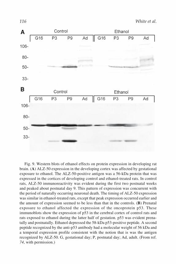
116 White et al.
Fig. 9. Western blots of ethanol effects on protein expression in developing ratbrain. (A) ALZ-50 expression in the developing cortex was affected by gestationalexposure to ethanol. The ALZ-50-positive antigen was a 56-kDa protein that wasexpressed in the cortices of developing control and ethanol-treated rats. In controlrats, ALZ-50 immunoreactivity was evident during the first two postnatal weeksand peaked about postnatal day 9. This pattern of expression was concurrent withthe period of naturally occurring neuronal death. The timing of ALZ-50 expressionwas similar in ethanol-treated rats, except that peak expression occurred earlier andthe amount of expression seemed to be less than that in the controls. (B) Prenatalexposure to ethanol affected the expression of the oncoprotein p53. Theseimmunoblots show the expression of p53 in the cerebral cortex of control rats andrats exposed to ethanol during the latter half of gestation. p53 was evident prena-tally and postnatally. Ethanol depressed the 58-kDa p53-positive peptide. A secondpeptide recognized by the anti-p53 antibody had a molecular weight of 56 kDa anda temporal expression profile consistent with the notion that is was the antigenrecognized by ALZ-50. G, gestational day; P, postnatal day; Ad, adult. (From ref.74, with permission.)

Apoptosis in Neurotoxicology 117
8.3.8. Concordance of the Biochemical and Anatomical Data
The biochemical data support stereological anatomical studies on neu-ronal number in somatosensory cortex and thalamus. Following prenatalexposure to ethanol, somatosensory cortex has 33% fewer neurons (85). Onthe other hand, the numbers of neurons in the somatosensory thalamus areunaffected by prenatal exposure to ethanol (86).
Ethanol affects the survival of neurons in vivo and in vitro. The in vivostudies show that ethanol toxicity is time and place dependent. This findingis true whether the ethanol exposure occurs during the period of neuronalgeneration (i.e., before the period of naturally occurring neuronal death andsynaptogenesis) or during the period of naturally occurring neuronal death(cf. refs. 66 and 68). In either case, some areas of the brain are particularlyaffected by ethanol (e.g., somatosensory cortex), whereas others seem to beprotected from ethanol-induced neuronal loss (e.g., somatosensory thala-mus). The ethanol-induced effects on neuronal survival have been modeledin vitro. Using such in vitro systems has permitted us to show that ethanol-induced death is mediated by neurotrophins; specifically for cortical neu-rons, the critical neurotrophin is NGF. In this regard, the toxic effects ofethanol are regulated by the low-affinity neurotrophin receptor, p75, andspecifically affects genes such as neg.
8.4. Studies of Organophosphorus Compounds8.4.1. Effects of Chlorpyrifos and Its Metaboliteson Developmental Exposure to PC12 Cells
As mentioned earlier, neural development in vitro can be examined usingPC12 cells. These cells were exposed to various concentrations of chlorpyrifos[CPF: O,O'-diethyl O-(3,5,6-trichloro-2-pyridyl) phosphorothionate], two ofits metabolites, chlorpyrifos-oxon and trichloropyridinol (TCP: 3,5,6-trichloro-2-pyridinol), and a fourth compound, Burroughs Wellcome (BW)284c51, a specific acetylcholinesterase inhibitor, to determine if this develop-mental process could be impacted (87). NGF-differentiated PC12 cells ex-posed to 0.1–100 nM CPF-oxon showed an increase in apoptosis in theabsence of NGF, and a dose-related increase in apoptosis was observed inthe presence of NGF. Differentiated PC12 cells exposed the non-cholinest-erase-inhibiting metabolite, TCP, at concentrations of 0.1–100 μM showed adose-related increase in apoptosis in the absence of NGF and at the highestdose in the presence of NGF. Similar paradigms were also used for CPF andthe BW compound exposures. No effect was seen on levels of apoptosis withincreasing doses of CPF. With BW compound exposure, again at 0.1–100μM, a significant increase in apoptosis was seen in the absence of NGF. A

118 White et al.
comparison of the amount of cell death induced by these compounds in bothdifferentiated and undifferentiated cells revealed that under all conditions,the differentiated cells had greater levels of fragmented DNA. Furthermore,the oxon induced apoptosis, with higher levels seen in differentiated cells,and NGF protected against apoptotic cell death.
Results from cell death ELISA assays were corroborated with TUNELstaining (see Fig. 10). Differentiated PC12 cells exposed to vehicle control,chlorpyrifos at 1 μM or 100 μM, and trichoropyridinol at 1 μM or 100 μMdemonstrated increased apoptosis of primed cells with NGF withdrawal.TCP appeared to induce apoptosis at 1 μM, whereas CPF did not until con-centrations of 100 μM were used.
Thus, in vitro exposure to CPF metabolites CPF-oxon or TCP in theabsence of NGF resulted in a dose-dependent induction of apoptotic deathwith effects observed as low as 0.1 nM for CPF-oxon or I.0 μM for TCP.Exposure to NGF (10 ng/mL) protected against cell death at doses of TCPand CPF-oxon below 100 μM and 1 nM, respectively. These data suggestthat differentiated PC12 cells are more vulnerable to apoptosis followingNGF withdrawal than undifferentiated cells and that NGF under certain con-ditions can protect against this pesticide-induced apoptosis.
8.4.2. In Vivo Developmental Exposure to Chlorpyrifos Affects Apoptosis
In parallel to the above in vitro work, apoptosis in the developing brain inresponse to CPF exposure was determined. The exposure paradigm for thiswork was gavage administration of pregnant dams beginning on GD14 andcontinuing through GD18. Pups were then removed at various times-pointsfollowing the last dose. Because earlier work from the laboratory showedthat at 5 h after the last dose of CPF, cholinesterase inhibition was greatestand TCP levels were highest in fetal brain (88), the 5-h time-point was usedfor apoptosis studies. Four doses of CPF were used: 3, 5, 7, or 10 mg/kg/d.Cell death by ELISA was used to determine that at both 7- and 10-mg/kg/ddoses, levels of fragmented DNA were significantly increased in GD18 brains(see Fig. 11). TUNEL staining on this tissue corroborated the ELISA results,wherein control animals exposed to vehicle showed basal levels of TUNEL-stained cells at GD18. Tissue from a fetus exposed to the 7-mg/kg/d dosedemonstrated apoptotic cells at levels greater than controls (see Fig. 12).
8.5. Contributions of Other Organophosphorus Compoundsto Cell Death
Organophosphorus (OP) compounds are neurotoxic by two major path-ways: inhibition of acetylcholinesterase (AChE) and induction of delayedneuropathy. AChE inhibition is the mechanism by which OP compounds exert

Apoptosis in Neurotoxicology 119
their insecticidal effects and is far more common than OP-induced delayedneuropathy (OPIDN). OPIDN requires initial inhibition of another esterase,neuropathy target esterase (NTE, or neurotoxic esterase). NTE inhibition must
Fig. 10. Effects of chlorpyrifos or TCP exposures (1 μM or 100 μM) in PC12cells in the absence and presence of NGF. Red arrows indicate apoptotic cells.Increased apoptosis is evident in TCP-exposed cells at 1 μM, but is not evident inCPF-exposed cells until 100 μM. Also note increased apoptosis in the absence ofNGF. Scale bars = 50 μm; n=4 per dose.
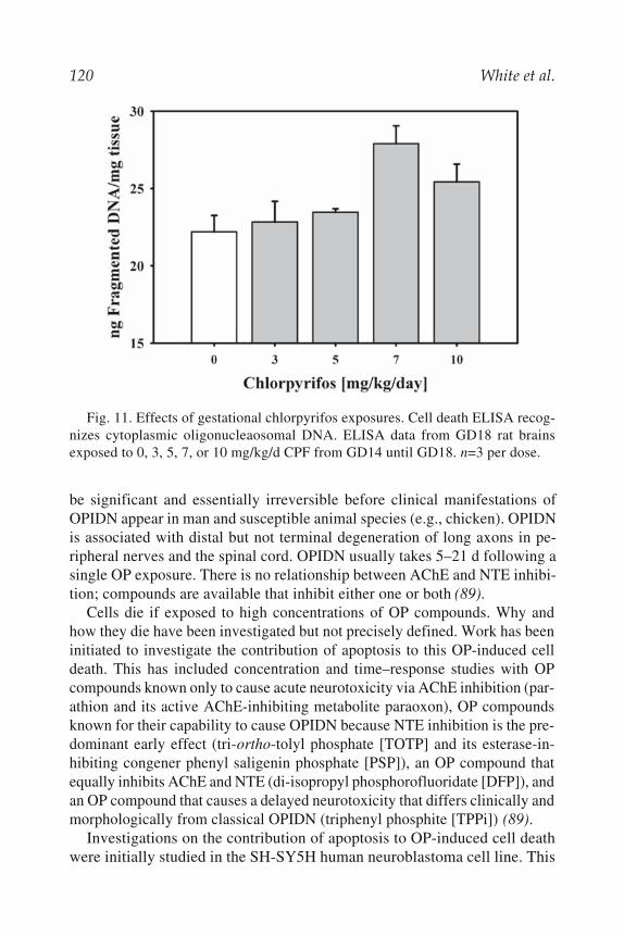
120 White et al.
be significant and essentially irreversible before clinical manifestations ofOPIDN appear in man and susceptible animal species (e.g., chicken). OPIDNis associated with distal but not terminal degeneration of long axons in pe-ripheral nerves and the spinal cord. OPIDN usually takes 5–21 d following asingle OP exposure. There is no relationship between AChE and NTE inhibi-tion; compounds are available that inhibit either one or both (89).
Cells die if exposed to high concentrations of OP compounds. Why andhow they die have been investigated but not precisely defined. Work has beeninitiated to investigate the contribution of apoptosis to this OP-induced celldeath. This has included concentration and time–response studies with OPcompounds known only to cause acute neurotoxicity via AChE inhibition (par-athion and its active AChE-inhibiting metabolite paraoxon), OP compoundsknown for their capability to cause OPIDN because NTE inhibition is the pre-dominant early effect (tri-ortho-tolyl phosphate [TOTP] and its esterase-in-hibiting congener phenyl saligenin phosphate [PSP]), an OP compound thatequally inhibits AChE and NTE (di-isopropyl phosphorofluoridate [DFP]), andan OP compound that causes a delayed neurotoxicity that differs clinically andmorphologically from classical OPIDN (triphenyl phosphite [TPPi]) (89).
Investigations on the contribution of apoptosis to OP-induced cell deathwere initially studied in the SH-SY5H human neuroblastoma cell line. This
Fig. 11. Effects of gestational chlorpyrifos exposures. Cell death ELISA recog-nizes cytoplasmic oligonucleaosomal DNA. ELISA data from GD18 rat brainsexposed to 0, 3, 5, 7, or 10 mg/kg/d CPF from GD14 until GD18. n=3 per dose.

Apoptosis in Neurotoxicology 121
culture system was chosen because previous studies demonstrated its use-fulness for differentiating biochemical effects of OP compounds causingacute neurotoxicity and OPIDN based on relative inhibitions of AChE andNTE, respectively, whether they were active esterase inhibitors orprotoxicants (90,91). Changes in nuclear morphology were used to assessthe contribution of apoptosis to cell death for the six OP compounds listed.
Fig. 12. Effects of gestational chlorpyrifos exposures. Apoptosis demonstratedby TUNEL staining of sagittal sections of GD18 brain exposed to either saline (A)or 7 mg/kg/d CPF (B). Brains were cut at 12 μm and stained according to kit proto-cols (Intergen). Tissue is counterstained with methyl green. Scale bars = 250 μm;n=4 per dose.

122 White et al.
Nuclear effects were determined by fluorescent microscopy. Results indi-cated that apoptotic nuclear budding occurred with parathion, paraoxon, andTPPi, but it was a relatively late event, requiring at least 16 h of exposure to0.1 mM concentrations of test compounds (see Fig. 13). PSP and TOTP athigher concentrations (1 mM) induced nuclear condensation and shrinkagewith little nuclear budding. Effects on the nucleus were suggestive of DNAfragmentation associated with apoptosis and were indicated by the presenceof discrete bands of DNA on agarose gels of cells exposed to paraoxon,parathion, TPPi, and TOTP. Increases in subG1 DNA fragmentationdetected by flow cytometry were another indication of changes in DNAintegrity associated with apoptosis, with concentrations for effect similar tothose needed for nuclear changes (44).
The contribution of apoptosis to these alterations in nuclear morphologyand DNA integrity was verified by examining OP-induced caspase activa-tion, as this is required for apoptotic cascades. For these studies, flowcytometry was used to assess activated caspase-3 by labeling cells withantiactive caspase-3 antibodies coupled to the fluorochrome phycoerythrin.Significant caspase-3 activation was found to occur prior to DNA fragmen-tation and nuclear budding following exposure of neuroblastoma cells tosome OP compounds (paraoxon, TOTP), but not to others (parathion, PSP,TPPi). DFP did not induce caspase-3 activation. OP-compound-induced ac-tivation of caspase-3 could be inhibited by treatment with protease inhibi-tors, including PMSF, a general protease inhibitor, and by more specificinhibitors of caspase-3 and caspase-8 (44).
The results of the above-described in vitro studies suggest that apoptosis isthe predominant form of death in neuronal cells in culture and that this occursindependent of specific esterase inhibition as protoxicants (e.g., TOTP, par-athion) and both NTE and AChE inhibitors could initiate its occurrence. Ad-ditional studies suggest that dysfunctional mitochondria might be involved indetermining whether cell death proceeds via apoptotic or necrotic pathways.These studies demonstrated changes in mitochondrial transmembrane poten-tial when the neuroblastoma cell line was exposed to OP compounds (45). Inaddition, a fluorescent dye impenetrable to undamaged mitochondria wasfound to enter this organelle in OP-exposed cultures of dissociated avian dor-sal root ganglia neurons. In the latter, the colocalization of dyes in the mito-chondria was greater in cells exposed to neuropathy-inducing OP compounds(mipafox, PSP) than it was in cells exposed to an OP only capable of inhibit-ing AChE (paraoxon) (see Fig. 14) (46). Disruption of mitochondria in bothculture systems was verified by electron microscopy.
Demonstration of apoptosis in OP-exposed animals has proven to be moredifficult than demonstration of this mode of cell death in vitro. Attempts

Apoptosis in Neurotoxicology 123
have been made on hens exposed to neuropathy-inducing OP compoundsand rats exposed to AChE and NTE inhibitors (Carlson, Jortner, and Ehrich,unpublished). Appearance of unequivocal apoptosis has been lacking. Thisis to be expected, as it is not always apparent that apoptosis occurred as cellswere dying if progress toward cell death is not followed over the entire timeit occurs. Detection of apoptosis in vivo involves the examination of a popu-lation of cells undergoing apoptosis asynchronously. Furthermore, the body
Fig. 13. Nuclear fluorescence to demonstrate apoptosis in SH-SY5Y cells. Cellswere exposed to triphenyl phosphite (TPPi) for up to 72 h before acetone/ethanolfixation and staining with 10 μg/mL Hoechst 33342 solution. Increased appearance ofapoptotic nuclear buds and fragments occurred as early as 4 h after exposure. Shrink-age and a loss of nuclear structure can be noted at 72 h. (Magnification = 400×.)

124 White et al.
has mechanisms for quickly removing dying cells, making timing for detec-tion of cells undergoing apoptosis a significant challenge. However, it islikely that the apoptosis noted in vitro is relevant to the in vivo situation.Further work is needed to improve the correlation.
9. CONCLUSIONS AND FUTURE DIRECTIONSThis review illustrates that known developmental neurotoxicants (EtOH
and mercury) and compounds that are clear neurotoxicants in adults(haloacetic acids and organophosphorus agents) affect apoptosis throughmultiple mechanisms (see Fig. 15). These mechanisms include suggestedalterations in neurotrophic signaling, protein phosphorylation, mitochondrialdysfunction and alterations in the balance of proapoptotic factors.
This chapter attempts to summarize the myriad methods used to detectapoptosis and describe their strengths and weaknesses with exemplar devel-opmental neurotoxicants. It is important to note that there are methodologi-cal challenges in designing in vitro experiments that retain tissue andorganismal relevance. It is not surprising that it is often difficult to detect
Fig. 14. Colocalization of fluorescent dyes in mitochondria of dorsal root gan-glia cells isolated from 9-d-old chick embryos. The cells were exposed to 10–6 Mconcentrations of the organophosphorus compounds paraoxon, mipafox, and PSP.All are significantly different from control (p<0.05 for paraoxon, p<0.001 formipafox, p<0.01 for PSP; analysis of variance followed by Tukey’s multiple com-parison test; n=5).

Apoptosis in Neurotoxicology 125
apoptosis in vivo in tissue, which predominantly occurs in an asynchronousfashion, versus in culture systems, in which, by design, cell death occurs ina more synchronous fashion. The methodological simplicity of in vitro sys-tems can be a strength when dealing with issues of signal detection, but itcan also be a weakness when attempting to extrapolate to a more heterog-enous cell population of a specific region of the nervous system.
This review chapter illustrates both the strengths and weakness of reduc-tionist test systems in which apoptosis can be studied. It addresses this issuefrom a toxicological perspective in which dose response to exogenous agentsis fundamental. Regarding developmental neurotoxicology, a critical con-
Fig. 15. Presumptive apoptotic pathways of various neurotoxicants includingethanol, organophosphates, haloacetic acids, and methylmercury. Mercury and etha-nol appear to act at least partially through receptor-mediated pathways, whereasother compounds appear to act downstream, affecting cell signaling and mitochon-drial function.

126 White et al.
sideration that permeates the study of cell death is the time-course of effects.Whereas in vitro systems are ideal for detection of markers of apoptosis,there needs to be clear linkage of effects on apoptosis and cell number andeventual cell connectivity in the nervous system in order to more fully un-derstand what the consequences of changes in these markers mean. It is en-tirely conceivable that a transient increase or decrease in apoptosis duringprogramed cell death can lead to adverse consequences. The proof of thisprinciple, however, remains a significant research need in order to improveextrapolation from effects observed in vitro to predicted effects in vivo inthe developing nervous system. Although this task sounds gargantuan, in-creased evidence from in vitro systems has shed significant light on the cellbiology and signaling pathways that regulate this process of cell death. Re-cent efforts to create mathematical models of caspase function in apoptosiscould provide clues to solving some of these in vitro to in vivo extrapolationissues in a quantitative fashion (92). In addition, employing such modelingefforts and validation of their predictions could help in future hypothesistesting with environmental agents that are suspected of mediating their tox-icity through this process. The clinical implication of dysregulation ofapoptosis in neurological, immunological diseases, and many cancers couldbe the study of gene and environmental interactions that are significant fornot only detection but possible future treatments.
ACKNOWLEDGMENTS
For work done in the Laboratory for Neurotoxicity Studies, Virginia-Maryland Regional College of Veterinary Medicine, the contributions ofDr. Kent Carlson, Dr. Christiane Massicotte, and Dr. Bernard S. Jortner areacknowledged. Extramural support was provided by US EPA grantRA825356 and a Novartis predoctoral fellowship through the Society ofToxicology. Michael Miller thanks the many collaborators that he has hadover the past dozen years, including Julie Jacobs, Jia Luo, Sandra Mooney,Gail Seabold, and the countless many in the neuroscience and alcohol re-search communities who continue to be his colleagues/teachers. He grate-fully acknowledges the N.I.A.A.A. and the Department of Veterans Affairsfor their generous support of this work. Sid Hunter acknowledges the workof Dr. Keith Ward and Ellen Rogers in the completion of his work. For workdone in the Neurotoxicology Division of the US Environmental ProtectionAgency, the contributions of Leon Lassiter, Kaberi Das, and Damani Parranare gratefully acknowledged.
The information in this document has been funded in part by the US En-vironmental Protection Agency. It has been reviewed by the National Health

Apoptosis in Neurotoxicology 127
and Environmental Effects Research Laboratory and approved for publica-tion. Approval does not signify that the contents reflect the views of theagency nor does mention of trade names or commercial products constituteendorsement or recommendation for use.
REFERENCES1. Kerr, J. F., Wyllie, A. H., and Currie, A. R. (1972) Apoptosis: a basic biologi-
cal phenomenon with wide-ranging implications in tissue kinetics. Br. J. Can-cer 26, 239–257.
2. Meier, P., Finch, A., and Evan, G. (2000) Apoptosis in development. Nature407, 796–801.
3. Offen, D., Elkon, H., and Melamed, E. (2000) Apoptosis as a general cell deathpathway in neurodegenerative diseases. J. Neural Transm. 58, Suppl. 153–166.
4. Savill, J. and Fadok, V. (2000) Corpse clearance defines the meaning of celldeath. Nature 407, 784–788.
5. Bortner, C. D., Hughes, F. M., and Cidlowski, J. A. Shi (1997) Cell volumeregulation, ions, and apoptosis, in Programmed Cell Death (Shi, Y.-B., Shi,Y., Xu, Y. and Scott, D., eds.), Plenum, New York, pp. 63–70.
6. Barrow, J. R., Stadler, H. S., and Capecchi, M. R. (2000) Roles of Hoxa1and Hoxa2 in patterning the early hindbrain of the mouse. Development 127,933–944.
7. Oppenheim, R. W. and Cowan, W. M. (1982) Neuronal cell death and somerelated regressive phenomena during neurogenesis. A selective historicalreview and a progress report, in Studies in Developmental Neurobiology. Es-says in Honor of Victor Hamburger ( Cowan, W.M., ed.), Oxford UniversityPress, New York, pp. 74–133.
8. Blaschke, A. J., Staley, K., and Chun, J. (1996) Widespread programmed celldeath in proliferative and postmitotic regions of the fetal cerebral cortex.Development 122, 1165–1174.
9. White, L. D. and Barone, S. J. (2001) Qualitatative and quantitative estimatesof apoptosis from birth to senescence in the rat brain. Cell Death Differ. 8,345–356.
10. Thomaidou, D., Mione, M. C., Cavanagh, J. F., and Parnavelas, J. G. (1997)Apoptosis and its relation to the cell cycle in the developing cerebral cortex. J.Neurosci. 17, 1075–1085.
11. Margolis, R. L., Chuang, D. M., and Post, R. M. (1994) Programmed cell death:implications for neuropsychiatric disorders. Biol. Psychiatry 35, 946–956.
12. Busciglio, J. and Yankner, B. A. (1995) Apoptosis and increased generation ofreactive oxygen species in Down’s syndrome neurons in vitro. Nature 378,776–779.
13. Selemon, L. D., Rajkowska, G., and Goldman-Rakic, P. S. (1998) Elevatedneuronal density in prefrontal area 46 in brains from schizophrenic patients:application of a three-dimensional, stereologic counting method. J. Comp.Neurol. 392, 402–412.

128 White et al.
14. Jarskog, L. F., Gilmore, J. H., Selinger, E. S., and Lieberman, J. A. (2000)Cortical bcl-2 protein expression and apoptotic regulation in schizophrenia.Biol. Psychiatry 48, 641–650.
15. Falkai, P., Bogerts, B., and Rozumek, M. (1988) Limbic pathology in schizo-phrenia: the entorhinal region—a morphometric study. Biol. Psychiatry 24,515–521.
16. Beckmann, H. and Lauer, M. (1997) The human striatum in schizophrenia. II.Increased number of striatal neurons in schizophrenics. Psychiatry Res. 8,99–109.
17. Highley, J. R., Esiri, M. M., McDonald, B., Cortina-Borja, M., Herron, B. M.,and Crow, T. J. (1999) The size and fibre composition of the corpus callosumwith respect to gender and schizophrenia: a post-mortem study. Brain 122(Pt.1), 99–110.
18. Fidler, D. J., Bailey, J. N., and Smalley, S. L. (2000) Macrocephaly in autism andother pervasive developmental disorders. Dev. Med. Child Neurol. 42, 737–740.
19. Piven, J., Arndt, S., Bailey, J., and Andreasen, N. (1996) Regional brain en-largement in autism: a magnetic resonance imaging study. J. Am. Acad. ChildAdolesc. Psychiatry 35, 530–536.
20. Sears, L. L., Vest, C., Mohamed, S., Bailey, J., Ranson, B. J., and Piven, J.(1999) An MRI study of the basal ganglia in autism. Prog.Neuropsychopharmacol. Biol. Psychiatry 23, 613–624.
21. Hardan, A. Y., Minshew, N. J., and Keshavan, M. S. (2000) Corpus callosumsize in autism. Neurology 55, 1033–1036.
22. Hashimoto, T., Tayama, M., Mori, K., Fujino, K., Miyazaki, M., and Kuroda,Y. (1989) Magnetic resonance imaging in autism: preliminary report.Neuropediatrics 20, 142–146.
23. Bailey, A., Luthert, P., Bolton, P., Le Couteur, A., Rutter, M., and Harding, B.(1993) Autism and megalencephaly. Lancet 341, 1225–1226.
24. Hengartner, M. O. (1999) Programmed cell death in the nematode C. elegans.Recent Prog. Horm. Res. 54, 213–222.
25. Lee, C. Y. and Baehrecke, E. H. (2000) Genetic regulation of programmed celldeath in Drosophila. Cell Res. 10, 193–204.
26. Nunez, G., Benedict, M. A., Hu, Y., and Inohara, N. (1998) Caspases: the pro-teases of the apoptotic pathway. Oncogene 17, 3237–3245.
27. Hengartner, M. O. (2000) The biochemistry of apoptosis. Nature 407, 770–776.28. Garcia, I., Martinou, I., Tsujimoto, Y., and Martinou, J. C. (1992) Prevention
of programmed cell death of sympathetic neurons by the bcl-2 proto-oncogene.Science 258, 302–304.
29. Allsopp, T. E., Wyatt, S., Paterson, H. F., and Davies, A. M. (1993) The proto-oncogene bcl-2 can selectively rescue neurotrophic factor-dependent neuronsfrom apoptosis. Cell 73, 295–307.
30. Chao, D. T. and Korsmeyer, S. J. (1998) BCL-2 family: regulators of cell death.Annu. Rev. Immunol. 16, 395–419.
31. Vekrellis, K., McCarthy, M. J., Watson, A., Whitfield, J., Rubin, L. L., andHam, J. (1997) Bax promotes neuronal cell death and is downregulated duringthe development of the nervous system. Development 124, 1239–1249.

Apoptosis in Neurotoxicology 129
32. van Lookeren, C. M. and Gill, R. (1998) Tumor-suppressor p53 is expressed inproliferating and newly formed neurons of the embryonic and postnatal ratbrain: comparison with expression of the cell cycle regulators p21Waf1/Cip1,p27Kip1, p57Kip2, p16Ink4a, cyclin G1, and the proto-oncogene Bax. J.Comp. Neurol. 397, 181–198.
33. Shimohama, S., Fujimoto, S., Sumida, Y., and Tanino, H. (1998) Differentialexpression of rat brain bcl-2 family proteins in development and aging.Biochem. Biophys. Res. Commun. 252, 92–96.
34. Mooney, S. M. and Miller, M. W. (2000) Expression of bcl-2, bax, and caspase-3 in the brain of the developing rat. Dev. Brain Res. 123, 103–117.
35. Krammer, P. H. (2000) CD95’s deadly mission in the immune system. Nature407, 789–795.
36. Rabizadeh, S., Oh, J., Zhong, L. T., et al. (1993) Induction of apoptosis by thelow-affinity NGF receptor. Science 261, 345–348.
37. Casaccia-Bonnefil, P., Kong, H., and Chao, M. V. (1998) Neurotrophins: thebiological paradox of survival factors eliciting apoptosis. Cell Death Differ. 5,357–364.
38. Li, H., Zhu, H., Xu, C. J., and Yuan, J. (1998) Cleavage of BID by caspase 8mediates the mitochondrial damage in the Fas pathway of apoptosis. Cell 94,491–501.
39. Luo, X., Budihardjo, I., Zou, H., Slaughter, C., and Wang, X. (1998) Bid, aBcl2 interacting protein, mediates cytochrome c release from mitochondria inresponse to activation of cell surface death receptors. Cell 94, 481–490.
40. Yin, X. M. (2000) Signal transduction mediated by Bid, a pro-death Bcl-2 fam-ily proteins, connects the death receptor and mitochondria apoptosis pathways.Cell Res. 10, 161–167.
41. Rich, T., Allen, R. L., and Wyllie, A. H. (2000) Defying death after DNA dam-age. Nature 407, 777–783.
42. Zucker, R. M., Hunter, S., and Rogers, J. M. (1998) Confocal laser scanningmicroscopy of apoptosis in organogenesis-stage mouse embryos. Cytometry33, 348–354.
43. Charriaut-Marlangue, C. and Ben-Ari, Y. (1995) A cautionary note on the useof the TUNEL stain to determine apoptosis. NeuroReport 7, 61–64.
44. Carlson, K., Jortner, B. S., and Ehrich, M. (2000) Organophosphorus com-pound-induced apoptosis in SH-SY5Y human neuroblastoma cells. Toxicol.Appl. Pharmacol. 168, 102–113.
45. Carlson, K. and Ehrich, M. (1999) Organophosphorus compound-inducedmodification of SH-SY5Y human neuroblastoma mitochondrial transmem-brane potential. Toxicol. Appl. Pharmacol. 160, 33–42.
46. Massicotte, C. (2001) The effects of neuropathy-inducing organophosphateesters on energy metabolism: a biochemical and morphological assessment,Ph.D. dissertation, Virginia Tech, Blacksburg, VA.
47. van Engeland, M., Nieland, L. J., Ramaekers, F. C., Schutte, B., andReutelingsperger, C. P. (1998) Annexin V-affinity assay: a review on an

130 White et al.
apoptosis detection system based on phosphatidylserine exposure. Cytometry31, 1–9.
48. Hunter, E. S., III, Rogers, E. H., Schmid, J. E., and Richard, A. (1996) Com-parative effects of haloacetic acids in whole embryo culture. Teratology 54,57–64.
49. Ward, K. W., Rogers, E. H., and Hunter, E. S., III (1998) Dysmorphogeniceffects of a specific protein kinase C inhibitor during neurulation. Reprod.Toxicol. 12, 525–534.
50. Ward, K. W., Rogers, E. H., and Hunter, E. S., III (2000) Comparative patho-genesis of haloacetic acid and protein kinase inhibitor embryotoxicity in mousewhole embryo culture. Toxicol. Sci. 53, 118–126.
51. Levi, A., Biocca, S., Cattaneo, A., and Calissano, P. (1988) The mode of actionof nerve growth factor in PC12 cells. Mol. Neurobiol. 2, 201–226.
52. Parran, D. K., White, L. D., and Barone, S., Jr. (2000) Methylmercury andmercuric chloride affect neuronal apoptosis in PC12 cell in a dose- and NGF-dependent fashion. Toxicol. Soc. 545, 115.
53. Nagashima, K. (1997) A review of experimental methylmercury toxicity inrats: neuropathology and evidence for apoptosis. Toxicol Pathol. 25, 624–631.
54. Seabold, G. K., Luo, J., and Miller, M. W. (1998) Effect of ethanol onneurotrophin-mediated cell survival and receptor expression in cultures of cor-tical neurons. Dev. Brain Res. 108, 139–145.
55. Miller, M. W. (2003) Describing the balance of cell proliferation and death: amathematical model. J. Neurobiol., in press.
56. Jacobs, J. S. and Miller, M. W. (2001) Neg, a nerve growth factor-promotedgene expressed by fetal neorcortical neurons that is down-regulated by ethanol.J. Comp. Neurol. 460, 212–222.
57. Chao, M. V. (1994) The p75 neurotrophin receptor. J. Neurobiol. 25, 1373–1385.58. Tscharke, D. C., Wilkinson, R., and Simmons, A. (2000) Use of mRNA differen-
tial display to study the action of lymphocyte subsets in vivo and application to amurine model of herpes simplex virus infection. Immunol. Lett. 74, 127–132.
59. Nagase, T., Seki, N., Ishikawa, K., Ohira, M., et al. (1996) Prediction of thecoding sequences of unidentified human genes. VI. The coding sequences of80 new genes (KIAA0201–KIAA0280) deduced by analysis of cDNA clonesfrom cell line KG-1 and brain. DNA Res. 3, 321–354.
60. West, J. R. (1987) Fetal alcohol-induced brain damage and the problem of de-termining temporal vulnerability: a review. Alcohol Drug Res. 7, 423–441.
61. Ward, G. R. and West, J. R. (1992) Effects of ethanol during development onneuronal survival and plasticity, in Development of the Central Nervous Sys-tem: Effects of Alcohol and Opiates (Miller, M.W., ed.), Wiley–Liss, NewYork, pp. 109–138.
62. Nornes, H. O. and Morita, M. (1979) Time of origin of the neurons in the caudalbrain stem of rat. An autoradiographic study. Dev. Neurosci. 2, 101–114.
63. Miller, M. W. and Muller, S. J. (1989) Structure and histogenesis of the princi-pal sensory nucleus of the trigeminal nerve: effects of prenatal exposure toethanol. J. Comp. Neurol. 282, 570–580.

Apoptosis in Neurotoxicology 131
64. Ashwell, K. W. and Waite, P. M. (1991) Cell death in the developing trigemi-nal nuclear complex of the rat. Dev. Brain Res. 63, 291–295.
65. Miller, M. W. and Al-Ghoul, W. M. (1993) Numbers of neurons in the devel-oping principal sensory nucleus of the trigeminal nerve: enhanced survival ofearly-generated neurons over late-generated neurons. J. Comp. Neurol. 330,491–501.
66. Miller, M. W. (1995) Effect of pre- or postnatal exposure to ethanol on thetotal number of neurons in the principal sensory nucleus of the trigeminal nerve:cell proliferation and neuronal death. Alcohol Clin. Exp. Res. 19, 1359–1363.
67. Al-Ghoul, W. M. and Miller, M. W. (1993) Development of the principal sen-sory nucleus of the trigeminal nerve of the rat and evidence for a transientsynaptic field in the trigeminal sensory tract. J. Comp. Neurol. 330, 476–490.
68. Miller, M. W. (1999) A longitudinal study of the effects of prenatal ethanolexposure on neuronal acquisition and death in the principal sensory nucleus ofthe trigeminal nerve: interaction with changes induced by transection of theinfraorbital nerve. J. Neurocytol. 28, 999–1015.
69. Wolozin, B. L., Pruchnicki, A., Dickson, D. W., and Davies, P. (1986) A neu-ronal antigen in the brains of Alzheimer patients. Science 232, 648–650.
70. Wolozin, B., Scicutella, A., and Davies, P. (1988) Reexpression of a develop-mentally regulated antigen in Down syndrome and Alzheimer disease. Proc.Natl. Acad. Sci. USA 85, 6202–6206.
71. Al-Ghoul, W. M. and Miller, M. W. (1989) Transient expression of Alz-50immunoreactivity in developing rat neocortex: a marker for naturally occur-ring neuronal death? Brain Res. 481, 361–367.
72. Valverde, F., Lopez-Mascaraque, L., and de Carlos, J. A. (1990) Distributionand morphology of Alz-50-immunoreactive cells in the developing visual cor-tex of kittens. J. Neurocytol. 19, 662–671.
73. Miller, M. W., Al-Ghoul, W. M., and Murtaugh, M. (1991) Expression of ALZ-50 immunoreactivity in the developing principal sensory nucleus of the trigemi-nal nerve: effect of transecting the infraorbital nerve. Brain Res. 560, 132–138.
74. Kuhn, P. E. and Miller, M. W. (1998) Expression of p53 and ALZ-50 immu-noreactivity in rat cortex: effect of prenatal exposure to ethanol. Exp. Neurol.154, 418–429.
75. Zhan, Q., Carrier, F., and Fornace, A. J., Jr. (1993) Induction of cellular p53activity by DNA-damaging agents and growth arrest. Mol. Cell Biol. 13,4242–4250.
76. Mosner, J., Mummenbrauer, T., Bauer, C., Sczakiel, G., Grosse, F., andDeppert, W. (1995) Negative feedback regulation of wild-type p53 biosynthe-sis. EMBO J. 14, 4442–4449.
77. Morrison, R. S., Wenzel, H. J., Kinoshita, Y., Robbins, C. A., Donehower, L.A., and Schwartzkroin, P. A. (1996) Loss of the p53 tumor suppressor geneprotects neurons from kainate-induced cell death. J. Neurosci. 16, 1337–1345.
78. Miller, M. W. and Kuhn, P. E. (1997) Neonatal transection of the infraorbitalnerve increases the expression of proteins related to neuronal death in the prin-cipal sensory nucleus of the trigeminal nerve. Brain Res. 769, 233–244.

132 White et al.
79. Mooney, S. M. and Miller, M. W. (2001) Effects of prenatal exposure on theexpression of bcl-2, bax, and caspase-3 in the developing rat trigeminal/soma-tosensory system. Brain Res. 911, 71–81.
80. Keane, R. W., Srinivasan, A., Foster, L. M., et al. (1997) Activation of CPP32during apoptosis of neurons and astrocytes. J. Neurosci. Res. 48, 168–180.
81. Armstrong, R. C., Aja, T. J., Hoang, K. D., et al. (1997) Activation of theCED3/ICE-related protease CPP32 in cerebellar granule neurons undergoingapoptosis but not necrosis. J. Neurosci. 17, 553–562.
82. Harada, J. and Sugimoto, M. (1999) Activation of caspase-3 in beta-amyloid-induced apoptosis of cultured rat cortical neurons. Brain Res. 842, 311–323.
83. Kirsch, D. G., Doseff, A., Chau, B. N., et al. (1999) Caspase-3-dependentcleavage of Bcl-2 promotes release of cytochrome c. J. Biol. Chem. 274,21,155–21,161.
84. Liu, X. and Zhu, X. Z. (1999) Roles of p53, c-Myc, Bcl-2, Bax and caspases inserum deprivation-induced neuronal apoptosis: a possible neuroprotectivemechanism of basic fibroblast growth factor. NeuroReport 10, 3087–3091.
85. Miller, M. W. and Potempa, G. (1990) Numbers of neurons and glia in maturerat somatosensory cortex: effects of prenatal exposure to ethanol. J. Comp.Neurol. 293, 92–102.
86. Mooney, S. M. and Miller, M. W. (1999) Effects of prenatal exposure to etha-nol on systems matching: the number of neurons in the ventrobasal thalamicnucleus of the mature rat. Dev. Brain Res. 117, 121–125.
87. Das, K. P., Barone, S., Jr., and White, L. D. (2000) Metabolites of chlorpyrifosinduce apoptosis in PC12 cells. Toxicol. Soc. 545, 117.
88. Hunter, D. L., Lassiter, T. L., and Padilla, S. (1999) Gestational exposure tochlorpyrifos: comparative distribution of trichloropyridinol in the fetus anddam. Toxicol. Appl. Pharmacol. 158, 16–23.
89. Ehrich, M. and Jortner, B. S. (2001) Organophosphorus-induced delayed neu-ropathy, in Handbook of Pesticide Toxicology (Krieger, R., ed.), Academic,San Diego, CA, pp. 987–1012.
90. Ehrich, M., Correll, L., and Veronesi, B. (1997) Acetylcholinesterase and neu-ropathy target esterase inhibitions in neuroblastoma cells to distinguish orga-nophosphorus compounds causing acute and delayed neurotoxicity. Fundam.Appl. Toxicol. 38, 55–63.
91. Barber, D., Correll, L., and Ehrich, M. (1999) Comparative effectiveness oforganophosphorus protoxicant activating systems in neuroblastoma cells andbrain homogenates. J. Toxicol. Environ. Health A 57, 63–74.
92. Fussenegger, M., Bailey, J. E., and Varner, J. (2000) A mathematical model ofcaspase function in apoptosis. Nature Biotechnol. 18, 768–774.

Toxicant-Induced Neurotransmitter Dysfunction 133
133
6Impairment of Neurotransmitter Metabolism
and Function by Neurotoxicants
Enzyme Pathways in Neurons and Astroglia
Michael Aschner and Ursula Sonnewald
1. INTRODUCTIONIn order to perform neurotoxicological studies, model systems have to be
established and techniques developed to analyze relevant parameters. Thepresent chapter describes the in vitro effects of the neurotoxicantsaminooxyacetic acid (AOAA), 3-nitropropionic acid (3-NPA), and methyl-mercury (MeHg) on glial cell neurotransmitters, and for 3-NPA, we alsodescribe the effects on cultured neurons. Major emphasis is directed at theeffects of these compounds on glutamate metabolism.
2. CELL CULTURESCells taken directly from the organism and subsequently grown for at
least 24 h in vitro are considered to be primary cultures. Different proce-dures have been used, often in combination, to enable the establishment ofmonotypic cultures from mixed-cell suspensions. They are obtained after aninitial mechanical and/or enzymatic dissociation of the nervous tissue ofneonatal or newborn rat, mouse, or chick. Because of the relative abundanceof cells, magnetic resonance spectroscopy (MRS) studies are often per-formed on cultures consisting of excitatory cerebellar granule cells orinhibitory -aminobutyric-acid-(GABA)ergic cerebral cortex neurons pre-pared as described by Schousboe et al. (1) and Hertz et al. (2). The choice ofthese particular preparations of cultured neurons should be viewed in lightof the fact that 90% of the synapses in the brain utilize either glutamate or
From: Methods in Pharmacology and Toxicology: In Vitro Neurotoxicology: Principles and ChallengesEdited by: E. Tiffany-Castiglioni © Humana Press Inc., Totowa, NJ

134 Aschner and Sonnewald
GABA as the neurotransmitter. As counterparts for the neurons, typicallyastrocytes from the same brain region can be maintained in culture as de-tailed by Hertz et al. (3).
Appreciation of the essential role of astrocytes in aiding neuronalmetabolism and maintaining and modifying the biochemical milieu of thecentral nervous system (CNS) has grown greatly in the last decades (4–6).Included among these interdependent processes between astrocytes andneurons are guidance of neuronal migration (7), the release of neurotrophicfactors (8), as well as glutamate uptake and its metabolism to glutamine(6,9–11). Because neurons are not capable of anaplerosis, it is essential thatthey obtain precursors from astrocytes for transmitter synthesis (6).Observations of additional astrocyte and neuron interactions include thedescription of an obligatory role for astrocytes in providing precursormolecules for neuronal glutathione (GSH) synthesis (12–15). More recentstudies demonstrate that few synapses form in the absence of glial cells andthat the few synapses that do form are functionally immature (16). Assuggested by Ullian et al. (16), astrocytes increase the number of maturefunctional synapses on the CNS neurons by sevenfold and are required forsynaptic maintenance in vitro. Thus, astrocytes are invoked to induce andstabilize synapses, raising the possibility that glia could actively participatein synaptic plasticity. Astrocytes also actively participate in synapticintegration by releasing glutamate via a calcium-regulated, exocytosislike process (17). This process follows activation of the receptor CXCR4 bythe chemokine stromal cell-derived factor 1 (SDF-1). An extraordinaryfeature of the ensuing signaling cascade is the rapid extracellular release oftumor necrosis factor (TNF). Bezzi et al. (17) also demonstrated that alteredglial communication has direct neuropathological consequences, identify-ing a new pathway for glia–glia and glia–neuron communication that isrelevant to both normal brain function and neurodegenerative diseases.
3. MAGNETIC RESONANCE SPECTROSCOPY (MRS)13C-MRS is a powerful tool for the analysis of brain metabolism and
metabolic trafficking between different brain compartments (18–20) and itis also useful in studying the effects of neurotoxicants on metabolism invivo (21,22) and in vitro (23–26). The nuclei that are most commonly usedin MRS for metabolic studies are 1H, 31P, and 13C. 1H and 31P are naturallyabundant isotopes and, therefore, the most common methods of studyinvolves examining differences in the natural abundance spectra under vari-ous metabolic states. In contrast, 13C has a natural abundance of 1.1%. This

Toxicant-Induced Neurotransmitter Dysfunction 135
disadvantage normally makes detection difficult and 13C-MRS is thus oflimited use for studies on endogenous metabolites, unless they occur in largeamounts. However, the low natural abundance can be an advantage in that13C-enriched precursors can be used for metabolic pathway mapping withlittle or no background interference from endogenous metabolites.
13C-MRS and [U-13C]glutamate have been used extensively for metabolicstudies in astrocytes (11,24,27–29) and cerebellar granule neurons (30). Itcould be shown that exogenous glutamate was metabolized in culturedastrocytes to a great extent. Labeled glutamine, aspartate, and glutathionesynthesized from [U-13C]glutamate were detected in cell extracts, whereasin medium, in addition to the added [U-13C]glutamate, labeled glutamine,aspartate, and lactate were also observed. In cerebellar granule neurons(glutamatergic) [U-13C]glutamate is metabolized to aspartate, glutathione,and, to a small extent, lactate (30). In both cell types, glutamate is also syn-thesized via the tricarboxylic acid (TCA) cycle from labeled glutamate. Theschematic presentation of the distribution of label in different metabolitesderived from TCA cycle intermediates is shown in Fig. 1. After uptake byastrocytes, [U-13C]glutamate can be either converted to [U-13C]glutaminedirectly by glutamine synthetase or enter the TCA cycle after conversion to2-oxoglutarate for energy production and/or the synthesis of other metabo-lites. [U-13C]oxaloactate is formed after several steps, and [U-13C]aspartatecan be synthesized thereafter. [U-13C]lactate can be derived after severalsteps of gluconeogenesis from [U-13C]oxaloacetate or directly via malicenzyme from [U-13C]malate. In the presence of unlabeled glucose, unla-beled pyruvate can be converted to acetyl-coenzyme A. The condensationof labeled oxaloacetate and unlabeled acetyl-coenzyme A will lead to thesynthesis of [1,2-13C]/ [3,4-13C]aspartate, [1,2,3-13C]glutamate, and [1,2,3-13C]glutamine via TCA cycle intermediates (see Fig. 1).
4. IMPAIRMENT OF NEUROTRANSMITTERMETABOLISM AND FUNCTIONBY NEUROTOXICANTS
4.1. AminooxyaceticAn increasing number of observations suggest that neurodegenerative
diseases might be associated with aberrations in energy metabolism and thehandling of glutamate (31). Glutamate metabolism is thus a central issue inseveral of the major brain pathologies. In order to obtain animal models ofneurodegenerative diseases, inhibitors of mitochondrial energy metabolismhave been used. The transaminase inhibitor aminooxyacetic acid (AOAA)

136 Aschner and Sonnewald
induces striatal lesions characteristic of neurodegenerative diseases (32).AOAA inhibits the major transaminases, aspartate aminotransferase (AAT)and alanine aminotransferase (ALAAT), both in cytosol and in mitochon-dria. AAT is an essential component of the malate–aspartate shuttle, whichis important also in brain. This shuttle transports reduction equivalents ofNADH from the cytosol into mitochondria. The lesions produced by AOAAare attenuated by NMDA antagonists or prior decortication. This and otherobservations have led to the formulation of an excitotoxic hypothesis forneurodegenerative disorders (33).
4.1.1. Effect of AOAA on [U-13C]Glutamate Metabolism in Astrocytes
Extracellular glutamate is very efficiently taken up into astrocytes (34).In keeping with this, a fourfold increase in the intracellular glutamate con-tent was observed when the cells were incubated in a glutamate-containingmedium (29). Conversion of extracellular glutamate to glutamine is a part ofthe so-called “glutamate–glutamine cycle” (35,36). Previous studies ofglutamate metabolism in astrocytes have clearly shown that oxidativemetabolism in the TCA cycle takes place in astrocytes (37–39). However,different conclusions with regard to the significance and mechanism of oxi-dation through the TCA cycle have been reached. In one study, it was dem-onstrated that the extent of oxidation is coupled to the glutamateconcentration (28). A prerequisite for entry of exogenous glutamate into theTCA cycle is the conversion of glutamate to 2-oxoglutarate (2-OG), whichcan take place via a transamination or a deamination. In order to probe thesignificance of transamination for the oxidative metabolism of glutamate,the transaminase inhibitor AOAA has been used (37–39). It has been shownthat transamination played a minor role for formation of 2-OG from
Fig. 1. The distribution of label in different metabolites derived form tricarboxy-lic acid cycle intermediates.

Toxicant-Induced Neurotransmitter Dysfunction 137
glutamate (29). It appears, however, to be the major pathway for the oppo-site reaction (i.e., formation of glutamate from 2-OG). Formation of [1,2,3-13C]glutamate and glutamine (a sign of TCA cycle activity) was stronglyreduced in the presence of AOAA. An explanation for the great decrease ofthese isotopomers is that transamination is the major pathway for glutamateformation from 2-OG, which is in accordance with the low affinity ofglutamate dehydrogenase (GDH) for ammonia (40). However, the impor-tance of GDH for normal brain function is underlined by the demonstrationthat the neurodegenerative disorder olivopontocerebellar atrophy is linkedto impairment of GDH activity (41).
4.2. 3-Nitropropionic Acid3-Nitropropionic acid (3-NPA) is an irreversible inhibitor of succinate
dehydrogenase, which is part of both the TCA cycle and complex II of themitochondrial electron transport chain. Accidental ingestion of 3-NPA inhumans or systemic administration to experimental animals results in selec-tive striatal lesions (42,43). 3-NPA showed an age-dependent neurotoxicityin young adult rodents, which is similar to the late age of onset forHuntington’s disease and other neurodegenerative diseases (42,43). It hasbeen shown that 3-NPA inhibited synaptosomal respiration in a dose-depen-dent manner (44). In the absence of glutamine, 3-NPA caused a decrease ininternal concentrations of aspartate and glutamate, whereas GABAincreased. No increase was found in the external levels of these amino acids.With glutamine and 3-NPA in the medium, both glutamate and GABAincreased inside the synaptosomes while the external concentration ofglutamate rose also (44), lending further evidence for an excitotoxic mecha-nism in neurodegeneration. In mice injected subcutaneously with 3-NPA,an increased GABA concentration was observed, whereas glutamate wasslightly decreased (21). Furthermore, using [1-13C]glucose or [2-13C]acetatein combination with MRS, it could be shown that 3-NPA inhibited neuronsmore than glial cells (21).
4.2.1. Effect of 3-NPA on [U-13C]Glutamate Metabolism in Neurons
In cerebellar granule neurons, TCA cycle activity was efficiently blockedby 3 mM of 3-NPA (27); the metabolism of [U-13C]glutamate was restrictedto the formation of succinate. Only the uniformly labeled isotopomer ofglutamate could be detected by MRS and the amount of labeled glutamatewithin the cells was decreased compared to the control (29), which agreeswell with observations in synaptosomes (44). Lactate labeling through theTCA cycle has been observed previously in cell culture (27) and mouse(21). This formation of labeled lactate in cerebellar granule neurons wasabolished by 3 mM of 3-NPA (30).

138 Aschner and Sonnewald
4.2.2. Effect of 3-NPA on [U-13C]Glutamate Metabolism in Astrocytes
3-Nitropropionic acid intoxication has also been studied in murine astro-cytes receiving 0.5 mM of [U-13C]glutamate in all cases and two differentconcentrations of 3-NPA (3 and 10 mM) (25). 3-NPA intoxication clearlyaffected glutamate metabolism in astrocytes. Succinate accumulated intracel-lularly and extracellularly, and intracellular glutamate and glutamine concen-trations were reduced. In the control group, the succinate concentration wastoo small to be detected by 13C-MRS or, alternatively, succinate was not la-beled from glutamate. After 3-NPA treatment, no label was detected inaspartate. However, label appeared in lactate in astrocytes receiving 3 mM of3-NPA, and intracellular [1,2,3-13C]glutamate and extracellular [1,2,3-13C]glutamine were also still present in cells receiving 3 mM of 3-NPA,although both were significantly reduced. Such labeling from [U-13C]glutamate is only possible using precursors from the TCA cycle, indicatingthat 3 mM of 3-NPA was not sufficient to achieve a complete TCA cycleinhibition. With 10 mM of 3-NPA, the TCA cycle conversion of[U-13C]glutamate to metabolites was restricted to the formation of succi-nate (25).
4.2.3. Effect of 3-NPA on Uptake and Degradation of Glutamate
Uptake and degradation of glutamate was not impaired, because no accu-mulation of extracellular or intracellular glutamate was observed with3-NPA (25). To the contrary, intracellular glutamate was significantlyreduced, consistent with previous findings both in cerebellar granule cells(30) and synaptosomes (44). In synaptosomes incubated in medium withoutglutamine, 3-NPA caused a small drop in internal concentrations ofglutamate and an overall decline in the sum of aspartate, glutamate, andGABA concentrations, suggesting that the glutamate dehydrogenase reac-tion was stimulated. There is evidence for stimulation of GDH activity alsoin astrocytes (25) because intracellular glutamate concentrations decreasedin the presence of 3-NPA, a reduction not related to inhibition of the uptakeof glutamate, as there was no accumulation of extracellular glutamate (27).In the same study, it was shown that intracellular glutamine also decreasedafter 3-NPA treatment. Thus, the decrease in glutamate was not related to anincrease in glutamine synthetase activity. Stimulation of glutamine syn-thetase is also unlikely because proportionally less glutamate was metabo-lized directly to glutamine in the presence of 3-NPA, whereas there wasmassive accumulation of succinate. Together, these data suggest that GDHactivity was stimulated by 3-NPA both in astrocytes and neurons.

Toxicant-Induced Neurotransmitter Dysfunction 139
4.2.4. Energy Metabolism in the Presence of 3-NPA
A significant fall in ATP has been observed, both in synaptosomes [1–2mM of 3-NPA for 5–30 min, (44)] and in neuronal cultures of murine frontalcortex [0.25–1 mM of 3-NPA for 2–4 h (30)]. Intrastriatal injection of3-NPA has also been reported to result in a reduced ATP content within 3 hin the area close to the injection site (45). In cortical astrocytes, only a small,statistically not significant decrease in ATP was observed after administra-tion of 3-NPA (25). These results suggested that astrocytic ATP stores wereless vulnerable to 3-NPA intoxication than neuronal ATP stores. A possibleexplanation is the glial localization of pyruvate carboxylase, which is essen-tial for de novo synthesis of glutamine in astrocytes and subsequentlyglutamate and GABA in neurons (46). Thus, astrocytes have the ability tofuel the TCA cycle with intermediates from glucose via pyruvate and oxalo-acetate, a pathway not present in neurons (46). Indeed, there are indicationsof an increased pyruvate carboxylase activity in 3-NPA-treated astrocytesbecause glucose consumption was increased although lactate production wasdecreased in media from cells that had received a high glutamate concentra-tion (25). In synaptosomes, where pyruvate carboxylase is not present,lactate concentration increased after 3-NPA treatment (44).
4.2.5. Conclusions
In conclusion, astrocyte metabolism is clearly modified by 3-NPA. How-ever, higher concentrations are necessary in order to elicit a complete inhi-bition of the astrocytic TCA cycle when compared to the TCA cycle ofcerebellar granule cells. Astrocytes managed remarkably well in the pres-ence of high concentrations of 3-NPA, because ATP levels and energy-de-manding processes such as glutamate uptake and glutamine synthesis werelargely maintained. This was probably the result of the fact that astrocyteshave the ability to produce oxaloacetate from glucose via pyruvate. Theresults support the hypothesis that 3-NPA can be used in animal models forHuntington’s disease, as neurons appear to be more sensitive to this com-pound than astrocytes.
4.3. MethylmercuryMethylmercury (MeHg) is a highly neurotoxic compound producing neu-
ronal death that can be at least in part attributable to glutamate (47,48) andoverproduction of reactive oxygen species (ROS) (49–51). Although MeHgproduces neuronal death and alters neuronal function (4,52), mercurialslocalize predominantly within astrocytes (53,54). Earlier studies by us and

140 Aschner and Sonnewald
others have shown that MeHg produces astrocytic swelling both in culturedprimary astrocytes (55) and in vivo (53,54), stimulates astrocytic efflux ofexcitatory amino acids (EAA) and inhibits the uptake of EAA (55,56). Com-bined, these data suggest that MeHg-induced neuronal toxicity is, at least inpart, mediated by astrocytes.
4.3.1. Effects of MeHg on Glumatergic Systems
As alluded to earlier, a common finding upon exposure to MeHg is theoverproduction of ROS and alterations in glutamatergic function (49–51).Excess extracellular glutamate, such as that seen during inhibition of astro-cytic uptake, might generate excess ROS (57), and it is well established thatoverproduction of ROS inhibits glutamate uptake (58–60). In addition, excessextracellular glutamate concentrations lead to inhibition of the uptake of cys-tine, a GSH precursor (61), and decreased intracellular GSH content, thusrendering cells more susceptible to both the toxic sequelae of MeHg and ROS.The effect of MeHg on cystine uptake were addressed because cystine uptakemight involve the same family of transporters, XAG–, which are inhibited byMeHg. Studies were designed to distinguish whether the primary toxic mecha-nism of MeHg-induced neurodegeneration is the result of the effects on theglutamatergic system or direct production of ROS. The interactions amongglutamate, ROS, GSH, and MeHg are shown in Fig. 2.
Overproduction of ROS is a common feature of MeHg toxicity, espe-cially in neuronal cultures (62). Neurons contain approx 10-fold lower GSHlevels compared with astrocytes (63) and, therefore, are likely more suscep-tible to MeHg toxicity that coincides with decreased intracellular GSH con-centrations. Previous studies have suggested that neurons rely exclusivelyon astrocytes for providing GSH precursors (12,13,15). Thus, it was rea-soned that if MeHg inhibited cystine transport in astrocytes, neurons wouldbe disadvantaged as well, given the deprivation of astrocyte-derived GSHprecursors.
Evaluation of 35S-cystine accumulation revealed that astrocytes as wellas neurons readily transported 35S-cystine (64). These data corroborate workby others who have demonstrated high-affinity 35S-cystine transport in cul-tured neurons (14,61), yet are inconsistent with reports in which culturedneurons displayed no 35S-cystine transport (12,13). The factors associatedwith the apparent differences between these findings are unclear, but theymay relate to culture conditions, such as the choice of medium supplementsor source of serum. In addition, the cell types in the various experimentswere obtained by different isolation methods, which might have influencedtheir physiological properties. Hippocampal neurons, such as those used inthe present study, have relatively high levels of GSH compared to those

Toxicant-Induced Neurotransmitter Dysfunction 141
found in other brain regions (63) and, therefore, might posses a more effi-cient transport mechanism for GSH precursor uptake.
Recent studies (64) established that 35S-cystine uptake was temperaturedependent, demonstrating an active transport process in both astrocytes andneurons. Pretreatment with MeHg produced a concentrate-dependentdecrease in 35S-cystine uptake in astrocytes, but had no effect on 35S-cystineuptake in neurons. These data suggest that cortical astrocyte and hippocam-pal neuron cultures utilize differential cystine transport mechanisms (64).Allen et al. (64) concluded that in astrocytes, uptake of 35S-cystine wasapprox 30% Na+ dependent. Interestingly, omission of Na+ from the assaybuffer had no effect on 35S-cystine transport in neurons (64).
A well-characterized cystine transporter is the Na+-independent SystemXC– (65). The system is driven by the intracellular to extracellular glutamategradient to transport cystine into the cell. It is inhibited by high levels ofextracellular glutamate (65). Treatment of astrocytes with glutamate produceda decrease in 35S-cystine uptake, suggesting a role for System XC– (64). How-ever, it has also been suggested that the Na+-dependent excitatory amino acidtransporter (EAAT) family (System XAG–) is also capable of transporting cys-tine (61,66) and that the transport of 35S-cystine by the same system may becompetitively inhibited by elevated concentrations of glutamate. As a firststep in determining the contribution of the two transporters to the overalluptake of cystine, cells were treated with quisqualate, a specific inhibitor of
Fig. 2. Interactions among increased extracellular glutamate (GLUE), GSH, ROS,and MeHg. Increased GLUE increases ROS production leading to decreased GSHlevels. Increased GLUE also inhibits uptake of the GSH precursor cystine, leadingto decreased GSH levels. Decreased GSH levels lowers intracellular antioxidantlevels, thus decreasing the ability to detoxify ROS. Increased ROS (H2O2) inhibitglutamate uptake, producing increased GLUE. MeHg interacts with each of thesesystems, magnifying the detrimental effects and resulting in increased toxicity.
142 Aschner and Sonnewald
System XC–, or threo- -hydroxyaspartate (THA), a specific inhibitor of Sys-tem XAG–. These studies revealed that in both neurons and astrocytes, SystemXC– transports cystine and that astrocytes also transport cystine by SystemXAG–. No change in neuronal cystine uptake was apparent following removalof Na+. Accordingly, the Na+-dependent System XAG– was ruled out as a con-tributor to cystine uptake in neurons.
MeHg potently inhibits the uptake of 3H-D-aspartate via System XAG–
(EAAT1) in cultured astrocytes (47,55). Because System XAG– is alsocapable of transporting cystine, it is likely that the inhibitory effects of MeHgare the result of the inhibition of System XAG– function. To test this hypoth-esis, astrocytes were incubated with a System XAG– blocker THA, MeHg, orTHA plus MeHg. The addition of MeHg to the uptake buffer produced nofurther inhibition of THA-mediated decrease in cystine uptake, suggestingthat THA and MeHg are acting upon the same target. Thus, MeHg’s inhibi-tion of cystine transport was the result of actions on System XAG– (64).
Intracellular glutathione (GSH) levels (67,68) are known to modulateMeHg toxicity. MeHg decreases intracellular GSH levels because ofincreased efflux and oxidation, and thus increases in the synthesis of GSHare required to maintain adequate reducing power within astrocytes. Allenet al. (64) report that buthionine sulfoximine (BSO), a GSH-depleting agent,induced increases in cystine transport, and this effect was abolished by theaddition of MeHg in a dose-dependent manner. Given that MeHg appears toselectively blocks cystine transport via system XAG–, these data suggest thatthe increased transport of cystine was largely the result of the increased func-tion of System XAG–. The consequences of these effects are that MeHg notonly decreases cystine uptake in astrocytes with normal GSH levels but alsoprevents a compensatory increase in cystine transport induced by decreasedGSH levels. MeHg decreases intracellular astrocyte GSH levels both as aresult of increased GSH efflux and ROS production. Lowered intracellularGSH levels further increase the toxic effects of MeHg by decreasing intrac-ellular antioxidant levels, propagating a feedforward system that likely con-tinues unabated, producing ROS and, ultimately, cell death.
Decreases in cystine transport, and thus GSH levels, are also seen withhigh levels of extracellular glutamate, because of the inhibition of the cys-tine : glutamate heteroexchange System XC– (69). Thus, decreased functionof the EAA System XAG– transporter by MeHg will not only induce a directdecrease in cystine transport but also an indirect inhibition of its transportbecause of disruption of the intracellular/extracellular glutamate gradient.
A recent study published by Bender et al. (66) corroborates the abovefindings, namely that cystine is transported via System XAG– in cultured

Toxicant-Induced Neurotransmitter Dysfunction 143
astrocytes. System XAG– accounted for nearly 95% of cystine uptake in cul-tured astrocytes (66), whereas in our hands, System XAG– represented approx25% of astrocytic cystine uptake. However, they treated cells with a cell-permeable cyclic AMP analog prior to studies of cystine uptake (66). Thecyclic AMP analog is known to dramatically increase levels of both EAAT1and EAAT2 (70), thus increasing the System XAG– to System XC– ratio,resulting in decreased contribution of System XC– to cystine transport.
The mechanisms of MeHg-mediated inhibition of System XAG– remainunknown. Acute (5-min) coincubation with MeHg and cystine produced noinhibitory effects on cystine uptake, suggesting a lack of direct interactionbetween MeHg and the EAA transporter (64). Studies examining the effectsof mercurials on D-aspartate transport suggest that intracellular GSH levelsare important mediators of MeHg toxicity (68). However, it is unclearwhether these effects are the result of antioxidant actions of GSH or to GSHacting as an intracellular buffer of MeHg that would have the effect of low-ering “free” MeHg concentrations within the cells.
To examine possible mechanisms of MeHg-mediated inhibition ofglutamate transport, an additional series of studies were conducted (71).As alluded to earlier, two leading hypotheses for mechanisms of MeHgtoxicity invoke overproduction of ROS (50,51) and direct interactions ofMeHg with protein thiol. Electrophysiological studies suggest that boththe thiol redox state and ROS might mediate glutamate-induced glutamatetransporter current (60).
To investigate the role of the thiol redox state on glutamate transporterfunction in cultured primary astrocytes, cells were incubated with a thiol-reducing agent, dithiothreitol (DTT), or a thiol-oxidizing agent, 5,5'-dithio-bis(2-nitrobenzoic) acid (DTNB) (71). In contrast to the effects seen onglutamate-induced EAA transporter current (60), no changes in the actualtransport of the glutamate analog, 3H-D-aspartate, were noted. MeHg-me-diated inhibition of 3H-D-aspartate uptake was found to be time dependent.No changes in 3H-D-aspartate uptake were noted after acute 5-mincotreatment with MeHg and 3H-D-aspartate. However, 3H-D-aspartate up-take was inhibited following 60-min MeHg pretreatment. This lack of anacute effect argues against an inhibitory action of MeHg on 3H-D-aspartatethat is directly mediated by the transport protein.
Although not specifically determined, the mechanism of MeHg-inducedROS-mediated inhibition of EAA transport is hypothesized to be the resultof the inhibition of Na+/K+-ATPase and the activation of a Na+/H+ exchanger(72). The net effect of these actions is to increase intracellular Na+ levels(72). Because glutamate transport relies on the extracellular/intracellular Na+

144 Aschner and Sonnewald
gradient to provide the driving force for a transport of glutamate against a10,000 : 1 intracellular to extracellular glutamate gradient, changes in intra-cellular Na+ levels might dramatically inhibit EAA function.
Taken together, these studies suggest a major role for inhibition of theEAA transporter (System XAG–; EAAT1) in mediating MeHg toxicity incultured astrocytes (see Fig. 2). Inhibition of this transporter results indiminished uptake of the GSH precursor cystine (73). This, in turn, willdiminish astrocytic GSH levels and increase MeHg toxicity, either as a resultof diminished release of MeHg that is complexed with GSH or the result ofreduced antioxidant capacity, enabling MeHg to target sensitive cellularsites. Given that GSH of astrocytic origin is degraded extracellularly and isvital in providing precursors for GSH synthesis in at least some neuronaltypes (12,15), the effect of MeHg on astrocytes will also indirectly lead toreduced neuronal GSH levels. The resultant decreased GSH levels increaseneuronal vulnerability to ROS that are generated either as the result of excessNMDA receptor activation or by MeHg. It is noteworthy that a good corre-lation exists between areas of MeHg toxicity, such as cerebellar granulecells and visual association cortex, and high levels of glutamatergic inner-vation and NMDA receptors (74,75).
4.3.2. Metabolic Effects of MeHg
The effect of methylmercury on glutamate metabolism has been studiedby 13C-MRS (76). Cerebral cortical astrocytes were pretreated with MeHg,1 μM for 24 h or 10 μM for 30 min, and subsequently with 0.5 mM of [U-13C]glutamate for 2 h. High-performance liquid chromatography (HPLC)analysis of amino acids showed no changes in concentrations betweengroups. Furthermore, the amounts of most metabolites synthesized from [U-13C]glutamate were also unchanged in the presence of MeHg. However, theformation of [U-13C]lactate was decreased in the 10-μM group. This was notobserved for labeled aspartate. It should be noted that both [U-13C]lactateand [U-13C]aspartate can only be derived from [U-13C]glutamate via mito-chondrial metabolism. [U-13C]glutamate enters the tricarboxylic acid cycleafter conversion to 2-[U-13C]oxoglutarate, and [U-13C]aspartate is formedfrom [U-13C]oxaloacetate, as is [U-13C]lactate. [U-13C]lactate can also beformed from [U-13C]malate. This differential effect on labeled aspartate andlactate indicated cellular compartmentation, as also shown in other studies(77), and thus selective vulnerability of mitochondria within the astrocytesto the effects of MeHg. The decreased lactate production from glutamatemight be detrimental for the surrounding cells because lactate has beenshown to be an important substrate for neurons.

Toxicant-Induced Neurotransmitter Dysfunction 145
4.3.3. Conclusions
The above-detailed studies suggest multiple directions for future studies.Consequences of decreased cystine uptake in vivo as a result of MeHg areunknown. Given the ability of neurons to readily transport cystine and thespecific effects of MeHg on astrocytic cystine transport, the cumulativeeffects on neuronal GSH levels present an unanswered question. The abilityof catalase to reverse the inhibitory effects of MeHg on cystine transport,such as that seen with aspartate transport, is hypothesized but untested in thepresent set of experiments. The studies described herein only examined acuteeffects of MeHg on cystine and aspartate transport. Longer exposure timeswith lower concentrations of MeHg might provide mechanisms of toxicitythat are different from those induced by acute MeHg treatment. Anotherunanswered question pertains to the selective inhibition of EAAT1 by MeHgor H2O2. To date, all studies have only examined astrocytic EAA transport-ers. MeHg may differentially affect neuronal EAA transporters, and theirability to transport cystine has not been studied. Although MeHg and H2O2inhibit EAA transport in astrocytes, the molecular mechanisms underlyingthese inhibitory actions remain elusive.
REFERENCES1. Schousboe, A., Meier, E., Drejer, J., and Hertz, L. (1989) Preparation of pri-
mary cultures of mouse (rat) cerebellar granule cells, in A Dissection and Tis-sue Culture Manual of the Nervous System (Shahar, A., De Vellis, J.,Vernadakis, A., and Haber B., eds.), Alan R. Liss, New York, pp. 183–186.
2. Hertz, E., Yu, A. C. H., Hertz, L., Juurlink, B. H. J., and Schousboe, A. (1989)Preparation of primary cultures of mouse (rat) neurons, in A Dissection andTissue Culture Manual of the Nervous System (Shahar, A., De Vellis, J.,Vernadakis, A., and Haber, B, eds.), Alan R Liss, New York, pp. 183–186.
3. Hertz, L., Juurlink, B. H. J., Hertz, E., Fosmark, H., and Schousboe, A. (1989)Preparation of primary cultures of mouse (rat) astrocytes, in A Dissection andTissue Culture Manual of the Nervous System (Shahar, A., De Vellis, J.,Vernadakis, A., and Haber, B., eds.), Alan R. Liss, New York, pp. 105–108.
4. Aschner, M. and Kimelberg, H. K. (eds.) (1996) The Role of Glia inNeurotoxicology, CRC, Boca Raton, FL.
5. Kettenmann, H. and Ransom, B. R. (eds.) (1995) Neuroglia, Oxford Univer-sity Press, New York.
6. Schousboe, A., Westergaard, N., Sonnewald, U., Peterson, S. B., Yu, A. C. H.,and Hertz, L. (1992) Regulatory role of astrocytes for neuronal biosynthesisand homeostasis of glutamate and GABA. Prog. Brain Res. 94, 199–211.
7. Rakic, R. (1971) Neuron–glia relationship during granule cell migration indeveloping cerebellar cortex. A Golgi and electronmicroscopic study inMacacus Rhesus. J. Comp. Neurol. 141, 283–312.

146 Aschner and Sonnewald
8. Rudge, J. S. (1993) Astrocyte-derived neurotrophic factors, in Astrocytes:Pharmacology and Function (Murphy, S., ed.), Academic, San Diego, CA, pp.267–308.
9. Martinez-Hernandez, A., Bell, K. P., and Norenberg, M. D. (1977) Glutaminesynthetase: glial localization in brain. Science 195, 1356–1358.
10. Schousboe, A., Westergaard, N., Sonnewald, U., Peterson, S. B., Huang, R.,and Peng, L. (1993) Glutamate and glutamine metabolism andcompartmentation in astrocytes. Dev. Neurosci. 15, 359–366.
11. Sonnewald, U., Westergaard, N., and Schousboe, A. (1997) Glutamate trans-port and metabolism in astrocytes. Glia 21, 56–63.
12. Dringen, R., Pfeiffer, B., and Hamprecht, B. (1999) Synthesis of the antioxi-dant glutathione in neurons: supply by astrocytes of CysGly as precursor forneuronal glutathione. J. Neurosci. 19, 562–569.
13. Sagara, J., Miura, K., and Bannai, S. (1993) Maintenance of neuronal glu-tathione by glial cells. J. Neurochem. 61, 1672–1676.
14. Sagara, Y. and Schubert, D. (1998) The activation of metabotropicglutamate receptors protects nerve cells from oxidative stress. J. Neurosci.18, 6662–6671.
15. Wang, X. F. and Cynader, M. S. (2000) Astrocytes provide cysteine to neuronsby releasing glutathione. J. Neurochem. 74, 1434–1442.
16. Ullian, E. M., Sapperstein, S. K., Christopherson, K. S., and Barres, B. A.(2001) Control of synapse number by glia. Science 291, 657–661.
17. Bezzi, P., Domercq, M., Brambilla, L., et al. (2001) CXCR4-activated astro-cyte glutamate release via TNFalpha: amplification by microglia triggers neu-rotoxicity. Nature Neurosci. 4, 702–710.
18. Hassel, B. and Sonnewald, U. (1995) Glial formation of pyruvate and lactatefrom TCA cycle intermediates: implications for the inactivation of transmitteramino acids. J. Neurochem. 65, 2227–2234.
19. Hassel, B., Sonnewald, U., and Fonnum F (1995) Glial–neuronal interactionsas studied by cerebral metabolism of [2-13C]acetate and [1-13C]glucose: an exvivo 13C NMR spectroscopy study. J. Neurochem. 64, 2773–2782.
20. Håberg, A., Qu, H., Haraldseth, O., Unsgård, G., and Sonnewald, U. (1998) Invivo injection of [1-13C]glucose and [1,2-13C]acetate combined with ex vivo13C nuclear magnetic resonance spectroscopy: a novel approach to the study ofmiddle cerebral artery occlusion in the rat. J. Cereb. Blood Flow Metab. 18,1223–1232.
21. Hassel, B. and Sonnewald, U. (1995) Selective inhibition of the TCA cycle ofGABAergic neurons with 3-nitropropionic acid in vivo. J. Neurochem. 65,1184–1191.
22. Hassel, B., Bachelard, H., Fonnum, F., and Sonnewald, U. (1997) Traffickingof amino acids between neurons and glia in vivo. Effects of inhibition of glialmetabolism by fluoroacetate. J. Cereb. Blood Flow Metab. 17, 1230–1238.
23. Hassel, B., Sonnewald, U., Unsgård, G., and Fonnum, F. (1994) NMR spec-troscopy of cultured astrocytes; synthesis of citrate and glutamine by different

Toxicant-Induced Neurotransmitter Dysfunction 147
TCA cycles. Effects of glutamine and the gliotoxin fluorocitrate. J. Neurochem.62, 2187–2194.
24. Bakken, I. J., White, L. R., Unsgård, G., Aasly, J., and Sonnewald, U. (1998)[U-13C]glutamate metabolism in astrocytes during hypoglycemia and hypoxia.J. Neurosci. Res. 51, 636–645.
25. Bakken, I. J., Johnsen, S. F., White, L. R., Unsgård, G., Åsly, J., andSonnewald, U. (1997) NMR spectroscopy study of the effect of 3-nitropropionic acid on glutamate metabolism in cultured astrocytes. J.Neurosci. Res. 47, 642–649.
26. Westergaard, W., Drejer, J., Schousboe, A., and Sonnewald, U. (1996) Evalu-ation of the importance of transamination versus deamination in astrocyticmetabolism of (U-13C(glutamate. Glia 17, 160–168.
27. Sonnewald, U., Westergaard, N., Petersen, S. B., Unsgård, G., and Schousboe,A. (1993) Metabolism of [U-13C]glutamate in astrocytes studied by 13C NMRspectroscopy: incorporation of more label into lactate than into glutamine dem-onstrates the importance of the TCA cycle. J. Neurochem. 61, 1179–1182.
28. McKenna, M. C., Sonnewald, U., Huang, X., Stevenson, J., and Zielke, R. H.(1996) Exogenous glutamate concentration regulates the metabolic fate ofglutamate in astrocytes. J. Neurochem. 66, 386–393.
29. Sonnewald, U., Westergaard, N., Drejer, J., and Schousboe, A. (1996) Evalua-tion of the importance of transamination versus deamination in astrocytic me-tabolism of [U-13C]glutamate: implications for neurodegenerative diseases.Glia 17, 160–168.
30. Sonnewald, U., White, L., Ødegård, E., et al. (1996) MRS study of glutamatemetabolism in cultured neurons/glia. Neurochem. Res. 21, 987–993.
31. Beal, M. F., Hyman, B. T., and Koroshetz, W. (1993) Do defects in mitochon-drial energy metabolism underlie the pathology of neurodegenerative diseases?Trends Neurosci. 16, 125–131.
32. Urbanska, E., Ikonomidou, C., Sieklucka, M., and Turski, W. A. (1991)Aminooxyacetic acid produces excitotoxic lesions in rat striatum. Synapse 9,129–135.
33. Beal, M. F., Swartz, K. J., Hyman, B. T., Storey, E., Finn, S. F., and Koroshetz,W. (1991) Aminooxyacetic acid results in excitotoxin lesions by a novel indi-rect mechanism. J. Neurochem. 57, 1068–1073.
34. Gegelashvili, G. and Schousboe, A. (1998) Cellular distribution andkinetic properties of high-affinity glutamate transporters. Brain Res. Bull.45, 233–238.
35. Hertz, L., Dringen, R., Schousboe, A., and Robinson, S. R. (1999) Astrocytes:glutamate producers for neurons. J. Neurosci. Res. 57, 417–428.
36. Shank, R. P. and Aprison, M. H. (1977) Glutamine uptake and metabolism bythe isolated toad brain: evidence pertaining to its proposed role as a transmitterprecursor. J. Neurochem. 28, 1189–1196.
37. Farinelli, S.E. and Nicklas, W. J. (1992) Glutamate metabolism in rat corticalastrocyte cultures. J. Neurochem. 58, 1905–1915.
148 Aschner and Sonnewald
38. Yu, A. C., Fisher, T. E., Hertz, E., Tildon, J. T., Schousboe, A., and Hertz, L.(1984) Metabolic fate of [14C]-glutamine in mouse cerebral neurons in primarycultures. J. Neurosci. Res. 11, 351–357.
39. Yudkoff, M., Nissim, I., Hummeler, K., Medow, M., and Pleasure, D. (1986)Utilization of [15N]glutamate by cultured astrocytes. Biochem. J. 234, 185–192.
40. Cooper, A. J. and Plum, F. (1987) Biochemistry and physiology of brain am-monia. Physiol. Rev. 67, 440–519.
41. Plaitakis, A. and Berl, S. (1988) Pathology of glutamate dehydrogenase, inGlutamine and glutamate in mammals (Kvamme, E., ed.), CRC, Boca Raton,FL, pp. 128–140.
42. Wullner, U., Young, A. B., Penney, J. B., and Beal, M. F. (1994) 3-Nitropropionic acid toxicity in the striatum. J. Neurochem. 63, 1772–1781.
43. Brouillet, E., Jenkins, B. G., Hyman, B. T., et al. (1993) Age-dependent vul-nerability of the striatum to the mitochondrial toxin 3-nitropropionic acid. J.Neurochem. 60, 356–359.
44. Erecinska, M. and Nelson, D. (1994) Effects of 3-nitropropionic acid on syn-aptosomal energy and transmitter metabolism: relevance to neurodegenerativebrain diseases. J. Neurochem. 63, 1033–1041.
45. Beal, M. F, Brouillet, E., Jenkins, B., Henshaw, R., and Hyman B. T. (1993)Age-dependent striatal excitotoxic lesions produced by the endogenous mito-chondrial inhibitor malonate. J. Neurochem. 61, 1147–1150.
46. Yu, A. C. H, Drejer, J., Hertz, L., and Schousboe, A. (1983) Pyruvate carboxy-lase activity in primary cultures of astrocytes and neurons. J. Neurochem. 41,1484–1487.
47. Brookes, N. and Kristt, D. A. (1989) Inhibition of amino acid transport andprotein synthesis by HgCl2 and methylmercury in astrocytes: selectivity andreversibility. J. Neurochem. 53, 1228–1237.
48. Kim, S. U., Park, S. T., Lim, K. T., and Chung, Y. T. (1996) Methylmercury-induced neurotoxicity in cerebral neuron culture is blocked by antioxidantsand NMDA receptor antagonists. Neurotoxicology 17, 37–45.
49. Ali, S. F., LeBel, C. P., and Bondy, S.C. (1992). Reactive oxygen species for-mation as a biomarker of methylmercury and trimethyltin neurotoxicity,Neurotoxicology 13, 637–648.
50. Ganther, H. E., Goudie, C., Sunde, M. L., et al. (1972) Selenium: relation todecreased toxicity of methyl mercury added to diet containing tuna. Science75, 1122–1124.
51. Yee, S. and Choi, B. H. (1996) Oxidative stress in neurotoxic effects of meth-ylmercury poisoning. Neurotoxicology 17, 17–26.
52. Atchison, W. D. and Hare, M. F. (1994) Mechanisms of methylmercury-in-duced neurotoxicity, FASEB J. 8, 622–629.
53. Charleston, J. S., Body, R. L., Mottet, N. K., Vahter, M. E., and Burbacher, T.M. (1995) Autometallographic determination of inorganic mercury distribu-tion in the cortex of the carlacrine sulcus of the monkey Macaca fascicularisfollowing long-term subclinical exposure to methylmercury and mercuric chlo-ride. Toxicol. Appl. Pharmacol. 132, 325–333.

Toxicant-Induced Neurotransmitter Dysfunction 149
54. Garman, R. H., Weiss, B., and Evans, H. L. (1975) Alkylmercurial encephal-opathy in the monkey (Saimiri sciureus and Macaca arctoides): a histopatho-logic and autoradiographic study. Acta Neuropathol. 32, 61–74.
55. Aschner, M., Eberle, N. B., Miller, K., and Kimelberg, H. K. (1990) Interac-tions of methylmercury with rat primary astrocyte cultures: inhibition of ru-bidium and glutamate uptake and induction of swelling. Brain Res. 530,245–250.
56. Aschner, M., Yao, C. P., Allen J. W., and Tan, K. H. (2000) Methylmercuryalters glutamate transport in astrocytes. Neurochem. Int. 37, 199–206.
57. Coyle, J. T. and Puttfarken, P. (1993) Oxidative stress, glutamate, andneurodegenerative disorders. Science 262, 689–695.
58. Volterra, A., Trotti, D., Tromba, C., Floridi, S., and Racagni, G. (1994)Glutamate uptake inhibition by oxygen free radicals in rat cortical astrocytes.J. Neurosci. 14, 2924–2932.
59. Volterra, A., Trotti, D., and Racagni, G. (1994) Glutamate uptake is inhibitedby arachidonic acid and oxygen radicals via two distinct and additive mecha-nisms. Mol. Pharm. 46, 986–992.
60. Trotti, D., Nussberger, S., Volterra, A., and Hediger, M. A. (1997) Differentialmodulation of the uptake currents by redox interconversion of cysteine resi-dues in the human neuronal glutamate transporter EAAC1. Eur. J. Neurosci. 9,236–124.
61. Murphy, T. H., Schnaar, R. L., and Coyle, J. T. (1990) Immature cortical neu-rons are uniquely sensitive to glutamate toxicity by inhibition of cystine up-take. FASEB J. 4, 1624–1633.
62. Sarafian, T. A., Vartavarian, L., Kane, D. J., Bredesen, D. E., and Verity, M. A.(1994) Bcl-2 expression decreases methyl mercury-induced free-radical gen-eration and cell killing in a neural cell line. Toxicol. Lett. 74, 149–155.
63. Philbert, M. A., Beiswanger, C. M., Waters, D. K., Reuhl, K. R., and Lowndes,H. E. (1991) Cellular and regional distribution of reduced glutathione in thenervous system of the rat: histochemical localization by mercury orange and o-phthaldialdehyde-induced histofluorescence. Toxicol. Appl. Pharmacol. 107,215–227.
64. Allen, J. W., Shanker, G., and Aschner, M. (2001) Methylmercury inhibits thein vitro uptake of the glutathione precursor, cystine, in astrocytes but not inneurons. Brain Res. 891, 148–157.
65. Bannai, S. (1984) Transport of cystine and cysteine in mammalian cells.Biochim. Biophys. Acta 779, 289–306.
66. Bender, A. S., Reichelt., W., and Norenberg, M. D. (2000) Characterization ofcystine uptake in cultured astrocytes. Neurochem. Int. 37, 269–276.
67. Miura, K. and Clarkson, T. W. (1993) Reduced methylmercury accumulationin a methylmercury-resistant rat pheochromocytoma PC12 cell line. Toxicol.Appl. Pharmacol. 118, 39–45.
68. Aschner, M., Mullaney, K. J., Wagoner, D, Lash, L. H., and Kimelberg, H. K.(1994) Intracellular glutathione (GSH) levels modulate mercuric chloride

150 Aschner and Sonnewald
(MC)- and methylmercuric chloride (MeHgCl)-induced amino acid releasefrom neonatal rat primary astrocytes cultures. Brain Res. 664, 133–140.
69. Bannai, S. and Kitamura, E. (1980) Transport interaction of L-cystine andL-glutamate in human diploid fibroblasts in culture. J. Biol. Chem. 255,2372–2376.
70. Schlag, B. D., Vondrasek, J. R., Munir, M., et al. (1998) Regulation of the glialNa+-dependent glutamate transporters by cyclic AMP analogs and neurons. J.Neurosci. 53, 355–369.
71. Allen, J. W., Mutkus, L. A., and Aschner, M. (2001) Methylmercury-mediatedinhibition of 3H-D-aspartate transport in cultured astrocytes is reversed by theantioxidant catalase. Brain Res. 902, 97–100.
72. Vitarella, D., Kimelberg, H. K., and Aschner, M. (1996) Inhibition of regula-tory volume decrease in swollen rat primary astrocyte cultures by methylmer-cury is due to increased amiloride-sensitive Na+ uptake. Brain Res. 732,169–178.
73. Kranich, O., Dringen, R., Sandberg, M., and Hamprecht, B. (1998) Utilizationof cysteine and cysteine precursors for the synthesis of glutathione in astroglialcultures: preference for cystine. Glia 22, 11–18.
74. Monaghan, D. T., Holets, V. R., Toy, V. W., and Cotman, C. W. (1983) Ana-tomical distributions of four pharmacologically distinct 3H-L-glutamate bind-ing sites. Nature 306, 176–179.
75. Monaghan, D. T. and Cotman, C. W. (1985) Distribution of NMDA-sensitive3H-L-glutamate binding sites in rat brain as determined by quantative autorad-iography. J. Neurosci. 5, 2909–2919.
76. Sonnewald, U., Hertz, L., and Schousboe, A. (1998) Mitochondrial hetero-geneity in the brain at the cellular level. J. Cereb. Blood Flow Metab. 18,231–237.
77. Allen, J. W., El-Oqayli, H., Aschner, M., Syversen, T., and Sonnewald, U.Methylmercury has a selective effect on mitochondria in cultured astrocytes inthe presence of [U-13C]glutamate. Brain Res. 908, 149–154.

Cell-Type Responses of the CNS to Pb 151
151
7Cell-Type Specific Responses
of the Nervous System to Lead
Evelyn Tiffany-Castiglioni and Yongchang Qian
1. CELLULAR BASIS FOR THE NEUROTOXICITYOF LEAD
Cells that make up the nervous system interact in complex, dynamic struc-tural and biochemical contexts to generate organ function. A neurotoxicantthat alters the activities of a particular cell type also induces secondarychanges in the interactions between this cell and other cells. All types of cellin the nervous system are potential primary or secondary targets for damageby neurotoxic substances. The purpose of this chapter is to examine thereported cell-specific effects of an archetypal environmental neurotoxicant,inorganic lead (Pb), on neurons and neuroglia. Pb is an archetype in thebroad sense that, like several environmental neurotoxicants, it affects mul-tiple cell types, employs multiple mechanisms of toxic action, produces sub-lethal functional impairment to cells at low doses, is widespread in theenvironment, and is metabolically nonessential. Pb was perhaps the earliestenvironmental contaminant to be recognized as a neurotoxicant and is themost thoroughly studied to date in vitro. Pb neurotoxicologists have chartedtheir own courses, often guided by progress in neuroscience and cell biol-ogy and sometimes pointing out new directions for neurobiology. The ap-proaches that Pb neurotoxicologists have taken or not taken, the roads, paths,and blind alleys, will be discussed in this chapter, in the hope that telling thestory will facilitate in vitro studies with other neurotoxicants. This work willbe limited to effects of Pb on neurons, astroglia, and myelinating glia (oligo-dendroglia and Schwann cells), as the effects of Pb on microglia are virtu-ally unstudied.
From: Methods in Pharmacology and Toxicology: In Vitro Neurotoxicology: Principles and ChallengesEdited by: E. Tiffany-Castiglioni © Humana Press Inc., Totowa, NJ

152 Tiffany-Castiglioni and Qian
Lead ranks second of 275 on the ATSDR/EPA Priority List of HazardousSubstances for 2001 (1). The severity of its detrimental effects on the humannervous system is associated with the degree by which blood Pb concentra-tions are elevated. Thus, Pb encephalopathy occurs at blood Pb levels inexcess of 80 μg/dL and peripheral neuropathies at greater than 50 μg/dL.Slowed nerve conduction velocity occurs at levels of 30 μg/dL or higher(2,3). Because of the reduction of Pb content in gasoline and paint andstricter regulation of occupational exposures, both the number and severityof these types of Pb-induced effects have declined over the past 20 yr. Nev-ertheless, Pb continues to be a pervasive contaminant in the environment,with significant health risks. Extensive epidemiologic evidence shows thatlow-level Pb exposure causes developmental neurotoxicity in children (3,4),characterized in part by reduced attention span, deficits in school perfor-mance (5), reduced IQ scores (0.25–0.5 units per 1 μg/dL in blood above 10μg/dL) (6,7), and increased aggression (8). The Centers for Disease Controlhave defined 10 μg/dL as a screening blood Pb level of pediatric healthconcern (9), yet, in the United States from 1992 to 1994, approx 4% of chil-dren aged 1–5 yr had blood Pb levels greater than 10 μg/dL (10). In addi-tion, long-term occupational exposure (> 20 yr) to Pb has been suggested asa risk factor in the development of Parkinson’s disease (11–14).
1.1. Relative Sensitivities of Cell TypesThe cellular bases for toxic effects of Pb on the central nervous system
(CNS) are likely to be complex and involve several cell types, as well asmultiple toxicologic mechanisms. Both neurons and neuroglia show mor-phologic and functional abnormalities after Pb exposure. Neuronal effectsinclude alterations in morphology, neurite growth, ion channels, and bothpresynaptic and postsynaptic neuronal function (15–28). Many of these ef-fects are discussed in other chapters of this volume. Effects on Schwanncells include partial inhibition of myelination (29) and ultrastructural abnor-malities (30). Oligodendrocyte progenitor cells show delayed differentia-tion as a result of exposure to Pb (31). Pb-induced effects on culturedastroglia or glial cell line models include intracellular accumulation of Pb(32), altered glutamate metabolism (33,34), altered homeostasis of calciumand copper ions (32,35–40), oxidative or mitochondrial stress (41–44), anda reduced basal respiratory rate (45,46). The breadth of cellular effects indi-cates that Pb interacts with diverse proteins to impair cell function.
Figure 1 depicts the relative sensitivities of various cell types to morpho-logical changes and cell death induced by Pb and lists characteristicresponses of each cell type. The idea that myelinating cells and neurons aremore sensitive than astroglia to Pb-induced morphological alterations or cell

Cell-Type Responses of the CNS to Pb 153
death was first proposed in 1993 (47) based on a comparison of all availablein vitro findings at that time. No single study had compared all three celltypes under identical conditions; therefore, relative sensitivities werededuced from several studies in which two cell types were compared. Thisranking continues to be supported by newer data, for example, the findingby Tang et al. (30) that Schwann cells are 10-fold more sensitive thanastroglia to Pb-induced ultrastructural alterations. However, sensitivitybased on morphologic changes or cell death does not predict relative sensi-tivities of critical unique cell functions to damage by Pb, and such compari-sons cannot be extracted readily from existing studies. The difficulty incomparing different in vitro studies is that they are usually carried out undernonequivalent conditions that have been optimized in individual laborato-ries for the cell type and/or end points to be measured. As useful as theseconditions are for the examination of circumscribed questions about the celltype, they obstruct rigorous comparisons between cell types.
Many of the above-listed cellular effects have been observed both in vivoand in vitro. These parallel observations help establish the relevancy of thein vitro results obtained and validate the utility of the in vitro system forfurther mechanistic explorations of the phenomenon. The caveat is that par-allel observations in vivo/in vitro are obtained under different exposure con-ditions and regimens, and although the end point affected might be the same,the mechanisms may be different. In vitro neurotoxicology is practiced under
Fig. 1. Relative sensitivities of neural cells to damage by inorganic Pb. Collec-tive in vivo and in vitro data suggest that myelinating cells (oligodendroglia andSchwann cells) and neurons are more sensitive to lead-induced morphologicalchanges or cell death than are astroglia. The major reported effects of subcytotoxiclead exposure on the various cell types are shown in this figure.

154 Tiffany-Castiglioni and Qian
this weight of uncertainty, which only the counterweight of sufficient datawill offset.
1.2. Lead Exposure Regimens In VitroOne of the most difficult validation problems facing in vitro
neurotoxicology is the replication of toxicologically relevant exposure regi-mens to the toxicant. This subject has been reviewed previously with regardto Pb (47,48) and is further considered in Chapter 9. Ideally, basal condi-tions such as the developmental stage at time of exposure, duration and in-tensity of exposure, and biological availability of the toxicant should bereproduced in the in vitro system. Some approaches with which in vitroneurotoxicologists have attempted to replicate these conditions are presentedin this chapter. Early developmental stages can be modeled, as will be de-scribed for effects of Pb on oligodendroglial and astroglial development,but effects of Pb on the aging nervous system in culture remain to be ex-plored. One-time or repeated exposure can also be carried out in cell culture,as will be illustrated for several cell types; however, the incremental or spo-radic nature of childhood exposure to Pb is typically ignored in vitro.
Biological availability is the most complex problem in vitro of the threeconditions outlined, as the availability of Pb to target cells is dependent onmany factors. Pb availability in vivo is dependent on diet, nutritional status,and age (49–51), which are just beginning to be explored in vitro. Biologicalavailability of Pb is also dependent on the poorly understood dynamics ofPb transport across the blood–brain barrier, its levels in interstitial fluid inthe brain, and its potential for uptake by brain cells. These facets have beenroughly approximated in vitro in the absence of sufficient information, butthey are confounded by in vitro artifacts. Even when known total amountsof Pb are added to cultures, the biological availability of Pb is decreased bysuch factors as precipitation in culture medium (38) and the presence ofserum in the medium (37,52,53), which detract from the achievement ofprecise, relevant exposure conditions in vitro. The species of Pb that is bio-logically relevant could hold the key to this dilemma, but it is a matter ofcontinued debate. Several investigators view the free-Pb cation (Pb2+) as theprincipal species of interest (21,54–57), although it has not been ruled outthat Pb bound to other molecules is also biologically active. Free-Pb ionconcentrations can be closely regulated in short-term experiments by theuse of appropriate buffers or chlelating agents (56,58), but these methodsare not suitable for long-term exposures in vitro because of the toxicity ofthe chelators and the lack of nutrients in the buffers.

Cell-Type Responses of the CNS to Pb 155
2. EFFECTS OF LEAD ON NEURONSNeuronal activities and properties that are vulnerable to disruption by Pb
in the developing brain include morphological differentiation of neurites,synaptogenesis, presynaptic neurotransmitter release, postsynaptic receptorfunction, intracellular signaling, and gene expression. This section will ad-dress neuritogenesis (morphology) and molecular mechanisms of differen-tiation. A detailed discussion of the effects of Pb on synaptic function can befound in Chapter 9. Audesirk and Tjalkens consider intracellular signalingin detail in Chapter 4 and Barone et al. offer an additional perspective on theeffects of Pb on neurite extension in Chapter 8.
2.1. Neuronal Morphology
Several studies in vivo have shown that low to moderate levels of Pbexposure alter the morphology of neurons in experimental animals. Earlierwork on neuronal morphology in lead encephalopathy is omitted from thisdiscussion. Whereas other cells in the body lack polarity or have simplecytoarchitecture characterized by apical, basal, and lateral surfaces, neuronshave an extremely complex morphology. The dendritic tree of a multipolarneuron receives both excitatory and inhibitory inputs from the efferent pro-cesses of as many as 150 other neurons, entailing the maintenance ofpostsynaptic membrane domains, each responsive to a specific neurotrans-mitter. The neuron’s axon can be millimeters or centimeters in length, de-pending on its function, and can be specifically targeted during developmentto form part of a fiber tract or nerve. The establishment of neuronal connec-tivity adds another dimension to development that includes four events: neu-rite extension, synapse formation, the pruning of overproduced synapses,and the development of synaptic strength through activity. The neuron thuspresents numerous sites of vulnerability through which toxicants might alterits morphology and, hence, its function.
The earliest studies reporting effects of moderate Pb levels on neuronalmorphology were those of Crofton, McCauley, and colleagues (15,17).These investigators showed that prenatal exposure (69.2 μg/dL blood Pb atbirth) in rats results in perturbation of normal cortical synaptic overproduc-tion and pruning. They found a depressed rate of synaptic density accumula-tion from postnatal days 11–15 but normal synaptic density on d 21.However, behavioral deficits were evident on d 15–21, suggesting that neu-ronal circuitry could have been improperly established as a result of the lossof synapses that should have been available for pruning. Several subsequent

156 Tiffany-Castiglioni and Qian
in vivo studies reported abnormal dendritic morphology in a variety of spe-cies exposed to Pb. Alfano and Petit (16) reported increased dendriticbranching close to the cell body and decreased total number of branches inhippocampal dentate granule cells of rats exposed postnatally to moderatelyhigh Pb levels. Reuhl et al. (19) found that arborization and volume densityof dendrites are decreased in pyramidal neurons in monkey visual cortexafter moderate Pb exposure from birth to 6 yr. Legare et al. (59) found in-creased dendritic branching in cortical pyramidal neurons of guinea pigsprenatally exposed to low Pb levels (10–30 μg/dL in blood). In addition,Patrick and Anderson (20) found hyperspinous distal apical dendrites in thecortical pyramidal neurons of kittens postnatally exposed to low Pb levels.
In most of the above studies, the search for statistically significant mor-phological alterations was laborious and painstaking, as many parametersmeasured appeared unaffected. Comparisons of dendritic morphology aresubject to technical limitations, rather like comparing the branching patternsof trees on the horizon in winter. Some types of analysis, such as Schollanalysis, are so labor intensive that sufficient sample size and statisticalpower are difficult to acquire (59). A major obstacle is that differences mightbe masked by the large variation in neuronal morphology that occurs in thenormal brain. However, taken together, these studies show morphologicalalterations in dendrites from several brain regions and species, supportingthe idea that Pb is a developmental teratogen, as originally proposed byRegan and colleagues (18,60–62).
Two elegant in vivo studies further support the teratogenic hypothesis,although in reference to axons rather than dendrites. In the first study, Clineet al. (23) used a technically sophisticated approach to measure the effectsof Pb on the developing retinotectal system of Rana pipiens tadpoles. Amatrix material (Elvax40P) was prepared in Pb acetate and surgically im-planted over the dorsal surface of each tectal lobe to expose the developingoptic tecta. Topographic measurements of arbor morphology showed no ef-fect on the organization of the retinotectal projection but a 25–90% stuntingof axonal arbor area and branch tip number after exposure to 0.1 nM to 1 μMPb for 6 wk. The data suggest that Pb does not interfere with branch tipadditions or arbor migration, but increases branch retractions. In a secondstudy, Wilson et al. (28) examined a discreet anatomical unit, the rat barrelfield cortex, for Pb-induced effects. Cortical barrels in the rat somatosen-sory cortex are functional units comprised of neuronal aggregates that re-ceive their input from the vibrissae of a rodent’s whisker pad. These largewhiskers are arranged in five rows that map topographically through thethalamus to the cortical barrels. The authors compared areas of the barrelfield and individual barrels between controls and rat pups exposed to Pb

Cell-Type Responses of the CNS to Pb 157
from postnatal days 0 through 10, a time period during which target neuronscluster into barrels. The organization of the topographic map was not alteredby Pb treatment. However, the authors found a 10–12% reduction in corticalbarrel field area in neonatal rats with low Pb exposure (20–30 μg/dL inblood). These results suggest stunting of the arborization of thalamocorticalaxons in columnar processing units of the immature neocortex. The two stud-ies are in good agreement that low-level Pb exposure during developmentalters axonal morphology in a manner that could impair connectivity andbehavior.
In vitro models theoretically offer opportunities for examining the spe-cific targets and mechanisms by which Pb alters neuronal morphology. Sev-eral such studies have been carried out that demonstrate a variety of effectson neurite development. As these models are refined, they should offer valu-able insight into the chronology and mechanisms of Pb-induced alterations.Audesirk and colleagues (63,64) demonstrated a significant decrease in neu-rite initiation by cultured rat hippocampal neurons exposed to 25–100 nMPb chloride. Axonal elongation and branching were not affected at theseconcentrations. The latter finding is at variance with the above-described invivo effects and might reflect the lack of three-dimensional tissue interac-tions that exist in vivo. However, Kern and Audesirk (21) replicated theirfindings by showing a 25% reduction of neurite initiation, but not “axon” or“dendrite” length or branching by cultured rat hippocampal neurons exposedto 100 nM Pb for 2 d. An axon was descriptively identified as the processtwofold longer than any other process on the cell. These authors providedevidence by the use of specific inhibitors that Pb might act by stimulatingprotein phosphorylation by Ca2+/calmodulin-dependent protein kinase orprotein kinase A. In contrast to these studies with cultured rat hippocampalneurons, studies with the PC12 rat pheochromocytoma cell line show thatcomparable Pb levels stimulate neurite outgrowth in cells treated with nervegrowth factor (NGF) (65,66). These differences might reflect differentialresponses of various types of neuron to Pb exposure, an area that meritsfurther investigation. The differences also reflect a molecular effect, spe-cifically a role for NGF and its receptor in responses of PC12 cells to Pb.NGF was not added to the primary cultures used by Audesirk and colleagues.Studies of molecular mechanisms by which Pb induces alterations in neu-ronal morphology and function are described in the next subsection.
2.2. Molecular Mechanisms in NeuronsThe mechanisms by which Pb affects neuritogenesis have been examined
both in vivo and in vitro. As diagrammed in Figure 2, neurite extension, theestablishment of connectivity, and synaptosomal structuring are complex

158 Tiffany-Castiglioni and Qian
processes that require the coordinate function of appropriate adhesion andsignaling molecules. Cell adhesion molecules are cell surface proteins thatserve as mechanical links for cell–cell or cell–substrate attachment (67). Pbis known to interfere with the expression of two of these molecules: theneuronal cell adhesion molecule (NCAM) and N-cadherin (18,68). BothNCAM and N-cadherin are integral plasma membrane proteins of neuronsthat interact through homophilic binding to regulate cell–cell adhesion dur-ing development. NCAM and cadherins are viewed as potential targets forseveral neurotoxic metals, including Pb, Cd, and Hg (69). Other possibletargets are developmental proteins, including growth-associated protein 43(GAP-43) and ornithine decarboxylase (ODC) (70–72).
The neuronal cell adhesion molecule has been studied as a reporter mol-ecule of synaptic elaboration in rats and birds exposed postnatally to Pb(18,68). NCAM consists of several isoforms derived from alternative splicingof one gene (73). Three major forms have been identified: NCAM-180, -140,and -120 (74). NCAM-120 is the predominant isoform in glial cells (75).
Fig. 2. Possible mechanisms of impairment of neurite growth and differentiationby lead. Shown are two pyramidal cells extending axons toward an interneuron.Growth cones at the tips of extending axons are guided along substrates to targetcells by cell–substrate adhesion molecules, such as N-cadherin. Neurites formbundles by homophilic binding between neuronal cell adhesion molecules(NCAMs) on the plasma membranes of adjacent parallel axons. Neurotrophic fac-tors and/or neurotransmitters attract growth cones of the developing axons to tar-gets for synapse formation. Homophilic binding between synaptic N-cadherinmolecules on presynaptic and postsynaptic membranes structurally stabilizes thesynapse. Lead interferes with most or all of these molecular signaling mechanismsin neuritogenesis.

Cell-Type Responses of the CNS to Pb 159
NCAM-180 and -140 are associated with neurite outgrowth. The embryonicform of NCAM is heavily sialylated with polysialic acid (PSA) residues con-nected by an 2,8 linkage, which are added to the protein core bysialyltransferase (ST) enzymes in the Golgi apparatus. The large negativelycharged volume formed by the PSAs prevents premature homophilic interac-tion with other NCAMs and subsequent contact-mediated events inneuritogenesis. Conversion of the embryonic to adult form occurs viadesialylation to coincide with synaptogenesis (76–78). The conversion of theembryonic to the adult form of NCAM is delayed in the cerebellum of ratsexposed postnatally to low levels of Pb through the dams’ drinking water(18,60,61). Similarly, the expression of synaptosomal PSA–NCAM (i.e., theembryonic form) is slightly increased on posthatching day 34 in herring gullsexposed to a single high dose of Pb acetate (100 mg/kg body weight) onposthatching day 2, but returns to the control level by d 44 (68). Both Pbtreatment regimens are associated with behavioral deficits without weight lossin their respective animal models. These findings suggest that the conversionof the embryonic to the adult form of NCAM is delayed by Pb, reflectingeither direct stimulation of the enzyme that sialylates NCAM, inhibition ofdesialylation, or some other primary effect of Pb. In both the rat and herringgull studies, ST activity was stimulated in the Pb-treated animals to coincidewith the prolonged appearance of PSA–NCAM expression.
Davey and Breen (79) examined the stimulation of ST activity by Pb intwo neuronal cell lines that model different developmental stages, desig-nated HN9 and HN25. HN9 and HN25 cells were generated respectively bythe fusion of mouse N18TG2 neuroblastoma cells with hippocampal cellsfrom an embryonic day 9 or a postnatal day 25 mouse. Both cell lines showeda twofold or greater stimulation of ST activity from continuous exposure toPb acetate, although at different concentrations. In HN9 cells, stimulationoccurred at 10–14 and 10–12 M Pb, but not at 10–10, 10–8, or 10–6 M. In con-trast, ST activity was stimulated in HN25 cells to similar degrees by 10–10,10–8, and 10–6 M Pb, but not by lower concentrations. Zinc had no effect onST activity. The authors showed that in HN9 cells, the increase in ST activ-ity was primarily from an increase in the protein level of 2,3(N) ST and not
2,6(N) ST, that an exponential increase in enzyme activity occurred be-tween 48 and 96 h of Pb exposure, and that de novo protein synthesis wasrequired for stimulation of enzyme activity. The lag time for enzyme induc-tion was attributed to the gradual intracellular accumulation of Pb, althoughthese levels were too low to be detectable. The cell lines used do not expressPSA–NCAM, but rather the desialylated forms. Therefore, Pb effects on theexpression of embryonic and adult forms of NCAM could not be measured.Nevertheless, this cell culture study is in agreement with the conclusion of

160 Tiffany-Castiglioni and Qian
in vivo studies that ST activity is stimulated by Pb in neural cells. The celllines provide models for further study of detailed cellular mechanisms bywhich Pb can impair the development of neuronal connectivity.
In the herring gull study discussed earlier, synaptosomal N-cadherin lev-els were significantly decreased on posthatching days 34 and 44, recoveringto control levels on d 55 (68). N-Cadherin has two roles in the establishmentof neuronal connectivity. First, during the outgrowth of neurites, N-cadherinmediates the movement of growth cones along extracellular substrates (e.g.,the surfaces of axon bundles) (80,81). N-Cadherin is expressed at high lev-els on neurites during axonal elongation and diminishes after the axon hassynapsed with its target (82). Second, N-cadherin is localized within syn-apses, forming adherens junctions that frame the neurotransmitter releasezone (67,83,84). The finding that Pb treatment decreases N-cadherin levelshighlights an additional target for the disruption of neuronal development.This target has not yet been examined in a cell culture model.
A number of other mechanisms by which Pb might disrupt normalneuritogenesis or synaptogenesis have been identified, including processesinvolving neurotrophic factors. These mechanisms might be important notonly in development but throughout life, because neurites are constantlyremodeled in adulthood. Two studies provide evidence for an interactionbetween Pb and NGF on neurons. Zhou et al. (85) found that nerve growthfactor (NGF) protects rats pups exposed to Pb (0.2% in dams’ drinking wa-ter) from the Pb-induced loss of cholinergic neurons in the septum. NGFconferred complete protection only if administered on postnatal day 2, butnot on d 4, 11, or 18. This finding implies a critical period of vulnerability ofthe developing rat septum that is amenable to rescue by NGF. The mecha-nism of rescue is unknown. In an in vitro study, Crumpton et al. (66) haveshown that Pb acetate (0.025–1.0 μM) or recombinant human NGF (0.3–50ng/mL) each stimulates similar increases in binding of the transcription fac-tor Sp1 to DNA in PC12 nuclear extracts, and their effects are not additivewhen administered concurrently. The authors report temporal evidence thatSp1 DNA binding is mechanistically linked to the stimulation of neuriteoutgrowth by NGF or Pb and speculate that Pb may possess neurotrophicactivity.
Additional studies indicate that Pb can interfere with other growth-asso-ciated proteins, including GAP-43 and ODC. GAP-43 is a major componentof neuronal growth cones that is expressed by neurons during neurite elon-gation (86). GAP-43 expression is repressed when neuronal process exten-sion is completed. Zawia and Harry (70) found that mRNA expression forGAP-43 is significantly stimulated in the cerebellum of rat pups postnatallyexposed to Pb via the dam’s drinking water (0.2% Pb acetate) on postnatal

Cell-Type Responses of the CNS to Pb 161
day 9. This finding suggests a disruption of the normal pattern of neuronalelongation. ODC is a critical growth-specific enzyme associated with braindamage and disease. ODC carries out the decarboxylation of L-ornithine toform putrescine, which is the precursor to the natural polyamines spermi-dine and spermine. These polyamines are critical for numerous metabolicprocesses in the developing and mature nervous systems. ODC is localizedto neurons in the nervous system, although astroglial expression occurs inreactive astrocytes (87). In rats exposed to Pb via the dams’ drinking water(0.2% Pb acetate), prenatal exposure (gestational day 13 to birth) to Pb al-ters the specific activity of ODC in the hippocampus, neocortex and cer-ebellum, whereas postnatal exposure (1–15 d after birth) stimulates ODCactivity in the cerebellum (71,72). The observation that ODC activity isstimulated in the hippocampus following prenatal exposure is of interestbecause an increase in ODC gene expression has been suggested as a prog-nostic factor for predicting recurrence in meningiomas (88). The mecha-nism of Pb-induced ODC activity remains to be explored, although it hasbeen observed that 0.01 and 0.1 μM Pb stimulate but 1 μM inhibits ODCactivity in vitro (72). Studies with PC12 cells suggest that ODC activity canbe regulated by protein kinase C (89), which is activated by Pb, as reviewedin Chapter 4.
3. EFFECTS OF LEAD ON MYELINATING CELLSLead has been known for decades to induce morphological changes in the
myelin sheath of axons in both the central and peripheral nervous systems.Hypomyelination or demyelination occurs in rats exposed to high lead lev-els during neonatal or early postnatal development (90–93), and peripheralneuropathies in occupationally exposed adults (94). In addition, both hu-mans and animal models show reduced nerve conduction velocity with Pbexposure, suggesting damage to the myelin sheath (95). As described in thenext sections, cell-specific responses of Schwann cells have been studiedvery little in vitro, although oligodendroglia have been the focus of a num-ber of recent studies from one team of investigators.
3.1. Schwann Cell Myelination and Morphology
The direct effects of Pb on Schwann cells have received little study in cellculture models, probably because of the lack of good models for myelina-tion in vitro (96). However, two studies indicate that Schwann cells are sen-sitive primary targets for Pb-induced damage. In the first study, it was shownthat partial inhibition of myelination in embryonic rat dorsal root ganglionexplants occurs in cultures exposed to 0.1 μM Pb for 28 d (29). Evidence

162 Tiffany-Castiglioni and Qian
from a second study shows ultrastructural abnormalities in rat Schwann cellsexposed in culture to 1 μM Pb acetate for 24–96 h. Alterations include anincrease of cell surface blebs, mitochondrial swelling, enlargement of roughendoplasmic reticulum cisternae, cytoplasmic vacuolization, and formationof myelinoid bodies. In contrast, cultured rat astroglia do not show compa-rable changes below 10 μM Pb (30). The latter study supports the idea thatmyelinating cells are more sensitive than astrocytes to Pb- induced damage.
3.2. Oligodendroglial DevelopmentStructural alterations of myelin in the CNS are associated with moderate
and high lead exposure. For example, ultrastructural abnormalities occur in90% of the myelin in juvenile rats chronically exposed to low lead levelsthat produce an average blood lead level of 38.2 μg/dL (97). In vitro modelsoffer a means for testing the possibility that these alterations represent adirect, rather than a secondary, effect of Pb on oligodendroglia. An earlystudy showed the reduction of glycerol phosphate dehydrogenase (GPDH)activity in cultured oligodendroglia from a single exposure to a high con-centration (100 μM) of Pb acetate (98). This finding is paralleled by thereduction of GPDH activity in fetal guinea pigs and their dams exposed tolow lead levels that produced blood lead levels of 11–39 μg/dL in blood(99). Whereas GPDH is a developmental enzyme of oligodendroglia (100),these findings suggest delayed development.
More recent work by Poretz and co-workers (31) clearly shows that Pbdelays the progression of oligodendrocyte precursor cells through differen-tiation. The results of their work are summarized in Fig. 3. These investiga-tors used well-characterized oligodendroglial cell cultures in whichdifferentiation was experimentally controlled and various stages of differ-entiation were distinguished by immunofluorescence labeling for the stage-specific markers A2B5, O4, galactocerebroside (GC), 2', 3'-cyclic nucleotide3'-phosphohydrolase (CNP), and myelin basic protein (MBP), listed in or-der of developmental appearance. Pb concentrations over a range compa-rable to low and moderate exposure in rats were tested. Oligodendrocyteprogenitor (OP) cells were prepared from neonatal rat mixed glial culturesand maintained in the presence of basic fibroblast growth factor (bFGF) andplatelet derived growth factor (PDGF) to prevent their differentiation. WhenbFGF and PDGF were no longer added to the medium, the OPs differenti-ated into oligodendrocytes (OLs). The investigators found that a single treat-ment with 1 μM Pb acetate 24 h before removal of the growth factors delaysprogression of OP cells from A2B5+ progenitor cells to O4+ late progenitorcells. However, by d 4 after withdrawal of growth factors, control and Pb-treated cells achieve the same stage of differentiation. Furthermore, this level

Cell-Type Responses of the CNS to Pb 163
of Pb has no effect on subsequent maturation of postmitotic young oligo-dendrocytes, indicating that differentiated oligodendrocytes are resistant tothe effects of Pb.
Poretz’ group also showed that Pb-exposed oligodendroglia in culture havereduced cell numbers, an effect that is dependent on degree of differentiation(31). OP cells continue to divide in culture, whereas OLs cease dividing.Total cell numbers in OP cultures are reduced by exposure to 1 μM Pb acetatefor 3–7 d, suggesting a decrease of proliferation or survival. Total cell num-bers are also reduced in nonproliferating OL cultures, although higher Pbconcentrations and longer exposures are needed, supporting the idea thatmature oligodendrocytes become resistant to some of the deleterious effectsof Pb. Protein kinase C activation appears to be required for both the inhibi-tion of differentiation and proliferative capacity in OP cells by Pb (101).
Fig. 3. Effects of lead on the developmental potential of oligodendrocyte lineagecells. Shown are four morphologically distinct stages of oligodendrocyte differen-tiation. A2B5+ is a surface ganglioside expressed by oligodendrocyte progenitorcells. The O4 antigen is a marker for late oligodendrocyte progenitors,galactocerebrocide (GalC) is expressed by postmitotic differentiated oligodendro-cytes at both immature (premyelinating) and mature stages, and myelin basic pro-tein (MPB) is a structural myelin protein of mature oligodendrocytes. Low-level Pbexposure has been shown in vitro to delay the differentiation of oligodendrocyteprogenitor cells, as evidenced by the delayed appearance of both a multibranchedmorphology and O4. However, the cells eventually differentiate, even in the con-tinued presence of Pb. Postmitotic immature oligodendrocytes progress at the nor-mal rate through differentiation in the presence of Pb.
164 Tiffany-Castiglioni and Qian
The same investigators examined the developmental levels of myelin-specific galactolipids and their metabolic enzymes in several experiments.In a cell culture experiment, galactolipids in cultured cells were metaboli-cally radiolabeled with 3H-D-galactose and ratios between pairs ofgalactolipids were determined. The cell type examined was a central glia-4-like (CG-4-like) cell line, which is a propagating cell line established fromprimary OP cultures by repeated passage in medium containing a high per-centage of neuroblastoma B104-conditioned medium. The ratio ofnonhydroxy fatty acid galactocerebroside to glucocerebroside normallyincreases during differentiation in untreated cells (102), but this increasewas significantly less in cultures treated with 5 μM Pb for 6 d (31). The ratioof hydroxy fatty acid to nonhydroxy fatty acid galactocerebroside and theratio of galactocerebrosides to sulfatides were also depressed. These find-ings suggest that Pb interferes with developmentally regulated galactolipidexpression in oligodendroglia. Similar reductions occur in brain tissue ofrats developmentally exposed to Pb, which substantiates the cell culture find-ing. However, the animals in the in vivo study were exposed to Pb levels sohigh as to cause significant weight loss, an important confounding factor(500 or 2000 ppm Pb in the dams’ drinking water during gestation and lacta-tion, as well as pups’ drinking water after weaning). The blood lead levels inthe pups were 62 and 63 μg/dL on d 7 in the two groups, respectively, risingto 58 and 152 μg/dL on d 14 (103). This study has not been repeated withlower Pb doses in vivo.
Changes in specific activities of galactolipid metabolic enzymes in braintissue of rats exposed to Pb during development (103) and in Pb-treated OLcells (104) confirm that Pb perturbs the galactolipid pathway. Rat pupsexposed to Pb as described in the preceding paragraph show dose-depen-dent decreases in CNP specific activity on postnatal days 14–56. This find-ing suggests a reduced number of mature oligodendrocytes. Biosyntheticand catabolic enzymes in the galactolipid pathway likewise show reducedactivities. As mentioned earlier, weight loss in the pups is a confoundingfactor. However, an in vitro study by the same investigators shows similarreductions in galactolipid metabolic enzymes and CNP in OP cultures treatedwith 1 μM Pb acetate 24 h before removal of the growth factors. After 6 d ofdifferentiation in the presence of Pb, enzyme levels recover to those of Na-treated controls, with the exception of arylsulfatase A (ARSA), which re-mains depressed (104). This finding is of interest, as Poretz and colleagues(105,106) reported that Pb causes a missorting of ARSA in human fibro-blasts and speculate that humans with an inherited deficiency in ARSA couldhave enhanced susceptibility to Pb-induced neurotoxicity.

Cell-Type Responses of the CNS to Pb 165
The significance of delays in the progression of oligodendrocyte progeni-tor cells through differentiation may extend beyond probable delays in theonset or extent of myelination. Evidence has recently been presented fromin vitro studies that oligodendrocytes enhance axonal growth (107) and neu-ronal survival (108), and stimulate Na+ channel clustering in neurons (109).There is also evidence in the demyelinating “twitcher” mutant mouse thatperineuronal oligodendroglia protect against neuronal apoptosis byupregulation of lipocalcin-type prostaglandin D synthase, an enzymeexpressed in mature oligodendrocytes (110). These intercellular dependen-cies emphasize the importance of understanding the effects of Pb on specificcell types in order to understand the mechanisms of Pb-induced develop-mental neurotoxicity.
3.3. Oligodendroglial Gene ExpressionThe delay in oligodendrocyte progenitor differentiation found by Poretz
and colleagues (31) would not have been predicted from the early onset ofmyelin gene expression found by Zawia and Harry (70,111) and, indeed, theopposite was predicted (i.e., accelerated oligodendroglial development).Zawia and Harry used an in vivo rat pup model that differed in some respectsfrom that of Deng and Poretz (103), the most important being the develop-mental stage of exposure. In the Zawia and Harry model, rats were exposedto 0.2% Pb acetate in the dams’ drinking water from birth to postnatal day20, and therefore were not exposed during gestation. In the first of two stud-ies with rats (111), a developmental pattern for gene expression in the brainfrontal lobes of control pups was established for MPB, CNP, and proteolipidprotein (PLP), a structural myelin protein. In controls, all three mRNA lev-els peaked on postnatal day 20, followed by a sharp decline. However, themRNA level for PLP significantly increased on d 20 in Pb-treated rats, butnot at other time-points (postnatal days 6, 9, 12, 15, and 25). Pb treatmentdid not significantly alter expression of the other two myelin mRNAs at anytime-point.
In a second study, Zawia and Harry performed a similar study of MPBmRNA expression in rat cerebellum, again finding a peak of mRNA levelson postnatal day 20, followed by a reduction on d 25 that remained levelthrough day 50 (70). Pb-treated animals differed in two respects from con-trols: the level of MBP mRNA expression was much lower than controlsfrom d 20 to d 50, and an early onset of gene expression was noted, begin-ning on postnatal day 9. These findings again suggest transiently acceler-ated oligodendroglial development, rather than delayed development. Onthe other hand, the finding that cerebellar mRNA levels for MBP were

166 Tiffany-Castiglioni and Qian
depressed after weaning and into adulthood by Pb treatment is consistentwith the loss of myelin markers observed in the study by Deng and Poretz(103). The developmental period of Pb exposure probably is a key factor inresponses of oligodendroglial linage cells to Pb exposure, and further inves-tigation will be required to clearly identify the targets that are affected by Pbduring each developmental event. Aside from its potential developmentalimplications, the finding that postnatal Pb exposure induces MBP and PLPgene expression offers intriguing evidence to support the proposal that Pb isa carcinogen. MBP and PLP have been suggested as molecular markers toidentify human astrocytomas or oligodendrogliomas (112). Pb is classifiedas a possible carcinogen (113–115).
Zawia and colleagues have continued their investigation of mechanisms bywhich Pb may alter the gene expression of MPB and PLP in oligodendrogliathrough studies of the zinc-finger protein Sp1. Sp1 is a component of a tran-scriptional complex that participates in the regulation of genes rich in GCelements. Some genes under Sp1 control are MBP (116), PLP (117), ODC(118), and NMDA receptor 1 (119). Sp1 levels are 100-fold higher in differ-entiating cells than mature cells (120), supporting the idea that this proteinhas an important role in cell differentiation. MBP and PLP genes containmultiple Sp1 promoter regions, and Sp1 plays a critical role during oligoden-drocyte development in the human brain (117). In cerebellar tissue from ratsexposed to 0.2% Pb acetate in the dams’ water from birth to weaning, thepeak developmental period of increased binding of the Sp1 consensus se-quence to nuclear extracts is shifted from postnatal days 20–30 to d 5–10.This shift corresponds temporally with the premature expression of mRNAsfor MBP and PLP (70,121). Neither DNA binding of the transcription factornecrosis factor (NF)- B nor expression of the tubulin gene is affected in Pb-treated animals, suggesting specificity of Pb for Sp1 and genes it regulates. InPC12 cell cultures, 0.1 μM Pb acetate added to the medium shifts maximalNGF-stimulated Sp1–DNA binding from about 48 h to 0.5 h (66,121), whichis congruent with in vivo results. The effect of Pb on Sp1 binding has not beenstudied as yet in oligodendroglial cultures. Studies with a recombinant pep-tide of the human Sp1 active domain containing a Zn-finger motif show thatPb, Hg, and Cd, but not Ca, can modulate Sp1 function through selectivebinding to the Zn-finger motif (122,123). A diagram of a possible interactionof Pb with Sp1 leading to upregulation of MPB and PLP expression is givenin Fig. 4. An additional consideration is that the transcription factor activatingprotein-1 (AP-1) is probably involved in the modulation of Sp1- regulated

Cell-Type Responses of the CNS to Pb 167
MBP gene expression, because an AP-1-like binding site has been identifiedin the promoter of the MBP gene (124).
4. LEAD STORAGE AND NEUROTOXICITYIN ASTROGLIA
Astroglia are generally considered the most resistant cell type in the ner-vous system to morphological and cytocidal effects of Pb and are the pre-sumed repositories for Pb deposition in the brain. Toxic effects of Pb toastroglia have been reviewed regularly over the past 20 yr, and the reader isreferred to these reviews for work prior to 1998. Among the effects reportedare minimal pathology or cell death at low and moderate exposure levels, mini-mal or slight elevations in glial fibrillary acidic protein, alterations in glutaminesynthetase activity, alterations in calcium ion homeostasis, and Cu accumula-tion (45,47,48,125–128). More recently, the view has been taken that someresponses of astroglia of Pb are stress responses, including induction of heatshock proteins (HSPs) (129–132) and the 78-kDa glucose regulated protein(GRP78) (42–44). The upregulation of glial fibrillary acidic protein (GFAP),which is the major intermediate filament cytoskeletal protein of mature astro-
Fig. 4. Modulation of gene expression by lead in oligodendroglia. Pb may alterbrain development through modulation of the zinc-finger protein Sp1. Sp1 is a com-ponent of a transcriptional complex for gene regulation. It targets MBP, PLP, andother genes. Sp1 levels are 100-fold higher in differentiating cells than mature cells.Premature Sp1–DNA binding and MBP and PLP mRNA expression occur in cer-ebella of rat pups exposed postnatally to Pb.

168 Tiffany-Castiglioni and Qian
cytes, can also be viewed as a response to stress, as it is a hallmark of reactivegliosis. The subjects will be considered in the remainder of the chapter.
4.1. Lead Deposition in AstrogliaThe brain accumulates Pb when the blood Pb level is elevated (133), but
has no known mechanisms for its removal. Astroglia are a key cell type inunderstanding Pb deposition and neurotoxicity in the brain because they arethe major site for Pb accumulation (45,134). Three seminal in vivo studiessupport the concept that Pb is selectively deposited in astroglia after it crossesthe blood–brain barrier. First, Thomas et al. (135) observed that 72 h after 1-d-old rat pups are injected with 210Pb, Pb is localized autoradiographically tocapillary endothelium and astroglial footplates in the cerebellum. Second,Shirabe and Hirano (136) demonstrated the presence of cytoplasmic and in-tranuclear inclusions in macrophages and astroglia by energy-dispersive X-ray (EDX) microanalysis with transmission electron microscopy in adult rats6 mo after implantation of Pb acetate pellets into their forebrains. Third,Holtzman et al. (45) showed that in weanling rats dosed daily with highamounts of Pb, a Pb-sequestering property is exhibited by astroglia in moredifferentiated brain tissue, but not in less mature brain tissue. The subcellulardistribution of Pb in cerebral tissue was found by EDX microanalysis to be inastroglial cytoplasm, lysosomes, and nuclei, but not in neurons. In contrast,Pb was distributed throughout the organelles of both astroglia and neurons inthe cerebellum. From these observations arose the hypothesis (45), subse-quently dubbed the “lead sink hypothesis” (125), that astroglia in situ, whenthey are sufficiently mature, have the capacity to take up Pb into nontoxicsubcellular sites, potentially protecting neurons. The study by Holtzman etal. (45) did not address long-term distribution (and possible redistribution)of Pb in brain nor did it address distribution of Pb in low-level exposureconditions.
Cell culture studies have been carried out to test the Pb sink hypothesis,with conflicting results. Early cell culture work from our laboratory withatomic absorption spectroscopy (AAS) showed that rat astroglia in culturetake up Pb from the surrounding medium and concentrate it up to 10,000-fold the extracellular level (32,38,125). This finding tended to support thePb sink hypothesis, although Pb accumulation was not examined in neu-rons. On the other hand, Zurich et al. (137) found that neuron-enrichedaggregating cell cultures from fetal rat brain accumulated twice as much Pbas glial-enriched cultures when both were treated with low Pb levels. A con-founding factor in the latter study was that only the neuron-enriched cul-tures were exposed to depolarizing levels of K+ during Pb treatment. The

Cell-Type Responses of the CNS to Pb 169
depolarization of the cultures by the high K+ concentration was consideredby the authors to be a possible influence on Pb accumulation, and indeeddepolarization has been shown to greatly enhance Pb uptake by a voltage-regulated Ca2+ channel (59).
In another study, we addressed the question of whether the apparent abil-ity of astroglia to serve as a Pb sink in the mature brain tissue might resulteither from the strategic location of astroglia between the blood–brain bar-rier and neurons or from intrinsic differences between the ability of astrogliaand neurons to take up this metal (134). We compared Pb accumulation, asmeasured by AAS, in the SY5Y human neuroblastoma cell line with that ofprimary cultures of astroglia established from neonatal rats. After treatmentwith 1 μM Pb daily for up to 1 wk, astroglia accumulated up to 14-fold moreintracellular Pb than did neuroblastoma cells. Furthermore, astroglial cellswere stimulated to accumulate more Pb by treatment with conditioned me-dium from SY5Y cultures. In contrast, SY5Y cells differentiated by humanrecombinant -NGF took up significantly less Pb than did undifferentiatedSY5Y cells, suggesting that differentiated neurons either lose the ability thetake up Pb or develop the capacity to exclude most of it. These findingswere further explored in a bicameral coculture (Millipore® filter) system inwhich astroglia were cocultured with SY5Y cells and both were exposed toPb in shared medium. In coculture, astroglia accumulated at least 14-foldmore Pb than did SY5Y cells. It appears that astroglia have an intrinsic abil-ity to take up substantially more Pb than do neuronal cells, an ability that isenhanced by interactions with neuronal cells. The idea that the Pb-seques-tering property of astroglia is specifically inducible in neuronal cells stronglysupports and further refines the original Pb sink hypothesis.
4.2. Interactions of Lead with Other MetalsSeveral characteristics of astroglia suggest that they have the capacity to
serve as depots or distribution sites for metals in the brain. First, astrogliaare histoarchitecturally positioned on the abluminal side of blood vessels tobe the first cells of the brain parenchyma that encounter metals crossing theblood–brain barrier (138). Second, astroglia have high cytosolic levels ofmetallothioneins I and II, in contrast to low levels in neurons (139,140).Metallothionein III has been localized to hippocampal neurons (141,142)and astrocytes (143). Through their high binding affinities for metals,metallothioneins could allow astroglia to chelate certain free metals in thebrain as a neuroprotective action (144). Metallothionein mRNA and proteinare inducible in cultured astrocytes by Cd2+, Hg2+, and Zn+, although not byPb2+ (145). Third, astroglia have higher cytosolic levels of the redox buffer-

170 Tiffany-Castiglioni and Qian
ing molecule glutathione (GSH) than do neurons (146–148). Fourth,astroglia have metal transport proteins, including ceruloplasmin (149,150),ATP7a (Cu-ATPase or Menkes protein) (151,152), and divalent cation trans-porter-1 (DCT-1) (44). Potential interactions between Pb and the mecha-nisms by which cells handle essential metals emphasize the importance ofexamining metal interactions when studying the mechanisms of action ofneurotoxic metals. This topic has recently been reviewed (127).
The metal-handling machinery of astroglia presents numerous possiblesites for interactions with Pb, some of which may be mechanisms for meta-bolic damage. Among them is interference with metal transport proteins. Theidea that Pb might disrupt the normal physiological balance of trace metals inbrain tissue is supported by animal studies showing an elevation of Cu inbrain tissue of Pb-exposed animals (99,153,154). An effect of Pb accumula-tion in astroglia that has been observed in cell cultures is the transient accu-mulation of Cu (38). We have further examined this phenomenon in the C6rat glioma cell line, which expresses the astroglial property of Pb accumula-tion (155), and found that Pb blocks Cu efflux, apparently by inhibiting theCu efflux pump ATP7a, also known as Menkes protein (39,40).
These observations regarding Cu have led our laboratory to a more gen-eral consideration of astroglia as key cells that accumulate several metals,including Cu and Mn, a property that gives these cells roles as either metaldepots, relay stations for metals, or sites for toxic damage. A predominantastroglial localization of Mn in the brain has been inferred from observa-tions that 80% of the Mn in brain is associated with glutamine synthetase(156), which is localized primarily to astroglia (157). However, Mn local-ization has not been confirmed by comparisons of age-matched neurons andastrocytes, either in vivo or in vitro. Astroglia have an emerging role in themaintenance of brain Cu homeostasis, based on observations with brindledand macular mouse models for Menkes disease. Astroglia are hypothesizedto transport Cu from the endothelial cells forming the blood–brain barrier toneurons. Neurons in patients with Menkes disease are deficient in Cu, lead-ing to neuronal death (158,159). Figure 5 depicts a hypothetical model inwhich Pb accumulation by astroglia could result in Cu accumulation andimpaired astroglial function. Whereas a role of astroglia might be to supplyneurons with Cu, their reduced ability to do so would compromise neuronalfunction (160). Also shown in the figure is signaling activity whereby neu-rons stimulate the capacity for astrocytes to accumulate Pb (134).
4.3. Stress Responses in AstrogliaSince recognition in the 1980s that astroglia apparently function as a depot
for Pb and possibly other metals, studies have focused on the role of stress

Cell-Type Responses of the CNS to Pb 171
proteins in Pb tolerance in these cells. Cultured astroglia exposed to lowlevels of Pb in culture show multiple indications of oxidative stress or endo-plasmic reticulum (ER) stress, including transient depletion and subsequentelevation of intracellular GSH levels (41), loss of mitochondrial mem-brane potential (41), increased binding of the ROS-activated transcriptionfactors AP-1 (161) and NF- B to DNA (43), and induction of stress pro-teins. Opanashuk and Finkelstein (129,130) found that Pb concentrations of5 and 50 μM induce de novo biosynthesis of a set of stress proteins, includ-ing HSP70, in cultured rat astroglia. The transient induction of HSP70 pro-tein was subsequently observed in brain tissue of 21- to 45-d-old ratsexposed to a moderately high Pb dose (1% in dams’ drinking water, as wellas pups’ drinking water after weaning) during gestation and up to postnatalday 160 (131).
Fig. 5. Functional effects of lead accumulation by astroglia. Mature astrocytesaccumulate Pb intracellularly to a much higher concentration than do mature neu-rons. Pb accumulation in glia is associated with the inhibition of Cu efflux via theATP7a (Menkes) protein and transient intracellular accumulation of Cu. Cu trans-port from astrocytes to neurons during development might, therefore, be impaired.Neurons provide a soluble signal to developing astrocytes that stimulates their abil-ity to accumulate Pb.

172 Tiffany-Castiglioni and Qian
Li and Rossman (132) showed that gene expression of HSP90 is increasedafter treatment of wild-type C6 rat glioma cells with very high concentra-tions of Pb nitrate (100–600 μM) for 24 h. As previously mentioned, the C6cell line is an astroglialike cell line that takes up and stores large quantities ofPb (155). They also noted that HSP90 was one of seven genes upregulated oroverexpressed in a Pb-resistant variant of C6 cells, PbR11, which is aboutsixfold more resistant to Pb-induced cytotoxity than wild-type C6 cells. How-ever, other genes that are upregulated in PbR11 cells compared to wild-typecells, which include thrombospondin-1, heparin sulfate 6-sulfotransferase,neuropilin-1, ubiqitinlike activating enzyme E1C, and rat endogenousretrovirus, were not induced by Pb in wild-type C6 cells. The selective sensi-tivity of HSP90 suggests that HSP90 may be particularly responsive to Pb,even in Pb-tolerant cells. HSP90 is a molecular chaperone that is required forthe proper folding of certain proteins (162,163). The Pb-resistant clone maybe a useful model in which to study induction of HSP90 by Pb.
Like HSP90, GRP78 is a molecular chaperone required for protein foldingand is induced by stress. We have reported the induction of gene expressionof GRP78 at both the mRNA and protein levels in C6 cells exposed to 1 μMPb acetate for 1 wk (42,43). Induction also occurs in rat primary astroglia(44). GRP78 is one of several ER-resident proteins, others being a 94-kDaglucose-regulated protein (GRP94), protein disulfide isomerase (PDI), andcalreticulin, that have been identified as ER stress markers (164–167). Underconditions of oxidative or chemical stress, the ER undergoes a stress responsetermed the unfolded (or misfolded) protein response (UPR) (168,169).GRP78 gene expression is highly inducible in a delayed fashion (e.g., 12 h)at the transcriptional level by chemicals that disrupt redox potential (170),organelle Ca2+ homeostasis (169), and protein phosphorylation (171). Ourfinding that Pb exposure upregulates gene and protein expression of GRP78in C6 cells (42,43) is in agreement with upregulation of gene expression seenin Pb-exposed HepG2 hepatoma cells (172). The mechanism for GRP78 in-duction is unknown. However, Pb can specifically bind to GRP78 in vitro(42). It remains to be clarified whether the induction of GRP78 by Pb expo-sure provides tolerance to Pb or is a pathological response, as GRP78 isoverexpressed in malignant human breast lesions (173). We also note that C6cells have much higher GRP78 mRNA and protein levels than rat primaryastroglia in culture (Qian et al., unpublished data). Furthermore, an increaseof tumor necrosis factor- (TNF- ) gene expression by Pb has been reportedin a human U-373MG glioma cell line (174). These observations suggestmechanisms for the potential carcinogenicity of Pb.
The mechanisms by which Pb induces oxidative and ER stress in astrogliaare open to speculation, as Pb has no redox potential, but exists in biological

Cell-Type Responses of the CNS to Pb 173
tissues at a single +2 valence state. The cellular homeostatic mechanismsperturbed by Pb that result in oxidative stress to the cell are probably mul-tiple. As yet, studies have not provided direct evidence to link a primaryeffect of Pb exposure with secondary alterations in the cellular redox state.Whereas Cu is highly redox reactive because of its shifts between thecuprous and cupric ionic forms, Cu accumulation might be the primary effectof Pb exposure that leads to oxidative stress. Alternatively, the binding ofPb to GRP78 might be sufficient to produce ER stress and subsequent oxi-dative stress.
As a result of stress conditions such as trauma, infection, some diseases,and some types of toxic exposure, astrocytes could exhibit a unique mor-phological stress response known as gliosis (175). Gliosis is characterizedby several features: astroglial proliferation, astroglial hypertrophy, scar for-mation involving astrocytes and meningeal fibroblasts, and an increase incytoskeletal intermediate filaments, specifically GFAP. In vivo and in vitrostudies tend to show little correlation with regard to gliotic responses ofastrocytes to Pb exposure. Gliosis does not occur in vitro in purifiedastroglial cultures treated with either low or high concentrations of Pb(46,125). In vivo, responses are more varied and appear to depend on dura-tion and developmental window of exposure, as well as brain region. Forexample, gene expression for GFAP is decreased from d 30 to d 50 in thecerebellum of rats exposed from birth to postnatal day 20 via 0.2% Pb acetatein the dams’ drinking water (70). A similar finding has been reported inhippocampus (176). GFAP protein expression is elevated in young ratschronically exposed to moderately high Pb levels (177,178). Acute exposureof adult rats to Pb acetate (25 mg/kg body weight intraperitoneally for 3 d)results in enhanced GFAP protein expression in the hippocampus and cere-bral cortex but not in cerebellum (179). One explanation for these results isthat mature astrocytes become gliotic in response to primary damage toneurons (126,179). It should be possible to clarify the signals required forPb-induced astrogliosis in cell culture models through the use of cocultureswith other cell types, including neurons, cerebrovascular endothelial cells,and microglia.
5. SUMMARY AND RESEARCH NEEDS
Information generated from in vitro studies is beginning to yield animproved understanding of the effects of Pb on neuronal and glial cells, ifnot of complete mechanisms, then at least of their complexity. Among thegeneral conclusions from these studies and parallel in vivo studies are thefollowing:

174 Tiffany-Castiglioni and Qian
• Lead affects neurons, oligodendroglia, Schwann cells, and astroglia at toxico-logically relevant concentrations in vivo and in vitro.
• Myelinating cells and neurons are more sensitive to Pb-induced structural dam-age or cytotoxicity than are astroglia, but functional sensitivities might be simi-lar among cell types.
• Developmental windows of cellular vulnerability to Pb are highly relevant butpoorly understood.
• Accumulation of Pb by astroglia is associated with sublethal alterations inmetabolism and gene expression. However, the mechanistic significanceof astroglial Pb accumulation with respect to Pb neurotoxicity is poorlyunderstood.
• Deleterious effects of Pb on neuronal development and function involve nu-merous mechanisms, including some mediated by direct effects upon glia.
These conclusions suggest an abundance of fruitful areas for further in-vestigation that should bring about a better understanding not only of cellu-lar mechanisms of neurotoxicity but also of intercellular interactions in thenervous system. Pb may be considered a tool for probing some of theseinteractions. Some suggested areas for further research are the following: amechanistic integration of the effects of lead on behavior with moleculareffects of Pb on neurons and glia; the morphologic, physiologic, and mo-lecular effects of Pb on synaptogenesis; the complete chronological fate ofPb distribution in brain (early vs late distribution in cell types); the mecha-nisms of Pb uptake, storage and release by astroglia; the basis for differ-ences in Pb handling between immature and mature astroglia; linkage ofneurobehavioral deficits Pb-delayed development of oligodendroglia; andmetabolic interactions of Pb with essential metals.
ACKNOWLEDGMENTSThe authors’ work is supported by National Institutes of Health grants
P42 ES04917, P30 ES09106 and T32 ES07273 and by ATSDR grant U61/ATU684505. We thank Dr. Ronald Poretz for his helpful comments on thesection on oligodendroglia.
REFERENCES1. ATSDR (2001) CERCLA List of Priority Hazardous Substances, ATSDR In-
formation Center, Division of Toxicology, Agency for Toxic Substances andDisease Registry, US Department of Health and Human Services, Atlanta, GA.
2. Banks, E. C., Ferretti, L. E., and Shucard, D. W. (1997) Effects of low levellead exposure on cognitive function in children: a review of behavioral, neu-ropsychological and biological evidence. Neurotoxicology 18, 237–282.
3. Markowitz, M. (2000) Lead poisoning: a disease for the new millennium. Curr.Probl. Pediatr. 30, 62–70.

Cell-Type Responses of the CNS to Pb 175
4. Tong, S., von Schirnding, Y. E., and Prapamontol, T. (2000) Environmentallead exposure: a public health problem of global dimensions. Bull. WorldHealth Organ. 78, 1068–1077.
5. Needleman, H. L., Gunnoe, C., Leviton, A., et al. (1979) Deficits in psycho-logic and classroom performance of children with elevated dentine lead levels.N. Engl. J. Med. 300, 689–695.
6. Stiles, K. M. and Bellinger, D. C. (1993) Neuropsychological correlates oflow-level lead exposure in school-age children: a prospective study.Neurotoxicol. Teratol. 15, 27–35.
7. Schwartz, J. (1994) Low-level lead exposure and children’s IQ: a meta-analy-sis and search for threshold. Environ. Res. 65, 42–6555.
8. Needleman, H. L., Riess, J. A., Tobin, M. J., Biesecker, G. E., and Greenhouse,J.B. (1996) Bone lead levels and delinquent behavior. J. Am. Med. Assoc. 275,363–369.
9. Centers for Disease Control and Prevention (1991) Preventing lead poisoningin young children: a statement by the Centers for Disease Control. US Depart-ment of Health and Human Services, Atlanta, GA.
10. United States Environmental Protection Agency (2000) America’s Childrenand the Environment: A First View of Available Measures, US EPA, ResearchTriangle Park, NC.
11. Duckett, S., Galle., P., and Kradin, R. (1977) The relationship betweenParkinson syndrome and vascular siderosis: an electron microprobe study. Ann.Neurol. 2, 225–229.
12. Gorell, J. M., Johnson, C. C., Rybicki, B. A., et al. (1999) Occupational expo-sure to manganese, copper, lead, iron, mercury and zinc and the risk ofParkinson’s disease. Neurotoxicology 20, 239–248.
13. Gorell, J. M., Rybicki, B. A., Johnson, C. C., and Peterson, E. L. (1999) Occu-pational metal exposures and the risk of Parkinson’s disease.Neuroepidemiology 18, 303–308.
14. Kuhn, W., Winkel, R., Woitalla, D., Meves, S., Przuntek, H., and M¸ller T.(1998) High prevalence of parkinsonism after occupational exposure to lead-sulfate batteries. Neurology 50, 885–1886.
15. Crofton, K. M., Taylor, D. H., Bull, R. J., Sivulka, D. J., and Lutkenhoff, S. D.(1980) Developmental delays in exploration and locomotor activity in malerats exposed to low level lead. Life Sci. 26, 823–831.
16. Alfano, D. P. and Petit, T. L. (1982) Neonatal lead exposure alters the dendriticdevelopment of hippocampal dentate granule cells. Exp. Neurol. 75, 275–288.
17. McCauley, P. T., Bull, R. J., Tonti, A. P., et al. (1982) The effect of prenataland postnatal lead exposure on neonatal synaptogenesis in rat cerebral cortex.Toxicol. Environ. Health 10, 639–651.
18. Cookman, G. R., King, W., and Regan, C. M. (1987) Chronic low-level leadexposure impairs embryonic to adult conversion of the neural cell adhesionmolecule. J. Neurochem. 49, 399–403.

176 Tiffany-Castiglioni and Qian
19. Reuhl, K. R., Rice, D. C, Gilbert, S. G., and Mallett, J. (1991) Effects of chronicdevelopmental lead exposure on monkey neuroanatomy: visual system.Toxicol. Appl. Pharmacol. 99, 501–509.
20. Patrick, G. W. and Anderson, W. J. (1995) Dendritic alterations of corticalpyramidal neurons in postnatally lead-exposed kittens: a Golgi–Cox study.Dev. Neurosci. 17, 219–229.
21. Kern, M. and Audesirk, G. (1995) Inorganic lead may inhibit neurite develop-ment in cultured rat hippocampal neurons through hyperphosphorylation.Toxicol. Appl. Pharmacol. 134, 111–123.
22. Ishihara, K., Alkondon, M., Montes, J. G., and Albuquerque, E. X. (1995) On-togenically related properties of NMDA receptors in rat hippocampal neuronsand the age-specific sensitivity of developing neurons to lead. J. Pharm. Exp.Ther. 273, 1459–1470.
23. Cline, H. T., Witte, S., and Jones, K. W. (1996) Low lead levels stunt neuronalgrowth in a reversible manner. Proc. Natl. Acad. Sci. USA 93, 9915–9920.
24. Gilbert, M. E., Mack, C. M., and Lasley, S. M. (1996) Chronic developmentallead exposure increases threshold for long-term potentiation in the rat dentategyrus in vivo. Brain Res. 736, 118–124.
25. Gilbert, M. E. and Mack, C. M. (1998) Chronic developmental lead exposureaccelerates decay of long-term potentiation in rat dentate gyrus in vivo. BrainRes. 789, 139–149.
26. Omelchenko, I. A., Nelson, C. S., Marino, J. L., and Allen, C. N. (1996) Thesensitivity of NMDA receptors to lead inhibition is dependent on the receptorsubunit composition. J. PET 278, 15–20.
27. Omelchenko, I. A., Nelson, C. S., and Allen, C. N. (1997) Lead inhibition ofNMDA receptors containing NR2A, NR2C and NR2D subunits. J. PET 282,1458–1464.
28. Wilson, M. A., Johnston, M. V., Goldstein, G. W., and Blue, M. E. (2000)Neonatal lead exposure impairs development of rodent barrel field cortex. Proc.Natl. Acad. Sci. USA 97, 5540–5545.
29. Windebank, A. J. (1986) Specific inhibition of myelination by lead in vitro;comparison with arsenic, thallium, and mercury. Exp. Neurol. 94, 203–212.
30. Tang, H.-W., Yan, H.-L., Hu, X.-H., Liang, Y.-X., and Shen, X.-Y. (1996)Lead cytotoxicity in primary cultured rat astrocytes and Schwann cells. J. Appl.Toxicol. 16, 187–196.
31. Deng, W., McKinnon, R. D., and Poretz, R. D. (2001) Lead exposure delaysthe differentiation of oligodendroglial progenitors in vitro. Toxicol. Appl.Pharmacol. 174, 235–244.
32. Tiffany-Castiglioni, E., Zmudzki, J., Wu, J.-N., and Bratton, G. R. (1987) Ef-fects of lead treatment on intracellular Cu and Fe in cultured astroglia. Metab.Brain Dis. 2, 61–79.
33. Engle, M. J. and Volpe, J. J. (1990) Glutamine synthetase activity of develop-ing astrocytes is inhibited in vitro by very low concentrations of lead. Dev.Brain Res. 55, 283–287.

Cell-Type Responses of the CNS to Pb 177
34. Sierra, E. M. and Tiffany-Castiglioni, E. (1991) Reduction of glutamine syn-thetase activity in astroglia exposed in culture to low levels of inorganic lead.Toxicology 65, 295–304.
35. Dave, V., Vitarella, D., Aschner, J. L., Fletcher, P., Kimelberg, H. K., andAschner, M. (1993) Lead increases inositol 1,4,5-trisphosphate levels but doesnot interfere with calcium transients in primary rat astrocytes. Brain Res. 618,9–18.
36. Kerper, L. E. and Hinkle, P. M. (1997) Cellular uptake of lead is activated bydepletion of intracellular calcium stores. J. Biol. Chem. 272, 8346–8352.
37. Legare, M. E., Barhoumi, R., Hebert, E., Bratton, G. R., Burghardt, R. C., andTiffany-Castiglioni, E. (1998) Analysis of Pb2+ entry into cultured astroglia.Toxicol. Sci. 46, 90–100.
38. Rowles, T. K., Womac, C., Bratton, G. R., and Tiffany-Castiglioni, E. (1989)Interaction of lead and zinc in cultured astroglia. Metab. Brain Dis. 4, 187–201.
39. Qian, Y., Tiffany-Castiglioni, E., and Harris, E. D. (1995) Copper transportand kinetics in cultured C6 rat glioma cells. Am. J. Physiol. 269, C892–C898.
40. Qian, Y., Mikeska, G., Harris, E. D., Bratton, G. R., and Tiffany-Castiglioni,E. (1999) Effect of lead exposure and accumulation on copper homeostasis incultured C6 rat glioma cells. Toxicol. Appl. Pharmacol. 158, 41–49.
41. Legare, M. E., Barhoumi, R., Burghardt, R. C., and Tiffany-Castiglioni, E.(1993) Low-level lead exposure in cultured astroglia: identification of cellulartargets with vital fluorescent probes. Neurotoxicology 14, 267–272.
42. Qian, Y., Harris, E. D., Zheng, Y., and Tiffany-Castiglioni, E. (2000) Leadtargets GRP78, a molecular chaperone, in C6 rat glioma cells. Toxicol. Appl.Pharmacol. 163, 260–266.
43. Qian, Y., Falahatpsheh, M. H., Zheng, Y., Ramos, K. S., and Tiffany-Castiglioni, E. (2001) Induction of 78 kD glucose-regulated protein (GRP 78)expression and redox-regulated transcription factor activity by lead and mer-cury in C6 rat glioma cells. Neurotox. Res. 3, 581– 589.
44. Qian, Y. and Tiffany-Castiglioni, E. (2003) Lead-induced endoplasmic reticu-lum (ER) stress responses in the nervous system. Neurochem. Res. 28, 153–162.
45. Holtzman, D., de Vries, C., Nguyen, H., Olson, J., and Bensch, K. (1984) Matu-ration of resistance to lead encephalopathy: cellular and subcellular mecha-nisms. Neurotoxicology 5, 97–124.
46. Holtzman, D., Olson, J., de Vries, C., and Bensch, K. (1987) Lead toxicity inprimary cultured cerebral astrocytes and cerebellar granule neurons. Toxicol.Appl. Pharmacol. 89, 211–235.
47. Tiffany-Castiglioni, E. (1993) Cell culture models for lead toxicity in neuronaland glial. Neurotoxicology 4, 513–536.
48. Tiffany-Castiglioni, E., Legare, M.E., Schneider, L. A., Hanneman, W. H.,Zenger, E., and Hong, S. J. (1996) Astroglia and neurotoxicity, in The Role of

178 Tiffany-Castiglioni and Qian
Glia in Neurotoxicity (Aschner, M. and Kimelberg, H. K., eds.) CRC, BocaRaton, FL, pp. 175–200.
49. Yip, R. and Dallman, P. R. (1984) Developmental changes in erythrocyteprotoporphyrin: the roles of iron deficiency and lead toxicity. J. Pediatr. 104,710–730.
50. Yip, R. (1990) Multiple interactions between childhood iron deficiency andlead poisoning: evidence that childhood lead poisoning is an adverse conse-quence of iron deficiency, in Recent Knowledge on Iron and Folate Deficien-cies in the World (Hercberg, S., Galan, P., and Dupin, H., eds.), ColloqueINSERM, Paris, pp. 523–532.
51. O’Flaherty, E. J. (1995) Physiologically based models for bone-seeking ele-ments. V. Lead absorption and disposition in childhood. Toxicol. Appl.Pharmacol. 131, 297–308.
52. Scortegagna, M., Chikhale, E., and Hanbauer, I. (1998) Lead exposure in-creases oxidative stress in serum deprived E14 mesencephalic cultures. Roleof metallothionein and glutathione. Restor. Neurol. Neurosci. 12, 95–101.
53. Liu, M. Y., Hsieh, W. C., and Yang, B. C. (2000) In vitro aberrant gene expres-sion as the indicator of lead-induced neurotoxicity in U-373MG cells. Toxicol-ogy 147, 59–64.
54. Long, G. J., Rosen, J. F., and Schanne, F. A. X. (1994) Lead activation ofprotein kinase C from rat brain. J. Biol. Chem. 269, 834–837.
55. Srivastava, D., Hurwitz, R. L., and Fox, D. A. (1995) Lead- and calcium-medi-ated inhibition of bovine rod cGMP phosphodiesterase: interactions with mag-nesium. Toxicol. Appl. Pharmacol. 134, 43–52.
56. Tomsig, J. L. and Suszkiw, J. B. (1995) Multisite interactions between Pb+2
and protein kinase C and its role in norepinephrine release from bovine adrenalchromaffin cells. J. Neurochem. 64, 2667–2773.
57. Westerink, R. H. S. and Vijverberg, H. P. M. (2002) Ca+2-independent vesicu-lar catecholamine release in PC12 cells by nanomolar concentrations of Pb+2.J. Neurochem. 80, 861–873.
58. Tomsig, J. L. and Suszkiw, J. B. (1990) Pb-induced secretion from bovine chro-maffin cells: fura-2 as a probe for Pb2+. Am. J. Physiol. 259, C762–C768.
59. Legare, M. E., Castiglioni, A. J., Jr., Rowles, T. K., Calvin, J. A., Snyder-Armstead, C., and Tiffany-Castiglioni, E. (1993) Morphological alterations ofneurons and astrocytes in guinea pigs exposed to low levels of inorganic lead.NeuroToxicology 14(1), 77–80.
60. Breen, K. and Regan, C. M. (1988) Developmental control of N-CAMsialylation state by Golgi sialyltransferase isoforms. Development 104,147–154.
61. Breen, K. and Regan, C. M. (1988) Lead stimulates Golgi sialyltransferase attimes coincident with the embryonic to adult conversion of the neural cell ad-hesion molecule. Toxicology 49, 71–76.
62. Regan, C. M. (1993) Neural cell adhesion molecules, neuronal development,and lead toxicity. Neurotoxicology 14, 69–74.

Cell-Type Responses of the CNS to Pb 179
63. Audesirk, T., Audesirk, G., Ferguson, C., and Shugarts, D. (1991) Effects ofinorganic lead on the differentiation and growth of cultured hippocampal andneuroblastoma cells. Neurotoxicology 12, 529–538.
64. Kern, M., Audesirk, T., and Audesirk, G. (1993) Effects of inorganic lead onthe differentiation and growth of cortical neurons in culture. Neurotoxicology14, 319–328.
65. Williams, T. M., Ndifor, A. M., Neary, J. T., and Reams-Brown, R. R. (2000)Lead enhances NGF-induced neurite outgrowth in PC12 cells by potentiatingERK/MAPK activation. Neurotoxicology 21, 1081–1090.
66. Crumpton, T., Atkins, D., Zawia, N., and Barone, S. (2001) Lead exposure inpheochromocytoma (PC12) cells alters neural differentiation and Sp1 DNA-binding. Neurotoxicology 22, 49–62.
67. Tepass, U., Truong, K., Godt, D., Ikura, M., and Peifer, M. (2000) Cadherinsin embryonic and neural morphogenesis. Nature Rev. 1, 91–100.
68. Dey, P. M., Burger, J., Gochfeld, M., and Reuhl, K. R. (2000) Developmentallead exposure disturbs expression of synaptic neural cell adhesion moleculesin herring gull brains. Toxicology 146, 137–147.
69. Prozialeck, W. C., Grunwald G. B., Dey, P. M., Reuhl, K. R., and Parrish, A.R. (2002) Cadherins and NCAM as potential targets in metal toxicity. Toxicol.Appl. Pharmacol. 182, 255–265.
70. Zawia, N. H. and Harry, G. J. (1996) Developmental exposure to lead inter-feres with glial and neuronal differential gene expression in the rat cerebellum.Toxicol. Appl. Pharmacol. 138, 43–47.
71. Zawia, N. H., Evers, L. B., and Harry, G. J. (1994) Developmental profiles ofornithine decarboxylase activity in the hippocampus, neocortex and cerebel-lum: modulation following lead exposure. Int. J. Dev. Neurosci. 12, 25–30.
72. Zawia, N. H., Evers, L. B., Kodavanti, P. R., and Harry, G. J. (1994) Modula-tion of developmental cerebellar ornithine decarboxylase activity by lead-ac-etate. Neurotoxicology 15, 903–911.
73. Murray, B. A., Hemperly, J. J., Prediger, E. A., Edelman, G. M., andCunningham, B. A. (1986) Alternatively spliced mRNAs code for differentpolypeptide chains of the chicken neural cell adhesion molecule (N-CAM). J.Cell Biol. 102, 189–193.
74. Jorgensen, O. S. (1995) Neural cell adhesion molecule (NCAM) as a quantita-tive marker in synaptic remodeling. Neurochem. Res. 20, 533–547.
75. Noble, M., Albrechtsen, M., Moller, C., et al. (1985) Glial cells express N-CAM/D2-CAM-like polypeptides in vitro. Nature 316, 725–748.
76. Moran, N. M. and Bock, E. (1988) Characterization of the kinetics of neuralcell adhesion molecule homophilic binding. FEBS Lett. 242, 121–124.
77. Edelman, G. M. and Chuong, C.-M. (1982) Embryonic to adult conversion ofneural cell adhesion molecules in normal and staggerer mice. Proc. Natl. Acad.Sci. USA 79, 7036–7040.
78. Ronn, L. C. B., Hartz, B. P., and Bock, E. (1998) The neural cell adhesionmolecule (NCAM) in development and plasticity of the nervous system. Exp.Gerontol. 33, 853–864.

180 Tiffany-Castiglioni and Qian
79. Davey, F. D. and Breen, K. C. (1998) Stimulation of sialyltransferase bysubchronic low-level lead exposure in the developing nervous system. A po-tential mechanism of teratogen action. Toxicol. Appl. Pharmacol. 151, 16–21.
80. Riehl, R., Johnson, K., Bradley, R., et al. (1996) Cadherin function is requiredfor axon outgrowth in retinal ganglion cells in vivo. Neuron 17, 837–848.
81. Inoue, A. and Sanes, J. R. (1997) Lamina-specific connectivity in the brain:regulation by N-cadherin, neurotrophins, and glycoconjugates. Science 276,1428–1431.
82. Tanaka, H., Shan, W., Phillips, G. R., et al. (2000) Molecular modification ofN-cadherin in response to synaptic activity. Neuron 25, 93–107.
83. Fannon, A. M. and Colman, D. R. (1996) A model for central synaptic junc-tional complex formation based on the differential adhesive specificities of thecadherins. Neuron 17, 423–434.
84. Uchida, N., Honjo, Y., Johnson, K. R., Wheelock, M. J., and Takeichi. M.(1996) The catenin/cadherin adhesion system is localized in synaptic junctionsbordering transmitter release zones. J. Cell Biol. 135, 767–779.
85. Zhou, M., Tian, X., and Suszkiw, J. B. (2000) Developmental stage-dependentprotective effect of NGF against lead cholinotoxicity in the rat septum. BrainRes. 866, 268–273.
86. Reinhard, E., Nedivi, E., Wegner, J., Skene, J. H. P., and Westerfield, M. (1994)Neural selective activation and temporal regulation of a mammalian GAP-43promotor in zebrafish. Development 120, 1767–1775.
87. Bernstein, H.-G. and Muller, M. (1999) The cellular localization of the L-orni-thine decarboxylase/polyamine sytemin normal and diseased central nervoussystem. Prog. Neurobiol. 57, 485–505.
88. Klekner, A., Rohn, A. G., Schillinger, G., Schroder, R., Klug, N., and Ernestus,R. I. (2001) ODC mRNA as a prognostic factor for predicting recurrence inmeningiomas. J. Neurooncol. 53, 67–75.
89. Hilliard, A., Ramesh, A., and Zawia, N. H. (1999) Correlation between lead-induced changes in cerebral ornithine decarboxylase and protein kinase C ac-tivities during development and in cultured PC12 cells. Int. J. Dev. Neurosci.17, 777–785.
90. Toews, A. D., Krigman, M. R., Thomas, D. J., and Morell, P. (1980) Effect ofinorganic lead exposure on myelination in the rat. Neurochem. Res. 5, 605–616.
91. Toews, A. D., Blaker, W. D., Thomas, D. J., et al. (1983) Myelin deficit pro-duced by early postnatal exposure to inorganic lead or triethyltin are persistent.J. Neurochem. 41, 816–822.
92. Harry, G. J., Toews, A. D., Kirgman, M. R., and Morell, P. (1985) The effectof lead toxicity and milk deprivation on myelination in the rat. Toxicol. Appl.Pharmacol. 77, 458–464.
93. Sundstrˆm, R. and Karlsson, B. (1987) Myelin basic protein in brains of ratswith low dose lead encephalopathy. Arch. Toxicol. 59, 341–345.
94. Seppalainen, A. M., Hernberg, S., Vesanto, R., and Kock, B. (1983) Earlyneurotoxic effects of occupational lead exposure: a prospective study.Neurotoxicology 4(2), 181–192.

Cell-Type Responses of the CNS to Pb 181
95. Araki, S., Sato, H., Yokoyama, K., and Murata, K. (2000) Subclinical neuro-physiological effects of lead: a review on peripheral, central, and autonomicnervous system effects in lead workers. Am. J. Ind. Med. 37, 193–204.
96. Harry, G. J., Billingsley, M., Bruinink, A., et al. (1998) In vitro techniquesfor the assessment of neurotoxicity. Environ. Health Perspect. 106(Suppl.),131–158.
97. Dabrowska-Bouta, B., Sulkowski, G., Bartosz, G., Walski, M., andRafalowska, U. (1999) Chronic lead intoxication affects the myelin mem-brane status in the central nervous system of adult rats. J. Mol. Neurosci. 13,127–139.
98. Wu, J.-N. and Tiffany-Castiglioni, E. (1987) Reduction by lead of hydrocorti-sone-induced glycerol phosphate dehydrogenase activity in cultured rat oligo-dendroglia. In Vitro Dev. Cell. Biol. 23, 765–774.
99. Sierra, E. M., Rowles, T. K., Martin, J., Bratton, G. R., Womac, C., and Tif-fany-Castiglioni, E. (1989) Low level lead neurotoxicity in a pregnant guineapig model: Neuroglial enzyme activities and brain trace metal concentrations.Toxicology 59, 81–96.
100. Gordon, M. N., Kumar, S., Espinosa de los Monteros, A., and de Vellis, J.(1992) Ontogeny of glycerol phosphate dehydrogenase-positive oligodendro-cytes in rat brain. Impaired differentiation of oligodendrocytes in the myelindeficient mutant rat. Int. J. Devel. Neurosci. 10, 243–253.
101. Deng, W. and Poretz, R. D. (2002) Protein kinase C activation is required forthe lead- induced inhibition of proliferation and differentiation of culturedoligodendroglial progenitor cells. Brain Res. 929, 87–95.
102. Yim, S. H., Farrer, R. G., and Quarles, R. H. (1995) Expression of glycolipidsand myelin- associated glycoprotein during the differentiation of oligoden-drocytes: comparison of the CG-4 glial cell line to primary cultures. Dev.Neurosci. 17, 171–180.
103. Deng, W. and Poretz, R. D. (2001) Lead exposure affects levels of galacto-lipid metabolic enzymes in the developing rat brain. Toxicol. Appl. Pharmacol.172, 98–107.
104. Deng, W. and Poretz, R. D. (2001) Lead alters the developmental profile ofthe galactolipid metabolic enzymes in cultured oligodendrocyte lineage cells.Neurotoxicology 22, 429–437.
105. Poretz, R. D., Yang, A., Deng, W., and Manowitz, P. (2000) The interactionof lead exposure and arylsulfatase A genotype affects sulfatide catabolism inhuman fibroblasts. Neurotoxicology 21, 379–387.
106. Chen, X. G. and Poretz, R. D. (2001) Lead causes human fibroblasts to mis-sort arylsulfatase A. Toxicology 163, 107–114.
107. S·nchez, I., Hassinger, L., Paskevich, P. A., Shine, H. D., and Nixon, R. A.(1996) Oligodendroglia regulate the regional expansion of axon caliber andlocal accumulation of neurofilaments during development independently ofmyelin formation. J. Neurosci. 16, 5095–5105.

182 Tiffany-Castiglioni and Qian
108. Sortwell, C. E., Daley, B. F., Pitzer, M. R., McGuire, S. O., Sladek, J. R., andCollier, T. J. (2000) Oligodendrocyte-type 2 astrocyte-derived trophic factorsincrease survival of developing dopamine neurons through the inhibition ofapoptotic cell death. J. Comp. Neurol. 426, 143–153.
109. Kaplan, M. R., Meyer-Franke, A., Lambert, S., et al. (1997) Induction of so-dium channel clustering by oligodendrocytes. Nature 386, 724–728.
110. Taniike, M., Mohri, I., Eguchi, N., Beuckmann, C. T., Suzuki, K., and Urade,Y. (2002) Perineuronal oligodendrocytes protect against neuronal apoptosisthrough the production of lipocalin-type prostaglandin D synthase in a geneticdemyelinating model. J. Neurosci. 22, 4885–4896.
111. Zawia, N. H. and Harry, G. J. (1995) Exposure to lead-acetate modulates thedevelopmental expression of myelin genes in the rat frontal lobe. Int. J. Dev.Neurosci. 13, 639–644.
112. Popko, B., Pearl, D. K., Walker, D. M., et al. (2002) Molecular markers thatidentify human astrocytomas and oligodendrogliomas. J. Neuropathol. Exp.Neurol. 61, 329–338.
113. Anttila A. Heikkila P. Nykyri E. et al. (1996) Risk of nervous system canceramong workers exposed to lead. J. Occup. Environ. Med. 38, 131–136.
114. Cohen, R. D., Bowser, D. H., and Costa, M. (1996) Carcinogenicity andgenotoxicity of lead, beryllium, and other metals, in Toxicology of Metals(Chang, L. W., Magos, L., and Suzuki, T., eds.), CRC/Lewis, Boca Raton, FL,pp. 253–284.
115. Johnson, F. M. (1998) The genetic effects of environmental lead. Mutat. Res.410, 123–140.
116. Gencic, S. and Hudson, L. D. (1990) conservative amino acid substitution inthe myelin proteolipid protein of jimpymsd mice. J. Neurosci. 10, 117–124.
117. Henson, J., Saffer, J., and Furneaux, H. (1992) The transcription factor Sp1binds to the JC virus promoter and is selectively expressed in glial cells inhuman brain. Ann. Neurol. 32, 72–77.
118. Kumar, A. P., Mar, P. K., Zhao, B., Montgomery, R. L., Kang, D. C., andButler, A. P. (1995) Regulation of rat ornithine decarboxylase promoter activ-ity by binding of transcription factor Sp1. J. Biol. Chem. 270, 4341–4348.
119. Bai, G. and Kusiak, J. W. (1995) Functional analysis of the proximal 5'-flank-ing region of the N-methyl-D-aspartate receptor subunit gene, NMDAR1. J.Biol. Chem. 270, 7737–7744.
120. Saffer, J. D., Jackson, S. P., and Annarella, M. B. (1991) Developmental ex-pression of Sp1 in the mouse. Mol. Cell Biol. 11, 2189–2199.
121. Zawia, N. H., Sharan, R., Brydie, M., Oyama, T., and Crumpton, T. (1998)Sp1 as a target site for metal-induced perturbations of transcriptional regula-tion of developmental brain gene expression. Dev. Brain Res. 107, 291–298.
122. Razmiafshari, M. and Zawia, N. H. (2000) Utilization of a synthetic peptide asa tool to study the interaction of heavy metals with the zinc finger domain ofproteins critical for gene expression in the developing brain. Toxicol. Appl.Pharmacol. 166, 1–12.

Cell-Type Responses of the CNS to Pb 183
123. Razmiafshari, M., Kao, J., d’Avignon, A., and Zawia, N. H. (2001) NMR iden-tification of heavy metal-binding sites in a synthetic zinc finger peptide: toxi-cological implications for the interactions of xenobiotic metals with zinc fingerproteins. Toxicol. Appl. Pharmacol. 172, 1–10.
124. Miskimins, R. and Miskimins, W. K. (2001) A role for an AP-1-like site in theexpression of the myelin basic protein gene during differentiation. Int. J. Dev.Neurosci. 19, 85–91.
125. Tiffany-Castiglioni, E., Sierra, E. M., Wu, J.-N., and Rowles, T. K. (1989)Lead toxicity in neuroglia. Neurotoxicology 10, 383–410.
126. Tiffany-Castiglioni, E., Legare, M. E., Schneider, L. A., et al. (1996) Heavymetal effects on glia, in Methods in Neurosciences, Volume 30 (Regino Perez-Polo, J., ed.) Academic, New York, pp. 135–165.
127. Tiffany-Castiglioni, E. and Qian, Y. (2001) Astroglia as metal depots: mo-lecular mechanisms for metal accumulation, storage and release.Neurotoxicology 22, 577–592.
128. Lidsky, T. I. and Schneider, J. S. (2003) Lead neurotoxicity in children: basicmechanisms and clinical correlates. Brain 126, 5–19.
129. Opanashuk, L. A. and Finkelstein, J. N. (1995) Induction of newly synthe-sized proteins in astroglial cells exposed to lead. Toxicol. Appl. Pharmacol.131, 21–30.
130. Opanashuk, L. A. and Finkelstein, J. N. (1995) Relationship of lead-inducedproteins to stress response proteins in astroglial cells. J. Neurosci. Res. 42,623–632.
131. Selvin-Testa, A., Capani, F., Loidl, C. F., Lopez, E. M., and Pecci-Saavedra,J. (1997) Prenatal and postnatal lead exposure induces 70 kDa heat shock pro-tein in young rat brain prior to changes in astrocyte cytoskeleton.Neurotoxicology 18, 805–817.
132. Li, P. and Rossman, T. G. (2001) Genes upregulated in lead-resistant gliomacells reveal possible targets for lead-induced developmental neurotoxicity.Toxicol. Sci. 64, 90–99.
133. Bradbury, M. W. B. and Deane, R. (1993) Permeability of the blood-brainbarrier to lead. Neurotoxicology 14, 131–136.
134. Lindahl, L., Bird, L., Legare, M. E., Mikeska, G., Bratton, G. R, and Tiffany-Castiglioni, E. (1999) Differential ability of astroglia and neuronal cells toaccumulate lead: dependence on cell type and on degree of differentiation.Toxicol. Sci. 50, 236–243.
135. Thomas, J. A., Dallenbeck, F. D., and Thomas, M. (1973) The distribution ofradioactive lead [210Pb] in the cerebellum of developing rats. J. Pathol. 109,45–50.
136. Shirabe, T. and Hirano, A. (1977) X-ray microanalytical studies of lead-im-planted rat brains. Acta Neuropathol. 40, 189–192.
137. Zurich, M. G., Monnet-Tschudi, F., Bérode, M., and Honegger, P. (1998) Leadacetate toxicity in vitro: dependence on the cell composition of the cultures.Toxicol. In Vitro 12, 191–196.

184 Tiffany-Castiglioni and Qian
138. Vaguera-Orte, J., Cervos-Navarro, J., Martin-Giron, F., and Becerra-Ratia, J.(1981) Fine structure of the perivascular-limiting membrane, in Cerebral Mi-crocirculation and Metabolism (Cervos-Navarro, J. and Fitschka, E., eds.),Raven, New York, pp. 129–138.
139. Young, J. K., Garvey, J. S., and Huang, P. C. (2000) Glial immunoreactivityfor metallothionein in the rat brain. Glia 4, 602–620.
140. Penkowa, M., Nielsen, H., Hidalgo, J., Bernth, N., and Moos, T. (1999) Dis-tribution of metallothionein I + II and vesicular zinc in the developing centralnervous system: Correlative study in the rat. J. Comp. Neurol. 412, 303–318.
141. Masters, B. A., Quaife, C. J., Erickson, J. C., et al. (1994) Metallothionein IIIis expressed in neurons that sequester zinc in synaptic vesicles. J. Neurosci.14, 5844–5857.
142. Erickson, J. C., Hollopeter, G., Thomas, S. A., Froelick, G. J., and Palmiter,R. D. (1997) Disruption of the metallothionein-III gene in mice: analysis ofbrain zinc, behavior, and neuron vulnerability to metals, aging, and seizures.J. Neurosci. 17, 1271–1281.
143. Carrasco, J., Giralt, M., Molinero, A., Penkowa, M., Moos, T., and Hidalgo, J.(1999) Metallothionein (MT)-III: generation of polyclonal antibodies, com-parison with MT-I + II in the freeze lesioned rat brain and in a bioassay withastrocytes, and analysis of Alzheimer’s disease brains. J. Neurotrauma 16,1115–1129.
144. Aschner, M., Conklin, D. R. Yao, C. P., Allen, J. W., and Tan, K. H. (1998)Induction of astrocyte metallothioneins (Mts) by zinc confers resistanceagainst the acute cytotoxic effects of methylmercury on cell swelling, Na+
uptake, and K+ release. Brain Res. 813, 254–261.145. Kramer, K. K., Zoelle, J. T., and Klaassen, C. D. (1996) Induction of
metallothionein mRNA and protein in primary murine neuron cultures.Toxicol. Appl. Pharmacol. 141, 1–7.
146. Raps, S. P., Lai, J. C., Hertz, L., and Cooper, A. J. (1989) Glutathione is presentin high concentrations in cultured astrocytes but not in cultured neurons. BrainRes. 493, 398–401.
147. Slivka, A., Mytilineou, C., and Cohen, G. (1987) Histochemical evaluation ofglutathione in brain. Brain Res. 409, 275–284.
148. Philbert, M. A., Beiswanger, C. M., Waters, D. K., Reuhl, K. R., and Lowndes,H. E. (1991) Cellular and regional distribution of reduced glutathione in thenervous system of the rat: histochemical localization by mercury orange ando-phthaldialdhyde-induced histofluorescence. Toxicol. Appl. Pharmacol. 107,215–227.
149. Klomp, L., Farhangrazi, Z., Dugan, L., and Gitlin, J. (1996) Ceruloplasmin geneexpression in the murine central nervous system. J. Clin. Invest. 98, 207–215.
150. Patel, B. N. and David, S. (1997) A novel glycosylphosphatidylinositol-an-chored form of ceruloplasmin is expressed by mammalian astrocytes. J. Biol.Chem. 272, 20,185–20,190.
151. Kaler, S. G. and Schwartz, J. P. (1998) Expression of the menkes disease ho-molog in rodent neuroglial cells. Neurosci. Res. Commun. 23, 61–66.

Cell-Type Responses of the CNS to Pb 185
152. Qian, Y., Tiffany-Castiglioni, E., and Harris, E. D. (1997) A Menkes P-typeATPase involved in copper homeostasis in the central nervous system of therat. Mol. Brain. Res. 48, 60–66.
153. Niklowitz, W. J. (1980) Toxicology of lead, in Advances in Neurotoxicology(Manso, L., Lory, N., Lacasse, Y., and Roche, L., eds.), Permagon, New York,pp. 27–43.
154. Rehman, S.-U. and Chandra, O. (1984) Regional interrelationships of zinc,copper, and lead in the brain following lead intoxication. Bull. Environ.Contam. Toxicol. 32, 157–165
155. Tiffany-Castiglioni, E., Garcia, D. M., Wu, J. N., Zmudzki, J., and Bratton, G.R. (1988) Effects of lead on viability and intracellular metal content of C6 ratglioma cells. J. Toxicol. Environ. Health 23, 267–279.
156. Wedler, F. C. and Denman, R. B. (1984) Glutamine synthetase: the majorMn(II) enzyme in mammalian brain. Curr. Topics Cell. Regul. 24, 153–169.
157. Norenberg, M. D. and Martinez-Hernandez, A. (1979) Fine structural localiza-tion of glutamine synthetase in astrocytes of rat brain. Brain Res. 161, 303–310.
158. Kodama, H., Meguro, Y., Abe, T., et al. (1991) Genetic expression of Menkesdisease in cultured astrocytes of the macular mouse. J. Inherit. Metab. Dis. 14,896–901.
159. Kodama, H. (1993) Recent developments in Menkes disease. J. Inherit. Metab.Dis. 16, 791–799.
160. Hartmann, H. A. and Evenson, M. A. (1992) Deficiency of copper can causeneuronal degeneration. Med. Hypotheses 38, 75–85.
161. Scortegagna, M. and Hanbauer, I. (2000) Increase AP-1 binding activity andnuclear REF-1 accumulation in lead-exposed primary cultures of astroglia.Neurochem. Res. 25, 861–866.
162. Buchner, J. (1999). Hsp90 & Co.—a holding for folding. Trends Biochem.Sci. 24, 36–141.
163. Caplan, A. J. (1999). Hsp90’s secrets unfold: new insights from structural andfunctional studies. Cell Biol. 9, 262–268.
164. Lee, A. S. (1992) Mammalian stress response: induction of the glucose-regu-lated protein family. Curr. Opin. Cell Biol. 4, 267–273.
165. Gething, M. J. (1997) Guidebook to Molecular Chaperones and Protein-Fold-ing Catalysts. Oxford University Press, Oxford.
166. Chapman, R., Sidrauski, C., and Walter, P. (1998) Intracellular signalingfrom the endoplasmic reticulum to the nucleus. Annu. Rev. Cell. Dev. Biol.14, 459–485.
167. Kaufman, R. J. (1999) Stress signaling from the lumen of the endoplasmicreticulum: coordination of gene transcriptional and translational controls.Genes Dev. 13, 1211–1233.
168. Kozutsumi, Y., Segal, M., Normington, K., Gething, M. J., and Sambrook, J.(1988) The presence of malfolded proteins in the endoplasmic reticulum sig-nals the induction of glucose-regulated proteins. Nature 332, 462–464.
169. Wooden, S. K., Li, L. J., Navarro, D., Qadri, I., Pereira, L., and Lee, A. S.(1991) Transactivation of the grp78 promoter by malfolded proteins,

186 Tiffany-Castiglioni and Qian
glycosylation block, and calcium ionophore is mediated through a proximalregion containing a CCAAT motif which interacts with CTF/NF-I. Mol. Cell.Biol. 11, 5612–5623.
170. Miyata, T., Kokame, K., Agarwala, K.L., and Kato, H. (1998) Analysis of geneexpression in homocysteine-injured vascular endothelial cells: demonstrationof GRP78/BiP expression, cloning and characterization of a novel reducingagent-tunicamycin regulated gene. Semin. Thromb. Hemost. 24, 285–291.
171. Cao, X., Zhou, Y., and Lee, A. S. (1995) Requirement of tyrosine- and serine/threonine kinases in the transcriptional activation of the mammalian grp78/BiP promoter by thapsigargin. J. Biol. Chem. 270, 494–502.
172. Tully, D. B., Collins, B. J., Overstreet, J. D., et al. (2000) Effects of arsenic,cadmium, chromium, and lead on gene expression regulated by a battery of 13different promoters in recombinant HepG2 cells. Toxicol. Appl. Pharmacol.168, 79–90.
173. Fernandez, P. M., Tabbara, S. O., Jacobs, L. K., et al. (2000) Overexpressionof the glucose-regulated stress gene GRP78 in malignant but not benign hu-man breast lesions. Breast Cancer Res. Treat. 59, 15–26.
174. Liu, H., Miller, E., van de Water, B., and Stevens, J. L. (1998) Endoplasmicreticulum stress proteins block oxidant-induced Ca2+ increases and cell death.J. Biol. Chem. 273, 12,858–12,862.
175. Norenberg, M. (1996) Reactive astrocytosis, in The Role of Glia in Neuro-toxicity (Aschner, M. and Kimelberg, H. K., eds.), CRC, Boca Raton, FL,pp. 93–107.
176. Stoltenburg-Didinger, I., Pünder, B., Peters, M., et al. (1996) Glial fibrillaryacidic protein and RNA expression in adult rat hippocampus following low-level lead exposure during development. Histochem. Cell Biol. 105, 431–442.
177. Harry, G. J., Schmitt, T. J., Gong, Z., Brown, H., Zawia, N., and Evans, H. L.(1996) Lead- induced alterations of glial fibrillary acidic protein (GFAP) inthe developing rat brain. Toxicol. Appl. Pharmacol. 139, 84–93.
178. Selvin-Testa, A., Loidl, C. F., Lopez, E. M., Capani, F., Lopez-Costa, J. J.,and Pecci- Saavedra, J. (1995) Prolonged lead exposure modifies astrocytecytoskeletal proteins in the rat brain. Neurotoxicology 16, 389–401.
179. Struzyñska, L. Bubko, I., Walski, M., and Rafalwska, U. (2001) Astroglialreaction during the early phase of acute lead toxicity in the adult rat brain.Toxicology 165, 121–131.

Effects of Toxicants on Neural Differentiation 187
187
8Effects of Toxicants on Neural Differentiation
Stanley Barone, Jr., Prasada R. S. Kodavanti,and William R. Mundy
1. INTRODUCTION1.1. What Is Differentiation and Why Is It an ImportantProcess for Studies of Developmental Neurotoxicology?
Differentiation is a complex process by which a terminal cell phenotypeis determined. During neural development, in vivo cells of the nervous sys-tem reach this terminal phenotype through both preprogrammed genetic sig-naling and epigenetic signaling. This genetic program can set up initialorganizational planes and an initial temporal sequence of events, but epige-netic signals drive much of the later gene expression and subsequent proteinexpression that determines different phases of differentiation. This epige-netic signaling can stimulate pluripotent cells to become more restricted intheir fate, usually leading to multipotent cells and eventually to a final ter-minal phenotype. Epigenetic signals include a number of morphogenic andneurotrophic molecules that determine phenotype based on (1) the level ofexposure to these endogenous substances, (2) the order of exposure, and (3)the mixture of exposure to these different epigenetic signaling molecules.These complex signaling events are being elucidated with advances in stemcell research in which the signals that stimulate multipotent cells to becomeneurons, glia, muscle, or bone are starting to be revealed (1). Because of thiscomplexity, it is difficult to tease these different signaling events apart inmany in vivo systems and this is why a reductionist approach with in vitrosystems is often favored.
From: Methods in Pharmacology and Toxicology: In Vitro Neurotoxicology: Principles and ChallengesEdited by: E. Tiffany-Castiglioni © Humana Press Inc., Totowa, NJ

188 Barone, Kodavanti, and Mundy
Many subtle but clinically important developmental disorders of the ner-vous system involve perturbation of neural differentiation at one stage ofthis process or another (e.g., autism, schizophrenia, Down syndrome; forreview, see ref. 2). These perturbations are believed to be associated withcritical windows in the ontogeny of this process. Thus, the question arises:If you are using a reductionist approach; what stages of the process of differ-entiation are you trying to model in vitro and what epigenetic signalingmolecules are pertinent for the experimental system you are studying? Un-derstanding that in vivo differentiation includes the determination of bothneuronal and glial lineages and noting that there are different time scales fordifferentiation of these two cell lineages and differences in time-scales evenwithin the same lineage among different regions of the nervous system isvery important (3).
1.2. Perturbation of What Mechanisms Leads to Effectson This Process?
In the case of neuronal development, this process involves the develop-ment of specific structures, properties, and connections. This includes differ-entiation of cell bodies, axons, and dendrites. This differentiation overlapsgreatly with other processes of neural development in vivo and these pro-cesses are regionally and temporally regulated (see Fig. 1). The process ofdifferentiation leads the neuron to a mature state in which neurons can per-form normal functions such as neurotransmitter synthesis, neurotransmitterrelease, synaptic plasticity, synaptic transmission, and electrical excitability.
As the nervous system goes through these critical phases of ontogeny,there are regional and cell-type-specific windows of unique vulnerability(“critical windows of exposure”) to toxicants. In contrast, when the processis complete, the window of vulnerability has closed (reviewed in ref. 2).Injury caused by exposure to toxicants at these critical times can result intransient or permanent changes in the structure and function of the nervoussystem. The complexity of multiple events occurring sequentially duringdevelopment also increases the number of targets to be affected by develop-mental neurotoxicants. This idea is supported by evidence that the initialphase of differentiation likely begins as soon as neuronal precursors com-plete their last division and are primed for migration to the cortical plate(4,5). Thus, disruption of an early process that causes delays in proliferationand migration theoretically can have downstream effects on cell differentia-tion because the temporal and regional coordination of neurotrophic signal-ing molecules of differentiation can be disrupted. Alteration in criticalregulatory molecules by toxicants can perturb the growth and developmentof the nervous system. These critical molecules include morphogenic sig-

Effects of Toxicants on Neural Differentiation 189
naling molecules like neurotransmitters, neurotrophic factors, substrate ad-hesion molecules, and cell adhesion molecules. Toxicant exposure can in-terfere with the expression of these gene products, their receptors, orintracellular second-messenger signaling.
During the process of differentiation, neuronal cells go through a numberof steps on their way to a mature and stable terminal state. In addition to thevisible change in appearance and morphology, typically characterized by anincrease in the size of the cell soma and extension of neurites (see Fig. 2),there are profound changes in the expression of specific genes that result inthe arrest of cell division and increased synthesis of proteins that underliethe unique morphologic, physiologic, and biochemical properties of matureneurons and glia. As well as increasing the expression of potential targetsfor neurotoxicants, this change in the cellular protein complement can beused as an indication of the state of differentiation.
Fig. 1. Critical developmental periods and processes. This illustration depictsdevelopmental periods in the upper portion of the panel and the general timetable ofdevelopmental processes that include organogenesis and histogenesis. The bottomtwo-thirds emphasizes the processes intrinsic to development of the nervous system(i.e., proliferation, migration, synaptogenesis, apoptosis, gliogenesis, and myelina-tion). Note that the time span is not specific for a given regionM9f the nervoussystem. Each region of the nervous system has its distinct progression through theseprocesses. As result, a punctate exposure to an environmental agent can affect mul-tiple regions and multiple processes that are occurring simultaneously.

190 Barone, Kodavanti, and Mundy
190
Fig
. 2. I
niti
atio
n vs
ela
bora
tion
. PC
12 c
ells
can
be
used
to e
xam
ine
diff
eren
tiat
ion.
Key
cha
ract
eris
tics
of
unpr
imed
PC
12 c
ells
naiv
e to
ner
ve g
row
th f
acto
r ex
posu
re a
re th
e sm
all c
ell b
ody
size
and
lim
ited
neu
rite
s. P
rim
ed P
C12
cel
ls h
ave
sign
ific
ant n
euri
teou
tgro
wth
and
muc
h la
rger
cel
l bod
ies.

Effects of Toxicants on Neural Differentiation 191
2. MORPHOLOGICAL INDICES2.1. What Do You Measure Morphologically and How?
In vivo, morphological assessments of specific changes in differentiationinclude elaboration of dendritic arbors, axonal growth, and synapse forma-tion and all require the use of advanced techniques, including immunocy-tochemical staining, microinjection of cell dyes, and/or electron microscopy.These approaches often require specialized equipment and software, can belabor intensive, and require specialized training. The methods used for mor-phological measurement of differentiation in vitro include assessment ofcell size, number of cells exhibiting neurites, extent of neurite outgrowth,and fiber branching. These measurements of differentiation can be simplerand more efficient than similar correlate measures made in vivo, particu-larly in cell lines or primary cultures plated at low densities.
Many in vitro studies have used neurite outgrowth as a measure of differ-entiation after trophic factor stimulation (6–10) or to investigate the effectsof drug or chemical exposure on this process in cell lines (11–15) and pri-mary (16–19) or slice cultures (20,21). Although many studies use measuresof neurite outgrowth, most of these methods are semiquantitative at best.Most of these studies employ some criteria of measurement of processesthat cross circular annulae that exceed two times the cell body diameter (e.g.,ref. 22). This approach is rapid and simple but is limited in dose–responseanalysis and focuses on neurite elaboration and not neurite initiation. Other,more quantitative approaches to measuring neurite outgrowth are applicablefor use in measuring neurite initiation and elaboration (8). This approachhas been used to measure neurite initiation in PC12 cells that have no his-tory of exposure to nerve growth factor (NGF) but receive acute exposure tothis differentiating agent and coexposure to test chemicals. This approachcan also reveal alterations in elaboration of neurite outgrowth in primedPC12 cells. Primed cells have had a history of NGF exposure and are differ-entiated to a point where they have a significant neurite network but then arereplated. These primed cells are genetically identical to unprimed cells butare different in that they have reached the cell size of a differentiated neu-ronal phenotype. These primed cells will rapidly elaborate neurites over a24-h period and recapitulate what would take approx 7 d of NGF stimula-tion to achieve in this same cell line without a history of NGF exposure (seeFig. 3). The methodological approach for measuring neurite outgrowth canutilize sophisticated image analysis software to objectively quantify cellsize, neurite out growth, number of branch points, and branch segmentlength. This approach employs utilizing differential thresholds in a way thatdoes not require drawing of the entire neurite network (see Fig. 4).

192 Barone, Kodavanti, and Mundy
Fig. 3. Time-course and NGF concentration study of neurite outgrowth in PC12cells. (A) Unprimed PC12 cells were cultured for different periods of time (24, 48,72, 96, 120 h), during the priming period, in the presence of NGF (50 ng/mL) andresulted in a time-dependent increase in neurite outgrowth. (B) Primed PC12 cellswere cultured for different periods of time (3, 6, 12, 24 h), during the exposureperiod, in the presence of NGF (50 ng/mL) and resulted in a time- dependent in-crease in neurite outgrowth. Note that it takes almost 24 h for primed cells to elabo-rate as much neurite outgrowth unprimed cells. The results depicted arerepresentative of at least six independent measures and are expressed as mean ± SE.

Effects of Toxicants on Neural Differentiation 193
2.2. Case Studies of In Vitro and In Vivo Leadand Methylmercury
Examination of results of numerous studies of neurite outgrowth data bymeans of the same analysis system with PC12 cells can be used to drawsome conclusions about relative potency of neurotoxicants on different as-pects of differentiation (see Table 1). Even within a single class of toxicants
Fig. 4. Image analysis can be used to quantify neurite outgrowth. (A) The cellbodies of neurons can be discerned from the background by differential thresholdsin digital images and mean cell body area can be determined (yellow). (B) The totalneurite network can be discerned from the background by differential thresholds indigital images and digitally processes (skeletonized) to one pixel width to deter-mine the mean total neurite outgrowth per cell (blue). (C) The skeletonized imageof the total neurite network can be processed to determine where more than threepixels come into contact (triple point) to quantify fiber branching and crossing. (D)Subtraction of triple points from skeletonized total neurite network can provide thefiber segments that can then be uniquely labeled to provide a mean fiber branchsegment length.

194 Barone, Kodavanti, and MundyT
able
1N
euri
te O
utg
row
th in
Rat
Ph
eoch
rom
ocyt
oma
(PC
12) C
ells
Cla
ssA
gent
Unp
rim
ed (
LO
EL
)P
rim
ed (
LO
EL
)
Pos
itiv
e co
ntro
lN
erve
gro
wth
fac
tor
(8)
Fib
robl
ast g
row
th f
acto
rN
ot te
sted
(8)
Con
trol
K25
2A (
8,23
) in
hibi
tsN
ot te
sted
inhi
bito
rstr
k au
toph
osph
oryl
atio
nU
0126
(23)
inhi
bits
Not
test
edM
AP
K p
hosp
hory
lati
onN
GF
ant
ibod
y (8
)N
ot te
sted
FG
F a
ntib
ody
(8)
Not
test
edN
euro
toxi
cant
sL
ead
acet
ate
(12)
(0.0
25μ
M)
(0.0
25μ
M)
met
als
(1μ
M)
biph
asic
(1μ
M)
biph
asic
Met
hylm
ercu
ry(1
3)(0
.3μ
M)
(0.0
1μ
M)
Mer
cury
chl
orid
e (1
3)(0
.1μ
M)
(0.0
1μ
M)
Tri
met
hylt
ina
Not
test
ed(4μ
M)
Mon
omet
hylt
ina
Not
test
edb
Dim
ethy
ltin
aN
ot te
ste d
(2μ
M)
Dib
utyl
tina
Not
test
e d(0
.1μ
M)
Cho
line
ste r
a se
Chl
orpy
rifo
s (1
1)(1μ
M)
inhi
bito
rs a
ndC
hlor
pyri
fos-
oxon
(11
)(1
nM
)m
e ta b
olit
e sT
ric h
lorp
yrid
inol
(11
)(1μ
M)
Bur
roug
hs W
e lc o
me
Not
test
e d(1μ
M)
c om
poun
d(1
1)
Not
e: A
ll e
ffec
ts o
n ne
urit
e ou
tgro
wth
wer
e se
en a
t co
ncen
trat
ions
did
not
pro
duce
cha
nges
in
cell
via
bili
ty a
t L
OE
Ls
(low
est
effe
ctiv
e le
vels
).a U
npub
lish
ed d
ata
from
Jen
kins
and
Bar
one.
b No
effe
ct s
een
at a
ny c
once
ntra
tion
s te
sted
.
194

Effects of Toxicants on Neural Differentiation 195
such as metals, the effects on initiation and elaboration can be qualitativelydifferent. For example, low levels of lead can actually stimulate molecularand morphological characteristics of differentiation in both unprimed andprimed PC12 cells at relatively similar potencies (12). On the other hand,methylmercury exposure inhibits differentiation but with much greaterpotency on elaboration versus initiation (13). The effect of low level lead ormercury chloride exposure to accelerate initiation might be related to poten-tiation of calcium-mediated signaling of differentiation (12). Accelerateddifferentiation should not be viewed uniformly as a good thing, but must beconsidered in the context of the full concentration–response curve. A devia-tion from normal timing of differentiation might be indicative of altered celldetermination. In addition, this premature differentiation can have second-ary consequences on pattern formation. Timing of differentiation has dra-matic effects on downstream and upstream synaptic targets.
The relevance of in vitro findings to in vivo effects can be demonstratedqualitatively with two prototypical developmental neurotoxicants that af-fect differentiation: lead and methylmercury. In the case of lead, numerousinvestigators have shown that in vitro lead exposure can have a wide rangeof effects on differentiation depending on the exposure (23). In our in vitroassay system, lead appears to have stimulatory effects at lower concentra-tions and inhibitory effects at higher concentrations (12). Recent evidencefrom our lab would also suggest that in vivo perinatal exposure to lead inrats also has a stimulatory effect on the differentiation of specific neurotrans-mitter systems during postnatal development. The transmitter systems ex-amined were the catacholaminegic and sertonergic systems that have beenshown to be affected by postnatal lead exposure using functional and bio-chemical analyses (25–27). The in vivo effects of perinatal lead exposureare consistent with premature differentiation, but it appears that this effectcan come with a cost, because markers of later dendritic and synaptic elabo-ration appear to be stunted when compared to controls (27). In the case ofmethylmercury, differentiation is inhibited at low concentrations in vitroand this inhibition might involve changes in neurotrophic factor signaling(13,28), tubulin polymerization (29), and cell adhesion molecule expression(30,31). In vivo, the effects of low-dose gestational exposure to methylmer-cury in rats also results in the inhibition of what is believed to be dendriticelaboration (32). Although these are studies of developmental neurotoxicityof two heavy metals, their proposed mechanisms of action are very differ-ent. Furthermore, the key point made here is that the in vitro effects can beused to design in vivo studies and can be useful in determining themechanism(s) by which a toxicant might be affecting a specific develop-mental process.

196 Barone, Kodavanti, and Mundy
3. NEUROCHEMICAL INDICESThe use of neurochemical methods to monitor differentiation and matu-
ration of neuronal cells in culture is not a new concept. In the developmentof cell culture systems for use in neuroscience, biochemical measurementshave been used to characterize the differentiation and maturation of primaryneuronal cells (33) and neuronal cell lines (34). However, although the as-sessment of specific proteins associated with developmental processes hasbeen used successfully to detect chemical-induced injury in the developingnervous system in vivo (35,36), it is only recently that neurochemical mea-surement of proteins associated with cellular differentiation was recognizedfor its potential as a marker of neurotoxicity in cell culture test systems(37,38). Although not widely applied to date, the use of neurochemical mea-sures of differentiation for in vitro neurotoxicity testing has the potential toadd to and improve upon the data obtained from traditional morphologicmeasures of differentiation. Neurochemical methods for assessing differen-tiation, including radioligand binding, immunoblotting, and ELISA (en-zyme-linked immunosorbent assay) might prove to be more efficient andare potentially amenable to high-throughput screening. A change in a par-ticular neurochemical marker can also provide information regarding thesite or mechanism of action of a neurotoxicant. The sensitivity of neuro-chemical markers compared to morphologic assessment of neuronal differ-entiation is a critical issue that remains to be determined.
The process of differentiation results in mature neurons with unique prop-erties that subserve their ultimate function: the transfer of information bothintercellularly from one cell to another and intracellularly within cells. Theseproperties include the elaboration of dendrites and axons (which contain spe-cific neuronal cytoskeletal elements), electrical excitability (which dependson the presence of ion channels), synthesis and release of neurotransmitters(based on the expression of particular neurotransmitter-metabolizing en-zymes), the expression of neurotransmitter receptors, and formation of thesynaptic terminal. In many cases, the process of differentiation results in anincrease in expression over time in the culture of the proteins that underliethese functions. Thus, by critically evaluating the relationship between ex-pression of these cell-specific proteins and the state of morphological or func-tional maturity within a particular cell culture model, a subset of proteins canbe identified that can serve as neurochemical markers of differentiation. Al-though it is clear that the synthesis of many proteins is increased in differen-tiating cells, it should be noted that the number is fewer than may be expectedfrom the dramatic change in structure and function that is observed. For ex-ample, in PC12 cells differentiated with NGF, examination of 700–800 indi-

Effects of Toxicants on Neural Differentiation 197
vidual proteins by one- or two-dimensional gel electrophoresis showed that aquantitative change was apparent in only 5% of the proteins (39–41).
Appropriate growth of axons underlies the development of connectionsin the nervous system. Molecules that promote axon growth comprise verydifferent classes of protein acting on distinct sets of neuronal receptors.Soluble growth factors such as neurotrophins and fibroblast growth factors(FGF) act mostly on receptor-type protein tyrosine kinases, whereas extra-cellular matrix (ECM) proteins such as laminin and fibronectin act onheterodimeric integrins and cell adhesion molecules. Although the proteinsinvolved in the stimulation of axon growth are varied, both tyrosine phos-phorylation and calcium mobilization are components of many pathwaysactivated by axonal growth-promoting molecules. The onset of critical pro-tein expression determines whether the developing neurite becomes a den-drite or an axon (42). The initial process is short and contains bothmicrotubule-associated protein 2 (MAP2) and growth- associated protein-43 (GAP-43) proteins. The critical step in axonal differentiation is the re-striction of GAP-43 and the appearance of tau protein in one neurite, and allthe rest become dendrites. After differentiation of the axon, other processesbegin to extend and take on the branching appearance of dendrites. Newproteins, such as phosphokinase A and calcium/calmodulin kinase, appeardifferentially in the dendrites. Neurofilaments are present both in dendritesand axons. They are much more numerous and more heavily phosphory-lated in axons.
There are numerous examples of in vitro studies examining the expressionof neuron- specific cytoskeletal proteins, ion channels, neurotransmitter-me-tabolizing enzymes, and neurotransmitter receptors in primary neuronal cul-tures or neuronal cell lines. However, the number of studies in which acorrelation is made between the ontogeny of these proteins and a morphologi-cal or functional measure of differentiation are far fewer. The following sub-sections will focus on those neurochemical parameters that are clearlyassociated with an independent measure of neuronal differentiation.
3.1. Neurotransmitter-Metabolizing EnzymesIncreases in various neurotransmitter-metabolizing enzymes are observed
in both clonal cell lines and primary neuronal cultures. The identity andfinal levels of these enzymes in vitro will depend on the cell type, develop-mental stage of the starting material, and culture conditions. The PC12 cellclone is probably the most widely used model for the study of neuronaldifferentiation. When cultured in the presence of NGF, PC12 cells stop di-viding, extend neuronal processes, and become electrically excitable (6).These changes are accompanied by increases in enzymes involved in acetyl-

198 Barone, Kodavanti, and Mundy
choline and catecholamine synthesis and degradation. The acetylcholine-metabolizing enzymes choline acetyltransferase (ChAT) and acetylcho-linesterase (AChE) are increased twofold to threefold upon treatment withNGF (43–47), whereas the catecholamine-synthesizing enzyme tyrosinehydroxylase (TH) is increased twofold (48). Increases in metabolizing en-zymes are also observed in primary neuronal cultures. For example, in pri-mary cultures from fetal rat telencephalon, increases in ChAT, AChE, andglutamic acid decarboxylase (GAD) increase over time in culture and areaccompanied by an increase in cell size, neurite outgrowth, and the appear-ance of mature synapses (33).
Recently, there has been much interest in the idea that AChE has a mor-phogenic role during neuronal development distinct from its enzymatic abil-ity to break down acetylcholine (49,50). Thus, even in cells that arenoncholinergic, AChE is expressed at high levels during periods of neuriteoutgrowth. In cultured dorsal root ganglion neurons prepared from rat em-bryos, the level of AChE expression correlates with neurite outgrowth,whereas pharmacologic inhibition of AChE or treatment with an anti-AChEantibody reduces neurite outgrowth (51–53).
3.2. Receptors and Ion ChannelsThe depolarization of PC12 cells in response to application of acetylcho-
line is increased by NGF-induced differentiation (54,55). This response is aresult of the presence of nicotinic cholinergic receptors in the PC12 cellmembrane (43,56), which show a sixfold increase with time in culture afterdifferentiation with NGF, as assessed by binding of a monoclonal antibody(57). PC12 cells also express muscarinic cholinergic receptors in the cellmembrane (58,59). In addition, muscarinic cholinergic receptors show a pro-gressive increase (twofold to fourfold) over time after differentiation whenmeasured by the specific binding of the antagonists quinuclidinyl benzilate(58) or N-methylscopolamine (60).
The N-methyl-D-aspartate (NMDA) subtype of glutamate receptor iswidely distributed in the central nervous system and increases with develop-ment in vivo (61). Several NMDA receptor subunits have been cloned, in-cluding the NR1 subunit and four NR2 (A–D) subunits (62,63). Thecoexpression of the NR1 subunit with one or more of the NR2 subunits isrequired for the generation of a functional receptor. In primary neurons fromfetal rat or mouse brain, the expression of the NMDA receptor subunits in-creases over time in culture as measured by expression of NMDA receptorsubunit mRNA or binding of the NMDA antagonist MK-801 (64–66). Thisincrease follows the morphologic maturation of neurons in culture and is

Effects of Toxicants on Neural Differentiation 199
thought to be related to the emergence of glutamate-stimulated calcium fluxand excitotoxicity in mature cultures (67).
Electrical excitability and the release of a neurotransmitter from presyn-aptic stores generally increase with differentiation and maturation of neu-rons in culture largely resulting from the increased expression ofvoltage-gated ion channels. In PC12 cells, NGF-induced differentiation re-sults in a 10- to 20-fold increase in the density of Na2+ channels (68), and thecells acquire electrical excitability. The expression of voltage-gated Ca2+
channels is also changed with differentiation and maturation in vitro. Theinflux of Ca2+ through voltage-gated Ca2+ channels plays an important rolein neurotransmitter release during synaptic transmission. In neurons, a num-ber of different Ca2+ channel subtypes have been identified, including the Ntype, L type, P type, and Q type (69,70). Three of these subtypes, the N-, P-,and Q-type Ca2+ channels, are involved in evoking fast neurotransmitterrelease (71,72). PC12 cells express at least two of these Ca2+ channel sub-types (73). Differentiation of PC12 cells results in both an increase in Ca2+
channel expression and a change in the predominant subtype. As measuredby electrophysiologic recordings and channel-type-selective inhibitors,differentiation of PC12 cells with NGF results in a shift from predominantlyL-type Ca2+ channels to predominantly N-type channels (74–76). Thischange in the predominant Ca2+ channel subtype is accompanied by an in-crease in Ca2+ channel expression of twofold to fourfold as determined bymeasuring Ca2+ current (76–79) or ligand binding to the Ca2+ channel(76,79). The effect of methylmercury on Ca2+ channels was examined indifferentiating PC12 cells. Cells exposed to 10 nM methylmercury during 7 dof differentiation with NGF showed a 36% decrease in Ca2+ current on DIV7 (80). However, a similar exposure resulted no change in ligand binding toCa2+ channels and no change in neurite outgrowth (80), suggesting theeffects of methylmercury were the result of the direct inhibition of ionchannel function. These results indicate that measurement of changes in ionchannel function may not be a good measure of differentiation in cases wherea toxicant can directly block the channel. A more limited set of studies hasexamined the changes in Ca2+ channels during differentiation and matura-tion of primary neuronal cultures. Porter et al. (81) examined Ca2+ channelsin fetal hippocampal neurons in culture. Morphologically, an increase in thesize of the cell soma and elongation of neurites was noted at 3 d in vitro(DIV), with more extensive networks formed by DIV 6. Ca2+ channel currentsincreased rapidly during the first 7 d in culture, then continued to rise moreslowly up to DIV 28. Further studies from the same laboratory showed thatthe initial increase was the result of Ca2+ flux through L-, N-, and P/Q- typechannels, whereas increases after DIV 7 were the result of L-type chan-nels (82).

200 Barone, Kodavanti, and Mundy
3.3. Cytoskeletal ProteinsThere are several cytoskeletal proteins that increase with neurite growth
that can be useful as markers of differentiation and growth as well as forvisualization of axons and dendrites. These include neurofilament proteins,MAPs, GAP-43, and proteins associated with the synaptic terminal (synapsin,synaptophysin, synuclein). The involvement of these proteins in the forma-tion of neuronal processes and synaptic maturation have made them particu-larly well suited for the study of developing cultures (see Fig. 5).Neurofilaments that belong to the class of intermediate filaments are amongthe most phosphorylated proteins in the cytoskeleton and are prominent inlarge axons playing a role in the maintenance of cell shape and axonal trans-port. They are composed of light (68 kDa), medium (160 kDa) and heavy(200 kDa) subunits.
Inhibition of neurite outgrowth assessed by an ELISA for the light andmedium neurofilament subunits has been examined in two cell lines. In themouse NB41A3 neuroblastoma cell line, treatment of the cells for 6 d withthe excitatory amino acid analogs -N-methyl-l amino alanine or kainatedecreased neurofilament proteins with a corresponding decrease in neuriteoutgrowth (38). Similar results were observed in human SK- N-SH neuro-blastoma cells exposed to mercuric chloride for 6 d (83).
Although both microfilaments and intermediate filaments are involved inneuronal development and are possible sites for toxicants (84), much of thefocus has been given to microtubules. Microtubules are long polymers com-posed of longitudinally aligned tubulin dimers ( and monomers). Micro-tubule-associated proteins (MAPs) are high-molecular-weight structuralproteins (>200 kDa) that are associated with and stabilize the microtubules.Microtubules exist in a number of isoforms that differ in the content of -and -tubulins and decoration by MAPs (85). There are several subtypes ofMAP that occur at high levels in neurons, including MAP2a and MAP2b(288 and 280 kDa, respectively; expressed mainly in nerve cell bodies anddendrites) and MAP2c (70 kDa; expressed transiently in developing axons).The developmental regulation of MAPs suggests that they are involved inneuronal morphogenesis (86,87). PC12 cells contain several MAP subtypes,including MAP5/1b, MAP1, MAP2, and MAP3 (88). As assessed byimmunobloting, at least one of these subtypes (MAP5/1b) shows a large(10- to 15-fold) and progressive increase after differentiation of PC12 cellswith NGF (88,89). In primary neuronal cultures, MAP2 is a developmen-tally regulated subtype that is generally restricted to the cell soma and den-drites. It is useful for staining and identifying dendritic processes, althoughit can be found in all the minor processes early in differentiation (51,86).

Effects of Toxicants on Neural Differentiation 201
MAP2 staining and MAP2 protein levels in primary neuronal cultures in-crease in association with increasing dendritic length (90–92). Severalneurotoxicants have been shown to affect the microtubule system. Meth-ylmercury has been shown to interfere with maturation of microtubules incultured cells (29,93,94). 2,4-Dichlorophenoxyacetic acid (2,4-D), a potentneurotoxic herbicide, has been shown to inhibit microtubule assembly anddecrease neurite outgrowth in a dose-dependent manner (95). Dichlorvos,an organophosphorous insecticide, induced hyperphosphorylation of tubu-lin and MAP2 which, in turn, destabilizes microtubule assembly (96).
A related protein, tau, is a microtubule-associated protein that appears toplay a major role in the polymerization and stabilization of microtubulesduring neuronal differentiation and axon elongation (97). In primary cellsdeveloping in culture, tau is found in neurons but not in glia, and it is ini-tially present in both dendrites and axons (98). However, as the neuronsmature in vitro, tau can become segregated to the axon (99). A number ofstudies in both PC12 cells (10,100,101) and primary neuronal cultures (101)indicate that a dramatic increase in tau protein levels is coincidental withneurite outgrowth.
Fig. 5. Changes in protein levels for cytoskeletal and synaptic proteins upon differ-entiation of PC12 cells. The elaboration of neurites and formation of synapses resultsin the upregulation of a number cytoskeletal and synaptic proteins. Unprimed (undif-ferentiated) PC12 cells were treated with 50 ng/mL NGF for 7 d. On d 7, cell lysateswere prepared and proteins separated using sodium dodecyl sulfate–polyaerylamidegel electrophoresis. Protein levels were determined using commercially available an-tibodies by Western blot analysis. The results depicted are representative of at leastthree independent experiments and are expressed as mean ± SE.

202 Barone, Kodavanti, and Mundy
3.4. Synapses
GAP-43 is a growth-associated phosphoprotein that has long been knownfor its role in growth cone function and axonal elongation, regeneration af-ter axonal damage, and synaptic plasticity in the mature nervous system(102). Neurons growing either in vivo or in vitro express high levels of GAP-43 coincident with the beginning of neurite outgrowth (103–106). It is pref-erentially distributed in the growth cone and elongating axon of developingneurons (104,107,108). In PC12 cells, differentiation with NGF results in a5- to 10-fold increase in GAP-43 levels that correlates with the increase inneurite outgrowth (109–111). Inhibition of NGF-induced neurite outgrowthwith dexamethasone (110) or the MAP kinase inhibitor U0126 (111) pro-duced a corresponding decrease in GAP-43 expression. GAP-43 levels canalso be correlated with axonal outgrowth in primary neuronal cultures (112).
There are a number of proteins that are preferentially distributed to thesynapse. The best characterized of these are synapsin and synaptophysin,proteins that are localized to the presynaptic membrane and involved in syn-aptic vesicle fusion resulting in neurotransmitter release (113).Synaptobrevin is a small protein that is anchored to the vesicle membraneand plays a key role in exocytosis. Synaptotagmin is an integral membraneprotein and also plays a key role in calcium-dependent exocytosis (113).These protein levels increase with the active formation of synapses in thedeveloping rat brain (36,114). The time-course of this in vivo increase par-allels that of synaptogenesis and suggests that these proteins can be used asmarkers of nerve terminal maturation. In addition, the study of O’Callaghanand Miller (36) examined the effect of developmental exposure in vivo toorganotins on the levels of synapsin and synaptophysin. Both proteins weredecreased in brain regions previously shown to be damaged by theorganotins, suggesting that synapsin and synaptophysin can be used as mark-ers of nerotoxicant-induced damage to developing nerve terminals. The lev-els of synapsin and synaptophysin also increase in neuronal cultures. InPC12 cells, differentiation with NGF results in a threefold increase insynapsin which correlates with the appearance of synaptic vesicles in ma-ture nerve terminals (115). In primary neurons, both synapsin andsynaptophysin increase with time in culture, coincident with extent of syn-apse formation (116) and the ability to evoke neurotransmitter release (117).Less well studied are the synucleins, another family of proteins expressedpredominantly in the brain and enriched in presynaptic terminals. Althoughtheir normal physiologic functions are not entirely known, they can act toregulate membrane stability and/or turnover (118). Unlike synapsin andsynaptophysin, -synuclein does not appear in synaptic terminals in cul-

Effects of Toxicants on Neural Differentiation 203
tured neurons until several days after functional synapses begin to form(119) and thus do not appear to be a constitutive component of synapses. -Synuclein is upregulated in areas of the brain where synaptic architecture isundergoing rearrangements associated with learning and memory (120–122). Thus, -synuclein can be a marker for mature synapses, and levelscorrelate with the degree of functional plasticity in the synapse. In the ro-dent brain, -synuclein levels undergo a large increase during the braingrowth spurt and remain high in adulthood (123). In both PC12 cells (124)and primary hippocampal neurons (119), -synuclein levels increase duringthe later stages of differentiation, after synapses have formed.
3.5. Cell Adhesion MoleculesCell adhesion and substrate adhesion are extremely important to differen-
tiation. Neural cell adhesion molecules (NCAMs) are the most widely stud-ied and well characterized of the adhesion molecules involved inneurogenesis. NCAM is a surface glycoprotein whose structure is highly con-served across species. There are three isoforms that are temporally and spa-tially regulated during development. The 120- and 140-kDa isoforms appearearly in neural development and the 180-kDa isoform appears first inpostmitotic, postmigratory neurons (125). Although NCAM plays a centralrole in brain morphogenesis (126,127), little is known about its role in devel-opmental neurotoxicity. Of the various classes of toxicants, heavy metalssuch as lead and methylmercury have been shown to clearly affect NCAM indeveloping animals (128,129), which might be related to defective arboriza-tion and synaptogenesis reported in brains of treated animals (129,130).
4. MOLECULAR METHODS FOR DETECTINGDIFFERENTIATION
Measurements of steady-state mRNA levels for developmentally regu-lated proteins are often useful as markers for their function. In addition,toxic insults that target a given cell type or function might be expected toresult in somewhat specific alterations in mRNA expression of proteins in-volved in those functions. Alterations in mRNA levels depend on the extentof toxicity caused by the chemical (i.e., mRNA expression might be re-pressed or be upregulated in some conditions by a mechanism compensatingfor toxicant exposure). Thus, measurements of steady-state mRNA levelsfor selected proteins can serve as molecular markers for altered nervous sys-tem function consequent to exposure to a toxicant. There are several probesavailable for specific proteins in neurons and axons. These includeneurofilament proteins, which are the sites of action of a number of

204 Barone, Kodavanti, and Mundy
neurotoxicants (131). Changes in mRNA levels for synapsin I have beennoted during brain development (132) in a number of neurological disorders(133) and following exposure to various pharmaceutical agents (86). Ex-pression of both GAP-43 and its mRNA is upregulated in neurons duringaxonal regeneration following injury (134). Measurement of mRNA levelsfor this protein can be useful as a general marker of axonal regeneration.Several neurotoxicants including lead have been shown to stimulate GAP-43 mRNA expression (135,136), suggesting that perturbations in levels ofthis growth-associated protein might play a role in the retarded nervous sys-tem development. In addition, mRNAs of the microtubule associated pro-teins tau and MAP-2 are also modulated during brain development (137).
There are several experimental approaches to measure mRNA. Advan-tages of using this measurement are that it is a uniform, relatively simple,rapid, and usually inexpensive approach. This contrasts with direct assaysof a protein of interest, which requires at the very least a good antibody forthe protein being examined. These are very expensive to generate or pur-chase, and assays are complicated and time-consuming. On the other hand,many RNA species can be measured simultaneously with relatively simpletechniques. Northern blot analysis is the most commonly used technique formRNA quantitation where RNA (total or mRNA) is electrophoreticallyseparated on denaturing agarose gels according to molecular size, transferredto a nylon or similar membrane, and then hybridized with labeled DNAprobes complimentary to the mRNA species being examined (138). Afterunbound probe is rinsed from these membranes, the amount of the label ineach sample can be determined either autoradiographically or with electronicimaging systems. The Northern blot analysis has an advantage over thequicker “dot-blot” or “slot-blot” analysis, where RNA is directly immobi-lized on the membrane without any size fractionation, because the specific-ity of probe hybridization can be confirmed by examining the sizes of mRNAspecies that bind the labeled cDNA probe. A further advantage is that ifmultiple mRNA transcripts are present (alternative splicing of RNA), levelsof each individual transcript can be quantified. Another approach, roughlyan order of magnitude more sensitive than Northern, is the ribonuclease(RNase) protection assay (139). In this assay, RNA prepared from the tissueis hybridized within solution with a labeled antisense RNA probe. Afterhybridization, samples will be subjected to ribonuclease digestion, whichremoves all RNA except double-stranded RNA formed when target mRNAbinds the labeled probe. These fragments will be separated electrophoreti-cally and the label in appropriate bands will be quantified. This assay allowsthe determination of a number of different messages at the same time.Another procedure that is a more sensitive, but less quantitative strategy is

Effects of Toxicants on Neural Differentiation 205
reverse transcriptase–polymerase chain reaction (RT-PCR). This methodol-ogy is very useful for examining genes that are expressed at very low levels,at or below the limits of detection of Northern analysis or RNase protectionassay. The new technology in this area is DNA microarray. In this proce-dure, differential expression of a large number of genes can be monitoredquickly and at high levels of sensitivity.
5. SUMMARY, CONCLUSIONS,AND FUTURE DIRECTIONS5.1. Summary and Conclusions
Morphological indices of differentiation can provide qualitative and quan-titative information about concentration responses in vitro depending on thetest system used. This chapter provides some review of methods but alsoprovides examples with numerous positive controls and test chemicals inthe same cell line with the same quantitative approach. The current morpho-logical approaches for examining differentiation can be associated with ef-fects on differentiation at relatively low-level exposures in vivo of tworeference compounds (lead and methylmercury) that produce developmen-tal neurotoxicity leading to functional deficits.
Biochemical measures of differentiation can provide quantitative infor-mation that recapitulates in vitro differentiation under defined conditions.Although some data exist for selected regions for biochemical markers ofdifferentiation, additional data are needed to show the sensitivity of regionaland temporal profiles to developmental neurological disorders and develop-mental neurotoxicants.
Molecular methods can provide qualitative evidence for mechanism ofaction on the process of differentiation; however, more data are needed ontoxicant-induced changes and dose–response assessment.
For all three approaches for examining differentiation, further evaluationis required to determine the reliability, sensitivity, and predictive validity ofassays to detect toxicant-induced changes in these markers of differentiation.
5.2. Future DirectionsFuture research should address in vitro to in vivo extrapolation of the
markers and methods for determining the differentiation state of multipleneural cell types. More attention needs to be given to evaluating the relation-ship between in vitro concentration responses and to in vivo target-tissuedoses. This would greatly enhance the predictive validity of these in vitroassays. Molecular indices of differentiation could be useful for elucidatingmechanism(s) of action for drugs and toxicants but more work is required to

206 Barone, Kodavanti, and Mundy
provide proof of principle of a multitude of molecular approaches. One ofthe most promising areas where this in vitro research could have an impact isin the arena of screening mixtures of toxicants found in the environment. Ifthese approaches have predictive validity, then for in vivo effects on the sameprocesses regardless of their mechanism of action, one might determine thateffects on this developmental process could serve as a mode of action fordevelopmental neurotoxicity. Moreover, this approach could provide morerapid screening of very complex environmental mixtures and constituents ofmixtures for cumulative dose–response assessment.
ACKNOWLEDGMENTSThe authors are grateful for the technical assistance of Lori White, Scott
Jenkins, Connie Meacham, and Theresa Freudenrich. The authors are alsograteful for the photographic assistance of Mr. Keith Tarpley and for theeditorial comments of Dr. Tim Shafer, Dr. Stephanie Padilla, and Dr. EvelynTiffany-Castiglioni on an earlier draft of this manuscript.
This manuscript was reviewed by the NHEERL, US Environmental Pro-tection Agency, and approved for publication. Mention of trade names orcommercial products does not constitute endorsement or recommendationfor use. The opinions expressed by the authors are not to be misconstrued asUS Environmental Protection Agency policy.
REFERENCES1. Panchision, D. M. and McKay, R. D. (2002) The control of neural stem cells
by morphogenic signals. Curr. Opin. Genet. Dev. 12, 478–487.2. Rice, D. C. and Barone, S. J. (2000) Critical Periods of vulnerability for the
developing nervous system: evidence from humans and animal models.Environ. Health Perspect. 108(Suppl. 3), 511–533.
3. Edenfeld, G., Pielage, J., and Klambt, C. (2002) Cell lineage specification inthe nervous system. Curr. Opin. Genet. Dev. 12, 473–477.
4. McConnell, S. K. (1990) The specification of neuronal identity in the mamma-lian cerebral cortex. Experientia 46, 922–929.
5. Eagleson, K. L., Lillien, L., Chan, A. V., and Levitt, P. (1997) Mechanismsspecifying area fate in cortex include cell-cycle-dependent decisions and thecapacity of progenitors to express phenotype memory. Development 124,1623–1630.
6. Greene, L. A. and Tischler, A. S. (1976) Establishment of a noradrenergicclonal line of rat adrenal pheochromocytoma cells which respond to nervegrowth factor. Proc. Natl. Acad. Sci. USA 73, 2424–2428.
7. Greene, L. A., Bernd, P., Black, M. M., et al. (1983) Genomic and non-ge-nomic actions of nerve growth factor in development. Prog. Brain Res. 58,347–357.

Effects of Toxicants on Neural Differentiation 207
8. Shaughnessy, L. W. and Barone, S. J. (1997) Damage to the NBM leads to asustained lesion-induced increase in functional NGF in the cortex. NeuroReport8, 2767–2774.
9. Bosco, A. and Linden, R. (1999) BDNF and NT-4 differentially modulate neuriteoutgrowth in developing retinal ganglion cells. J. Neurosci. Res. 57, 759–769.
10. Drubin, D. G., Feinstein, S. C., Shooter, E. M., and Kirschner, M. W. (1985)Nerve growth factor-induced neurite outgrowth in PC12 cells involves the co-ordinate induction of microtubule assembly and assembly-promoting factors.J. Cell Biol. 101, 1799–1807.
11. Das, K. D. and Barone, S. J. (1999) Neuronal differentiation in PC12 cells isinhibited by chlorpyrifos and its metabolites: is acetylcholinesterase inhibitionthe site of action? Toxicol. Appl. Pharmacol. 160, 217–230.
12. Crumpton, T. L., Atkins, D., Zawia, N., and Barone, S., Jr. (2001) Lead expo-sure in pheochromocytoma (PC12) cells alters neural differentiation and Sp1DNA-binding. Neurotoxicology 22, 49–62.
13. Parran, D. K., Mundy, W. R., and Barone, S. J. (2001) Effects of methylmer-cury and mercuric chloride on differentiation and cell viability in PC12 cells.Toxicol. Sci. 59, 278–290.
14. Smith, S. L., Sadler, C. J., Dodd, C. C., et al. (2001) The role of glutathione inthe neurotoxicity of artemisinin derivatives in vitro. Biochem. Pharmacol. 61,409–416.
15. Brat, D. J. and Brimijoin, S. (1992) A paradigm for examining toxicant effectson viability, structure, and axonal transport of neurons in culture. Mol.Neurobiol. 6, 125–135.
16. Audesirk, T. and Cabell, L. (1999) Nanomolar concentrations of nicotine andcotinine alter the development of cultured hippocampal neurons via non-ace-tylcholine receptor-mediated mechanisms. Neurotoxicology 20, 639–646.
17. Mariussen, E., Myhre, O., Reistad, T., and Fonnum, F. (2002) The polychlori-nated biphenyl mixture aroclor 1254 induces death of rat cerebellar granulecells: the involvement of the N- methyl-D-aspartate receptor and reactive oxy-gen species. Toxicol. Appl. Pharmacol. 179, 137–144.
18. Layer, P. G. (1991) Cholinesterases during development of the avian nervoussystem. Cell Mol. Neurobiol. 11, 7–33.
19. Layer, P. G., Weikert, T., and Alber, R. (1993) Cholinesterases regulate neu-rite growth of chick nerve cells in vitro by means of a non-enzymatic mecha-nism. Cell Tissue Res. 273, 219–226.
20. Bywood, P. T. and Johnson, S. M. (2000) Dendrite loss is a characteristic earlyindicator of toxin-induced neurodegeneration in rat midbrain slices. Exp.Neurol. 161, 306–316.
21. Lotto, R. B. and Price, D. J. (1996) Effects of subcortical structures on thegrowth of cortical neurites in vitro. NeuroReport 7, 1185–1188.
22. Bilsland, J., Rigby, M., Young, L., and Harper, S. (1999) A rapid method forsemi- quantitative analysis of neurite outgrowth from chick DRG explants us-ing image analysis. J. Neurosci. Methods 92, 75–85.

208 Barone, Kodavanti, and Mundy
23. Audesirk, G. and Audesirk, T. (1998) Neurite development, in Handbook ofDevelopmental Neurotoxicology (Slikker, W. J. and Chang, L. W., eds.), Aca-demic, San Diego, pp. 61–86.
24. Cory-Slechta, D. A. (1995) Relationships between lead-induced learning im-pairments and changes in dopaminerigic, cholinerigic, and glutamatergic neu-rotransmitter system functions. Annu. Rev. Pharmacol. Toxicol. 35, 391–415.
25. Lasley, S. M. and Lane, J. D. (1988). Diminished regulation of mesolimbicdopaminergic activity in rat after chronic inorganic lead exposure. Toxicol.Appl. Pharmacol. 95, 474–483.
26. Lasley, S. M., Greenland, R. D., Minnema, D. J., and Michaelson, I. A. (1984)Influence of chronic inorganic lead exposure on regional dopamine and 5-HTturnover in rat brain. Neurochem. Res. 9, 1675–1688.
27. Moreira, E. G., Vassilieff, V. S., Vassilieff, I., et al. (2002) Developmentallead exposure: neurochemical and neuroanatomical effects in the rat. Toxicolo-gist 66, 125.
28. Parran, D. K., Barone, S., Jr., and Mundy, W. R. (2003) Methylmercury inhibitsNGF-induced TrkA autophosphorylation and neurite outgrowth in PC12 cells.Dev. Brain Res. 141(1-2), 71–81.
29. Graff, R. D., Falconer, M. M., Brown, D. L., and Reuhl, K. R. (1997) Alteredsensitivity of posttranslationally modified microtubules to methylmercury indifferentiating embryonal carcinoma-derived neurons. Toxicol. Appl.Pharmacol. 144, 215–224.
30. Lagunowich, L. A., Bhambhani, S., Graff, R. D., and Reuhl, K. (1991) Celladhesion molecules in the cerebellum: Targets of methylmercury toxicity? Soc.Neurosci. 17, 515.
31. Dey, P. M., Gochfeld, M., and Reuhl, K. R. (1999) Developmental methylmer-cury administration alters cerebellar PSA-NCAM expression and Golgisialyltransferase activity. Brain Res. 845, 139–151.
32. Barone, S. J., Haykal-Coates, N., Parran, D. K., and Tilson, H. A. (1998) Ges-tational exposure to methylmercury alters the developmental pattern of trk-likeimmunoreactivity in the rat brain and results in cortical dysmorphology. Dev.Brain Res. 109, 13–31.
33. Honegger, P. (1985) Biochemical differentiation in serum-free aggregatingbrain cell cultures, in Cell Culture in the Neurosciences (Bottenstein, J. E. andSato, G., eds.), Plenum, New York, pp. 223–243.
34. Guroff, G. (1985) PC12 cells as a model of neuronal differentiation, in CellCulture in the Neurosciences (Bottenstein, J. and Sato, G., eds.), Plenum, NewYork, pp. 245–272.
35. O’Callaghan, J. P. (1988) Neurotypic and gliotypic proteins as biochemicalmarkers of neurotoxicity. Neurotoxicol. Teratol. 10, 445–452.
36. O’Callaghan, J. P. and Miller, D. B. (1989) Assessment of chemically-inducedalterations in brain development using assays of neuron- and glia-localizedproteins. Neurotoxicology 10, 393–406.
37. Reinhardt, C. A. (1993) Neurodevelopmental toxicity in vitro: primary cell cul-ture models for screening and risk assessment. Reprod. Toxicol. 7(Suppl. 1),165–170.

Effects of Toxicants on Neural Differentiation 209
38. Abdulla, E. M. and Campbell, I. C. (1993) L-BMAA and kainate-inducedmodulation of neurofilament concentrations as a measure of neurite outgrowth:implications for an in vitro test of neurotoxicity. Toxicol. In Vitro 7, 341–344.
39. Garrels, J. I. and Schubert, D. (1979) Modulation of protein synthesis by nervegrowth factor. J. Biol. Chem. 254, 7978–7985.
40. McGuire, J. C., Greene, L. A., and Furano, A. V. (1978) NGF stimulates incor-poration of fucose or glucosamine into an external glycoprotein in cultured ratPC12 pheochromocytoma cells. Cell 15, 357–365.
41. McGuire, J. C. and Greene, L. A. (1980) Stimulation by nerve growth factor ofspecific protein synthesis in rat PC12 pheochromocytoma cells. Neuroscience5, 179–189.
42. Goslin, K. and Banker, G. (1989) Experimental observations on the developmentof polarity by hippocampal neurons in culture. J. Cell Biol. 108, 1507–1516.
43. Greene, L. A. and Rein, G. (1977) Synthesis, storage and release of acetylcho-line by a noradrenergic pheochromocytoma cell line. Nature 268, 349–351.
44. Edgar, D. H. and Thoenen, H. (1978) Selective enzyme induction in a nervegrowth factor- responsive pheochromocytoma cell line (PC 12). Brain Res.154, 186–190.
45. Lucas, C. A., Czlonkowska, A., and Kreutzberg, G. W. (1980) Regulation ofacetylcholinesterase by nerve growth factor in the pheochromocytoma PC12cell line. Neurosci. Lett. 18, 333–337.
46. Rieger, F., Shelanski, M. L., and Greene, L. A. (1980) The effects of nervegrowth factor on acetylcholinesterase and its multiple forms in cultures of ratPC12 pheochromocytoma cells: increased total specific activity and appear-ance of the 16 S molecular form. Dev. Biol. 76, 238–243.
47. Greene, L. A. and Rukenstein, A. (1981) Regulation of acetylcholinesteraseactivity by nerve growth factor. Role of transcription and dissociation fromeffects on proliferation and neurite outgrowth. J. Biol. Chem. 256, 6363–6367.
48. Hatanaka, H. (1981) Nerve growth factor-mediated stimulation of tyrosinehydroxylase activity in a clonal rat pheochromocytoma cell line. Brain Res.222, 225–233.
49. Layer, P. G. and Willbold, E. (1995) Novel functions of cholinesterases indevelopment, physiology and disease. Prog. Histochem. Cytochem. 29, 1–94.
50. Bigbee, J. W., Sharma, K. V., Gupta, J. J., and Dupree, J. L. (1999) Morpho-genic role for acetylcholinesterase in axonal outgrowth during neural develop-ment. Environ. Health Perspect. 107(Suppl. 1), 81–87.
51. Caceres, A., Banker, G., Steward, O., Binder, L., and Payne, M. (1984) MAP2is localized to the dendrites of hippocampal neurons which develop in culture.Brain Res. 315, 314–318.
52. Dupree, J. L. and Bigbee, J. W. (1994) Retardation of neuritic outgrowth andcytoskeletal changes accompany acetylcholinesterase inhibitor treatment incultured rat dorsal root ganglion neurons. J. Neurosci. Res. 39, 567–575.
53. Sharma, K. V. and Bigbee, J. W. (1998) Acetylcholinesterase antibody treat-ment results in neurite detachment and reduced outgrowth from cultured neu-

210 Barone, Kodavanti, and Mundy
rons: further evidence for a cell adhesive role for neuronal acetylcholinest-erase. J. Neurosci. Res. 53, 454–464.
54. Dichter, M. A., Tischler, A. S., and Greene, L. A. (1977) Nerve growth factor-induced increase in electrical excitability and acetylcholine sensitivity of a ratpheochromocytoma cell line. Nature 268, 501–504.
55. Ifune, C. K. and Steinbach, J. H. (1990) Regulation of sodium currents andacetylcholine responses in PC12 cells. Brain Res. 506, 243–248.
56. Boyd, N. D. (1987) Two distinct kinetic phases of desensitization of acetyl-choline receptors of clonal rat PC12 cells. J. Physiol. 389, 45–67.
57. Whiting, P. J., Schoepfer, R., Swanson, L. W., Simmons, D. M., and Lindstrom,J. M. (1987) Functional acetylcholine receptor in PC12 cells reacts with amonoclonal antibody to brain nicotinic receptors. Nature 327, 515–518.
58. Jumblatt, J. E. and Tischler, A. S. (1982) Regulation of muscarinic ligand bind-ing sites by nerve growth factor in PC12 phaeochromocytoma cells. Nature297, 152–154.
59. Cross, A. J., Johnson, J. A., Frith, C., and Taylor, G. R. (1984) Muscariniccholinergic receptors in a rat pheochromocytoma cell line. Biochem. Biophys.Res. Commun. 119, 163–167.
60. Viana, G. B., Davis, L. H., and Kauffman, F. C. (1988) Effects of organophos-phates and nerve growth factor on muscarinic receptor binding number in ratpheochromocytoma PC12 cells. Toxicol. Appl. Pharmacol. 93, 257–266.
61. McDonald, J. W., Johnston, M. V., and Young, A. B. (1990) Differential onto-genic development of three receptors comprising the NMDA receptor/channelcomplex in the rat hippocampus. Exp. Neurol. 110, 237–247.
62. Ishii, T., Moriyoshi, K., Sugihara, H., et al. (1993) Molecular characterizationof the family of the N-methyl-D-aspartate receptor subunits. J. Biol. Chem. 268,2836–2843.
63. Monyer, H., Sprengel, R., Schoepfer, R., et al. (1992) Heteromeric NMDAreceptors: molecular and functional distinction of subtypes. Science 256,1217–1221.
64. Williams, K., Russell, S. L., Shen, Y. M., and Molinoff, P. B. (1993) Develop-mental switch in the expression of NMDA receptors occurs in vivo and in vitro.Neuron 10, 267–278.
65. Zhong, J., Russell, S. L., Pritchett, D. B., Molinoff, P. B., and Williams, K.(1994) Expression of mRNAs encoding subunits of the N-methyl-D-aspartatereceptor in cultured cortical neurons. Mol. Pharmacol. 45, 846–853.
66. Mizuta, I., Katayama, M., Watanabe, M., Mishina, M., and Ishii, K. (1998)Developmental expression of NMDA receptor subunits and the emergence ofglutamate neurotoxicity in primary cultures of murine cerebral cortical neu-rons. Cell. Mol. Life Sci. 54, 721–725.
67. Cheng, C., Fass, D. M., and Reynolds, I. J. (1999) Emergence ofexcitotoxicity in cultured forebrain neurons coincides with larger glutamate-stimulated [Ca(2+)](i) increases and NMDA receptor mRNA levels. BrainRes. 849, 97–108.

Effects of Toxicants on Neural Differentiation 211
68. Rudy, B., Kirschenbaum, B., Rukenstein, A., and Greene, L. A. (1987) Nervegrowth factor increases the number of functional Na channels and inducesTTX-resistant Na channels in PC12 pheochromocytoma cells. J. Neurosci. 7,1613–1625.
69. Nowycky, M. C., Fox, A. P., and Tsien, R. W. (1985) Three types of neu-ronal calcium channels with different calcium agonist sensitivity. Nature 316,440–443.
70. Tsien, R. W., Lipscombe, D., Madison, D. V., Bley, K. R., and Fox, A. P.(1988) Multiple types of neuronal calcium channels and their selective modu-lation. Trends Neurosci. 11, 431–438.
71. Takahashi, T. and Momiyama, A. (1993) Different types of calcium channelsmediate central synaptic transmission. Nature 366, 156–158.
72. Wheeler, D. B., Randall, A., and Tsien, R. W. (1994) Roles of N-type and Q-type Ca2+ channels in supporting hippocampal synaptic transmission. Science264, 107–111.
73. Shafer, T. J. and Atchison, W. D. (1991) Methylmercury blocks N- and L-typeCa++ channels in nerve growth factor-differentiated pheochromocytoma(PC12) cells. J. Pharmacol. Exp. Ther. 258, 149–157.
74. Streit, J. and Lux, H. D. (1987) Voltage dependent calcium currents in PC12growth cones and cells during NGF-induced cell growth. Pflugers Arch. 408,634–641.
75. Plummer, M. R., Logothetis, D. E., and Hess, P. (1989) Elementary propertiesand pharmacological sensitivities of calcium channels in mammalian periph-eral neurons. Neuron 2, 1453–1463.
76. Usowicz, M. M., Porzig, H., Becker, C., and Reuter, H. (1990) Differentialexpression by nerve growth factor of two types of Ca2+ channels in rat pheo-chromocytoma cell lines. J. Physiol. 426, 95–116.
77. Furukawa, K., Onodera, H., Kogure, K., and Akaike, N. (1993) Time-depen-dent expression of Na and Ca channels in PC12 cells by nerve growth factorand cAMP. Neurosci. Res. 16, 143–147.
78. Lewis, D. L., De Aizpurua, H. J., and Rausch, D. M. (1993) Enhanced expres-sion of Ca2+ channels by nerve growth factor and the v-src oncogene in ratpheochromocytoma cells. J. Physiol. 465, 325–342.
79. Bouron, A., Becker, C., and Porzig, H. (1999) Functional expression of volt-age-gated Na+ and Ca2+ channels during neuronal differentiation of PC12 cellswith nerve growth factor or forskolin. Naunyn Schmiedebergs Arch.Pharmacol. 359, 370–377.
80. Shafer, T. J., Meacham, C. A., and Barone, S. (2002) Effects of prolongedexposure to nanomolar concentrations of methylmercury on voltage-sensitivesodium and calcium currents in PC12 cells. Dev. Brain Res. 136, 151–164.
81. Porter, N. M., Thibault, O., Thibault, V., Chen, K. C., and Landfield, P. W.(1997) Calcium channel density and hippocampal cell death with age in long-term culture. J. Neurosci. 17, 5629–5639.
82. Blalock, E. M., Porter, N. M., and Landfield, P. W. (1999) Decreased G-pro-tein-mediated regulation and shift in calcium channel types with age in hippoc-ampal cultures. J. Neurosci. 19, 8674–8684.

212 Barone, Kodavanti, and Mundy
83. Abdulla, E. M., Calaminici, M., and Campbell, I. C. (1995) Comparison ofneurite outgrowth with neurofilament protein subunit levels in neuroblastomacells following mercuric oxide exposure. Clin. Exp. Pharmacol. Physiol. 22,362–363.
84. Clarkson, T. W., Sager, P. R., and Syversen, T. L. (1986) The Cytoskeleton. ATarget for Toxic Agents, Plenum, New York.
85. Nunez, J. (1986) Differential expression of microtubule components duringbrain development. Dev. Neurosci. 8, 125–141.
86. Matus, A., Bernhardt, R., Bodmer, R., and Alaimo, D. (1986) Microtubule-associated protein 2 and tubulin are differently distributed in the dendrites ofdeveloping neurons. Neuroscience 17, 371–389.
87. Kobayashi, N. and Mundel, P. (1998) A role of microtubules during the forma-tion of cell processes in neuronal and non-neuronal cells. Cell Tissue Res. 291,163–174.
88. Brugg, B. and Matus, A. (1988) PC12 cells express juvenile microtubule-asso-ciated proteins during nerve growth factor-induced neurite outgrowth. J. CellBiol. 107, 643–650.
89. Greene, L. A., Liem, R. K., and Shelanski, M. L. (1983) Regulation of a highmolecular weight microtubule-associated protein in PC12 cells by nerve growthfactor. J. Cell Biol. 96, 76–83.
90. Caceres, A., Banker, G. A., and Binder, L. (1986) Immunocytochemical local-ization of tubulin and microtubule-associated protein 2 during the develop-ment of hippocampal neurons in culture. J. Neurosci. 6, 714–722.
91. Fletcher, T. L., De Camilli, P., and Banker, G. (1994) Synaptogenesis in hip-pocampal cultures: evidence indicating that axons and dendrites become com-petent to form synapses at different stages of neuronal development. J.Neurosci. 14, 6695–6706.
92. Guo, X., Chandrasekaran, V., Lein, P., Kaplan, P. L., and Higgins, D. (1999)Leukemia inhibitory factor and ciliary neurotrophic factor cause dendritic re-traction in cultured rat sympathetic neurons. J. Neurosci. 19, 2113–2121.
93. Sager, P. R. and Matheson, D. W. (1988) Mechanisms of neurotoxicity relatedto selective disruption of microtubules and intermediate filaments. Toxicology49, 479–492.
94. Hunter, A. M. and Brown, D. L. (2000) Effects of microtubule-associated pro-tein (MAP) expression on methylmercury-induced microtubule disassembly.Toxicol. Appl. Pharmacol. 166, 203–213.
95. Rosso, S. B., Caceres, A. O., de Duffard, A. M., Duffard, R. O., and Quiroga,S. (2000) 2,4- Dichlorophenoxyacetic acid disrupts the cytoskeleton and disor-ganizes the Golgi apparatus of cultured neurons. Toxicol. Sci. 56, 133–140.
96. Choudhary, S., Joshi, K., and Gill, K. D. (2001) Possible role of enhancedmicrotubule phosphorylation in dichlorvos induced delayed neurotoxicity inrat. Brain Res. 897, 60–70.
97. Paglini, G., Peris, L., Mascotti, F., Quiroga, S., and Caceres, A. (2000) Tauprotein function in axonal formation. Neurochem. Res. 25, 37–42.

Effects of Toxicants on Neural Differentiation 213
98. Dotti, C. G., Banker, G. A., and Binder, L. I. (1987) The expression and distri-bution of the microtubule-associated proteins tau and microtubule-associatedprotein 2 in hippocampal neurons in the rat in situ and in cell culture. Neuro-science 23, 121–130.
99. Litman, P., Barg, J., Rindzoonski, L., and Ginzburg, I. (1993) Subcellular lo-calization of tau mRNA in differentiating neuronal cell culture: implicationsfor neuronal polarity. Neuron 10, 627–638.
100. Rasouly, D., Rahamim, E., Ringel, I., et al. (1994) Neurites induced bystaurosporine in PC12 cells are resistant to colchicine and express high levelsof tau proteins. Mol. Pharmacol. 45, 29–35.
101. Smith, C. J., Anderton, B. H., Davis, D. R., and Gallo, J. M. (1995) Tauisoform expression and phosphorylation state during differentiation of cul-tured neuronal cells. FEBS Lett. 375, 243–248.
102. Benowitz, L. I. and Routtenberg, A. (1997) GAP-43: an intrinsic determinantof neuronal development and plasticity. Trends Neurosci. 20, 84–91.
103. Perrone-Bizzozero, N. I., Finklestein, S. P., and Benowitz, L. I. (1986) Syn-thesis of a growth-associated protein by embryonic rat cerebrocortical neu-rons in vitro. J. Neurosci. 6, 3721–3730.
104. Meiri, K. F., Willard, M., and Johnson, M. I. (1988) Distribution and phos-phorylation of the growth-associated protein GAP-43 in regenerating sympa-thetic neurons in culture. J. Neurosci. 8, 2571–2581.
105. Costello, B., Meymandi, A., and Freeman, J. A. (1990) Factors influencingGAP-43 gene expression in PC12 pheochromocytoma cells. J. Neurosci. 10,1398–1406.
106. Dani, J. W., Armstrong, D. M., and Benowitz, L. I. (1991) Mapping the devel-opment of the rat brain by GAP-43 immunocytochemistry. Neuroscience 40,277–287.
107. McGuire, C. B., Snipes, G. J., and Norden, J. J. (1988) Light-microscopicimmunolocalization of the growth- and plasticity-associated protein GAP-43in the developing rat brain. Brain Res. 469, 277–291.
108. Goslin, K., Schreyer, D. J., Skene, J. H., and Banker, G. (1990) Changes in thedistribution of GAP-43 during the development of neuronal polarity. J.Neurosci. 10, 588–602.
109. Jap Tjoen, S. E., Schmidt-Michels, M. H., Spruijt, B. M., Oestreicher, A. B.,Schotman, P., and Gispen, W. H. (1991) Quantitation of the growth-associ-ated protein B-50/GAP-43 and neurite outgrowth in PC12 cells. J. Neurosci.Res. 29, 149–154.
110. Jap Tjoen, S. E., Schmidt-Michels, M., Oestreicher, A. B., Schotman, P., andGispen, W. H. (1992) Dexamethasone-induced effects on B-50/GAP-43expression and neurite outgrowth in PC12 cells. J. Mol. Neurosci. 3, 189–195.
111. Das, K. P., Freudenrich, T. M., and Mundy, W. R. (2001) Evaluation of pro-tein markers for neuronal differentiation in PC12 cells. Toxicologist 61, 373.
112. Przyborski, S. A. and Cambray-Deakin, M. A. (1994) Developmental changesin GAP-43 expression in primary cultures of rat cerebellar granule cells. Mol.Brain Res. 25, 273–285.
214 Barone, Kodavanti, and Mundy
113. Thiel, G. (1993) Synapsin I, synapsin II, and synaptophysin: marker proteinsof synaptic vesicles. Brain Pathol. 3, 87–95.
114. Knaus, P., Betz, H., and Rehm, H. (1986) Expression of synaptophysin duringpostnatal development of the mouse brain. J. Neurochem. 47, 1302–1304.
115. Romano, C., Nichols, R. A., Greengard, P., and Greene, L. A. (1987) SynapsinI in PC12 cells. I. Characterization of the phosphoprotein and effect of chronicNGF treatment. J. Neurosci. 7, 1294–1299.
116. Ferreira, A., Kao, H. T., Feng, J., Rapoport, M., and Greengard, P. (2000)Synapsin III: developmental expression, subcellular localization, and role inaxon formation. J. Neurosci. 20, 3736–3744.
117. Ehrhart-Bornstein, M., Treiman, M., Hansen, G. H., Schousboe, A., Thorn, N.A., and Frandsen, A. (1991) Parallel expression of synaptophysin and evokedneurotransmitter release during development of cultured neurons. Int. J. Dev.Neurosci. 9, 463–471.
118. George, J. M. (2002) The synucleins. Genome Biol. 3, REVIEWS3002.119. Withers, G. S., George, J. M., Banker, G. A., and Clayton, D. F. (1997) De-
layed localization of synelfin (synuclein, NACP) to presynaptic terminals incultured rat hippocampal neurons. Dev. Brain Res. 99, 87–94.
120. George, J. M., Jin, H., Woods, W. S., and Clayton, D. F. (1995) Characteriza-tion of a novel protein regulated during the critical period for song learning inthe zebra finch. Neuron 15, 361–372.
121. Maroteaux, L. and Scheller, R. H. (1991) The rat brain synucleins; family ofproteins transiently associated with neuronal membrane. Mol. Brain Res. 11,335–343.
122. Shibayama-Imazu, T., Okahashi, I., Omata, K., et al. (1993) Cell and tissuedistribution and developmental change of neuron specific 14 kDa protein(phosphoneuroprotein 14). Brain Res. 622, 17–25.
123. Hsu, L. J., Mallory, M., Xia, Y., et al. (1998) Expression pattern of synucleins(non-Abeta component of Alzheimer’s disease amyloid precursor protein/al-pha-synuclein) during murine brain development. J. Neurochem. 71, 338–344.
124. Stefanis, L., Kholodilov, N., Rideout, H. J., Burke, R. E., and Greene, L. A.(2001) Synuclein-1 is selectively up-regulated in response to nerve growthfactor treatment in PC12 cells. J. Neurochem. 76, 1165–1176.
125. Pollerberg, G. E., Burridge, K., Krebs, K. E., Goodman, S. R., and Schachner,M. (1987) The 180-kD component of the neural cell adhesion molecule N-CAM is involved in a cell–cell contacts and cytoskeleton-membrane interac-tions. Cell Tissue Res. 250, 227–236.
126. Edelman, G. M. (1986) Cell adhesion molecules in the regulation of animalform and tissue pattern. Annu. Rev. Cell Biol. 2, 81–116.
127. Brackenbury, R., Sorkin, B. C., and Cunningham, B. A. (1987) Molecularfeatures of cell adhesion molecules involved in neural development. Res. Publ.Assoc. Res. Nerv. Ment. Dis. 65, 155–167.
128. Cookman, G. R., King, W., and Regan, C. M. (1987) Chronic low-level leadexposure impairs embryonic to adult conversion of the neural cell adhesionmolecule. J. Neurochem. 49, 399–403.

Effects of Toxicants on Neural Differentiation 215
129. Regan, C. M. (1993) Neural cell adhesion molecules, neuronal developmentand lead toxicity. Neurotoxicology 14, 69–74.
130. Reuhl, K. R., Rice, D. C., Gilbert, S. G., and Mallett, J. (1989) Effects ofchronic developmental lead exposure on monkey neuroanatomy: visual sys-tem. Toxicol. Appl. Pharmacol. 99, 501–509.
131. Pyle, S. J. and Reuhl, K. R. (1997) Cytoskeletal elements in neurotoxicity, inNervous System and Behavioral Toxicology (Lowndes, H. E. and Reuhl, K.R., eds.), Elsevier, New York, Vol. II, pp. 79–97.
132. Zurmohle, U. M., Herms, J., Schlingensiepen, R., Schlingensiepen, K. H., andBrysch, W. (1994) Changes of synapsin I messenger RNA expression duringrat brain development. Exp. Brain Res. 99, 17–24.
133. Tcherepanov, A. A. and Sokolov, B. P. (1997) Age-related abnormalities inexpression of mRNAs encoding synapsin 1A, synapsin 1B, and synaptophysinin the temporal cortex of schizophrenics. J. Neurosci. Res. 49, 639–644.
134. Curtis, R., Green, D., Lindsay, R. M., and Wilkin, G. P. (1993) Up-regulationof GAP-43 and growth of axons in rat spinal cord after compression injury. J.Neurocytol. 22, 51–64.
135. Schmitt, T. J., Zawia, N., and Harry, G. J. (1996) GAP-43 mRNA expressionin the developing rat brain: alterations following lead-acetate exposure.Neurotoxicology 17, 407–414.
136. Zawia, N. H. and Harry, G. J. (1996) Developmental exposure to lead inter-feres with glial and neuronal differential gene expression in the rat cerebel-lum. Toxicol. Appl. Pharmacol. 138, 43–47.
137. Ginzburg, I., Scherson, T., Giveon, D., Behar, L., and Littauer, U. Z. (1982)Modulation of mRNA for microtubule-associated proteins during brain devel-opment. Proc. Natl. Acad. Sci. USA 79, 4892–4896.
138. Thomas, P. S. (1980) Hybridization of denatured RNA and small DNA frag-ments transferred to nitrocellulose. Proc. Natl. Acad. Sci. USA 77, 5201–5205.
139. Berk, A. J. and Sharp, P. A. (1977) Sizing and mapping of early adenovirusmRNAs by gel electrophoresis of S1 endonuclease-digested hybrids. Cell 12,721–732.
Lead-Impaired Synaptic Function 217
217
From: Apoptosis Methods in Pharmacology and Toxicology: In Vitro Neurotoxicology: Principles and ChallengesEdited by: Evelyn Tiffany-Castiglioni © Humana Press Inc., Totowa, NJ
9Impairment of Synaptic Function
by Exposure to Lead
Stephen M. Lasley and Mary E. Gilbert
1. INTRODUCTIONOf all known neurotoxicants, lead (Pb) has received by far the most
research attention. Because of increasing awareness of its untoward effects,investigation of the metal’s central nervous system (CNS) actions hasextended over several decades and across multiple experimental species,methods, and approaches. As a result, numerous actions of lead on the brainhave been uncovered at the cellular and systems levels (e.g., ref. 1). None-theless, progress toward defining the specific bases of the neurotoxicityobserved in exposed young children has been slow and inefficient and hasnot been commensurate with the magnitude of effort invested. Multiplefactors have limited the development of this new information, but one of themost prominent has been the difficulty in linking findings obtained with invitro approaches to neurotoxicity present in vivo.
Cellular mechanisms of toxicity are most readily examined employing invitro systems, and in recent years many neurotoxicologists have come torecognize that in vitro approaches and lead exposure parameters must betterrelate to exposure in the intact animal for the results to have meaningfulsignificance. In this chapter we will examine two methodological approachesthat have been successful in meeting these criteria. Studies utilizing acuteexposure to Pb2+ in vitro and expressing the effects of the metal in terms offree Pb2+ concentrations have produced results on retinal and synaptic activ-ity corresponding well to independent measures of function in exposed intactanimals. In addition, experiments utilizing hippocampal slices harvested from

218 Lasley and Gilbert
chronically exposed animals have generated similarly valuable findings onsynaptic physiology, suggesting that these slices constitute a valid model ofhippocampal function in the whole animal when studied ex vivo. We willreview results generated in investigations of lead neurotoxicity utilizing eachof these approaches, compare the findings to those from analogous studiesconducted in vivo, and evaluate each methodology for its ability to elucidateneurotoxic mechanisms in the intact animal. It is our belief that more focuseduse of specific in vitro methodologies such as these will permit progress inthis research area to proceed at a greater pace.
2. UTILIZATION OF FREE IONCONCENTRATIONS IN IN VITRO SYSTEMS
Studies utilizing acute exposure to Pb2+ in vitro have most often employednominal metal ion concentrations, thereby ignoring the affinity of Pb2+ toform complexes with other ions in physiological buffer systems. Because ofthe complexity of metal–ligand equilibria in these solutions, control of freePb2+-ion speciation is necessary to ensure the reliability of data concerningthe actions of this metal ion. Furthermore, this approach provides a commonbasis for comparison of results across studies or laboratories investigatingPb2+ effects and establishes a potential basis for linking data from in vitroand in vivo systems once metal speciation in vivo is known. This methodol-ogy has been employed in several studies of the actions of metal ions oncellular processes (2–6).
The desired concentrations of free Pb2+ are set by use of a chelating agentpresent in excess, metal–ligand stability constants, and computer software.A number of chelators have been utilized including citrate, nitrilotriacetate,EDTA, and EGTA; the choice is based on their affinity for Pb2+ and on thedesired range of free ion concentrations. These compounds also serve tobuffer physiological media against any contaminant sources of Pb2+ in tis-sue or reagents.
Stability constants are obtained from reference compilations or from theoriginal research reports (e.g., refs. 7,8). Figure 1 shows the relationshipamong a chelating agent, stability constant, and nominal and free metal ionconcentrations. Because ions such as Pb2+ can form complexes with severalcomponents of physiological buffer systems, equations such as that in Fig. 1must be expressed for each Pb2+–ligand combination. Computer software isgenerally required to calculate the Pb2+ and ligand concentrations necessaryto simultaneously solve all of the stability relationships. The software uti-lized for this purpose has included Chelator (9), a program provided byFabiato (10), and commercially available products (11).

Lead-Impaired Synaptic Function 219
There is little direct evidence that the biologically active moiety of lead isthe free ion, as some other positively charged complexed form of Pb2+ couldbehave similarly. However, the ability to describe the actions of free Pb2+
utilizing classical modeling approaches (e.g., sigmoidal concentration–effectcurves as opposed to multiphasic curves) suggests that the free ion is theactive form. In addition, results obtained with free ion concentrations com-puted by use of Pb2+–ligand stability constants agree well with findingsbased on concentrations determined by other means (see refs. 2 and 4), andfree Pb2+ levels have also been verified by independent analytical methods(e.g., ref. 11).
3. CANDIDATE MECHANISMSOF Pb NEUROTOXICITY DERIVEDFROM IN VITRO SYSTEMS3.1. Neurotransmitter Release
It has long been known that acute addition of Pb2+ to incubation mediacontaining nerve synapse preparations inhibits Ca2+-dependent neurotrans-mission by decreasing depolarization-evoked acetylcholine release (e.g.,refs. 12,13). Subsequent work demonstrated that Pb2+ added in vitro com-petitively inhibits Ca2+ influx through voltage-sensitive channels in ratbrain synaptosomes (nominal Ki ~1 μM) and diminishes the associated ace-tylcholine release (14). A generalized action of Pb2+ on neuronal function isindicated by extension of these findings to synaptosomal preparations inwhich dopamine (15) and GABA (16) release are reduced with nominal con-centrations of 1–10 μM Pb2+. In contrast, nominal concentrations of 1–30μM Pb2+ increase spontaneous release in a concentration-dependent man-
Fig. 1. Chemical equation expressing a metal–ligand equilibrium utilizing achelating agent and resulting in a free metal ion concentration. In this manner anominal metal concentration is converted to a free ion concentration. Stability con-stants (e.g., Kcitrate) are found in reference compilations and in original researchpublications. M and M2+ are a metal and its divalent cation, such as lead, zinc, orcalcium; CH3COO- is an acetate ion.
220 Lasley and Gilbert
ner, a phenomenon that persists in the presence of Ca2+ channel blockers orabsence of Ca2+ in the perfusion medium (14–17). Pb2+ added to Ca2+-defi-cient medium has also been shown to directly trigger exocytosis of acetyl-choline with potency much greater than that of Ca2+ (18), indicative of aCa2+-mimetic action of Pb2+.
The above studies largely preceded the use of free Pb2+ concentrations tostudy the actions of the metal on transmitter release. It was left to these latterefforts to identify the sensitivity of various synaptic mechanisms to the actionsof Pb2+ and thereby implicate those effects associated with environmentallyrelevant exposures. Multiple laboratories have investigated the inhibition ofdepolarization-induced Ca2+ currents produced by acute exposure of culturedcells, resulting in free Pb2+ IC50 values in the range of 0.3–1.3 μM (e.g., refs.19,20). Other workers examined the stimulation of spontaneous transmitterrelease by acute exposure of permeabilized synaptosomes or cultured cells(18,21) and reported a free Pb2+ EC50 of 4 nM. Westerink and Vijverberg (22)addressed this same question using fluorescent dyes and confocal laser scan-ning microscopy of permeabilized PC12 cells, an independent approach alsobased on the determination of free Pb2+ concentrations. They observed athreshold for acute Pb2+ to induce exocytosis of between 10 and 20 nM. It hasbeen proposed that the synaptic concentrations of free Pb2+ in experimentalanimals chronically exposed to environmentally relevant levels of the metalare in the low nanomolar range (23), indicating the importance of the mecha-nism of Pb2+-induced transmitter release.
Recent studies utilizing nominal Pb2+ concentrations and patch clampingof cultured hippocampal cells identified these same mechanisms as underly-ing the metal’s actions on GABA and glutamate release (24,25). Braga et al.(24) reported a nominal Pb2+ IC50 of 68 nM to block the evoked release ofthese transmitters. Somewhat higher Pb2+ concentrations ( 100 nM) wererequired to increase tetrodotoxin-insensitive spontaneous glutamate andGABA release (25). The patch clamp technique is sensitive to much lowernominal Pb2+ levels than the older literature cited earlier.
Analogous studies conducted in intact animals employing intracerebralmicrodialysis have identified the same release components as being affectedas a result of chronic lead exposure. Lasley and co-workers have establishedthat environmentally relevant exposure diminishes K+-stimulated hippo-campal glutamate and GABA release, an effect that can be traced to theactions of exposure on the Ca2+-dependent component (26–28). On the otherhand, these investigators have also discerned an increase in K+-stimulatedglutamate and GABA release when extracellular Ca2+ levels and Ca2+ influxare minimized (i.e., the Ca2+-independent release component), although thiseffect is observable only at higher exposure levels [blood lead 62 μg/100

Lead-Impaired Synaptic Function 221
mL [28]). Clearly, there is good agreement between the synaptic mecha-nisms identified utilizing chronically exposed animals and acutely exposedin vitro preparations.
In summary, the historical interest in the actions of Pb2+ on transmitterrelease resulted in some elucidation of the mechanisms involved prior to theemergence of investigative approaches utilizing free ion concentrations.Thus, these latter studies have focused more on refining the observationsreported earlier and delineating the sensitivity of various synaptic processesto the actions of Pb2+. Although experiments performed in the context offree Pb2+ levels have precisely defined the transmitter release componentsaffected by exposure, they have had little opportunity to extend what isknown of the actions of the metal on this synaptic process.
3.2. NMDA ReceptorsBecause of the importance of the NMDA receptor channel in cognitive
function and synaptic plasticity, the effect of Pb2+ on this receptor has beenone focus of attempts to define the bases of lead-induced cognitive impair-ments. In general, these studies have addressed the issue of whether envi-ronmental exposure can be mediated through direct effects of this ion onchannel function. Most investigators have approached this question by em-ploying brief exposure to nominal concentrations of Pb2+ in vitro. Alkondonet al. (29) reported that this form of Pb2+ exposure produced decreases inNMDA receptor-mediated currents in patch-clamped fetal hippocampal cellsby decreasing the frequency of NMDA-activated channel openings. De-creases in the use-dependent binding of the noncompetitive NMDA antago-nist MK-801 to its receptor channel site have been noted (29–31). IC50 valuesdetermined in rat hippocampal (29,30) or mouse forebrain preparations (31)ranged from 7 to 10 μM Pb2+ based on nominal metal concentrations.
Similarities in the inhibitory effects of Pb2+ and Zn2+ on access to theNMDA receptor channel have also been found (29,32). An increase in theIC50 of Pb2+ to inhibit MK-801 binding has been observed in the presence ofZn2+ (31,33), suggesting competition for the same binding site. Consistentwith this proposal, Pb2+ has been reported to decrease the rate of dissocia-tion of MK-801 binding similarly to Zn2+ (32). However, using nominalmetal concentrations in forebrain membranes from adult animals, the pres-ence of Pb2+ has also been reported not to affect the Zn2+ IC50 for MK-801binding (32). Control of free metal ion speciation is necessary to ensure thereliability of data concerning Pb2+–Zn2+ interactions and has clarified someof the findings of these studies. This approach assumes free Pb2+ is the activeform of the ion and there is some evidence to support this assumption (seeSubheading 2.), although the actual speciation in vivo is unknown.

222 Lasley and Gilbert
Lasley and Gilbert (34) addressed these issues through experiments toidentify the effects of acute exposure in vitro to free Pb2+ and/or Zn2+ onaccess to the NMDA receptor channel. Their results indicated that the prop-erties of these free metal ions in adult rat cortical membranes are similar ininhibiting channel access to MK-801, but Pb2+ is more potent, exhibiting anIC50 of 0.55 μM versus 1.30 μM for Zn2+. Moreover, interaction studiesclearly demonstrated that these metal ions have independent sites of actionon the receptor channel and indicated a combined inhibitory effect whenboth Pb2+ and Zn2+ are present. As noted earlier, the synaptic concentrationsof free Pb2+ in experimental animals chronically exposed to environmen-tally relevant levels of the metal are thought to be in the low nanomolarrange (23). The fact that the Pb2+ IC50 for access to the NMDA receptorchannel is in the low micromolar range indicates that the effect on the recep-tor of chronic environmental exposure to this metal is most likely mediatedvia an indirect mechanism.
Studies examining the actions of chronic exposure on NMDA receptorfunction have also been performed. The majority of these efforts have uti-lized continuous lead administration throughout development with testingconducted in adult animals. Ma et al. (35) continuously exposed female ratsfrom just after conception to adulthood and found 15–41% increases com-pared to controls in NMDA receptor numbers in cortical and hippocampalareas. This increase in receptor number was determined by MK-801 autora-diography in the presence of blood lead values of 39 μg/100 mL. Guilarte etal. (36) also reported a 31% increase in forebrain NMDA receptors in ani-mals exposed continuously from conception until sacrifice as adults em-ploying Scatchard analyses of MK-801 binding. These increases wereobserved at blood lead concentrations of 14 μg/100 mL. Lasley et al. (37)examined hippocampal tissue and also performed Scatchard analyses ofMK-801 binding. These workers observed 30–38% increases in NMDA re-ceptor density in groups with blood lead values of 39 and 62 μg/100 mL,findings consistent with Ma et al. (35). In addition, Chen et al. (38) utilizedautoradiography and reported 19% increases in NMDA receptor number inhippocampal CA1 in animals exhibiting blood lead values of 30 μg/100 mL.
Whereas exposure-induced alterations of NMDA receptors have beenobserved in multiple laboratories, there has not been uniform agreement asto the direction of this change. Cory-Slechta et al. (39,40) employed autora-diography to investigate NMDA receptors in adult rats and found 15–30%decreases in MK-801 binding in several brain areas in exposed groups ex-hibiting blood lead values of 16–74 μg/100 mL. A plausible basis for thesediscrepancies is not evident, as Ma et al. (35) and Chen et al. (38) utilizedsimilar methodology. Of course, it is possible that the distinction is based on

Lead-Impaired Synaptic Function 223
the use of a postweaning exposure protocol as opposed to continual expo-sure initiated during gestation or at birth. However, it is worth noting thatbehavioral findings of enhanced responsiveness to NMDA (41,42) and di-minished sensitivity to MK-801 (43,44) are consistent with increases in re-ceptor density. Thus, the weight of the evidence appears to indicate thatchronic exposure increases NMDA receptor numbers.
In comparing results on NMDA receptor function obtained employingacute exposure to free Pb2+ in vitro with those produced utilizing chronicexposure, some conclusions are apparent. It is clear that the increase inNMDA receptor density observed in the majority of reports involvingchronic exposure is not a direct effect of Pb2+ on access to the receptor chan-nel, but must be the result of some other cellular mechanism. As thesechanges induced by chronic lead are an opposite reflection of those found inevoked glutamate release, a link between these two observations has beenproposed (37,45). Furthermore, the evidence suggests that direct inhibitionof the receptor channel does not occur with typical environmental expo-sures. Thus, although alterations induced by acute exposure in terms of freePb2+ concentrations elucidate the changes seen after chronic exposure, theydo not parallel them.
3.3. Protein Kinase CThe ability of Pb2+ to stimulate protein kinase C (PKC) activity with high
potency has received substantial research interest because of its potentialimplications for cellular and synaptic toxicity and because of its apparentimportance at environmental exposure levels. Using nominal metal concen-trations Markovac and Goldstein (46) found that Pb2+ could selectivelystimulate PKC activity at picomolar levels in partially purified enzyme fromrat brain compared to the micromolar levels of Ca2+ required to elicit thesame effect. Studies conducted in immature rat brain microvessels also em-ployed nominal concentrations to demonstrate that micromolar Pb2+ stimu-lates PKC activity and causes translocation of the kinase from the soluble tothe particulate fraction (47). This potency was equivalent to that of Ca2+ andsuggested that Pb2+ activated PKC in a Ca2+-mimetic fashion.
Subsequently, Long et al. (2) quantified the properties of Pb2+ activationby utilizing a chelating agent to buffer free Pb2+ and Ca2+ in the PKC assaymixture and then measuring free ion concentrations by fluorine-19 nuclearmagnetic resonance spectroscopy. These workers found the Km for free Pb2+
stimulation of PKC in rat brain cortical extracts to be 55 pM, whereas theanalogous value for Ca2+ was 0.26 μM. These mechanisms were further elu-cidated by Tomsig and Suszkiw (4) employing chelating agents to set freePb2+/Ca2+ concentrations in permeabilized adrenal chromaffin cells. Three

224 Lasley and Gilbert
Pb2+ interaction sites with PKC were discriminated: an activation site with aKm of 2.4 pM (compared to 1.0 μM for Ca2+) and competitive and noncom-petitive inhibitory sites with Km’s of 7.1 nM and 0.28 μM, respectively. Theopposing actions at these interactive sites resulted in a maximal efficacy forPb2+ activation of PKC that was less than half that of Ca2+, in agreementwith Long et al. (2), leading to the proposal that Pb2+ was a partial agonist ofthe kinase (4). Further work utilizing free ion concentrations and recombi-nant human PKC isozymes demonstrated that the picomolar potency of Pb2+
to activate the kinase resided in the Ca2+-dependent or conventional isoforms(48), whereas the inhibitory micromolar affinity interactions were evidentin all of the Ca2+-dependent and Ca2+-independent isozymes examined. Thedata indicated that the picomolar affinity activation of PKC by Pb2+ occurredat the Ca2+-binding sites, and thus these observations underscored the im-portance of the C2 domain of the enzyme as a molecular target of the metalion.
Attempts have been made to build on the observed effects of Pb2+ onPKC in vitro by obtaining more functional measures of the induced changesin kinase activity. Capillary-like structure formation within astroglial–endothelial cell cocultures was inhibited with a nominal Ki of 0.5 μM Pb2+,whereas the metal was found to increase membrane-associated PKC (49).These effects mimicked the actions of phorbol esters in activating PKC,suggesting a stimulation of kinase activity by Pb2+. Similarly, Lu et al. (50)found in human astrocytoma cells that Pb2+ induced a concentration-depen-dent increase in DNA synthesis that was mediated by activation of the -isoform of PKC. Ca2+-independent isozymes of the kinase were notinvolved. On the other hand, Kim et al. (51) demonstrated that exposure ofPC12 cells to Pb2+ induced the expression of immediate early genes such asc-fos by a PKC-dependent mechanism. This induction was associated withactivation of PKC - and -isoforms (Ca2+-independent), but not the - and
-isoforms (Ca2+-dependent).The effects of chronic lead exposure on PKC signaling have been more
difficult to discriminate. Most investigators have utilized broken cell prepa-rations and measures of either kinase translocation or enzyme activity. Thereis no basis on which to simulate in a broken cell preparation the intracellularmilieu that existed in a chronically exposed intact animal. In the preparationof a tissue extract for determination of kinase activity, the unbound Pb2+ isremoved or greatly diluted, so that the resulting activity measure largelyreflects changes in total PKC expression resulting from the exposure; thatis, this measure does not identify a synaptic pool of PKC or necessarilyrepresent the pool of kinase involved in signal transduction. Alternatively,the translocation of kinase from a cytosolic to membrane cellular fraction is

Lead-Impaired Synaptic Function 225
a somewhat nonspecific measure and observed changes should be indepen-dently confirmed. For example, chronic exposure has been reported toreduceexpression of the -isozyme of PKC in rat hippocampal cytosolic and mem-brane fractions, but this change was not manifested in phorbol ester bindingin tissue slices from these animals or in measures of Ca2+-dependent or Ca2+-independent PKC activity (52). In other work, phorbol ester-stimulated PKCtranslocation was assessed in hippocampal slices and found to be enhancedin chronically exposed animals (53), suggesting a lead-induced activation ofthe kinase. However, the number of phorbol ester binding sites in themembrane fraction was decreased as was expression of the PKC -isoform,leading the authors to suggest the presence of a downregulation of theenzyme. Reinholz et al. (54) reported decreased PKC activity in chronicallyexposed neonatal rats at postnatal day 8, but found enhanced expression ofthe -isozyme at this same time point.
From the effects of acute Pb2+ exposure in vitro, it is abundantly clearthat PKC is a toxicologically significant intracellular target for the metalion. However, various investigators have been unable to define how thisacute effect translates, if at all, to chronic exposure in the intact animal.Neither is it evident how one could discriminate inhibition of PKC activity(e.g., resulting from decreased efficacy relative to that associated with Ca2+)from a downregulated enzyme from prolonged stimulation. Judgment as towhether in vitro approaches employing free Pb2+ concentrations have theability to elucidate neurotoxic mechanisms involving PKC in intact chroni-cally exposed animals awaits the results of future experiments.
3.4. Neurite Initiation
Neurite initiation is known to be highly sensitive to neurotoxic com-pounds and has been the focus of studies examining morphological alter-ations caused by exposure to Pb2+ in vitro. Kern and Audesirk (3) assessedthis endpoint in cultured rat hippocampal neurons exposed acutely to nomi-nal 100 nM Pb2+ in combination with kinase or calmodulin inhibitors. Theyfound that Pb2+ inhibited neurite initiation, and on the basis of the resultswith inhibitors concluded that this occurred by inappropriate stimulation ofprotein phosphorylation by Ca2+-calmodulin-dependent (CaMKII) or cyclicAMP-dependent (PKA) protein kinases, possibly through stimulation ofcalmodulin. Intracellular free Ca2+ concentrations were measured in theseneurons by fura-2 spectrofluorometry and were not altered by up to 48 hexposure to nominal 100 nM Pb2+, leading these workers to propose that thestimulation of CaMKII, PKA, or calmodulin was not via increased Ca2+ butattributable to intracellular Pb2+ concentrations.

226 Lasley and Gilbert
In contrast, Crumpton et al. (55) have observed a biphasic potentiation ofneurite outgrowth in PC12 cells after exposure for up to 72 h to nominal0.025–10 μM Pb2+. These workers found that Pb2+ stimulated differentia-tion, but produced an effect of greater magnitude when the cells had beenprimed by nerve growth factor. Also, 1 and 10 μM Pb2+ were not as effectiveas lower concentrations. These investigators proposed that Pb2+ initiatesneuronal differentiation via exposure-induced increases observed in bind-ing of the zinc-finger protein Sp1 to DNA.
Subsequent work demonstrated that concentrations of free Pb2+ as low as100–300 pM activates calmodulin and that in the presence of physiologicalconcentrations of free Ca2+, free Pb2+ stimulates calmodulin at levels below50 pM (56). In agreement with these observations, Pb2+ was also shown toactivate the Ca2+-calmodulin-dependent phosphatase, calcineurin, with athreshold of approx 100 pM free ion, although concentrations >200 pMreduced activity (57). Combined with subsaturating Ca2+ concentrations, aslittle as 20 pM free Pb2+enhanced activity of this phosphatase. Furthermore,exposure of cultured hippocampal neurons to nominal 100 nM Pb2+ resultedin 9–16% decreases in the free Ca2+/fura-2 signal at periods up to 2 d ofexposure, indicating that Pb2+ decreased intracellular free Ca2+ levels (58).Other experiments on these cells demonstrated a transient calmodulin-de-pendent increase in Ca2+ efflux, probably through stimulation of Ca2+ extru-sion by plasma membrane Ca2+-ATPase.
Other reported actions of Pb2+ related to neurite initiation have been asso-ciated with Ca2+-activated enzymes, but do not involve calmodulin.Audesirk et al. (59) demonstrated that Pb2+ inhibited the Ca2+-dependentcysteine protease μ-calpain, which is thought to be important in neuronaldifferentiation. Free Pb2+ alone did not affect calpain activity, but it com-peted with Ca2+ for the binding sites on the enzyme and exhibited the prop-erties of a noncompetitive inhibitor. Potential mechanisms underlying theinhibition of neurite initiation by free Pb2+ are summarized in Table 1.
Evidence of Pb2+-induced inhibition of neurite outgrowth is in generalagreement with observations made after chronic exposure to lead employ-ing in vivo models. Cline et al. (60) employed an exposure protocol of 0.1nM to 100 μM nominal Pb2+ for 6 wk localized to the retinotectal system offrog tadpoles and observed a severely reduced area and branch tip numberof retinal ganglion cell axon arborizations within the optic tectum atnanomolar Pb2+ concentrations. Reuhl et al. (61) exposed primates to 2 mglead/kg/d from infancy to 6 yr of age and found that neuronal volume den-sity was reduced in primary visual area V1 and in visual projection area V2compared to a group exposed to 25 μg lead/kg/d. Moreover, a relative

Lead-Impaired Synaptic Function 227
227
Tab
le 1
Pot
enti
al M
ech
anis
ms
Un
der
lyin
g L
ead
-In
du
ced
In
hib
itio
nof
Neu
ron
al D
evel
opm
ent
Mec
hani
smS
ourc
e
Inap
prop
riat
e st
imul
atio
n of
CaM
KII
, PK
A a
ctiv
ity
Ker
n an
d A
udes
irk,
(3)
Pb2+
-ind
uced
act
ivat
ion
of c
alm
odul
inK
ern
et a
l. (5
6)A
ctiv
atio
n of
cal
cine
urin
at l
ow [
Pb2+
]K
ern
and
Aud
esir
k (5
7)S
tim
ulat
ion
of C
a2+ e
fflu
x vi
a C
a2+-A
TP
ase
Fer
guso
n et
al.
(58)
Non
com
peti
tive
inhi
biti
on o
f μ
-cal
pain
Aud
esir
k et
al.
(59)

228 Lasley and Gilbert
decrease in the number of arborizations among pyramidal neurons in bothareas V1 and V2 was observed in the higher dose group.
Thus, there is good correspondence between reports that acute Pb2+ expo-sure in vitro and extended exposure in animal models in vivo results indiminished neuronal growth and differentiation at Pb2+ concentrations ofapparent environmental relevance. Although studies employing intact ani-mals have not progressed to the investigation of specific cellular mecha-nisms underlying these effects, it is apparent that the use of in vitro systemshas identified the level of detail needed. Moreover, the use of free Pb2+ lev-els has specified a set of actions of the metal that could readily account forthe changes observed in neuronal development after chronic exposure. It isleft to future studies in intact animals to verify the accuracy of the cellulareffects determined by in vitro work, but certainly there are well-definedpoints at which to begin.
3.5. Rod Photoreceptors
The actions of lead on retinal cells have been a focus of research investi-gation for over two decades. It has long been recognized that Pb2+ exhibits aselective effect on rod cells (62) and, more recently, that the associated lossof rod and bipolar cells was the result of exposure-induced apoptotic changes(e.g., ref. 63). These observations have been linked with exposure-relatedalterations in rod-mediated visual function, and in vitro studies utilizing freeion concentrations have done much to elucidate the mechanistic bases ofthese observations.
These latter efforts have established the concentration-dependent inhibi-tion of cyclic GMP (cGMP) hydrolysis by free Pb2+, in addition to increasesin retinal cGMP and rod Ca2+ levels (e.g., ref. 11). Kinetic studies utilizingpurified rod cGMP phosphodiesterase have shown that picomolar free Pb2+
concentrations competitively inhibit the enzyme relative to millimolar con-centrations that are required for Mg2+ cofactor activity, thus binding with104 to 106-fold higher affinity than Mg2+ and preventing cGMP hydrolysis(11,64). When retinas are incubated in free Ca2+ and/or Pb2+ in vitro, therods selectively die by apoptosis associated with mitochondrial depolariza-tion, release of mitochondrial cytochrome-c, and increased caspase activity(65,66). Fox et al. (65) have proposed that apoptosis is triggered by Ca2+ andPb2+ overload as a result of altered cGMP phosphodiesterase activity. Sub-sequent work found the elevations in free Ca2+ and Pb2+ to be localized tophotoreceptors and determined that the effects of the two ions were additiveand blocked by a mitochondrial permeability transition pore inhibitor (66).This suggested that the two ions bind to the internal metal binding site ofthis pore and thereby initiate the apoptosis cascade.
Lead-Impaired Synaptic Function 229
These mechanisms are entirely consistent with electroretinogram (ERG)changes observed in animals chronically exposed during early development:decreases in maximal ERG amplitude, decreases in absolute ERG sensitivity,and increases in mean ERG latency that were selective for rod photoreceptors(67,68). Also in agreement with these mechanisms are observed elevations inretinal cGMP levels and reductions in light-activated cGMP phosphodi-esterase activity. Moreover, the exposure level-dependent degeneration of rodand bipolar cells exhibited the classical morphological features of apoptoticcell death (63). Other measures of visual function in chronically exposed ani-mals also have been found to be consistent with the mechanistic data. Long-term dose-dependent elevations in response thresholds are present but only atscotopic (i.e., rod-mediated) backgrounds, and dark adaptation is delayed (69).In addition, exposure-induced decreases in rhodopsin content that were pro-portional to the loss of rod cells have been reported (63) as well as dose-dependent decreases in retinal Na+, K+-ATPase activity (70).
These studies investigating rod photoreceptors are perhaps the bestexamples of the ability to correlate data obtained in vitro in terms of freePb2+/Ca2+ concentrations with findings derived from in vivo exposure andwith changes in visual physiology. In multiple instances the same cellularmechanisms are affected with each approach and are consistent with ERGand rod-mediated functional measures. The use of free ion concentrationshas thus contributed greatly to our understanding of the effects of chronic Pbexposure on visual function. These relationships are summarized in Table 2.
4. THE HIPPOCAMPAL SLICE AND Pb-INDUCEDALTERATIONS IN SYNAPTIC PLASTICITY
The hippocampal slice has been widely used throughout neuroscience re-search in studies measuring biochemical activity or physiological function. Inparticular, field potential recordings made in these slices have proven to be avaluable model system for identifying mechanisms of toxicity that are presentin vivo, when the tissue is taken from chronically exposed animals, i.e., whenthe tissue is examined ex vivo (see Fig. 2). These slices retain components oflocal neuronal circuitry as well as much of the extracellular and intracellularmilieu present in vivo. Thus, the hippocampal slice constitutes a capable insitu model of tissue in exposed intact animals, and valuable findings on synap-tic physiology have resulted. The use of these slices to generate the observa-tions described in Subheading 4.1. is based on these principles.
4.1. Long-Term PotentiationHippocampal long-term potentiation (LTP) is a widely accepted cellular
model of learning and memory that is characterized by a persistent increase

230 Lasley and Gilbert
Tab
le 2
Mec
han
ism
s of
Pb
-In
du
ced
Im
pai
rmen
t of
Ret
inal
Fu
nct
ion
In v
itro
evi
denc
eIn
viv
o ev
iden
ceP
hysi
olog
ical
cha
nges
Com
peti
tive
inhi
biti
on o
f cG
MP
PD
EIn
crea
sed
reti
nal c
GM
P in
crea
sed
reti
nal c
GM
PD
ecre
ased
sti
mul
ated
cG
MP
PD
ED
ecre
ased
max
imal
ER
G a
mpl
itud
eac
tivi
tyD
ecre
ased
abs
olut
e E
RG
sen
siti
vity
Incr
ease
d ro
d [C
a2+]
Incr
ease
d m
ean
ER
G la
tenc
y
Apo
ptos
is f
rom
incr
ease
d ph
otor
ecep
tor
Mor
phol
ogic
al f
eatu
res
of a
popt
otic
Incr
ease
d re
spon
se th
resh
olds
at
Ca2+
/Pb2+
via
bin
ding
to m
itoc
hond
rial
rod,
bip
olar
cel
l dea
thsc
otop
ic b
ackg
roun
dspe
rmea
bili
ty tr
ansi
tion
por
eD
ecre
ased
rho
dops
in p
ropo
rtio
nal t
oD
elay
ed d
ark
adap
tati
once
ll lo
ssD
ecre
ased
ret
inal
Na+
,K+-A
TP
ase
acti
vity
Dec
reas
ed r
etin
al N
a+,K
+-A
TP
ase
acti
vity
Abb
revi
atio
ns:
PD
E, p
hosp
hodi
este
rase
; ER
G, e
lect
rore
tino
gram
.
230

Lead-Impaired Synaptic Function 231
Fig
. 2. D
iagr
am d
epic
ting
the
dist
inct
ions
bet
wee
n ac
ute
Pb2+
expo
sure
in v
itro
and
chr
onic
lead
exp
osur
e in
viv
o in
the
cond
uct
of n
euro
phys
iolo
gica
l st
udie
s in
hip
poca
mpu
s. F
or a
cute
exp
osur
e in
vit
ro, s
lice
s ar
e ha
rves
ted
from
con
trol
ani
mal
s an
d P
b2+is
appl
ied
in p
erfu
sing
sol
utio
ns s
uch
as s
how
n in
the
flas
k. F
or c
hron
ic d
evel
opm
enta
l exp
osur
e in
viv
o, le
ad is
adm
inis
tere
d to
the
dam
thr
ough
the
dri
nkin
g w
ater
pri
or t
o w
eani
ng o
f th
e pu
ps,
and
test
ing
is p
erfo
rmed
at
som
e la
ter
tim
e po
int
on i
ndiv
idua
lof
fspr
ing
mai
ntai
ned
on l
ead
wat
er. E
x vi
vo s
tudi
es u
tili
ze i
n vi
tro
syst
ems
to e
xam
ine
tiss
ue (
such
as
hipp
ocam
pal
slic
es)
from
thes
e ch
roni
call
y ex
pose
d of
fspr
ing,
whe
reas
in
vivo
stu
dies
em
ploy
mea
sure
men
ts m
ade
dire
ctly
in
inta
ct a
nim
als.
The
wav
e-fo
rms
disp
lay
typi
cal
exci
tato
ry p
osts
ynap
tic
pote
ntia
ls a
nd p
opul
atio
n sp
ikes
and
the
ind
icat
ed r
ecor
ding
loc
atio
ns.
The
CA
1,C
A3,
and
den
tate
gyr
us (
DG
) su
breg
ions
are
als
o in
dica
ted.
231

232 Lasley and Gilbert
in synaptic efficacy following delivery of brief tetanic stimulation (71,72).Several lines of evidence support the hypothesis that the processes of LTPprovide a neurophysiological substrate for learning and information stor-age. Although the link between this increase in synaptic efficacy and learn-ing is far from conclusive (see e.g., refs. 73–75), LTP is thought to utilizethe same synaptic mechanisms as the learning process.
Findings from studies investigating the neurophysiological effects ofchronic lead exposure have been remarkably consistent, whether utilizingchronically exposed intact animals or hippocampal slices examined ex vivo.No changes in baseline field potential measures (i.e., excitatory postsynap-tic potential [EPSP] or population spike [PS]) evoked by single pulse stimu-lation have been observed (e.g., refs. 76–78). On the other hand, LTPrequires more complex patterns of stimulation for initiation. In studiesfocused on the effects on LTP in hippocampal subregions CA1 and dentategyrus, whether in intact animals or tissue slices studied ex vivo, there hasbeen 100% concordance that exposure diminishes potentiation. A summaryof these reports organized by hippocampal subregion is provided in Table 3.
Three distinct actions of chronic lead exposure on measures of LTP havebeen identified. It is evident from the studies listed in Table 3 that exposureincreases the threshold for induction and reduces the magnitude of potentia-tion, but exposure has also been shown to shorten LTP duration by acceler-ating its rate of decay (82). Clearly, diminished LTP magnitude is commonlyobserved whether using in vivo or ex vivo preparations. Although an eleva-tion in the threshold for induction of potentiation has been evaluated only inwhole animal studies (Table 3), a decreased incidence of LTP induction hasbeen reported in tissue slices also (91), suggesting an impairment of induc-tion processes. On the other hand, a meaningful comparison of decay oflong-lasting LTP in intact animals and hippocampal slices is not possible:investigation of the decay of potentiation required a period of weeks (82),whereas tissue slices are only viable for periods up to 6–8 h. Nonetheless, itis apparent that there is a good correspondence between the effects of chroniclead exposure on LTP when actions of the metal are studied in vivo and exvivo. Tissue slices from chronically exposed animals therefore present aneffective means to further investigate the cellular and/or biochemical basesof the actions of lead on LTP.
Moreover, measures of hippocampal LTP obtained from chronicallyexposed animals as a function of developmental period and exposure level(76,81) have exhibited striking similarities to data collected in parallel stud-ies which quantified hippocampal glutamate release. These observationshave suggested that lead-induced changes in stimulated glutamate release

Lead-Impaired Synaptic Function 233
(such as the diminished Ca2+-dependent responses) are an important factorin the exposure-related alterations seen in LTP (28,45). Moreover, theseeffects on glutamate release are intuitively consistent with the observeddecreases in LTP magnitude and elevations in induction threshold(76,77,81). These lead actions could also relate to impairments in ontogen-esis of the barrel field cortex in rats (a model of developmental plasticity)resulting from exposure during early development (92), as the mechanismsinvolved in barrel field plasticity have been closely linked to those underly-ing LTP (93). Other workers have also drawn parallels between the plastic-ity that guides establishment and maintenance of synaptic connections incortical structures during brain ontogeny and the induction and maintenanceof LTP in mature organisms (94).
Table 3Hippocampal LTP and Chronic Lead Exposure
Exposure Blood BrainDurationa Pbb Pbc Model Effect of exposure on LTP Ref.
Dentate gyrus
PN0—PN85–105 37.5 378 In vivo Impaired induction Lasley et al. (79)PN0—PN90–120 37.2 ND In vivo Elevated induction threshold Gilbert et al. (77)PN0—PN90–115 30.1 180 In vivo Diminished magnitude Ruan et al. (80)G16—PN130–210 40.2 378 In vivo Elevated induction threshold Gilbert et al. (76)PN30—PN130–210 38.7 350 and diminished magnitudeG16—PN120–180 26.8d 220 in vivo Elevated induction threshold Gilbert et al. (81)
40.2 378 and diminished magnitude61.8 670
G16—PN210–540 ND ND In vivo Accelerated decay Gilbert and Mack (82)G0—PN50 31.9 587 In vivo Diminished magnitude Nihei et al. (83)
CA1
G0—PN70–210 14.3 160 Slices Blocked, required exposure Altmann et al. (84)during early development
56–70 de 24.1 ND Slices Blocked in presence of strong Grover and Frye (78)tetanus
G16—PN91 31.5 ND In vivo Diminished magnitude Zaiser and Miletic (85)PN0—PN21 30.1f 333 Slices Diminished magnitude Xu et al. (86)G0—PN90–130 16.0 135 Slices Diminished magnitude Gutowski et al. (87)PN0—PN21 30.1 776 Slices Diminished magnitude Zhao et al. (88)G16—PN28 23d ND In vivo Diminished magnitude Zaiser and Miletic (89)
50PN0–PN21 33 364 Slices Diminished magnitude Cai et al. (90)
aExposure duration in terms of gestational (G) or postnatal (PN) days of age; PN0 = day of birth.bValues expressed as μg/deciliter.cValues expressed as ng/g tissue.dDifferent blood Pb values generated by differing levels of exposure.eDuration of exposure in young adult rats, ages not reported.
fBlood and brain Pb values determined at weaning; all others determined at the age of testing.

234 Lasley and Gilbert
5. SUMMARY
In this chapter, we have demonstrated the value of two methodologicalapproaches that better relate in vitro exposure parameters to those present ina chronically exposed intact animal, thus producing in vitro results of moremeaningful significance. Investigations utilizing acute exposure to Pb2+ invitro and expressing the effects of the metal in terms of free Pb2+ concentra-tions have clarified several aspects of lead effects on synaptic function. Thisapproach has contributed greatly to our understanding of the effects ofchronic exposure on visual function by uncovering evidence of the cellularactions of Pb2+ on rod photoreceptors that correspond to analogous findingsderived from in vivo exposure and with changes in visual physiology. It isapparent that acute Pb2+ exposure in vitro and extended exposure in vivoresults in diminished neuronal growth and differentiation at Pb2+ concentra-tions of environmental relevance, and the use of in vitro systems and freePb2+ levels have identified mechanisms that could account for these alter-ations. This approach has resulted in refinement of earlier observations ofPb2+-induced changes in transmitter release and in definition of the releasecomponents that are affected. Furthermore, the alterations in NMDA recep-tor function induced by acute exposure in terms of free Pb2+ concentrationshave significantly clarified the changes seen after chronic lead administra-tion. Although it is quite clear from in vitro studies that PKC is a toxicologi-cally significant intracellular target for Pb2+, judgment as to the validity ofthese observations awaits scientifically sound future experiments.
In addition, experiments utilizing hippocampal slices harvested fromchronically exposed animals to investigate changes in LTP have producedfindings on synaptic physiology that have corresponded well to observa-tions made in vivo, suggesting that these slices constitute a valid model ofhippocampal function in the whole animal when studied ex vivo. As such,they represent untapped potential to further investigate the cellular and/orbiochemical bases of the actions of lead on LTP.
ACKNOWLEDGMENT
This document has been subjected to review by the National Health andEnvironmental Effects Research Laboratory and approved for publication.Approval does not signify that the contents reflect the views of the Agency,nor does mention of trade names or commercial products constitute endorse-ment or recommendation for use.

Lead-Impaired Synaptic Function 235
REFERENCES1. Cory-Slechta, D. A. (1995) Relationships between lead-induced learning im-
pairments and changes in dopaminergic, cholinergic, and glutamatergic neu-rotransmitter system functions. Annu. Rev. Pharmacol. Toxicol. 35, 391–415.
2. Long, G. J., Rosen, J. F., and Schanne, F. A. X. (1994) Lead activation ofprotein kinase C from rat brain. J. Biol. Chem. 269, 834–837.
3. Kern, M. and Audesirk, G. (1995) Inorganic lead may inhibit neurite develop-ment in cultured rat hippocampal neurons through hyperphosphorylation.Toxicol. Appl. Pharmacol. 134, 111–123.
4. Tomsig, J. L. and Suszkiw, J. B. (1995) Multisite interactions between Pb2+
and protein kinase C and its role in norepinephrine release from bovine adrenalchromaffin cells. J. Neurochem. 64, 2667–2773.
5. Paoletti, P., Ascher, P., and Neyton, J. (1997) High-affinity zinc inhibition ofNMDA NR1-NR2A receptors. J. Neurosci. 17, 5711–5725.
6. Traynelis, S. F., Burgess, M. F., Zheng, F., Lyuboslavsky, P., and Powers, J. L.(1998) Control of voltage-independent zinc inhibition of NMDA receptors bythe NR1 subunit. J. Neurosci. 18, 6163–6175.
7. Smith, R. M. and Martell, A. E. (1976) Critical Stability Constants, Plenum,New York.
8. Simons, T. J. B. (1985) Influence of lead ions on cation permeability in humanred cell ghosts. J. Membr. Biol. 84, 61–71.
9. Schoenmakers, T. J. M., Visser, G. J., Flik, G., and Theuvenet, A. P. R. (1992)Chelator: an improved method for computing metal ion concentrations in physi-ological solutions. BioTechniques 12, 870–879.
10. Fabiato, A. (1988) Computer programs for calculating total from specified freeor free from specified total ionic concentrations in aqueous solutions contain-ing multiple metals and ligands. Methods Enzymol. 157, 378–417.
11. Srivastava, D., Hurwitz, R. L., and Fox, D. A. (1995) Lead- and calcium-medi-ated inhibition of bovine rod cGMP phosphodiesterase: interactions with mag-nesium. Toxicol. Appl. Pharmacol. 134, 43–52.
12. Kober, T. E. and Cooper, G. P. (1976) Lead competitively inhibits calcium-dependent synaptic transmission in the bullfrog sympathetic ganglion. Nature262, 704–705.
13. Manalis, R. S., Cooper, G. P., and Pomeroy, S. L. (1984) Effects of lead onneuromuscular transmission in the bullfrog. Brain Res. 294, 95–109.
14. Suszkiw, J., Toth, G., Murawsky, M., and Cooper, G. P. (1984) Effects of Pb2+
and Cd2+ on acetylcholine release and Ca2+ movements in synaptosomes andsubcellular fractions from rat brain and Torpedo electric organ. Brain Res. 323,31–46.
15. Minnema, D. J., Greenland, R. D., and Michaelson, I. A. (1986) Effect of invitro inorganic lead on dopamine release from superfused rat striatal synapto-somes. Toxicol. Appl. Pharmacol. 84, 400–411.

236 Lasley and Gilbert
16. Minnema, D. J. and Michaelson, I. A. (1986) Differential effects of inorganiclead and delta-aminolevulinic acid in vitro on synaptosomal gamma-aminobutyric acid release. Toxicol. Appl. Pharmacol. 86, 437–447.
17. Minnema, D. J., Michaelson, I. A., and Cooper, G. P. (1988) Calcium effluxand neurotransmitter release from rat hippocampal synaptosomes exposed tolead. Toxicol. Appl. Pharmacol. 92, 351–357.
18. Shao, Z. and Suszkiw, J. B. (1991) Ca2+ surrogate action of Pb2+ on acetylcho-line release from rat brain synaptosomes. J. Neurochem. 56, 568–574.
19. Audesirk, G. and Audesirk, T. (1991) Effects of inorganic lead on voltage-sensitive calcium channels in N1E-115 neuroblastoma cells. NeuroToxicology12, 519–528.
20. Sun, L. R. and Suszkiw, J. B. (1995) Extracellular inhibition and intracellularenhancement of Ca2+ currents by Pb2+ in bovine adrenal chromaffin cells. J.Neurophysiol. 74, 574–581.
21. Tomsig, J. L. and Suszkiw, J. B. (1996) Metal selectivity of exocytosis in-toxin-permeabilized bovine chromaffin cells. J. Neurochem. 66, 644–650.
22. Westerink, R. H. S. and Vijverberg, H. P. M. (2002) Ca2+-independent vesicu-lar catecholamine release in PC12 cells by nanomolar concentrations of Pb2+.J. Neurochem. 80, 861–873.
23. Goldstein, G. W. (1993) Evidence that lead acts as a calcium substitute in sec-ond messenger metabolism. NeuroToxicology 14, 97–102.
24. Braga, M. F. M., Pereira, E. F. R., and Albuquerque, E. X. (1999) Nanomolarconcentrations of lead inhibit glutamatergic and GABAergic transmission inhippocampal neurons. Brain Res. 826, 22–34.
25. Braga, M. F. M., Pereira, E. F. R., Marchioro, M., and Albuquerque, E. X.(1999) Lead increases tetrodotoxin-insensitive spontaneous release ofglutamate and GABA from hippocampal neurons. Brain Res. 826, 10–21.
26. Lasley, S. M. and Gilbert, M. E. (1996) Presynaptic glutamatergic function indentate gyrus in vivo is diminished by chronic exposure to inorganic lead. BrainRes. 736, 125–134.
27. Lasley, S. M., Green, M. C., and Gilbert, M. E. (1999) Influence of exposureperiod on in vivo hippocampal glutamate and GABA release in rats chronicallyexposed to lead. NeuroToxicology 20, 619–630.
28. Lasley, S. M. and Gilbert, M. E. (2002) Rat hippocampal glutamate and GABArelease exhibit biphasic effects as a function of chronic lead exposure level.Toxicol. Sci. 66, 139–147.
29. Alkondon, M., Costa, A. C. S., Radhakrishnan, V., Aronstam, R. S., and Albu-querque, E. X. (1990) Selective blockade of NMDA-activated channel cur-rents may be implicated in learning deficits caused by lead. FEBS Lett. 261,124–130.
30. Guilarte, T. R. and Miceli, R. C. (1992) Age-dependent effects of lead on[3H]MK-801 binding to the NMDA receptor-gated ionophore: in vitro and invivo studies. Neurosci. Lett. 148, 27–30.
Lead-Impaired Synaptic Function 237
31. Schulte, S., Muller, W. E., and Friedberg, K. D. (1995) In vitro and in vivoeffects of lead on specific 3H-MK-801 binding to NMDA receptors in the brainof mice. NeuroToxicology 16, 309–318.
32. Guilarte, T. R., Miceli, R. C., and Jett, D. A. (1995) Biochemical evidence ofan interaction of lead at the zinc allosteric sites of the NMDA receptor com-plex: effects of neuronal development. NeuroToxicology 16, 63–72.
33. Guilarte, T. R., Miceli, R. C., and Jett, D. A. (1994) Neurochemical aspects ofhippocampal and cortical Pb2+ neurotoxicity. NeuroToxicology 15, 459–466.
34. Lasley, S. M. and Gilbert, M. E. (1999) Lead inhibits the rat N-methyl-D-aspar-tate receptor channel by binding to a site distinct from the zinc allosteric site.Toxicol. Appl. Pharmacol. 159, 224–233.
35. Ma, T., Chen, H-H., Chang, H. L., Hume, A. S., and Ho, I. K. (1997) Effects ofchronic lead exposure on [3H]MK-801 binding in the brain of rat. Toxicol. Lett.92, 59–66.
36. Guilarte, T. R., Miceli, R. C., Altmann, L., Weinsberg, F., Winneke, G., andWiegand, H. (1993) Chronic prenatal and postnatal Pb2+ exposure increases[3H]MK-801 binding sites in adult rat forebrain. Eur. J. Pharmacol. 248,273–275.
37. Lasley, S. M., Green, M. C., and Gilbert, M. E. (2001) Rat hippocampal NMDAreceptor binding as a function of chronic lead exposure level. Neurotoxicol.Teratol. 23, 185–189.
38. Chen, H-H., Ma, T., and Ho, I. K. (2001) Effects of developmental lead expo-sure on inhibitory avoidance learning and glutamate receptors in rats. Environ.Toxicol. Pharmacol. 9, 185–191.
39. Cory-Slechta, D. A., Garcia-Osuna, M., and Greenamyre, J. T. (1997) Lead-induced changes in NMDA receptor complex binding: correlations with learn-ing accuracy and with sensitivity to learning impairments caused by MK-801and NMDA administration. Behav. Brain Res. 85, 161–174.
40. Cory-Slechta, D. A., McCoy, L., and Richfield, E. K. (1997) Time course andregional basis of Pb-induced changes in MK-801 binding: Reversal by chronictreatment with the dopamine agonist apomorphine but not the D1 agonist SKF-82958. J. Neurochem. 68, 2012–2023.
41. Cohn, J. and Cory-Slechta, D. A. (1994) Lead exposure potentiates the effectsof NMDA on repeated learning. Neurotoxicol. Teratol. 16, 455–465.
42. Cory-Slechta, D. A., Pokora, M. J., and Johnson, J. L. (1996) Postweaning leadexposure enhances the stimulus properties of N-methyl-D-aspartate: Possibledopaminergic involvement? NeuroToxicology 17, 509–522.
43. Cohn, J. and Cory-Slechta, D. A. (1993) Subsensitivity of lead-exposed rats tothe accuracy-impairing and rate-altering effects of MK-801 on a multipleschedule of repeated learning and performance. Brain Res. 600, 208–218.
44. Cory-Slechta, D. A. (1995) MK-801 subsensitivity following postweaning leadexposure. NeuroToxicology 16, 83–96.

238 Lasley and Gilbert
45. Lasley, S. M. and Gilbert, M. E. (2000) Glutamatergic components underlyinglead-induced impairments in hippocampal synaptic plasticity. NeuroToxicology21, 1057–1068.
46. Markovac, J. and Goldstein, G. W. (1988) Picomolar concentrations of leadstimulate brain protein kinase C. Nature 334, 71–73.
47. Markovac, J. and Goldstein, G. W. (1988) Lead activates protein kinase C inimmature rat brain microvessels. Toxicol. Appl. Pharmacol. 96, 14–23.
48. Sun, X., Tian, X., Tomsig, J. L., and Suszkiw, J. B. (1999) Analysis of differ-ential effects of Pb2+ on protein kinase C isozymes. Toxicol. Appl. Pharmacol.156, 40–45.
49. Laterra, J., Bressler, J. P., Indurti, R. R., Belloni-Olivi, L., and Goldstein, G. W.(1992) Inhibition of astroglia-induced endothelial differentiation by inorganiclead: a role for protein kinase C. Proc. Natl. Acad. Sci. USA 89, 10,748–10,752.
50. Lu, H., Guizzetti, M., and Costa, L. G. (2001) Inorganic lead stimulates DNAsynthesis in human astrocytoma cells: role of protein kinase C. J. Neurochem.78, 590–599.
51. Kim, K-A., Chakraborti, T., Goldstein, G. W., and Bressler, J. P. (2000) Imme-diate early gene expression in PC12 cells exposed to lead: requirement for pro-tein kinase C. J. Neurochem. 74, 1140–1146.
52. Nihei, M. K., McGlothan, J. L., Toscano, C. D., and Guilarte, T. R. (2001)Low level Pb2+ exposure affects hippocampal protein kinase C gene and pro-tein expression in rats. Neurosci. Lett. 298, 212–216.
53. Chen, H-H., Ma, T., and Ho, I. K. (1999) Protein kinase C in rat brain is alteredby developmental lead exposure. Neurochem. Res. 24, 415–421.
54. Reinholz, M. M., Bertics, P. J., and Miletic, V. (1999) Chronic exposure to leadacetate affects the development of protein kinase C activity and the distributionof the PKC isozyme in the rat hippocampus. NeuroToxicology 20, 609–618.
55. Crumpton, T., Atkins, D. S., Zawia, N. H., and Barone, S. (2001) Lead expo-sure in pheochromocytoma (PC12) cells alters neural differentiation and Sp1DNA-binding. NeuroToxicology 22, 49–62.
56. Kern, M., Wisniewski, M., Cabell, L., and Audesirk, G. (2000) Inorganic leadand calcium interact positively in activation of calmodulin. NeuroToxicology21, 353–364.
57. Kern, M. and Audesirk, G. (2000) Stimulatory and inhibitory effects of inor-ganic lead on calcineurin. Toxicology 150, 171–178.
58. Ferguson, C., Kern, M., and Audesirk, G. (2000) Nanomolar concentrations ofinorganic lead increase Ca2+ efflux and decrease intracellular free Ca2+ ionconcentrations in cultured rat hippocampal neurons by a calmodulin-depen-dent mechanism. NeuroToxicology 21, 365–378.
59. Audesirk, T., Pedersen, C., Audesirk, G., and Kern, M. (1998) Low levels ofinorganic lead noncompetitively inhibit μ-calpain. Toxicology 131, 169–174.
60. Cline, H. T., Witte, S., and Jones, K. W. (1996) Low lead levels stunt neuronalgrowth in a reversible manner. Proc. Natl. Acad. Sci. USA 93, 9915–9920.

Lead-Impaired Synaptic Function 239
61. Reuhl, K. R., Rice, D. C., Gilbert, S. G., and Mallett, J. (1989) Effects ofchronic developmental lead exposure on monkey neuroanatomy: visual sys-tem. Toxicol. Appl. Pharmacol. 99, 501–509.
62. Fox, D. A. and Sillman, A. J. (1979) Heavy metals affect rod, but not cone,photoreceptors. Science 206, 78–80.
63. Fox, D. A., Campbell, M. L., and Blocker, Y. S. (1997) Functional alterationsand apoptotic cell death in the retina following developmental or adult leadexposure. NeuroToxicology 18, 645–664.
64. Fox, D. A. and Srivastava, D. (1995) Molecular mechanism of the lead-induced inhibition of rod cGMP phosphodiesterase. Toxicol. Lett. 82-83,263–270.
65. Fox, D. A., He, L., Poblenz, A. T., Medrano, C. J., Blocker, Y. S., andSrivastava, D. (1998) Lead-induced alterations in retinal cGMP phosphodi-esterase trigger calcium overload, mitochondrial dysfunction and rod photore-ceptor apoptosis. Toxicol. Lett. 102-103, 359–361.
66. He, L., Poblenz, A. T., Medrano, C. J., and Fox, D. A. (2000) Lead and calciumproduce rod photoreceptor cell apoptosis by opening the mitochondrial perme-ability transition pore. J. Biol. Chem. 275, 12,175–12,184.
67. Fox, D. A. and Farber, D. B. (1988) Rods are selectively altered by lead: I.Electrophysiology and biochemistry. Exp. Eye Res. 46, 597–611.
68. Lilienthal, H., Lenaerts, C., Winneke, G., and Hennekes, R. (1988) Alterationof the visual evoked potential and the electroretinogram in lead-treated mon-keys. Neurotoxicol. Teratol. 10, 417–422.
69. Fox, D. A., Srivastava, D., and Hurwitz, R. L. (1994) Lead-induced alterationsin rod-mediated visual functions and cGMP metabolism: new insights.NeuroToxicology 15, 503–512.
70. Fox, D. A., Rubinstein, S. D., and Hsu, P. (1991) Developmental lead exposureinhibits adult rat retinal, but not kidney, Na+,K+-ATPase. Toxicol. Appl.Pharmacol. 109, 482–493.
71. Barnes, C. (1995) Involvement of LTP in memory: are we “searching underthe street light?” Neuron 15, 751–754.
72. Bliss, T. V. P. and Collingridge, G. L. (1993) A synaptic model of memory:long-term potentiation in the hippocampus. Nature 361, 31–39.
73. Cain, D. P., Hargreaves, E. L., Boon, F., and Dennison, Z. (1993) An exami-nation of the relations between hippocampal long-term potentiation, kin-dling, afterdischarge, and place learning in the water maze. Hippocampus 5,153–163.
74. Sutherland, R. J., Dringenberg, H. C., and Hoesing, J. M. (1993) Induction oflong-term potentiation at perforant path dentate synapses does not affect placelearning or memory. Hippocampus 3, 141–148.
75. Hargreaves, E. L., Cain, D. P., and Vanderwolf, C H. (1990) Learning andbehavioral long-term potentiation: importance of controlling for motor activ-ity. J. Neurosci. 10, 1472–1478.

240 Lasley and Gilbert
76. Gilbert, M. E., Mack, C. M., and Lasley, S. M. (1999) The influence of devel-opmental period of lead exposure on long-term potentiation in the adult ratdentate gyrus in vivo. NeuroToxicology 20, 57–70.
77. Gilbert, M. E., Mack, C. M., and Lasley, S. M. (1996) Chronic developmentallead exposure increases threshold for long-term potentiation in the rat dentategyrus in vivo. Brain Res. 736, 118–124.
78. Grover, C. A. and Frye, G. D. (1996) Ethanol effects on synaptic neurotrans-mission and tetanus-induced synaptic plasticity in hippocampal slices ofchronic in vivo lead-exposed adult rats. Brain Res. 734, 61–71.
79. Lasley, S. M., Polan-Curtain, J., and Armstrong, D. L. (1993) Chronic expo-sure to environmental levels of lead impairs in vivo induction of long-termpotentiation in rat hippocampal dentate. Brain Res. 614, 347–351.
80. Ruan, D., Chen, J., Zhao, C., Xu, Y., Wang, M., and Zhao, W. (1998) Impair-ment of long-term potentiation and paired-pulse facilitation in rat hippocampaldentate gyrus following developmental lead exposure in vivo. Brain Res. 806,196–201.
81. Gilbert, M. E., Mack, C. M., and Lasley, S. M. (1999) Chronic developmentallead exposure and hippocampal long-term potentiation: biphasic dose-responserelationship. NeuroToxicology 20, 71–82.
82. Gilbert, M. E. and Mack, C. M. (1998) Chronic developmental lead exposureaccelerates decay of long-term potentiation in rat dentate gyrus in vivo. BrainRes. 789, 139–149.
83. Nihei, M. K., Desmond, N. L., McGlothan, J. L., Kuhlmann, A. C., andGuilarte, T. R. (2000) N-Methyl-D-aspartate receptor subunit changes are asso-ciated with lead-induced deficits of long-term potentiation and spatial learn-ing. Neuroscience 99, 233–242.
84. Altmann, L., Weinsberg, F., Sveinsson, K., Lilienthal, H., Wiegand, H., andWinneke, G. (1993) Impairment of long-term potentiation and learning fol-lowing chronic lead exposure. Toxicol. Lett. 66, 105–112.
85. Zaiser, A. E. and Miletic, V. (1997) Prenatal and postnatal chronic exposure tolow levels of inorganic lead attenuates long-term potentiation in the adult rathippocampus in vivo. Neurosci. Lett. 239, 128–130.
86. Xu, Y., Ruan, D., Wu, Y., et al. (1998) Nitric oxide affects LTP in area CA1and CA3 of hippocampus in low-level lead-exposed rat. Neurotoxicol. Teratol.20, 69–73.
87. Gutowski, M., Altmann, L., Sveinsson, K., and Wiegand, H. (1998) Synapticplasticity in the CA1 and CA3 hippocampal region of pre- and postnatally lead-exposed rats. Toxicol. Lett. 95, 195–203.
88. Zhao, W., Ruan, D., Xu, Y., Chen, J., Wang, M., and Ge, S. (1999) The effectsof chronic lead exposure on long-term depression in area CA1 and dentategyrus of rat hippocampus in vitro. Brain Res. 818, 153–159.
89. Zaiser, A. E. and Miletic, V. (2000) Differential effects of inorganic lead onhippocampal long-term potentiation in young rats in vivo. Brain Res. 876,201–204.

Lead-Impaired Synaptic Function 241
90. Cai, L., Ruan, D-Y., Xu, Y-Z., Liu, Z., Meng, X-M., and Dai, X-Q. (2001)Effects of lead exposure on long-term potentiation induced by 2-deoxy-D-glucose in area CA1 of rat hippocampus in vitro. Neurotoxicol. Teratol. 23,481–487.
91. Altmann, L., Gutowski, M., and Wiegand, H. (1994) Effects of maternal leadexposure on functional plasticity in the visual cortex and hippocampus of im-mature rats. Dev. Brain Res. 81, 50–56.
92. Wilson, M. A., Johnston, M. V., Goldstein, G. W., and Blue, M. E. (2000)Neonatal lead exposure impairs development of rodent barrel field cortex. Proc.Natl. Acad. Sci. USA 97, 5540–5545.
93. Rema, V. and Ebner, F. F. (1999) Effect of enriched environment rearing onimpairments in cortical excitability and plasticity after prenatal alcohol expo-sure. J. Neurosci. 19, 10,993–11,006.
94. Gilbert, M. E. and Lasley, S. M. (2002) Long-term consequences of develop-mental exposure to lead or polychlorinated biphenyls: synaptic transmissionand plasticity in the rodent CNS. Environ. Toxicol. Pharmacol. 12, 105–117.

Aggregating Brain Cell Cultures 243
243
10Aggregating Brain Cell Cultures
for Neurotoxicological Studies
Marie-Gabrielle Zurich, Florianne Monnet-Tschudi,Lucio G. Costa, Benoît Schilter, and Paul Honegger
1. INTRODUCTIONBecause of the limited accessibility of the brain for experimentation, but
also for ethical and economical reasons, there is considerable interest in cul-ture models suitable for neurotoxicological research. Although it is gener-ally accepted that in vitro models cannot cover the entire spectrum of brainfunctions, they have proven to be indispensable for investigations in the lifesciences since the early work of Harrison (1). To date, many in vitro modelsof various complexity are available, ranging from monolayer cultures ofimmortalized cell lines to organotypic cultures. Each of these culture sys-tems has its particularities, therefore, it is of great importance to select themodel that is most appropriate for the question to be solved.
Although many biological problems can be addressed by the use of simplecell culture systems, neurotoxicological research often requires more com-plex models that allow for interactions between the different cell types presentin the nervous system. In addition to the synaptic interactions among neurons,cell-to-cell signaling and metabolic interactions have been observed alsobetween neurons and glial cells, as well as between the different types of glialcell (i.e., astrocytes, oligodendrocytes, and microglial cells). When studyingthe effect of a potential neurotoxicant in the brain, it is necessary to considersecondary effects that ensue from the primary impact of this toxicant on agiven cell type. Such secondary reactions can attenuate neurotoxicity, for ex-ample, through the activation of neurotrophic systems, but in most cases they
From: Methods in Pharmacology and Toxicology: In Vitro Neurotoxicology: Principles and ChallengesEdited by: E. Tiffany-Castiglioni © Humana Press Inc., Totowa, NJ

244 Zurich et al.
will exacerbate neurotoxicity. Both cases are often observed as a consequenceof the reactivity of astrocytes and/or microglial cells.
For many neurotoxicological investigations, it appears therefore of interestto make use of in vitro models that offer maximal cell-to-cell interactions, aswell as easy handling and high reproducibility. These characteristics are typi-cal for rotation-mediated aggregating brain cell cultures. We have adopted andrefined the methodology of this culture system introduced by Moscona (2) andwe have subsequently applied it for neurotoxicological investigations. Aggre-gating brain cell cultures are primary, three-dimensional cell cultures consist-ing of even-sized, spherical structures that are maintained in suspension byconstant gyratory agitation. Because of the avidity of freshly dissociated fetalcells to attach to their counterparts, cell aggregates form spontaneously andrapidly under appropriate culture conditions. The cells are able to migratewithin the formed aggregates and to interact with each other by direct cell–cellcontact, as well as through exchange of nutritional and signaling factors. Thistissue-specific environment enables aggregating neural cells to differentiateand to develop specialized structures, such as synapses and compact myelin,resembling those of brain tissue in situ. This maturation process makes theaggregates a valuable model for studying the interferences of potentialneurotoxicants with critical developmental stages that might cause irrevers-ible structural and functional alterations. The ability of the cells to synthetizeand compact myelin around axons also permits one to investigate the potentialdemyelinating effects of toxicants and to study the power of regeneration ofthe cells after the insult. The fact that the aggregates can be maintained forlong periods of time in culture allows one to study not only acute toxicity atrelatively high toxicant concentrations but also chronic exposure to low toxi-cant concentrations, as well as delayed toxic processes.
The assessment of human health risks from chemical exposure is animportant aspect in neurotoxicology. In most cases, the risks for humans areevaluated from biological responses found in experimental animals, basedon the assumption that the mechanism of action is similar in all speciesregardless the doses of exposure. Aggregating brain cell cultures might beof great help in that matter, because mechanisms of action can be studied insuch cultures prepared from several species, including man.
2. AGGREGATING BRAIN CELL CULTURES2.1. Preparation and Maintenance of Serum-Free AggregatingBrain Cell Cultures
Aggregating brain cell cultures can be prepared routinely from 16-d-oldembryonic rat telencephalon (3,4). The entire dissection and dissociation

Aggregating Brain Cell Cultures 245
procedure is performed in ice-cold, sterile, modified Puck’s salt solution D(137 mM NaCl, 5.4 mM KCl, 0.2 mM Na2HPO4, 0.2 mM KH2PO4, 5.6 mMD-glucose, 58.4 mM sucrose, pH 7.4, 340 mOsm). The excised telencepha-lons are pooled and dissociated in a two-step process. First, the tissue isforced with a glass rod through a nylon mesh bag with 200-μm pores. Thedispersed tissue is then gently triturated with a plastic pipet and filtered bygravity flow through a nylon mesh bag with 115-μm pores. The resultingsuspension is sedimented by centrifugation (300g, 15 min at 4°C) andwashed twice in solution D. After the last centrifugation, the cells are resus-pended in cold serum-free culture medium to obtain a cell density of 7.5 ×10⁶ cells/mL. Aliquots (4 mL) of this suspension are transferred to 25-mLErlenmeyer flasks. The chemically defined medium is prepared fromDulbecco’s modified Eagle’s medium (DMEM) powder, containing highglucose (4.5 g/L) and L-glutamine, but no pyruvate. It is supplemented withinsuline, triiodothyronine, transferrine, hydrocortisone-21-phosphate, traceelements, and vitamins. The flasks are placed onto a rotating gyratory shaker,in an atmosphere of 10% CO2 and 90% humidified air, at 37°C. The initialfrequency of agitation (68 rpm) is progressively increased to reach 77 rpmafter the transfer of the cultures at day in vitro 2 (DIV 2) to 50-mL Erlenm-eyer flasks and the addition of 4 mL of fresh prewarmed culture medium.The final frequency of agitation (80 rpm) is reached at DIV 5.
With the telencephalon of 16-d-old rat embryos as a source, as many as100 flasks of aggregating brain cell cultures can be prepared from the littersof 12 pregnant rats, each flask containing more than 1000 individual aggre-gates with a final diameter of 300–400 μm. After DIV 5, the aggregates ofseveral flasks can be pooled and aliquoted so that for each initial flask, sixreplicate cultures can be obtained. This means that starting with 12 pregnantrats, up to 600 cultures can be generated in one batch. In addition to themixed brain cell aggregates, cultures highly enriched either in neurons or inglial cells can be prepared, through an easy treatment with either arabino–furanosil–cytosine (Ara-C) or cholera toxin, respectively (5).
2.2. Characteristics of Serum-Free AggregatingBrain Cell Cultures
Mechanically dissociated embryonic brain cells appear to reaggregate ina random fashion, whereas once the three-dimensional structures are formed,the cells undergo migration and they reorganize in a tissue-specific way.The final aggregates contain all types of brain cell (i.e., neurons, astrocytes,oligodendrocytes, and microglia) (see Fig. 1), whereas they are devoid offibroblasts (Monnet-Tschudi and Honegger, unpublished observation) andof blood-borne macrophages (6).

246 Zurich et al.
Fig. 1. Aggregate sections immunolabeled with cell-type specific markers illus-trating the histotypic organization. (A) Neuronal cell bodies, immunostained forMAP-2, are often arranged in patches and are localized throughout the aggregates,except in the periphery and in the center; DIV 21, bar = 150 μm; bright field. (B)Neuronal processes, immunolabeled for NF-H, are densely packed in an externalring; DIV 35, bar = 150 μm; bright field. (C) Synapses, immnunolabeled withsynaptophysin, are superimposed on the neuronal patches seen in (a); DIV 21, bar =50 μm; fluorescence. (D) Astrocytes, immunolabeled with GFAP, are found through-out the aggregates. Moreover, the external surface of the aggregates is formed by alayer of astrocytes. DIV 15, bar = 150 μm; bright field. (E) Oligodendrocytes, labeledwith MBP, are found in the same regions as neurons. Larger fluorescent areas showsites of myelin synthesis in oligodendrocyte cell bodies, whereas small fluorescentdots represent myelin around neuronal processes. DIV 35, bar = 50 μm; fluores-cence. (F) Microglia, labeled with the isolectin B4 of Griffonia simplicifolia, arefound scattered troughout the aggregates; DIV 15, bar = 150 μm; bright field.

Aggregating Brain Cell Cultures 247
Many neuronal cell bodies tend to be localized more toward the center ofthe aggregate (Fig. 1A) and their processes are more oriented toward theperiphery, forming an external layer of fibers (Fig. 1B). The cell bodies ofoligodendrocytes are found intermingled with neuronal bodies, with theirprocess-forming myelin sheaths around axons (Figs. 1E and 2), whereasastrocytes occur throughout the three-dimensional structure (Fig. 1D). Asmall population of microglial cells is found scattered throughout the aggre-gates (Fig. 1F). Most of the glial cells arise from the proliferation of glioblastsduring the first 2 wk in vitro, with a maximal mitotic activity around DIV 5.This proliferative period has been characterized by measuring total proteinand total DNA content, as well as by the incorporation of [3H] thymidine (7).Neurons are mostly postmitotic, although autoradiographic examinations atthe electron microscopic level showed that some of them are able to incorpo-rate radiolabeled thymidine into their DNA (Honegger and Favrod, unpub-lished observation). The expression of differentiated characteristicsprogresses for several weeks, as illustrated by the gradual increase in cell-
Fig. 2. Electron micrographs showing the histotypic appearance of the aggre-gates. (A) Presence of synapses (arrowheads on each side of synaptic cleft) and ofcompact myelin sheaths (arrow); bar = 120 nm. (B) Myelin fine structure. Thepreparation shows normal compaction of the myelin membranes around the axon(AX) with formation of major dense lines (open arrows) and intraperiod lines. Filledarrow shows the axolemma. Bar = 40 nm.

248 Zurich et al.
type-specific enzyme activities (7), cytoskeletal and synaptic proteins (8),and Na+,K+-ATPase subtypes expression (9,10) similar to that observed invivo. In parallel, extensive morphological maturation is also observed:growth of neuropil and appearance of mature synapses (Figs. 1C and 2A), aswell as the accumulation of myelin basic protein and the formation of myelinlamellae around axons (Figs. 1E and 2). Myelination starts in the third weekand attains a maximum after 4 wk of culture. With the progression of matura-tion, the metabolic rate increases substantially and the neurons exhibit spon-taneous electrical activity (4,7,11).
2.3. Choice of End PointsAggregating brain cell cultures can be employed for a large range of ap-
plications in neurotoxicology, ranging from routine screening to mechanis-tic studies. Therefore, the criteria (end points) to be used for analysis need tobe in accord with the aim of the study. To keep time-consuming analyses toa strict minimum, a strategy was adopted by which the toxic effects of chemi-cals is analyzed in sequential steps, starting out with the establishment of adose–response test with a restricted set of criteria for general cytotoxicityand then proceeding to criteria specific for cell types and for subcellularcomponents. The nature of aggregate cultures, free-floating in the culturemedium, greatly facilitates reproducible sampling. Furthermore, the highamount of material permits multidisciplinary analyses of toxicant effects, aswell as repetitive sampling for time-course analyses. Biochemical and mor-phological methods, as well as techniques of molecular biology, such asWestern blot, Northern blot, and in situ hybridization, are currently used toanalyze the effects of neurotoxicants.
General cytotoxicity, or unspecific toxicity, is evaluated by measuringlactate dehydrogenase (LDH) release in culture medium and the remainingintracellular LDH content, as well as total protein content. Increase in LDHrelease as well as decrease in LDH and protein content may indicate basalcytotoxicity, originally defined as toxicity to common cellular functions andstructures, assuming that all types of cells are similarly affected. However,the selective loss of one particular cell type cannot be excluded. Therefore,additional parameters are applied to detect differences in susceptibilitybetween the diverse cell types and to distinguish between the cell-type-spe-cific effects.
Neuron-specific effects are assessed by measuring the activities of neu-ronal enzymes such as choline acetyl transferase (ChAT), specific for cho-linergic neurons, and glutamic acid decarboxylase (GAD), specific forGABAergic neurons. The levels of activity of these enzymes give indica-tions of the maturational state of the different neuronal subtypes, as well as

Aggregating Brain Cell Cultures 249
of possible cell-type-specific structural and/or functional changes or cellloss. Because these enzymes are mainly located in synapses, decreases intheir activity in the absence of clues suggesting general cytotoxicity mightindicate a selective loss of synapses. In that case, structural effects on syn-apses can be further evaluated by assaying for synapse-specific componentssuch as synapsin or synaptophysin on whole aggregates and, if indicated, onsynaptical fractions, by means of immunohistochemistry and Western blot-ting. Furthermore, structural effects on neuronal processes can be assessedby immunohistochemistry and Western blotting of microtubule-associatedprotein (MAP2) and neurofilaments (NF-M, NF-H). These parameters giveindications of morphological changes and/or changes in the density of pro-cesses. The reversibility of decreases in such structural parameters can thenbe examined by analyzing the same structural parameters 1 and 2 wk later.If type-specific cell loss is observed, follow-up investigations could be con-ducted in order to determine whether the apoptotic processes are implicatedin the neuronal cell death. In that case, soluble nucleosome content and DNAfragmentation assessed by the Tdt-mediated dUPT Nick End Labeling(TUNEL) technique are quantified. In addition to structural changes,neurotoxicants could also induce specific functional changes in neurons.They can be evaluated for example, by measuring 2-deoxyglucose uptake(10), the liberation of neurotransmitters or the modulation of neuronalcytoskeletal protein phosphorylation induced by stimulation, or depolariza-tion (12,13). Intracellular and extracellular recordings of neuronal electricalactivity is currently under examination and may eventually become a sensi-tive endpoint for future studies.
Oligodendrocyte-specific effects are evaluated by measuring the activityof 2',3'-cyclic nucleotide 3'-phosphohydrolase (CNP) or by immunohis-tochemistry for galactocerebroside, myelin/oligodendrocyte glycoprotein(MOG), or myelin basic protein (MBP), as well as by radioimmunoassay ofMBP. These markers allow monitoring of oligodendrocyte maturation,myelination, demyelination, and remyelination processes. Parallel decreasesof CNP activity and MBP content in the absence of significant cell deathstrongly indicates demyelination. In this case, myelin can be extracted fromthe cultures, quantified, and further analyzed. The reversibility of demyeli-nation can be tested by further sampling and analysis of the same param-eters one and 2 wk later, after the end of treatment with the neurotoxicant.
Compared to neurons and oligodendrocytes, astrocytes and microglialcells display a high sensitivity and reactivity to brain injury and neurotoxicinsults. Both of these glial cell types are well known to undergo morpho-logical changes corresponding to their process of activation and to synthe-size and release numerous signaling molecules in response to a great variety

250 Zurich et al.
of stimuli. Such reactions can occur at concentrations of neurotoxicants forwhich no direct effect can be observed on neurons or oligodendrocytes. Forthis reason, these two types of glial cell are recognized as sensitive indica-tors of toxicity.
Astrocyte-specific effects are routinely evaluated by measuring the activ-ity of glutamine synthetase (GS). Follow-up analyses include immunohis-tochemistry for vimentin, GS, and the glial fibrillary acidic protein (GFAP),and quantification of GFAP by enzyme-linked immunosorbent assay(ELISA). All of these criteria are useful markers of the maturational state,whereas GFAP, in addition, is taken as a marker of astrocyte reactivity. In-jury to the brain causes the transformation of resting to reactive astrocytes(14), the hallmark of which is an increase in GFAP, the major intermediatefilament protein of this cell type. GFAP has been proposed by O’Callaghan(15) as a biomarker of neurotoxicity. This implies that a sensitive in vitromodel for neurotoxicity should enable the detection of reactive gliosis inresponse to a chemical insult. Astrocytes in monolayer cultures express ab-normally high levels of GFAP, presumably resulting from spontaneous acti-vation (16,17), whereas the presence of neurons on the confluent glialmonolayers attenuates the high expression of GFAP (16,18). Aggregatingbrain cell cultures contain all types of neuron and glial cell present in theoriginal tissue. These cells interact by direct cell–cell contact as well asthrough the exchange of nutritional and signaling factors. This could ex-plain why chemically induced astrogliosis can be observed in this culturesystem. A number of examples of chemically induced reactive astrogliosishave already been reported in this model [e.g., after treatment withtrimethyltin (19) (Fig. 3), mercuric chloride, monomethylmercury chloride(20) and parathion (21)]. In this context, it is worth noting that in our hands,the immunocytochemical analysis of GFAP is a more sensitive marker oftoxicity than the measure of the GFAP content by ELISA. Generally, weobserved that the increase in GFAP immunostaining was detectable at con-centrations 100 times lower than changes in total GFAP content measuredby ELISA. Aquino et al. (22) using both monoclonal and polyclonal anti-bodies reported a temporal delay between the increase in GFAP observed byimmunocytochemistry and changes in the content by Western blot analysisin vivo. They attributed the rapid increase in GFAP immunostaining to in-creased availability of epitopes, perhaps resulting to physical changes in thefilament bundles following astrocytic swelling.
Microglial-cell-specific effects are best analyzed by taking advantage ofthe specific binding of isolectin B4 of Griffonia simplicifolia (23). Micro-glial cells, also termed resident macrophages, are recognized for their par-ticipation in numerous pathological processes of the brain (24,25), and they

Aggregating Brain Cell Cultures 251
251
Fig
. 3.
Eff
ects
of
TM
T o
n G
FA
P i
n im
mat
ure
aggr
egat
e cu
ltur
es.
Cul
ture
s w
ere
trea
ted
from
DIV
5 t
o D
IV 1
4 an
dim
mun
oflu
ores
cenc
e st
aini
ng w
as e
xam
ined
at
DIV
14
wit
h a
mon
oclo
nal
anti
body
. ( A
) U
ntre
ated
con
trol
s; (
B)
10–9
MT
MT
;(C
) 10–8
MT
MT
; (D
) 10–7
MT
MT
; (E
) 10–6
MT
MT
. Bar
= 5
0 μ
m. (
Rep
rint
ed fr
om re
f. 1
9w
ith
perm
issi
on fr
om E
lsev
ier S
cien
ce.)

252 Zurich et al.
are known to affect other cell types in the brain by releasing various biologi-cally active molecules. Microglial cells are also highly sensitive to signalsemitted by various types of cell. One of the most commonly described char-acteristics of microglial activation is the alteration in cellular morphology.In response to sublethal neuronal injury, the resting ramified microglial cellsbecome hyperplasic and adopt distinct morphological features such as swol-len cell bodies and stouter processes (26,27). In more severe injuries, micro-glial cells can migrate toward dying neurons and act as phagocyticscavengers. Although primary reactive microglia are able to revert to theresting form, the phagocytic microglia will probably undergo cell death(25,28–30). Because of their polyvalent nature, the role played by micro-glial cells in the neurotoxic action of chemicals needs to be examined ineach case. Microglia are present in the aggregates and have been shown tobe highly responsive to trimethyltin treatment (31). The microglial reaction,characterized by an increase in the number and/or clustering of GSI-B4 lec-tin-positive cells, was elicited by low concentrations of trimethylin (TMT),which caused no detectable changes in either neuronal or astroglial param-eters. These results suggested that microglial activation might provide aneven more sensitive indicator of TMT neurotoxicity than GFAP measure-ments. This view is in accord with in vivo observations by McCann et al.(32), who reported that microglial and astroglial reactions following TMT-induced neuronal necrosis were separated in time, with microglial activa-tion clearly preceding astrogliosis. In brain cell aggregate cultures, highsensitivity of microglial cells was found also after treatment with low con-centrations of ochratoxin A (33) and mercury compounds (20) (see Fig. 4),the latter in accord with observation in vivo by Charleston et al. (34).
2.4. Use of Aggregating Brain Cell Culturesfor Neurotoxicological Studies
2.4.1. Dose–Response Relationships, First Screening
For the initial evaluation of a potential neurotoxicant, our experience hasshown that it is useful to establish a dose–response relationship for a widerange of concentrations (generally covering five orders of magnitude) usingonly a restricted set of criteria. Treatment with the potential neurotoxicantstarts at DIV 5, on four replicate cultures for each concentration. Aggre-gates are treated after each medium change (every 2 d) and are harvested atDIV 15. For each concentration of the toxicant, half of the aggregates con-tained in each replicate culture are homogenized, aliquoted for different bio-chemical analyses, and kept at –80°C; the remaining fractions are harvestedfor morphological analyses.

Aggregating Brain Cell Cultures 253
The first series of analyses concerns general cytotoxicity, determined bymeasuring the remaining LDH activity and total protein content. Test condi-tions that cause more than 50% decrease of both criteria are consideredcytotoxic and are not further analyzed. The remaining samples are assayedfor subcytotoxic effects by measuring cell-type-specific parameters as de-scribed in Subheading 2.3. If indicated by the results obtained, subsequentanalyses involve criteria indicative for astrocytosis (GFAP) and microglialreactivity to determine the lowest concentrations of the neurotoxicant caus-ing an effect. Based on the combined results obtained from this dose–re-sponse assay, the concentration range will be adjusted for follow-up studies,for which additional end points, including immunocytochemical and mor-phological criteria, will be considered.
2.4.2. Maturation-Dependent Toxicity
Screening tests as outlined in Subheading 2.4.1. allow the first rapidevaluation of a series of chemicals for their potential toxicity. The culturesused for this purpose contain brain cells in an early stage of differentiation.However, the sensitivity of cells to a given neurotoxicant can change as a
Fig. 4. Representative examples for the histochemical evaluation of microgliastained with the GSI-B4 isolectin bound to horseradish peroxidase. Undifferenti-ated cultures were treated from DIV 5 to DIV 15 with HgCl2. (A) Section of anaggregate from untreated cultures; (B) section of an aggregate from cultures treatedwith HgCl2 (10–10 M) shows a characteristic clustering of stained cells; (C) sectionof an aggregate from cultures treated with HgCl2 (10–8 M) shows a large clusteringof stained cells typically found under these conditions. Bar = 50 μm. (Reprintedfrom ref. 20 with permission.)

254 Zurich et al.
function of cell maturation. Furthermore, certain toxicants can affect devel-opmental processes that occur only at more advanced stage of maturation,such as synaptogenesis and myelination. Thus, for a more complete evalua-tion of maturation-dependent toxicity, further experiments are required inorder to study drug effects on successive developmental events. Aggregat-ing brain cell cultures provide a particularly suitable model for studying theeffects of toxicants on developmental processes, because, after the prolif-erative period, extensive maturation confers on the cells a high degree ofdifferentiation.
To extend the screening approach described previously to the more ad-vanced maturational events, aggregates are treated with toxicants during thelate period of differentiation (DIV 25–35) characterized by synaptogenesisand myelination. A first set of criteria, as described in Subheading 2.4.1., isused for a general evaluation of neurotoxicity. Then, according to the re-sults, specific criteria, as described in Subheading 2.3., are applied to evalu-ate the effects of the toxicant on synaptogenesis and/or myelination.
By the use of this approach, the toxic effects of several metal compoundswere found to be maturation dependent. Based on general and cell-type-specific criteria, it was found that several metal compounds (i.e., mercuricchloride, triethyltin chloride, and thallium chloride) were more toxic in im-mature cultures, whereas bismuth sodium tartrate, dimethylmercury, andnickel chloride were more effective in differentiated cultures (35). Usingthe same criteria, a typical development-dependent effect was also observedin aggregate cultures treated with diphenylhydantoin (DPH), a commonlyused anticonvulsant (36). The results showed that neurons were sensitive toDPH during both developmental periods tested, whereas glial cells weresensitive to DPH only during the early developmental stage. Because mostglial cells are mitotically active only during the first 2 wk in culture, thisfinding suggests that the glia toxicity of DPH was restricted to glioblasts.
The mechanism(s) involved in the higher sensitivity of young animals toorganophosporus pesticides (OPs) is still not fully understood. It has beenproposed that it might be the result of differences in the maturation of detoxi-cation mechanisms mainly localized in the liver (37–39). Aggregates wereused to analyze the intrinsic maturation-dependent sensitivity of brain cellsto chronic exposure of the OPs parathion, chlorpyrifos, and their oxygenanalogs (40). It was found that OPs exerted toxic effects, although only atconcentrations that resulted in a high degree of inhibition of acetylcholinest-erase (AChE). In general, neurons were more sensitive to OPs than glialcells. Neurons showed distinct maturation-dependent sensitivities to differ-ent OPs. Cholinergic neurons were more affected during an early matura-

Aggregating Brain Cell Cultures 255
tional stage by chlorpyrifos and chlorpyrifos-oxon, whereas differentiatedGABAergic neurons showed higher sensitivity to paraoxon. These resultsare in accord with recent findings of Liu et al. (41), indicating that a group ofcompounds with similar chemical structure and reactivity could exert dif-ferent maturation-dependent and cell type-specific effects. Selective neuro-toxicity can be further analyzed with respect to glial reactivity and structuralchanges in neurons.
In case of TMT, a neurotoxicant occurring as a byproduct in the manufac-ture of plastics, development-dependent effects were observed on glial cellsand on neurons (19,31). Detailed analyses showed that at noncytotoxic con-centrations, TMT induced astrocytic and microglial reactions and decreasedthe content of synaptic proteins, whereas the growth cone-associated pro-tein GAP-43 was affected only at cytotoxic concentrations (31).
2.4.3. Long-Term and Delayed Toxicity
The consequences of long-term exposure to low levels of xenobiotics aredifficult to assess in the living individual. The effects may not exceed thelimits of normality and thus remain undetected. Some neurotoxicants mightrequire cellular accumulation before exerting adverse effects, whereas oth-ers might induce very subtle sequential modifications at the molecular andcellular level that will ultimately become pathogenic. At any rate, it can beexpected that the mechanism(s) of action underlying chronic drug effectsdiffer from those of acute intoxication. Aggregating cultures provide aunique model to study long-term effects of toxicants because they are ableto maintain a highly differentiated state for months in vitro. The followingexamples illustrate how the prolonged exposure of brain cells to low dosesof chemicals might increase the selective toxic effects.
Brain cell aggregates were treated with 6-aminonicotinamide (42) at 1–4μM, for either 9 or 29 d. It was found that 9 d of treatment had very little effecton the cell-type-specific enzyme activities measured (CAT, GAD, GS, andCNP), even at the highest dose (4 μM). In contrast, after 29 d of treatment,CNP was affected at a much lower concentration (1 μM), and at 4 μM, all ofthe enzymatic activities measured were drastically reduced. These resultsshowed that prolonged exposure to relatively low concentrations of 6-aminonicotinamide increased the toxic action on brain cells. Similarly, aggre-gates treated with lead acetate at very low concentration (10–7 M) showedconsiderable decrease of enzymatic parameters (GS, CNP, and GAD) after 50d of treatment, whereas no toxic effects were found after 10 and 30 d of treat-ment (43). This finding is in line with the work of Tiffany-Castiglioni et al.(44,45), showing that lead accumulates in cells of the nervous system.

256 Zurich et al.
A neurotoxicant could selectively affect one or several molecules in theearly development of the brain, but the consequences would be manifestonly when development is completed. This important aspect of “delayedtoxicity” can also be studied in aggregates. After short- or long-term treat-ment during different stages of differentiation, part of the cultures are har-vested immediately at the end of the period of treatment; others are onlywashed free of toxicant and then kept longer in culture until the harvest.This approach allowed us to observe a delayed degradation of neuronalparameters after an early treatment with lead acetate (unpublished observa-tions). Furthermore, after treatment with OPs, we observed delayed effectson the neuronal parameters ChAT and GAD, in spite of a partial recovery ofthe activity of acetylcholinesterase (unpublished observations).
2.4.4. Demyelination/Remyelination Studies
In the aggregates, oligodendrocyte proliferation is restricted essentiallyto the first 2 wk in culture. Thereafter, oligodendrocytes progressively dif-ferentiate, and within 3–4 wk in culture, they form compact sheaths of my-elin around axons (46). The use of a chemically defined medium enabled theidentification of a series of factors that enhance oligodendrocyte maturationand myelination. The three-dimensional structure of the aggregates is notonly essential for the myelination of axons, but it also permits oligodendro-cytes to undergo cell–cell interactions that have been shown to play animportant role in oligodendroglial signal transduction pathways (47).
In highly differentiated cultures, demyelination can be induced by anti-bodies directed against the myelin/oligodendrocyte glycoprotein (MOG) inthe presence of complement. This demyelination is followed by a significantincrease in mitotic activity, ultimately leading to remyelination and the for-mation of compact myelin (48). Demyelination induced by several cytokines,such as interleukin-1 , interferon- , and tumor necrosis factor- has alsobeen investigated (6). These features and the extensive characterization ofmyelination and demyelination make the aggregate cultures a unique in vitrosystem to assess the potential effects of toxic agents on these processes. Forexample, fumonisin B1, a mycotoxin often present in corn-based food prod-ucts, has been shown to selectively affect glial cells. In particular, fumosinB1 delayed oligodendrocyte development and impaired myelin formation andmyelin deposition (49) (see Table 1). Noncytotoxic concentrations of the or-ganophosphorus insecticide parathion interfered with the myelination pro-cess and/or with already deposited myelin (21), whereas protein kinase C(PKC) activators, such as mezerein and phorbol 12-myristate acetate, induceddemyelination (48). The potential of remyelination of the aggregate culturesmight allow for the identification of toxicants slowing down this process.

Aggregating Brain Cell Cultures 257
2.4.5. Modulation of Toxic Effects by Cell–Cell Interactions
Signals provided by the cellular environment are recognized as essentialcomplements to genetic determinants for the development of the nervoussystem (for review, see ref. 50). Some of these extracellular modulatory orregulatory signals, termed “epigenetic factors,” are supplied by the circula-tion, others by neighboring cells. Neurons as well as non-neuronal cells ap-pear to respond to a wide spectrum of signaling molecules, which modulatecritical processes in cellular development (51) and pattern formation (52).With respect to the development of neurons and glial cells, a wealth of con-vincing evidence indicates that epigenetic factors, including hormones,trophic factors, extracellular matrix components, secreted proteases or pro-tease inhibitors, neurotransmitters, and cytokines, specifically influence cellproliferation, differentiation, and survival. Furthermore, in analogy with theepigenetic modulation of neuronal development by glia-derived factors, itseems that glial cell development depends on neuronal influence and on glia–glia interactions. In addition to their important role during development, atleast part of these epigenetic factors are thought to be essential also for maturecellular functions and maintenance. In this context, it can be expected that atoxic insult to the brain will probably affect not only one single cell type, butwill most likely induce perturbations in the entire system by chain reactions.Furthermore, among the various paracrine molecules, some could attenuatethe neurotoxicity, whereas others will exacerbate it.
Toxic effects might be modulated by cellular interactions in several ways.For example, a type of cell might provide protection to other cell types by
Table 1Radioimmunoassay for MBP After Fumonisin B1 Treatmentat Two Developmental Periods
[Fumonisin B1] (μM) DIV 18–28 DIV 25–35
0 5.4 ± 0.2 5.0 ± 0.43 3.8 ± 0.1** 6.6 ± 1.510 1.8 ± 0.2** 4.4 ± 0.440 1.8 ± 0.1** 3.9 ± 0.7
Note: Aggregate cultures at two developmental periods were treated for 10 dwith fumonisin B1. At the end of the treatment, aggregates were homogenizedand MBP content was measured by radioimmunoassay. Values represent themean (μg MBP/flask) of five to six replicate cultures ± SEM. Statistical evalua-tion was made by analysis of variance followed by Mann–Whitney posttest.
**p<0.002.Source: Reprinted from ref. 49 with permission.

258 Zurich et al.
forming a physical barrier, thus limiting toxicant access. Alternatively, sig-naling factors, such as neurotrophins, growth factors, and cytokines, releasedby one cell type might modulate the toxic effects on another cell type. Acti-vated astrocytes and microglial cells are well known sources of such factors.
Microglial cells are known to react to several pathological events in thecentral nervous system and therefore are considered as early markers of tox-icity (28,31,53,54). Microglial activation is generally characterized by agradual transition from a quiescent stellate form to a macrophagelike form,which is accompanied by the upregulation of surface antigens and by theformation of clusters (27,29,55). Lipopolysaccharide is a potent direct acti-vator of microglial cells (56). However, microglia can also be activated byindirect pathways, involving the release of molecules by injured neurons(54,57), dying astrocytes (58), or cell debris occurring during demyelination(59). In turn, activated microglial cells are able to upregulate the formationof several bioactive molecules, including reactive oxygen species and nitro-gen intermediates, proteolytic enzymes, glutamate, and cytokines such asinterleukins, tumor necrosis factor- (TNF- ) or transforming growth fac-tor- (TGF- ) (28,60,61). They were also shown to release neurotrophinssuch as nerve growth factor (NGF) and brain-derived neurotrophic factor(BDNF) (62–64).
Trimethyltin at a concentration as low as 10–9 M was shown to inducemicroglial reactions in brain cell aggregate cultures, whereas it caused as-trocytic reactions at 10–8 M and neuronal cell death at 10–6 M (19,31). Incontrast, the use of isolated cultures did not permit the direct observation ofeffects of TMT on microglial morphology, proliferation, and viability forconcentrations up to 10–6 M. In monolayer cocultures, 10–6 M of TMTinduced morphological changes in microglial cells in the presence of neu-rons but not in the presence of astrocytes. These results suggest that thepresence of neurons is necessary to activate microglial cells under TMTtreatment. Furthermore, they show that aggregating brain cell cultures, al-lowing extensive cell–cell interactions, permitted the observation of micro-glial activation in response to TMT with a very high sensitivity (65).
Methylmercury (MeHgCl) was shown to induce microglial clustering inthe aggregates at concentration as low as 10–10 M, astrocytic reactions at 10–
8 M, and neuronal death at 10–6 M (20). In the vicinity of the clusters ofmicroglial cells, astrocytes were prevalent and immunoreactivity for theneuronal cytoskeletal marker MAP-2 was decreased. Addition of interleukin(IL)-6 prevented the decrease in MAP-2 staining and caused an increase inGFAP staining. In isolated cultures, methylmercury directly activated mi-croglial cells at concentrations ranging from 10–10 to 10–7 M. Furthermore,treatment with MeHgCl increased the release of IL-6 by cocultures of astro-

Aggregating Brain Cell Cultures 259
cytes with a high proportion of microglial cells. Together, these results sug-gest that microglial cells are directly activated by methylmercury and that,in a histotypic environment, clusters of these activated microglial cells mightinteract with neighboring astrocytes leading to local increase of IL-6 release.The released IL-6 can, in turn, induce astrocytic reactions and protect neu-rons from methylmercury toxicity (66).
2.4.6. Potential Use of Aggregating Brain Cell Culturesin the Risk Assessment Process
Humans are exposed to a multitude of potentially toxic natural and syn-thetic chemicals. Risk assessment strategies have been developed to addressthe significance for human health of such exposures, which combine toxico-logical data with estimated degrees of exposure to evaluate the probabilityof adverse effects for the human population. Risk assessment is usuallydivided into four major steps: (1) hazard identification, (2) hazard charac-terization, (3) exposure assessment, and (4) risk characterization. Hazardcharacterization assesses the dose–effect relationships and leads to theestablishment of safety standards such as the acceptable daily intake (ADI).Up to now, safety standards have relied heavily on animal studies, and invitro data have not been exploited significantly (67,68). Several aspects ofthe hazard characterization process could clearly benefit from the applica-tion of in vitro test systems such as the aggregating brain cell cultures.
Whereas human safety standards are generally based on animal data,interspecies extrapolation constitutes a key issue. In the food safety domain,it has been suggested that the application of sensitive and diagnostic mark-ers of toxicity in a combination of animal and human test systems couldimprove the extrapolation process (68,69). In such a complementaryapproach, the first step would involve animal studies aimed at identifyingkey toxic effects and appropriate markers for toxicity prediction. The nextstep should aim at the establishment of in vitro systems able to reproducethe toxicity observed in vivo. This step can be achieved by investigating theeffects of the test compound on selected markers in cell cultures preparedfrom the specific target tissues of the same animal species as that employedin the in vivo study. In the following step, an equivalent human in vitrosystem would be employed to investigate the species specificity of the toxiceffects and to check the relevance to humans of the effects and mechanismsidentified in the animal in vivo and in vitro models. If integrated in such anapproach, aggregating brain cell cultures might play a significant role in theassessment of neurotoxicological risks. In theory, aggregates can be pre-pared from any animal species. So far, neurotoxicological studies have beenreported in aggregate cultures derived from chick (70), rat (71–73; our

260 Zurich et al.
work), and man (74–76). These studies have covered a wide range of neuro-toxic agents, including metals, mycotoxins, organophosphorous insecticides,excitatory amino acids, drugs, and human immunodeficiency virus. Theseinvestigations have involved many diagnostic markers of neurotoxicity (e.g.,astrogliosis, microglial reaction) that allow a direct comparison of the ef-fects obtained in in vivo and in vitro test systems. In this context, it is worthmentioning that aggregating brain cell cultures can be used as an efficienttool to develop and validate new biochemical markers of neurotoxicity.
Aggregating brain cell cultures can also be used to confirm the suitabilityof safety standards already established. For example, concerns have beenraised about the adequacy of current safety standards (e.g., ADIs) to coverall of the potential neurotoxicological and neurodevelopmental effects ofOPs. Usually, it is considered that the ADIs for OPs can be derived on thebasis of brain AChE inhibition in mature animals. However, it has beenquestioned whether brain AChE inhibition is the most sensitive marker ofthe toxic effects of OPs and whether it should be considered as the criticalmechanism for the ADI setting. Moreover, the suitability of data obtained inadult animals to cover risk assessment for infants has been challenged. Stud-ies conducted in aggregating brain cell cultures indicated that the cytotoxiceffects of OPs were unrelated to AChE inhibition (40) and that other mecha-nisms of toxicity, which may be compound-specific, are likely to exist. How-ever, all toxic effects on neuronal and glial markers were found at OPconcentrations that exceeded the IC50 values for AChE inhibition. In addi-tion, age-dependent susceptibility to toxic effects was found to be com-pound-specific, whereas no maturation-dependent differences wereobserved for AChE inhibition. Overall, these data support brain AChE inhi-bition as a suitable and conservative end point to derive human safety stan-dards such as the ADI.
3. CONCLUSIONSBecause of their three-dimensional architecture, aggregating brain cell cul-
tures are unique in their ability to establish a tissue-specific cellular organiza-tion and to reproduce very closely the expression of the in vivo phenotype.The large number of replicate cultures that can be prepared in a single batchmakes this procedure suitable for routine tests of a whole series of compounds.Both the general cytotoxic effects and cell-type-specific toxicity can be deter-mined by selecting a set of representative diagnostic criteria. Maturation-de-pendent toxic effects can be examined at well-defined developmental stagesof the cultures, such as periods of mitotic activity, synaptogenesis, or myeli-nation, with the help of very sensitive and reliable markers. The ability tomaintain the aggregates in a highly differentiated state for prolonged periods

Aggregating Brain Cell Cultures 261
in culture also allows long-term observations of toxic drug action, such as theeffect of chronic exposure to low concentrations of a given neurotoxicant orthe long-term consequences of an acute toxic insult during a critical period ofdevelopment. In addition to the possibility of testing compounds in a screen-ing approach, aggregating brain cell cultures also offer the possibility of study-ing mechanisms of action at the cellular and molecular levels. All of thesefeatures make the brain cell aggregates a suitable, versatile, and relevant invitro system for neurotoxicological studies.
REFERENCES1. Harrison, R.G. (1907) Observations on the living developing nerve fiber. Anat.
Rec. 1, 116–118.2. Moscona, A. A. (1960) Patterns and mechanisms of tissue reconstruction from
dissociated cells, in Developing Cell Systems and their Control (Rudnick, D.,ed.), Ronald, New York, pp. 45–70.
3. Honegger, P., Lenoir, D., and Favrod, P. (1979) Growth and differentiationof aggregating fetal brain cells in a serum-free defined medium. Nature 282,305–308.
4. Honegger, P. and Monnet-Tschudi, F. (1997) Aggregating neural cell cultures,in Protocols for Neural Cell Culture (Fedoroff, S. and Richardson, A., eds.),Humana, Totowa, NJ, pp. 25–49.
5. Honegger, P. and Werffeli, P. (1988) Use of aggregating cell cultures for toxi-cological studies. Experientia 44, 817–822.
6. Loughlin, A. J., Honegger, P., Woodroofe, M. N., Comte, V., Matthieu, J.-M.,and Cuzner, M. L. (1994) Myelination and cytokine-induced demyelination inaggregating rat brain cell cultures: effects of macrophage-enrichment. .Neurosci. Res. 37, 647–653.
7. Honegger, P. (1985) Biochemical differentiation in serum-free aggregatingbrain cell cultures, in Cell Culture in the Neurosciences (Bottenstein, J. E. andSato, G., eds.), Plenum, New York, pp. 223–243.
8. Riederer, B. M., Monnet-Tschudi, F., and Honegger, P. (1992) Developmentand maintenance of the neuronal cytoskeleton in aggregated cell cultures offetal rat telencephalon and influence of elevated K+ concentrations. J.Neurochem. 58, 649–658.
9. Corthésy-Theulaz, I., Mérillaz, A. M., Honegger, P., and Rossier, B. C. (1990)Na+-K+-ATPase gene expression during in vitro development of rat fetal fore-brain. Am. J. Physiol. 258, C1062–C1069.
10. Honegger, P. and Pardo, B. (1999) Separate neuronal and glial Na+,K+-AT-Pase isoforms regulate glucose utilization in response to membrane depolar-ization and elevated extracellular potassium. J. Cereb. Blood Flow Metab. 19,1051–1059.
11. Honegger, P. and Matthieu, J.-M. (1990) Aggregating brain cell cultures: amodel to study myelination and demyelination, in Cellular and Molecular

262 Zurich et al.
Biology of Myelination (Jeserich, G., Althaus, H. H., and Waehneldt, T. V.,eds.), Springer-Verlag, Berlin, pp. 155–170.
12. Honegger, P. and Richelson, E. (1979) Neurotransmitter synthesis, storage andrelease by aggregating cell cultures of rat brain. Brain Res. 62, 89–101.
13. Mata, M., Honegger, P., and Fink, D. J. (1997) Modulation of phosphorylationof neuronal cytoskeletal proteins by neuronal depolarization. Cell. Mol.Neurobiol. 17, 129–140.
14. Norton, W. T., Aquino, D. A., Hozumi, I., Chiu, F.-C., and Brosnan, C. F.(1992) Quantitative aspects of reactive gliosis: a review. Neurochem. Res. 17(9), 877–885.
15. O’Callaghan, J. P. (1991) Assessment of neurotoxicity: use of glial fibrillaryacidic protein as a biomarker. Biomed. Environ. Sci. 4, 197–206.
16. McMillian, M. K., Thai, L., Hong, J.-S., O’Callaghan, J. P., and Pennypacker,K. R. (1994) Brain injury in a dish: a model for reactive gliosis. TINS 17,138–142.
17. Wu, V. W. and Schwartz, J. P. (1998) Cell culture models for reactive gliosis:new perspectives. J. Neurosci. Res. 51, 675–681.
18. Rozovsky, I., Laping, N. J., Krohn, K., Teter, B., O’Callaghan, J. P., and Finch,C. E. (1995) Transcriptional regulation of glial fibrillary acidic protein by cor-ticosterone in rat astrocytes in vitro is influenced by the duration of time inculture and by astrocyte-neuron interactions. Endocrinology 136, 2066–2073.
19. Monnet-Tschudi, F., Zurich, M.-G., Riederer, B. M., and Honegger, P. (1995)Effects of trimethyltin (TMT) on glial and neuronal cells in aggregate cultures:dependence on the developmental stage. NeuroToxicology 16, 97–104.
20. Monnet-Tschudi, F., Zurich, M.-G., and Honegger, P. (1996) Comparison ofthe developmental effects of two mercury compounds on glial cells and neu-rons in aggregate cultures of rat telencephalon. Brain Res. 741, 52–59.
21. Zurich, M.-G., Honegger, P., Schilter, B., Costa, L. G., and Monnet-Tschudi,F. (2000) Use of aggregating brain cell cultures to study developmental effectsof organophosphorus insecticides. NeuroToxicology 21, 599–606.
22. Aquino, D.A., Chiu, F.-C., Brosnan, C. F., and Norton, W. T. (1988) Glialfibrillary acidic protein increases in the spinal cord of Lewis rats with acuteexperimental autoimmune encephalomyelitis. J. Neurochem. 51, 1085–1095.
23. Streit, W. J. and Kreutzberg, G. W. (1987) Lectin binding by resting and reac-tive microglia. J. Neurocytol. 16, 249–260.
24. Giulian, D. (1987) Ameboid microglia as effectors of inflammation in the cen-tral nervous system. J. Neurosci. Res. 18, 155–171.
25. Perry, V. H. and Gordon, S. (1991) Macrophage and the nervous system. Int.Rev. Cytol. 125, 203–244.
26. Morioka, T. and Streit, W. J. (1991) Expression of immunomolecules on mi-croglial cells following neonatal sciatic nerve axotomy. J. Neuroimmunol. 35,21–30.
27. Marty, S., Dusart, I., and Peschanski, M. (1991) Glial changes following anexcitotoxic lesion in the CNS-I. Microglia/macrophages. Neuroscience 45,529–539.

Aggregating Brain Cell Cultures 263
28. Stoll, G. and Jander, S. (1999) The role of microglia and macrophages in thepathophysiology of the CNS. Prog. Neurobiol. 58, 233–247.
29. Streit, W. J., Walter, S. A., and Pennell, N. A. (1999) Reactive microgliosis.Prog. Neurobiol. 57, 563–581.
30. Gehrmann, J. and Kreutzberg, G. W. (1995) Microglia in experimental neuro-pathology, in Neuroglia (Kettenmann, H. and Ransom, B. R., eds.), OxfordUniversity Press, New York, pp. 883–904.
31. Monnet-Tschudi, F., Zurich, M.-G., Pithon, E., van Melle, G., and Honegger,P. (1995) Microglial responsiveness as a sensitive marker for trimethyltin(TMT) neurotoxicity. Brain Res. 690, 8–14.
32. McCann, M. J., O’Callaghan, J. P., Martin, P. M., Bertram, T., and Streit, W. J.(1996) Differential activation of microglia and astrocytes following trimethyltin-induced neurodegeneration. Neuroscience 72, 273–281.
33. Monnet-Tschudi, F., Sorg, O., Honegger, P., Zurich, M.-G., Huggett, A. C.,and Schilter, B. (1997) Effects of naturally occurring food mycotoxin ochra-toxin A on brain cells in culture. NeuroToxicology 18, 831–840.
34. Charleston, J.S., Bolender, R.P., Mottet, N.K., Body, R.L., Vahter, M.E. andBurbacher, T.M. (1994) Increases in the number of reactive glia in the visualcortex of Macaca fascicularis following subclinical long-term methyl mercuryexposure. Toxicol. Appl. Pharmacol. 129, 196–206.
35. Monnet-Tschudi, F., Zurich, M.-G., and Honegger, P. (1993) Evaluation of thetoxicity of different metal compounds in the developing brain using aggregat-ing cell cultures as a model. Toxic. In Vitro 7, 335–339.
36. Honegger, P. and Schilter, B. (1992) Serum-free aggregate cultures of fetal ratbrain and liver cells: methodology and some practical application inneurotoxicology, in The Brain in Bits and Pieces. In Vitro Techniques in Neu-robiology, Neuropharmacology and Neurotoxicology (Zbinden, G., ed.), MTCVerlag, Zollikon, Switzerland, pp. 51–79.
37. Benke, G. M. and Murphy, S. D. (1975) The influence of age on the toxicityand metabolism of methyl parathion and parathion in male and female rats.Toxicol. Appl. Pharmacol. 31, 254–269.
38. Mortensen, S. R., Chanda, S. M., Hooper, M. J., and Padilla, S. (1996) Matura-tional differences in chlorpyrifos-oxonase activity may contribute to age-re-lated sensitivity to chlorpyrifos. J. Biochem. Toxicol. 11, 279–287.
39. Lassiter, T. L., Padilla, S., Mortensen, S. R., Chanda, S. M., Moser, V. C., andBarone, S., Jr. (1998) Gestational exposure to chlorpyrifos: apparent protec-tion of the fetus? Toxicol. Appl. Pharmacol. 152, 56–65.
40. Monnet-Tschudi, F., Zurich, M.-G., Schilter, B., Costa, L. G., and Honegger,P. (2000) Maturation-dependent effects of chlorpyrifos and parathion and theiroxygen analogs on acetylcholinesterase and neuronal and glial markers in ag-gregating brain cell cultures. Toxicol. Appl. Pharmacol. 165, 175–183.
41. Liu, J., Olivier, K., and Pope, C. N. (1999) Comparative neurochemical effectsof repeated methyl parathion or chlorpyrifos exposures in neonatal and adultrats. Toxicol. Appl. Pharmacol. 158, 186–196.

264 Zurich et al.
42. Honegger, P. and Schilter, B. (1995) The use of serum-free aggregating braincell cultures in neurotoxicology, in Neurotoxicology: Approaches & Methods(Chang, L. W., ed.), Academic, New York, pp. 507–516.
43. Zurich, M.-G., Monnet-Tschudi, F., and Honegger, P. (1994) Long-term treat-ment of aggregating brain cell cultures with low concentrations of lead acetate.NeuroToxicology 15(3), 715–720.
44. Tiffany-Castiglioni, E., Zmudzki, J., and Bratton, G. R. (1986) Cellular targetsof lead neurotoxicity: in vitro models. Toxicology 42, 303–315.
45. Tiffany-Castiglioni, E., Zmudzki, J., Wu, J. N., and Bratton, G. R. (1987) Ef-fects of lead treatment on intracellular iron and copper concentrations in cul-tured astroglia. Metab. Brain Dis. 2(1), 61–79.
46. Guentert-Lauber, B., Monnet-Tschudi, F., Omlin, F. X., Favrod, P., andHonegger, P. (1985) Serum-free aggregate cultures of rat CNS glial cells: bio-chemical, immunocytochemical and morphological characterization. Dev.Neurosci. 7, 33–44.
47. He, M., Howe, D. G., and McCarthy, K. D. (1996) Oligodendroglial signaltransduction systems are regulated by neuronal contact. J. Neurochem. 67,1491–1499.
48. Pouly, S., Storch, M. K., Matthieu, J.-M., Lassman, H., Monnet-Tschudi, F.,and Honegger, P. (1997) Demyelination induced by protein kinase C-activat-ing tumor promoters in aggregating brain cell cultures. J. Neurosci. Res. 49,121–132.
49. Monnet-Tschudi, F., Zurich, M.-G., Sorg, O., Matthieu, J.-M., Honegger, P.,and Schilter, B. (1999) The naturally occuring food mycotoxin fumonisin B1impairs myelin formation in aggregating brain cell culture. NeuroToxicology20, 41–48.
50. Arenender, A. T. and De Vellis, J. (1989) Basic Neurochemistry: Molecular,Cellular, and Medical Aspects, Raven, New York, pp. 479–506.
51. Hunt, R. K. (1987) Neural Development, Part IV: Cellular and Molecular Dif-ferentiation, Current Topics in Developmental Biology, Vol. 21, Academic,New York.
52. Steindler, D. A., Faissner, A. and Schachner, M. (1989) Brain “cordones”: tran-sient boundaries of glia and adhesion molecules that define developing func-tional units. Comments on Developmental Neurobiology 1, 29–60.
53. Thomas, W. E. (1992) Brain macrophages: evaluation of microglia and theirfunctions. Brain Res. Rev. 17, 61–74.
54. Kreutzberg, G. W. (1996) Microglia: a sensor for pathological events in theCNS. TINS 19, 312–318.
55. Sawada, M., Kondo, N., and Marunouchi, T. (1989) Production of tumornecrosis factor-alpha by microglia and astrocytes in culture. Brain Res. 491,394–397.
56. Finsen, B. R., Jorgensen, M. B., Diemer, N. H., and Zimmer, J. (1993) Micro-glial MHC antigen expression after ischemic and kainic acid lesions of theadult rat hippocampus. Glia 7, 41–49.
Aggregating Brain Cell Cultures 265
57. Beyer, M., Gimsa, U., Eyüpoglu, I. Y., Hailer, N. P., and Nitsch, R. (2000)Phagocytosis of neuronal or glial debris by microglial cells: upregulation ofMHC class II expression and multinuclear giant cell formation in vitro. Glia31, 262–266.
58. Ashwell, K. (1990) Microglia and cell death in the developing mouse cerebel-lum. Dev. Brain Res. 55, 219–230.
59. Lassmann, H., Schmied, M., Vass, K., and Hickey, W. F. (1993) Bone marrowderived elements and resident microglia in brain inflammation. Glia 7, 19–24.
60. Banati, R. B., Gehrmann, J., Schubert, P., and Kreutzberg, G. W. (1993) Cyto-toxicity of microglia. Glia 7, 111–118.
61. Mizuno, T., Sawada, M., Suzumura, A., and Marunouchi, T. (1994) Expres-sion of cytokines during glial differentiation. Brain Res. 656, 141–146.
62. Mallat, M., Houlgatte, R., Brachet, P., and Prochiantz, A. (1989) Lipopolysac-charide-stimulated rat brain macrophages release NGF in vitro. Dev. Biol. 133,309–311.
63. Miwa, T., Furukawa, S., Nakajima, K., Furukawa, Y., and Kohsaka, S. (1997)Lipopolysaccharide enhances synthesis of brain-derived neurotrophic factor incultured rat microglia. J. Neurosci. Res. 50, 1023–1029.
64. Elkabes, S., diCicco-Bloom, E. M., and Black, I. B. (1996) Brain microglia/macrophages express neurotrophins that selectively regulate microglial prolif-eration and function. J. Neurosci. 16, 2508–2521.
65. Eskes, C. (2000) Implication of microglial cells in the cascade of cell–cell in-teractions following subchronic neurotoxicant treatments: a multiple in vitrostudy. Thesis dissertation, University of Lausanne.
66. Eskes, C., Honegger, P., Juillerat-Jeanneret, L., and Monnet-Tschudi, F.Microglial reaction induced by non-cytotoxic methylmercury treatment leadsto neuroprotection via interactions with astrocytes and IL-6 release. Glia37, 43–52.
67. Walton, K., Walker, R., van de Sandt, J. J. M., et al. (1999) The application ofin vitro data in the derivation of the Acceptable Daily Intake (ADI) of foodadditives. Food Chem. Toxicol. 37, 1175–1197.
68. Schilter, B., Holzhäuser, D., Cavin, C., and Huggett, A. C. (1996) An inte-grated in vivo/in vitro strategy to improve food safety evaluation. Trends FoodSci. Technol. 7, 327–332.
69. Huggett, A. C., Schilter, B., Roberfroid, M., Antignac, E., and Koemen, J. H.(1996) Comparative methods of toxicity testing. Food Chem. Toxicol. 34,183–192.
70. Funk, K. A., Liu, C.-H., Higgins, R. J., and Wilson, B. W. (1994) Avianembryonic brain reaggregate culture system II. NTE activity discriminatesbetween effects of a single neuropathic or nonneuropathic organophosphoruscompound exposure. Toxicol. Appl. Pharmacol. 124, 159–163.
71. Atterwill, C. K., Hillier, G., Johnston, H., and Thomas, S. M. (1992) A tieredsystem for in vitro neurotoxicity testing: a place for neural cell line andorganotypic cultures? in The Brain in Bits and Pieces (Zbinden, G., ed.), MTCVerlag, Zollikon, Switzerland, pp. 81–114.

266 Zurich et al.
72. Fox, R. M., Davenport Jones, J. E., and Atterwill, C. K. (1995) Oxidative stressinduces neurotoxicity in rat brain reaggregate cultures that can be prevented by
-tocopherol. NeuroToxicology 16, 558.73. Fox, R. M., Davenport Jones, J. E., and Atterwill, C. K. (1995) Excitatory
amino acids produce toxic glial responses in whole rat brain reaggregate cul-tures. NeuroToxicology 16, 559.
74. Hayes, G. M., Fox, R. M., Cuzner, M. L., and Griffin, G. E. (2000) Humanrotation-mediated fetal mixed brain cell aggregate culture: characterization andN-methyl-D-aspartate. Neurosci. Lett. 287, 146–150.
75. Pulliam, L., Gascon, R., Stubblebine, M., McGuire, D., and McGrath, M. S.(1997) Unique monocyte subset in patients with AIDS dementia. Lancet 349,692–695.
76. Pulliam, L., Stubblebine, M., and Hyun, W. (1998) Quantification of neurotox-icity and identification of cellular subsets in a three-dimensional brain model.Cytometry 32, 66–69.

Methods to Assess Neuroendocrine Disruptors 267
267
11Use of Complimentary In Vitro and In Vivo
Methods for Assessing NeuroendocrineDisruptors
W. Les Dees, Jill K. Hiney, Robert K. Dearth,and Vinod K. Srivastava
1. INTRODUCTIONIt is often necessary to use a variety of techniques to more completely assess
a specific area of study. In recent years, this has been increasingly true for thefield of neuroendocrinology. Anatomical and molecular methodologies areoften used in conjunction with in vitro and in vivo physiological approachesto gain meaningful information regarding factors controlling or altering neu-roendocrine functions. When used together in a given study, in vitro and invivo methods can be complimentary to one another and, thus, provide vitalinformation. The focus of this chapter will be to demonstrate how both invitro and in vivo techniques can be used in this complimentary fashion toprovide new and important insights into basic neuroendocrine events and theirmechanisms of action. Furthermore, we will demonstrate how these tech-niques can be used to help better understand the sites of action and the effectsof specific toxic substances that alter neuroendocrine function.
2. NEUROENDOCRINE ACTIONS OF INSULIN-LIKE GROWTH FACTOR-1 DURING PUBERTY2.1. Endocrine Perspective: Initial Basic In Vivo Studies
Dees and Skelley (1) first reported that the ethanol (ETOH)-induced delayin female puberty was associated with depressed growth hormone (GH) and
From: Methods in Pharmacology and Toxicology: In Vitro Neurotoxicology: Principles and ChallengesEdited by: E. Tiffany-Castiglioni © Humana Press Inc., Totowa, NJ

268 Dees et al.
luteinizing hormone (LH). Interestingly, the GH levels were depressed priorto LH, suggesting to us that perhaps insulin-like growth factor-1 (IGF-1), aperipheral signal that mediates many of the effects of GH (2–6), might beable to act at the level of the hypothalamus to induce the peripubertalincrease in luteinizing hormone-releasing hormone (LHRH) and, hence, playan early role in the initiation of puberty. The localization of the type 1 IGFreceptors in the median eminence (ME) region of the hypothalamus pro-vides an anatomical basis to support a potential regulatory action of IGF-1to elicit LHRH release from the neuron terminals in this area (7,8). There-fore, the first step was to test the hypothesis that IGF-1 could induce theprepubertal release of hypothalamic LHRH.
2.2. In Vitro Studies: IGF-1-Induced LHRH ReleaseTo test our above hypothesis, we chose to utilize the static in vitro incu-
bation system first described by Negro-Vilar and Ojeda over 20 yr ago whenthey reported the stimulatory influence of norepinephrine (NE) on LHRHrelease (9). Importantly, these initial in vitro results have been validated byin vivo studies over the years, and the involvement of NE in LHRH secre-tion is now well accepted, hence further demonstrating the usefulness of thistechnique for assessing factors controlling or altering the secretions of thisneuropeptide. A distinct advantage of this protocol is that it allows theinvestigator to use very small volumes (250–400 μL) of medium, which willallow accurate measurement of picogram amounts of peptide released.Briefly, the in vitro procedure calls for the removal of the ME region fromthe base of the hypothalamus, then transferring the tissue immediately to anincubation well containing Krebs bicarbonate glucose buffer (pH 7.4) in anatmosphere of 95% O2 and 5% CO2. The tissues are preincubated for 30 minto allow for equilibration. Tissues are then incubated for 1–3 h so that basalLHRH release into the medium could be assessed and compared to thatrelease following a challenge with a specific substance that is known orsuspected of being able to induce the secretion of the peptide.
Using the above-described static incubation system, we investigatedwhether IGF-1 could stimulate LHRH release from ME fragments of prepu-bertal female rats (10). Figure 1 demonstrates that IGF-1 stimulated LHRHrelease in a dose-dependent manner, suggesting a possible link betweensomatic development and the activation of the LHRH/LH releasing system.Whether this effect of IGF-1 on LHRH release is the result of a direct effecton the LHRH neuron terminals or an indirect action on glial networks of theME is not known. Perhaps future studies designed using cell culture meth-odologies might be able to generate useful information, especially sinceDuenas et al. (11) demonstrated that glial elements localized in the ME have

Methods to Assess Neuroendocrine Disruptors 269
high levels of IGF-1 immunoreactivity during first proestrus, suggesting thiswas the result of the uptake of the peptide from the circulation. Whateverthe mechanism, it was apparent from our in vitro study that a potentiallyimportant physiological action of IGF-1, other than cell differentiation andproliferation, might exist, thus demonstrating a critical need to validate thisfinding in vivo.
2.3. In Vivo Studies: IGF-1-Induced LH ReleaseTo further test our hypothesis that IGF-1 is involved in the hypothalamic
control of LHRH/LH secretion at the time of puberty, we utilized anapproach (12) that has been used for many years to assess the effects ofsubstances thought to act within the hypothalamus to subsequently influ-ence pituitary hormone secretion. The technique requires a stainless-steelcannula to be implanted into the third ventricle of the brain. After severaldays of recovery, a cannula is also implanted in the external jugular vein 1–2 d before the experiment (13) in order to take blood samples every 10 minprior to and after the central administration of test substances. This method-ology allows for assessing pulsatile hormone secretion from freely moving,
Fig. 1. Effect of IGF-1 on LHRH secretion in vitro. Fragments of median emi-nence from prepubertal female rats were exposed to IGF-1 in a short-term staticincubation system, and the amount of LHRH released into the medium was mea-sured. IGF-1 significantly stimulated LHRH release in a dose-dependent manner.Bars represent the mean values ± SEM; N= 7–25. *p<0.05 and **p<0.01 vs basalvalue.

270 Dees et al.
unanesthetized animals. By utilizinig these techniques, we discovered thatintraventricular administration of IGF-1 was capable of stimulating LHrelease from late juvenile and peripubertal animals (14). Figure 2 demon-strates the combined data from each group of immature female rats showingthe arithmetic means of the areas under the LH curves of secretion. In thisregard, the 2-, 20-, and 200-ng doses of IGF-1 all caused significantincreases in LH levels. In order to determine if this action of IGF-1 was acentrally mediated or a pituitary effect, a similar in vivo study was designed(14). We immunoneutralized hypothalamic LHRH, via third ventricularadministration of a LHRH antiserum, further demonstrating the hypotha-lamic site of action of IGF-1 to induce LH release. Figure 3 depicts thatanimals that received the normal rabbit serum (NRS) showed a significantincrease in LH after IGF-1 stimulation, whereas this increase was blockedin the animals that received the antiserum. These results indicate thatimmunoneutralization of hypothalamic LHRH inhibits the increase in LHreleased following the intraventricular injection of IGF-1; thus, demonstrat-
Fig. 2. Effect of IGF-1 on the mean areas under the LH curves of secretion invivo. Prepubertal female rats were injected with IGF-1 through a cannula into thethird ventricle of the brain, and blood was sampled for LH from a jugular cannula inunanethetized, freely moving animals. The hatched bar represents the mean (±SEM)area under the basal segment of the LH curve, and the solid bars represent the arith-metic mean (±SEM) of the area under the postsaline or post-IGF-1 injection seg-ment of the LH curve. Note that saline-injected animals showed no increase in LHsecretion, whereas those animals injected with IGF-1 showed significant increasesin LH release. *p<0.05 and ***p<0.001; N= 9–15.

Methods to Assess Neuroendocrine Disruptors 271
ing in vivo that the ability of IGF-1 to stimulate the LH release is a centrallymediated effect.
We continued to use an in vivo approach to determine whether IGF-1administration could advance the onset of female puberty (14). In order tosimulate the enhanced afternoon increase in LH levels that occurs prior topuberty (15–17), we administered IGF-1 into the third ventricle of juvenileanimals twice daily at 1300 and 1500 h. This method advanced vaginal open-ing and first ovulation by 5 d (p<0.001) in the animals given IGF-1 (34.0±.36d of age) compared to animals given saline (38.9±0.4 d of age). These resultshave been confirmed by in vivo experiments using transgenic mice and pri-mates. Specifically, in the GH receptor knockout mouse, IGF-1 levels arelow and puberty is delayed, but administration of IGF-1 increased their IGF-1 levels and advanced puberty (18). In primates, administration of IGF-1was shown to advance the timing of first ovulation (19).
2.4. In Vitro Assessment of IGF-1’s Mechanism of ActionKnowing that IGF-1 plays an important early role in the prepubertal pro-
cess, questions regarding its mechanism of action to activate LHRH secre-
Fig. 3. Effect of in vivo immunoneutralization of LHRH biological activity onIGF-1-induced LH release. Prepubertal animals treated before and after receivingeither normal rabbit serum (NRS) (�) plus IGF-1 (200 ng) or anti-LHRH serum (�)plus IGF-1. Each point represents the mean ±SEM LH values. Note that the animalsreceiving NRS showed a significant post-IGF increase in LH release, whereas thisincrease was blocked in the animals that received the anti-LHRH serum. The asteriskrepresents the maximum LH response determined by Prism software. The arrowheadrepresents the third ventricular injection of NRS or anti-LHRH serum. The arrowdenotes the injection of IGF-1 after the fourth sample. *p<0.05; N=8–11.

272 Dees et al.
tion become important. Because our initial in vitro and subsequent in vivostudies were supportive of one another, we felt that additional in vitro stud-ies would allow us to assess mechanistic questions. In this regard, we usedthe static incubation system to show that IGF-1-induced LHRH release ismediated by prostaglandin E2 (PGE2). Figure 4A,B illustrates that IGF-1can induce PGE2 and LHRH release from the same ME fragments.
To further demonstrate the necessity of PGE2, we added indomethacin, aprostaglandin synthesis inhibitor, to the incubation media. Figure 5demontrates that indomethacin blocks IGF-1-induced LHRH release, indi-cating that PGE2 mediates IGF-1-induced LHRH release.
2.5. Summary of the IGF-1 Influence on PubertyThe results from our initial in vitro study (10) led to subsequent in vivo
studies in our lab (14), as well as others (18,19), that support the hypothesis
Fig. 4. Induction of PGE2 (A) and LHRH (B) release by IGF-1 in vitro. Frag-ments of median eminence from prepubertal female rats were placed in a staticincubation system. Fragments were treated with IGF-1, and the secretion of PGE2
and LHRH was assessed. Bars represent the mean ±SEM of the respective hormonereleased into the medium. Note that IGF-1 significantly stimulated both PGE2 andLHRH release from the same samples. **p<0.01; N=15.

Methods to Assess Neuroendocrine Disruptors 273
that IGF-1 is a metabolic signal capable of providing the link between thesomatotrophic axis and the central activation of the LHRH/LH-releasingsystem at the time of puberty. Furthermore, those in vivo studies lead usback to additional in vitro studies to begin assessing mechanisms of action.Taken together, these data indicate strongly that IGF-1 plays a crucial, if notpivotal, role in the initiation of the female pubertal process. Importantly,these studies demonstrate that in vitro and in vivo methodologies can beused in an alternative fashion to begin new initiatives, validate their impor-tance in the animal, and begin assessing mechanisms of action. Not only canthese techniques be used to advance basic science in specific body systems,but they can also be used to assess the effects of toxic substances that mayalter those systems. The remainder of this chapter will address this issue.
3. EFFECTS OF ETOH ON IGF-1 ACTIONSDURING PUBERTY3.1. Endocrine Perspective
Several different in vitro protocols, such as cell culture (20–22) andstatic incubation of tissue explants (23–26), have been utilized to test theeffects of ETOH on different systems. We showed that NE-induced LHRHrelease was blocked by ETOH, an effect resulting from a decrease in the
Fig. 5. Effect of indomethacin on IGF-1-induced LHRH release in vitro. Thisexperiment was carried out as described in the legend of Fig. 4. Indomethacinblocked the ability of IGF-1 to stimulate LHRH release, indicating that PGE2 medi-ates IGF-1-induced LHRH release. Bars represent mean ±SEM. **p<0.01; N=8–9.

274 Dees et al.
production of PGE2 (23). Because this was later observed following stimu-lation with other neurotransmitters that influence LHRH release (24–26),we aimed to determine if prepubertal IGF-1-induced LHRH release couldalso be blocked by ETOH.
3.2. In Vitro ETOH/IGF-1 Studies
The median eminence region of juvenile female rats were tested in thestatic incubation system described earlier. The MEs were incubated inmedium containing ETOH (50 mM), or in buffer only, prior to and alongwith IGF-1 stimulation. PGE2 and LHRH were measured from the samesamples of medium. Figure 6A,B demonstrate that IGF-1 stimulated therelease of PGE2 (p<0.001) and LHRH (p<0.01) from the MEs in the absenceof ETOH; however, their respective release was blocked by the drug (27).We showed previously that the addition of PGE2 to the medium overridesthe effect of ETOH and induces LHRH release (23). Thus, we have demon-strated that PGE2 is involved in IGF-1-induced LHRH release and contrib-utes to a growing body of evidence suggesting that the principal action ofETOH to block LHRH release is the result of altering PGE2 formation.
3.3. In Vivo ETOH/ IGF-1 StudiesThe aforementioned in vitro and in vivo studies demonstrating the influ-
ence of IGF-1 on the initiation of puberty and the subsequent in vitro datashowing the potential effect of ETOH on IGF-1-induced LHRH releaseprompted us to conduct in vivo studies to more completely assess the toxiceffects of ETOH on the IGF-1 system. Results from earlier studies showingthat chronic ETOH exposure delays puberty in the female rat (1,28) and thatacute ETOH exposure disrupts the pulsatile release of these two hormones(29–31) made further in vivo studies assessing potential ETOH/IGF-1 inter-actions necessary. To begin, we administered ETOH by a specific diet regi-men for 5 d to rats as they approached puberty (32). Results demonstratedETOH’s ability to suppress expression of IGF-1 mRNA in the liver, but notin the hypothalamus. This hepatic effect was subsequently associated withsignificant depressions in the serum levels of IGF-1 and LH. Thus, we sug-gested that the detrimental effects exerted by ETOH on growth rates, LHlevels, and the female pubertal process are associated, at least in part, withthis drug’s ability to alter the peripheral synthesis of IGF-1. Consequently,suppressed circulating levels of the peptide cause an insufficient amount ofthe peptide to stimulate LHRH release from the nerve terminals in the ME.We have recently showed that suppressed serum IGF-1 and LH levels alsooccur in primates following chronic exposure to ETOH (33).

Methods to Assess Neuroendocrine Disruptors 275
Although the above-described in vivo studies provided important infor-mation, based on our earlier in vitro study showing that ETOH blocked IGF-1-induced LHRH release (27), it was still necessary to assess this potentialeffect more directly, by measuring the effect of ETOH on the in vivo releaseof LH after intraventricular administration of IGF-1. In this regard, we dem-onstrated that IGF-1-induced prepubertal LH secretion and this release wasblocked by a single, moderate dose of ETOH (27).
3.4. Summary of ETOH/IGF-1 InteractionsThe combined use of in vivo and in vitro approaches have shown that
ETOH can acutely block IGF-1-induced LH release during late prepubertal
Fig. 6. Effects of ETOH on IGF-1-induced PGE2 and LHRH release from theMEs of prepubertal female rats in vitro. (A) The corelease of PGE2 and LHRH fromthe ME tissue incubated without ETOH. (B) The corelease of the two substancesfrom ME tissues incubated in the presence of ETOH (50 mM). Note that in thecontrol vials, IGF-1 stimulated significant increases in both PGE2 (***p<0.001) andLHRH (**p<0.01). In test vials, ETOH did not alter basal release of either sub-stance, but it blocked the IGF-1 induced release of both PGE2 and LHRH. N=8–10.

276 Dees et al.
development. Furthermore, this is a centrally mediated action that is the resultof decreased PGE2 formation resulting in suppressed release of LHRH.Importantly, by using in vitro and in vivo techniques together, as well asassessing both the chronic and acute effects of ETOH, we determined thatthe drug appears to have more than one effect to alter LH release and thesubsequent progression of the pubertal process. This depends on the timingof exposure relative to the phase of pubertal development and the duration ofexposure. For example, studies assessing the acute effects of ETOH demon-strated the central action of ETOH to alter IGF-1-induced LHRH/LH releaseat the transition between juvenile and peripubertal phases of development(27), and studies assessing effects of chronic ETOH exposure showed de-layed entry into the peripubertal period (32). Importantly, this action wasassociated with the ability of the drug to decrease peripheral IGF-1 synthesisand subsequent release of the peptide into systemic circulation (32).
4. EFFECTS OF ETOH ON NMDA-RECEPTORACTIVATION DURING PUBERTY4.1. Endocrine Perspective: Initial Basic In Vivoand In Vitro Studies
Activation of hypothalamic N-methyl-DL-aspartic acid (NMDA) recep-tors (NMDA-R) causes the stimulation of LH secretion via a hypothalamicaction to release LHRH and not by any direct action on the pituitary (34–41). Also, NMDA was shown to effectively induce LHRH release in vitro(24,41–43) and caused precocious puberty in rats (34) and primates (35)when given systemically, suggesting its involvement in the pubertal pro-cess. Because of these actions and because of the known effects of ETOH onthe NMDA-R in the brain (24,44–46), we decided to determine if ETOHcould (1) alter prepubertal NMDA-induced LHRH release in vitro and (2)alter the ability of NMDA to advance puberty in vivo (47).
4.2. In Vitro ETOH/NMDA StudiesThe in vitro incubations were identical to those described earlier in this
chapter except for two methodological differences. First, the tissue frag-ment used included the arcuate nucleus (AN) still attached to the ME. Usingthis AN–ME fragment is important because the AN contains the NMDA-Rand, thus, it must be left attached to the ME for NMDA to stimulate LHRHrelease in vitro. Second, the MgSO4 content of the medium was reduced to0.4 mM, because it has been shown that concentrations above 1 mM can

Methods to Assess Neuroendocrine Disruptors 277
block the stimulatory effects of NMDA (41). Our results (24) as depicted inFig. 7 demonstrated that an ETOH dose of 50 mM blocked NMDA-R-acti-vated LHRH release from prepubertal female rats, thus further suggestingthat ETOH might alter the timing of puberty.
4.3. In Vivo ETOH/NMDA StudiesFor the initial experiment (24), prepubertal female rats began receiving
saline or saline–ETOH (3 g/kg) solution by gastric gavage daily at 1230 h.Each day at 1400 and 1600 h, the animals received subcutaneous injectionsof saline or NMDA in saline. The timing of puberty was assessed in allanimals by monitoring vaginal opening and subsequent cytology. Figure 8demonstrates that NMDA advanced vaginal opening and the acute exposureto ETOH significantly attenuated this response. Additionally, first ovulationwas also delayed in those animals. This combination of in vitro and in vivo
Fig. 7. Effects of ETOH on basal and NMDA-induced LHRH release from theAN–ME tissue of prepubertal female rats in vitro. Note that basal LHRH levelswere similar in both control (A) and test (B,C) vials. Media containing ETOH hadno effect on basal secretion. During the challenge period, the addition of NMDA(20 mM) significantly elicited LHRH release in the control vials and those AN–ME’s exposed to 30 mM ETOH (B). However, the 50-mM dose of ETOH blockedthe NMDA-induced release of LHRH. *p<0.01; N=32 for control vials, N= 12 for30-mM ETOH vials; N=23 for 50-mM ETOH vials.

278 Dees et al.
methods shows that ETOH exposure of rats during late prepubertal develop-ment clearly alters NMDA-R-activated events associated with the progres-sion of puberty. In a later study (47), we showed that there was an increasein NMDA-R mRNA in the preoptic area of the brain at the time of firstproestrus (Fig. 9). Importantly, this is the brain region containing most ofthe LHRH neurons and the increased mRNA at this time was associatedwith an increased ability for NMDA to stimulate LH release in vivo. It wasalso shown (47) that ETOH can block this NMDA-induced responsivenessduring proestrus (see Fig. 10).
4.4. Summary of ETOH/NMDA-R Interactions
Initial in vivo and in vitro studies demonstrated that NMDA-R activationcould induce mammalian puberty by an action at the hypothalamic level tostimulate LHRH release. The continued use of these methods enabled us todemonstrate that ETOH is a toxin that detrimentally affects the function ofthese receptors, causing depressed LHRH secretion and thereby altering theprogression of puberty.
Fig. 8. Effects of ETOH on NMDA-induced puberty in vivo. Bars represent meanage (±SEM) at vaginal opening (VO). Note that NMDA injections advanced theonset of puberty, whereas exposure to ETOH significantly attenuated NMDA’sability to advance puberty. N=10; +p <0.01, *p <0.01.

Methods to Assess Neuroendocrine Disruptors 279
5. EFFECTS OF ETOH ON LEPTIN ACTIONSDURING PUBERTY5.1. Endocrine Perspectives: Initial Basic In Vitroand In Vivo Studies
Leptin, a peptide derived from adipose tissue, is an important metabolicsignal involved in maintaining reproductive function. Mutant mice unableto produce leptin (ob/ob) or its receptor (db/db) are obese, infertile, andunable to secrete sufficient amounts of gonadotropin (48–51). Leptinadministration to mutant animals restores reproductive function and stimu-lates gonadotropin secretion (52–54). In normal rodents, leptin administra-tion has been shown to induce precocious puberty (55,56). Because the adultob/ob mouse has a gonadotropin secretory pattern similar to that of prepu-bertal animals, investigators hypothesized that leptin might play a role in thecontrol of gonadotropin secretion.
Initial studies (57) used adult male rats and found that leptin induced therelease of LHRH from the hypothalamic AN–ME explants and LH releasefrom anterior pituitaries in vitro. Furthermore, the peptide stimulated LHrelease in vivo following delivery into the third ventricle. These resultsclearly depicted a central action of leptin to induce LH secretion in adultanimals. About this same time, it was shown that leptin is a metabolic signal
Fig. 9. NMDA-R1 mRNA in the POA of 34- to 36-d-old peripubertal femalerats, as assessed by densitometric analysis. In the POA region, NMDA-R1 mRNAexpression increased significantly on the day of first proestrus. *p<0.05; bars rep-resent the mean (±SEM) of four determinations.

280 Dees et al.
necessary for puberty (58,59); however, the timing and site of action werenot known. Therefore, we conducted in vivo studies to address these issues(60). The ability of leptin to act centrally to induce LH secretion was ob-served in late juvenile rats (see Fig. 11), an effect not observed during laterphases of pubertal development. Importantly, this effect in late juvenile ratswas paralleled by an increase in serum leptin levels (see Fig. 12). Thus, wesuggested the main effect of this peptide was early in the pubertal process.
5.2. In Vivo and In Vitro ETOH/Leptin StudiesBased on the above early action of leptin and because reproductive prob-
lems associated with leptin deficiency are similar to those that occur in theprepubertal female rat after chronic ETOH exposure (1), we assessed thepotential effects of ETOH on leptin (61). When administered chronically,ETOH significantly lowered serum leptin (see Fig.13), IGF-1, and LH lev-els. Leptin replacement to ETOH-treated animals did not restore serum IGF-1 levels. However, leptin effectively restored LH levels to normal, but didnot advance the timing of puberty. When administered acutely, ETOHblocked leptin-induced LH release following central administration of thepeptide (see Fig. 14). Conversely, halved anterior pituitaries removed from
Fig. 10. In vivo effect of NMDA on LH secretion during first proestrus afterintragastric administration of saline (open bar) or ETOH (solid bar). Mean basalLH was not altered by saline nor was the ability of NMDA to induce a markedincrease in LH release (post-NMDA). In contrast, ETOH blocked the ability ofNMDA to stimulate LH release (post-NMDA). Basal represents the mean (±SEM)basal secretion derived using the means of the first three samples taken from eachanimal. Pre-NMDA represents the mean (±SEM) of the single blood sample takenfrom each animal 90 min after saline or ETOH treatment. Post-NMDA representsthe mean (±SEM) peak LH response in each animal 10 or 20 min post-NMDAtreatment. **p<0.001; N=16–24.

Methods to Assess Neuroendocrine Disruptors 281
Fig. 11. Effect of leptin on the mean areas under the respective LH curves of secre-tion from prepubertal rats in vivo. Open bar represents the arithmetic mean (±SEM)of the area under the basal or preinjection segment of the LH curve. The solid barrepresents the arithmetic mean (±SEM) of the area under the postsaline or postleptininjection segment of the LH curve. Note that leptin, not saline, induced postinjectionincreases in the areas under the respective LH curves. **p<0.01; N=9-13.
Fig. 12. Serum leptin levels during the phases of the pubertal development. Notethe increased levels of leptin in the older juvenile (late prepubertal) animals whencompared to the younger juvenile and the peripubertal aged animals. Bars indicatethe mean (±SEM) levels of the peptide. Juv-1, juvenile animals 28–30 d old; Juv-2,juvenile animals 32–34 d old; EP, early proestrus; LP, late proestrus; E, estrus; D1,first diestrus. *p<0.05; N=7–23.

282 Dees et al.
control and 5-d ETOH-treated animals were incubated in vitro in the samestatic system used for hypothalamic incubation, and significantly releasedequal amounts of LH in response to leptin (see Fig. 15).
5.3. Summary of ETOH/Leptin InteractionsThis combination of in vivo and in vitro experiments showed that ETOH
acts not only to suppress peripheral levels of leptin, but also to block itscentral action to facilitate LH secretion. Leptin replacement can reverse theETOH-induced suppression of LH by a direct action at the level of the pitu-itary, but it cannot elevate serum IGF-1, a peripheral signal that acts cen-trally to stimulate LHRH/LH release during the juvenile–peripubertaltransition period and thus accelerate the initiation of female puberty. To-gether, these in vivo and in vitro methods allowed us to further dissect outthe complex actions and interactions of multiple hormones and sites of theiractions involved in the pubertal process, as well as the vulnerability of theiractions to the toxic effects of ETOH.
6. IMPACT OF IN VITRO STUDIESTO ASSESS MECHANISMS OF ACTION
Once initial in vitro results are confirmed by in vivo observations, in vitromethods can again be used to isolate and characterize mechanisms of action.It is known that NE, NMDA-R activation, leptin, and IGF-1 all stimulateLHRH release by increasing PGE2 synthesis/release (10,23,24,58). In 1991,we showed that ETOH blocked PGE2 release in vitro (23). Subsequent invitro studies have shown (62,63) that NE and NMDA-R activation result in
Fig. 13. Effect of ETOH administration on mean prepubertal levels of leptin invivo. Note that serum leptin levels were significantly decreased in the ETOH-treatedgroup when compared to the control group. **p<0.01; N=22 for controls, N=11 forETOH treated rats.
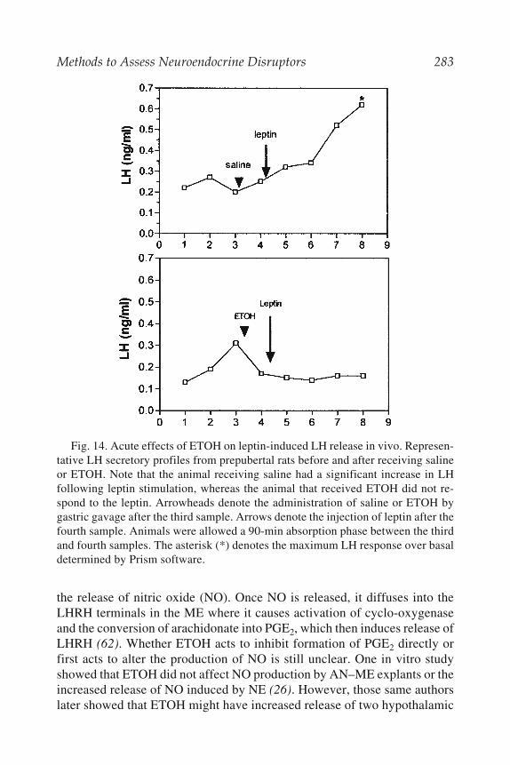
Methods to Assess Neuroendocrine Disruptors 283
the release of nitric oxide (NO). Once NO is released, it diffuses into theLHRH terminals in the ME where it causes activation of cyclo-oxygenaseand the conversion of arachidonate into PGE2, which then induces release ofLHRH (62). Whether ETOH acts to inhibit formation of PGE2 directly orfirst acts to alter the production of NO is still unclear. One in vitro studyshowed that ETOH did not affect NO production by AN–ME explants or theincreased release of NO induced by NE (26). However, those same authorslater showed that ETOH might have increased release of two hypothalamic
Fig. 14. Acute effects of ETOH on leptin-induced LH release in vivo. Represen-tative LH secretory profiles from prepubertal rats before and after receiving salineor ETOH. Note that the animal receiving saline had a significant increase in LHfollowing leptin stimulation, whereas the animal that received ETOH did not re-spond to the leptin. Arrowheads denote the administration of saline or ETOH bygastric gavage after the third sample. Arrows denote the injection of leptin after thefourth sample. Animals were allowed a 90-min absorption phase between the thirdand fourth samples. The asterisk (*) denotes the maximum LH response over basaldetermined by Prism software.

284 Dees et al.
inhibitory peptides that then caused a decrease in NO production (63).Regardless of the specific action on NO, it is clear that the overall effect ofETOH to decrease PGE2 formation is responsible for the drug’s ability todiminish LHRH release. Taken together, the above results demonstrate theusefulness and importance of in vitro methodology in discerning mecha-nisms of hormone action, as well as mechanisms by which toxic substances,such as ETOH, can alter hormone actions.
7. CONCLUSIONS
In this chapter, we have illustrated using the neuroendocrine system thatin vitro and in vivo methodologies can be used in a complimentary fashionto investigate specific scientific questions. We have provided severalexamples depicting the alternating use of these methods to address basicneuroendocrine responses, as well as more mechanistic questions. For ex-ample, not only have we described the use of this approach to initiate a newarea of study (i.e., central actions of IGF-1 at puberty) and to advance ourbasic understanding of the neuroendocrinology of puberty but also to assessneuroendocrine influences of both acute and chronic ETOH exposure. Wesuggest that experimental designs using these techniques in a complimen-tary fashion would also be beneficial to other studies assessing potentialneuroendocrine deficits that might be caused by toxins such as lead, 2,3,7,8-
Fig. 15. The effect of leptin on LH release from prepubertal anterior pituitariesof control and ETOH-treated rats incubated in vitro. The solid bar represents thebasal secretion of LH; the hatched bar represents the leptin-induced secretion ofLH from the anterior pituitaries of the respective control and ETOH-treated groups.Note that control and ETOH-treated pituitaries showed the same increase in LHrelease in response to leptin, indicating that pituitary responsiveness was unalteredby ETOH. **p<0.01; N=7 control, N=11 ETOH-treated rats.
Methods to Assess Neuroendocrine Disruptors 285
tetrachlorodibenzo-p-dioxins, polychlorinated biphenyls, and organophos-phate compounds.
REFERENCES1. Dees, W. L. and Skelley, C. W. (1990) Effects of ethanol during the onset of
female puberty. Neuroendocrinology 51, 64–692. Tannenbaum, G. S., Guyda, H. J., and Posner, B. I. (1983) Insulin-like growth
factors: a role in growth hormone negative feedback and body weight regula-tion via brain. Science 220, 77–79
3. Schoenle, E., Zapf, J., Hauri, C., Steiner, T., and Froesch, E. R. (1985) Com-parison of in vivo effects of insulin-like growth factor I and II and of growthhormone in hypophysectomized rats. Acta Endocrinol. 108, 167–174
4. Philipps, A. F., Persson, B., Hall, K., et al. (1988) The effects of biosyntheticinsulin-like growth factor 1. Supplementation of somatic growth, maturation,and erthropoiesis on neonatal rat. Pediatr. Res. 23, 298–305
5. Hizuka, N., Takano, K., Shizume, K., et al. (1986) Insulin-like growth factor Istimulates growth in normal growing rats. Eur. J. Pharmacol. 125, 143–146
6. Behringer, R. R., Lewin, T. L., Quaife, C. J., Palmiter, R. D., Brinster, R. L.,and D‘Ercole, A. J. (1990) Expression of insulin-like growth factor 1 stimu-lates normal somatic growth in growth hormone deficient transgenic mice.Endocrinology 127, 1033–1040
7. Bohannon, N. J., Figlewicz, D. P., Corp, E. S., Wilcox, J., Porte, C. D., Jr., andBaskin, D G. (1986) Identification of binding sites for an insulin-like growthfactor (IGF-1) in the median eminence of the rat brain by quantitative autorad-iography. Endocrinology 119, 943–945
8. Marks, J. L., Porte, D., Jr., and Baskin, D. G. (1991) Localization of type 1insulin-like growth factor receptor messenger RNA in the adult rat brain by insitu hybridization. Mol. Endocrinol. 5, 1158–1168
9. Negro-Vilar, A., Ojeda, S. R., and McCann, S. M. (1979) Catecholaminergicmodulation of luteinizing hormone-releasing hormone release by median emi-nence terminals in vitro. Endocrinology 128, 1749–1757.
10. Hiney, J. K., Ojeda, S. R., and Dees, W. L. (1991) Insulin-like growth factor-1:a possible metabolic signal involved in the regulation of female puberty. Neu-roendocrinology 54, 420–423
11. Duenas, M., Luquin, S., Chowen, J. A., Torres-Aleman, I., Naftolin, F., andGarcia-Segura, L. M. (1994) Gonadal hormone regulation of insulin-likegrowth factor-1-like immunoreactivity in hypothalamic astroglia of develop-ing and adult rats. Neuroendocrinology 59, 528–538.
12. Antunes-Rodrigues, J. and McCann, S. M. (1970) Water, sodium chloride, andfood intake induced by injections of cholinergic and adrenergic drugs into thethird ventricle of the rat brain. Proc. Soc. Exp. Biol. Med. 133, 1464–1469.
13. Harms, P. G. and Ojeda, S. R. (1974) Methods for cannulation of the rat jugu-lar vein. J. Appl. Physiol. 309, 261–263.

286 Dees et al.
14. Hiney, J. K., Srivastava, V., Nyberg, C. L., Ojeda, S. R., and Dees, W. L.(1996) Insulin-like growth factor-1 of peripheral origin acts centrally to accel-erate the initiation of female puberty. Endocrinology 137, 3717–3727.
15. Andrews, W. W. and Ojeda, S. R. (1981) A detailed analysis of the serum LHsecretory profile in conscious, free-moving female rats during the time ofpuberty. Endocrinology 109, 2032–2039.
16. Terasawa, E., Bridson, W. E., Nass, T. E., Noonan, J. J., and Dierschke, D. J.(1984) Developmental changes in the luteinizing hormone secretory pattern inperipubertal female rhesus monkeys: comparisons between gonadally intactand ovariectomized animals. Endocrinology 115, 2233–2240.
17. Urbanski, H. F. and Ojeda, S. R. (1985) The juvenile peripubertal transitionperiod in the female rat: Establishment of a diurnal pattern of pulsatile LHsecretion. Endocrinology 117, 644–649.
18. Danilovich, N., Wernsing, D., Coschigano, J. J., Kopchick, J. J., and Bartke,A. (1998) Deficits in female reproductive function in GH-R-KO mice; role ofIGF-1. Endocrinology 140, 2637–2640.
19. Wilson, M. E. (1998) Premature elevation in serum insulin-like growth factor-1 advances first ovulation in rhesus monkeys. J. Endocr. 158, 247–257.
20. Xiaowei, X., Ingram, R. L., and Sonntag, W. E. (1995) Ethanol suppressesgrowth hormone-mediated cellular responses in liver slices. Alcoholism: Clin.Exp. Res. 19, 1246–1251.
21. De, A., Boyadjieva, N. I., and Sarkar, D. K. (1999) Effects of ethanol on alph-adrenergic and beta-adrenergic agonist-stimulated beta endorphin release andcAMP production in hypothalamic cells in primary cultures. Alcoholism: Clin.Exper Res. 23, 46-51.
22. Frasor, J. M., Anderson, R. A., and Russell, L. D. (1992) Ethanol impairs secre-tory activity of Sertoli celss in conventional and bicameral culture. Biol.Reprod. 46(Suppl. 1), 171.
23. Hiney, J. K. and Dees, W. L. (1991) Ethanol inhibits luteinizing hormone-releasing hormone from the median eminence of prepubertal female rats invitro: Investigation of its actions on norepinephrine and prostaglandin-E2.Endocrinology 128, 1404–1408.
24. Nyberg, C. L., Hiney, J. K., Minks, J. E., and Dees, W. L. (1993) Ethanol altersN-methyl-DL-aspartic acid -induced LH secretion of luteinizing hormonereleasing hormone and the onset of puberty in the female rat. Neuroendocri-nology 57, 863–868.
25. Rettori,, V., Kamat, A., and McCann, S. M. (1994) Nitric oxide mediates thestimulation of luteinizing-hormone releasing hormone release induced byglutamic acid in vitro. Brain Res. Bull. 33, 501–503.
26. Canteros, G., Rettori, V., Franchi, A., et al. (1995) Ethanol inhibits luteinizinghormone-releasing hormone (LHRH) secretion by blocking the response ofLHRH neuronal terminals to nitric oxide. PNAS 92, 3416–3420.
27. Hiney, J. K., Srivastava, V., Lara, T., and Dees, W. L. (1998) Ethanol blocksthe central action of IGF-1 to induce luteinizing hormone secretion in the pre-pubertal female rat. Life Sci. 62, 301–308.

Methods to Assess Neuroendocrine Disruptors 287
28. Dees, W. L., Skelley, C. W., Hiney, J. K., and Johnston, C. A. (1990) Actionsof ethanol on hypothalamic and pituitary hormones in prepubertal female rats.Alcohol 7, 21–25
29. Soszynski, P. A. and Frohman, L. A. (1992) Inhibitory effects of ethanol on thegrowth hormone (GH)-releasing hormone-GH-Insulin-like growth factor-I axisin the rat. Endocrinology 131, 2603–2608.
30. Dees, W. L., Rettori, V., Kozlowski, G. P., and McCann, S. M. (1985) Ethanoland the pulsatile release of luteinizing hormone, follicle stimulating hormoneand prolactin in ovariectomized rats. Alcohol 2, 641–646.
31. Dees, W. L., Skelley, C. W., Rettori, V., Kentroti, M. S., and McCann, S. M.(1988) Influence of ethanol on growth hormone secretion in adult and prepu-bertal female rats. Neuroendocrinology 48, 495–499.
32. Srivastava, V., Hiney, J. K., Nyberg, C. L., and Dees, W. L. (1995) Effect ofethanol on the synthesis of insulin-like growth factor 1 (IGF-1) and the IGF-1receptor in late prepubertal female rats: a correlation with serum IGF-1. Alco-holism: Clin. Exp. Res. 19, 1467–1473.
33. Dees, W. L., Dissen, G. A., Hiney, J. K., Lara, F., and Ojeda, S. R. (2000)Alcohol ingestion inhibits the increased secretion of puberty-related hormonesin the developing female Rhesus monkey. Endocrinology 141, 1325–1331.
34. Urbanski, H. F. and Ojeda, S. R. (1987) Activation of luteinizing hormone-releasing hormone release advances the onset of female puberty. Neuroendo-crinology 46, 273–276.
35. Gay, V. L. and Plant, T. M. (1987) N-Methyl-D,L-aspartate elicits hypotha-lamic gonadotropin-releasing hormone release in prepubertal male rhesus mon-keys (Macaca mulatta). Endocrinology 120, 2289–2296.
36. Claypool, L. E. and Terasawa, E. (1989) N-Methyl-DL-aspartate (NMDA) in-duces LHRH release as measured by in vivo push-pull perfusion in the stalk-median eminence of pre and peripubertal female rhesus monkeys. Biol. Reprod.40(Suppl.), 83.
37. Price, M. T., Olney, J. W., and Cicero, T. J. (1978) Acute elevations of serumluteinizing hormone induced by kainic acid, N-methyl-aspartic acid orhomosysteic acid. Neuroendocrinology 26, 352–358.
38. Ondo, J. G., Wheeler, D. D., and Dom, R. M. (1988) Hypothalamic site ofaction for N-methyl-D-aspartate (NMDA) on LH secretion. Life Sci. 43,2283–2286.
39. Wilson, R. C. and Knobil, E. (1982) Acute effects of N-methyl-DL-aspartate onthe release of pituitary gonadotropins and prolactin in the adult female rhesusmonkey. Brain Res. 248, 177–179.
40. Tal, J., Price, M T., and Olney, J. W. (1983) Neuroactive amino acids influencegonadotropin output by a suprapituitary mechanism in either rodents or pri-mates. Brain Res. 273, 170–182.
41. Bourgiuignon, J. P., Gerard, A., and Franchimont, P. (1989) direct activationof gonadotropin-releasing hormone secretion through different receptors toneuroexcitatory amino acids. Neuroendocrinology 49, 402–408

288 Dees et al.
42. Donoso, A. O., Lopez, F. J., and Negro-Vilar, A. (1990) Glutamate receptorsof the non-N-methyl-D-aspartic acid type mediate the increase in luteinizinghormone-releasing hormone release by excitatory amino acids in vitro. Endo-crinology 126, 414–420.
43. Lopez, F. J., Donoso, A. O., and Negro-Vilar, A. (1990) Endogenous excita-tory amino acid neurotransmission regulates the estradiol-induced LH surge inovariectomized rats Endocrinology 126, 1771–1773.
44. Lovinger, D. M., White, G., and Weight, F. F. (1989) Ethanol inhibits NMDA-activated ion current in hippocampal neurons. Science 128, 1541–1547.
45. Leslie, S. W., Brwon, L. M., Dildy, J. E., and Sims, J. S. (1990) Ethanol andneuronal calcium channels. Alcohol 7, 233–236.
46. Simson, P. E., Criswell, H. E., Johnson, K. B., Hicks, R. E., and Breese, G. R.(1991) Ethanol inhibits NMDA-evoked electrophysiological activity in vivo.J. Pharmacol. Exp. Ther. 257, 225–231.
47. Nyberg, C. L., Srivastava, V., Hiney, J. K., Lara, F., and Dees, W. L. (1995) N-Methyl-aspartic acid receptor messenger ribonucleic acid levels and luteiniz-ing hormone release in immature female rats. Effects of stage of pubertaldevelopment and exposure to ethanol. Endocrinology 136, 2874–2880.
48. Swerdloff, R., Batt, R., and Bray, G. (1976) Reproductive hormonal functionin the genetically obese (ob/ob) mouse. Endocrinology 103, 542–547.
49. Swerdloff, R. S., Peterson, M., Vera, A., Batt, R. A. L., Heber, D., and Bray,G. (1978) The hypothalamic–pituitary axis in genetically obese (ob/ob) mice:Response to luteinizing hormone-releasing hormone. Endocrinology 103,542–547.
50. Johnson, L. M. and Sidnam, R. L. (1979) A reproductive endocrine profile inthe diabetes (db) mutant mouse. Biol. Reprod. 20, 552–559.
51. Batt, R., Everard, D., Gillies, G., Wilkinson, M., Wilson, C., and Yeo, T. (1982)Investigation into the hypogonadism of the obese mouse (genotype ob\ob). J.Reprod. Fertil. 64, 363–371.
52. Chehab, F. F., Lim, M. E., and Ronghua, L. (1996) Correction of the sterilitydefect in homozygous obese female mice. Nature Genet. 12, 318–320.
53. Barash, I. A., Cheung, C. C., Weigle, D. S., et al. (1996) Leptin is a metabolicsignal to the reproductive system. Endocrinology 137, 3144–3147.
54. Mounzith, K., Ronghua, L., and Chehab, F. F. (1997) Leptin treatment rescuesthe sterility of genetically obese ob/ob males. Endocrinology 138, 1190–1193.
55. Chehab, F. F., Mounzih, K., Ronghua, L., and Lim, M. E. (1997) Early onset ofreproductive function in normal female mice treated with leptin. Science 275,88–90.
56. Ahima, R. S., Dushay, J., Flier, S. N., Prabakaran, D., and Flier, J. S. (1997)Leptin accelerates the onset of puberty in normal female mice. J. Clin. Invest.99, 391–395.
57. Yu, W. H., Kimura, M., Walczewska, A., Karanth, S., and McCann, S. M.(1997) Role of leptin in hypothalamic-pituitary function. Proc. Natl. Acad. Sci.USA 94, 1023–1028.

Methods to Assess Neuroendocrine Disruptors 289
58. Carro, E., Pinilla, L., Seoane, L. M., et al. (1997) Influence of endogenousleptin tone on the estrous cycle and luteinizing hormone pulsatility in femalerats. Neuroendocrinology 66, 375–377.
59. Cheung, C. C., Thornton, J. E., Kuijper, J. L., Weigle, D. S., Clifton, D. K., andSteiner, R. A. (1997) Leptin is a metabolic gate for the onset of puberty in thefemale. Endocrinology 138, 855–858.
60. Dearth, R. K., Hiney, J. K., and Dees, W. L. (2000) Leptin acts centrally toinduce the prepubertal secretion of luteinizing hormone in the female rat. Pep-tides 21, 387–392.
61. Hiney, J. K., Dearth, R. K., Lara, F., Wood, S., Srivastava, V., and Dees, W. L.(1999) Effects of ethanol on leptin secretion and the leptin-induced luteinizinghormone (LH) release from the late juvenile female rats. Alcoholism: Clin.Exp. Res. 23, 1785–1792.
62. Rettori, V., Gimeno, M., Lyson, K., and McCann, S. M. (1992) Nitric oxidmediates norepinephrine-induced prostaglandin E2 release from the hypothala-mus. Proc. Natl. Acad. Sci. USA 89, 11,543–11,546.
63. Lomniczi, A., Mastronardi, C. A., Faletti, A. G., et al. (2000) Inhibitory path-ways and the inhibition of luteinizing hormone-releasing hormone release byalcohol. Proc. Natl. Acad. Sci. USA 97, 2337-2342.

In Vitro Models to Study Endogenous Neurotoxicants 291
291
12Establishing In Vitro Models
to Study Endogenous Neurotoxicants
Heather D. Durham
1. INTRODUCTIONAdvances in molecular genetics over the last decade have resulted in the
identification of genetic mutations responsible for several inherited neuro-logical diseases. Not only has cloning of these genes led to methods fordiagnosis of patients and identification of carriers but also to the establish-ment of animal and cell culture models to study mechanisms by which mu-tant proteins induce toxicity in vulnerable cell types. Early neuropathologicalstudies of autopsy tissue from patients with degenerative neurological dis-eases commonly revealed the presence of inclusion bodies in affected neu-ronal populations (see Table 1; 1–44). These include tangles and plaques inAlzheimer’s disease, Lewy bodies in Parkinson’s disease, and nuclear orcytoplasmic aggregates in the trinucleotide repeat diseases (spinal bulbarmuscular atrophy, Huntington’s disease, spinocerebellar ataxia 1 and 3,dentato-pallidoluysian atrophy) and cytoplasmic inclusions in familial andsporadic motor neuron diseases (45,46). That similar inclusions are observedin both sporadic and hereditary forms of neurological diseases suggestedthat similar pathways might be involved in pathogenesis whether proteinabnormalities result from inherited sequence differences, DNA damage, orposttranslational modifications. The presence in inclusions of ubiquitin, astress protein required for targeting abnormal proteins for degradation, sug-gested failure of proteolytic processing to rid cells of aberrant proteins. How-ever, the primary or secondary role of these inclusions in the pathogenesisof disease could not be surmised from studies of postmortem tissue at end-stage disease. Once genes responsible for familial forms were cloned, gene
From: Methods in Pharmacology and Toxicology: In Vitro Neurotoxicology: Principles and ChallengesEdited by: E. Tiffany-Castiglioni © Humana Press Inc., Totowa, NJ

292 Durham
292
Tab
le 1
Exa
mp
les
of P
rote
otox
ican
ts R
esu
ltin
g in
Gen
etic
Mu
tati
ons
Res
pon
sib
le f
or H
um
an N
euro
logi
cal D
isea
se
Mut
ant p
rote
inH
uman
dis
ease
Incl
usio
n bo
dies
Cel
ls m
ost a
ffec
ted
Ref
.
Am
yloi
d pr
ecur
sor
prot
ein
Alz
heim
er’s
Ext
race
llul
ar
-am
yloi
d in
Lim
bic
and
asso
ciat
ion
cort
ices
,1–
4pl
ague
s, n
euro
fibr
illa
ryhi
ppoc
ampu
sta
ngle
sT
auF
ront
otem
pora
l dem
enti
asP
aire
d he
lica
l fil
amen
ts in
Fro
ntot
empo
ral c
orti
cal n
euro
ns5–
7M
ulti
syst
em a
trop
hyne
urof
ibri
llar
y ta
ngle
sP
rese
nili
n 1
and
2A
lzhe
imer
’sA
myl
oid
plaq
ues
Lim
bic
and
asso
ciat
ion
cort
ices
,3,
4,8–
10hi
ppoc
ampu
s-S
ynuc
lein
Par
kins
on’s
Lew
y bo
dies
Sub
stan
tia
nigr
a6,
11L
ewy
body
dem
enti
aC
orti
cal p
yram
idal
neu
rons
Cu/
Zn-
supe
roxi
deC
hrom
osom
e 21
-lin
ked
Cyt
opla
smic
incl
usio
nsU
pper
and
mot
or m
otor
neu
rons
,12
–15
dism
utas
e (S
OD
-1)
am
yotr
ophi
c la
tera
l scl
eros
isas
troc
ytes
(A
LS
)H
igh-
mol
ecul
ar-w
eigh
tR
are
case
s of
fam
ilia
l AL
SH
yali
ne a
nd s
kein
like
Upp
er a
nd lo
wer
mot
or n
euro
ns16
neur
ofil
amen
t pr
otei
nin
clus
ions
, Bun
ina
bodi
es(N
F-H
)*H
unti
ngti
nH
unti
ngto
n’s
Nuc
lear
and
cyt
opla
smic
Str
iatu
m, c
ereb
ral c
orte
x17
–20
inc l
usio
ns*A
ndro
gen
rece
ptor
Ken
nedy
’s d
isea
seN
ucle
ar a
nd c
ytop
lasm
icL
ower
mot
or n
euro
ns, d
orsa
l18
–22
incl
usio
ns
root
gan
glia

In Vitro Models to Study Endogenous Neurotoxicants 293
293
*Ata
xin-
1S
pino
cere
bell
ar a
taxi
a (S
CA
1)E
osin
ophi
llic
sph
eroi
ds,
Cer
ebel
lar
Pur
kinj
e, d
enta
te18
–20,
23nu
clea
r in
clus
ion
body
nucl
eus,
bra
inst
em*A
taxi
n-2
SC
A2
Incr
ease
d m
utan
t pr
otei
n,C
ereb
ella
r P
urki
nje,
bra
in-
18–2
0,24
–26
but
no i
nclu
sion
sst
em, f
ront
o- t
empo
ral
lobe
s*A
taxi
n-3
SC
A3
/ Mac
hado
–Jos
eph
Nuc
lear
incl
usio
nsC
ereb
ella
r de
ntat
e ne
uron
s,18
–20,
27di
seas
eba
sal
gang
lia,
bra
inst
em,
spin
al c
ord
81A
-sub
unit
of
volt
age-
SC
A6
Cyt
opla
smic
incl
usio
nsC
ereb
ella
r P
urki
nje
and
18–2
0,28
,29
dep
ende
nt c
alcu
mgr
anul
e ne
uron
s, d
enta
te c
hann
elnu
cleu
s, i
nfer
ior
oliv
e*A
taxi
n-7
SC
A7
Nuc
lear
incl
usio
nC
ereb
ellu
m, b
rain
stem
,18
–20,
30m
acul
a, v
isua
l co
rtex
*Atr
ophi
n-1
Den
toru
brop
alli
dolu
ysia
nN
ucle
ar in
clus
ion
Cer
ebel
lum
, cer
ebra
l cor
tex,
18–2
0,31
,32
atro
phy
basa
l ga
ngli
a**
Pol
y(A
) bi
ndin
g pr
otei
n 2
Ocu
loph
aryn
geal
dys
trop
hyN
ucle
ar in
clus
ion
Ske
leta
l mus
cle
33N
euro
serp
inF
amil
ial d
emen
tia/
prog
ress
ive
Col
lins
bod
ies
Cor
tica
l neu
rons
, sub
cort
ical
34–3
6m
yocl
onus
epi
leps
ynu
clei
Pri
on p
rote
inC
reut
zfel
d-Ja
cob
(CJD
),P
rPS
c de
posi
tion
in p
laqu
esC
orte
x, b
asal
gan
glia
2,37
–39
Ger
stm
ann-
Stä
ussl
er–
Cer
ebel
lum
, cer
ebru
m,
Sch
eink
er d
isea
sebr
a ins
tem
Fat
al f
amil
ial i
nsom
nia
Kur
uM
ulti
ple
Cer
ebel
lum
, cer
ebru
m,
brai
nste
mP
MP
22, P
0C
harc
ot-M
arie
-Too
thA
ccum
ulat
ion
inS
chw
ann
cell
s40
–44
endo
plas
mic
ret
icul
um
Not
e: T
rinu
cleo
tide
rep
eat d
isea
ses
wit
h ex
pans
ion
of *
poly
glut
amin
e or
**p
olya
lani
ne tr
acts
.

294 Durham
transfer and gene knockout technologies could be used to examine the pro-gression of toxicity in cell culture and animal models. These studies havealso extended our knowledge about how cells handle proteins with inheritedor acquired structural abnormalities.
This chapter will focus on the challenges of establishing and interpretingdata from cell culture models to study proteotoxicity, an emerging field oftoxicology. The discussion commences with a review of the types ofproteotoxicants in the nervous system, then turns to issues of modeling ge-netic disorders in cell culture, including choice of cell lines or primary cul-tures, methodology for gene transfer, and technological developments thatare facilitating studies of toxicity in cultured neural tissue. Specific examplesof in vitro studies will be used to illustrate general concepts, but the refer-ence list will focus on recent review articles as a means of referring thereader to the literature relevant to specific proteins.
2. PROTEINACEOUS NEUROTOXICANTSToxic proteins can be expressed in cells as a result of genetic mutation or
generated through posttranslational modification. Recessively inherited orX-linked diseases are usually the result of loss of function resulting frominterference with synthesis, transport, stability, or enzymatic activity of theproduct. Although initial misfolding might be responsible for failure of pro-tein stability or proper targeting to its site of action, normal expression fromone allele is sufficient to render heterozygotes free of disease.
On the other hand, dominantly inherited diseases result when a toxic gainof function is conferred to the protein by the genetic mutation. Examples arepresented in Table 1 and include mutations in amyloid precursor protein andpresenilins in Alzheimer’s disease, mutations in Cu/Zn-superoxidedismutase (SOD-1) in familial amyotrophic lateral sclerosis, and expansionof a polyglutamine repeat domain in huntingtin in Huntington’s disease. Insuch cases, normal function may or may not be compromised in the mutantprotein, but toxicity is not abrogated by expression of the normal allele orexperimentally by gene knockout. However, a dominantly inherited loss offunction disease can result when protein derived form the mutant allele ex-erts a dominant negative effect on normal protein. This occurs in the periph-eral sensory-motor neuropathy, Charcot-Marie Tooth disease type 1A(CMT1A) and the more severe Déjérine–Scottas syndrome resulting frommutations in the gene encoding a peripheral myelin protein, PMP22. The so-called Trembler-J (TrJ) and Trembler (Tr) point mutations result in inter-ruption of trafficking through the endoplasmic reticulum, but transport ofthe wild-type protein is also prevented by heterodimer formation with mu-tant proteins (40). In the case of prion diseases (Creutzfeld–Jacob,

In Vitro Models to Study Endogenous Neurotoxicants 295
spongiform encephalopathies), prion protein transitions from a primarilyhelical structure to a -pleated sheet. The abnormal conformation can occurspontaneously as a result of genetic mutation or by conversion through asso-ciation with other abnormally transformed prion molecules, which may evenbe transmitted from other hosts in the case of bovine spongiform encephal-opathy. Finally, exposing cells to chemical toxicants can induce modifica-tions to normal proteins (e.g., carbonylation, peroxidation, nitrosylation,glycoxidation). The requirement to catabolize and replace these modifiedproteins could stress cells and contribute to toxicity.
What these dominantly inherited mutant proteins have in common is thepropensity to adopt altered conformations and to self-associate and aggre-gate. The terms “conformation diseases” or “protein folding diseases” havebeen coined to categorize these disorders. Theories on the toxic propertiesconferred by such mutations include enhancement of alternate enzymaticproperties, binding and sequestering wild-type protein or other key cellularproteins, and disrupting the function of proteasomes, the major effectors ofproteolysis of abnormal proteins as well as turnover of most normal cytoso-lic proteins. For information about mechanisms of toxicity for specific mu-tant proteins, the reader is referred to the references provided in Table 1.
3. STUDY OF PROTEOTOXICANTSIN CULTURE MODELS
The same considerations apply to the choice of culture model to studyproteotoxicants as chemical toxicants (discussed in other chapters of thisvolume) with the added considerations of the methods to be used for genetransfer and detection of gene expression. The major types of culture prepa-rations used in neurotoxicological investigations are listed in Table 2 alongwith the advantages and disadvantages for experimental investigation andthe techniques of gene transfer that can be applied to express mutant pro-teins. Because toxic proteins act in a dominant fashion regardless of expres-sion of normal alleles, ‘proteinopathies’ can be modeled simply by expressinga cDNA encoding the mutant protein in cells from normal organisms.
3.1. Vectors for Gene Transfer to Neural Cells3.1.1. Delivery of Plasmid DNA
In general, plasmid DNA can be delivered into cells chemically (by cat-ionic lipid or cationic polymer-mediated transfection), physically (microin-jection or biolistics/gene gun), or by electroporation. Primary cultures ofneural tissue more closely replicate the conditions in the intact animal rela-tive to dividing tumor cell lines; however, primary cells, particularly those

296 Durham
296
Tab
le 2
Cu
ltu
re M
odel
s U
sed
in N
euro
toxi
city
Stu
die
s
Met
hods
for
gen
eC
ultu
re ty
peA
dvan
tage
sD
isad
vant
ages
tr
ansf
er
Who
le-e
mbr
yoC
ytoa
rchi
tect
ure
pres
erve
dL
arge
num
bers
of
anim
als
requ
ired
Pla
smid
DN
Acu
ltur
esM
etab
olic
act
ivit
y pr
eser
ved
Lim
ited
mic
rosc
opic
vis
uali
zati
on•
Bio
list
ics
(gen
e gu
n)E
xcel
lent
to s
tudy
fet
al d
evel
opm
ent a
ndin
livi
ng s
tate
• E
lect
ropo
rati
onte
rato
geni
city
Lim
ited
dur
atio
n of
cul
ture
Vir
al v
ecto
rs•
Loc
al in
ject
ion
Org
anot
ypic
/R
easo
nabl
e pr
eser
vati
on o
f cy
toar
chit
ectu
reM
icro
scop
y of
indi
vidu
al c
ells
dif
ficu
ltP
lasm
id D
NA
expl
ant/
slic
e(n
eura
l ne
twor
ks)
in li
ving
cul
ture
s•
Bio
list
ics
(gen
e gu
n)cu
ltur
esE
xten
sion
of
nerv
e tr
acts
to a
ppro
pria
te ta
rget
sL
imit
ed m
ater
ial f
or b
ioch
emic
al a
naly
sis
• E
lect
ropo
rati
onG
ood
mye
lina
tion
Tim
e-co
nsum
ing
to m
aint
ain
Vir
al v
ecto
rsC
ultu
res
viab
le f
or m
onth
s•
Loc
al in
ject
ion
Ele
ctro
phys
iolo
gica
l rec
ordi
ngs
poss
ible
Mic
rosc
opic
vis
uali
zati
on o
f pe
riph
eral
exp
lant
Rea
ggre
gate
Lar
ge n
umbe
r pe
r an
imal
Cyt
oarc
hite
ctur
e no
t nor
mal
Pla
smid
DN
Acu
ltur
esG
ener
al c
ell–
cell
inte
ract
ions
dev
elop
Dif
ficu
lt to
vis
ulai
ze m
icro
sopi
call
y•
Bio
list
ics
Mye
lina
tion
in
the
livi
ng s
tate
• E
lect
ropo
rati
onL
onge
vity
Mat
eria
l for
bio
chem
ical
ana
lysi
sD
isso
ciat
edE
xcel
lent
vis
uali
zati
on in
livi
ng s
tate
Nor
mal
cyt
oarc
hite
ctur
e no
t pre
serv
edV
iral
vec
tors
mon
ola y
e rV
arie
ty o
f ce
ll ty
pes
and
phys
iolo
gica
l pro
cess
esM
yeli
nati
on p
oor
Pla
smid
DN
Acu
ltur
esM
ulti
ple
asse
ssm
ents
in th
e sa
me
cell
or
cult
ure
• M
icro
inje
ctio
n•
Bio
list
ics
• E
lect
ropo
rati
onL
arge
num
ber
per
anim
al•
Lim
ited
tran
sfec
tion
Mai
ntai
ned
in c
ultu
re f
or w
eeks
/mon
ths
wit
h ca
lciu
m p
hosp
hate
Sub
ject
to m
orph
olog
ical
, neu
roph
ysio
logi
cal
or
cati
onic
lipi
dsan
d bi
oche
mic
al a
naly
ses

In Vitro Models to Study Endogenous Neurotoxicants 297
297
Em
bryo
nic
stem
Bot
h pr
olif
erat
ive
and
diff
eren
tiat
edD
iffi
cult
to e
stab
lish
line
s in
itia
lly
Pla
smid
DN
Ace
ll/
phen
otyp
esR
equi
re s
peci
al c
ultu
re c
ondi
tion
s to
• C
atio
nic
lipi
d/D
NA
imm
orta
lize
dE
xpre
ss n
euro
nal p
rope
rtie
sdi
ffer
enti
ate
tran
sfec
tion
neur
onal
lin
esS
uita
ble
for
mor
phol
ogic
al, n
euro
phys
iolo
gica
l,M
any
cell
–cel
l int
erac
tion
s m
issi
ng•
Rec
epto
r li
gand
-lip
id/
and
bioc
hem
ical
ana
lyse
s D
NA
com
plex
es•
Bio
list
ics
• E
lect
ropo
rati
on•
Mic
roin
ject
ion
Vir
al v
ecto
rsO
ther
cel
l lin
esP
roli
fera
te to
larg
e nu
mbe
rsL
imit
ed r
epre
sent
atio
n of
cel
l typ
es a
ndP
lasm
id D
NA
(tum
or l
ines
,E
ase
of c
ultu
repr
oper
ties
in
situ
• C
atio
nic
lipi
d/D
NA
hybr
id l
ines
)tr
ansf
ecti
onM
ater
ial f
or b
ioch
emic
al a
naly
ses
Los
s of
nor
mal
cyt
oarc
hite
ctur
e •
Bio
list
ics
Man
y ca
n be
dif
fere
ntia
ted
to n
euro
nal
Cel
lula
r pr
oper
ties
may
not
be
stab
le o
ver
• E
lect
ropo
rati
onph
enot
ype
ti
me
• M
icro
inje
ctio
nV
iral
vec
tors
Neu
ral t
issu
eC
ontr
ol o
f ce
lls
expr
essi
ng p
rote
in o
f in
tere
stE
xpen
sive
and
labo
r in
tens
ive
to g
ener
ate
Gen
e of
inte
rest
isfr
om t
rans
geni
cC
an b
e an
alyz
ed e
x vi
vo o
r pl
aced
in c
ultu
retr
ansg
enic
mic
e an
d m
aint
ain
colo
nies
inte
grat
ed i
nto
geno
me
anim
als
Arc
hite
ctur
al in
tegr
ity,
via
bili
ty, e
ase
ofm
anip
ulat
ion
depe
ndin
g on
pre
para
tion

298 Durham
that are postmitotic, are difficult to transfect with plasmid DNA. This condi-tion results from the various barriers that must be crossed for DNA to pen-etrate the nuclear compartment and be transcribed (47,48). The basis oftransfection is the condensation of negatively charged plasmid DNA withcationic lipids or cationic polymers; the resulting positively charged com-plexes have affinity for anionic cell membranes, which facilitates the inter-action and uptake through endocytosis. This process can be assisted byincorporating a surface-receptor ligand (e.g., transferrin) into the complex[(e.g., transferrin–polylysine (49) or transferrin–polyethylenimines (47)]such that endocytosis becomes receptor mediated. Whether taken up by bulkor receptor-mediated endocytosis, the DNA must then escape endosomal/lysosomal compartments, which varies with the physico-chemical proper-ties of the complex. Coexposure to lysosomotropic agents, [e.g., chloroquine(50), peptides (51), or replication-deficient viruses (52)], has been used tofacilitate plasmid release, but toxicity can be an issue, particularly in neu-ronal cultures.
The greatest impediment to transfection of postmitotoic cells is thenuclear membrane. When cells are dividing, this barrier is removed and thecDNA can integrate into the host genome to produce stable transfectants.Even when plasmid DNA is microinjected into the cytoplasm of motor neu-rons in long-term culture, no expression is detected (Durham, unpublishedobservations). Other studies have shown that nuclear import of linear DNAis limited to less than 1.5 kb (48). Transport of linear DNA is facilitated bynon-covalent attachment to nuclear localization amino acid sequences, butthis has not improved transfection efficiency of plasmid DNA (48). For theabove reasons, transfection efficiencies have typically been low (a few per-cent) in primary cultured neurons, although this has been sufficient in manylaboratories to obtain results in specific neuronal populations, particularlyhippocampal neurons, cortical neurons, and dorsal root ganglion neurons. Arecent study reports transfection efficiencies of 20–25% in cortical neuronsand 25–30% in hippocampal neurons with LIPOFECT-AMINE2000™ (53).In our hands, this and other transfecting agents have failed to transfect mo-tor neurons in spinal cord cultures that have been maintained several weeksin vitro (see Fig. 1A). Use of commonly employed cationic lipids, calciumphosphate precipitation, and transferrin–polylysine–DNA complexes led toexpression of a CMVlacZ plasmid in some glial cells, but not in neuronalcells (see Fig. 1).
Alternative physical methods for bypassing the membranous barriers togene transfer are biolistics (gene gun) and intranuclear microinjection. Par-ticle-mediated gene transfer or biolistic transfection has been used for genetransfer into several different culture preparations of nervous tissue, includ-

In Vitro Models to Study Endogenous Neurotoxicants 299
Fig. 1. Comparison of three methods of transferring a reporter gene construct(lacZ) into spinal motor neurons in dissociated cultures of embryonic murine spinalcord. (A) Liposome-mediated transfection of plasmid DNA (CMVlacZ) bylipofectamine. Only background glial cells express -galactosidase 3 d followingtransfection. (B) Expression of -galactosidase following microinjection ofCMVlacZ plasmid DNA into nuclei of motor neurons. Concentration of plasmid isadjusted to result in expression in 80–100% of injected cells. (C) Replication-defi-cient adenoviral recombinant (AdCMVlacZ) transduces all cell types in the spinalcord culture with high efficiency. Both percentage of cells transduced and level ofexpression are proportional to viral titer, but can be limited by toxicity of the vectorand nonspecific toxicity of protein product, particularly in long-term experiments.(From ref. 54.) For additional information, see ref. 54.

300 Durham
ing dissociated, organotypic, and slice cultures (55–57). Plasmid DNA-coated gold or tungsten particles are “shot” into cultures at high velocity.Usually, compressed helium is used for particle acceleration. Transfectionefficiency is determined by the number of particles entering the cell, butlimited by the cellular damage inflicted by membrane disruption. Althoughbetter than conventional transfection, efficiency in neurons is still in theorder of less than 10% in most studies, but 34% was achieved in cerebellarorganotypic slice cultures under optimized conditions (58).
In our laboratory, we have had success using intranuclear microinjectionof plasmid expression vectors for gene transfer into primary cultured neu-rons (12,59,60) (see also Fig. 1B). Glass micropipets with inner filamentsfor quick filling, the same as used for electrophysiological recording, arepulled and a small amount of plasmid DNA in TRIS/EDTA is placed in thebottom of the shank with Eppendorf microloaders. Pressure injection is usedto force the solution out of the pipet tip and, when the microelectrode tip ismicromanipulated into the cell nucleus, into the nucleoplasm. Suppliers ofpressure microinjectors include Eppendorf, Narashige, World PrecisionInstruments, and Applied Scientific Instrumentation. The procedure requiresthat cultures be maintained outside of the incubator on the microscope stage.Because neural cultures are particularly sensitive to pH changes, they areplaced in minimum essential medium without bicarbonate, pH adjusted to7.2, for the period of manipulation and transferred at the end of the proce-dure to fresh culture medium containing 0.75% gentamycin, pre-equilibratedin the incubator to 37°C, pH 7.2–7.4. The disadvantages of microinjectionare that considerable training is required to develop expertise; a certain num-ber of cells will be killed by the procedure depending on the skill of thepersonnel performing the injections; it is time-consuming, and the numberof cells injected is too small for conventional biochemical analyses. On theother hand, the number of micromethods available to analyze ion concentra-tions, activity of signaling pathways, and gene expression in single cells israpidly growing. These include activity-dependent antibodies, optical probesfor microfluorometric assays, fluorescence resonance energy transfer(FRET), and single cell reverse transcription–polyacrylamide gel electro-phoresis (RT-PCR). In addition, with laser capture microdissection, specificcells can be removed from cultures for analysis of gene expression and cor-relation with morphological markers.
3.1.2. Gene Transfer Using Viral Vectors
The ability of viruses to penetrate membranous barriers in cells and utilizethe host to express viral genes has been exploited for transferring foreignDNA into cells. The most commonly used viruses include replication-

In Vitro Models to Study Endogenous Neurotoxicants 301
defective adenovirus (ADV), herpes simplex virus (HSV), and adeno-asso-ciated virus (AAV). For a comprehensive discussion of the properties, ad-vantages, and disadvantages of various viral vectors, the reader is referredto refs. 61–63. High efficiency of transduction of neuronal and glial cells isachievable by viral vector-mediated gene transfer (see Fig. 1C). However,the titer or multiplicity of infection (MOI) that can be used is limited bydirect toxicity of the vector’s lysosomotropic effect and over-expression ofgene product in cells transduced with high copy number (54). Preliminaryexperiments are required with each culture system, recombinant, and ex-perimental duration to optimize protocols.
The major inconveniences with viral vectors are the effort and timerequired for production and limitations on the size of cDNA that can beinserted. ADV was initially rendered replication defective by removal of theE1 region of the viral genome, allowing a maximal cDNA insert size of 7.5kb. This size can be extended somewhat by deletion of other “E” regions.Insert size can be increased to 36 kb by use of a gutted vector containing cis-acting DNA sequences necessary for viral replication and packaging but noviral coding sequences (64). However, replication of gutted vectors requiresa helper virus to provide the necessary viral proteins in trans and it can bedifficult to obtain high titres. For most purposes, in vitro E1- or E1/E3-de-leted ADV is adequate. To produce recombinants, the expression cassettecontaining the cDNA of interest is introduced by homologous recombinationand the virus is replicated in mammalian packaging lines, usually HEK293cells, that stably express the viral genes required for replication that havebeen deleted from the vector. The recombinant ADV must be plaque purifiedand verified not to have incorporated genes of replication by recombinationevents in the packaging line. A simplified method for generating recombi-nant ADV has been developed that eliminates the need for plaque purifica-tion. A recombinant adenoviral plasmid is generated by homologousrecombination in bacteria rather than eukaryotic cells (65). After transfec-tion of this plasmid into the mammalian packing line, viral production isfollowed with green fluorescent protein encoded by a gene incorporated intothe viral backbone. This system is available as the Stratagene AdEasy kit.
Two types of HSV1-derived vectors are recombinant nonlytic HSV1,which can accommodate up to 30 kb of cDNA, and plasmid-based amplicons(66). Although slightly more toxic than ADV, HSV1-expression vectorshave been utilized considerably in primary neuronal cultures. AAV is a non-pathogenic human parvovirus that can integrate into the genome of dividingcells and is relatively nontoxic (67). Although requiring helper virus forreplication, AAV recombinants can be purified free of helper virus. Themajor insert size is limited (about 5 kb).

302 Durham
3.1.3. RNA Delivery Methods
Messenger RNA can be transfected into nondividing cells with cationiclipids or peptide-modified low-molecular-weight polycations, overcomingthe nuclear membrane barrier to gene transfer (68). The major disadvan-tages over plasmid DNA transfection are the difficulty of mRNA produc-tion and susceptibility to degradation, both prior to and following transferinto cells. Intracellular stability is increased by incorporating a 5' cap and asubstantial 3' poly(A) tail (68). Only recently have studies been carried outto optimize delivery methods. An alternative is Simian Forest virus, a self-replicating and self-transcribing RNA molecule (69). However, cytotoxic-ity is a limiting factor. Few studies have utilized RNA-based transfection toexpress proteins, given the relative ease of DNA-based methodologies.
3.1.4. Culture of Neural Tissue from Transgenic Mice
Studies of proteotoxicity in vivo are conducted by producing micetransgenic for the mutant gene of interest, or for experimental controls, thewild-type gene. Gene knockout can confirm whether mutations confer a toxicgain of function to the protein or loss of function is the mechanism of toxic-ity. Any of the in vitro preparations listed in Table 2 can be produced fromtransgenic mice and can be valuable for mechanistic studies or screening ofpotential therapeutic agents if a mutant phenotype can be defined. For prepa-rations requiring culture of embryonic tissue, individual embryos must becultured separately and the embryonic tissue subsequently genotyped unlessthe transgenic mice have been bred to homozygosity. Examples include thedemonstration of increased vulnerability to glucose deprivation and chemi-cal hypoxia in cortical neurons cultured from presenilin 1 mutant mice (70),increased glutamate sensitivity of motor neurons cultured from G93A mu-tant SOD-1 mice (71), and analysis of Ca2+ handling in Purkinje cells ofcerebellar slices prepared from spinocerebellar ataxia type 1 (SCA1)transgenic mice overexpressing ataxin 1 with an expanded polyglutaminerepeat (72).
3.2. DosimetryFor cells undergoing mitosis, levels of short-term expression will be pro-
portional to the strength of the promotor element utilized to drive gene ex-pression, the number of copies of plasmid or viral vector accessing thenuclear compartment, and the turnover of the specific mRNA and protein.However, maintenance of expression for more than a few days depends onintegration of plasmid or viral DNA into the genome of the host cell or useof self-replicating plasmid (e.g., pCEP4, Invitrogen). Retroviral and AAVviral vectors can integrate, whereas ADV and HSV recombinants do not. In

In Vitro Models to Study Endogenous Neurotoxicants 303
postmitotic cells, integration is neither possible nor an issue because thecells do not divide to dilute out the copies of vector. We have found thatexpression persists for several weeks in cultured neurons following micro-injection of plasmid DNA or transduction with viral vectors.
In the case of plasmid transfection or transduction of viral vectors, proto-cols must be optimized for the amount of plasmid or titer of viral recombi-nant to add to the culture and for how long. In the case of microinjection, themajor determinant is concentration of plasmid DNA in the injectate becausethe volume of fluid injected is restricted and should be minimized. To moni-tor the efficiency of transfer of plasmid DNA by microinjection, we include70-kDa dextran (neutral or anionic) conjugated to fluorescein or rhodaminein the injectate at 20 mg/mL as a nontoxic marker of injected cells (conju-gates to other epifluorescent tags are also available; Molecular Probes). Thenumber of neurons containing the marker can be counted easily in livingcultures under epifluorescence microscopy and compared to the number ofcells expressing the protein of interest by immunocytochemistry. The lowestconcentration of plasmid DNA giving detectable protein expression in 80–100% of the surviving injected cells is determined in preliminary studies.Our fixation protocol is 8 min in 3% paraformaldehyde, followed by 1-minpermeabilization in 0.5% Nonidet P-40 and subsequent fixation for 2 min inparaformaldehyde. With this protocol some of the 70-kDa dextran markerusually persists in the nuclear compartment after fixation. Lysine-fixabledextrans are not recommended because they precipitate plasmid DNA intooligomeric complexes incompatible with the microinjection procedures.
For studies of proteotoxicity, the issues of dosimetry are similar to thoseimportant for studies of chemical toxicity. Toxicity will be proportional tothe dose (level of gene expression) and duration of the exposure. How doesthe scientist model experimentally low levels of expression of a mutant pro-tein over decades and are the same mechanisms responsible for toxicity withhigh-level expression over a short-term experiment as in the disease? Moststudies have utilized gene transfection in short-term studies (i.e., 24 h to afew days). Commercially available expression systems have incorporatedviral gene promoters such as cytomegalovirus (CMV) to achieve high-levelgene expression. However, most proteins will be toxic if highlyoverexpressed (just as any chemical is toxic if administered at a sufficientlyhigh concentration) and viral promoters might be too strong if the experi-ment is to be conducted in a time frame of weeks rather than a few days. Iflower levels of expression are required, this can be achieved by use ofeukaryotic promoters (e.g., neuron-specific enolase [NSE], neurofilament-L,phosphoglycerokinase [PGK], glial fibrillary acidic protein elongation factor1B [EF1B], or tubulin). On the other hand, the duration of experiments in

304 Durham
culture is limited by their viability and lower-level expression might not besufficient to achieve a phenotype, depending on the end points being mea-sured. Preliminary studies with the appropriate controls are necessary toidentify the appropriate conditions for each case. For example, in our labo-ratory, expression and toxicity of mutant SOD-1 proteins in motor neuronsof dissociated spinal cord cultures was examined over a 1- to 2-wk periodfollowing intranuclear microinjection of plasmid expression vectors (12,59).In this case, either pCEP4 or pcDNA3 plasmids, which contain the CMVpromoter could be utilized because expression of wild-type SOD-1 fromthese vectors was not toxic (see Fig. 2B). However, in recent experiments toestablish a similar model of Kennedy’s disease resulting from trinucleotiderepeat expansion in the androgen receptor gene, it was necessary to utilize aweaker promoter (PGK) to drive expression because the CMV-driven levelof androgen-receptor protein with a normal number of glutamine repeatswas toxic and resulted in inclusion formation (unpublished observations). Acaveat of overexpression is that nonspecific protein aggregation can occurthat is not related to the disease-causing mechanism.
In many toxic gain-of-function neurological diseases, clinical onset isdelayed into adulthood and certain neural cell populations are preferentiallyvulnerable to toxicity. This indicates that aging effects and exposure to otherstresses are important in the development of the disease phenotype and hasimplications for modeling these diseases in vitro. Manifestations of toxicitywill be different with acute expression of the mutant gene product than withlong-term stable expression in cell lines or neural tissue cultured fromtransgenic mice. In the former, toxicity eventually manifests in lethality (asillustrated in Fig. 2), whereas cells stably expressing the mutant gene musteither express lower levels of the toxic protein or have mustered protectivemechanisms in order to remain viable. For example, motor neurons in disso-ciated spinal cord cultures prepared from transgenic mice overexpressingthe G93A mutant human SOD-1 remain viable, but are more sensitive toadditional toxic stresses and have altered calcium homeostasis (59,71).Upregulation of heat shock proteins correlates with the ability of NIH 3T3cells to survive stable expression of mutant SOD-1 (60). To overcome thedifficulty in establishing stable cell lines that express toxic proteins, induc-ible expression systems have been used, including Tet-On (73) and ecdys-one-inducible (74) mammalian expression systems.
Thus, the context of mutant protein expression must be considered in designand interpretation of experiments. Paradigms of acute expression (transienttransfection, inducible expression systems, microinjection) prejudice the out-come to the toxic gain-of-function of the mutant protein, whereas long-term,

In Vitro Models to Study Endogenous Neurotoxicants 305
Fig. 2. Monitoring expression and toxicity of neurotoxic proteins following genetransfer by intranuclear microinjection. (A) Postfixation immunolabeling providesqualitative analysis of protein expression and detects changes in trafficking anddistribution. Wild-type human SOD-1 (left) and G93A mutant SOD-1 (right) detectedby immunolabeling with antibody specific for human SOD-1 (SD-G6; Sigma-Aldrich) 3 d following microinjection of SOD-1 pCEP4 expression vector (200 μg/mL).Mutant SOD-1 is detected in cytoplasmic inclusions in approx 30% of expressingmotor neurons, whereas wild-type protein is always distributed diffusely. (B) Moni-toring viability of motor neurons following microinjection of SOD-1 expression vec-tors. Seventy kilo-Daltons dextran–FITC (20 mg/mL) was coinjected with SOD-1expression vector. Motor neurons containing the marker were counted underepifluorescence microscopy and the number expressed as a percentage of thosepresent the day after microinjection. For further methodological details, see ref. 12.

306 Durham
stable expression provides a better opportunity to study interactions with otherphysiological and environmental stressors.
3.3. ControlsMandatory controls for transfer of mutant genes are (1) wild-type cDNA
in the same vector to control for nonspecific toxicity resulting fromoverexpression, (2) “empty” vector to control for toxicity of the vector orgene transfer protocol, and (3) cultures that have not been subjected to genetransfer to control for normal attrition. Once a gene transfer protocol hasbeen optimized, the latter might not be necessary because a general culturecondition will be manifested in the “empty” vector control. Many studiesalso incorporate transfer of an additional reporter gene such as lacZ or greenfluorescent protein (GFP); however, potential toxicity of the reporter proteinis a consideration, particularly in longer-term studies. When cotransferredwith the gene of interest, either as a separate vector, as a fusion tag, orbicistronic with the gene of interest in the same construct, expression of thesesequences monitors the efficiency of gene transfer. Transfer of the reporterconstruct alone provides an additional control for overexpression; however,given the difference in toxicity and turnover rates of proteins, we considerthe latter unnecessary when the wild-type counterpart to the cDNA of inter-est is being investigated as the control for mutants. Following gene transfer,the levels of protein expression should be monitored at various times overthe duration of the experiment. Monitoring expression of other housekeepinggenes controls for nonspecific suppression of endogenous gene expression.
3.4. Monitoring Gene Expression and Toxicity3.4.1. Detecting Transfected Cells
The most conventional method of monitoring gene expression ispostfixation by immunocytochemistry using antibody specific to thetransgenic protein. An example is illustrated in Fig. 2B. Plasmid expressionvector for the G93A mutant of human SOD-1, responsible for a familiarform of motor neuron disease (13), was microinjected into nuclei of motorneurons of dissociated cultures of murine spinal cord. Antibody specific tohuman SOD-1 (Sigma-Aldrich) was used to visualize expression of humanSOD-1. In most cases, species-specific antibodies are not available or thetransferred gene encodes the sequence from the same species as the culturedcells. In such cases, antibody labeling of the protein produced from the trans-ferred gene is superimposed upon the endogenous gene product.Coexpression of a reporter gene or coinjection of a fluorescent dextran aidsin identifying transfected cells. An alternative is to incorporate sequencesencoding epitope tags (e.g., HA, Flag, his) or GFP (or its derivatives) as a

In Vitro Models to Study Endogenous Neurotoxicants 307
fusion tag in the cDNA sequence inserted into the expression vector. Theadvantage of GFP is that the distribution of the protein can be visualized byepifluorescence microscopy in living cells, providing information on traf-ficking and changes in distribution over time in the same cell. In the exampleillustrated in Fig. 2, aggregation of mutant human SOD-1 protein into inclu-sions was detected by immunocytochemistry after fixation in some neuronsexpressing mutant protein, but never in those expressing wild-type humanSOD-1. To quantitate this manifestation of toxicity, the number of neuronscontaining aggregates was counted and expressed as a percentage of thetotal number of cells expressing the mutant protein (12,59). In those experi-ments, different cultures were evaluated at each time period followingmicroinjection of expression vectors. If a GFP fusion tag were to be incor-porated into the vector, the time-course of aggregate formation could befollowed over time in a single neuron. GFP and enhanced GFP (EGFP) tagshave been utilized to study formation of intracellular inclusions of disease-causing polyglutamine repeat expansions (e.g., constructs of EGFG-exon 1of huntingtin containing the trinucleotide repeat sequence) and their asso-ciation with heat shock proteins (73).
With the use of GFP fusion tags or comicroinjection of fluorescent dext-ran, precise viability studies can be conducted in each culture over time,because these markers leak out from dead cells. This is illustrated for themutant SOD-1 example in Fig. 2B; the number of cells containing the markerwere counted under epifluorescence microscopy, expressed as a percentageof those present on the day following microinjection (to exclude cells dyingof the procedure) and plotted against time. Viable morphology can be veri-fied under phase contrast microscopy. Viability curves are particularly use-ful to assess the effectiveness of potential neuroprotective therapies (59). Apossible disadvantage of fusion tags, which must be controlled for, is thatthey could alter conformation of the protein in ways that significantly affectturnover, activity, and toxicity. However, HA and Flag epitope tags and GFPfusion tags have been used successfully in numerous studies ofpolyglutamine-expanded proteins. The disadvantage of fluorescent dextranmarkers is that they are slowly phagocytosed over time and cleared from thecell, limiting the time they can be detected microscopically for screeningpurposes to less than 2 wk.
3.4.2. Monitoring Toxicity in Specific Neural Cell Typesin Mixed Neural Cultures
Important aspects of neurotoxicity include the differences in susceptibil-ity of specific neural cell populations to toxicity and the importance of cell–cell interactions in determining the physiological conditions responsible for

308 Durham
this preferential vulnerability. To reproduce the mechanisms of neurotoxic-ity operating in the intact organism, more and more attention is being paid toassessing the effects of toxicants on neuron–neuron and neuron–glial inter-actions. In organtotypic and even in dissociated cultures of nervous tissue,many of these cellular interactions can be preserved and evaluated; how-ever, this requires analysis at the single-cell level. There have been manyrecent advances in the ability to assess gene expression and biochemicalpathways in individual cells. These studies will be important in understand-ing why expression of mutant proteins is differentially toxic to cell types.
With laser capture microdissection, cells with a particular morphologycan be removed from cultures and tissues for quantitiation of mRNA ex-pression. Using DNA microarray technology, analysis of gene expressionfrom a few, or even single, cells is possible (75). Analysis of protein expres-sion in single cells is only possible in situ at this time, but considerableinformation on protein trafficking and signaling can be obtained with cur-rently available antibodies, particularly antibodies recognizing specificphosphorylated epitopes or cleavage products. Advances in microfluidics,two-dimensional gel electrophoresis detection systems, and antibodymicroarrays are reducing the sample size required for detection of proteinsusing proteomic approaches. Two-dimensional gel electrophoresis can beperformed on material captured by laser microdissection, but thousands ofcells are required (76).
Microfluorometric assays of membrane potential, pH, calcium ion con-centrations, and so forth are routinely as vital end points for toxicity. Instandard methods, cell-permeant esterified indicators are added to culturemedium, cross the plasma membrane, and are trapped within cells followingcleavage by intracellular esterases. Long-term and repeated measures aredifficult because the indicators distribute widely and often alter cell physi-ology (e.g., calcium indicators are powerful calcium chelators). Techniqueshave been developed to incorporate the indicators into nanosphere matrices,which greatly improves compatibility with long-term and repeated measures.Once nanospheres are introduced into the cell by transfection, ballistics, ormicroinjection, they can serve as bystanders to monitor parameters withoutsignificantly interfering with the biology of the cell (77).
Green fluorescent protein fusion tags can be used to monitor changes incellular compartmentalization that may coincide with activation [e.g., nucleartranslocation of transcription factors, retention of protein kinase C isoformsat cell membranes in response to activation by diacylglycerol (78)]. With thegeneration of GFP mutants with different excitation and emission spectra,more specific assays have been developed based on FRET, a technique thatpermits the study of protein–protein interactions and protein conformation
In Vitro Models to Study Endogenous Neurotoxicants 309
changes in vivo (79). FRET can occur between two fluorophores in nanom-eter proximity. Excitation of a donor fluorophore can transfer energy to andexcite an acceptor fluorophore with a longer wavelength of emission. Eventsincreasing the distance between the donor and acceptor fluorophores or thatbring fluorophores together can be monitored by measuring changes inFRET. Activity-based assays have been developed by incorporating bothdonor and acceptor fluorophores in the same construct separated by a linkercontaining, for example, protease cleavage, calcium/calmodulin binding, orphosphorylation domains of specific proteins in proteolytic or signaling cas-cades (intramolecular FRET) (79–81). When expressed in cells by gene trans-fer, such designer macromolecules can be used to assess dynamic effects ofneurotoxicants. Donor and acceptor fluorophores are incorporated into dif-ferent expression constructs to study protein–protein interactions. Mutantproteins have a propensity to adopt abnormal conformations and to aggre-gate and localize to inclusion bodies along with normal proteins such as heatshock proteins and ubiquitin. Intermolecular FRET analysis can be used totest how closely these proteins interact.
4. CONCLUDING REMARKSDuring the past decade there has been an explosion in identifying genetic
mutations that result in synthesis of toxic proteins in cells. Considerableprogress has been made in applying in vitro culture systems to model andstudy proteotoxicity. Although the same principles apply as with the studyof chemical toxicants, specific challenges include controlling the dosage andquantitating gene expression over time. New technologies are improvingour capability to monitor the effect of both chemical and protein toxicantson gene expression, morphology, and biochemical pathways in postmitotic,adherent cells in mixed cultures or tissue slices. These will extend the capa-bility to examine how expression of proteotoxicants affects complex inter-actions between neural cell types that are integral to neural function.
REFERENCES
1. Fidani, L. and Goate, A. (1992) Mutations in APP and their role in -amyloiddeposition. Prog. Clin. Biol. Res. 379, 195–214.
2. Carrell, R. W. and Gooptu, B. (1998) Conformational changes and disease—serpins, prions and Alzheimer’s. Curr. Opin. Struct. Biol. 8, 799–809.
3. Selkoe, D. J. (2001) Alzheimer’s disease: genes, proteins, and therapy. Physiol.Rev. 81, 741–766.
4. Tanzi, R. E. and Bertram, L. (2001) New frontiers in Alzheimer’s diseasegenetics. Neuron 32, 181–184.

310 Durham
5. Goedert, M., Crowther, R. A., and Spillantini, M. G. (1998) Tau mutationscause frontotemporal dementias. Neuron 21, 955–958.
6. Spillantini, M. G. and Goedert, M. (2000) The alpha-synucleinopathies:Parkinson’s disease, dementia with Lewy bodies, and multiple system atrophy.Ann. NY Acad. Sci. 920, 16–27.
7. Spillantini, M. G. and Goedert, M. (2001) Tau gene mutations and tau pathol-ogy in frontotemporal dementia and parkinsonism linked to chromosome 17.Adv. Exp. Med. Biol. 487, 21–37.
8. Sherrington, R., Rogaev, E. I., Liang, Y., et al. (1995) Cloning of a gene bear-ing missense mutations in early-onset familial Alzheimer’s disease. Nature375, 754–760.
9. Levy-Lahad, E., Wasco, W., Poorkaj, P., et. al. (1995) Candidate gene for thechromosome 1 familial Alzheimer’s disease locus. Science 269, 973–977.
10. Mattson, M. P., Chan, S. L., and Camandola, S. (2001) Presenilin mutationsand calcium signaling defects in the nervous and immune systems. BioEssays23, 733–744.
11. Polymeropoulos, M. H., Lavedan, C., Leroy, E., et al. (1997) Mutation in thealpha-synuclein gene identified in families with Parkinson’s disease. Science276, 2045–2047.
12. Durham, H. D., Roy, J., Dong, L., and Figlewicz, D. A. (1997) Aggregation ofmutant Cu/Zn superoxide dismutase proteins in a culture model of ALS. J.Neuropathol. Exp. Neurol. 56, 523–530.
13. Rosen, D. R., Siddique, T., Patterson, D., et al. (1993) Mutations in Cu/Znsuperoxide dismutase gene are associated with familial amyotrophic lateralsclerosis. Nature 362, 59–62.
14. Bruijn, L. I., Becher, M. W., Lee, M. K., et al. (1997) ALS-linked SOD1 mutantG85R mediates damage to astrocytes and promotes rapidly progressive diseasewith SOD1-containing inclusions. Neuron 18, 327–338.
15. Verbeke, P., Fonager, J., Clark, B. F., and Rattan, S. I. (2001) Heat shockresponse and ageing: mechanisms and applications. Cell Biol. Int. 25, 845–57.845–857.
16. Figlewicz, D. A., Krizus, A., Martinoli, M. G., et al. (1994) Variants of theheavy neurofilament subunit are associated with the development of amyo-trophic lateral sclerosis. Hum. Mol. Genet. 3, 1757–1761.
17. The Huntington’s Disease Collaborative Research Group (1993) A novel genecontaining a trinucleotide repeat that is expanded and unstable on Huntington’sdisease chromosomes. Cell 72, 971–983.
18. Zoghbi, H. Y. and Orr, H. T. (2000) Glutamine repeats and neurodegeneration.Annu. Rev. Neurosci. 23, 217–247.
19. Orr, H. T. (2001) Beyond the Qs in the polyglutamine diseases. Genes Dev. 15,925–932.
20. Fischbeck, K. H. (2001) Polyglutamine expansion neurodegenerative disease.Brain Res. Bull. 56, 161–163.
21. La Spada, A. R., Wilson, E. M., Lubahn, D. B., Harding, A. E., and Fischbeck,K. H. (1991) Androgen receptor gene mutations in X-linked spinal and bulbarmuscular atrophy. Nature 352, 77–79.
22. Merry, D. E. (2001) Molecular pathogenesis of spinal and bulbar muscularatrophy. Brain Res. Bull. 56, 203–207.

In Vitro Models to Study Endogenous Neurotoxicants 311
23. Orr, H. T., Chung, M. Y., Banfi, S., et al. (1993) Expansion of an unstabletrinucleotide CAG repeat in spinocerebellar ataxia type 1. Nature Genet. 4,221–226.
24. Imbert, G., Saudou, F., Yvert, G., et al. (1996) Cloning of the gene for spinoc-erebellar ataxia 2 reveals a locus with high sensitivity to expanded CAG/glutamine repeats. Nature Genet. 14, 285–291.
25. Pulst, S. M., Nechiporuk, A., Nechiporuk, T., et al. (1996) Moderate expansionof a normally biallelic trinucleotide repeat in spinocerebellar ataxia type 2.Nature Genet. 14, 269–276.
26. Sanpei, K., Takano, H., Igarashi, S., et al. (1996) Identification of the spinocer-ebellar ataxia type 2 gene using a direct identification of repeat expansion andcloning technique, DIRECT. Nature Genet. 14, 277–284.
27. Kawaguchi, Y., Okamoto, T., Taniwaki, M., et al. (1994) CAG expansions in anovel gene for Machado-Joseph disease at chromosome 14q32.1. Nature Genet.8, 221–228.
28. Zhuchenko, O., Bailey, J., Bonnen, P., et al. (1997) Autosomal dominant cer-ebellar ataxia (SCA6) associated with small polyglutamine expansions in thealpha 1A-voltage-dependent calcium channel. Nature Genet. 15, 62–69.
29. Frontali, M. (2001) Spinocerebellar ataxia type 6: channelopathy or glutaminerepeat disorder? Brain Res. Bull. 56, 227–231.
30. David, G., Abbas, N., Stevanin, G., et al. (1997) Cloning of the SCA7 genereveals a highly unstable CAG repeat expansion. Nature Genet. 17, 65–70.
31. Koide, R., Ikeuchi, T., Onodera, O., et al. (1994) Unstable expansion of CAGrepeat in hereditary dentatorubral-pallidoluysian atrophy (DRPLA). NatureGenet. 6, 9–13.
32. Nagafuchi, S., Yanagisawa, H., Sato, K., et al. (1994) Dentatorubral andpallidoluysian atrophy expansion of an unstable CAG trinucleotide on chro-mosome 12p. Nature Genet. 6, 14–18.
33. Brais, B., Bouchard, J. P., Xie, Y. G., et al. (1998) Short GCG expansions inthe PABP2 gene cause oculopharyngeal muscular dystrophy. Nature Genet.18, 164–167.
34. Davis, R. L., Holohan, P. D., Shrimpton, A. E., et al. (1999) Familial encepha-lopathy with neuroserpin inclusion bodies. Am. J. Pathol. 155, 1901–1913.
35. Davis, R. L., Shrimpton, A. E., Holohan, P. D., et al. (1999) Familial dementiacaused by polymerization of mutant neuroserpin. Nature 401, 376–379.
36. Takao, M., Benson, M. D., Murrell, J. R., et al. (2000) Neuroserpin mutationS52R causes neuroserpin accumulation in neurons and is associated with pro-gressive myoclonus epilepsy. J. Neuropathol. Exp. Neurol. 59, 1070–1086.
37. Knight, R. (2001) Creutzfeldt–Jakob disease: a protein disease. Proteomics 1,763–766.
38. Chiesa, R. and Harris, D. A. (2001) Prion diseases: what is the neurotoxicmolecule? Neurobiol. Dis. 8, 743–763.
39. Lovestone, S. and McLoughlin, D. M. (2002) Protein aggregates and demen-tia: is there a common toxicity? J. Neurol. Neurosurg. Psychiatry 72, 152–161.

312 Durham
40. Tobler, A. R., Notterpek, L., Naef, R., Taylor, V., Suter, U., and Shooter, E. M.(1999) Transport of Trembler-J mutant peripheral myelin protein 22 is blockedin the intermediate compartment and affects the transport of the wild-type pro-tein by direct interaction. J. Neurosci. 19, 2027–2036.
41. Roa, B. B., Garcia, C. A., Suter, U., et al. (1993) Charcot–Marie–Tooth diseasetype 1A. Association with a spontaneous point mutation in the PMP22 gene. N.Engl. J. Med. 329, 96–101.
42. Notterpek, L., Ryan, M. C., Tobler, A. R., and Shooter, E. M. (1999) PMP22accumulation in aggresomes: implications for CMT1A pathology. Neurobiol.Dis. 6, 450–460.
43. Notterpek, L., Ryan, M. C., Tobler, A. R., and Shooter, E. M. (1999) PMP22accumulation in aggresomes: implications for CMT1A pathology. Neurobiol.Dis. 6, 450–460.
44. Tobler, A. R., Liu, N., Mueller, L., and Shooter, E. M. (2002) Differentialaggregation of the Trembler and Trembler J mutants of peripheral myelin pro-tein 22. Proc. Natl. Acad. Sci. USA 99, 483–488.
45. Goedert, M., Spillantini, M. G., and Davies, S. W. (1998) Filamentous nervecell inclusions in neurodegenerative diseases. Curr. Opin. Neurobiol. 8,619–632.
46. Paulson, H. L. and Fischbeck, K. H. (1998) Trinucleotide repeats inneurogenetic disorders. Annu. Rev. Neurosci. 19, 79–107.
47. Kircheis, R., Wightman, L., and Wagner, E. (2001) Design and gene deliveryactivity of modified polyethylenimines. Adv. Drug Deliv. Rev. 53, 341–358.
48. Cartier, R. and Reszka, R. (2002) Utilization of synthetic peptides containingnuclear localization signals for nonviral gene transfer systems. Gene Ther. 9,157–167.
49. Wagner, E., Zenke, M., Cotten, M., Geug, H., and Birnstiel, M. L. (1990)Transferrin–polycation conjugates as carriers for DNA uptake into cells. Proc.Natl. Acad. Sci. USA 87, 3410–3414.
50. Ciftci, K. and Levy, R. J. (2001) Enhanced plasmid DNA transfection withlysosomotropic agents in cultured fibroblasts. Int. J. Pharm. 218, 81–92.
51. Plank, C., Oberhauser, B., Mechtler, K., Koch, C., and Wagner, E. (1994) Theinfluence of endosome-disruptive peptides on gene transfer using synthetic vi-rus-like gene transfer systems. J. Biol. Chem. 269, 12,918–12,924.
52. Curiel, D. T., Agarwal, S., Wagner, E., and Cotten, M. (1991) Adenovirus en-hancement of transferrin–polylysine-mediated gene delivery. Proc. Natl. Acad.Sci. USA 88, 8850–8854.
53. Ohki, E. C., Tilkins, M. L., Ciccarone, V. C., and Price, P. J. (2001) Improvingthe transfection efficiency of post-mitotic neurons. J. Neurosci. Methods 112,95–99.
54. Durham, H. D., Lochmüller, H., Jani, A., Acsadi, G., Massie, B., and Karpati,G. (1996) Toxicity of replication-defective adenoviral recombinants in disso-ciated cultures of nervous tissue. Exp. Neurol. 140, 14–20.

In Vitro Models to Study Endogenous Neurotoxicants 313
55. Biewenga, J. E., Destree, O. H., and Schrama, L. H. (1997) Plasmid-mediatedgene transfer in neurons using the biolistics technique. J. Neurosci. Methods71, 67–75.
56. Wellmann, H., Kaltschmidt, B., and Kaltschmidt, C. (1999) Optimized proto-col for biolistic transfection of brain slices and dissociated cultured neuronswith a hand-held gene gun. J. Neurosci. Methods 92, 55–64.
57. Usachev, Y. M., Khammanivong, A., Campbell, C., and Thayer, S. A. (2000)Particle-mediated gene transfer to rat neurons in primary culture. PflugersArch. 439, 730–738.
58. Murphy, R. C. and Messer, A. (2001) Gene transfer methods for CNSorganotypic cultures: a comparison of three nonviral methods. Mol. Ther. 3,113–121.
59. Roy, J., Minotti, S., Dong, L., Figlewicz, D. A., and Durham, H. D. (1998)Glutamate potentiates the toxicity of mutant Cu/Zn-superoxide dismutase inmotor neurons by postsynaptic calcium-dependent mechanisms. J. Neurosci.18, 9673–9684.
60. Bruening, W., Roy, J., Giasson, B., Figlewicz, D. A., Mushynski, W. E., andDurham, H. D. (1999) Upregulation of protein chaperones preserves viabilityof cells expressing toxic Cu/Zn-superoxide dismutase mutants associated withamyotrophic lateral sclerosis. J. Neurochem. 72, 693–699.
61. Karpati, G., Lochmüller, H., Nalbantoglu, J., and Durham, H. (1995) The prin-ciples of gene therapy for the nervous system. TINS 19, 49–54.
62. Slack, R. S. and Miller, F. D. (1996) Viral vectors for modulating gene expres-sion in neurons. Curr. Opin. Neurobiol. 6, 576–583.
63. Janson, C. G., McPhee, S. W., Leone, P., Freese, A., and During, M. J. (2001)Viral-based gene transfer to the mammalian CNS for functional genomic stud-ies. TINS 24, 706–712.
64. Hartigan-O’Connor, D., Barjot, C., Salvatori, G., and Chamberlain, J. S. (2002)Generation and growth of gutted adenoviral vectors. Methods Enzymol. 346,224–246.
65. He, T. C., Zhou, S., da Costa, L. T., Yu, J., Kinzler, K. W., and Vogelstein, B.(1998) A simplified system for generating recombinant adenoviruses. Proc.Natl. Acad. Sci. USA 95, 2509–2514.
66. Simonato, M., Manservigi, R., Marconi, P., and Glorioso, J. (2000) Gene trans-fer into neurones for the molecular analysis of behaviour: focus on herpes sim-plex vectors. TINS 23, 183–190.
67. Grimm, D., Kern, A., Rittner, K., and Kleinschmidt, J. A. (1998) Novel toolsfor production and purification of recombinant adenoassociated virus vectors.Hum. Gene Ther. 9, 2745–2760.
68. Bettinger, T., Carlisle, R. C., Read, M. L., Ogris, M., and Seymour, L. W.(2001) Peptide-mediated RNA delivery: a novel approach for enhanced trans-fection of primary and post-mitotic cells. Nucleic Acids Res. 29, 3882–3891.
69. Simons, M., De Strooper, B., Multhaup, G., Tienari, P. J., Dotti, C. G., andBeyreuther, K. (1996) Amyloidogenic processing of the human amyloid pre-

314 Durham
cursor protein in primary cultures of rat hippocampal neurons. J. Neurosci. 16,899–908.
70. Mattson, M. P., Zhu, H., Yu, J., and Kindy, M. S. (2000) Presenilin-1 mutationincreases neuronal vulnerability to focal ischemia in vivo and to hypoxia andglucose deprivation in cell culture: involvement of perturbed calcium homeo-stasis. J. Neurosci. 20, 1358–1364.
71. Kruman, I. I., Pedersen, W. A., Springer, J. E., and Mattson, M. P. (1999)ALS-linked Cu/Zn-SOD mutation increases vulnerability of motor neurons toexcitotoxicity by a mechanism involving increased oxidative stress and per-turbed calcium homeostasis. Exp. Neurol. 160, 28–39.
72. Inoue, T., Lin, X., Kohlmeier, K. A., Orr, H. T., Zoghbi, H. Y., and Ross, W.N. (2001) Calcium dynamics and electrophysiological properties of cerebellarPurkinje cells in SCA1 transgenic mice. J. Neurophysiol. 85, 1750–1760.
73. Wyttenbach, A., Carmichael, J., Swartz, J., et al. (2000) Effects of heat shock,heat shock protein 40 (HDJ-2), and proteasome inhibition on protein aggrega-tion in cellular models of Huntington’s disease. Proc. Natl. Acad. Sci. USA 97,2898–2903.
74. Wang, G. H., Mitsui, K., Kotliarova, S., et al. (1999) Caspase activation duringapoptotic cell death induced by expanded polyglutamine in N2a cells.Neuroreport 10, 2435–2438.
75. Mills, J. C., Roth, K. A., Cagan, R. L., and Gordon, J. I. (2001) DNAmicroarrays and beyond: completing the journey from tissue to cell. NatureCell Biol. 3, E175–E178.
76. Craven, R. A. and Banks, R. E. (2001) Laser capture microdissection andproteomics: possibilities and limitation. Proteomics 1, 1200–1204.
77. Clark, H. A., Barker, S. L. R., Basuel, M., et al. (1998) Subcellularoptochemical nanobiosensors: probes encapsulated by biologically localisedembedding (PEBBLEs). Sensors Actuators B 51, 12–16.
78. Ng, T., Squire, A., Hansra, G., et al. (1999) Imaging protein kinase Calphaactivation in cells. Science 283, 2085–2089.
79. Truong, K. and Ikura, M. (2001) The use of FRET imaging microscopy todetect protein–protein interactions and protein conformational changes in vivo.Curr. Opin. Struct. Biol. 11, 573–578.
80. Mere, L., Bennett, T., Coassin, P., et al. (1999) Miniaturized FRET assays andmicrofluidics: key components for ultra-high-throughput screening. DrugDiscov. Today 4, 363–369.
81. Zacharias, D. A., Baird, G. S., and Tsien, R. Y. (2000) Recent advances intechnology for measuring and manipulating cell signals. Curr. Opin. Neurobiol.10, 416–421.

Annotated Reading List 315
315
Appendix
Annotated Reading List
Evelyn Tiffany-Castiglioni, Lucio G. Costa,Marion Ehrich, William R. Mundy, Gerald J. Audesirk,
Michael Aschner, Prasada R. S. Kodavanti,and Stephen M. Lasley
This appendix contains a critically reviewed list of works related to invitro neurotoxicology. The list has been carefully selected and annotated bythe contributors to include reference books on neurotoxicology, books andessays on in vitro neurotoxicology, books and chapters on related methods,and important review articles that have appeared in the past 10 yr.
BOOKS ON NEUROTOXICOLOGYAschner, M. and Kimelberg, H. K. (eds.) (1996) The Role of Glia in Neurotox-icity, CRC, New York
This is the first book to focus exclusively on the roles of neuroglia in neu-rotoxicity. Contributors review and explore potential sites for neurotoxicaction in glial–neuronal interactions in both the central and peripheral ner-vous systems. Individual chapters address methodologies and concepts ofneurotoxicology, including many examples of in vitro approaches. Thispublication was intended to fill a gap in the literature. With the increasingimportance of glia, a journal, Glia, has been dedicated to studies on thesecells. In addition, many recent textbooks have included discussion of glia,encompassing neurophysiology, neuroanatomy, neuroscience, neurochem-istry, and neuropharmacology. None, however, included the role of glia inneurotoxicity, a timely topic and a subject in its own right.
Chang, L.W. (ed.) (1994) Principles of Neurotoxicology, Marcel Dekker, NewYork
This standard reference provides a comprehensive overview of principlesand modern concepts of neurotoxicology. The work contains major sec-tions on the central nervous system, behavioral neurotoxicology, biochemi-cal and molecular neurotoxicology, and developmental neurotoxicology.
From: Methods in Pharmacology and Toxicology: In Vitro Neurotoxicology: Principles and ChallengesEdited by: E. Tiffany-Castiglioni © Humana Press Inc., Totowa, NJ

316 Tiffany-Castiglioni et al.
Costa, L. and Manzo, L. (eds.) (1998) Occupational Neurotoxicology, CRC,New York
This book provides a concise overview of important concerns of the rela-tively new specialty of occupational neurotoxicology. Among them arecommonly encountered workplace neurotoxicants, signs and symptoms ofneurotoxicity, detection and monitoring of human exposure by biomarkers,epidemiology, and diagnostic methods.
Harry, G. J. (ed.) (1994) Developmental Neurotoxicology, CRC, New YorkThis book examines the biological characteristics of the developing ner-vous system that increase its vulnerability to damage by exposure to envi-ronmental toxicants. Contributing authors discuss functional alterations thatoccur at exposure levels too low to produce structural teratogenesis.
Massaro, E. J. (ed.) (2002) Handbook of Neurotoxicology, Volumes I and II,Humana, Totowa, NJ
This two-volume set provides a current overview of important topics incontemporary neurotoxicology. In Volume I, 28 topics are covered under 4sections: pesticides, metals, microbial toxins, and animal toxins (venoms).In Volume II, 21 topics are covered under 4 sections: developmentalneurotoxicology, drugs of abuse, imaging, and neurobehavioral assessmentmethods. Section editors and chapter contributors are international expertsfrom academia, industry, and government agencies. Volume I, in particu-lar, offers excellent further reading on topics relevant to in vitroneurotoxicology, such as pesticide effects on ion channels, mechanisms oflead neuronal toxicity, interactions of metals with the zinc-finger motif,and the blood–brain barrier in metal toxicity.
Slikker, W. B. and Chang, L. W. (eds.) (1998) Handbook of DevelopmentalNeurotoxicology, Academic Press, New York
This highly comprehensive multidisciplinary reference addresses themechanisms and relevance of the developmental toxicity of chemicals. Thesubject is divided into seven major sections on cellular and molecular mor-phogenesis, developmental biology and toxicology, synaptogenesis andneurotransmission, nutrient and chemical disposition, behavioral assess-ment, clinical assessment and epidemiology, specific neurotoxic syn-dromes, and risk assessment.
Tilson, H. A. and Harry, G. J. (1999) Neurotoxicology, 2nd ed., Target OrganToxicology Series, Taylor & Francis, Philadelphia
The major focuses of this edition are the discovery of sites and mechanismsof neurotoxicity and their value in improving risk assessment. The text fo-cuses primarily on the neurobiological basis underlying neurotoxic sitesand modes of action. Contributing authors provide 15 chapters on topicsspanning molecular biological approaches in neurotoxicology, in vitroneurotoxicology, specific cellular and biochemical processes damaged bytoxicants, effects on learning and behavior, and emerging concepts in riskassessment.

Annotated Reading List 317
BOOKS AND POSITION PAPERS ON IN VITRONEUROTOXICOLOGY
Aschner, M., Allen, J. W., Kimelberg, H. K., LoPachin, R. M., and Streit, W.J. (1999) Glial cells in neurotoxicity development. Annu. Rev. Pharmacol.Toxicol. 39, 151–173
Experts on each of the major classes of neuroglia (astrocytes, oligodendro-cytes, microglia, and Schwann cells) present models for neurotoxic sites ofaction that involve glia. Glial interactions with neurons and other glia thatunderlie nervous system development and function are examined. The workdescribed is based on in vitro and in vivo models.
Aschner, M. and Kerper, L. E. (2000) Transport of metals in the nervous sys-tem, in Molecular Biology and Toxicology of Metals (Koropatnick, D.J. andZalups, R. K., eds.), Taylor & Francis, London, pp. 276–299
The blood–brain barrier (BBB) is a specialized structure responsible forthe maintenance of the neuronal microenvironment. A pivotal function ofthe endothelial cells comprising the blood–brain barrier is to regulate theselective transport and metabolism of substances from blood to brain, aswell as their transport in the opposite direction. This chapter addresses thedevelopment of the blood–brain barrier, with emphasis on the crosstalkbetween astrocytes and endothelial cells, as well as known mechanisms ofmetal transport by endothelial cells.
Audesirk, G. J. (1997) In vitro systems in neurotoxicological studies, in Ner-vous System and Behavioral Toxicology, Comprehensive Toxicology, Volume11 (Lowndes, H. E. and Reuhl, K. R., eds.), Elsevier Science, Amsterdam, pp.431–446
This chapter focuses on complementarity between in vitro and in vivoapproaches. A concise overview of in vitro systems is provided. Of specialinterest are discussions of acute versus semichronic neurotoxicity and theproblem of concentration and duration of exposure in vitro.
Costa, L. G. (1998) Neurotoxicity testing: a discussion of in vitro alternatives.Environ. Health Perspect. 106(Suppl.), 505–510.
In addition to briefly discussing the advantages and disadvantages of invitro systems, the author thoughtfully discusses in vitro systems for mecha-nistic studies and neurotoxicity screening. Tiered approaches are suggested,because no single invitro system can reliably detect all possible end points.
Costa, L. G. (1998) Biochemical and molecular neurotoxicology: relevance tobiomarker development, neurotoxicity testing and risk assessment, Toxicol.Lett. 102–103, 417–421
Biomarkers are generally divided into three categories: biomarkers ofexposure, effect, and susceptibility. This commentary addresses biomarkersof effect and their cross-disciplinary use in animal toxicity studies, epide-miology, and in vitro toxicity testing. The example of organophosphorusinsecticide neurotoxicity is explored.

318 Tiffany-Castiglioni et al.
Deng, W. and Poretz, R. D. (2003) Oligodendroglia in developmental neuro-toxicity. Neurotoxicology 24, 161–178
This is the first contemporary review to address the roles of oligodendro-glia in developmental neurotoxicity. Topics covered are the developmentallineage of oligodendrocytes, maturational characteristics in vivo and invitro, and modulation of differentiation in cell culture models. The well-defined oligodendrocyte lineage is presented as an advantageous systemfor investigations of developmental neurotoxicity. Recent work from theauthors’ laboratory on lead neurotoxicity is reviewed.
Ehrich, M. and Veronesi, B. (1998) In vitro neurotoxicology, in Neurotoxi-cology (Tilson, H. A. and Harry, G. J., eds.), Taylor & Francis, Philadelphia,pp. 37–50
This chapter provides a general review of methods and examples of theiruse for neurotoxicology. The biological basis underlying neurotoxic sitesand modes of action is addressed.
Harry, G. J., Billingsley, M., Bruinink, A., Campbell, I. L., Classen, W.,Dorman, D. C., Galli, C., Ray, D., Smith, R. A., and Tilson, H. A. (1998) Invitro techniques for the assessment of neurotoxicity. Environ. Health Perspect.106(Suppl.), 131–158
This work is an extensive review and discussion of the topic prepared as adocument for the International Program on Chemical Safety (IPCS) andcosponsored by the United Nations Environment Program, World HealthOrganization, and International Labor Organization. The focus of this re-view is the usefulness of in vitro techniques for the identification of neuro-toxic hazards. End points receive particular attention because of their usein distinguishing between a pharmacologic and neurotoxic response. Thiswork is also valuable as an introductory resource for the reader new toculture techniques, as several common techniques, cell lines, and problemsencountered in culture are discussed.
Harry, G. J. and Tilson, H. A. (ed.) (1999) Neurodegeneration Methods andProtocols, Humana, Totowa, NJ
The objective of this book is to develop an understanding of and technicalability in various cellular and molecular techniques for studying many as-pects of nervous system cell biology. The protocols in this book span amultidisciplinary range of cellular and molecular approaches and shouldallow investigators to address research questions directed toward under-standing nervous system function, injury, degeneration, and the repair/re-generative process.
Pentreath, V. W. (ed.) (1999) Neurotoxicology In Vitro, Taylor & Francis,Philadelphia
This excellent work is suggested as a companion volume to the currentbook. The book contains concise reviews of principles of neurobiology,commonly used cell lines, and selected in vitro techniques. The uses of invitro methods for mechanisms versus screening studies are also thought-fully addressed.

Annotated Reading List 319
Philbert, M. A. and Aschner, M. (1997) Glial cells, in Nervous System andBehavioral Toxicology, Comprehensive Toxicology, Volume 11 (Lowndes, H.E. and Reuhl, K. R., eds.), Elsevier Science, Amsterdam, pp. 217–236
The dynamic role of glia in the maintenance of normal neural tissues andtheir potential involvement in degenerative disease processes and follow-ing exposure to xenobiotics are discussed. This review provides a generaloverview of glia as targets and mediators of neurotoxicity in the nervoussystem.
Tiffany-Castiglioni, E. and Qian, Y. (2001) Astroglia as metal depots: mo-lecular mechanisms for metal accumulation, storage and release.Neurotoxicology 22, 577–592
This review extends the lead-sink hypothesis for astroglia to other metals.In vivo and in vitro evidence that mercury, manganese, and copper mightbe selectively accumulated by astroglia is examined.
Tiffany-Castiglioni, E., Ehrich, M., Dees, W. L, Costa, L.G., Kodavanti, P. R.S., Lasley, S. M., Oortgiesen, M., and Durham, H. D. (1999) Bridging the gapbetween in vitro and in vivo models for neurotoxicology. Toxicol. Sci. 51,178–183
The authors wrote this commentary as a result of their participation as pan-elists in a poster-discussion session on the complementarity and usefulnessof in vitro and in vivo approaches to neurotoxicity testing. The session washeld in the 1998 meeting of the Society for Toxicology. This paper servedas a catalyst for the present volume.
Trotti, D., Danbolt, N. C., and Volterra, A. (1998). Glutamate transporters areoxidant-vulnerable: a molecular link between oxidative and excitotoxicneurodegeneration? Trends Pharmacol. Sci. 19, 328–334
The authors discuss the idea that glutamate transporters in the brain can beoxidized by biological agents, leading to decreased glutamate uptake andextracellular accumulation of neurotoxic glutamate. This phenomenon isof interest to neurotoxicologists in that a similar process can occur whencells are exposed to chemical oxidants. The possible involvement of oxida-tive alterations of specific glutamate transporters in pathologies (amyo-trophic lateral sclerosis, Alzheimer’s disease, brain trauma, and ischaemia)is reviewed.
METHODS AND MODEL SYSTEMS APPLICABLETO IN VITRO NEUROTOXICOLOGY
Aschner, M., Kimelberg, H. K., and Vitarella, D. (1995) Selective techniquesdesigned to evaluate neurotoxicity, in Neurotoxicology: Approaches and Meth-odologies (Chang, L. W., ed.), Academic, New York, pp. 439–444
This chapter presents a new version of an available technique for dynamicmeasurement of changes in cell volume of substratum-attached monolayercell cultures. When combined with release measurements of endogenouscell markers, it affords a powerful tool for rapid measurements of cytotox-

320 Tiffany-Castiglioni et al.
icity and, potentially, a high throughput screening method for variousneurotoxicants.
Banker, G. and Goslin, K. (eds.) (1998) Culturing Nerve Cells, 2nd ed., MITPress, Cambridge, MA
This manual, now in its second edition, offers an outstanding resource forculture of vertebrate neural tissue and cells. It contains several eloquentlywritten chapters on underlying principles, as well as detailed recipes andprotocols for culturing specific cell types and mixed cultures. Contributorsprovide first-hand tutorials on the techniques developed in their laborato-ries, including advantages, limitations, and troubleshooting.
Boulton, A. A., Baker, G. B., and Bateson, A. N. (eds.) (1999) In Vitro Neuro-chemical Techniques, Humana, Totowa, NJ
This collection of contemporary techniques for neurochemical and molecu-lar neurobiology research includes assays that are useful for the measure-ment of cell injury and cell death.
Buznikov, G. A., Nikitina, L. A., Bezuglov, V. V., Lauder, J. M., Padilla, S.,and Slotkin, T. A. (2001) An invertebrate model of the developmental neuro-toxicity of insecticides: effects of chlorpyrifos and dieldrin in sea urchin em-bryos and larvae. Environ. Health Perspect. 109, 651–661.
This article describes an interesting new approach to in vitro developmen-tal neurotoxicity testing that utilizes an invertebrate model.
Ehrich, M. (1998) Human cells as in vitro alternatives for toxicological re-search and testing: neurotoxicity studies. Comments Toxicol. 6, 189–197
Sources of cells as well as advantages and disadvantages of the use of cellsof human origin are discussed.
Freshney, R. I. (2000) Culture of Animal Cells: A Manual of Basic Technique,4th ed., Wiley–Liss, New York
This book well deserves its common aphorism as the bible of tissue cultureusers. It is thorough, clearly written, well and generously illustrated, andfrequently updated. Included are theoretical and practical considerations ofcell, tissue, and organ culture, as well as detailed protocols of commonprocedures, such as sterile technique, preparation of medium, cytotoxicityassays, and cryopreservation. Detailed protocols are provided for two typesof nervous system culture (cerebellar granule neurons and olfactory bulbensheathing cells), but more specialized works would need to be consultedfor other culture protocols.
Gad, S. C. (2000) Neurotoxicology in vitro, in In Vitro Toxicology, 2nd ed.,(Gad, C. G., ed.), Taylor & Francis, New York, pp. 188–221
This article surveys the tools of in vitro neurotoxicology, including typesof cell culture preparation used, specific examples of their use, methods oftissue culture, morphological and functional toxicity assays, and the designof neurotoxicant screening systems. Specific neurotoxicologic studies arebriefly discussed as examples to illustrate the problems and potential ad-vantages of in vitro approaches. the subjects selected are anticonvulsants,heavy metals, and excitoxins, mostly from articles published in the 1980s.

Annotated Reading List 321
Gilbert, M. E. (2000) In vitro systems as simulations of in vivo conditions: thestudy of cognition and synaptic plasticity in neurotoxicology. Ann. NY Acad.Sci. 919, 119–13.
The effects of regional brain stimulation and ablation on behavior have ledto inferences on the impact of these manipulations on psychological con-structs of “learning” and “memory.” This review describes how an electro-physiological property, long-term potentiation (LTP), greatly expanded theability to probe cellular aspects of the representation of memories in thebrain. The study of plasticity in this manner is an excellent example of howin vivo phenomena translate to more simplified in vitro test systems todirectly address cellular and biochemical mechanisms of information stor-age in the brain.
Maines, M., Costa, L. G., and Reed, D. J. (eds.) (2002) Current Protocols inToxicology Wiley, New York
This two-volume collection offers detailed laboratory procedures for theassessment of toxicity at multiple levels of biological complexity rangingfrom whole organisms to biochemical pathways. It is available in updatablelooseleaf, CD-ROM, and Web-based formats. Chapter 12, “Biochemicaland Molecular Neurotoxicology,” contains units written by authorities intheir fields on several in vitro topics, including the development of an invitro blood–brain barrier, culture of rat hippocampal neurons and rat corti-cal astrocytes, and analytical techniques for cytology and imaging.
O’Hare, S. and Atterwill, C. K. (eds.) (1995) In Vitro Toxicity Testing Proto-cols, Humana, Totowa, NJ
This collection of detailed protocols includes the preparation and use ofcultured astrocytes for assays of gliotoxicity, as well as several chapters ongeneral and topical toxicity.
Tyson, C., Witschi, H., and Frazier, J., (1994) In Vitro Toxicity Indicators,Methods in Toxicology Vol. 1B, Academic, New York
This book contains detailed testing procedures for assessing cell injury andcell death. The chapters do not specifically address neural cells, but proto-cols can be adapted for use in neurotoxicity testing.
Zurich, M. G., Honegger, P., Schilter, B., Costa, L. G., and Monnet-Tschudi,F. (2000) Use of aggregating brain cell cultures to study developmental effectsof organophosphorus insecticides. Neurotoxicology 21, 599–606
This experimental study shows how aggregating cultures of brain cells canbe used to assess developmental neurotoxicity.
REVIEWS ON NEUROTOXIC SUBSTANCESSTUDIED IN VITRO
Costa, L. G., Guizzetti, M., Lu, H., Bordi, F., Vitalone, A., Tita, B., Palmery,M., Valeri, P., and Silvestrini, B. (2001) Intracellular signal transduction path-ways as targets for neurotoxicants. Toxicology 160, 19–26

322 Tiffany-Castiglioni et al.
This review focuses on the interactions of lead, ethanol, and polychlori-nated biphenyls with signal transduction pathways, especially proteinkinase C isoenzymes. The potential importance of such pathways in neuro-toxic processes is discussed.
Gilbert, M. E. and Lasley, S. M. (2002) Long-term consequences of develop-mental exposure to lead or polychlorinated biphenyls: synaptic transmissionand plasticity in the rodent CNS. Environ. Toxicol. Pharmacol. 12, 105–117
The authors review current evidence concerning the effects of exposure tolead or polychlorinated biphenyls (PCBs) on hippocampal synaptic trans-mission and use-dependent plasticity, particularly effects that persist longafter exposure has ended. Long-term potentiation (LTP) is thought to rep-resent a physiological substrate for memory, and during ontogeny, this typeof plasticity guides the establishment and maintenance of synaptic connec-tions in cortical structures. It is proposed that in the developing nervoussystem PCB or lead perturb activity-dependent plasticity leading to organi-zational changes in brain. The aberrant connectivity resulting during devel-opment is manifested as impaired LTP and cognitive ability in the matureorganism.
Guerri, C., Pascual, M., and Renau-Piqueras, J. (2001) Glia and fetal alcoholsyndrome. Neurotoxicology 22, 593–559
The article reviews evidence obtained in vivo and in culture that ethanoldirectly damages astrocytes and radial glia, impairing neuronal migrationin the developing brain.
Kodavanti, P. R. S., and Tilson, H. A. (2000). Neurochemical effects of envi-ronmental chemicals: in vitro and in vivo correlations on second messengerpathways. Ann. NY Acad. Sci. 919, 97–105
This article focuses on correlating changes in second-messenger pathwaysfollowing in vitro and in vivo exposure to persistent environmental chemi-cals such as polychlorinated biphenyls (PCBs). Second messengers, includ-ing calcium, protein kinase C, and inositol phosphates, are critical fornervous system development and function. This article reports changes inthese pathways in in vitro neuronal cultures at concentrations that are bio-logically relevant.
Tiffany-Castiglioni, E., Legare, M. E., Schneider, L.A., Hanneman, W.H.,Zenger, E., and Hong, S. (1996) Astroglia and lead neurotoxicity, in The Roleof Glia in Neurotoxicity (Aschner, M. and Kimelberg, H. K., eds.), CRC, BocaRaton, FL, pp. 175–200
This chapter reviews in vivo and in vitro work on the effects of lead on mam-malian astroglia dating from 1993 to 1996. Topics include calcium homeo-stasis, glutathione metabolism, morphology, and cytoskeletal proteins.
Tiffany-Castiglioni, E. (1993) Cell culture models for lead toxicity in neu-ronal and glial cells. Neurotoxicology 14, 513–536
This article critically reviews most of the work to 1993 on the effects oflead on astroglia, oligodendroglia, Schwann cells, and neurons in culture.
Annotated Reading List 323
Mammalian and invertebrate models are included. The work reviewed isorganized historically into three phases: the exploratory, expansion, andintensification stages of in vitro lead neurotoxicology. These phases arecharacterized by progressive refinement of end points from lethal responsesat millimolar doses to physiologically relevant molecular responses atsubmicromolar doses. The article also contains a still timely detaileddiscussion on problems with lead concentrations and exposure protocols inin vitro.
Veronesi, B., Ehrich, M., Blusztain, J. K., Oortgiesen, M., and Durham, H.(1996) Cell culture models of interspecies selectivity to organophosphorusinsecticides. Neurotoxicology 18, 283–298
The article presents an integrated summary of studies by the authors inwhich interspecies differences in responses of nervous tissue to organo-phosphorus insecticides were examined in vitro. By the use of human andmouse cell lines, as well as homogenized tissue, the underlying mecha-nisms for interspecies differences were shown to include targets not previ-ously recognized in vivo, including cellular metabolism, target enzymebaseline activities, and receptor-medicated cell-signaling pathways.

Index 325
325
Index
A
Acetylcholinesterase,inhibitors, see Organophosphatesneural differentiation marker, 198
Aggregating brain cell culture,cell–cell interactions in toxic effect
modulation, 257–259cell types, migration, and
proliferation, 245, 247, 248demyelination/remyelination studies,
256dose–response relationships in first
screening, 252, 253end points for neurotoxicology
studies,astrocyte-specific effects, 250general cytotoxicity, 248microglia-specific effects, 250, 252neuron-specific effects, 248, 249oligodendrocyte-specific effects,
249, 250long-term and delayed toxicity
studies, 255, 256maturation-dependent toxicity
studies, 253–255preparation and maintenance of
serum-free cultures, 244, 245rationale for neurotoxicology
studies, 243, 244risk assessment prospects, 259, 260
ALZ-50, immunoreactivity studies ofethanol effects, 114, 115
Aminooxyacetic acid, glutamatemetabolism studies inastrocytes, 135–137
Amyloid precursor protein,neurotoxicity studies, seeProteotoxicants
Apoptosis,
assays,annexin cell membrane assays, 106caspase-3 assays, 105enzyme-linked immunosorbent
assay, 104flow cytometry, 104fluorescence microscopy, 105gel electrophoresis of DNA
ladders, 104immunohistochemistry, 105mitochondrial transition pore
evaluation, 106terminal transferase-mediated
dUTP nick end labeling, 104transmembrane potential, 106whole-mount staining with
Lysotracker Red, 103, 104death receptor pathway, 101–103embryo studies of xenobiotic effects,
107, 109ethanol studies,
ALZ-50 immunoreactivity,114, 115
Bcl protein expression, 115caspase-3 expression, 115correlation of biochemical and
anatomical data, 117cultured cortical neuron apoptosis
and neurotrophin interactions,111–114
principal sensory nucleus cellnumbers, 114
methylmercury effects on PC12 cellapoptosis, 109, 110
mitochondrial pathway, 103necrosis comparison, 96, 97neuropathology, 99, 100organophosphate studies,
animal study challenges, 122–124

326 Index
chlorpyrifos effects indevelopment,animal studies, 118PC12 cells, 117, 118
neuroblastoma cell line toxicitystudies, 120–122
programmed cell death duringnervous system development,97–99
prospects for neurotoxicologystudies, 124–126
regulation,Bcl, 101Caenorhabditis elegans, 100, 101caspases, 101Drosophila melanogaster, 101
stimuli, 95Astrocyte,
aggregating brain cell culture, seeAggregating brain cell culture
glutamate metabolism studies in cellculture,
aminooxyacetic acid effects,135–137
methylmercury studies, 144, 1453-nitropropionic acid effects, 138
Astroglia,lead effects,
deposition, 168, 169interactions with other metals,
169, 170stress protein response, 167, 168,
170–173metal-binding proteins and transport
systems, 169, 170
B
Biologically based dose–responsemodels, development and use,45, 51
Blood–brain barrier,limitations of in vitro testing, 32models, 7
C
Calcium channels, neural differentiationmarkers, 199
Calcium flux,extrusion mechanisms, 66, 67fluorescent dye studies, 68–70heavy metal effects, 59, 67influx pathways, 65, 66mitochondrial respiration regulation
by calcium, 72signaling pathways in homeostasis,
63, 65storage and release of calcium from
intracellular organelles, 67, 68Chlorpyrifos, see OrganophosphatesCholine acetyltransferase, neural
differentiation marker, 198Cyclic GMP phosphodiesterase, lead
effects in rod photoreceptors,228, 229
Cystine transporter, methylmercuryeffects in neurons, 141–143
E
Ethanol,apoptosis studies,
ALZ-50 immunoreactivity, 114,115
Bcl protein expression, 115caspase-3 expression, 115correlation of biochemical and
anatomical data, 117cultured cortical neuron apoptosis
and neurotrophin interactions,111–114
principal sensory nucleus cellnumbers, 114
puberty effects,insulin-like growth factor-1
modulation of luteinizinghormone-releasing hormonerelease,in vitro studies, 274

Index 327
in vivo studies, 274, 275overview, 273, 274, 276
leptin effects, 279, 280, 282N-methyl-D-aspartate receptor
effects,in vitro studies, 276, 277in vivo studies, 277, 278overview, 276, 278
toxicity mechanisms, 5Exposure–dose–response paradigm,
computational modeling, 50–53overview, 41, 42toxicogenomics integration, 49, 50
F
Fluorescence resonance energy transfer(FRET), protein–proteininteractions in transfectedcells, 308, 309
FRET, see Fluorescence resonanceenergy transfer
G
GAP-43,lead stimulation in neurons, 160, 161neural differentiation marker, 202
GFAP, see Glial fibrillary acidic proteinGFP, see Green fluorescent proteinGlia, see also specific cells,
toxicant effects, 6, 7types, 6
Glial fibrillary acidic protein (GFAP),lead response in astroglia, 173
Glial guidance theory, development, 2Glutathione,
antioxidant defense, 73, 74methylmercury effects in neurons,
140, 142Green fluorescent protein (GFP),
expression reporter intransfected cells, 306, 307
GRP78cellular stress protection, 77
lead interactions, 78lead response in astroglia, 172, 173
H
Heat shock proteins (HSPs),chaperone functions, 76lead response in astroglia, 171, 172
Heme oxygenase-1 (HO-1),cellular stress protection, 76–78expression regulation, 76functions, 76induction in neurons, 76–78
Hippocampal slice, see Long-termpotentiation
HO-1, see Heme oxygenase-1HSPs, see Heat shock proteinsHuntingtin, neurotoxicity studies, see
Proteotoxicants
I
IGF-1, see Insulin-like growth factor-1Insulin-like growth factor-1 (IGF-1),
ethanol effects in puberty,in vitro studies, 274in vivo studies, 274, 275overview, 273, 274, 276
puberty role, luteinizing hormone-releasing hormone releaseinduction,
in vitro studies, 268, 269in vivo studies, 269–271prostaglandin E2 modulation,
272, 282–284overview, 272, 273
In vitro neurotoxicology,examples of model system studies,
cell lines, 8, 9, 61, 62ex vivo preparations, 12heterogeneous cell interactions,
10, 11isolated cells, 11primary cell cultures, 9, 10, 61
historical perspective, 1–3, 45–47
328 Index
interactions of exposure, target,physiology, and toxicants,13–16
paradigm of accumulated damage,7, 12
research needs, 18, 19risk assessment, see Risk
assessment, neurotoxicitytrends, 15, 17, 18
L
Lead,astroglia effects,
deposition, 168, 169interactions with other metals,
169, 170stress protein response, 167, 168,
170–173calcium fluorescent dye interference,
69, 70calcium pump effects, 67exposure regimens in vitro, 154free ion concentrations and binding
equilibria of in vitro systems,217–219, 234
hippocampal slice studies of long-term potentiation effects, 229,232, 233
nervous system cell typesensitivities, 152–154
neurite outgrowth studies in PC12cells, 193–195
neuron effects,adhesion molecules, 157–160GAP-43 stimulation, 160, 161morphology, 155–157nerve growth factor protection, 160ornithine decarboxylase
stimulation, 161sialyltransferase stimulation,
159, 160neurotoxicity mechanisms,
N-methyl-D-aspartate receptorinhibition, 221–223
neurite initiation, 225–228neurotransmitter release studies,
219–221overview, 4, 151, 152protein kinase C effects, 223–225rod photoreceptor studies, 228, 229
oligodendrocyte effects,differentiation, 162, 163, 165gene expression studies, 165–167myelination, 164
oxidative stress induction, 74, 75prospects for neurotoxicity studies,
173, 174Schwann cell effects,
morphology, 162myelination inhibition, 161, 162
stress protein induction andinteractions, 77, 78
Leptin, ethanol effects in puberty, 279,280, 282
LHRH, see Luteinizing hormone-releasing hormone
Linearized multistage model,development and use, 44
Long-term potentiation (LTP),hippocampal slice studies oflead effects, 229, 232, 233
LTP, see Long-term potentiationLuteinizing hormone-releasing hormone
(LHRH),ethanol effects in puberty,
in vitro studies, 274in vivo studies, 274, 275overview, 273, 274, 276
insulin-like growth factor-1induction of release,
in vitro studies, 268, 269in vivo studies, 269–271overview, 272, 273prostaglandin E2 modulation,
272, 282–284

Index 329
M
Magnetic resonance spectroscopy(MRS), glutamate metabolismstudies in cell culture,
aminooxyacetic acid effects onastrocytes, 135–137
methylmercury studies, 144, 1453-nitropropionic acid effects,
astrocytes, 138neurons, 137
overview, 133–135MAPs, see Microtubule-associated
proteinsMercury, toxicity mechanisms, 4N-Methyl-D-aspartate (NMDA)
receptor,ethanol effects in puberty,
in vitro studies, 276, 277in vivo studies, 277, 278overview, 276, 278
lead inhibition, 221–223neural differentiation marker,
198, 199Methylmercury,
cystine transporter effects inneurons, 141–143
effects on PC12 cell apoptosis,109, 110
glutamate metabolism studies, 144,145
glutamatergic system effects inneurons, 140–144
neurite outgrowth studies in PC12cells, 193–195
Microtubule-associated proteins(MAPs), neural differentiationmarkers, 200, 201
Moolgavkar–Venzon–Knudsen (MVK)model, development and use,45, 51
MRS, see Magnetic resonancespectroscopy
MVK model, see Moolgavkar–Venzon–Knudsen model
N
Nerve growth factor (NGF),ethanol studies of cultured cortical
neuron apoptosis andneurotrophin interactions,111–114
lead effect protection in neurons, 160PC12 cell apoptosis prevention, 109
Neural differentiation,critical developmental periods and
processes, 188, 189epigenetic signaling, 187gene expression analysis, 203–205neurite outgrowth as morphological
index,lead effects, 225–228measurement, 191PC12 studies on neurotoxicant
effects, 193–195neurochemical indices,
adhesion molecules, 203assay techniques, 196cytoskeletal proteins, 200, 201growth factors, 197neurotransmitter-metabolizing
enzymes, 197, 198receptors and ion channels,
198, 199synapse markers, 202, 203
prospects for neurotoxicologystudies, 205, 206
NGF, see Nerve growth factorNitric oxide (NO),
lead effects on synthesis, 75neurotoxicity mechanisms, 73reactive nitrogen species, 72, 73S-nitrosylation, 72synthases, 72, 73
3-Nitropropionic acid,

330 Index
energy metabolism effects inneurons, 139
glutamate metabolism studies in cellculture,
astrocytes, 138neurons, 137uptake and degradation studies,
138NMDA receptor, see N-Methyl-D-
aspartate receptorNO, see Nitric oxide
O
ODC, see Ornithine decarboxylaseOligodendrocyte,
aggregating brain cell culture, seeAggregating brain cell culture
lead effects,differentiation, 162, 163, 165gene expression studies, 165–167myelination, 164
Organophosphates,apoptosis studies,
animal study challenges, 122–124chlorpyrifos effects in
development,animal studies, 118PC12 cells, 117, 118
neuroblastoma cell line toxicitystudies, 120–122
toxicity mechanisms, 3, 4, 118–120Ornithine decarboxylase (ODC), lead
stimulation in neurons, 161Oxidative stress,
antioxidant defenses, 73, 74mitochondrial respiration regulation
by calcium, 72neurotoxicant induction, 60, 74, 75reactive nitrogen species, 72, 73reactive oxygen species sources, 70toxicant effects on free radical
production and clearance,74, 75
P
PCBs, see Polychlorinated biphenylsPhysiologically based pharmacokinetic
modeling, development anduse, 4
PKC, see Protein kinase CPolychlorinated biphenyls (PCBs),
calcium sequestration inhibition, 68toxicity mechanisms, 4, 5
Presenelins, neurotoxicity studies, seeProteotoxicants
Protein kinase C (PKC), lead effects,223–225
Proteotoxicants,controls for mutant gene transfer, 306culture model types and
comparisons, 286, 297dosimetry of mutant protein
expression, 302–304, 306gene transfer vectors,
plasmid transfection, 295, 298, 300RNA transfection, 302transgenic mice and neural tissue
culture, 302viral vectors, 300, 301
toxicity monitoring, 307–309transfected cell detection, 306, 307types and diseases, 291–295
Puberty,ethanol effects, see Ethanolinsulin-like growth factor-1
modulation, see Insulin-likegrowth factor-1
leptin modulation, see LeptinN-methyl-D-aspartate receptor
modulation, see N-Methyl-D-aspartate receptor
R
Radiation-induced toxicity,chronological continuum ofneurotoxic events, 12, 13
Reactive nitrogen species,

Index 331
neurotoxicity mechanisms, 73sources, 72, 73toxicant effects on production and
clearance, 74, 75Reactive oxygen species, see Oxidative
stressRisk assessment, neurotoxicity,
aggregating brain cell cultureprospects, 259, 260
animal testing, 30, 31computational modeling, 50–53definition, 29government regulation, 30historical perspective, 42–45in vitro testing,
dose–response studies, 35, 36end points, 33limitations, 32mechanistic models, 32, 34rationale, 31, 32validation, 37
integration of mechanistic data, 47, 48prospects, 53–56steps, 29
Rod photoreceptor, lead studies,228, 229
S
Schwann cell, lead effects,morphology, 162myelination inhibition, 161, 162
Sialyltransferase, lead stimulation inneurons, 159, 160
Synapsin, neural differentiation marker,202
Synaptophysin, neural differentiationmarker, 202
-Synuclein,neural differentiation marker,
202, 203neurotoxicity studies, see
Proteotoxicants
T
Tau,neural differentiation marker,
201neurotoxicity studies, see
ProteotoxicantsToxicogenomics,
prospects, 53–56risk assessment integration, 49, 50
Related Documents