Review Acta Neurobiol Exp 2011, 71: 113–128 © 2011 by Polish Neuroscience Society - PTBUN, Nencki Institute of Experimental Biology INTRODUCTION The blood-brain barrier (BBB) is an active interface between the circulation and the central nervous system (CNS) which restricts the free movement of different substances between the two compartments and plays a crucial role in the maintenance of the homeostasis of the CNS. The BBB has a dual, a barrier and a carrier function. The barrier function means that the BBB restricts the transport of potentially toxic or harmful substances from the blood to the brain which is achieved through a fourfold defense line: 1) The paracellular barrier formed by interendothe- lial junctions restricts the free movement of water soluble compounds between two adjacent cells. 2) The transcellular barrier is made possible by the low level of endocytosis and transcytosis characteristic for brain endothelial cells and inhibits transport of substances through the cytoplasm. 3) The enzymatic barrier is provided by a complex set of enzymes, including acetylcholinesterase, alka- line phosphatase, gamma-glutamyl transpeptidase, monoamine oxidases, and other drug metabolizing enzymes capable to degrade different chemical com- pounds. 4) In addition to these, the cerebral endothelium expresses a large number of efflux transporters (ABC, ATP-binding cassette transporters) like ABCB1 (P-glycoprotein), ABCC1, ABCC4 and ABCG2 (BCRP). Besides the barrier function the BBB possesses an important carrier function which is responsible for the transport of nutrients to the brain and removal of metab- olites. Small lipid-soluble molecules and blood gases like oxygen and carbon dioxide diffuse passively the BBB, while essential polar nutrients like glucose and amino acids require specific transport proteins (solute carriers, SLC transporters) in order to reach the brain. The BBB plays a crucial role in the clinical practice as well. On the one side there is a large number of neu- rological disorders including cerebral ischemia, brain In vitro models of the blood-brain barrier Imola Wilhelm, Csilla Fazakas, and Istvan A. Krizbai* Institute of Biophysics, Biological Research Center, Hungarian Academy of Sciences, Szeged, Hungary, *Email: [email protected] The blood-brain barrier (BBB) is an active interface between the circulation and the central nervous system (CNS) with a dual function: the barrier function restricts the transport from the blood to the brain of potentially toxic or harmful substances; the carrier function is responsible for the transport of nutrients to the brain and removal of metabolites. The BBB plays a crucial role in the clinical practice as well. On the one side there is a large number of neurological disorders including cerebral ischemia, brain trauma and tumors, neurodegenerative disorders, in which the permeability of the BBB is increased. On the other hand due to the relative impermeability of the barrier many drugs are unable to reach the CNS in therapeutically relevant concentration, making the BBB one of the major impediments in the treatment of CNS disorders. The significant scientific and industrial interest in the physiology and pathology of the BBB led to the development of several in vitro models of the BBB. These models are mainly based on the culture of cerebral endothelial cells. The best in vitro models which mimic the best way the in vivo anatomical conditions are the co-culture models in which brain endothelial cells are co-cultured with astrocytes and/or pericytes. Our in vitro BBB model is characterized by high transendothelial electrical resistance (TEER regularily above 200 Ohm×cm 2 ), low permeability and expression of several transporters. Our experiments have proven that the model is suitable for basic research and for testing the interaction between the BBB and potential drug candidates (toxicity, permeability, interaction with efflux transporters) as well. Key words: cerebral endothelial cells, blood-brain barrier, in vitro models, permeability, tight junction Correspondence should be addressed to: I. A. Krizbai E-mail: [email protected] Received 20 December 2010, accepted 26 January 2011

Welcome message from author
This document is posted to help you gain knowledge. Please leave a comment to let me know what you think about it! Share it to your friends and learn new things together.
Transcript

Review Acta Neurobiol Exp 2011, 71: 113–128
© 2011 by Polish Neuroscience Society - PTBUN, Nencki Institute of Experimental Biology
INTRODUCTION
The blood-brain barrier (BBB) is an active interface between the circulation and the central nervous system (CNS) which restricts the free movement of different substances between the two compartments and plays a crucial role in the maintenance of the homeostasis of the CNS. The BBB has a dual, a barrier and a carrier function. The barrier function means that the BBB restricts the transport of potentially toxic or harmful substances from the blood to the brain which is achieved through a fourfold defense line:
1) The paracellular barrier formed by interendothe-lial junctions restricts the free movement of water soluble compounds between two adjacent cells.
2) The transcellular barrier is made possible by the low level of endocytosis and transcytosis characteristic for brain endothelial cells and inhibits transport of
substances through the cytoplasm.3) The enzymatic barrier is provided by a complex
set of enzymes, including acetylcholinesterase, alka-line phosphatase, gamma-glutamyl transpeptidase, monoamine oxidases, and other drug metabolizing enzymes capable to degrade different chemical com-pounds.
4) In addition to these, the cerebral endothelium expresses a large number of efflux transporters (ABC, ATP-binding cassette transporters) like ABCB1 (P-glycoprotein), ABCC1, ABCC4 and ABCG2 (BCRP).
Besides the barrier function the BBB possesses an important carrier function which is responsible for the transport of nutrients to the brain and removal of metab-olites. Small lipid-soluble molecules and blood gases like oxygen and carbon dioxide diffuse passively the BBB, while essential polar nutrients like glucose and amino acids require specific transport proteins (solute carriers, SLC transporters) in order to reach the brain.
The BBB plays a crucial role in the clinical practice as well. On the one side there is a large number of neu-rological disorders including cerebral ischemia, brain
In vitro models of the blood-brain barrierImola Wilhelm, Csilla Fazakas, and Istvan A. Krizbai*
Institute of Biophysics, Biological Research Center, Hungarian Academy of Sciences, Szeged, Hungary, *Email: [email protected]
The blood-brain barrier (BBB) is an active interface between the circulation and the central nervous system (CNS) with a dual function: the barrier function restricts the transport from the blood to the brain of potentially toxic or harmful substances; the carrier function is responsible for the transport of nutrients to the brain and removal of metabolites. The BBB plays a crucial role in the clinical practice as well. On the one side there is a large number of neurological disorders including cerebral ischemia, brain trauma and tumors, neurodegenerative disorders, in which the permeability of the BBB is increased. On the other hand due to the relative impermeability of the barrier many drugs are unable to reach the CNS in therapeutically relevant concentration, making the BBB one of the major impediments in the treatment of CNS disorders. The significant scientific and industrial interest in the physiology and pathology of the BBB led to the development of several in vitro models of the BBB. These models are mainly based on the culture of cerebral endothelial cells. The best in vitro models which mimic the best way the in vivo anatomical conditions are the co-culture models in which brain endothelial cells are co-cultured with astrocytes and/or pericytes. Our in vitro BBB model is characterized by high transendothelial electrical resistance (TEER regularily above 200 Ohm×cm2), low permeability and expression of several transporters. Our experiments have proven that the model is suitable for basic research and for testing the interaction between the BBB and potential drug candidates (toxicity, permeability, interaction with efflux transporters) as well.
Key words: cerebral endothelial cells, blood-brain barrier, in vitro models, permeability, tight junction
Correspondence should be addressed to: I. A. Krizbai E-mail: [email protected]
Received 20 December 2010, accepted 26 January 2011

114 I. Wilhelm et al.
trauma, tumors, neurodegenerative disorders in which the permeability of the BBB is increased (Weiss et al. 2009). On the other hand, due to the relative imperme-ability of the barrier many drugs are unable to reach the CNS in therapeutically relevant concentration, making the BBB one of the major impediments in the treatment of CNS disorders (Jeffrey and Summerfield 2010).
CELLULAR COMPONENTS OF THE BBB
The principal components of the BBB are the endothelial cells, astrocytes, pericytes (Fig. 1). Some other cellular elements like neurons or microglia may also play a significant role in the function of BBB.
Endothelial cells
From the point of view of the permeability the most important cell types of the BBB are cerebral endothelial cells (CECs) which form a continuous sheet covering the inner surface of the capillaries. Endothelial cells are interconnected by tight junctions which form a belt-like structure at the apical region of the cells. Brain endothe-lial cells have both endothelial-like features (i.e., expres-sion of von Willebrand factor, uptake of acetylated LDL, high activity of alkaline phosphatase and gamma-glu-tamyl transpeptidase) and epithelial-like features like continuous line of tight junctions, low level of pinocyto-sis, high transendothelial electrical resistance (TEER).
Pericytes
Endothelial cells are sitting on the basal membrane which consists mainly of collagen IV, fibronectin, lami-
nin and proteoglycans. Engulfed in the basal membrane are the pericytes which cover approximately 22-32% of the endothelium. Pericytes play an important role in the regulation of endothelial proliferation, angiogenesis and inflammatory processes (for review see: Dore-Duffy 2008). In the absence of pericytes an abnormal vasculo-genesis, endothelial hyperplasia and increased permea-bility occurs in the brain (Armulik et al. 2010).
Astrocytes
Astrocytes are also important components of the BBB and are capable to induce BBB properties in endothelial cells (for review see: Abbott et al. 2006). Endfeet of astrocytes cover a significant part of the endothelial surface (Kacem et al. 1998). Astrocytes are sources of important regulatory factors like TGF-beta, GDNF, bFGF, IL-6. Astrocytes deficient in GFAP with incomplete functionality are not able to induce BBB properties (Pekny et al. 1998).
Other cellular components associated with the BBB
Although neurons are not directly involved structur-ally in the formation of the BBB, cerebral capillaries are innervated by different noradrenergic, serotonergic, cholinergic or GABA-ergic neurons (Hawkins and Davis 2005). Neurons can regulate important aspects of BBB function and can induce the expression of BBB-related enzymes in cultured cerebral endothelial cells (Tontsch and Bauer 1991). Microglial cells are also found in the perivascular space, playing a very impor-tant immunological role, however, their contribution to the BBB properties is not well characterized.
DETERMINING ELEMENTS OF BBB PERMEABILITY
Intercellular junctions
From the point of view of paracellular permeability the interendothelial junctions including tight junctions (TJs) and adherens junctions (AJs) play a crucial role.
Tight junctions
Tight junctions of CECs act as a physical barrier forcing most molecular traffic to take a transcellular Fig. 1. Cellular structure of the BBB.

In vitro models of the blood-brain barrier 115
route across the BBB, rather than moving paracellu-larly through the junctions, as in most endothelia. Presence of a continuous line of tight junctions at the cell-cell borders is one of the most important elements of the BBB phenotype of CECs. In this respect brain endothelial cells resemble epithelial cells. TJs are responsible for the separation of the apical and the basolateral membrane domain leading to the polariza-tion of the cell (‘fence function’), and for the restriction of the paracellular pathway (‘gate function’) (for review see: Gonzalez-Mariscal et al. 2003).
The molecular components of the TJs can be sepa-rated into transmembrane and cytoplasmic plaque proteins. Transmembrane proteins of endothelial TJs include occludin (Furuse et al. 1993), junctional adhe-sion molecules (Martin-Padura et al. 1998) and mem-bers of the claudin family (Furuse et al. 1998). Brain endothelial cells express claudin-5 (Morita et al. 1999) and to a smaller extent claudin-3, -10, -12, and possibly other subtypes (Ohtsuki et al. 2008). Plaque proteins link transmembrane proteins to the actin cytoskeleton and include PDZ-containing proteins, like zonula occludens (ZO)-1 (Stevenson et al. 1986), ZO-2 (Gumbiner et al. 1991) and non-PDZ proteins like cin-gulin (Citi et al. 1988, 1989) or JACOP (junction-asso-ciated coiled-coil protein)/paracingulin (Ohnishi et al. 2004).
Adherens junctions
AJs are ubiquitous in the vasculature and mediate adhesion of endothelial cells to each other, contact inhibition during vascular growth and remodeling, initiation of cell polarity, and – in part – regulation of paracellular permeability. The transmembrane pro-teins of the adherens junctions are the cadherins, in the case of vascular endothelial cells mainly VE-cadherin (Breier et al. 1996), which is linked through the caten-ins (alpha, beta and gamma) to the cytoskeleton. A proper function of the adherens junction is needed for tight junction formation (Schulze and Firth 1993). In addition, TJs and AJs may be even structurally inter-connected, since it has been shown that ZO-1 and ZO-2 can interact with alpha-catenin (Itoh et al. 1997, 1999).
Paracellular permeability has been reported to be increased in several neurological disorders including cerebral ischemia, brain tumors, and neurodegenera-tive disorders. These functional changes are usually
associated with changes in the expression and localiza-tion of principal TJ proteins (for review see: Weiss et al. 2009).
Efflux transporters
From the point of view of drug permeability efflux transporters are of special importance. A large number of drugs are substrates of ABC transporters and therefore cannot reach their CNS target in adequate concentrations. Brain endothelial cells are able to express a whole set of ABC transporters. One of the most important is P-glycoprotein (ABCB1) which is able to transport a large variety of lipophilic drugs out of CECs (for review see: Demeule et al. 2002, Begley 2004). In addition, other members of the ABC transporters may also have a sig-nificant contribution. CECs have been shown to express members of the multidrug resistance proteins (MRPs, or according to the new nomenclature the ABCC family) like MRP1, MRP4, MRP5, MRP6 and expression of BCRP (ABCG2) is also well documented (for review see: Loscher and Potschka 2005, Shen and Zhang 2010). Expression profile of these transporters largely determines permea-bility properties and their functionality is an important requirement for the quality of the in vitro models.
SLC transporters
The SLC family of membrane transport proteins includes over 300 members organized in 48 subfamilies. Brain endothelial cells express high amounts of glucose transporter 1 (GLUT1, SLC2 family) and a large number of other transporters as well. These include transporters of the organic anion/cation transporter family (OAT or SLC22 family), organic anion transporter family (OATP or SLC21 family), cationic amino acid transporters (SLC7 family members), monocarboxilate transporters (MCT or SLC16 family members) and members of the proton/oligopeptyde transporters like PEPT1 and 2, PHT1 and 2 (SLC15) (Carl et al. 2010). The importance of these transporters is not yet fully understood, how-ever, it is evident that drugs which are substrates of these transporters can reach high concentrations in the CNS.
Other mechanisms
The negatively charged endothelial glycocalyx is also involved in maintaining low vascular permeability and the asymmetrically distributed enzymes (enzy-

116 I. Wilhelm et al.
matic barrier) contribute to inactivation of their spe-cific substrates. Under physiological conditions the vesicular transcytosis in CECs is considerably limited, however, in some pathological conditions the number of pinocytotic vesicles may rise leading to an increased permeability (Cipolla et al. 2004).
DETERMINATION OF PARACELLULAR BARRIER CHARACTERISTICS IN VITRO
Measurement of TEER
There are several methods to determine the barrier properties of in vitro models. The paracellular perme-ability can be relatively monitored using measurement of transendothelial electrical resistance (TEER). The most widely used “classical” method to determine TEER is based on the culture of endothelial cells on semipermeable filters which define two compart-ments: the apical, upper compartment which can be considered as “blood-side” and the basolateral, lower compartment which is the “brain side”. For the mea-surement of TEER two electrodes are used, one being placed in the upper and the other in the lower chamber and the electrodes are separated by the endothelial layer. Since the surface of the filter should be taken into consideration the results are expressed in Ohm×cm2. Good models have values in the order of magnitude of hundreds. The other method is the use of ECIS method (electrical cell substrate impedance sens-ing). Here, endothelial cells are cultured on special electrode surfaces and the resistance is determined based on the voltage drop between these electrodes and a larger counter electrode placed in the medium. A monolayer with low paracellular permeability has resistance valued in the order of magnitude of 10000 Ohm (Hartmann et al. 2007). This method was not designed to use with co-culture models. Resistance values referred in the present manuscript were obtained with the “classical” method.
Measurement of permeability
Besides “electrical” methods permeability of tracer substances with known molecular weight can also be used. Sodium fluorescein (MW = 376 Da) and FITC-dextran are the most widely used tracers. The apparent permeability is determined using the following for-mula: Papp = dQ/(dT×A×C0), where dQ is the transported
amount, dT the incubation time, A the surface of filter and C0 the initial concentration. Permeability values in the order of magnitude of 10-6 cm/s for sodium fluores-cein are considered good values.
IN VITRO MODELS USED FOR THE STUDY OF THE BBB
The significant scientific and industrial interest in the physiology and pathology of the BBB led to the development of several in vitro models of the BBB.
Models based on cells of non-cerebral origin
There is a significant number of publications using different types of epithelial cells or endothelial cells of non-cerebral origin for the study of different aspects of BBB function. Especially MDCK (Madin-Darby canine kidney) cells are relatively widely used in this respect. MDCK cells have good paracellular permea-bility characteristics; however, there are major differ-ences between MDCK and brain endothelial cells. Although epithelial and endothelial TJ structures show considerable similarities, in MDCK cells claudin-1 is the principal claudin whereas in CECs claudin-5 is the most important, moreover, ZO-3 is not expressed in CECs. Further differences are in transporter expres-sion which may determine brain penetration of drugs. Despite these disadvantages studies on MDCK cells can be used with limitations in the study of paracellu-lar permeability. Furthermore, since they are easy to culture and to transfect, modified MDCK cells (over-expressing P-glycoprotein or LRP) have been used for permeability screens (Wang et al. 2005, Nazer et al. 2008). Human umbilical endothelial cells (HUVECs) have also been used as BBB models (Langford et al. 2005). The advantage of HUVECs is that they are a human cell line, however, not of cerebral origin.
Taken together, these models might be suitable to address some specific questions of BBB function; however, they cannot be considered real BBB models.
In vitro BBB models based on the culture of cerebral endothelial cells
An important step towards an in vitro BBB model was the succesful isolation of brain capillaries (Joó and Karnushina 1973) which was followed a few years later by the first cerebral endothelial cultures (Panula

In vitro models of the blood-brain barrier 117
et al. 1978, DeBault et al. 1979). Soon the first cultures on semipermeable supports appeared (Bowman et al. 1983) which after recognition of the importance of astrocytic factors opened the way to the co-culture models (Tao-Cheng et al. 1987, Laterra et al. 1990, Rubin et al. 1991).
Primary cultures
Primary cultures of endothelial cells isolated from a relatively broad spectrum of mammals are in use nowadays. The most widely used are brain endothelial cells of rat, mouse, pig and bovine origin (for review see: Deli et al. 2005). A great advantage of the rodent models is the availability of experimental animals and the possibility to use transgenic animals in the case of mouse models. Furthermore, these animals are the best characterized ones, and the availability of antibodies, cloned genes also favors their use. However, due to their small size, relatively low amounts of endothelial cells can be obtained from them. The advantage of using pig or bovine endothelial cells relies on the large quantity of cells obtainable, good permeability proper-ties, however, the access to these animals is restricted and they are not so well characterized from biochemi-cal or molecular point of view. The use of human pri-mary cells is also restricted by the unavailability of experimental material. Brain tissue for isolation usu-ally originates from surgical material (Persidsky et al. 1997, Bernas et al. 2010) which often cannot be consid-ered as a “healthy” tissue.
Cell lines
The relatively high costs and special skills required for the isolation of brain endothelial cells led to the development of several cell lines. Brain endothelial cells are able to form spontaneus cell lines without transformation. Such cell lines originating from mice and pig were used up to 35-40 passages (DeBault et al. 1981, Tontsch and Bauer 1989).
One of the best characterized brain endothelial cell lines is RBE4 obtained by the transfection of rat brain microvessel endothelial cells with a plasmid contain-ing the E1A adenovirus gene (Roux et al. 1994). RBE4 cells have been shown to retain many BBB character-istics like high alkaline phosphatese and gamma-glu-tamyl transpeptidase activity (Roux et al. 1994) and expression of P-glycoprotein (Régina et al. 1998). This
cell line has been used in the study of signalling char-acteristics of brain endothelial cells (Krizbai et al. 1995, Fabian et al. 1998, Smith and Drewes 2006, Zhang et al. 2009), regulation of P-glycoprotein (Pilorget et al. 2007, Yu et al. 2007), cell migration (Barakat et al. 2008) and permeability studies (Pan et al. 2005).
A well characterized rat brain endothelial cell line is GP8 as well, which was obtained by immortaliza-tion of rat cerebral endothelial cells using SV40 large T antigen (Greenwood et al. 1996). This cell line has also been used successfuly in different signalling studies (Etienne et al. 1998, Vandamme et al. 2004, Lupo et al. 2005) and regulation of P-glycoprotein activity (Régina et al. 1999, Deli et al. 2001). Several studies in which GP8 or its derivatives were used par-allelly with RBE4 show similar characteristics for both cell lines (Fabian et al. 1998, Etienne et al. 1998, Vandamme et al. 2004). The GPNT cell line was obtained from GP8 cells retransfected with a selection
Fig. 2. Expression of junctional proteins in CECs. Phase contrast images (A, B) show the spindle shaped morphology of RBECs in culture. Transmembrane tight junction proteins claudin-5 (C), occludin (D) and the plaque protein ZO-1 (E, F) show a continuous membrane staining at the cell-cell borders. Arrows indicate interdigitations at contact sites characteristic for the endothelium (F).
118 I. Wilhelm et al.
plasmid containing the puromycin resistance gene (Régina et al. 1999).
Another widely used cerebral endothelial cell line is the commercially available b-end 3-5 of murine origin also used mainly in signalling studies (Lee et al. 2010) but permeability studies as well (Tan et al. 2001, Omidi et al. 2003). Porcine (Neuhaus et al. 2006) and bovine (Sobue et al. 1999) cell lines are also available but far less well characterized.
Due to the restricted availability of human material for the isolation of primary human CECs, development of a reliable human brain cerebral endothelial cell line was of primordial importance. The best characterized human cell line is the hCMEC/D3 which has been shown to retain important BBB characteristics, like expression of junctional proteins and efflux transport-ers (Weksler et al. 2005). Besides widespread signal-ling studies (Schreibelt et al. 2007, Lim et al. 2008, Wilhelm et al. 2007, 2008, Zhong et al. 2008, Fischer et al. 2009) transporter regulation was also investigat-ed (Dauchy et al. 2009, Zastre et al. 2009, Carl et al. 2010) and has been proposed as a model system for drug transport investigations as well (Poller et al. 2008). Recently, a new conditionally immortalized human microvascular endothelial cell line was estab-lished using a temperature sensitive SV40-T antigen. These cells express occludin and claudin-5 at the cell boundaries as well as several influx and efflux trans-porters (Sano et al. 2010).
Co-cultures
Although cerebral endothelial cells are the principal components of the BBB several other cell types play
important regulatory roles in the induction and main-tenance of a properly functioning BBB. This led to the inclusion of glial cells, pericytes and even neurons in different BBB models (for review see: Deli et al. 2005).
Co-culture of endothelial cells with glial cells
Most in vitro co-culture models use brain endothe-lial cells and glial cells. A great step towards the understanding of the BBB was the discovery that glial cells are able to induce BBB properties (DeBault and Cancilla 1980). Since the interaction of cerebral endothelial cells with astrocytes has been extensively studied (Abbott et al. 2006) endothelial glial co-culture models have became the most widespread models. Endothelial cells are usually primary cells of rat, bovine or mouse origin; however, even cell lines are used. Glial cells can also be primary cells or the C6 cell line, which has been intensively used for the study of gliomas (Hu et al. 2010). Some models are singeneic, where both brain endothelial cells and glial cells come from mouse (Stamatovic et al. 2005), or rat (Veszelka et al. 2007, Hutamekalin et al. 2008). Other models use cells of different origin: for example bovine endothe-lial cells and rat astrocytes.
High TEER (500-600 Ohm×cm2) and low permea-bility values have been obtained with bovine endothe-lial cells co-cultured with astrocytes (Dehouck et al. 1990, Zysk et al. 2001), C6 glioma cells (Raub 1996) or cultured in the presence of astrocyte-conditioned medium (Rubin et al. 1991). Similarily, good results were obtained with models based on rat or mouse endothelial cells and astrocytes (Kis et al. 2001, Deli et al. 2003, Hutamekalin et al. 2008). There is a consider-able effort to establish human models but success is limited by the difficulty to access human brain tissue. A further difficulty is that the available tissue is usu-ally of surgical origin, which can be considered intact only with serious limitations. These difficulties are reflected by the high variability of the permeability of these models; however, TEER values of 200-300 Ohm×cm2 have been measured (Megard et al. 2002). Porcine models proved to be also useful (Franke et al. 2000), co-culture of porcine brain endothelial cells with C6 astroglioma showed TEER values up to 900 Ohm×cm2 (Smith et al. 2007).
Using porcine brain endothelial cells and astrocytes in contact and without contact Cohen-Kashi Malina
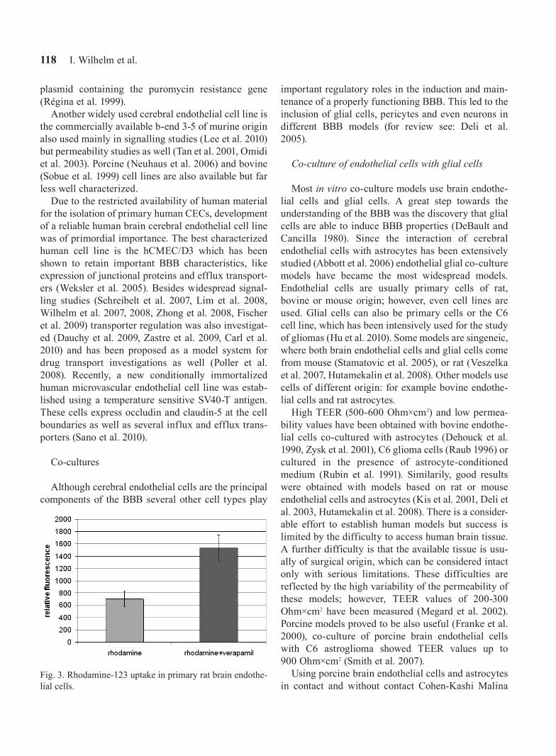
Fig. 3. Rhodamine-123 uptake in primary rat brain endothe-lial cells.

In vitro models of the blood-brain barrier 119
and coworkers (2009) have shown that a direct contact between endothelial cells and astrocytes is necessary to obtain good TEER values. Other systems using no direct contact between endothelial cells and astrocytes showed low pemeability values. Based on the method of Dehouck and others (1990) a modified BBB model was introduced suitable for high throughput screening in which a “BBB-inducing medium” is used. This BBB-inducing medium contains 1% conditioned medi-um from the glial-endothelial co-culture, harvested 48 h after refreshing the co-culture system medium and frozen until further use (Culot et al. 2007).
Endothelial cell lines in co-culture with glial cells have also been used as in vitro BBB models. The high-est controversy is linked to the use of ECV304 cells. This cell line although shows endothelial characteris-tics (Kiessling et al. 1999, Suda et al. 2001) and in co-culture with glial cells can reach relatively high TEER values, proved to be a non-endothelial human cell line (Brown et al. 2000). Despite this controversy this model is still used as an in vitro BBB model (Kuhlmann et al. 2006, Wang et al. 2010). The mouse cerebral endothelial cell lines b-end 3 and 5 in co-culture with C6 glioma cells exhibit TEER values well below 100 Ohm×cm2. Similarly RBE4 cells and rat astrocytes or C6 cells also show high permeability values (in the range of 10-5 cm/s for sucrose). A triple culture model using RBE4.B cells has demonstrated that neurons and astrocytes are able to induce a significant decrease in the permeability for sucrose (Schiera et al. 2005).
Co-culture of endothelial cells with pericytes
Pericytes are in close contact with endothelial cells therefore a co-culture of endothelial cells with pericytes in in vitro models is plausible. Co-culture of brain endothelial cells with pericytes was shown to increase TEER in a rat model (Hayashi et al. 2004), and peri-cytes have been shown to induce MRP6 expression (Berezowski et al. 2004) in endothelial cells. Surprisingly, other data indicate that pericytes induce MMP secretion in endothelial cells (Zozulya et al. 2008). In the interac-tion between pericytes and endothelial cells endothe-lin-1 (Dehouck et al. 1994), TGF-beta (Dohgu et al. 2005) and angiopoetin-1 (Hori et al. 2004) may play a significant role. More recently, a triple co-culture model including endothelial cells, astrocytes and pericytes was characterized and shown to have high TEER and low permeability (Nakagawa et al. 2007, 2009).
Co-culture of endothelial cells with other cells
Neurons have been shown to induce blood-brain barrier related enzymes in cultured cerebral endothe-lial cells (Tontsch and Bauer 1991). A co-culture model of RBE4.B cells and cortical neurons was established by Cestelli and colleagues (2001) and it has been dem-onstrated that it is not necessary a direct contact among endothelial cells and neurons for the induction of occludin expression. Moreover, not only mature neurons but differentiating embryonic neural progeni-tor cells are also able to induce BBB properties in cerebral endothelial cells (Weidenfeller et al. 2007). Interestingly, Zenker and coauthors (2003) found that the TEER of brain capillary endothelial cells was increased by 50% in a non-contact co-culture with macrophages.
Dynamic models
There is increasing evidence that shear stress is able to affect endothelial barrier function (Tarbell 2010). This led to the development of dynamic in vitro models. For this purpose usually hollow fibers are used which mimic capillaries and allow co-culture of other cell types as well. The first models were based on the co-culture of bovine aortic endothelial cells and glial cells (Stanness et al. 1997, Cucullo et al. 2002). Recently a dynamic model allowing pulsatile flow and using the human cerebral endothelial cell line hCMEC/D3 and astrocytes was developed. The model showed TEER around 1000 Ohm×cm2. Interestingly, the same cells used in a static model provided only TEER values around 70 Ohm×cm2 (Cucullo et al. 2008). A further modification of the dynamic BBB model was described in which hol-low fibers were used with transmural microholes (2 to 4
Fig. 4. Expression of transporters in CECs. RT-PCR was performed using cDNA of RBECs and specific primer pairs for rat transporters.

120 I. Wilhelm et al.
micrometers) making the model suitable for immune cell transmigration studies. The model retained high TEER and low permeability properties (Cucullo et al. 2010).
Similar results were obtained by other investigators as well. Human fetal astrocytes and especially astro-cyte-conditioned medium reduced significantly the permeability of an in vitro BBB model based on the culture of primary human brain endothelial cells. Under static conditions TEER values of about 500 Ohm×cm2 could be reached. Fluid shear stress of 1-2 dyne/cm2 reduced the permeability to approximately 50% (Siddharthan et al. 2007).
The large number of different in vitro models suggests that there is no „perfect” model so far and – depending on the studied question – one or the other model can be more advantageous. Studies on intracellular signalling have been performed in the majority of cases on monocultures cultured in Petri dishes. This method has the advantage that endothelial cells can be used in large quantities for biochemical studies, physiological investigations (migra-tion, proliferation) and the optical characteristics of the plastic Petri dishes are superior to different filters. Besides this, the experimental costs are also lower. The disadvan-tage of these approaches lies in that cellular interactions within the BBB cannot be taken into consideration. Primary cultures from different species have been succes-fully used including human, bovine, porcine; rat or mouse
origin. The use of cell lines is also widespread, however they cannot be recommended when investigations on tight junction structure and function are in focus.
The culture on different filter systems is usually the method of choice when cellular interactions are impor-tant and these can be regarded as true BBB models. These models are mainly used when barrier properties (TEER, permeability) are investigated or in drug trans-port studies. TEER and permeability are two important parameters in the determination of the quality of the in vitro model. Unfortunately, under culture conditions brain endothelial cells do not show the same character-istics as in vivo, especially regarding the barrier proper-ties (Wolburg et al. 1994). However, under carefully chosen experimental conditions in vitro models may have good barrier characteristics and are useful tools in drug permeability and transport experiments. It has been demonstrated that above TEER values of 120-130 Ohm×cm2 the permeability for sodium fluorescein and FITC-labeled dextrane (4 kDa) does not decrease with the increase of TEER (Gaillard and deBoer 2000) indi-cating that barriers with at least 120-130 Ohm×cm2 can be used for transport experiments. Best results are obtained with models based on primary cerebral endothelial cells; the major limitation of the models using endothelial cell lines is their relatively high para-cellular permeability (Roux and Couraud 2005).
Fig. 5. In vitro model of the BBB.

In vitro models of the blood-brain barrier 121
CHARACTERIZATION OF A RAT IN VITRO BBB MODEL
Culture of primary cerebral endothelial cells
Our in vitro BBB model is based on the culture of primary rat brain endothelial cells (RBECs). Endothelial cells are isolated from cortices of 2-3-week old Wistar rats, using a two step enzymatic digestion and cen-trifugation on Percoll gradient (Wilhelm et al. 2007, Hutamekalin et al. 2008, Nagyoszi et al. 2010). Following plating on collagenIV/fibronectin-coated Petri dishes cells reach confluency within 5 days. Cells show a spindle shaped morphology (Fig. 2A, B) and form a continuous line of tight junctions at the cell borders as revealed by staining with antibodies against the TJ proteins claudin-5, occludin and ZO-1 (Fig. 2C-F).
Besides junctional proteins brain endothelial cells express a whole set of transporters as well. One of the most important efflux transporters with a decisive impact on drug delivery is P-glycoprotein (P-gp/mdr1/Abcb1). The presence of a functional P-glycoprotein was tested using rhodamine-123 uptake. Verapamil, a specific inhibitor of P-gp was able to induce a more than twofold increase in the rhodamine-123 uptake indicating that this transporter is present and active in RBECs (Fig. 3). RT-PCR experiments have demon-strated the expression of other transporters as well, like ABCC5, ABCG2, MVP (major vault protein) and RLIP (non-ABC transporter RalA binding protein) as well (Fig. 4).
Co-culture systems
For the co-culture system pericytes are obtained from cerebral microvessels plated onto non-coated dishes. Within 10 days an almost pure pericytic culture is obtained as assessed by staining with alpha-actin antibody (Fig. 5).
Glial cultures, consisting mainly of astrocytes (Fig. 5) are prepared from newborn rats and are cultured on poly-L-lysine coated surfaces.
As in vitro BBB models both double and triple co-culture systems can be used, based on the culture of endothelial cells with astrocytes and/or pericytes. The triple co-culture system, which is the closest to the in vivo situation, is prepared as follows: rat brain peri-cytes are plated onto the backside of 12-well Transwell
filters (pore size: 0.4 μm; 1.5×10⁴ cells/filter). On the next day endothelial cells are plated onto the upper surface of the filters. After reaching confluency, the endothelial monolayer is supplied with 550 nM hydro-cortisone, 250 μM CPT-cAMP and 17.5 μM RO-201724 and placed into dishes containing glial cultures for 24 h. In the lower compartment astrocyte-conditioned medium can also be used with similar barrier charac-teristics of the model. We have obtained TEER values of 264 ± 67 Ohm×cm2 when astrocytes were used vs. 252 ± 32 Ohm×cm2 when astrocyte-conditioned medi-um was used, calculated from four independent exper-iments.
Measurement of TEER
We can measure TEER using chopstick electrodes and an EVOM epithelial voltohmmeter (World Precision Instruments, Sarasota, Florida, USA). The disadvantage of this method is that the plates contain-ing the cultures need to be removed from the thermo-state for measurements, which can be made only at certain time points.
The cellZscope system (NanoAnalytics, Münster, Germany) allows for continuous monitoring of the transendothelial electric resistance of up to 24 filters. After reaching confluency on the filter inserts endothe-lial cells already develop TEER values above 100 Ohm×cm2. At this point, the endothelial monolayer is supplied with 550 nM hydrocortisone, 250 μM CPT-
Fig. 6. Induction of TEER by hydrocortisone, cAMP and astrocyte-conditioned medium. Confluent RBECs grown on semipermeable filter inserts were supplied with 550 nM hydrocortisone, 250 μM CPT-cAMP and 17.5 μM RO-201724 from the apical side and astrocyte-conditioned medium from the basolateral side. TEER was monitored using the CellZ-scope system.

122 I. Wilhelm et al.
cAMP and 17.5 μM RO-201724 and placed into the wells of the instrument containing astrocyte-condi-tioned medium. TEER values increase gradually reach-ing values above 200 Ohm×cm2 (Fig. 6).
Permeability measurements
The barrier function can also be evaluated by mea-suring the permeability of the cells to sodium-fluores-cein (SF, MW = 376 Da) and Evans blue labeled albu-min (EBA, MW = 67 kDa). Transwell filters contain-ing endothelial cells and pericytes are washed with Ringer-HEPES solution (pH 7.4). Ringer-HEPES is
added to the abluminal side of the filter. The luminal side is loaded with Ringer-HEPES containing 10 µg/ml SF, 170 µg/ml Evans blue and 10 mg/ml BSA. The cells are then incubated at 37ºC for 1 h with gentle shaking, and samples are collected from the abluminal side. Concentration of SF and EBA can be measured using a fluorescent microplate reader with an excita-tion wavelength of 485 nm and an emission wavelength of 520 nm for SF, and 584/680 nm excitation/emission wavelengths for EBA. Apparent permeability (Papp) is calculated as described previously. We have achieved permeability values of 2.48×10⁻⁶ (± 1.87×10⁻⁶) cm/s for SF and 8.32×10⁻⁷ (± 2.26×10⁻⁷) for EBA (8 independent
Fig. 7. Use of the in vitro model for brain metastasis investigations. Fluorescently labeled cancer cells are plated on conflu-ent endothelial cultures (A). At the end of co-culture (B) cells from the upper compartment are removed (C). Fluorescence microscopy images of the tumor cells (A2058 melanoma cells) at the end of co-culture (D) and after wiping off the cells from the apical side of the filter (transmigrated cells, E).

In vitro models of the blood-brain barrier 123
experiments, TEER values of 278 ± 58 Ohm×cm2). Our results are in line with previous literature data (Gaillard and deBoer 2000) showing that a monolayer with a TEER above 120-130 Ohm×cm2 is accompanied by a low paracellular permeability.
Using the same experimental setup, the apparent permeability of test drugs can also be evaluated. In this case the test drug is added to the luminal side and its concentration is measured in the abluminal buffer. Calculations are performed similarly.
This model is also suitable for testing substrates of efflux transporters. The test chemical is applied to either the apical (A) or the basolateral (B) side of the endothelial monolayer in the presence or absence of known inhibitors of specific transporters. Samples are taken from the opposite chamber. Papp (A-to-B) and Papp (B-to-A) is calculated. If the ratio between them is >1 when the test compound is given alone, and close to 1 when the test compound is given together with the specific efflux transporter inhibitor, the test drug is probably a substrate of the transporter and in vivo will reach lower concentrations in the brain.
Metastasis model
In order to study the routes and mechanisms of transmigration of tumor cells through the BBB we have constructed a transmigration experimental setup consisting of brain endothelial cells cultured on large pore size filter inserts (Fig. 7).
Primary RBECs are gently trypsinized and passed onto fibronectin/collagen-coated filter inserts (8 μm pore size, 1.13 cm2, Millipore) which are placed into 12-well plates. After reaching confluency, endothe-lial cells are supplemented with hydrocortisone and cAMP from the apical side and astrocyte-condi-tioned medium from the basolateral side for 24 h in order to tighten the junctions. The following day 105 fluorescently labeled tumor cells (A2058 melanoma cells) are plated into the upper compartment onto the endothelial monolayer (Fig. 7A). Tumor cells are able to migrate through the endothelial cell layer and the pores of the filter and accumulate on the other side of the filter (Fig. 7B). After 5-24 h cells are fixed. Cells from the upper compartment are removed with a cotton swab (Fig. 7C) and tumor cells migrated through the endothelial monolayer and the pores of the filter can be counted using a fluorescence micro-scope (Fig. 7E).
CONCLUSION
In summary, our in vitro BBB model is charac-terized by high transendothelial electrical resis-tance (TEER regularily over 200 Ohm×cm2), low permeability and expression of different transport-ers. Our experiments have proven that the model is suitable for the study of different aspects of BBB function in basic research and for testing the inter-action between the BBB and potential drug candi-dates (toxicity, permeability, interaction with efflux transporters) as well.
ACKNOWLEDGEMENT
This work was partially supported by the project REG-DA-09-1-2009-0004, BBBTEER9 (I.K.).
REFERENCES
Abbott NJ, Rönnbäck L, Hansson E (2006) Astrocyte-endothelial interactions at the blood-brain barrier. Nat Rev Neurosci 7: 41–53.
Armulik A, Genové G, Mäe M, Nisancioglu MH, Wallgard E, Niaudet C, He L, Norlin J, Lindblom P, Strittmatter K, Johansson BR, Betsholtz C (2010) Pericytes regulate the blood-brain barrier. Nature 468: 557–561.
Barakat S, Turcotte S, Demeule M, Lachambre MP, Régina A, Baggetto LG, Béliveau R (2008) Regulation of brain endothelial cells migration and angiogenesis by P-glycoprotein/caveolin-1 interaction. Biochem Biophys Res Commun 372: 440–446.
Begley DJ (2004) ABC transporters and the blood-brain bar-rier. Curr Pharm Des 10: 1295–1312.
Berezowski V, Landry C, Dehouck MP, Cecchelli R, Fenart L (2004). Contribution of glial cells and pericytes to the mRNA profiles of P-glycoprotein and multidrug resis-tance-associated proteins in an in vitro model of the blood-brain barrier. Brain Res 1018: 1–9.
Bernas MJ, Cardoso FL, Daley SK, Weinand ME, Campos AR, Ferreira AJ, Hoying JB, Witte MH, Brites D, Persidsky Y, Ramirez SH, Brito MA (2010) Establishment of primary cultures of human brain microvascular endothelial cells to provide an in vitro cellular model of the blood-brain barrier. Nat Protoc 5: 1265–1272.
Bowman PD, Ennis SR, Rarey KE, Betz AL, Goldstein GW (1983) Brain microvessel endothelial cells in tissue cul-ture: a model for study of blood-brain barrier permeabil-ity. Ann Neurol 14: 396–402.

124 I. Wilhelm et al.
Breier G, Breviario F, Caveda L, Berthier R, Schnürch H, Gotsch U, Vestweber D, Risau W, Dejana E (1996) Molecular cloning and expression of murine vascular endothelial-cadherin in early stage development of car-diovascular system. Blood 87: 630–641.
Brown J, Reading SJ, Jones S, Fitchett CJ, Howl J, Martin A, Longland CL, Michelangeli F, Dubrova YE, Brown CA (2000) Critical evaluation of ECV304 as a human endothe-lial cell model defined by genetic analysis and functional responses: a comparison with the human bladder cancer derived epithelial cell line T24/83. Lab Invest 80: 37–45.
Carl SM, Lindley DJ, Couraud PO, Weksler BB, Romero I, Mowery SA, Knipp GT (2010) ABC and SLC transporter expression and pot substrate characterization across the human CMEC/D3 blood-brain barrier cell line. Mol Pharm 7: 1057–1068.
Cestelli A, Catania C, D’Agostino S, Di Liegro I, Licata L, Schiera G, Pitarresi GL, Savettieri G, De Caro V, Giandalia G, Giannola LI (2001) Functional feature of a novel model of blood brain barrier: studies on permeation of test compounds. J Control Release 76: 139–147.
Cipolla MJ, Crete R, Vitullo L, Rix RD (2004) Transcellular transport as a mechanism of blood-brain barrier disrup-tion during stroke. Front Biosci 9: 777–785.
Citi S, Sabanay H, Jakes R, Geiger B, Kendrick-Jones J (1988) Cingulin, a new peripheral component of tight junctions. Nature 333: 272–276.
Citi S, Sabanay H, Kendrick-Jones J, Geiger B (1989) Cingulin: characterization and localization. J Cell Sci 93: 107–122.
Cohen-Kashi Malina K, Cooper I, Teichberg VI (2009) Closing the gap between the in-vivo and in-vitro blood-brain barrier tightness. Brain Res 1284: 12–21.
Cucullo L, McAllister MS, Kight K, Krizanac-Bengez L, Marroni M, Mayberg MR, Stanness KA, Janigro D (2002) A new dynamic in vitro model for the multidimen-sional study of astrocyte-endothelial cell interactions at the blood-brain barrier. Brain Res 951: 243–254.
Cucullo L, Couraud PO, Weksler B, Romero IA, Hossain M, Rapp E, Janigro D (2008) Immortalized human brain endothelial cells and flow-based vascular modeling: a marriage of convenience for rational neurovascular stud-ies. J Cereb Blood Flow Metab 28: 312–328.
Cucullo L, Marchi N, Hossain M, Janigro D (2010) A dynamic in vitro BBB model for the study of immune cell trafficking into the central nervous system. J Cereb Blood Flow Metab [Epub ahead of print].
Culot M, Lundquist S, Vanuxeem D, Nion S, Landry C, Delplace Y, Dehouck MP, Berezowski V, Fenart L,
Cecchelli R (2008) An in vitro blood-brain barrier model for high throughput (HTS) toxicological screening. Toxicol In Vitro 22: 799–811.
Dauchy S, Miller F, Couraud PO, Weaver RJ, Weksler B, Romero IA, Scherrmann JM, De Waziers I, Declèves X (2009) Expression and transcriptional regulation of ABC transporters and cytochromes P450 in hCMEC/D3 human cerebral microvascular endothelial cells. Biochem Pharmacol 77: 897–909.
DeBault LE, Kahn LE, Frommes SP, Cancilla PA (1979) Cerebral microvessels and derived cells in tissue culture: isolation and preliminary characterization. In Vitro 15: 473–487.
DeBault LE, Cancilla PA (1980) gamma-Glutamyl transpep-tidase in isolated brain endothelial cells: induction by glial cells in vitro. Science 207: 653–655.
DeBault LE, Henriquez E, Hart MN, Cancilla PA (1981) Cerebral microvessels and derived cells in tissue culture: II. Establishment, identification, and preliminary charac-terization of an endothelial cell line. In Vitro 17: 480–494.
Dehouck MP, Méresse S, Delorme P, Fruchart JC, Cecchelli R (1990) An easier, reproducible, and mass-production method to study the blood-brain barrier in vitro. J Neurochem 54: 1798–1801.
Dehouck MP, Vigne P, Torpier G, Breittmayer JP, Cecchelli R, Frelin C (1997) Endothelin-1 as a mediator of endothe-lial cell-pericyte interactions in bovine brain capillaries. J Cereb Blood Flow Metab 17: 464–469.
Deli MA, Abrahám CS, Takahata H, Niwa M (2001) Tissue plasminogen activator inhibits P-glycoprotein activity in brain endothelial cells. Eur J Pharmacol 411: R3–R5.
Deli MA, Abrahám CS, Niwa M, Falus A (2003) N,N-diethyl-2-[4-(phenylmethyl)phenoxy] ethanamine increases the permeability of primary mouse cerebral endothelial cell monolayers. Inflamm Res 52 Suppl 1: S39–40.
Deli MA, Abrahám CS, Kataoka Y, Niwa M (2005) Permeability studies on in vitro blood-brain barrier mod-els: physiology, pathology, and pharmacology. Cell Mol Neurobiol 25: 59–127.
Demeule M, Régina A, Jodoin J, Laplante A, Dagenais C, Berthelet F, Moghrabi A, Béliveau R (2002) Drug trans-port to the brain: key roles for the efflux pump P-glycoprotein in the blood-brain barrier. Vascul Pharmacol 38: 339–348.
Dohgu S, Takata F, Yamauchi A, Nakagawa S, Egawa T, Naito M, Tsuruo T, Sawada Y, Niwa M, Kataoka Y (2005) Brain pericytes contribute to the induction and up-regula-

In vitro models of the blood-brain barrier 125
tion of blood-brain barrier functions through transform-ing growth factor-beta production. Brain Res 1038: 208–215.
Dore-Duffy P (2008) Pericytes: pluripotent cells of the blood brain barrier. Curr Pharm Des 14: 1581–1593.
Etienne S, Adamson P, Greenwood J, Strosberg AD, Cazaubon S, Couraud PO (1998) ICAM-1 signaling path-ways associated with Rho activation in microvascular brain endothelial cells. J Immunol 161: 5755–5761.
Fábián G, Szabó CA, Bozó B, Greenwood J, Adamson P, Deli MA, Joó F, Krizbai IA, Szucs M (1998) Expression of G-protein subtypes in cultured cerebral endothelial cells. Neurochem Int 33: 179–185.
Fischer S, Nishio M, Peters SC, Tschernatsch M, Walberer M, Weidemann S, Heidenreich R, Couraud PO, Weksler BB, Romero IA, Gerriets T, Preissner KT (2009) Signaling mechanism of extracellular RNA in endothelial cells. FASEB J 23: 2100–2109.
Franke H, Galla H, Beuckmann CT (2000) Primary cultures of brain microvessel endothelial cells: a valid and flexible model to study drug transport through the blood-brain barrier in vitro. Brain Res Brain Res Protoc 5: 248–256.
Furuse M, Hirase T, Itoh M, Nagafuchi A, Yonemura S, Tsukita S, Tsukita S (1993) Occludin: a novel integral membrane protein localizing at tight junctions. J Cell Biol 123: 1777–1788.
Furuse M, Fujita K, Hiiragi T, Fujimoto K, Tsukita S (1998) Claudin-1 and -2 novel integral membrane proteins local-izing at tight junctions with no sequence similarity to occludin. J Cell Biol 141: 1539–1550.
Gaillard PJ, de Boer AG (2000) Relationship between per-meability status of the blood-brain barrier and in vitro permeability coefficient of a drug. Eur J Pharm Sci 12: 95–102.
González-Mariscal L, Betanzos A, Nava P, Jaramillo BE (2003) Tight junction proteins. Prog Biophys Mol Biol 81: 1–44.
Greenwood J, Pryce G, Devine L, Male DK, dos Santos WL, Calder VL, Adamson P (1996) SV40 large T immortal-ised cell lines of the rat blood-brain and blood-retinal barriers retain their phenotypic and immunological char-acteristics. J Neuroimmunol 71: 51–63.
Gumbiner B, Lowenkopf T, Apatira D (1991) Identification of a 160-kDa polypeptide that binds to the tight junction protein ZO-1. Proc Natl Acad Sci USA 88: 3460–3464.
Hartmann C, Zozulya A, Wegener J, Galla HJ (2007) The impact of glia-derived extracellular matrices on the bar-rier function of cerebral endothelial cells: an in vitro study. Exp Cell Res 313: 1318–1325.
Hawkins BT, Davis TP (2005) The blood-brain barrier/neu-rovascular unit in health and disease. Pharmacol Rev 57: 173–185.
Hayashi K, Nakao S, Nakaoke R, Nakagawa S, Kitagawa N, Niwa M (2004) Effects of hypoxia on endothelial/peri-cytic co-culture model of the blood-brain barrier. Regul Pept 123: 77–83.
Hori S, Ohtsuki S, Hosoya K, Nakashima E, Terasaki T (2004) A pericyte-derived angiopoietin-1 multimeric complex induces occludin gene expression in brain capil-lary endothelial cells through Tie-2 activation in vitro. J Neurochem 89: 503–513.
Hu JG, Wang XF, Zhou JS, Wang FC, Li XW, Lü HZ (2010) Activation of PKC-alpha is required for migration of C6 glioma cells. Acta Neurobiol Exp (Wars) 70: 239–245.
Hutamekalin P, Farkas AE, Orbók A, Wilhelm I, Nagyoszi P, Veszelka S, Deli MA, Buzás K, Hunyadi-Gulyás E, Medzihradszky KF, Meksuriyen D, Krizbai IA (2008) Effect of nicotine and polyaromtic hydrocarbons on cere-bral endothelial cells. Cell Biol Int 32: 198–209.
Itoh M, Nagafuchi A, Moroi S, Tsukita S (1997) Involvement of ZO-1 in cadherin-based cell adhesion through its direct binding to alpha catenin and actin filaments. J Cell Biol 138: 181–192.
Itoh M, Morita K, Tsukita S (1999) Characterization of ZO-2 as a MAGUK family member associated with tight as well as adherens junctions. J Biol Chem 274: 5981–5986.
Jeffrey P, Summerfield S (2010) Assessment of the blood-brain barrier in CNS drug discovery. Neurobiol Dis 37: 33–37.
Joó F, Karnushina I (1973) A procedure for the isolation of capillaries from rat brain. Cytobios 8: 41–48.
Kacem K, Lacombe P, Seylaz J, Bonvento G (1998) Structural organization of the perivascular astrocyte end-feet and their relationship with the endothelial glucose transporter: a confocal microscopy study. Glia 23: 1–10.
Kiessling F, Kartenbeck J, Haller C (1999) Cell-cell contacts in the human cell line ECV304 exhibit both endothelial and epithelial characteristics. Cell Tissue Res 297: 131–140.
Kis B, Deli MA, Kobayashi H, Abrahám CS, Yanagita T, Kaiya H, Isse T, Nishi R, Gotoh S, Kangawa K, Wada A, Greenwood J, Niwa M, Yamashita H, Ueta Y (2001) Adrenomedullin regulates blood-brain barrier functions in vitro. Neuroreport 12: 4139–4142.
Krizbai I, Szabó G, Deli M, Maderspach K, Lehel C, Oláh Z, Wolff JR, Joó F (1995) Expression of protein kinase C family members in the cerebral endothelial cells. J Neurochem 65: 459-462.

126 I. Wilhelm et al.
Kuhlmann CR, Lessmann V, Luhmann HJ. Fluvastatin stabi-lizes the blood-brain barrier in vitro by nitric oxide-de-pendent dephosphorylation of myosin light chains (2006) Neuropharmacology 51: 907–913.
Langford D, Hurford R, Hashimoto M, Digicaylioglu M, Masliah E (2005) Signalling crosstalk in FGF2-mediated protection of endothelial cells from HIV-gp120. BMC Neurosci 6:8.
Laterra J, Guerin C, Goldstein GW (1990) Astrocytes induce neural microvascular endothelial cells to form capillary-like structures in vitro. J Cell Physiol 144: 204–215.
Lee HT, Chang YC, Tu YF, Huang CC. CREB activation medi-ates VEGF-A’s protection of neurons and cerebral vascular endothelial cells (2010) J Neurochem 113: 79–91.
Lim JC, Kania KD, Wijesuriya H, Chawla S, Sethi JK, Pulaski L, Romero IA, Couraud PO, Weksler BB, Hladky SB, Barrand MA (2008) Activation of beta-catenin sig-nalling by GSK-3 inhibition increases p-glycoprotein expression in brain endothelial cells. J Neurochem 106: 1855–1865.
Löscher W, Potschka H (2005) Blood-brain barrier active efflux transporters: ATP-binding cassette gene family. NeuroRx 2: 86–98.
Lupo G, Nicotra A, Giurdanella G, Anfuso CD, Romeo L, Biondi G, Tirolo C, Marchetti B, Ragusa N, Alberghina M (2005) Activation of phospholipase A(2) and MAP kinases by oxidized low-density lipoproteins in immortal-ized GP8.39 endothelial cells. Biochim Biophys Acta 1735: 135–150.
Martìn-Padura I, Lostaglio S, Schneemann M, Williams L, Romano M, Fruscella P, Panzeri C, Stoppacciaro A, Ruco L, Villa A, Simmons D, Dejana E. (1998) Junctional adhesion molecule, a novel member of the immunoglobulin super-family that distributes at intercellular junctions and modu-lates monocyte transmigration. J Cell Biol 142: 117–127.
Megard I, Garrigues A, Orlowski S, Jorajuria S, Clayette P, Ezan E, Mabondzo A (2002) A co-culture-based model of human blood-brain barrier: application to active transport of indinavir and in vivo-in vitro correlation. Brain Res 927: 153–167.
Morita K, Sasaki H, Furuse M, Tsukita S (1999) Endothelial claudin: claudin-5/TMVCF constitutes tight junction strands in endothelial cells. J Cell Biol 147: 185–194.
Nagyoszi P, Wilhelm I, Farkas AE, Fazakas C, Dung NT, Haskó J, Krizbai IA (2010) Expression and regulation of toll-like receptors in cerebral endothelial cells. Neurochem Int 57: 556–564.
Nakagawa S, Deli MA, Nakao S, Honda M, Hayashi K, Nakaoke R, Kataoka Y, Niwa M (2007) Pericytes from
brain microvessels strengthen the barrier integrity in pri-mary cultures of rat brain endothelial cells. Cell Mol Neurobiol 27: 687–694.
Nakagawa S, Deli MA, Kawaguchi H, Shimizudani T, Shimono T, Kittel A, Tanaka K, Niwa M (2009) A new blood-brain barrier model using primary rat brain endothe-lial cells, pericytes and astrocytes. Neurochem Int 54:253–263.
Nazer B, Hong S, Selkoe DJ (2008) LRP promotes endocy-tosis and degradation, but not transcytosis, of the amy-loid-beta peptide in a blood-brain barrier in vitro model. Neurobiol Dis 30: 94–102.
Neuhaus W, Lauer R, Oelzant S, Fringeli UP, Ecker GF, Noe CR (2006) A novel flow based hollow-fiber blood-brain barrier in vitro model with immortalised cell line PBMEC/C1-2. J Biotechnol 125: 127–141.
Ohnishi H, Nakahara T, Furuse K, Sasaki H, Tsukita S, Furuse M (2004) JACOP, a novel plaque protein localiz-ing at the apical junctional complex with sequence simi-larity to cingulin. J Biol Chem 279: 46014–46022.
Ohtsuki S, Yamaguchi H, Katsukura Y, Asashima T, Terasaki T (2008) mRNA expression levels of tight junction protein genes in mouse brain capillary endothelial cells highly puri-fied by magnetic cell sorting. J Neurochem 104: 147–154.
Omidi Y, Campbell L, Barar J, Connell D, Akhtar S, Gumbleton M (2003) Evaluation of the immortalised mouse brain capillary endothelial cell line, b.End3, as an in vitro blood-brain barrier model for drug uptake and transport studies. Brain Res 990: 95–112.
Pan W, Yu Y, Cain CM, Nyberg F, Couraud PO, Kastin AJ (2005) Permeation of growth hormone across the blood-brain barrier. Endocrinology 146: 4898–4904.
Panula P, Joó F, Rechardt L (1978) Evidence for the pres-ence of viable endothelial cells in cultures derived from dissociated rat brain. Experientia 34: 95–97.
Pekny M, Stanness KA, Eliasson C, Betsholtz C, Janigro D (1998) Impaired induction of blood-brain barrier proper-ties in aortic endothelial cells by astrocytes from GFAP-deficient mice. Glia 22: 390–400.
Persidsky Y, Stins M, Way D, Witte MH, Weinand M, Kim KS, Bock P, Gendelman HE, Fiala M (1997) A model for monocyte migration through the blood-brain barrier dur-ing HIV-1 encephalitis. J Immunol 158: 3499–3510.
Pilorget A, Demeule M, Barakat S, Marvaldi J, Luis J, Béliveau R (2007) Modulation of P-glycoprotein function by sphingosine kinase-1 in brain endothelial cells. J Neurochem 100: 1203–1210.
Poller B, Gutmann H, Krähenbühl S, Weksler B, Romero I, Couraud PO, Tuffin G, Drewe J, Huwyler J (2008) The

In vitro models of the blood-brain barrier 127
human brain endothelial cell line hCMEC/D3 as a human blood-brain barrier model for drug transport studies. J Neurochem 107: 1358–1368.
Raub TJ (1996) Signal transduction and glial cell modula-tion of cultured brain microvessel endothelial cell tight junctions. Am J Physiol 271: C495–503.
Régina A, Koman A, Piciotti M, El Hafny B, Center MS, Bergmann R, Couraud PO, Roux F (1998) Mrp1 multi-drug resistance-associated protein and P-glycoprotein expression in rat brain microvessel endothelial cells. J Neurochem 71: 705–715.
Régina A, Romero IA, Greenwood J, Adamson P, Bourre JM, Couraud PO, Roux F (1999) Dexamethasone regula-tion of P-glycoprotein activity in an immortalized rat brain endothelial cell line, GPNT. J Neurochem 73: 1954–1963.
Roux F, Durieu-Trautmann O, Chaverot N, Claire M, Mailly P, Bourre JM, Strosberg AD, Couraud PO (1994) Regulation of gamma-glutamyl transpeptidase and alkaline phos-phatase activities in immortalized rat brain microvessel endothelial cells. J Cell Physiol 159: 101–113.
Roux F, Couraud PO (2005) Rat brain endothelial cell lines for the study of blood-brain barrier permeability and transport functions. Cell Mol Neurobiol 25: 41–58.
Rubin LL, Hall DE, Porter S, Barbu K, Cannon C, Horner HC, Janatpour M, Liaw CW, Manning K, Morales J, Tanner L, Tomaselli KJ, and Bardet F (1991) A cell culture model of the blood-brain barrier. J Cell Biol 115: 1725–1735.
Sano Y, Shimizu F, Abe M, Maeda T, Kashiwamura Y, Ohtsuki S, Terasaki T, Obinata M, Kajiwara K, Fujii M, Suzuki M, Kanda T (2010) Establishment of a new con-ditionally immortalized human brain microvascular endothelial cell line retaining an in vivo blood-brain bar-rier function. J Cell Physiol 225: 519–528.
Schiera G, Sala S, Gallo A, Raffa MP, Pitarresi GL, Savettieri G, Di Liegro I (2005) Permeability properties of a three-cell type in vitro model of blood-brain barrier. J Cell Mol Med 9: 373–379.
Schreibelt G, Kooij G, Reijerkerk A, van Doorn R, Gringhuis SI, van der Pol S, Weksler BB, Romero IA, Couraud PO, Piontek J, Blasig IE, Dijkstra CD, Ronken E, de Vries HE (2007) Reactive oxygen species alter brain endothelial tight junction dynamics via RhoA, PI3 kinase, and PKB signaling. FASEB J 21: 3666–3676.
Schulze C, Firth JA (1993) Immunohistochemical localiza-tion of adherens junction components in blood-brain bar-rier microvessels of the rat. J Cell Sci 104: 773–782.
Shen S, Zhang W (2010) ABC transporters and drug efflux at the blood-brain barrier. Rev Neurosci 21: 29–53.
Siddharthan V, Kim YV, Liu S, Kim KS (2007) Human astrocytes/astrocyte-conditioned medium and shear stress enhance the barrier properties of human brain microvas-cular endothelial cells. Brain Res 1147: 39–50.
Smith JP, Drewes LR (2006) Modulation of monocarboxylic acid transporter-1 kinetic function by the cAMP signaling pathway in rat brain endothelial cells. J Biol Chem 281: 2053–2060.
Smith M, Omidi Y, Gumbleton M (2007) Primary porcine brain microvascular endothelial cells: biochemical and functional characterisation as a model for drug transport and targeting. J Drug Target 15: 253–268.
Sobue K, Yamamoto N, Yoneda K, Hodgson ME, Yamashiro K, Tsuruoka N, Tsuda T, Katsuya H, Miura Y, Asai K, Kato T (1999) Induction of blood-brain barrier properties in immortalized bovine brain endothelial cells by astro-cytic factors. Neurosci Res 35: 155–164.
Stamatovic SM, Shakui P, Keep RF, Moore BB, Kunkel SL, Van Rooijen N, Andjelkovic AV. (2005) Monocyte chemoattractant protein-1 regulation of blood-brain bar-rier permeability. J Cereb Blood Flow Metab 25: 593–606.
Stanness KA, Westrum LE, Fornaciari E, Mascagni P, Nelson JA, Stenglein SG, Myers T, Janigro D (1997) Morphological and functional characterization of an in vitro blood-brain barrier model. Brain Res 771: 329–342.
Stevenson BR, Siliciano JD, Mooseker MS, Goodenough DA (1986) Identification of ZO-1: a high molecular weight polypeptide associated with the tight junction (zonula occludens) in a variety of epithelia. J Cell Biol 103: 755–766.
Suda K, Rothen-Rutishauser B, Günthert M, Wunderli-Allenspach H (2001) Phenotypic characterization of human umbilical vein endothelial (ECV304) and uri-nary carcinoma (T24) cells: endothelial versus epithe-lial features. In Vitro Cell Dev Biol Anim 37: 505–514.
Tao-Cheng JH, Nagy Z, Brightman MW (1987) Tight junc-tions of brain endothelium in vitro are enhanced by astro-glia. J Neurosci 7: 3293–3299.
Tarbell JM (2010) Shear stress and the endothelial transport barrier. Cardiovasc Res 87: 320–330.
Tan KH, Dobbie MS, Felix RA, Barrand MA, Hurst RD (2001) A comparison of the induction of immortalized endothelial cell impermeability by astrocytes. Neuroreport 12: 1329–1334.
Tontsch U, Bauer HC (1989) Isolation, characterization, and long-term cultivation of porcine and murine cerebral cap-illary endothelial cells. Microvasc Res 37:148–161.

128 I. Wilhelm et al.
Tontsch U, Bauer HC (1991). Glial cells and neurons induce blood-brain barrier related enzymes in cultured cerebral endothelial cells. Brain Res 539: 247–253.
Vandamme W, Braet K, Cabooter L, Leybaert L (2004) Tumour necrosis factor alpha inhibits purinergic calcium signalling in blood-brain barrier endothelial cells. J Neurochem 88: 411–421.
Veszelka S, Pásztói M, Farkas AE, Krizbai I, Ngo TK, Niwa M, Abrahám CS, Deli MA (2007) Pentosan polysulfate protects brain endothelial cells against bacterial lipopoly-saccharide-induced damages. Neurochem Int 50: 219–228.
Wang Q, Rager JD, Weinstein K, Kardos PS, Dobson GL, Li J, Hidalgo IJ (2005) Evaluation of the MDR-MDCK cell line as a permeability screen for the blood-brain barrier. Int J Pharm 288: 349–359.
Wang Q, Luo W, Zhang W, Liu M, Song H, Chen J (2010) Involvement of DMT1 +IRE in the transport of lead in an in vitro BBB model. Toxicol In Vitro [Epub ahead of print].
Weidenfeller C, Svendsen CN, Shusta EV (2007) Differentiating embryonic neural progenitor cells induce blood-brain bar-rier properties. J Neurochem 101: 555–565.
Weiss N, Miller F, Cazaubon S, Couraud PO (2009) The blood-brain barrier in brain homeostasis and neurological diseases. Biochim Biophys Acta 1788: 842–857.
Weksler BB, Subileau EA, Perrière N, Charneau P, Holloway K, Leveque M, Tricoire-Leignel H, Nicotra A, Bourdoulous S, Turowski P, Male DK, Roux F, Greenwood J, Romero IA, Couraud PO (2005) Blood-brain barrier-specific properties of a human adult brain endothelial cell line. FASEB J 19: 1872–1874.
Wilhelm I, Farkas AE, Nagyoszi P, Váró G, Bálint Z, Végh GA, Couraud PO, Romero IA, Weksler B, Krizbai IA (2007) Regulation of cerebral endothelial cell morphology by extracellular calcium. Phys Med Biol 52: 6261–6274.
Wilhelm I, Nagyoszi P, Farkas AE, Couraud PO, Romero IA, Weksler B, Fazakas C, Dung NT, Bottka S, Bauer H, Bauer HC, Krizbai IA (2008) Hyperosmotic stress induc-
es Axl activation and cleavage in cerebral endothelial cells. J Neurochem 107: 116–126.
Wolburg H, Neuhaus J, Kniesel U, Krauss B, Schmid EM, Ocalan M, Farrell C, Risau W (1994) Modulation of tight junction structure in blood-brain barrier endothelial cells. Effects of tissue culture, second messengers and cocul-tured astrocytes. J Cell Sci 107: 1347–1357.
Yu C, Kastin AJ, Tu H, Waters S, Pan W (2007) TNF acti-vates P-glycoprotein in cerebral microvascular endothe-lial cells. Cell Physiol Biochem 20:853–858.
Zastre JA, Chan GN, Ronaldson PT, Ramaswamy M, Couraud PO, Romero IA, Weksler B, Bendayan M, Bendayan R (2009) Up-regulation of P-glycoprotein by HIV protease inhibitors in a human brain microvessel endothelial cell line. J Neurosci Res 87: 1023–1036.
Zenker D, Begley D, Bratzke H, Rübsamen-Waigmann H, von Briesen H (2003) Human blood-derived macrophag-es enhance barrier function of cultured primary bovine and human brain capillary endothelial cells. J Physiol 551: 1023–1032.
Zhang Y, Wu X, He Y, Kastin AJ, Hsuchou H, Rosenblum CI, Pan W (2009) Melanocortin potentiates leptin-induced STAT3 signaling via MAPK pathway. J Neurochem 110: 390–399.
Zhong Y, Smart EJ, Weksler B, Couraud PO, Hennig B, Toborek M (2008) Caveolin-1 regulates human immuno-deficiency virus-1 Tat-induced alterations of tight junc-tion protein expression via modulation of the Ras signal-ing. J Neurosci 28: 7788–7796.
Zozulya A, Weidenfeller C, Galla HJ (2008) Pericyte-endothelial cell interaction increases MMP-9 secretion at the blood-brain barrier in vitro. Brain Res 1189: 1–11.
Zysk G, Schneider-Wald BK, Hwang JH, Bejo L, Kim KS, Mitchell TJ, Hakenbeck R, Heinz HP (2001) Pneumolysin is the main inducer of cytotoxicity to brain microvascular endothelial cells caused by Streptococcus pneumoniae. Infect Immun 69: 845–852.
Related Documents


![Beyond the Blood-Brain Barrier - UCLA CTSI · Beyond the Blood-Brain Barrier: ... Circumventing the blood-brain barrier ... K30 presentation final clean.ppt [Read-Only] Author:](https://static.cupdf.com/doc/110x72/5b0543887f8b9a0a548e9fa1/beyond-the-blood-brain-barrier-ucla-ctsi-the-blood-brain-barrier-circumventing.jpg)


