In vitro comparison of the photothermal anticancer activity of graphene nanoparticles and carbon nanotubes Zoran M. Markovic a, 1 , 2 , Ljubica M. Harhaji-Trajkovic b, 2 , Biljana M. Todorovic-Markovic a , Dejan P. Kepi c a , Katarina M. Arsikin c , Svetlana P. Jovanovi c a , Aleksandar C. Pantovic c , Miroslav D. Drami canin a , Vladimir S. Trajkovic c, * a Vinca Institute of Nuclear Sciences, University of Belgrade, Belgrade, Serbia b Institute for Biological Research, University of Belgrade, Belgrade, Serbia c Institute of Microbiology and Immunology, School of Medicine, University of Belgrade, Dr. Subotica 1, Belgrade 11000, Serbia article info Article history: Received 24 September 2010 Accepted 15 October 2010 Available online 10 November 2010 Keywords: Graphene Carbon nanotubes Cancer Hyperthermia Apoptosis abstract The present study compared the photothermal anticancer activity of near-infrared (NIR)-excited graphene nanoparticles and carbon nanotubes (CNT). Despite lower NIR-absorbing capacity, suspension of poly- vinylpyrrolidone-coated graphene sheets exposed to NIR radiation (808 nm, 2 W/cm 2 ) generated more heat than DNA or sodium dodecylbenzenesulfonate-solubilized single-wall CNT under the same conditions. Accordingly, graphene nanoparticles performed significantly better than CNT in inducing photothermal death of U251 human glioma cells in vitro. The superior photothermal sensitivity of graphene sheets could be largely explained by their better dispersivity, which has been supported by a simple calculation taking into account thermodynamic, optical and geometrical properties of the two type of carbon nanoparticles. The mechanisms of graphene-mediated photothermal killing of cancer cells apparently involved oxidative stress and mitochondrial membrane depolarization resulting in mixed apoptotic and necrotic cell death characterized by caspase activation/DNA fragmentation and cell membrane damage, respectively. Ó 2010 Elsevier Ltd. All rights reserved. 1. Introduction The selective thermal ablation of malignant tissue at tempera- tures above 40 C is a feasible alternative treatment option when surgical resection is not possible [1,2]. Unfortunately, major thermal ablative strategies such as laser, ultrasound, microwave or radio frequency treatment do not possess inherent specificity for malig- nant cells. Therefore, the selective sensitization of malignant cells to irradiation is required to increase efficiency and reduce toxicity of thermal ablation. One of the promising strategies is photothermal therapy, in which nanoparticles such as carbon nanotubes (CNT) or gold nanoparticles are used to convert near-infrared (NIR) radiation to vibrational energy, thus generating heat sufficient for cancer cell killing [3,4]. The use of NIR light in the 700-1100-nm range for the induction of hyperthermia is particularly attractive, because biolog- ical systems mostly lack chromophores that absorb in the NIR region [5,6]. Hence, lesions can be treated without the need for direct access to the tumor site, with the NIR light effectively and safely penetrating normal tissue and selectively destroying cancer cells to which the nanoparticles are attached. CNT are cylindrical structures up to hundreds of nanometers in diameter and up to few micrometers in length, consisting of atom- ically thin sheets of carbon known as “graphene”. Graphene was merely considered as part of graphite’s crystal structure until 2004, when graphene layers isolated by mechanically peeling sheets off graphite crystals have been shown to possess some surprising electrical properties [7]. During the past few years, graphene’s unique electronic structure and properties have started to threaten the dominance of CNT in potential applications from electronics to sensors. Moreover, the in vitro toxicity of graphene towards human cells appears to be lower than that of CNT [8], thus supporting its possible use in biomedicine. Accordingly, graphene oxide nano- particles have been considered as potential cancer biomarker sensors and nanoplatforms for cancer-selective drug delivery [9,10]. On the other hand, while photothermal anticancer effects of CNT have been well-documented [11e 17], the possibility of direct tumor cell killing by graphene-based nanoparticles has remained largely unexplored. Namely, only one very recent study has so far demon- strated the ability of intravenously injected polyethylene glycol- coated pure graphene sheets to reduce tumor size in mice by * Corresponding author. Tel.: þ381 11 3643 233; fax: þ381 11 3643 235. E-mail addresses: [email protected] (Z.M. Markovic), [email protected] (V.S. Trajkovic). 1 Tel.: þ381 11 2455 451; fax: þ381 11 3440 100. 2 These authors equally contributed to the work. Contents lists available at ScienceDirect Biomaterials journal homepage: www.elsevier.com/locate/biomaterials 0142-9612/$ e see front matter Ó 2010 Elsevier Ltd. All rights reserved. doi:10.1016/j.biomaterials.2010.10.030 Biomaterials 32 (2011) 1121e1129

Welcome message from author
This document is posted to help you gain knowledge. Please leave a comment to let me know what you think about it! Share it to your friends and learn new things together.
Transcript

lable at ScienceDirect
Biomaterials 32 (2011) 1121e1129
Contents lists avai
Biomaterials
journal homepage: www.elsevier .com/locate/biomater ia ls
In vitro comparison of the photothermal anticancer activity of graphenenanoparticles and carbon nanotubes
Zoran M. Markovic a,1,2, Ljubica M. Harhaji-Trajkovic b,2, Biljana M. Todorovic-Markovic a, Dejan P. Kepi�c a,Katarina M. Arsikin c, Svetlana P. Jovanovi�c a, Aleksandar C. Pantovic c, Miroslav D. Drami�canin a,Vladimir S. Trajkovic c,*
aVinca Institute of Nuclear Sciences, University of Belgrade, Belgrade, Serbiab Institute for Biological Research, University of Belgrade, Belgrade, Serbiac Institute of Microbiology and Immunology, School of Medicine, University of Belgrade, Dr. Subotica 1, Belgrade 11000, Serbia
a r t i c l e i n f o
Article history:Received 24 September 2010Accepted 15 October 2010Available online 10 November 2010
Keywords:GrapheneCarbon nanotubesCancerHyperthermiaApoptosis
* Corresponding author. Tel.: þ381 11 3643 233; faE-mail addresses: [email protected] (Z.M. Marko
Trajkovic).1 Tel.: þ381 11 2455 451; fax: þ381 11 3440 100.2 These authors equally contributed to the work.
0142-9612/$ e see front matter � 2010 Elsevier Ltd.doi:10.1016/j.biomaterials.2010.10.030
a b s t r a c t
The present study compared the photothermal anticancer activity of near-infrared (NIR)-excited graphenenanoparticles and carbon nanotubes (CNT). Despite lower NIR-absorbing capacity, suspension of poly-vinylpyrrolidone-coated graphene sheets exposed to NIR radiation (808 nm, 2W/cm2) generatedmore heatthan DNA or sodium dodecylbenzenesulfonate-solubilized single-wall CNT under the same conditions.Accordingly, graphene nanoparticles performed significantly better than CNT in inducing photothermaldeath of U251 human glioma cells in vitro. The superior photothermal sensitivity of graphene sheets couldbe largely explained by their better dispersivity, which has been supported by a simple calculation takinginto account thermodynamic, optical and geometrical properties of the two type of carbon nanoparticles.The mechanisms of graphene-mediated photothermal killing of cancer cells apparently involved oxidativestress and mitochondrial membrane depolarization resulting in mixed apoptotic and necrotic cell deathcharacterized by caspase activation/DNA fragmentation and cell membrane damage, respectively.
� 2010 Elsevier Ltd. All rights reserved.
1. Introduction
The selective thermal ablation of malignant tissue at tempera-tures above 40 �C is a feasible alternative treatment option whensurgical resection is not possible [1,2]. Unfortunately, major thermalablative strategies such as laser, ultrasound, microwave or radiofrequency treatment do not possess inherent specificity for malig-nant cells. Therefore, the selective sensitization of malignant cells toirradiation is required to increase efficiency and reduce toxicity ofthermal ablation. One of the promising strategies is photothermaltherapy, in which nanoparticles such as carbon nanotubes (CNT) orgold nanoparticles are used to convert near-infrared (NIR) radiationto vibrational energy, thus generating heat sufficient for cancer cellkilling [3,4]. The use of NIR light in the 700-1100-nm range for theinduction of hyperthermia is particularly attractive, because biolog-ical systemsmostly lack chromophores that absorb in the NIR region[5,6]. Hence, lesions can be treatedwithout the need for direct access
x: þ381 11 3643 235.vic), [email protected] (V.S.
All rights reserved.
to the tumor site, with the NIR light effectively and safely penetratingnormal tissue and selectively destroying cancer cells to which thenanoparticles are attached.
CNT are cylindrical structures up to hundreds of nanometers indiameter and up to few micrometers in length, consisting of atom-ically thin sheets of carbon known as “graphene”. Graphene wasmerely considered as part of graphite’s crystal structure until 2004,when graphene layers isolated by mechanically peeling sheets offgraphite crystals have been shown to possess some surprisingelectrical properties [7]. During the past few years, graphene’sunique electronic structure and properties have started to threatenthe dominance of CNT in potential applications from electronics tosensors. Moreover, the in vitro toxicity of graphene towards humancells appears to be lower than that of CNT [8], thus supporting itspossible use in biomedicine. Accordingly, graphene oxide nano-particles have been considered as potential cancer biomarkersensors and nanoplatforms for cancer-selective drug delivery [9,10].On the other hand, while photothermal anticancer effects of CNThave been well-documented [11e17], the possibility of direct tumorcell killing by graphene-based nanoparticles has remained largelyunexplored. Namely, only one very recent study has so far demon-strated the ability of intravenously injected polyethylene glycol-coated pure graphene sheets to reduce tumor size in mice by

Fig. 1. AFM analysis of carbon nanomaterials. (A) Large-scale and (B) small-scale AFM images of graphene nanoparticles and CNTSDBS are presented together with (C) the surfaceprofile of the individual nanoparticles.
Z.M. Markovic et al. / Biomaterials 32 (2011) 1121e11291122
NIR-stimulated hyperthermia [18]. However, it has not been reportedhow the photothermal efficiency of graphene compares to that ofcarbon nanotubes, or which cell death mechanisms are responsiblefor graphene-mediated photothermal killing of cancer cells.
In the present study, we used an in vitro system for NIR-inducedhyperthermia to directly compare the photothermal anticancerefficiency of graphene nanoparticles and carbon nanotubes, as wellas to investigate the type and molecular mechanisms of graphene-mediated photothermal cancer cell death.
2. Materials and methods
2.1. Preparation and characterization of graphene nanoparticles and carbonnanotubes
A stable water suspension of graphene nanoparticles was prepared by aqueous-phase exfoliation of graphite in the presence of non-ionic/non-toxic macromoleculepolyvinylpyrrolidone (PVP) [19], while single-wall CNT were made water-soluble by
functionalization with DNA or anionic detergent sodium dodecylbenzenesulfonate(SDBS), as previously described [20,21]. Briefly, 500 mg of crystalline graphitepowder (synthetic, 6 mm; Timcal, Bodio, Switzerland) were suspended in 100 ml ofdeionized water containing 1 g PVP (SigmaeAldrich, St. Louis, MO), while 50 mg ofsingle-wall CNT (95%; BuckyUSA, Houston, TX) were added to 100 ml of deionizedwater containing 1 mg of heat-denatured single-stranded DNA or 2 mg SDBS (bothfrom SigmaeAldrich, St. Louis, MO). The graphene and CNT suspensions weresonicated in a 750 W ultrasonic bath for 3 h, centrifuged at 4000 rpm for 1 h toremove large aggregates, and the resulting clear colloids were collected. The totalcarbon particle concentration in the obtained suspensions was determined usinggravimetric method, by drying 10 ml of colloid at 60 �C in air and measuring the dryresidue. The following concentrations were obtained: 22 mg/ml (graphene/PVP -GPVP), 25 mg/ml (CNTDNA) and 180 mg/ml (CNTSDBS). Atomic force microscopy (AFM)measurements were performed using an AFM microscope (Quesant InstrumentCorp., Agoura Hills, CA) operating in tapping mode in air at room temperature.Suspensions of carbon nanoparticles were deposited on silicon substrate by spincoater and the images were obtained after drying in air and vacuum annealing at60 �C, using standard silicon tips (NanoAndMore GMBH,Wetzlar, Germany)with theforce constant of 40 N/m. The UVevis spectra of the nanocarbon-based suspensionswere scanned within the wavelength range of 500e1100 nm using Avantes UVevis
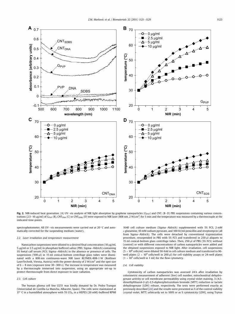
Fig. 2. NIR-induced heat generation. (A) UVevis analysis of NIR light absorption by graphene nanoparticles (GPVP) and CNT. (BeD) PBS suspensions containing various concen-trations (2.5e10 mg/ml) of GPVP (B), CNTDNA (C) or CNTSDBS (D) were exposed to NIR laser (808 nm, 2 W/cm2) for 5 min and the temperature was measured by a thermocouple at theindicated time-points.
Z.M. Markovic et al. / Biomaterials 32 (2011) 1121e1129 1123
spectrophotometer. All UVevis measurements were carried out at 20 �C and auto-matically corrected for the suspending medium (water).
2.2. Laser irradiation and temperature measurement
Nanocarbon suspensions were diluted to a desired final concentration (10 mg/ml,5 mg/ml or 2.5 mg/ml) in phosphate buffered saline (PBS; SigmaeAldrich) containing5% foetal calf serum (FCS; SigmaeAldrich) in the absence or presence of cells. Thesuspensions (500 ml) in 15 ml conical-bottom centrifuge glass tubes were illumi-nated with a 808 nm continuous-wave NIR laser RLTMDL-808-1 W (RoithnerLaserTechnik, Vienna, Austria) with the power density of 2 W/cm2 and the spot sizeof 6 � 8 mm (exposure time 30e300 s). The increase in temperature was measuredby a thermocouple immersed into suspension, using an appropriate set-up toprotect thermocouple from direct exposure to laser radiation.
2.3. Cell culture
The human glioma cell line U251 was kindly donated by Dr. Pedro Tranque(Universidad de Castilla-La Mancha, Albacete, Spain). The cells were maintained at37 �C in a humidified atmosphere with 5% CO2, in a HEPES (20 mM)-buffered RPMI
1640 cell culture medium (SigmaeAldrich) supplemented with 5% FCS, 2 mML-glutamine,10 mM sodium pyruvate, and 100 IU/ml penicillin and streptomycin (allfrom SigmaeAldrich). The cells were detached by conventional trypsinizationprocedure, resuspended in PBS with 5% FCS and transferred in 250 ml aliquots to15 ml conical-bottom glass centrifuge tubes. Then, 250 ml of PBS (5% FCS) without(control) or with different concentrations of carbon nanoparticles were added andthe obtained suspensions exposed to NIR light. After irradiation, cell suspensions(5�106 cells/ml) were diluted 50-fold in cell culture medium and transferred to 96-well plates (2 � 104 cells/well in 200 ml) for cell viability assays or 24-well plates(1 � 105 cells/well in 1 ml) for the flow cytometry.
2.4. Cell viability
Cytotoxicity of carbon nanoparticles was assessed 24 h after irradiation bycolorimetric measurement of adherent (live) cell number, mitochondrial dehydro-genase activity or cell membrane permeability using crystal violet staining, 3-(4,5-dimethylthiazol-2-yl)-2,5-diphenyltetrazolium bromide (MTT) reduction or lactatedehydrogenase (LDH) release, respectively. The tests were performed exactly aspreviously described [22] and the results were presented as % of the control viability(crystal violet, MTT) arbitrarily set to 100% or as % cytotoxicity (LDH), using Tryton
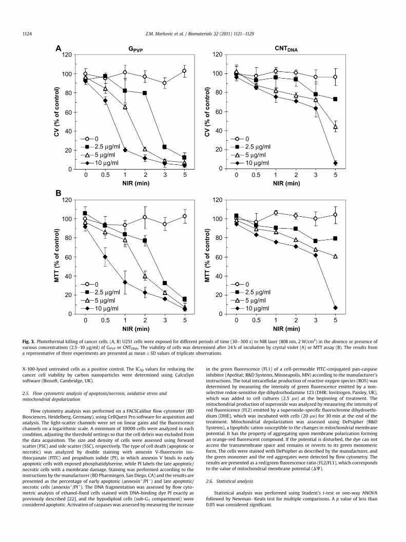
Fig. 3. Photothermal killing of cancer cells. (A, B) U251 cells were exposed for different periods of time (30e300 s) to NIR laser (808 nm, 2 W/cm2) in the absence or presence ofvarious concentrations (2.5e10 mg/ml) of GPVP or CNTDNA. The viability of cells was determined after 24 h of incubation by crystal violet (A) or MTT assay (B). The results froma representative of three experiments are presented as mean� SD values of triplicate observations.
Z.M. Markovic et al. / Biomaterials 32 (2011) 1121e11291124
X-100-lysed untreated cells as a positive control. The IC50 values for reducing thecancer cell viability by carbon nanoparticles were determined using CalcuSynsoftware (Biosoft, Cambridge, UK).
2.5. Flow cytometric analysis of apoptosis/necrosis, oxidative stress andmitochondrial depolarization
Flow cytometry analysis was performed on a FACSCalibur flow cytometer (BDBiosciences, Heidelberg, Germany), using CellQuest Pro software for acquisition andanalysis. The light-scatter channels were set on linear gains and the fluorescencechannels on a logarithmic scale. A minimum of 10000 cells were analyzed in eachcondition, adjusting the threshold settings so that the cell debris was excluded fromthe data acquisition. The size and density of cells were assessed using forwardscatter (FSC) and side scatter (SSC), respectively. The type of cell death (apoptotic ornecrotic) was analyzed by double staining with annexin V-fluorescein iso-thiocyanate (FITC) and propidium iodide (PI), in which annexin V binds to earlyapoptotic cells with exposed phosphatidylserine, while PI labels the late apoptotic/necrotic cells with a membrane damage. Staining was performed according to theinstructions by themanufacturer (BD Pharmingen, San Diego, CA) and the results arepresented as the percentage of early apoptotic (annexinþ/PI�) and late apoptotic/necrotic cells (annexinþ/PIþ). The DNA fragmentation was assessed by flow cyto-metric analysis of ethanol-fixed cells stained with DNA-binding dye PI exactly aspreviously described [22], and the hypodiploid cells (sub-G1 compartment) wereconsidered apoptotic. Activation of caspases was assessed bymeasuring the increase
in the green fluorescence (FL1) of a cell-permeable FITC-conjugated pan-caspaseinhibitor (ApoStat; R&D Systems, Minneapolis, MN) according to the manufacturer’sinstructions. The total intracellular production of reactive oxygen species (ROS) wasdetermined by measuring the intensity of green fluorescence emitted by a non-selective redox-sensitive dye dihydrorhodamine 123 (DHR; Invitrogen, Paisley, UK),which was added to cell cultures (2.5 mM) at the beginning of treatment. Themitochondrial production of superoxide was analyzed by measuring the intensity ofred fluorescence (FL2) emitted by a superoxide-specific fluorochrome dihydroethi-dium (DHE), which was incubated with cells (20 mM) for 30 min at the end of thetreatment. Mitochondrial depolarization was assessed using DePsipher (R&DSystems), a lipophilic cation susceptible to the changes in mitochondrial membranepotential. It has the property of aggregating upon membrane polarization formingan orange-red fluorescent compound. If the potential is disturbed, the dye can notaccess the transmembrane space and remains or reverts to its green monomericform. The cells were stained with DePsipher as described by the manufacturer, andthe green monomer and the red aggregates were detected by flow cytometry. Theresults are presented as a red/green fluorescence ratio (FL2/FL1), which correspondsto the value of mitochondrial membrane potential (DJ).
2.6. Statistical analysis
Statistical analysis was performed using Student’s t-test or one-way ANOVAfollowed by NewmaneKeuls test for multiple comparisons. A p value of less than0.05 was considered significant.
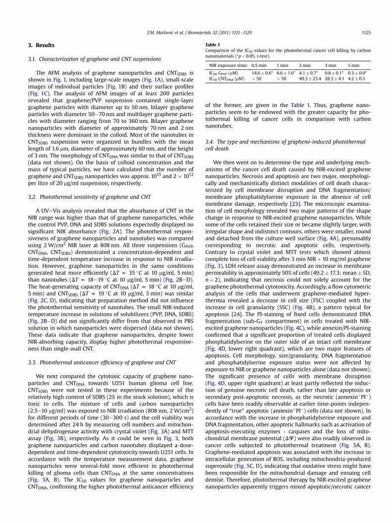
Table 1Comparison of the IC50 values for the photothermal cancer cell killing by carbonnanomaterials (*p< 0.05, t-test).
NIR exposure time: 0.5 min 1 min 2 min 3 min 5 min
IC50 GPVP (mM) 14.6� 0.6* 6.6� 1.6* 4.1� 0.7* 0.8� 0.1* 0.3� 0.0*IC50 CNTDNA (mM) > 50 > 50 49.3� 23.4 28.3� 4.1 4.2� 0.3
Z.M. Markovic et al. / Biomaterials 32 (2011) 1121e1129 1125
3. Results
3.1. Characterization of graphene and CNT suspensions
The AFM analysis of graphene nanoparticles and CNTSDBS isshown in Fig. 1, including large-scale images (Fig. 1A), small-scaleimages of individual particles (Fig. 1B) and their surface profiles(Fig. 1C). The analysis of AFM images of at least 200 particlesrevealed that graphene/PVP suspension contained single-layergraphene particles with diameter up to 50 nm, bilayer grapheneparticles with diameter 50e70 nm and multilayer graphene parti-cles with diameter ranging from 70 to 360 nm. Bilayer graphenenanoparticles with diameter of approximately 70 nm and 2 nmthickness were dominant in the colloid. Most of the nanotubes inCNTSDBS suspension were organized in bundles with the meanlength of 1.6 mm, diameter of approximately 60 nm, and the heightof 3 nm. The morphology of CNTDNA was similar to that of CNTSDBS(data not shown). On the basis of colloid concentration and themass of typical particles, we have calculated that the number ofgraphene and CNTSDBS nanoparticles was approx. 1015 and 2 � 1012
per litre of 20 mg/ml suspension, respectively.
3.2. Photothermal sensitivity of graphene and CNT
A UVeVis analysis revealed that the absorbance of CNT in theNIR range was higher than that of graphene nanoparticles, whilethe control PVP, DNA and SDBS solutions expectedly displayed nosignificant NIR absorbance (Fig. 2A). The photothermal respon-siveness of graphene nanoparticles and nanotubes was comparedusing 2 W/cm2 NIR laser at 808 nm. All three suspensions (GPVP,CNTDNA, CNTSDBS) demonstrated a concentration-dependent andtime-dependent temperature increase in response to NIR irradia-tion. However, graphene nanoparticles in the same conditionsgenerated heat more efficiently (DT z 35 �C at 10 mg/ml, 5 min)than nanotubes (DT z 18e19 �C at 10 mg/ml, 5 min) (Fig. 2BeD).The heat-generating capacity of CNTDNA (DT z 18 �C at 10 mg/ml,5 min) and CNTSDBS (DT z 19 �C at 10 mg/ml, 5 min) was similar(Fig. 2C, D), indicating that preparation method did not influencethe photothermal sensitivity of nanotubes. The small NIR-inducedtemperature increase in solutions of solubilizers (PVP, DNA, SDBS)(Fig. 2BeD) did not significantly differ from that observed in PBSsolution in which nanoparticles were dispersed (data not shown).These data indicate that graphene nanoparticles, despite lowerNIR-absorbing capacity, display higher photothermal responsive-ness than single-wall CNT.
3.3. Photothermal anticancer efficiency of graphene and CNT
We next compared the cytotoxic capacity of graphene nano-particles and CNTDNA towards U251 human glioma cell line.CNTSDBS were not tested in these experiments because of therelatively high content of SDBS (2% in the stock solution), which istoxic to cells. The mixture of cells and carbon nanoparticles(2.5e10 mg/ml) was exposed to NIR irradiation (808 nm, 2 W/cm2)for different periods of time (30e300 s) and the cell viability wasdetermined after 24 h by measuring cell numbers and mitochon-drial dehydrogenase activity with crystal violet (Fig. 3A) and MTTassay (Fig. 3B), respectively. As it could be seen in Fig. 3, bothgraphene nanoparticles and carbon nanotubes displayed a dose-dependent and time-dependent cytotoxicity towards U251 cells. Inaccordance with the temperature measurement data, graphenenanoparticles were several-fold more efficient in photothermalkilling of glioma cells than CNTDNA at the same concentrations(Fig. 3A, B). The IC50 values for graphene nanoparticles andCNTDNA, confirming the higher photothermal anticancer efficiency
of the former, are given in the Table 1. Thus, graphene nano-particles seem to be endowed with the greater capacity for pho-tothermal killing of cancer cells in comparison with carbonnanotubes.
3.4. The type and mechanisms of graphene-induced photothermalcell death
We then went on to determine the type and underlying mech-anisms of the cancer cell death caused by NIR-excited graphenenanoparticles. Necrosis and apoptosis are two major, morphologi-cally and mechanistically distinct modalities of cell death charac-terized by cell membrane disruption and DNA fragmentation/membrane phosphatidylserine exposure in the absence of cellmembrane damage, respectively [23]. The microscopic examina-tion of cell morphology revealed two major patterns of the shapechange in response to NIR-excited graphene nanoparticles. Whilesome of the cells retained their size or became slightly larger, withirregular shape and indistinct contours, others were smaller, roundand detached from the culture well surface (Fig. 4A), presumablycorresponding to necrotic and apoptotic cells, respectively.Contrary to crystal violet and MTT tests which showed almostcomplete loss of cell viability after 3 min NIRþ 10 mg/ml graphene(Fig. 3), LDH release assay demonstrated an increase in membranepermeability in approximately 50% of cells (49.2�17.3; mean� SD,n¼ 2), indicating that necrosis could not solely account for thegraphene photothermal cytotoxicity. Accordingly, a flow cytometricanalysis of the cells that underwent graphene-mediated hyper-thermia revealed a decrease in cell size (FSC) coupled with theincrease in cell granularity (SSC) (Fig. 4B), a pattern typical forapoptosis [24]. The PI-staining of fixed cells demonstrated DNAfragmentation (sub-G1 compartment) in cells treated with NIR-excited graphene nanoparticles (Fig. 4C), while annexin/PI-stainingconfirmed that a significant proportion of treated cells displayedphosphatidylserine on the outer side of an intact cell membrane(Fig. 4D, lower right quadrant), which are two major features ofapoptosis. Cell morphology, size/granularity, DNA fragmentationand phosphatidylserine exposure status were not affected byexposure to NIR or graphene nanoparticles alone (data not shown).The significant presence of cells with membrane disruption(Fig. 4D, upper right quadrant) at least partly reflected the induc-tion of genuine necrotic cell death, rather than late apoptosis orsecondary post-apoptotic necrosis, as the necrotic (annexinþPIþ)cells have been readily observable at earlier time-points indepen-dently of “true” apoptotic (annexinþPI�) cells (data not shown). Inaccordance with the increase in phosphatidylserine exposure andDNA fragmentation, other apoptotic hallmarks such as activation ofapoptosis-executing enzymes - caspases and the loss of mito-chondrial membrane potential (DJ) were also readily observed incancer cells subjected to photothermal treatment (Fig. 5A, B).Graphene-mediated apoptosis was associated with the increase inintracellular generation of ROS, including mitochondria-producedsuperoxide (Fig. 5C, D), indicating that oxidative stress might havebeen responsible for the mitochondrial damage and ensuing celldemise. Therefore, photothermal therapy by NIR-excited graphenenanoparticles apparently triggers mixed apoptotic/necrotic cancer

Fig. 4. The type of graphene-induced photothermal cell death. (AeD) U251 cells were exposed for 3 min to NIR laser (808 nm, 2 W/cm2) in the absence or presence of GPVP (10 mg/ml). After 24 h, the cell morphology was examined by inverted microscopy (A), while flow cytometry was used to assess the cell size (FSC) and granularity (SSC) (B), DNA frag-mentation (C) or externalization of phosphatidylserine and membrane permeability (Ann/PI-staining) (D). The representative photomicrographs, histograms and dot plots from oneof three experiments are presented (no significant changes were observed in cell cultures exposed to NIR or GPVP alone - not shown).
Z.M. Markovic et al. / Biomaterials 32 (2011) 1121e11291126

Fig. 5. The intracellular mechanisms of graphene-induced photothermal cell death. (AeD) U251 cells were exposed for 3 min to NIR laser (808 nm, 2 W/cm2) in the absence orpresence of GPVP (10 mg/ml). After 24 h, flow cytometry was used to assess caspase activation (ApoStat staining; A), while the mitochondrial membrane potential (DJ) (DePsipherstaining; B), production of ROS (DHR staining; C) and superoxide (DHE staining; D) were measured after 4 h. The representative photomicrographs, histograms and dot plots areshown. The results are mean� SD values from three experiments (*p< 0.05 refers to untreated cells and cells treated with NIR or GPVP alone; ANOVA).
Z.M. Markovic et al. / Biomaterials 32 (2011) 1121e1129 1127
Z.M. Markovic et al. / Biomaterials 32 (2011) 1121e11291128
cell death associated with oxidative stress, mitochondrial depo-larization and caspase activation.
4. Discussion
The present study demonstrates the in vitro photothermalanticancer activity of NIR-excited graphene nanoparticles coatedwith biocompatible polymer PVP. The observed effect was morepronounced than that of carbon nanotubes and involved mixedapoptotic/necrotic cancer cell death associated with the inductionof oxidative stress and mitochondrial membrane depolarization.These data are consistent with the recently demonstrated hightumour uptake of polyethylene glycol-solubilized graphene nano-particles and the efficient photothermal ablation of cancer tissue indifferent xenograft tumour mouse models [18].
The superior photothermal efficiency of graphene nanoparticlesin comparison with nanotubes might seem somewhat unexpected,having in mind that the latter displayed better NIR-absorbingcapacity in the present study. Moreover, the mass-specific heatcapacity of single-wall CNT (0.65 J/gK) [25] is three-fold lower thanthat of graphene (2 J/gK) [26], indicating that nanotubes shouldgenerate heat faster than graphene. However, the fact that gra-phene nanoparticles were apparently better dispersed than single-wall CNT could presumably influence the photothermal sensitivityof the two carbon nanomaterials. To compare the experimentalresults with the predicted heating capacities of graphene andsingle-wall CNT, we have used the following equations, taking intoaccount the thermodynamic, optical and geometrical properties ofcarbon nanoparticles:
DQ ¼ mcDT ¼ Nm1cDT (1)
AzNSEf (2)
where DQ is the developed heat, m is the mass of graphene(nanotube) particles, c is the mass-specific heat capacity, DT is thetemperature increase, N is the number of particles in suspension,m1 is the mass of the representative graphene bilayer or nanotubebundle, A is the absorption at 808 nm determined by UVevis, S isthe surface of graphene/nanotube that absorbs light and Ef is theefficiency of light absorption for graphene (z2.3% per layer) [27]and single-wall CNT (z20% per bundle) [28]. For simplicity, weassumed that the developed heat is identical for nanotubes andgraphene. On the basis of relation (1) and (2), the followingsimplified relation for the relative temperature increase of gra-phene and nanotube suspensions is derived:
DTGDTCNT
¼ EfG � ACNT � cCNT � p dCNT4
EfCNT � AG � cG � hG(3)
where dCNT and hG are the average diameter of CNT bundle andheight of graphene nanoparticles, respectively, as determined byAFM analysis. The calculated DTG/DTCNT value ofz3.5 is close to theexperimentally observed value of z2 (corresponding to the DTGand DTCNT of 35 �C and 18e19 �C, respectively e Fig. 2), indicatingthat the smaller size of graphene nanoparticles could indeed morethan counterbalance the inferior photothermal performance ofgraphene. One might argue that the observed difference in gra-phene and CNT dispersivity was simply due to the inappropriatechoice of solubilizers (PVP vs. DNA or SDBS). However, while PVPwas the optimal choice for graphene solubilization, SDBS and DNAin our hands actually performed better in solubilizing nanotubeswhen compared to PVP or other dispersants such as hydrox-yethylcellulose and melamine sulfonate (Markovic et al., unpub-lished data). While our preliminary results suggest that multi-wall
CNT, in accordance with their more metallic nature, could havebetter photothermal sensitivity than their single-wall counterparts,they still generated less heat than graphene nanoparticles (Mar-kovic et al., unpublished observation). Our data are consistent withthe known tendency of carbon nanotubes to aggregate into largeropes or bundles due to attractive van der Waals interactions, thusmaking the production of suspensions with well-dispersed singlenanotubes a non-trivial, if not an insurmountable task whenconventional low cost methodology and as-produced CNT areused [29]. Therefore, although a single nanotube in theory couldoutperform a single graphene layer in terms of photothermalsensitivity, it nevertheless might be easier to produce the suspen-sions of graphene nanoparticles with satisfactory photothermalperformance.
In accordance with the excellent photothermal responsiveness,graphene nanoparticles in our experiments efficiently killed cancercells by hyperthermia. However, the type and mechanisms oftumour cell death induced by an anticancer agent are importantdeterminants of its efficiency. For example, induction of apoptosis,a programmed cell death characterized by DNA fragmentation inthe absence of cell membrane disruption is frequently associatedwith the development of tumor cell resistance to therapy [30]. Onthe other hand, necrotic cell death could lead to a beneficialenhancement of the immune response against tumor [23].Although it has been generally assumed that photothermal therapyinduces necrotic cell death by causing irreversible protein dena-turation and/or cell membrane damage [14,31e33], some recentstudies indicate that, depending on temperature increase and/orexposure time, it can also trigger apoptosis [33e37]. Accordingly,the present study demonstrates that hyperthermia induced by NIR-excited graphene nanoparticles causes mixed apoptotic/necroticcancer cell death associated with oxidative stress/superoxideproduction and mitochondrial membrane depolarization. Thesefindings are consistent with the fact that the loss of mitochondrialmembrane potential is a common initial event in both apoptosisand necrosis, the final outcome mainly depending on the intra-cellular ATP level [38]. Mitochondrial depolarization is usuallycaused by ROS [38] and it further potentiates oxidative stress andcell demise through electron leaking from the respiratory chain andsubsequent generation of harmful superoxide anion [39], whichwas also observed during graphene-mediated photothermal cyto-toxicity in our experiments. Expectedly, oxidative stress-mediatedmitochondrial damage and apoptotic DNA fragmentation in thepresent study were accompanied by activation of caspases, thekey apoptosis-executing intracellular enzymes activated by smallmolecules such as cytochrome C, leaking from damaged mito-chondria [40]. The potential significance of these findings lies inthe possibility that pharmacological conversion of apoptosis tonecrosis by interference with caspase activity and/or loweringATP levels could increase therapeutic efficiency of NIR-excitedgraphene through stimulation of antitumour immune response andsuppression of development of therapeutic resistance.
5. Conclusions
The data presented here demonstrate the in vitro photothermalanticancer activity of graphene nanoparticles, mediated throughinduction of oxidative stress and mitochondrial damage resultingin both apoptotic and necrotic death of tumor cells. Importantly,our results indicate that the photothermal anticancer efficiency ofgraphene, due to the better dispersivity/smaller size of graphenenanoparticles, is superior to that of its structural sibling andpotential future rival in cancer treatment, carbon nanotube. This, inaddition to the large surface area, low toxicity and extremely low

Z.M. Markovic et al. / Biomaterials 32 (2011) 1121e1129 1129
cost, makes graphene-based nanoparticles promising candidatesfor the photothermal therapy of cancer.
Appendix
Figures with essential color discrimination. Fig. 1 in this article isdifficult to interpret in black and white. The full color images can befound in the online version, at doi:10.1016/j.biomaterials.2010.10.030.
References
[1] Wust P, Hildebrandt B, Sreenivasa G, Rau B, Gellermann J, Riess H, et al.Hyperthermia in combined treatment of cancer. Lancet Oncol 2002;3:487e97.
[2] Fiorentini G, Szasz A. Hyperthermia today: electric energy, a new opportunityin cancer treatment. J Cancer Res Ther 2006;2:41e6.
[3] Huang X, Jain PK, El-Sayed IH, El-Sayed MA. Gold nanoparticles: interestingoptical properties and recent applications in cancer diagnostics and therapy.Nanomedicine (Lond) 2007;2:681e93.
[4] Son SJ, Bai X, Lee SB. Inorganic hollow nanoparticles and nanotubes innanomedicine Part 2: imaging, diagnostic, and therapeutic applications. DrugDiscov Today 2007;12:657e63.
[5] Frangioni JV. In vivo near-infrared fluorescence imaging. Curr Opin Chem Biol2003;7:626e34.
[6] Weissleder R. A clearer vision for in vivo imaging. Nat Biotechnol2001;19:316e7.
[7] Novoselov KS, Geim AK, Morozov SV, Jiang D, Zhang Y, Dubonos SV, et al.Electric field effect in atomically thin carbon films. Science 2004;306:666e9.
[8] Zhang Y, Ali SF, Dervishi E, Xu Y, Li Z, Casciano D, et al. Cytotoxicity effects ofgraphene and single-wall carbon nanotubes in neural phaeochromocytoma-derived PC12 cells. ACS Nano 2010;4:3181e6.
[9] Yang W, Ratinac KR, Ringer SP, Thordarson P, Gooding JJ, Braet F. Carbonnanomaterials in biosensors: should you use nanotubes or graphene? AngewChem Int Ed Engl 2010;49:2114e38.
[10] Jagadeesan D, Eswaramoorthy M. Functionalized carbon nanomaterialsderived from carbohydrates. Chem Asian J 2010;5:232e43.
[11] Kam NW, O’Connell M, Wisdom JA, Dai H. Carbon nanotubes as multifunc-tional biological transporters and near-infrared agents for selective cancer celldestruction. Proc Natl Acad Sci U S A 2005;102:11600e5.
[12] Torti SV, Byrne F, Whelan O, Levi N, Ucer B, Schmid M, et al. Thermal ablationtherapeutics based on CN(x) multi-walled nanotubes. Int J Nanomedicine2007;2:707e14.
[13] Zhou F, Xing D, Ou Z, Wu B, Resasco DE, Chen WR. Cancer photothermaltherapy in the near-infrared region by using single-walled carbon nanotubes.J Biomed Opt 2009;14:021009.
[14] Wang CH, Huang YJ, Chang CW, Hsu WM, Peng CA. In vitro photothermaldestruction of neuroblastoma cells using carbon nanotubes conjugated withGD2 monoclonal antibody. Nanotechnology 2009;20:315101.
[15] Burke A, Ding X, Singh R, Kraft RA, Levi-Polyachenko N, Rylander MN, et al.Long-term survival following a single treatment of kidney tumors withmultiwalled carbon nanotubes and near-infrared radiation. Proc Natl Acad SciU S A 2009;106:12897e902.
[16] Xiao Y, Gao X, Taratula O, Treado S, Urbas A, Holbrook RD, et al. Anti-HER2 IgYantibody-functionalized single-walled carbon nanotubes for detection andselective destruction of breast cancer cells. BMC Cancer 2009;9:351.
[17] Moon HK, Lee SH, Choi HC. In vivo near-infrared mediated tumor destructionby photothermal effect of carbon nanotubes. ACS Nano 2009;3:3707e13.
[18] Yang K, Zhang S, Zhang G, Sun X, Lee ST, Liu Z. Graphene in mice: ultrahigh invivo tumor uptake and efficient photothermal therapy. Nano Lett2010;10:3318e23.
[19] Bourlinos AB, Georgakilas V, Zboril R, Steriotis TA, Stubos AK, Trapalis C.Aqueous-phase exfoliation of graphite in the presence of poly-vinylpyrrolidone for the production of water-soluble graphenes. Solid StateCommun 2009;149:2172e6.
[20] Jovanovi�c SP, Markovi�c ZM, Kleut DN, Romcevi�c NZ, Trajkovi�c VS,Drami�canin MD, et al. A novel method for the functionalization of gamma-irradiated single wall carbon nanotubes with DNA. Nanotechnology2009;20:445602.
[21] Markovi�c Z, Jovanovi�c S, Kleut D, Rom�cevi�c N, Jokanovi�c V, Trajkovi�c V, et al.Comparative study on modification of single wall carbon nanotubes bysodium dodecylbenzene sulfonate and melamine sulfonate superplasticiser.Appl Surf Sci 2009;255:6359e66.
[22] Raicevic N, Mladenovic A, Perovic M, Harhaji L, Miljkovic D, Trajkovic V. Ironprotects astrocytes from 6-hydroxydopamine toxicity. Neuropharmacology2005;48:720e31.
[23] Edinger AL, Thompson CB. Death by design: apoptosis, necrosis and autoph-agy. Curr Opin Cell Biol 2004;16:663e9.
[24] Vermes I, Haanen C, Reutelingsperger C. Flow cytometry of apoptotic celldeath. J Immunol Methods 2000;243:167e90.
[25] Hone J, Batlogg B, Benes Z, Johnson AT, Fischer JE. Quantized phonon spectrumof single-wall carbon nanotubes. Science 2000;289:1730e3.
[26] Mounet N, Marzari N. First-principles determination of the structural, vibra-tional and thermodynamic properties of diamond, graphite, and derivatives.Phys Rev B 2005;71:205214.
[27] Kuzmenko AB, van Heumen E, Carbone F, van der Marel D. Universal opticalconductance of graphite. Phys Rev Lett 2008;100:117401.
[28] Hsu IK, Pettes MT, Bushmaker A, Aykol M, Shi L, Cronin SB. Optical absorptionand thermal transport of individual suspended carbon nanotube bundles.Nano Lett 2009;9:590e4.
[29] Coleman JN. Liquid-Phase exfoliation of nanotubes and graphene. Adv FunctMater 2009;19:3680e95.
[30] Hersey P, Zhang XD. Overcoming resistance of cancer cells to apoptosis. J CellPhysiol 2003;196:9e18.
[31] Feyh J, Gutmann R, Leunig A, Jäger L, Reiser M, Saxton RE, et al. MRI-guidedlaser interstitial thermal therapy (LITT) of head and neck tumors: progresswith a new method. J Clin Laser Med Surg 1996;14:361e6.
[32] Koller MR, Hanania EG, Stevens J, Eisfeld TM, Sasaki GC, Fieck A, et al. High-throughput laser-mediated in situ cell purification with high purity and yield.Cytometry A 2004;61:153e61.
[33] Lee C, Hong C, Kim H, Kang J, Zheng HM. TiO2 nanotubes as a therapeuticagent for cancer thermotherapy. Photochem Photobiol 2010;86:981e9.
[34] Kirui DK, Rey DA, Batt CA. Gold hybrid nanoparticles for targeted photo-therapy and cancer imaging. Nanotechnology 2010;21:105105.
[35] Kasili PM, Vo-Dinh T. Photothermal treatment of human carcinoma cells usingliposome-encapsulated gold nanoshells. Nanobiotechnology 2005;1:245e52.
[36] Tong L, Cheng JX. Gold nanorod-mediated photothermolysis inducesapoptosis of macrophages via damage of mitochondria. Nanomedicine (Lond)2009;4:265e76.
[37] Nikfarjam M, Muralidharan V, Malcontenti-Wilson C, Christophi C. Theapoptotic response of liver and colorectal liver metastases to focal hyper-thermic injury. Anticancer Res 2005;25:1413e9.
[38] Kim JS, He L, Lemasters JJ. Mitochondrial permeability transition: a commonpathway to necrosis and apoptosis. Biochem Biophys Res Commun2003;304:463e70.
[39] Turrens JF. Mitochondrial formation of reactive oxygen species. J Physiol2003;552:335e44.
[40] Chen M, Wang J. Initiator caspases in apoptosis signaling pathways. Apoptosis2002;7:313e9.
Related Documents