This article appeared in a journal published by Elsevier. The attached copy is furnished to the author for internal non-commercial research and education use, including for instruction at the authors institution and sharing with colleagues. Other uses, including reproduction and distribution, or selling or licensing copies, or posting to personal, institutional or third party websites are prohibited. In most cases authors are permitted to post their version of the article (e.g. in Word or Tex form) to their personal website or institutional repository. Authors requiring further information regarding Elsevier’s archiving and manuscript policies are encouraged to visit: http://www.elsevier.com/copyright

Welcome message from author
This document is posted to help you gain knowledge. Please leave a comment to let me know what you think about it! Share it to your friends and learn new things together.
Transcript

This article appeared in a journal published by Elsevier. The attachedcopy is furnished to the author for internal non-commercial researchand education use, including for instruction at the authors institution
and sharing with colleagues.
Other uses, including reproduction and distribution, or selling orlicensing copies, or posting to personal, institutional or third party
websites are prohibited.
In most cases authors are permitted to post their version of thearticle (e.g. in Word or Tex form) to their personal website orinstitutional repository. Authors requiring further information
regarding Elsevier’s archiving and manuscript policies areencouraged to visit:
http://www.elsevier.com/copyright

Author's personal copy
In vitro biocompatibility and bacterial adhesionof physico-chemically modified Ti6Al4V surface
by means of UV irradiation
Amparo M. Gallardo-Moreno a,b, Miguel A. Pacha-Olivenza b,a, Laura Saldana b,c,Ciro Perez-Giraldo d,b, Jose M. Bruque a,b, Nuria Vilaboa c,b, M. Luisa Gonzalez-Martın a,b,*
a Department of Applied Physics, Faculty of Science, University of Extremadura, Avda. Elvas s/n, 06071 Badajoz, Spainb Networking Research Center on Bioengineering, Biomaterials and Nanomedicine, CIBER-BBN, Spain
c Research Unit, University Hospital La Paz, Paseo de la Castellana 261, 28046 Madrid, Spaind Department of Microbiology, Faculty of Medicine, University of Extremadura, Av. Elvas s/n, 06071 Badajoz, Spain
Received 1 February 2008; received in revised form 22 July 2008; accepted 23 July 2008Available online 6 August 2008
Abstract
UV irradiation leads to a ‘‘spontaneous” wettability increase of the Ti6Al4V surface while preserving bulk properties of the alloy thatare crucial for its performance as an orthopedic and dental implant. We hypothesized that UV treatment of Ti6Al4V may impair bac-terial adhesion without compromising the good response of human bone-forming cells to this alloy. The in vitro biocompatibility of theTi6Al4V surface, before and after UV irradiation, was analyzed by using human cells related to the osteoblastic phenotype. The adhesionprocesses of bacterial strains related to clinical orthopedic infections, i.e., Staphylococcus aureus and Staphylococcus epidermidis, werestudied theoretically and in vitro, under dynamic and static conditions as well as in the presence or absence of shear forces. While humancell adhesion was not altered by UV irradiation of Ti6Al4V alloy, this treatment reduced not only initial bacterial adhesion rates but alsothe number of bacteria retained on the surface after the passage of two air–liquid interfaces on the previously adhered bacteria. Thisstudy proposes the use of UV treatment prior to implantation protocols as an easy, economic and effective way of reducing bacterialadhesion on the Ti6Al4V surface without compromising its excellent biocompatibility.� 2008 Acta Materialia Inc. Published by Elsevier Ltd. All rights reserved.
Keywords: Ti6Al4V; Ultraviolet; Biocompatibility; Bacterial adhesion
1. Introduction
Microbial infection is one of the most destructive com-plications related to orthopedic implants because antimi-crobial therapy usually lacks efficacy at the point whenthe infection process is detected [1,2]. Controlled antibioticrelease from the biomaterial and antibiotic loading on thebiomaterial surface are strategies employed to overcomethis problem, but there is concern about the increased
microbial resistance to antibiotics that these proceduresmay induce. An alternative approach in the fight againstbacterial adhesion focuses on the modification of phys-ico-chemical surface properties of the biomaterial, suchas hydrophobicity, surface tension and electrical surfacepotential, because they are crucial in the initial approachand further retention of bacterial cells to various surfaces[3–5]. On the other hand, adequate adhesion of osteoblastsand their progenitors to the implant surface ultimatelyinfluences their capacity to proliferate and perform theirspecific functions. Surface characteristics of materials, suchas chemistry and surface energy, play an essential role incell adhesion to biomaterials. Thus, any modification ofthe surface characteristics introduced in order to diminish
1742-7061/$ - see front matter � 2008 Acta Materialia Inc. Published by Elsevier Ltd. All rights reserved.
doi:10.1016/j.actbio.2008.07.028
* Corresponding author. Address: Department of Applied Physics,Faculty of Science, University of Extremadura, Avda. Elvas s/n, 06071Badajoz, Spain. Tel.: +34 924289532; fax: +34 924289651.
E-mail address: [email protected] (M.L. Gonzalez-Martın).
Available online at www.sciencedirect.com
Acta Biomaterialia 5 (2009) 181–192
www.elsevier.com/locate/actabiomat

Author's personal copy
adhesion of microorganisms to a biomaterial should notcompromise bone-forming cell adhesion.
The Ti6Al4V alloy is widely used in orthopedic and den-tal applications due to its low density, excellent mechanicaland anti-corrosive properties, and good biocompatibility[6,7]. The spontaneous passivation of this alloy forms athin outer layer, predominantly composed of amorphousor poorly recrystallized TiO2. We have recently shown thatthe presence of TiO2 changes the physico-chemical surfaceproperties of Ti6Al4V after UV irradiation [8]. Interest-ingly, titanium, TiO2 surfaces and anodized titanium alloywith a thin film of anatase show anti-bacterial propertiesunder UV treatment [9–11]. However, to date there is noinformation available on the affinity of human cells andbacteria for physico-chemically modified Ti6Al4V surfacesafter UV treatment. We hypothesized that UV treatment ofTi6Al4V may impair bacterial adhesion without compro-mising the behavior of human bone-forming cells on thisalloy. The present study reports on the in vitro biocompat-ibility of the Ti6Al4V surface after UV irradiation by eval-uating the behavior of three types of human cells related tothe osteoblastic phenotype, including the osteoblastic Saos-2 cell line, mesenchymal cells from bone marrow (hMSC)and primary osteoblasts (hOB). We address the in vitro ini-tial adhesion behavior and further retention of three staph-ylococci strains directly involved in implant-relatedinfections, i.e., Staphylococcus aureus ATCC29213, Staph-
ylococcus epidermidis ATCC35984 (producer of an extra-cellular polysaccharide substance, EPS) and S.
epidermidis HAM892 (mutant of S. epidermidisATCC35984, non-producer of EPS) [12–14]. The behaviorsof human cells and bacterial strains on the titanium alloysurface after irradiation are extensively discussed in rela-tion to UV-induced physico-chemical changes on thesurface.
2. Materials and methods
2.1. Ti6Al4V
Disks of Ti6Al4V were cut from bars of 25 or 20 mm indiameter kindly supplied by SURGIVAL S.A. (Valencia,Spain). A TIMETAL 6–4 ELI alloy was processed as ahot rolled annealed bar at 700 �C for 2 h, then air cooled.The disks were abraded on successively finer silicon carbidepapers, mechanically polished with diamond paste, and fin-ished with silica colloidal.
Prior to their use, the Ti6Al4V disks were carefullycleaned with distilled water at 60 �C, vigorously rubbedwith a smooth cotton cloth, then rinsed repeatedly, firstwith distilled water and finally with distilled and deionisedwater (Milli-Q system); they were then immersed in a bea-ker with distilled and deionized water and sonicated for10 min, rinsed again with distilled and deionized water,dried in an oven at 40 �C for 1 h and stored in a desiccatorfor no longer than 24 h. Samples used as controls were notsubjected to further treatment. A second set of samples was
exposed to an UV source for 15 h. This period was suffi-cient to guarantee a complete hydrophilization of the sur-face, as we have recently shown [8]. G15-T8 UV lampswere kindly provided by Philips (Philips Iberica, Spain).The lamps emitted predominantly at a wavelength of257.7 nm and the lamp glass avoided the production ofozone, which is produced by wavelengths lower than200 nm. The disks were positioned at 10 cm from the lightsource and centered, receiving an intensity of2.6 mW cm�2. The irradiation installation was inside anopaque wood chamber to prevent interference from theroom or daylight, or damage to the users.
2.2. Cell culture
Human osteosarcoma Saos-2 cells (ECACC, Salisbury,Wiltshire, UK) were grown in DMEM medium supple-mented with 10% (v/v) heat-inactivated foetal bovineserum (FBS), 500 UI ml�1 of penicillin and 0.1 mg ml�1
of streptomycin. Human mesenchymal stem cells frombone marrow (hMSC) were purchased from Cambrex BioScience (Verviers, Belgium) and maintained in growth med-ium (Cambrex). Primary bone cells (hOB) were obtainedfrom human bone specimens aseptically collected duringorthopedic knee surgery and were cultured as has beendescribed previously [15]. Bone samples were obtainedfrom four patients aged 72 ± 5 years old. Each bone sam-ple was processed in a separated primary culture and exper-iments were performed using cultures obtained fromindependent patients. Patients enrolled in this researchsigned Informed Consent and all procedures using humantissue designated ‘‘surgical waste” were approved by theHuman Research Committee of Hospital La Paz (Date ofApproval: 02-14-2007). hOB were cultured in DMEM con-taining 15% (v/v) FBS, 500 UI ml�1 of penicillin and0.1 mg ml�1 of streptomycin. Cells were maintained at37 �C under 5% CO2 and 95% air in a humidifiedincubator.
For cell culture, Ti6Al4V disks of 20 mm in diameterwere used. Ti6Al4V samples were routinely sterilised underUV light in a laminar flow hood for 15 min on each sideand stored until use. Immediately before use in cell cultureexperiments, parallel sets of samples were left untreated ortreated with UV light as described above.
2.3. Cell spreading assays
Cells were seeded on Ti6Al4V samples treated oruntreated with UV light in 12-well plates (1.5 � 105 Saos-2 cells/well, 6 � 104 hMSC/well and 6 � 104 hOB/well) asdescribed above and cultured for 3 or 24 h. Attached cellswere fixed with 4% formaldehyde in PBS and permeabili-zed with 0.1% Triton X-100 in PBS. The cells were thenstained with PBS containing 4 � 10�7 M phalloidine-TRITC (Sigma) to visualize filamentous actin and withPBS containing 3 � 10�6 M 4,6-diamidino-2-phenylindole(DAPI, Sigma) for nuclear DNA. Cells were examined
182 A.M. Gallardo-Moreno et al. / Acta Biomaterialia 5 (2009) 181–192

Author's personal copy
using a fluorescence microscope (Leica AF6000, Wetzlar,Germany). High-resolution fluorescence images from rep-resentative fields of each sample were captured and digi-tally deconvolved using Huygens software (ScientificVolume Imaging, Hilversum, The Netherlands). A totalof 30 cells randomly selected from five representativeimages per sample were manually outlined, and cell areaswere measured using ImageJ v1.34 image analysis software.
2.4. Bacterial strains and culture
Staphylococcus aureus ATCC29213 (S. aureus), S. epi-
dermidis ATCC35984 (S. epidermidis4) and S. epidermidis
HAM892 (S. epidermidis2) were stored at �80 �C in por-ous beads (Microbank, Pro-Lab Diagnostics, Austin, TX,USA). S. epidermidis HAM892 is a negative extracellularpolysaccharide substance producer (EPS-negative)mutant derived by acriflavine mutagenesis from S. epide-
rmidis ATCC35984 (EPS-positive) and it was kindly pro-vided by L. Baldassarri from the Laboratorio diUltrastrutture, Istituto Superiore di Sanita, Rome, Italy.From the frozen stock, blood agar plates were inoculatedand incubated at 37 �C to obtain cultures. From thesecultures, tubes of 4 ml of Trypticase Soy Broth (TSB)(BBL, Becton Dickinson, Cockeysville, Maryland, USA)were inoculated for 10 h at 37 �C and then 25 ll of thispre-culture was used again to inoculate 50 ml of TSB at37 �C for 14 h. This amount of time was sufficient toguarantee that all strains were at the stationary phaseof growing (previously checked with the growing curvesfor the three strains).
The bacteria were then harvested by centrifugation for5 min at 1000g (Sorvall TC6, Dulont, Newtown, Pennsyl-vania, USA) and washed three times with PBS conditionedat 37 �C. Then the bacteria were re-suspended in PBS at aconcentration of 3 � 108 bacteria ml�1 for flow experi-ments and contact angle assays, and of approximately107 bacteria ml�1 for zeta potential tests.
2.5. Bacterial adhesion–retention experiments
Flow experiments were carried out at 37 �C in a paral-lel plate flow chamber previously described in detail [16]by using an inverted metallographic microscope (OlympusGX51, Barcelona, Spain). Ti6Al4V disks of 25 mm werefixed in the center of one of the plates of the chamberto ensure laminar conditions for the liquid flow. Theadhesion process was analyzed by using two differentexperimental settings. In order to study the dynamicadhesion of bacteria on Ti6Al4V, the flow chamber wasplaced so that the alloy surface was on the top side ofthe flow channel and the bacteria were allowed to attachwhile flowing at a constant rate of 2 ml min�1 for 20 min.Live adhesion was followed as time passed by recordingthe images with a video camera connected to a computer.Initial adhesion rates (j0) were given as bacteria adheredper square centimeter and second after counting the
adhered bacteria in each image. To evaluate whether pro-gression of adhesion was modified in static conditions, thealloy sample was placed on the bottom side of the flowchannel, the flow was stopped and the bacteria were leftto deposit on the surface for 2 min. Then the parallelplate flow chamber was turned upside down in order totake images of the adhered bacteria in different areas ofthe sample and to count them. This last procedure wasrepeated until 20 min of adhesion was reached. At theend of each static adhesion period and after taking imagesof the adhered bacteria, the flow was slowly opened inorder to homogenize the bacterial concentration insidethe flow channel, and then stopped again. Previous exper-iments showed that adhered bacteria were not detachedduring this process. We will refer to this last experimentalsetting as static-time-function. Static-time adhesion rates(j) were given as bacteria adhered per square centimeterand second.
Once the adhesion process was finished, and with a finalnumber of bacteria nf on the titanium alloy surface, the sur-face was exposed to the passing of two liquid–air interfacesin order to check the strength of the bacterial retention.After the first and second liquid–air interfaces the numberof remaining bacteria on the surface was counted anddenoted as nI–1 and nI–2, respectively.
2.6. Physico-chemical surface characterization
2.6.1. Contact angle measurements
For the surface tension evaluation, the contact angles ofwater (hW) (Milli-Q Plus), formamide (hF) (puriss>99.0%,Fluka, Switzerland) and diiodomethane (hD) (purum>98%,Fluka, Switzerland) were determined on the Ti6Al4V sur-face and on bacterial lawns using the sessile droptechnique.
Bacterial lawns were prepared by filtering a bacterialsuspension though 0.45 lm pore size filters (Millipore,Molsheim, France) using negative pressure [17]. Filterswere left to air-dry at 37 �C for 60 min for S. aureus, and30 min for S. epidermidis4 and S. epidermidis2. The timefor drying was the time at which residual PBS disappearedbetween the cells and the ‘‘plateau contact angles” (h) wereobtained. This time was checked for the three strains priorto the experiments [17]. Then the filters were introducedinto an environmental chamber G211 (Kruss, Hamburg,Germany), connected to a thermostat to maintain the tem-perature at 37 �C. Before measuring the contact angle witha probe liquid, the chamber was allowed to become satu-rated with the vapor of the liquid employed. Subsequently,the contact angles were obtained by analysing the imagescaptured with a computer.
2.6.2. Zeta potential measurements
For the electrical surface potential characterization, thezeta potentials (f) of bacteria suspended in PBS were mea-sured at 37 �C with a laser Doppler velocimeter (CoulterDELSA 440, Langley Ford Instruments, Amherst, USA),
A.M. Gallardo-Moreno et al. / Acta Biomaterialia 5 (2009) 181–192 183
Author's personal copy
which uses scattering of incident laser light to provide thebacterial electrophoretic mobility (le) from the velocity ofthe particles inside an electric field [18]. f were related tole through the Helmholtz–Smoluchowski equation
f ¼ 4pge
le ð1Þ
where g and e are the medium viscosity and permittivity,respectively.
The zeta potential of Ti6Al4V was obtained from thestreaming potential [18] by using an electrokinetic analyzer(EKA, Anton Paar, Austria). The measurements weremade with the electrolyte 0.001 M KCl setting a ramp pres-sure of 600 mb. The pressure gradient (DP) between bothends of the clamping cell provoked movement of the elec-trolyte inside the electrokinetic channel, which, in turn,was reflected in a potential difference between both ends(DV). Both magnitudes were related to provide the valueof f
f ¼ DV � g � j0:1M � R0:1M
DP � e � R0:001M
ð2Þ
with R0.001M being the resistivity of the 0.001 M KCl elec-trolytic solution and j0.1M and R0.1M the electrical conduc-tivity and resistivity of a 0.1 M solution of KCl needed forcorrecting the surface conductivity of the sample.
2.7. X-DLVO analysis
The surface tension components, cLW (Lifshitz–van derWaals), cAB ¼ 2
ffiffiffiffiffiffiffiffiffiffiffiffifficþ � c�p
(acid–base, with c� the electron-donor and c+ the electron-acceptor parameters) were calcu-lated from the contact angles by applying the Young–Dup-re equation to each probe liquid (L)
cLðcos hþ 1Þ ¼ 2ffiffiffiffiffiffiffiffiffiffiffiffiffiffiffiffiffiffifficLW � cLW
L
qþ 2
ffiffiffiffiffiffiffiffiffiffiffiffifficþ � c�L
pþ 2
�ffiffiffiffiffiffiffiffiffiffiffiffiffic� � cþL
qð3Þ
where cL ¼ cLWL þ cAB
L is the surface tension of the probe li-quid [19].
At 37 �C these values were the following: for watercLW = 20.3 mJ m�2, c� = 25.0 mJ m�2, c+ = 25.0 mJ m�2,cAB = 50.0 mJ m�2; for formamide cLW = 36.7 mJ m�2,c� = 38.3 mJ m�2, c+ = 2.2 mJ m�2, cAB = 18.4 mJ m�2
for diiodomethane cLW = 48.1 mJ m�2, c� = 0.0 mJ m�2,c+ = 0.7 mJ m�2, cAB = 0.0 mJ m�2 [19,20].
The total interaction free energy, DG, between microor-ganisms and substrata through water (W) as a function ofthe separation distance (d) was calculated by the sum of theLifshitz–van der Waals (LW), the acid–base (AB) and theelectrical interaction free energies (DGLW, DGAB and DGEL,
respectively), as proposed by the extended DLVO theory(X-DLVO) and calculated as follows:
DGLWðdÞ ¼ �A6
adþ a
d þ 2aþ ln
dd þ 2a
� �� �ð4Þ
where A is the Hamaker constant, A ¼ �12pd20DGLW
d0, a is
the microorganisms radius, d0 the distance of the closestapproach between two surfaces and DGLW
d0was obtained
through the Lifshitz–van der Waals surface free energy asfollows:
DGLWd0¼
ffiffiffiffiffiffiffifficLW
B
q�
ffiffiffiffiffiffiffifficLW
S
q� �2
�ffiffiffiffiffiffiffifficLW
B
q�
ffiffiffiffiffiffiffiffifficLW
PBS
q� �2
�ffiffiffiffiffiffiffifficLW
S
q�
ffiffiffiffiffiffiffiffifficLW
PBS
q� �2
ð5Þ
Subscripts B and S are bacterium and substratum, respec-tively, and the surface free energy of the suspending liquid,PBS, is considered nearly equal to that of water.
DGAB ¼ 2pad0DGABd0
expðd0 � dÞ
d0
ð6Þ
where
and finally,
DGELðdÞ¼pe0eraðf2B
þf2SÞ
2fBfS
f2Bþf2
S
ln1þexpð�jdÞ1�expð�jdÞþlnð1�expð�2jdÞÞ
" #
ð8Þe0 is the dielectric constant of the vacuum and er the relativedielectric constant of the suspending liquid. fB and fS arethe zeta potentials of bacteria and substrata, respectively.
Accordingly to the X-DLVO theory, interaction freeenergies predict favorable adhesion when they are negativeand absence of adhesion when they are positive.
2.8. Statistical analysis
The statistical analysis of the samples was undertakenusing a one-tail unpaired t-test and one-way ANOVAs.All data reported are means ± SD of at least three indepen-dent experiments. The confidential range selected was 95%,which gives statistical differences when P-values <0.05.
3. Results and discussion
The changes observed in the Ti6Al4V surface after UVirradiation have already been discussed in a recent studyby our group [8]. Table 1 exhibits those important changesin hW and hF, the polar liquids, before and after irradiation(P < 0.05). Taking into account that hW can be considered
DGABd0¼ 2
ffiffiffiffiffiffiffiffifficþPBS
p ffiffiffiffiffic�Bp þ ffiffiffiffiffi
c�Sp � ffiffiffiffiffiffiffiffiffi
c�PBS
p� �þ ffiffiffiffiffiffiffiffiffi
c�PBS
p ffiffiffiffifficþB
pþ
ffiffiffiffifficþS
p�
ffiffiffiffiffiffiffiffifficþPBS
p �ffiffiffiffiffiffiffiffiffiffi
c�BcþSp
�ffiffiffiffiffiffiffiffiffifficþBc�S
p24
35 ð7Þ
184 A.M. Gallardo-Moreno et al. / Acta Biomaterialia 5 (2009) 181–192

Author's personal copy
a quantitative indicator of hydrophobicity, the control sur-face behaves as hydrophobic, while the UV-treated surfaceis presented as highly hydrophilic. The excitation of thesemiconductor TiO2 present on the surface of Ti6Al4Vgenerates electron–hole pairs, which are responsible forthe changes observed. The subsequent well-describedchemical reactions lead to the substitution of oxygen atomsof the TiO2 lattice for OH� groups, making the surfacehydrophilic [21–23]. Such hydrophilicity is also detected
in the values of the surface tension, mainly in the acid–basecomponent and the parameters c� and c+. As we haveshown, the increment in the electron-donor capacity isrelated to the presence of hydroxyl ions on the surface [8].
3.1. Effect of UV treatment on cell behavior
Among other surface characteristics, wettability isthought to play a pivotal role in cell response to materials.
Sao
s-2
hM
SC
hOB
Control UV Control UV
Sao
s-2
hM
SC
hOB
0
500
1000
1500
2000
2500
3000
3500
4000
4500
Saos
hMSC
hOB
Control UV
Cel
l are
a (µ
m2 )
0
500
1000
1500
2000
2500
3000
3500
4000
4500
Cel
l are
a (µ
m2 )
* *
hOB
Saos-2
hMSC
Saos
hMSC
hOB
* *
Control UV
hOB
Saos-2
hMSC
Fig. 1. Cell behaviour on Ti6Al4V surfaces. Cells were cultured on Ti6Al4V before irradiation (control) and after irradiation (UV) for 3 h (a) or 24 h (b).Actin cytoskeleton and nuclei staining of cells cultured on Ti6Al4V surfaces. Representative images of three independent experiments with similar results.Bars = 50 lm. The graphics show the cell area measured on Ti6Al4V surfaces and results represent the mean ± SD of three independent experiments.*P < 0.05 compared to hMSC and hOB.
Table 1Physico-chemical surface characterization of the Ti6Al4V surface before (control) and after 15 h of UV light treatment (UV) and for the three bacteriastudied
hW (�) hF (�) hD (�) f (mV) cLW (mJ m�2) c� (mJ m�2) c+ (mJ m�2) cAB (mJ m�2) c (mJ m�2)
Control 88 ± 3 66 ± 2 49 ± 1 �31 ± 2 32.3 3.2 0.2 1.5 33.8UV 9 ± 1 3.9 ± 0.2 42 ± 1 �30 ± 2 28.2 51.6 4.9 31.7 59.9
S. aureus 72 ± 3 58 ± 2 44 ± 1 �8 ± 2 30.4 13.8 0.3 3.9 34.3S. epidermidis4 74 ± 2 60 ± 2 49 ± 1 �6 ± 2 28.2 12.6 0.4 4.4 32.6S. epidermidis2 58 ± 2 65 ± 2 49 ± 1 �10 ± 2 24.8 40.0 0.01 0.7 25.5
Contact angles of water, formamide and diiodomethane (hW,hF,hD), zeta potential (f) and Lifshitz–van der Waals (cLW), acid base (cAB) and total (c)surface free energy and electron-donor (c�) and electron-acceptor (c+) parameters for all the samples.
A.M. Gallardo-Moreno et al. / Acta Biomaterialia 5 (2009) 181–192 185

Author's personal copy
In this sense, self-assembling monolayers (SAMs) of alkan-ethiols or alkylsilanes have provided suitable model sur-faces to study the influence of wettability and surfacefunctional groups on cell attachment. While it has beengenerally assumed that increased hydrophilicity favors celladhesion to surfaces [24], available data on mammaliancells adhesion to SAMs are far from conclusive [25–28].The very diverse cell types employed to assess the influenceof wettability on SAMs attachment could explain the lackof uniformity of the data, as cells from different originsmay respond differently to materials.
In order to study whether UV-induced changes ofTi6Al4V surface affect the ability of human bone-formingcells to adhere to the alloy, we examined the behavior ofthree types of cells related to the osteoblastic phenotype(Saos-2, hMSC and hOB). Examination of microphoto-graphs from cells incubated for 3 h on the surfaces revealedthat UV treatment had no significant effect on the adhesionability of any tested cell type (Fig. 1a). Quantification ofcells attached to the surfaces by means of a standardMTT assay confirmed these observations (data not shown).Information on the influence of wettability on cell behavioron titanium-based surfaces is quite limited, mainly due tothe fact that reported treatments which modify surfacehydrophilicity also lead to subtle changes in roughness.For example, heating or treatment of Ti6Al4V alloy withperoxide results in highly hydrophilic surfaces, withincreased roughness, which does not modify the rate ofattachment of human osteoblastic MG-63 cells [29]. How-ever, rabbit osteoblasts preferentially attached to heated Tisurfaces with an increased number of hydroxyl groups androughness, as compared to untreated samples [30]. Rough-ness of the surface was not affected after UV treatment(data not shown), and according to our data, early attach-ment of several culture models of human osteoblasts is notaffected by UV-induced changes in the wettability ofTi6Al4V alloy surfaces.
Fibroblasts attachment equalled rates on hydrophobicor hydrophilic SAMs but cell spreading was sensitive towettability, as it was impaired on highly hydrophilic sur-faces [26]. Therefore, we considered it of interest to studywhether changes in the alloy surface after UV treatmentof Ti6Al4V samples modified spreading patterns of Saos-2 cells, hMSC or hOB in the short culture period of 3 h.To this end, we quantified the area of cells cultured onthe surfaces and examined the ability of cells to form actinstress fibers. The average cell areas of both hMSC and hOBwere higher than those measured in Saos-2 cells (Fig. 1a).The quantified areas of cells cultured on UV-treated sur-faces were similar to those measured on surfaces withouttreatment, regardless of the cell type. Microscopic exami-nation of actin-stained cells revealed a higher organizationof actin cytoskeleton in both hMSC and hOB than in Saos-2 cells (Fig. 1a). While hMSC and hOB exhibited radiallypositioned bundles of actin filaments, no defined-stressfibers appeared in Saos-2 cells. Interestingly, the patternof actin distribution was unaffected by UV treatment in
any of the cell types. Examination of microphotographsrevealed that cells have markedly different shapes at 24 h(Fig. 1b). While Saos-2 cells had a typical polygonal mor-phology with well-formed actin filaments, hMSC andhOB expanded to an elongated shape in which actin fila-ments organized in well-defined-stress fibers mostly ori-ented in a parallel direction following the main cellularaxis. Actin network organization was unaffected by UVtreatment on Ti6Al4V samples. Rapid initial cell spreadingdetected in either hMSC or hOB was not significantly mod-ified after culturing cells for 24 h (Fig. 1b). However, theaverage area of Saos-2 cells increased greatly when the cul-ture period was extended. At 24 h, changes induced onTi6Al4V samples by UV treatment did not modify thespreading of any cell type. Interestingly, visualization ofSaos-2 cells, hMSC and hOB adhered to Ti6Al4V alloyafter 24 h indicated that cell viability was not affected byUV-induced changes of the surface. A recently describedmodified sand-blasted and acid-etched titanium surface(modSLA) with an extremely hydrophilic characterresulted in a reduction of viability of MG-63 cells, as com-pared to unmodified SLA [31]. ModSLA and SLA exhibitsimilar roughness, suggesting that cell colonization ofblasted surfaces could be inversely related to surface hydro-philicity. However, under our experimental conditions we
0
200
400
600
800
1000
1200
UVcontrol
j (b
acte
ria
cm-2
s-1)
* ** *
S. aureus
S. epidermidis 4
S. epidermidis 2
0
50
100
150
200
250
300
UVcontrol
j 0 (
bact
eria
cm
-2s-1
)
*
*
**
*
S. aureus
S. epidermidis 4
S. epidermidis 2
Fig. 2. Initial adhesion rates of the three bacterial strains to the Ti6Al4Vsurface located at the top of the parallel plate flow chamber, j0, (a) andadhesion rates when the sample is located at the bottom, j, (b), beforeirradiation (control) or after 15 h of irradiation (UV). *P < 0.05 specificcomparisons inside the text.
186 A.M. Gallardo-Moreno et al. / Acta Biomaterialia 5 (2009) 181–192

Author's personal copy
did not detect that osteoblastic viability is impaired on pol-ished, highly hydrophilic UV-treated Ti6Al4V alloy. Insummary, UV treatment of Ti6Al4V does not impair itsgood in vitro biocompatibility.
3.2. Effect of UV treatment on bacterial adhesion
Fig. 2 displays the values of the adhesion rates for thetwo processes, i.e., dynamic or j0 and static-time-functionor j, respectively, before (control) and after UV irradiation(UV). Initial adhesion rates (j0), calculated from the adhe-sion at the top of the surface, are lower than the adhesionrates (j), calculated from the adhesion at the bottom,regardless of the presence or absence of UV irradiation
(P < 0.05). These data suggest that bacterial adhesionstrongly depends on the relative position of the surfaceand the flow conditions of the surrounding media: staticadhesion, mainly mediated by deposition of bacteria, ishigher than dynamic adhesion, which avoids deposition.Measured differences between j and j0 can be as high as fif-teen-fold as detected for S. epidermidis2 after irradiation.Some experiments were also carried out in flow-dynamicconditions with the surface placed at the bottom plate inorder to check the contribution of the flow conditions tothe observed adhesion differences. The results (not shown)demonstrated that the final state of the substratum in flowconditions was the same as in static-time-function condi-tions, which implies that only the position of the surface,
Fig. 3. Representative images of the bacterial coverage state at the end of the dynamic flow experiment at top (a) and at the end of the static-time-functionadhesion experiment at bottom (b).
A.M. Gallardo-Moreno et al. / Acta Biomaterialia 5 (2009) 181–192 187

Author's personal copy
relative to the bacterial suspension, plays an important rolein adhesion tendency. This behavior was also observed insimilar experiments dealing with glass surfaces [32], whichmeans that deposition of bacteria plays an important rolein promoting adhesion.
Comparisons among the three strains show that thehighest adhesion was detected for S. aureus, followed byS. epidermidis4, while the lowest adhesion was for themutant strain S. epidermidis2. Differences were, withoutexception, statistically significant (P < 0.05). The resultsobtained could be in agreement with multiple data whichshow that S. aureus is the dominant organism associatedwith infected metallic implants while S. epidermidis is morefrequently isolated from non-metallic devices [13,33,34].The differences observed between both S. epidermidis
strains could be related to their different EPS production.In the case of S. epidermidis2 the suppression of its EPSproduction capacity makes it less adherent because EPSpromotes not only adhesion to different surfaces but alsoco-adhesion between bacteria [35,36]. Fig. 3 presents somerepresentative images of the final state of the Ti6Al4V sur-face when it is located at the top (a) and at the bottom (b).Image analysis of each experiment suggests that bacterialco-adhesion is the main reason for the differences betweenboth S. epidermidis strains at the end of the experiment.Despite sonication of all bacterial suspensions prior to flowexperiments, the number of bacteria counted in the case ofS. epidermidis4 was always increased by the large cumulusof bacterial cells in the images, while the density of singleattached bacteria seemed to be similar for S. epidermidis4and S. epidermidis2 (Fig. 3b). Nevertheless, initial adhesionof S. epidermidis4 always took place with single bacterialcells (Fig. 3a). The differences observed for j0 between bothS epidermidis strains were around 60% before irradiation,and 70% after irradiation (Fig. 2a), which means thatEPS production may also contribute to the higher adhesionof S. epidermidis4 to host surfaces.
The differences between Fig. 2a and b reveal that UVirradiation changes the adhesion rates of the bacteriastrains studied. With the exception of j for S. epidermidis2,the adhesion of bacteria decreased on the irradiated surfacecompared to the control sample regardless of the type ofadhesion test carried out. However, adhesion changes weremore evident when deposition was avoided (Fig. 2a). Thus,j0 diminished by 16% for S. aureus, by 28% for S. epidermi-
dis4 and by 45% for S. epidermidis2. In an attempt to find arelationship between adhesion rates before and after irradi-ation, we plotted j0 and j before UV against j0 and j afterUV. As shown in Fig. 4, a very good linear relationshipwith the slope of 1 and R2 = 0.9929 was found. This meansthat after irradiation both j0 and j decrease by about 33cells per square centimeter and second (independent termof the linear fitting) as compared to their correspondingvalues for the non-irradiated alloy. Although it is difficult,at present, to account for a specific empirical relationship,it could be speculated that whatever the properties or par-ticular characteristics of the bacterial strain, bacteria
y = 1.0072x - 32.944
R2 = 0.99290
100200300400500600700800900
0 200 400 600 800
j0 , j (control) (bacteria cm-2s-1)
j 0 ,
j (U
V)
(ba
cter
ia c
m-2
s-1)
Fig. 4. Relationship between the initial adhesion rates at top (j0, squares)and the adhesion rates at bottom (j, rhomboids) before (control) and after15 h of irradiation (UV). White symbols: S. epidermidis2, grey symbols: S.
epidermidis4, black symbols: S. aureus.
0
20
40
60
80
100
120
140
nf
nI-1
nI-2
* * *
*
n (b
acte
ria
cm-2
)10
-4n
(bac
teri
a cm
-2)
10-4
n (b
acte
ria
cm-2
)10
-4
UVcontrol
S. aureus
nI-2
nf
nI-1
0
20
40
60
80
100
120
140
nf
nI-1
nI-2
UVcontrol
S. epidermidis4
*
**
* nI-2
nf
nI-1
0
20
40
60
80
100
120
140
nf
nI-1
nI-2
UVcontrol
*
*
S. epidermidis2
nI-2
nf
nI-1
Fig. 5. Number of the final bacteria (nf) adhered to the Ti6Al4V surface atthe end of the experiment (bottom) and number of the remaining bacteriaafter the passing of the first liquid–air interface (nI�1) and after the secondliquid–air interface (nI�2) without irradiation (control) or after 15 h ofirradiation (UV) for S. aureus (a), S. epidermidis4 (b) and S. epidermidis2(c). *P < 0.05 specific comparisons inside the text.
188 A.M. Gallardo-Moreno et al. / Acta Biomaterialia 5 (2009) 181–192
Author's personal copy
‘‘sense” a similar Ti6Al4V surface change induced by UVirradiation.
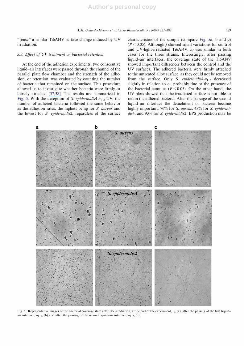
3.3. Effect of UV treatment on bacterial retention
At the end of the adhesion experiments, two consecutiveliquid–air interfaces were passed through the channel of theparallel plate flow chamber and the strength of the adhe-sion, or retention, was evaluated by counting the numberof bacteria that remained on the surface. This procedureallowed us to investigate whether bacteria were firmly orloosely attached [37,38]. The results are summarized inFig. 5. With the exception of S. epidermidis4-nI–2-UV, thenumber of adhered bacteria followed the same behavioras the adhesion rates, the highest being for S. aureus andthe lowest for S. epidermidis2, regardless of the surface
characteristics of the sample (compare Fig. 5a, b and c)(P < 0.05). Although j showed small variations for controland UV-light-irradiated Ti6Al4V, nf was similar in bothcases for the three strains. Interestingly, after passingliquid–air interfaces, the coverage state of the Ti6Al4Vshowed important differences between the control and theUV surfaces. The adhered bacteria were firmly attachedto the untreated alloy surface, as they could not be removedfrom the surface. Only S. epidermidis4-nI–2 decreasedslightly in relation to nf, probably due to the presence ofthe bacterial cumulus (P < 0.05). On the other hand, theUV plots showed that the irradiated surface is not able toretain the adhered bacteria. After the passage of the secondliquid–air interface the detachment of bacteria becamehighly important: 76% for S. aureus, 43% for S. epidermi-
dis4, and 93% for S. epidermidis2. EPS production may be
Fig. 6. Representative images of the bacterial coverage state after UV irradiation, at the end of the experiment, nf, (a), after the passing of the first liquid–air interface, nI�1, (b) and after the passing of the second liquid–air interface, nI�2, (c).
A.M. Gallardo-Moreno et al. / Acta Biomaterialia 5 (2009) 181–192 189

Author's personal copy
the reason for the highest retention observed in S. epidermi-
dis4. In fact, suppression of EPS production in S. epidermi-
dis2 gave the lowest retention. Fig. 6 presents an illustrativeimage of the state of the surface at the end of the experimentand after the passing of the two liquid–air interfaces. In thecase of S. epidermidis4 cumulus of bacteria appear evenafter the passing of the liquid–air interfaces.
The novel results presented above suggest that despiteunavoidable attachment of bacterial cells to Ti6Al4V, irra-diation of its surface with UV light prior to its use wouldfacilitate easy removal of most of the adhered bacteria byshear forces, which can be present in target sites wherethe implants are placed.
3.4. Physico-chemical approach to bacterial adhesion and
retention
Table 1 shows that hW was similar for S. aureus and S.
epidermidis4 (P > 0.05), both of which are hydrophobic,while hW was lower for S. epidermidis2 (P < 0.05). hF, hD
and f values were very similar for all the strains. S. epide-
rmidis2 had a very strong electron-donor capacity com-pared to the other two strains. Nevertheless, due to itslow c+ value, cAB and c were the lowest.
As a first and qualitative attempt to relate the physico-chemical parameters to in vitro bacteria adhesion, hydro-phobicity of the interacting surfaces can be taken as a pre-dictor of higher or lower adhesion. Although there is stillsome controversy about the role of substratum hydropho-bicity (or surface tension) in bacterial adhesion, [39,40]data from the present study indicate that substratumhydrophobicity plays a crucial role in the adhesion ratesof bacteria. Regardless of the strain selected, hydrophilicUV-irradiated Ti6Al4V presents lower adhesion rates ascompared with hydrophobic control surfaces (Fig. 3). Inaddition, the UV-irradiated Ti6Al4V surface also shows alow ability to retain bacterial cells, and liquid–air interfacescan remove more than three-quarters of the adhered bacte-ria. Working with yeast cells, we have also observed thatsilicone hydrophobic surfaces have lower detachment ratescompared with hydrophilic glass surfaces [38].
Despite the shortage of X-DLVO in its application tothe analysis of bacterial adhesion, this model could givesome indications on the behavior of the adhesion process.If we consider bacteria as colloidal particles of approxi-mately 900 nm in diameter, with surface tension and zetapotential as evaluated from contact angle and electropho-retic mobility measurements, interacting with a flat surface,the application of Eqs. (4)–(8) gives the dependence of theinteraction free energy with distance between particle andsurface. Fig. 7 presents DG, in kT units, as a function ofthe separation distance for S. epidermidis4 before and afterirradiation (Fig. 7a and b, respectively). The other twostrains had a similar pattern in the X-DLVO plots (datanot shown). A summary of the most relevant energeticinformation extracted from all the X-DLVO figures isgiven in Table 2. The most important feature related to
the data in Fig. 7 is the great change in the plot patternafter UV. For control surfaces, DG showed two favorableadhesion minima, a secondary minimum and a primaryminimum related with a potential well. For the UV-irradi-ated sample, despite a secondary minimum still appearing,the primary minimum was substituted by an energy barrier.
The assumption that bacteria are colloidal particles hasthe important shortcoming of supposing that real bacteriaare able to reach distances as close to the substratum sur-face as 1.57 A, which is the closest approach between twosurfaces described by the colloidal theory. For example,Li and Logan [41] have recently pointed out that Esche-
richia coli bacteria approach host surfaces to a distanceof within 50 A (distance roughly equal to the length ofthe core polysaccharide on Gram-negative bacteria), while
0 10 20 30 40 50 60
ΔG (
kT)
-90
-75
-60
-45
-30
-15
0
15
30
45ΔG
(kT
)
-90
-75
-60
-45
-30
-15
0
15
30
45
ΔGEL
ΔGAB
ΔGLW
ΔG
d (Å)
0 10 20 30 40 50 60
d (Å)
ΔGEL
ΔGAB
ΔGLW
ΔG
Fig. 7. X-DLVO plots of the total interaction free energy (DG) betweenS. epidermidis4 and the Ti6Al4V surface before (a) and after 15 h ofirradiation (b). Lifshitz–van der Waals (DGLW), acid–base (DGAB) andelectrical (DGEL) components of DG.
190 A.M. Gallardo-Moreno et al. / Acta Biomaterialia 5 (2009) 181–192

Author's personal copy
Sharma and Hanumantha assume that interaction dis-tances should be located at 6.57 A [42]. On this basis, thepredictions of X-DLVO model at the minimal distance,d0, seem unrealistic. Therefore, the secondary adhesionminimum has to play an important role in the bacterialadhesion process. The favorable interaction, resulting inadhesion to the Ti6Al4V surface, must take place whenbacteria fall into the secondary adhesion minimum at dis-tances of 26.6 A for S. aureus, 30.6 A for S. epidermidis4and 44.1 A for S. epidermidis2, for which DG is negativein all cases, as shown in Table 2. The plot of the adhesionrates against DG values of the secondary minimum in Fig. 8shows a very good relationship for j0–DG, whereas for j–DG
it is more difficult to define such a clear tendency. Thisresult can be related to the differences observed betweenboth adhesion rates. Bacterial adhesion to the surfacelocated at the top of the flux can be a consequence onlyof the initial adhesion tendency of bacteria, quantified byDG, while the results on the surface located at the bottomalso account for deposition.
After this first initial adhesion step, bacteria wouldstrengthen their linkage to the control surfaces. In the caseof the irradiated surface, differences in the surface proper-ties can prevent a firm retention to the surface, making theadhered cells vulnerable to any shear force, as proved bythe results obtained after the passage of two air–liquidinterfaces (Fig. 5).
4. Conclusions
UV irradiation of the Ti6Al4V surface does not compro-mise the excellent in vitro biocompatibility of the alloy, asmeasured by culturing human Saos-2 cells, primary osteo-blasts and mesenchymal stem cells on treated surfaces. Thephysico-chemical changes occurring on the surface induce areduction in adhesion rates of S. aureus ATCC29213, S.
epidermidis ATCC35984 and S. epidermidis HAM892under flow conditions. Despite the limitations that the X-DLVO theory has in its application to bacterial adhesion,it has been observed a good correlation between the initialadhesion rates and DG at the secondary minima for theTi6Al4V surface before and after irradiation. The observedlooser adhesion to the irradiated surface can be also relatedto the modification of the surface properties of the system.From a practical point of view, this study proposes UVtreatment prior to implantation protocols as an easy, eco-nomic and effective way of reducing the bacterial adhesionto the Ti6Al4V surface without altering the excellent bio-compatibility of the alloy.
Acknowledgments
This work was supported by Grants MAT2006-12948-C04-02-03-04 from the Ministerio Espanol de Ciencia yTecnologıa, Grant 3PR05A021 from the Consejerıa deEconomıa, Comercio e Innovacion of the Junta deExtremadura, Grant CAM S-0505/MAT/000324 fromComunidad de Madrid, and Fundacion Mutua Madrilena.N.V. is supported by program I3SNS from Fondo deInvestigaciones Sanitarias. The authors thank the medicalstaff of the Orthopedic Department (Hospital UniversitarioLa Paz, Madrid, Spain) for providing us with bone sam-ples, and SURGIVAL S.A. (Valencia, Spain) for supplyingus the Ti6Al4V alloy samples used in this study.
References
[1] Mack D, Rohde H, Harris LG, Davies AP, Horstkotte MA,Knobloch JK. Biofilm formation in medical device-related infection.Int J Artif Organs 2006;29:343–59.
[2] Edupuganti OP, Antoci V, King SB, Jose B, Adams CS, Parvizi J,et al. Covalent bonding of vancomycin to Ti6Al4V alloy pins
Table 2Total interaction free energies (DG) at the minimal separation distance (d0) and energetic information of singular points observed in the X-DLVO graphics:presence or absence of maxima, locations and energy of secondary minima and existence of primary minima
d0 MAX Secondary minimum Primary minimum
DG (mJ m�2) DG (kT) d (A) DG (kT) d (A) DG (kT)
Control S. aureus �42.96 �4504.94 16.1 �8.48 26.6 �9.92 YesS. epidermidis4 �43.26 �4532.99 15.1 �3.62 30.6 �7.32 YesS. epidermidis2 �20.66 �2084.21 10.6 28.55 44.1 �3.27 Yes
UV S. aureus 10.73 1065.11 No 33.1 �5.90 NoS. epidermidis4 9.76 961.92 No 36.1 �4.44 NoS. epidermidis2 28.21 2981.23 No 48.1 �2.06 No
Data are given for the interaction of the three bacterial strains with the Ti6Al4V surface before irradiation (control) and after irradiation (UV).
0
50
100
150
200
-10 -9 -8 -7 -6 -5 -4 -3 -2 -1 0
ΔG at secondary minimum (kT)
j 0 (b
acte
ria
cm-2
s-1 )
400
500
600
700
800
900
1000
j (b
acte
ria
cm-2
s-1 )
Fig. 8. Relationships between the initial adhesion rates at the top (j0,circles) and the adhesion rates at the bottom (j, triangles) against theinteraction free energy (DG) calculated at the secondary energy minima.White symbols: S. epidermidis2, grey symbols: S. epidermidis4, blacksymbols: S. aureus.
A.M. Gallardo-Moreno et al. / Acta Biomaterialia 5 (2009) 181–192 191

Author's personal copy
provides long-term inhibition of Staphylococcus aureus colonization.Bioorg Med Chem Lett 2007;17:2692–6.
[3] An YH, Friedman RJ. Handbook of bacterial adhesion. Principles,methods and applications. Totowa, NJ: Humana Press Inc.; 2000.
[4] Bos R, Van der Mei HC, Busscher HJ. Physico-chemistry of initialmicrobial adhesive interactions – its mechanisms and methods forstudy. FEMS Microbiol Rev 1999;23:179–230.
[5] Mendez-Vilas A, Gallardo-Moreno AM, Calzado-Montero R, Gon-zalez-Martın ML. AFM probing in aqueous environment of Staph-
ylococcus epidermidis cells naturally immobilised on glass: Physico-chemistry behind the successful immobilisation. Colloids Surf B2008;63:101–9.
[6] Brunette DM, Tengwall P, Textor M, Thomsen P. Titanium inmedicine. London: Springer-Verlag; 2001.
[7] De Giglio E, Guascito MR, Sabbatini L, Zambonin G. Electropo-lymerization of pyrrole on titanium substrates for the futuredevelopment of new biocompatible surfaces. Biomaterials2001;22:2609–16.
[8] Pacha-Olivenza MA, Gallardo-Moreno AM, Mendez-Vilas A, Bru-que JM, Gonzalez-Carrasco JL, Gonzalez-Martın ML. Effect of UVirradiation on the surface Gibbs energy of Ti6Al4V and Ti6Al4Voxidized alloys. J Colloid Interface Sci 2008;320:117–24.
[9] Cho M, Chung H, Choi W, Yoon J. Different inactivation behavioursof MS-2 Phage and Escherichia coli in TiO2 photocatalytic disinfec-tion. Appl Environ Microbiol 2005;71:270–5.
[10] Maness P-C, Smolinski S, Blake DM, Huang Z, Wolfrum EJ, JacobyW. Bactericidal activity of photocatalytic TiO2 reaction: toward anunderstanding of its killing mechanism. Appl Environ Microbiol1999;65:4094–8.
[11] Gopal J, George RP, Muraleedharan P, Kalavathi S, Banerjee S,Dayal RK, et al. Photocatalytic inhibition of microbial fouling byanodized Ti6Al4V alloy. J Mater Sci 2007;42:5152–8.
[12] Vogely HCh. Infections in orthopaedic surgery. Clinical and exper-imental studies. Ph.D. Thesis. Universiteit Utrecht; 2000.
[13] Holgers KM, Ljungh A. Cell surface characteristics of microbiolog-ical isolates from human percutaneous titanium implants in the headand neck. Biomaterials 1999;20:1319–26.
[14] Ozturk O, Sudagidan M, Turkan U. Biofilm formation by Staphy-
lococcus epidermidis on nitrogen ion implanted CoCrMo alloymaterial. J Biomed Mater Res A 2007;81:663–8.
[15] Saldana L, Vilaboa N, Valles G, Gonzalez-Cabrero J, Munuera L.Osteoblast response to thermally oxidized Ti6Al4V alloy. J BiomedMater Res A 2005;73:97–107.
[16] Sjollema J, Busscher HJ, Weerkamp AH. Real time enumeration ofadhering microorganisms in a parallel plate flow cell using automatedimage analysis. J Microbiol Methods 1989;9:73–8.
[17] Busscher HJ, Weerkamp AH, Van der Mei HC, Van Pelt AWJ, deJong HP, Arends J. Measurements of the surface free energy ofbacterial cell surfaces and its relevance for adhesion. Appl EnvironMicrob 1984;48:980–3.
[18] Shaw DJ. Introduction to colloid and surface chemistry. London: But-terworths; 1989.
[19] Van Oss CJ. Interfacial forces in aqueous media. New York: MarcelDekker; 1994.
[20] Gonzalez-Martın ML, Janczuk B, Labajos-Broncano L, Bruque JM.Determination of the carbon black surface free energy componentsfrom the heat of immersion measurements. Langmuir 1997;13:5991–4.
[21] Diebold U. The surface of titanium dioxide. Surf Sci Rep2003;48:53–229.
[22] Wu K-R, Wang J-J, Liu W-C, Chen Z-S, Wu J-K. Deposition ofgraded TiO2 films featured both hydrophobic and photo inducedhydrophilic properties. Appl Surf Sci 2006;252:5829–38.
[23] Zhang P, Tay BK, Zhang YB, Lau SP, Yung KP. The reversiblewettability of Ti containing amorphous carbon films by UV irradi-ation. Surf Coat Technol 2005;198:184–8.
[24] Garcia AJ, Keselowsky BG. Biomimetic surfaces for control of celladhesion to facilitate bone formation. Crit Rev Eukaryot Gene2002;12:151–62.
[25] Arima Y, Iwata H. Effect of wettability and surface functional groupson protein adsorption and cell adhesion using well-defined mixed self-assembled monolayers. Biomaterials 2007;28:3074–82.
[26] Faucheux N, Schweiss R, Lutzow K, Werner C, Groth T.Self-assembled monolayers with different terminating groups asmodel substrates for cell adhesion studies. Biomaterials2004;25:2721–30.
[27] Keselowsky BG, Collard DM, Garcia AJ. Surface chemistry modu-lates focal adhesion composition and signaling through changes inintegrin binding. Biomaterials 2004;25:5947–54.
[28] Liu L, Chen S, Giachelli CM, Ratner BD, Jiang S. Controllingosteopontin orientation on surfaces to modulate endothelial celladhesion. J Biomed Mater Res A 2005;74:23–31.
[29] MacDonald DE, Rapuano BE, Deo N, Stranick M, SomasundaranP, Boskey AL. Thermal and chemical modification of titanium–aluminum–vanadium implant materials: effects on surface properties,glycoprotein adsorption, and MG63 cell attachment. Biomaterials2004;25:3135–46.
[30] Feng B, Weng J, Yang BC, Qu SX, Zhang XD. Characterization ofsurface oxide films on titanium and adhesion of osteoblast. Bioma-terials 2003;24:4663–70.
[31] Zhao G, Schwartz Z, Wieland M, Rupp F, Geis-Gerstorfer J,Cochran DL, et al. High surface energy enhances cell response totitanium substrate microstructure. J Biomed Mater Res A.2005;74:49–58.
[32] Sjollema J, Busscher HJ, Weerkamp AH. Deposition of oralstreptococci and polystyrene lattices onto glass in a parallel flow cell.Biofouling 1988;1:101–12.
[33] Barth E, Mirivik QM, Wagner W, Gristina AG. In vitro and in vivocomparative colonization of Staphylococcus aureus and Staphylococ-
cus epidermidis on orthopaedic implant materials. Biomaterials1989;10:325–8.
[34] Fortun J, Navas E. A critical approach to the pathogenesis,diagnosis, treatment and prevention of catheter-related bloodstreaminfections and nosocomial endocarditis. Clin Microbiol Infect1999;5:2S40–50.
[35] Kodjikian L, Burillon C, Chanloy C, Bostvironnois V, Pellon G,Mari E, et al. In vivo study of bacterial adhesion to five types ofintraocular lenses. Invest Ophthalmol Vis Sci 2002;43:3717–21.
[36] Dunne WM. Bacterial adhesion: seen any good biofilms lately? ClinMicrobiol Rev 2002;15:155–66.
[37] Gomez-Suarez C, Busscher HJ, Van der Mei HC. Analysis ofbacterial detachment from substratum surfaces by the passage of air–liquid interfaces. Appl Environ Microbiol 2001;67:2531–7.
[38] Gallardo-Moreno AM, Gonzalez-Martın ML, Bruque JM, Perez-Giraldo C. The adhesion strength of Candida parapsilosis to glass andsilicone as a function of hydrophobicity, roughness and cellmorphology. Colloids Surf A 2004;249:99–103.
[39] Gallardo-Moreno AM, Van der Mei HC, Busscher HJ, Gonzalez-Martın ML, Bruque JM, Perez-Giraldo C. Adhesion of Enterococcus
faecalis grown under subinhibitory concentrations of ampicillin andvancomycin to a hydrophilic and a hydrophobic substratum. FEMSMicrobiol Lett 2001;203:75–9.
[40] Cerca N, Pier GB, Vilanova M, Oliveira R, Azeredo J. Quantitativeanalysis of adhesion and biofilm formation on hydrophilic andhydrophobic surfaces of clinical isolates of Staphylococcus epidermi-
dis. Res Microbiol 2005;156:506–14.[41] Li B, Logan BE. Bacterial adhesion to glass and metal-oxide surfaces.
Colloids Surf B 2004;36:81–90.[42] Sharma PK, Hanumantha Rao K. Adhesion of Paenibacillus poly-
myxa on chalcopyrite and pyrite: surface thermodynamics andXDLVO theory. Colloids Surf B 2003;29:21–38.
192 A.M. Gallardo-Moreno et al. / Acta Biomaterialia 5 (2009) 181–192
Related Documents