_____________________________________________________________________________________________________ *Corresponding author: Email: [email protected]; British Journal of Pharmaceutical Research 6(2): 76-87, 2015, Article no.BJPR.2015.051 ISSN: 2231-2919 SCIENCEDOMAIN international www.sciencedomain.org In vitro Antimicrobial Activity of Agents from Spilanthes filicaulis and Laportea ovalifolia against Some Drug Resistant Bacteria Jane-Francis Tatah Kihla Akoachere 1,2 , Yvonne Suylika 1 , Ajeck James Mbah 3 , Aponglen Godfred Ayimele 3 , Jules Clement Nguedia Assob 4 , Siméon Pierre Chegaing Fodouop 5,6 , Norbert Kodjio 6 and Donatien Gatsing 6* 1 Department of Microbiology and Parasitology, Faculty of Science, University of Buea, Cameroon. 2 Laboratory for Emerging Infectious Diseases, Faculty of Science, University of Buea, Cameroon. 3 Department of Chemistry, Faculty of Science, University of Buea, Cameroon. 4 Department of Biomedical Sciences, Faculty of Health Sciences, University of Buea, Cameroon. 5 Department of Biomedical Sciences, Faculty of Science, University of Ngaoundéré, Cameroon. 6 Department of Biochemistry, Faculty of Science, University of Dschang, P.O.Box 67 Dschang, Cameroon. Authors’ contributions This work was carried out in collaboration between all authors. Author JFTKA was the field investigator and drafted part of the manuscript; author DG designed the study, supervised the work and drafted part of the manuscript; authors YS, JCNA, SPCF and NK contributed to the evaluation of antimicrobial properties; authors AJM and AGA performed the fractionation of extracts and the structural elucidation of the isolated compounds. All authors read and approved the final manuscript. Article Information DOI: 10.9734/BJPR/2015/15582 Editor(s): (1) Rahul S. Khupse, Pharmaceutical Sciences, University of Findlay, USA. Reviewers: (1) Said Laarabi, Department of Biology, Laboratory of Plant Physiology, University Mohammed V-Agdal, Morocco. (2) Anonymous, Slovakia. Complete Peer review History: http://www.sciencedomain.org/review-history.php?iid=983&id=14&aid=8165 Received 5 th December 2014 Accepted 23 rd January 2015 Published 17 th February 2015 ABSTRACT Aims: In a search for natural substances with potential for the treatment of typhoid fevers and urinary tract infections (UTI), extracts and compounds obtained from S. filicaulis and L. ovalifolia were tested for antibacterial activity. Study Design: Extraction, fractionation, isolation and identification of compounds, antibacterial evaluation. Original Research Article

Welcome message from author
This document is posted to help you gain knowledge. Please leave a comment to let me know what you think about it! Share it to your friends and learn new things together.
Transcript
_____________________________________________________________________________________________________ *Corresponding author: Email: [email protected];
British Journal of Pharmaceutical Research 6(2): 76-87, 2015, Article no.BJPR.2015.051
ISSN: 2231-2919
SCIENCEDOMAIN international www.sciencedomain.org
In vitro Antimicrobial Activity of Agents from Spilanthes filicaulis and Laportea ovalifolia against
Some Drug Resistant Bacteria
Jane-Francis Tatah Kihla Akoachere1,2, Yvonne Suylika1, Ajeck James Mbah3, Aponglen Godfred Ayimele3, Jules Clement Nguedia Assob4,
Siméon Pierre Chegaing Fodouop5,6, Norbert Kodjio6 and Donatien Gatsing6*
1Department of Microbiology and Parasitology, Faculty of Science, University of Buea, Cameroon. 2Laboratory for Emerging Infectious Diseases, Faculty of Science, University of Buea, Cameroon.
3Department of Chemistry, Faculty of Science, University of Buea, Cameroon. 4Department of Biomedical Sciences, Faculty of Health Sciences, University of Buea, Cameroon. 5Department of Biomedical Sciences, Faculty of Science, University of Ngaoundéré, Cameroon.
6Department of Biochemistry, Faculty of Science, University of Dschang, P.O.Box 67 Dschang,
Cameroon.
Authors’ contributions
This work was carried out in collaboration between all authors. Author JFTKA was the field investigator and drafted part of the manuscript; author DG designed the study, supervised the work
and drafted part of the manuscript; authors YS, JCNA, SPCF and NK contributed to the evaluation of antimicrobial properties; authors AJM and AGA performed the fractionation of extracts and the
structural elucidation of the isolated compounds. All authors read and approved the final manuscript.
Article Information
DOI: 10.9734/BJPR/2015/15582 Editor(s):
(1) Rahul S. Khupse, Pharmaceutical Sciences, University of Findlay, USA. Reviewers:
(1) Said Laarabi, Department of Biology, Laboratory of Plant Physiology, University Mohammed V-Agdal, Morocco. (2) Anonymous, Slovakia.
Complete Peer review History: http://www.sciencedomain.org/review-history.php?iid=983&id=14&aid=8165
Received 5th
December 2014 Accepted 23
rd January 2015
Published 17th February 2015
ABSTRACT
Aims: In a search for natural substances with potential for the treatment of typhoid fevers and urinary tract infections (UTI), extracts and compounds obtained from S. filicaulis and L. ovalifolia were tested for antibacterial activity. Study Design: Extraction, fractionation, isolation and identification of compounds, antibacterial evaluation.
Original Research Article
Akoachere et al.; BJPR, 6(2): 76-87, 2015; Article no. BJPR.2015.051
77
Place and Duration of Study: University of Buea and University of Dschang, Cameroon, between February 2012 and December 2013. Methodology: The most active extracts were fractionated by column and thin layer chromatographic (TLC) techniques, and tested for antibacterial activity, using agar diffusion and broth dilution methods. The structures of pure compounds obtained were elucidated by NMR spectroscopy, physical data and by comparing these values with published data. Results: Methylene chloride extract of S. filicaulis was the most active (diameters of 12-15 mm). Methylene chloride (12-16 mm) and methanol (13-16 mm) extracts of L. ovalifolia were active against all isolates. MIC values ranged from 0.08 - 1.25 mg/ml for the fractions of S. filicaulis and 0.16 - 1.25 mg/mL for L. ovalifolia fractions. Streptococcus sp was the most sensitive microbial agent (MIC = 0.08 mg/mL) for fraction I
S of S. filicaulis.
Staphylococcus aureus and
Streptococcus
sp recorded the lowest MIC for fraction ML of L. ovalifolia. Two compounds isolated from S.
filicaulis were identified as β-stigmasterol (1) and hexadecanoic acid (2) while one from L. ovalifolia was identified as β-sitosterol (3). These three compounds and two other secondary metabolites not yet identified (GL2 and GL3) showed antibacterial activity. The in vitro activity of β-stigmasterol against Klebsiella pneumoniae and Salmonella Typhimurium is reported herein for the first time. Conclusion: These plants may contain lead molecules for the formulation of drugs useful for the management of UTI and typhoid fevers caused by resistant pathogens.
Keywords: Urinary tract infection; typhoid fevers; drug resistance; natural substances; antibacterial
activity.
1. INTRODUCTION
Urinary tract infections (UTI), with its diverse clinical syndromes, are considered as a worldwide health problem, affecting both men and women. The prevalence UTI increases progressively [1], particularly in rural areas, the major contributing factors being inadequate sanitary conditions and lack of appropriate hygiene. The pathogenesis of UTI is complex and depends on many factors, some linked to the host and others associated with the pathogens.
Despite the existence of potent antibiotics for treatment, resistant strains of uropathogens are continuously appearing, posing a formidable challenge in terms of disease management and prevention. There are reports on high prevalence of Extended-spectrum β-lactamase (ESBL) in uropathogens [2] with some exhibiting multiple resistances to antibiotics of other classes [3]. Typhoid fever continues to be a serious public health burden, particularly in sub-Saharan Africa where it is endemic [4]. The causative agents of typhoid, paratyphoid A and paratyphoid B fevers are Salmonella Typhi, S. Paratyphi A and S. Paratyphi B, respectively [5]. Conventional antityphoid drugs are becoming more and more unavailable to the common man in Africa due to increased cost [6]. Moreover, Salmonella Typhi has rapidly become resistant to the previously efficacious drugs like ciprofloxacin [7]. Hence,
there is a need for new agents that can be used in the treatment of urinary tract infections and typhoid fever.
Spilanthes filicaulis, commonly called Creeping Spot flower, belongs to the family Asteraceae, also known as Daisies, and is generally distributed in the tropics. It is a creeping annual herb with prostrate stems rooting from the nodes. It reproduces from seeds. The leaves are alternate and ovate. The blade is closely attached to the stem on a short slightly hairy petiole. The inflorescence consists of ovoid flower heads in short axillary peduncles. Both the ray and disc flowers are yellow in colour. The plant is relatively undemanding in cultivation and has been shown to possess medicinal potentials [8]. Laportea ovalifolia, commonly called Stinging Nettle, belongs to the family Urticaceae. It is widespread in tropical Africa. It is Monoecious, perennial herb, with scattered stinging hairs and prostrate main stem. The leaves are alternate and simple. The young leaves are covered with weak hairs that cause an active burning sensation on contact with the skin. Inflorescence is unisexual: male inflorescence and female inflorescence. Flowers are unisexual: male flowers and female flowers. The fruit is an ovoid achene.
Ethnobotanical and ethnopharmacological surveys in some parts of the North-West region of Cameroon reveal that S. filicaulis is used for
Akoachere et al.; BJPR, 6(2): 76-87, 2015; Article no. BJPR.2015.051
78
the treatment of toothache, stomachache, gastritis and malaria [9]. A decoction of the leaves of L. ovalifolia is widely used in other parts of the country for the treatment of several illnesses including headaches, pneumonia, dysentery and epilepsy [10], and as a cure for diabetes mellitus, rheumatism and internal ulcers [11]. Crude extracts obtained from the root of a related species, Laportea crenulata, have been reported to exhibit remarkable antibacterial activities against both Gram (+) and Gram (-) bacteria [12], although reports on the antibacterial activity of S. filicaulis and L. ovalifolia are lacking. However, there are claims by traditional healers in some parts of the North-West and South-West regions of Cameroon on the treatment of urinary tract infections and typhoid fever by extracts of S. filicaulis and L. ovalifolia. This study was therefore carried out to evaluate the antibacterial activity of S. filicaulis and L. ovalifolia in order to provide scientific justification for their use in the management of UTI and typhoid fever, and to determine their potential as lead molecules in the development of new antibacterial drugs against infections due to resistant pathogens.
2. MATERIALS AND METHODS
2.1 Preparation of Crude Extracts
Fresh plants were harvested in collaboration with traditional healers. Plants were identified and authenticated as S. filicaulis and L. ovalifolia by Dr. Chuyong G. of the Department of Botany and Plant Physiology, University of Buea, and the voucher specimens were deposited at the Herbarium of the Limbe Botanic Garden (Ref. numbers SCA10813 for L. ovalifolia and SCA2420 for S. filicaulis). Whole plants were washed with several changes of clean tap water, air dried and ground. Powder (5.0 kg) of S. filicaulis was macerated separately first in hexane (2 x 10 L each), then in methylene chloride (2 x 10 L each) and finally in methanol (2 x 10 L each). Slurry was prepared with each solvent and allowed to stand for 3 days at ambient temperature and then filtered using Whatman No. 1 filter paper into separate clean glass containers. The filtrates were concentrated under reduced pressure in a rotavapor (BUCHI Rotavapor R200, Switzerland) to recover the solvent. The crude extracts obtained were weighed, labeled and stored in sterile universal bottles at 4°C.
2.2 Fractionation of Extracts and Isolation of Compounds
All three extracts from S. filicaulis were tested and only the hexane and methylene chloride extracts were biologically active. These two extracts were then combined (41 g), fractionated using vacuum liquid chromatography (VLC) on silica gel and eluted with a gradient of ethyl acetate (EtOAc [0–80%]) in hexane (Table 1). Sixty-four fractions (400 mL each) were collected and pooled according to their thin layer chromatography (TLC) profile into eleven main fractions (As–Ks). These fractions were also tested and A
s, B
s, I
s and J
s were active. Fractions
As and Bs, obtained by eluting with Hex-EtOAc (98:2 and 95:5 respectively), were combined (4.5 g) and passed through a Sephadex LH-20 column using methylene chloride–methanol (30:70) as eluent. The partially chlorophyll-free fractions resulting were regrouped and further chromatographed on SiO2 with a gradient of EtOAc in hexane to afford ASF1 (1, 40 mg). Repeated separation of the subfractions on silica followed by purification on Sephadex LH-20 yielded ASF2 (2, 36 mg) and ASF3 (7 mg). Characterization of the secondary metabolites was done using spectroscopic techniques, melting points and comparison of these data with those found in literature. Compound 1: mp 165°C,
13C NMR (400 MHz,
CDCl3),ppm): 12.0 (C-29), 12.2 (C-18), 19.0 (C-26), 19.4 (C-19), 21.1 (C-11, C-21), 21.2 (C-27), 24.5 (C-15), 25.4 (C-28), 28.93 (C-16), 31.6 (C-2), 31.9 (C-7, C-8, C-25), 36.5 (C-10), 37.2 (C-1), 39.7 (C-12), 40.5 (C-20), 42.3 (C-4), 42.4 (C-13), 50.1 (C-9), 51.2 (C-24), 56.0 (C-17), 56.9 (C-14), 71.8 (C-3), 121.7(C-6), 129.3 (C-23), 138.3 (C-22), 140.8 (C-5). 1H NMR (400 MHz, CDCl3), (ppm): 0.67 (3H, s, CH3-13), 0.81 (3H, d, CH3-26), 0.85 (3H, d, CH3-27), 0.91 (3H, d, CH3-20), 1.00 (3H, s, CH3-10), 3.51 (1H, m, H-3), 5.03 (1H, dd, H-23), 5,13 (1H, dd, H-22), 5.35 (1H, m, H-6). Compound 2: mp 63°C,
13C NMR (400 MHz,
CDCl3), (ppm): 14.1 (C-16), 22.7 (C-15), 31.9 (C-14), 29.0-29.7 (C-4 to C-13), 24.7 (C-3), 33.9 (C-2), 178.9 (C-1). For L. ovalifolia, only stem and leaves were used. A sample (5 kg) of L. ovalifolia powder was macerated at room temperature, successively in hexane, methylene chloride and methanol (10 L) for 2 x 3 days each. The methylene chloride
Akoachere et al.; BJPR, 6(2): 76-87, 2015; Article no. BJPR.2015.051
79
extract (50 g) which was active against all the isolates, was fixed on 80.0 g of Celite and subjected to vacuum liquid chromatography on 400 g of silica gel. Elution was done with increasing gradients of hexane in ethyl acetate and lastly with methanol (Table 1). This afforded fourteen fractions. Fraction F (3.9 g), obtained from hexane in ethyl acetate [9:1], was fixed on 6 g of Celite and subjected to column chromatography on 40 g of silica gel eluting with a step–wise gradient of ethyl acetate in hexane as the mobile phase to obtain 122 sub fractions. Sub fractions 25- 44 obtained from 99% hexane/ethyl acetate afforded GL1 (3, 33 mg) as a white solid that was recrystallised in methanol. Sub-fractions 45-51 yielded GL2 (5 mg) after recrystallising in acetone. Fraction H, obtained from hexane in ethyl acetate [8:2], was subjected to Sephadex, eluting with methanol/methylene chloride [2:8] to remove chlorophyll. After drying and weighing, 3.9 g of chlorophyll free material was obtained which was subjected for further separation by column chromatography on 40 g of silica gel, eluting with increasing gradients of ethyl acetate in hexane to obtain 100 sub-fractions of 100 mL each. Sub-fractions 82-100, which were treated on Sephadex eluted with methylene chloride, afforded 30 sub-fractions of 2 mL each. Sub-fractions 14-30 gave GL3 (7 mg) after allowing the solvent to evaporate overnight.
Compound 3: mp 137°C, 13C NMR (400 MHz,
CDCl3), ppm): 11.8 (C-18, C-29), 18.7 (C-26), 19.0 (C-21), 19.4 (C-19), 19.8 (C-27), 21.1 (C-11,), 23.0 (C-28), 24.3 (C-15), 26.1 (C-25), 28.2 (C-16), 29.1 (C-23), 31.7 (C-2), 31.9 (C-7, C-8), 33.9 (C-22), 36.1 (C-20), 36.5 (C-10), 37.2 (C-1), 39.8 (C-12), 42.3 (C-4, C-13), 45.8 (C-24), 50.1 (C-9), 56.0 (C-17), 56.8 (C-14), 71.8 (C-3), 121.7(C-6), 140.8 (C-5). 1H NMR (400 MHz, CDCl3), ppm): 0.72 (3H, s, CH3-18), 0.85 (3H, d, CH3-27), 0.87 (3H, d, CH3-26), 0.89 (3H, t, CH3-29), 0.96 (3H, d, CH3-21), 1.05 (3H, s, CH3-19), 3.50 (1H, m, H-3), 5.40 (1H, m, H-6).
2.3 Test Bacteria and Culture Media
Multi-drug resistant bacteria species, including Escherichia coli, Klebsiella oxytoca, Enterobacter cloacae, Pseudomonas aeruginosa, Proteus vulgaris, Serratia marcescens, Citrobacter fruendii, Klebsiella pneumoniae, Staphylococcus aureus, Enterococcus faecalis, Proteus mirabilis, and Streptococcus species isolated from patients with UTI [13], Salmonella Typhi, Salmonella paratyphi A and Salmonella typhimurium
obtained from Pasteur Centre, Yaoundé, Cameroon, were used as test organisms. The culture media used, namely Mueller Hinton Agar (MHA), Salmonella-Shigella Agar (SSA) and Mueller Hinton Broth (MHB) were manufactured by Accumix TM (Belgique). SSA was used for the screening of contaminants from the inoculum and for the activation and isolation of the Salmonella species. For antibacterial tests, Mueller Hinton Agar and Mueller Hinton Broth were used.
2.4 Preparation of Inoculum
Bacterial isolates were suspended in 20 mL of Mueller-Hinton broth at 37 °C overnight. The concentration of organisms in the broth culture was adjusted to 0.5 McFarland Standard (1 x 10⁸
CFU/mL) by diluting fresh cultures and comparing with 0.5 McFarland standard and further diluted 100 fold to obtain a concentration of 1 x 10
5 to 10
6 CFU/mL for each microorganism.
2.5 Evaluation of Antibacterial Activity of the Crude Extracts
Crude extracts (50 mg/mL) were analyzed for antibacterial activity on isolates by the agar diffusion method [14]. This test was performed as a primary screening for susceptibility of the isolates to crude extracts. One milliliter (1 mL) of standard inoculum was uniformly spread over the entire surface of Mueller-Hinton agar plates. The plates were left on the bench for 5 minutes after which they were dried at 30°C for 1 hr. Four equidistant wells of 6 mm diameter were bored round the plates using a sterile cork borer. The extracts were dissolved in 10% dimethyl sulfoxide (10% DMSO) and then diluted in sterile distilled water. Volumes of 125 µL were then measured separately into each well. 10% DMSO was used as negative control. The plates were left for 1 hr on the bench and then incubated for 24 hrs at 37°C. The diameters of zones of inhibition were measured (in millimeters). All tests were done in duplicate and the inhibition zone diameter was recorded as the average of the two replicates. Active extracts were further fractionated by column and thin layer chromatography techniques and the fractions (50 mg/mL) again tested for antimicrobial activity by the agar hole diffusion assay.
2.6 Determination of Minimum Inhibitory Concentration (MIC) of Active Fractions
MICs of active fractions were determined by the broth dilution assay [13]. A loop full of the
Akoachere et al.; BJPR, 6(2): 76-87, 2015; Article no. BJPR.2015.051
80
bacterial culture was inoculated in 20 mL of Mueller-Hinton broth and incubated at 37°C for 24 hrs. This was then diluted in fresh broth and its concentration standardized by visually comparing with the 0.5 McFarland standard corresponding to 10
8 CFU/mL. This was further
submitted to 100-fold dilution to obtain 106 CFU/mL for each bacteria inoculum. Each fraction was dissolved in 10% DMSO and two-fold serially diluted with Mueller Hinton Broth (MHB) to obtain the following concentrations: 1.25, 0.63, 0.32, 0.16 and 0.08 mg/mL. Mueller-Hinton broth served as negative control. One hundred and fifty microlitres (150 L) of inoculum (106 CFU/mL) was transferred to all the six bottles. The contents of the bottles were thoroughly mixed and incubated at 37 °C for 24 hrs. The bottles were examined for growth (turbidity) visually by comparing with the negative control. The MIC was read from the tube with the lowest concentration of fraction that inhibited visible bacterial growth.
2.7 Determination of the MIC of the Isolated Compounds
The MIC values of the various compounds on the studied bacteria were determined using rapid INT (p-Iodonitrotetrazolium chloride) colorimetric assay [15,16]. Briefly, the test sample was dissolved in 10% DMSO and the solution obtained was added to Mueller Hinton Broth (MHB), and two-fold serially diluted (in a 96-wells microplate). This was followed by the addition of 100 μL of inoculum (1.5 × 10
6 CFU/mL) prepared
in MHB. The plates were sealed and then the contents of the wells were mixed using a shaker and incubated at 37°C for 24 hrs. Wells containing MHB and 100 μL of inoculum served as negative control. The total volume in each well was 200 μL. Ciprofloxacin (Sigma-Aldrich, St Quentin Fallavier, France) was used as reference antibiotic. After the period of incubation, the MICs of samples were detected following addition of INT (40 μL; 0.2 mg/mL) and re-incubation at 37°C for 30 minutes. Viable bacteria changed the colour of the yellow dye to pink. MIC was defined as the lowest concentration of the compound that completely inhibited the microbial growth and prevented this colour change.
3. RESULTS
3.1 Identification of Compounds Isolated Due to similarity in activity of methanol and methylene chloride crude extracts of L. ovalifolia,
only the methylene chloride extract was fractionated. Repeated column chromatography as described above afforded three secondary metabolites (GL1, GL2 and GL3). GL1 had a melting point of 137 °C and comparison of its 1H and
13C NMR data with report by Suparb and
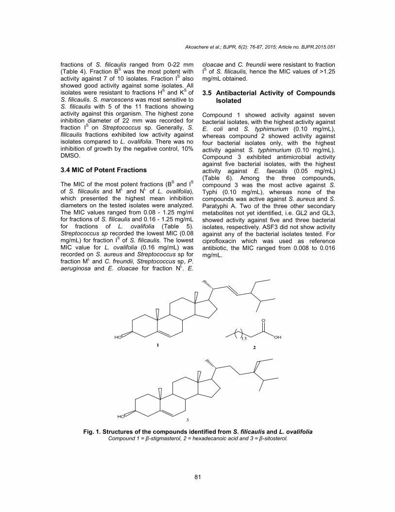
Amorn [17] enabled GL1 to be identified as -sitosterol (3) (Fig. 1). Methylene chloride and hexane extracts of S. filicaulis were combined prior to fractionation to improve yield as methylene chloride extract was small. Three secondary metabolites (ASF1, ASF2 and ASF3) were isolated and two of them (ASF1 and ASF2) were identified as β-stigmasterol (1) [18] (mp 165°C) and hexadecanoic acid (palmitic acid, 2) [19] (mp 63°C), respectively (Fig. 1).
3.2 Antibacterial Activity of Crude Extracts
Crude extracts of S. filicaulis and L. ovalifolia were screened for antibacterial activity against resistant pathogens. Hexane, methylene chloride and methanol extracts were tested against the pathogens by the agar diffusion method. With the exception of K. oxytoca and S. aureus which were resistant to hexane extract, all isolates demonstrated susceptibility to methylene chloride (zone size 12-15 mm) and hexane extracts of S. filicaulis (11-13 mm) (Table 2). Methylene chloride (zone size 12-16 mm) and methanol (zone size 13-16 mm) crude extracts of L. ovalifolia were active against all isolates (Table 2). Isolates were completely resistant to methanol extract of S. filicaulis and hexane extract of L. ovalifolia with the exception of E. coli, P. vulgaris and S. marcescens. S. marcescens was sensitive to five of the six crude extracts analyzed. Cut-off point for sensitivity was 14 mm compared with intermediate activity of ciprofloxacin [20]. There was no inhibition by the negative control, 10% DMSO.
3.3 Antibacterial Activity of Fractions
The susceptibility of isolates to 14 fractions of L. ovalifolia and 11 fractions of S. filicaulis was analyzed by the agar diffusion assay. The tests were done in duplicates and the averages recorded. An inhibition zone of 14 mm was taken as cut-off by comparing with ciprofloxacin, the standard antibiotic. Zone sizes (excluding the well diameter) for L. ovalifolia fractions ranged from 0-20 mm (Table 3). Seven of the 14 fractions of L. ovalifolia (HL-NL) exhibited potent antibacterial activity. Zone sizes of the 11
Akoachere et al.; BJPR, 6(2): 76-87, 2015; Article no. BJPR.2015.051
81
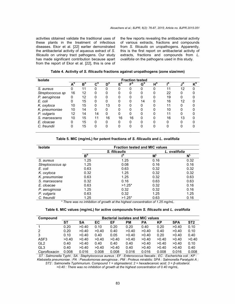
fractions of S. filicaulis ranged from 0-22 mm (Table 4). Fraction BS was the most potent with activity against 7 of 10 isolates. Fraction I
S also
showed good activity against some isolates. All isolates were resistant to fractions HS and KS of S. filicaulis. S. marcescens was most sensitive to S. filicaulis with 5 of the 11 fractions showing activity against this organism. The highest zone inhibition diameter of 22 mm was recorded for fraction IS on Streptococcus sp. Generally, S. filicaulis fractions exhibited low activity against isolates compared to L. ovalifolia. There was no inhibition of growth by the negative control, 10% DMSO.
3.4 MIC of Potent Fractions The MIC of the most potent fractions (BS and IS of S. filicaulis and M
L and N
L of L. ovalifolia),
which presented the highest mean inhibition diameters on the tested isolates were analyzed. The MIC values ranged from 0.08 - 1.25 mg/ml for fractions of S. filicaulis and 0.16 - 1.25 mg/mL for fractions of L. ovalifolia (Table 5). Streptococcus sp recorded the lowest MIC (0.08 mg/mL) for fraction IS of S. filicaulis. The lowest MIC value for L. ovalifolia (0.16 mg/mL) was recorded on S. aureus and Streptococcus sp for fraction M
L and C. freundii, Streptococcus sp, P.
aeruginosa and E. cloacae for fraction NL. E.
cloacae and C. freundii were resistant to fraction IS of S. filicaulis, hence the MIC values of >1.25 mg/mL obtained.
3.5 Antibacterial Activity of Compounds Isolated
Compound 1 showed activity against seven bacterial isolates, with the highest activity against E. coli and S. typhimurium (0.10 mg/mL), whereas compound 2 showed activity against four bacterial isolates only, with the highest activity against S. typhimurium (0.10 mg/mL). Compound 3 exhibited antimicrobial activity against five bacterial isolates, with the highest activity against E. faecalis (0.05 mg/mL) (Table 6). Among the three compounds, compound 3 was the most active against S. Typhi (0.10 mg/mL), whereas none of the compounds was active against S. aureus and S. Paratyphi A. Two of the three other secondary metabolites not yet identified, i.e. GL2 and GL3, showed activity against five and three bacterial isolates, respectively. ASF3 did not show activity against any of the bacterial isolates tested. For ciprofloxacin which was used as reference antibiotic, the MIC ranged from 0.008 to 0.016 mg/mL.
HO
1
OH
O
13
2
HO3
Fig. 1. Structures of the compounds identified from S. filicaulis and L. ovalifolia Compound 1 = β-stigmasterol, 2 = hexadecanoic acid and 3 = β-sitosterol.
Akoachere et al.; BJPR, 6(2): 76-87, 2015; Article no. BJPR.2015.051
82
Table 1. Fractionation of extracts of S. filicaulis and L. ovalifolia
S. filicaulis (Combined methylene chloride and hexane )
L. ovalifolia (methylene chloride)
Eluent Fraction Grouping Eluent Fraction Grouping Hex, 100:EtOAc,0 1-9 1-9 (A
S) Hex, 100:EtoAc, 0 1-6 1-5 (A
L), 6-7(B
L),
95:5 10-16 10-15 (BS) 95:5 7-14 8-12 (C
L), 13-15 (D
L)
90:10 17-24 16-17 (CS) 90:10 15-21 16-18 (E
L), 19-21 (F
L)
85:15 25-30 18-26 (DS) 80:20 22-28 22-24 (G
L), 25-30 (H
L)
80:20 31-36 27-31 (ES) 60:40 29-35 31-41 (I
L)
70:30 37-42 32-38 (FS) 40:60 36-45 42-43 (J
L)
60:40 43-47 39-44 (GS) 20:80 46-56 44-59 (K
L)
50:50 48-50 45-52 (HS) MeOH 57-68 60-61 (L
L), 63-67 (M
L),
67-68 (NL)
40:60 51-56 53-56 (IS)
20:80 57-60 57-60 (JS)
MeOH 61-64 61-64 (KS) Hex = hexane; EtOAc = ethyl acetate; MeOH = methanol
Table 2. Antimicrobial activity of crude extracts of plants (zone size/mm)
Isolate S. filicaulis extracts L. ovalifolia extracts
Hexane Methylene chloride
Methanol Hexane Methylene chloride
Methanol
E. coli 12 15 12 14 16 14 P. vulgaris 12 14 12 14 15 15 S. marcescens 13 15 15 15 16 14 K. pneumoniae 12 14 0 0 13 16 C. freundii 11 15 0 0 16 15 P. aeruginosa 12 15 0 0 15 14 E. cloacae 12 14 0 0 16 13 K. oxytoca 0 14 0 0 12 16 S. aureus 0 14 0 0 16 14 Streptococcus sp 11 12 0 0 13 13
Table 3. Activity of L. ovalifolia fractions against uropathogens (zone size/mm)
Isolate Fraction tested
AL BL CL DL EL FL GL HL IL JL KL LL ML NL S. aureus 12 0 12 0 11 11 11 14 14 15 13 16 19 16 Streptococcus sp 11 0 0 0 0 0 0 14 0 15 0 15 16 15 E. coli 11 10 0 10 10 11 10 17 17 15 15 17 20 20 K. oxytoca 15 0 0 0 12 11 11 18 17 15 17 15 20 19 K. pneumoniae 14 0 0 0 11 0 0 15 14 0 13 14 14 16 P. vulgaris 12 11 12 12 12 13 11 13 13 15 13 15 15 15 E. cloacae 14 11 0 11 14 13 0 15 20 16 16 16 14 19 S. marcescens 11 11 12 11 0 10 0 15 15 16 14 16 14 14 C. freundii 14 0 0 0 0 0 0 14 15 14 14 15 14 15 P. aeruginosa 12 0 0 0 0 0 0 14 14 15 11 15 17 15
4. DISCUSSION In this study, ciprofloxacin was used as reference antibiotic. As outlined by CLSI [21] guidelines, bacterial isolates with MIC value ≥ 4 μg/ml are defined as resistant isolates. The MIC values of ciprofloxacin for all the bacterial isolates tested were ≥ 4 μg/ml (Table 6), confirming that they
were really drug resistant bacteria, as observed in a previous work [13]. We evaluated the in vitro antibacterial activity of crude extracts, fractions and compounds from two medicinal plants, Spilanthes filicaulis and Laportea ovalifolia, on drug resistant typhoid pathogens and uropathogens. The antibacterial
Akoachere et al.; BJPR, 6(2): 76-87, 2015; Article no. BJPR.2015.051
83
activities obtained validate the traditional uses of these plants in the treatment of infectious diseases. Ekor et al. [22] earlier demonstrated the antibacterial activity of aqueous extract of S. filicaulis on urinary tract pathogens. Our study has made significant contribution because apart from the report of Ekor et al. [22], this is one of
the few reports revealing the antibacterial activity of various extracts, fractions and compounds from S. filicaulis on uropathogens. Apparently, this is the first report on antibacterial activity of extracts, fractions and compounds from L. ovalifolia on the pathogens used in this study.
Table 4. Activity of S. filicaulis fractions against uropathogens (zone size/mm)
Isolate Fraction tested
AS B
S C
S D
S E
S F
S G
S H
S I
S J
S K
S
S. aureus 0 11 0 0 0 0 0 0 11 12 0 Streptococcus sp 16 12 0 0 0 0 0 0 22 0 0 P. aeruginosa 0 12 0 0 0 0 0 0 19 0 0 E. coli 0 15 0 0 0 0 14 0 16 12 0 K. oxytoca 10 15 0 13 0 0 0 0 11 0 0 K. pneumoniae 10 14 0 0 0 0 0 0 10 0 0 P. vulgaris 12 14 14 0 0 0 0 0 11 0 0 S. marcescens 10 15 11 16 16 16 0 0 16 13 0 E. cloacae 0 15 0 0 0 0 0 0 0 0 0 C. freundii 0 15 0 0 0 0 0 0 0 0 0
Table 5. MIC (mg/mL) for potent fractions of S. filicaulis and L. ovalifolia
Isolate Fraction tested and MIC values
S. filicaulis L. ovalifolia B
S IS
ML
NL
S. aureus 1.25 1.25 0.16 0.32 Streptococcus sp 1.25 0.08 0.16 0.16 E. coli 0.63 0.63 0.32 0.32 K. oxytoca 0.32 1.25 0.32 0.32 K. pneumoniae 0.63 1.25 0.32 0.63 S. marcescens 0.32 0.16 0.63 0.63 E. cloacae 0.63 >1.25* 0.32 0.16 P. aeruginosa 1.25 0.32 0.32 0.16 P. vulgaris 0.63 0.32 1.25 1.25 C. freundii 1.25 >1.25* 0.63 0.16
* There was no inhibition of growth at the highest concentration of 1.25 mg/mL.
Table 6. MIC values (mg/mL) for active compounds from S. filicaulis and L. ovalifolia
Compound Bacterial isolates and MIC values ST SA EC EF PM PA KP SPA ST2
1 0.20 >0.40 0.10 0.20 0.20 0.40 0.20 >0.40 0.10 2 0.20 >0.40 >0.40 0.40 >0.40 >0.40 0.40 >0.40 0.10 3 0.10 >0.40 0.40 0.05 >0.40 >0.40 0.20 >0.40 0.40 ASF3 >0.40 >0.40 >0.40 >0.40 >0.40 >0.40 >0.40 >0.40 >0.40 GL2 0.40 >0.40 0.40 0.40 0.40 >0.40 >0.40 >0.40 0.10 GL3 0.40 >0.40 >0.40 >0.40 0.40 >0.40 >0.40 >0.40 0.40 Ciprofloxacin 0.008 0.016 0.008 0.008 0.016 0.016 0.008 0.016 0.008 ST : Salmonella Typhi ; SA : Staphylococcus aureus ; EF : Enterococcus faecalis ; EC : Escherichia coli ; KP :
Klebsiella pneumoniae ; PA : Pseudomonas aeruginosa ; PM : Proteus mirabilis; SPA : Salmonella Paratyphi A ; ST2 : Salmonella Typhimurium. Compound 1 = stigmasterol; 2 = hexadecanoic acid; 3 = β-sitosterol.
>0.40 : There was no inhibition of growth at the highest concentration of 0.40 mg/mL.
Akoachere et al.; BJPR, 6(2): 76-87, 2015; Article no. BJPR.2015.051
84
Plant crude extracts are mixtures of many compounds/constituents and the biological activity shown by a given extract may reflect the contributions from a number of these phytochemicals. Consequently, the biological activity of a plant extract is usually followed by bioassay-guided fractionation, which is a method designed to isolate and identify the active principle(s). The most active extracts of plants were fractionated to ascertain the antibacterial properties of the active ingredients. It was interesting to note that S. aureus, which is known to have a high level of resistance to antibiotics [23], was susceptible to 6 fractions of L. ovalifolia, but was resistant to fractions of S. filicaulis. Also, Streptococcus sp showed susceptibility to some fractions of L. ovalifolia and only two fractions of S. filicaulis (A
s and I
s).
Our findings indicate that L. ovalifolia contains compounds with high potency against gram positive and gram negative organisms. Some of the active components may also be distributed unevenly in all the fractions explaining why some fractions of L. ovalifolia exhibited weak activities. Data on the antimicrobial characteristics of L. ovalifolia are rare although it has been studied for its anti-diabetic and antioxidative characteristics [24]. The antimicrobial property of S. filicaulis is probably due to lipid soluble active substances as fraction BS (most potent) was obtained by elution with 100% and 95% hexane. Hexane is a non-polar solvent which can easily extract the fat soluble phytochemicals such as essential oils, sterols and triterpenes [25]. In addition, the 40% hexane: 60% ethyl acetate fraction (I
S) of S.
filicaulis also showed high activity, suggesting that some of the active components had intermediary polarity. In addition to this antibacterial activity, S. filicaulis has been reported to also possess analgesic properties [22], the probable reason for its use in treatment of toothache and stomachache in herbal medicine, and anti-inflammatory characteristics [9]. A related species to S. filicaulis, S. acmella Linn has been reported to possess remarkable inhibitory activity against human and agricultural fungal pathogens [26]. The MIC values of the most active fractions were also determined. The lowest MIC (0.08 mg/mL) in this study was recorded for fraction I
S of S.
filicaulis on Streptococcus sp. It is noteworthy that this corroborates the activity of fraction I
S on
Streptococcus sp. tested by diffusion technique, which gave the largest inhibition zone diameter
of 22 mm. MIC values of >1.25 mg/mL observed with fraction IS of S. filicaulis on E. cloacae and C. fruendii indicated resistance. This is not surprising as these were the only isolates that showed no activity with fraction IS during antibacterial susceptibility testing of fractions. These isolates could possess intrinsic resistance to compounds present in fraction I
S. However,
despite this resistance, E. cloacae and C. fruendii were susceptible to other fractions whose MICs were studied. Streptococcus sp was resistant to five antibiotics [13] but had the lowest MIC for 3 fractions (I
S, M
L and N
L) of the 4 tested.
Our findings indicate the presence of highly potent antistreptococcal compounds in these fractions. Importantly, our results also demonstrated the activity of fractions against two prominent multidrug resistant pathogens - Pseudomonas aeruginosa and Staphylococcus aureus - with a high preponderance in both clinical and environmental samples [27]. MICs of 0.08-1.25 mg/ml observed in fractions against both gram positive and gram-negative multi-drug resistant uropathogens suggest the possible presence of constituents with a wide spectrum of activity that could be exploited for development of more potent antimicrobial chemotherapeutic agents for the eradication of multi-drug resistant pathogens in our community and also justifies their use in traditional medicine. S. filicaulis and L. ovalifolia are annual plants with a wide distribution and therefore are not at risk of extinction. These plants are thus available for exploitation. The amount of active compounds in plant crude extracts may be diluted by the presence of other compounds, but when fractionated, these compounds become concentrated and therefore exhibit significant activity. This may explain the larger zone sizes observed with the fractions. The activities of the fractions of S. filicaulis and L. ovalifolia may provide answers to this phenomenon of resistance in our study area. Inexhaustive fractionation or smearing of some active components throughout the column could also account for the weak activities obtained. Compounds 1, 2, 3, GL2 and GL3 exhibited various degrees of antibacterial activity against some of the bacteria used. The in vitro antibacterial activity of β-stigmasterol has been reported against Staphylococcus aureus, Streptococcus mutans, Streptococcus salivarius, Streptococcus mitis, Streptococcus anginosus and Lactobacillus acidophilus [28], but no report was seen for Klebsiella pneumoniae, while palmitic acid (ASF2) has shown in vitro activity
Akoachere et al.; BJPR, 6(2): 76-87, 2015; Article no. BJPR.2015.051
85
against some strains of bacteria [29]. There are several reports [30,31] on the activity of β-sitosterol from different plants against both Gram-positive and Gram-negative bacterial isolates similar to those used in our study. Thus β-stigmasterol and palmitic acid should be the active principles in S. filicaulis, whereas β-sitosterol should be one of the active compounds in L. ovalifolia. Among the three compounds identified, β-sitosterol (3) was more active against Salmonella Typhi than the two other compounds, whereas β-stigmasterol (1) and hexadecanoic acid (2) showed similar activities against Salmonella Typhimurium, and were more active than β-sitosterol against this bacterial isolate. β-sitosterol and palmitic acid isolated from other plant species have already been reported to exhibit antibacterial activity against S. Typhi and S. Typhimurium [32]. The in vitro activity of β-stigmasterol against S. Typhi and S. Typhimurium is herein reported for the first time. The main causative agent of typhoid fever is S. Typhi [5,16]; therefore, these compounds (especially compound 3) could be developed and used in the treatment of typhoid fever, if they are exempt of toxicity. The activity of ciprofloxacin (standard drug) against all the bacterial isolates tested was much greater than those of compounds 1, 2 and 3. The MIC values of ciprofloxacin were generally more than 13 times lower than those of these active compounds, suggesting that compounds 1, 2 and 3 may be about 13 times less active than ciprofloxacin against the bacteria used.
5. CONCLUSION The spectrum of activity exhibited by extracts, fractions and compounds obtained from Laportea ovalifolia and Spilanthes filicaulis against drug resistant bacterial pathogens (UTI and typhoid fever pathogens) as observed in our study indicate that they may contain lead molecules for the synthesis of drugs useful in the management of UTIs and typhoid fevers. Our results justify the use of these plants in traditional medicine as broad spectrum anti-infectious agents. In vivo anti-infective effects and toxicity of these extracts, fractions and compounds should be studied in order to further ascertain their therapeutic effect and their safety. Being that related species to these plants have been reported with antifungal properties, further studies testing the antimycotic characteristics of
these plants will add value to their economic, medicinal and agricultural importance.
CONSENT It is not applicable.
ETHICAL APPROVAL It is not applicable.
ACKNOWLEDGEMENTS We would like to express our gratitude to Dr (Mrs) Fonkoua Marie-Christine, Pasteur Centre, Yaoundé, Cameroon, for her cooperation.
COMPETING INTERESTS Authors have declared that no competing interests exist.
REFERENCES 1. Kunin CM. Urinary tract infections:
Detection, Prevention and Management. 5th edition. Baltimore: Williams and Wilkin ; 1997.
2. Tankhiwale SS, Jalgaonkoar SV, Ahamad S, Hassani U. Evaluation of extended spectrum beta lactamase in urinary isolates. Indian J. Med. Res. 2004; 120:553-556.
3. Mshana SE, Kamugisha E, Mirambo M, Chakraborty T, Lyamuya EF. Prevalence of multiresistant gram-negative organisms in a tertiary hospital in Mwanza, Tanzania. BMC Research Notes. 2009;2:49.
4. Gatsing D, Aliyu R, Meli WB, Adoga GI, Tchouanguep MF. Phytochemical profile and antisalmonellal properties of Allium sativum bulb extract. West African Journal of Biological Sciences. 2003;14:29-36.
5. Gatsing D, Mbah JA, Garba IH, Tane P, Djemgou P, Nji-Nkah BF. An antisalmonellal agent from the leaves of Glossocalyx brevipes Benth (Monimiaceae). Pakistan Journal of Biological Sciences. 2006;9(1):84-87.
6. Gatsing D, Adoga GI. Antisalmonellal activity and phytochemical screening of the various parts of Cassia petersiana Bolle (Caesalpiniaceae). Research Journal of Microbiology. 2007;2(11):876-880.
7. Madhulika U, Harish BN, Parija SC. Current pattern in antimicrobial
Akoachere et al.; BJPR, 6(2): 76-87, 2015; Article no. BJPR.2015.051
86
susceptibility of Salmonella Typhi isolates in Pondicherry. Indian J. Med. Res. 2004;120:111-114.
8. Bussmann RW, Sharon D. Traditional medicinal plants in Northern Peru: tracking two thousand years of healing culture. J. Ethnobiol. Ethnomed. 2006;2:1-47.
9. Simbo DJ. An ethnobotanical survey of medicinal plants in Babungo, Northwest Region, Cameroon. J. Ethnobiol. Ethnomed. 2010;6:8.
10. Adjanohoun JE, Aboubaka N, Dramane K, Ebot ME, Ekpere KA, Enow-Orack EG, Focho D, Gbile ZO, Kamanyi A, Kamsu KJ, Keita A, Mbenkum T, Mbi CN, Mbiele AL, Mbome IL, Mubine NK, Nancy WL, Nkongmeneck B, Satabie B, Sofowora A, Tamze V, Wirmum CK. Traditional Medicine and pharmacopeia: Contribution to Ethnobotanical and Floristical Studies in Cameroon. Organisation of African Unity/Scientific, Technical and Research Commission. Porto-NOVO: Centre National de Production de Manuels Scolaires.1996;36-52.
11. Focho DA, Ndam WT, Fonge BA. Medicinal plants of Aguambu-Bamumbu in the Lebialem highlands, Southwest Province of Cameroon. Afr. J. Pharmaco. 2009;3(1):1-13.
12. Rahman MM, Khan A, Haque ME, Rahman MM. Antimicrobial and cytotoxic activities of Laportea crenulata. Fitoterapia. 2008;79(7-8):584-6.
13. Akoachere J-FTK, Suylika Y, Njom HA, Esemu NK. Etiologic profile and antimicrobial susceptibility of community-acquired urinary tract infection in two Cameroonian towns. BMC Research Notes. 2012;5:219.
14. Tata EF, Agba IM, Chukwedo AA, Mgbojikwe L, Wirkom KV. Susceptibilities of aerobic bacteria associated with bovine mastitis to leaf extracts of Bauhinia reticulata. Online Vet. J. 2009;4(2):1-42.
15. Eloff JN. A sensitive and quick microplate method to determine the minimal inhibitory concentration of plant extracts for bacteria. Planta Medica. 1998;64:711-713.
16. Kengni F, Tala DS, Djimeli NM, Fodouop SPC, Kodjio N, Magnifouet NH, Gatsing D. In vitro antimicrobial activity of Harungana madagascriensis and Euphorbia prostrata extracts against some pathogenic Salmonella sp. International Journal of Biological and Chemical Sciences. 2013; 7(3):1106-1118.
17. Suparb B, Amorn P. Chemical constituents of the roots of Bridellia tomentosa BL. J. Sci. Soc. Thailand. 1991;17:61-69.
18. Jamal A, Yaacob W, Laily B. A chemical study on Phyllanthus columnaris. Euro. J. Sci. Res. 2009;28:76-81.
19. Neol BK, Rahayu UU, Nordin HL, Tai YC, Tu YL, Mohd AS. Chemical constituents from two weed species of Spermacoce (Rubiaceae). The Malaysian J. Anal. Sci. 2010;14:6-11.
20. NCCLS (National Committee for Clinical Laboratory Standards). Performance standards for antimicrobial susceptibility testing. 13th Informational supplement, M100-S13. Wayne, PA, USA: NCCLS ; 2002.
21. CLSI (Clinical and Laboratory Standards Institute). Performance standards for anti-microbial susceptibility testing. Eleventh Informational Supplement; Document M100-S11. Wayne, PA: CLSI; 2001.
22. Ekor M, Ashorobi RB, Ibitoye SF, Kasimi LS. Acute toxicity, analgesic potential and preliminary antimicrobial studies of the aqueous plant extract of Spilanthes filicaulis. Nigerian J. Health Biomed. Sci. 2005;4(1):30-34.
23. Vila J, Pal T. Update on Antibacterial Resistance in Low-Income Countries: Factors Favoring the Emergence of Resistance. The Open Infectious Diseases Journal. 2010;4:38-54.
24. Iffen TS, Usoro CAO. The effect of ethanolic extract of Laportea ovalifolia plants growing in Calabar on antioxidants status of streptozocin induced diabetic rats. Glob. J. Pharmaco. 2010;4(1):01-05.
25. Cowan MM. Plant products as antimicrobial agents. Clinical Microbiology Reviews. 1999;12(4):564-582.
26. Rani SA, Murty SU. Antifungal potential of flower head extract of Spilanthes acmella Linn. Afr. J. Biomedl. Res. 2006;9:67-69.
27. Nkwelang G, Akoachere J-FTK, Kamga LH, Emmanuel DF, Ndip RN. Staphylococcus aureus isolates from clinical and environmental samples in a semi-rural area of Cameroon: phenotypic characterization of isolates. Afr. J. Microbiol. Res. 2009;3(11):731-736.
28. Koteswara PR, Bhaskar DR, Ravi CHK, Ravindra MN, Madhavi Y, Raghava TR. In vitro antibacterial activity of Moringa oleifera against dental plaque bacteria. J. Pharmacy Res. 2011;4:695-697.
Akoachere et al.; BJPR, 6(2): 76-87, 2015; Article no. BJPR.2015.051
87
29. Ndip RN, Dilonga HM, Ndip LM, Akoachere J-FK, Nkuo-Akenji T. Pseudomonas aeruginosa isolates recovered from clinical samples in Buea, Cameroon: current status on biotyping and antibiogram. Trop. Med. Int. Health. 2005; 10(1):74–81.
30. Mokbel MS, Hashinaga F. Evaluation of the antibacterial activity of extract from Buntan (Citrus grandis Osbeck) fruit peel. Pak. J. Biol. Sci. 2005;8:1090-1092.
31. Singh S, Tejovathi G. Analysis of IR, NMR
and antimicrobial activity of -sitosterol isolated from Momordica charantia. Sci. Secure J. Biotechn. 2012;1:9-13.
32. Silva-Belmares Y, Rivas-Morales C, Viveros-Valdez E, Guadalupe de la Cruz-Galicia M, Carranza-Rosales P. Antimicrobial and cytotoxic activities from Jatropha dioica roots. Pakistan Journal of Biological Sciences. 2014;17:748-750.
© 2015 Akoachere et al.; This is an Open Access article distributed under the terms of the Creative Commons Attribution License (http://creativecommons.org/licenses/by/4.0), which permits unrestricted use, distribution, and reproduction in any medium, provided the original work is properly cited.
Peer-review history: The peer review history for this paper can be accessed here:
http://www.sciencedomain.org/review-history.php?iid=983&id=14&aid=8165
Related Documents