In Situ Detection of PHIP at 48 mT: Demonstration Using a Centrally Controlled Polarizer Kevin W. Waddell, Aaron M. Coffey, and Eduard Y. Chekmenev * Vanderbilt University Institute of Imaging Science, 1161 21st Avenue South, Nashville, Tennessee 37232, United States Abstract Presented here is a centrally controlled, automated parahydrogen-based polarizer with in situ detection capability. A 20% polarization, corresponding to a 5 000 000-fold signal enhancement at 48 mT, is demonstrated on 2-hydroxyethyl-1- 13 C-propionate-d 2,3,3 using a double-tuned antenna and pulsed polarization transfer. In situ detection is a refinement of first-generation devices enabling fast calibration of rf pulses and B 0 , quality assurance of hyperpolarized contrast agents, and stand-alone operation without the necessity of high-field MR spectrometers. These features are essential for biomedical applications of parahydrogen-based hyperpolarization and for clinical translation. We demonstrate the flexibility of the device by recording 13 C signal decay due to longitudinal relaxation of a hyperpolarized contrast agent at 48 mT corresponding to 2 MHz proton frequency. This appears to be the longest recorded T 1 (101 ± 7 s) for a 13 C hyperpolarized contrast agent in water. Introduction Nuclear magnetic resonance (NMR) is a relatively insensitive method for detecting metabolites in vivo due to low nuclear spin polarization and broad resonances that arise from susceptibility gradients across heterogeneous tissues. Methods for increasing polarization toward the theoretical limit of unity are becoming more widespread and can potentially decrease the metabolite MR detection threshold at available clinical fields from 1 μmol/cm 3 to sub-nmol/cm 3 . For example, the spin polarization available at 3 T and in vivo temperatures is P = 2.6 × 10 −4 % for 13 C versus P = 20% for hyperpolarized samples. These hyperpolarized spin states relax to the equilibrium according to an exponential decay constant T 1 . Despite this shortcoming, useful contrast agents with T 1 relaxation times on the order of 10–120 s have enabled pathway-specific flux measurements with hyperpolarized samples at unprecedented temporal and spatial resolution in vivo. 1 As a result, subsecond metabolic imaging of hyperpolarized agents 1 has the potential to be a sensitive and specific tool for imaging abnormal metabolism such as that of many cancers. 2,3 Not surprisingly, the ongoing research efforts of many groups have been focused on human cancer imaging in preclinical models with Investigational New Drug (IND) and U.S. Food and Drug Adminsitration (FDA) clearance approved 4 for the first clinical trial using hyperpolarized 13 C-pyruvate (University of California San Fransisco). 5 When these methods are refined and improved so as to afford safe administration, hyperpolarized MR is likely to become a pivotal complement to fluorodeoxyglucose-positron emission tomography (FDG- PET) for cancer imaging. 6 © 2011 American Chemical Society * [email protected] . NIH Public Access Author Manuscript J Am Chem Soc. Author manuscript; available in PMC 2011 May 20. Published in final edited form as: J Am Chem Soc. 2011 January 12; 133(1): 97–101. doi:10.1021/ja108529m. NIH-PA Author Manuscript NIH-PA Author Manuscript NIH-PA Author Manuscript

Welcome message from author
This document is posted to help you gain knowledge. Please leave a comment to let me know what you think about it! Share it to your friends and learn new things together.
Transcript

In Situ Detection of PHIP at 48 mT: Demonstration Using aCentrally Controlled Polarizer
Kevin W. Waddell, Aaron M. Coffey, and Eduard Y. Chekmenev*
Vanderbilt University Institute of Imaging Science, 1161 21st Avenue South, Nashville,Tennessee 37232, United States
AbstractPresented here is a centrally controlled, automated parahydrogen-based polarizer with in situdetection capability. A 20% polarization, corresponding to a 5 000 000-fold signal enhancement at48 mT, is demonstrated on 2-hydroxyethyl-1-13C-propionate-d2,3,3 using a double-tuned antennaand pulsed polarization transfer. In situ detection is a refinement of first-generation devicesenabling fast calibration of rf pulses and B0, quality assurance of hyperpolarized contrast agents,and stand-alone operation without the necessity of high-field MR spectrometers. These featuresare essential for biomedical applications of parahydrogen-based hyperpolarization and for clinicaltranslation. We demonstrate the flexibility of the device by recording 13C signal decay due tolongitudinal relaxation of a hyperpolarized contrast agent at 48 mT corresponding to 2 MHzproton frequency. This appears to be the longest recorded T1 (101 ± 7 s) for a 13C hyperpolarizedcontrast agent in water.
IntroductionNuclear magnetic resonance (NMR) is a relatively insensitive method for detectingmetabolites in vivo due to low nuclear spin polarization and broad resonances that arise fromsusceptibility gradients across heterogeneous tissues. Methods for increasing polarizationtoward the theoretical limit of unity are becoming more widespread and can potentiallydecrease the metabolite MR detection threshold at available clinical fields from 1 μmol/cm3
to sub-nmol/cm3. For example, the spin polarization available at 3 T and in vivotemperatures is P = 2.6 × 10⁻⁴ % for 13C versus P = 20% for hyperpolarized samples. Thesehyperpolarized spin states relax to the equilibrium according to an exponential decayconstant T1. Despite this shortcoming, useful contrast agents with T1 relaxation times on theorder of 10–120 s have enabled pathway-specific flux measurements with hyperpolarizedsamples at unprecedented temporal and spatial resolution in vivo.1 As a result, subsecondmetabolic imaging of hyperpolarized agents1 has the potential to be a sensitive and specifictool for imaging abnormal metabolism such as that of many cancers.2,3 Not surprisingly, theongoing research efforts of many groups have been focused on human cancer imaging inpreclinical models with Investigational New Drug (IND) and U.S. Food and DrugAdminsitration (FDA) clearance approved4 for the first clinical trial usinghyperpolarized 13C-pyruvate (University of California San Fransisco).5 When these methodsare refined and improved so as to afford safe administration, hyperpolarized MR is likely tobecome a pivotal complement to fluorodeoxyglucose-positron emission tomography (FDG-PET) for cancer imaging.6
© 2011 American Chemical Society* [email protected] .
NIH Public AccessAuthor ManuscriptJ Am Chem Soc. Author manuscript; available in PMC 2011 May 20.
Published in final edited form as:J Am Chem Soc. 2011 January 12; 133(1): 97–101. doi:10.1021/ja108529m.
NIH
-PA Author Manuscript
NIH
-PA Author Manuscript
NIH
-PA Author Manuscript
The two dominant technologies for hyperpolarization of exogenous contrast agents arereferred to as dynamic nuclear polarization (DNP)7 and para-hydrogen-induced polarization(PHIP).8 The latter utilizes facile production of pure singlet states of diatomic hydrogen,whereas the former relies upon polarization transfer from highly polarized electrons inglasses at high magnetic fields and cryogenic temperatures. Despite significant technologicalchallenges and relatively high cost, the DNP technique has progressed to commerciallyavailable systems capable of producing batches of hyperpolarized solution every 1–3 h.During this time, polarization buildup is monitored by a high-resolution NMR probe,allowing for optimization of hyperpolarized conditions and quality assurance (QA).
Compared to long preparation periods associated with DNP in current commercialimplementations, PHIP can be used to prepare hyperpolarized metabolic contrast agentssuch as 13C-succinate in aqueous media in seconds.9,10 First-generation PHIP polarizerprototypes10,11 are currently in use by a few scientific groups and typically require tediouscalibrations. Earlier designs covered the essential experimental demands for proof ofconcept, but refinements to ease operator demands and to improve robustness will facilitateextension of PHIP as a routine preclinical tool for studying in vivo metabolism. A typicalPHIP workflow requires calibration of the electromagnet field10 where catalytichydrogenation takes place and tedious optimization of applied radiofrequency fields (rf) toaccomplish efficient transfer in sequences with 10 or more pulses.12 Lacking onboardreceivers and comprehensive central control, first-generation PHIP devices often requiretime-consuming calibrations using detection on external high-field spectrometers.13 Thesedraw-backs limit PHIP technology to a few sites with skilled MR spectroscopists and inhibita widespread use of this technology for multidisciplinary imaging communities, especiallyamong the biologists, biochemists, and medical researchers.
The instrument presented here addresses additional considerations required for high-throughput in vivo studies and is based on an earlier prototype, which operates at a staticfield of 12 mT (Waddell, K. W., unpublished results). The design is streamlined fromcommercially available controllers, and the added functionality reduces experimentaldemands and improves reliability. We provide the overall polarizer design and the basicsteps necessary for quick fine-tuning. Sensitivity at 48 mT is sufficient for QA ofhyperpolarized contrast agents. The polarizer operates as a stand-alone device to producehyperpolarized contrast agents for biomedical imaging. Other research groups can readilyreplicate this polarizer design, because it is based on commercially available low-field NMRequipment. To demonstrate the utility of in situ detection and the overall instrument setup,we have measured the lifetime of the hyperpolarized contrast agent, 2-hydroxyethyl-1-13C-propionate-d2,3,3 (HEP), at 48 mT, and the resulting T1 appears to be the longest recorded T1for a 13C hyperpolarized contrast agent in protonated aqueous medium.
MethodsParahydrogen (para-H2) Gas.
para-H2 (97.5 ± 0.5%) was prepared by passage of high-purity hydrogen gas through a 14 Kchamber14 filled with iron oxide catalyst10 using an in-house automated para-H2 generatorsetup. The fresh para-H2 obtained over the course of an hour’s production was collected in a10 L aluminum storage tank (200 psi or 14 bar). Percentage of para-H2 conversion wasmeasured by 1H spectroscopy using a Bruker 500 MHz high-resolution NMRspectrometer.15
Waddell et al. Page 2
J Am Chem Soc. Author manuscript; available in PMC 2011 May 20.
NIH
-PA Author Manuscript
NIH
-PA Author Manuscript
NIH
-PA Author Manuscript

RF Circuit.A double-tuned rf circuit operating at 2.020 MHz (1H) and 0.508 MHz (13C) was designedon the basis of a previously published high-field rf circuit.16 The circuit consists of two coilswith elements for achieving suitable tuning range and impedance matching for each channel.The air core inductors were wound from 20 AWG magnet wire. Inductances were LS = 39.6μH for the sample coil and LG = 30.8 μH for the ground coil with impedance matching onthe 1H channel achieved via LM1H = 617 nH as measured on an Agilent E5071C networkanalyzer. A combination of fixed C22CF series capacitors (Dielectric Laboratories,Cazenovia, NY) were used in parallel with variable capacitors (model NMTM120C,Voltronics, Denville, NJ). The optimal value of 1H tuning capacitance was CT1H = 391 pF +0–120 pF. For 13C, the optimal tune and match capacitances were CT13C = 1100 pF + 0–120pF and CM13C = 270 pF + 0–120 pF. Because the measured isolation between channels was−5 dB, an in-line parallel LC circuit serving as a low pass filter for the 1H frequency wasadded to the 13C channel to improve the isolation between the channels.
Molecular Addition of para-H2 Using Rh-Based Catalyst.The molecular addition of para-H2 gas to the tracer was conducted in the previouslydescribed chemical reactor at a 120 psi (8 bar) para-H2 pressure over a time span of 4 sunder the conditions of continuous wave (CW) proton decoupling. The hydrogenationreaction was more than 95% complete using the Rh-based molecular catalyst.17,18 Thecatalyst in aqueous medium was prepared using a previously developed protocol13 to a finalcatalyst concentration of 2.5–3.0 mM. The molecular precursor, 2-hydroxyethyl-1-13C-acrylate-d2,3,3 (HEA; product number 676071, Sigma-Aldrich-Isotec, Miamisburg, OH),was added to freshly prepared catalyst solution resulting in 8 mM HEA. The prepared stocksolution containing catalyst and HEA was used for chemical reaction with 97.5% para-H2.The reactor pressure after spraying the liquid using high-pressure N2 gas into an atmosphereof para-H2 gas was 250 psi or 17 bar.
Polarization Transfer from para-H2.Spin order of para-H2 singlet state was converted to 13C hyperpolarization using thepolarization transfer sequence developed by M. Goldman et al.12 The rf pulses wereoptimized using the method described in the Results section. Filling the reactor chamberwith para-H2 gas, injecting an aqueous mixture of molecular precursor and catalyst,chemical reaction under CW 1H decoupling, and heteronuclear polarization transfer weretimed from the central controller (Magritek, Wellington, New Zealand). In situ NMRdetection of hyperpolarized compounds was started immediately after polarization transferfrom para-H2 to 13C. These contrast agents are expected to be used in high-field in vivoimaging, and the automated ejection along with filtration can be seamlessly integrated andsystematically controlled with respect to all other experimental events with this setup.Although the catalytic hydrogenation is typically conducted at elevated temperature (>60°C),9,15 the ejected material decreases the temparture to 30–35 °C due to samplemanipulation, which is compatible with in vivo use.
ResultsA commercially available double-channel MR spectrometer (Magritek, Wellington, NewZealand) synchronizes rf transmission, chemical shuttling, and detection in the 48 mT fieldof a Halbach array. TTL lines are accessed within the pulse program itself and routedthrough a microcontroller to switch solenoid valves that control gas and chemical delivery tothe high-pressure reactor (Figure 1).
Waddell et al. Page 3
J Am Chem Soc. Author manuscript; available in PMC 2011 May 20.
NIH
-PA Author Manuscript
NIH
-PA Author Manuscript
NIH
-PA Author Manuscript

rf and B0 Calibration.The frequency response of the 1H and 13C channels of a dual-channel probe (described inMethods) is presented in Figure 2. The circuit design achieved a quality factor Q = 66 forthe 1H channel at 2.02 MHz and Q = 43 for the 13C channel operated at 0.508 MHz. Radiofrequency calibration using a solution of 29 g of sodium 1-13C-acetate in 99.9% D2O in a100 mL spherical phantom yielded a 90° 1H excitation pulse width of 37 μs at 57 W. Thecorresponding pulse width on 13C was 150 μs at 14 W. 1H rf calibration was conductedusing an automated routine supplied by the manufacturer of the low-field spectrometer. Thecalibration of resonant frequencies is conducted by direct proton detection and can be shiftedup to ~1 kHz in extreme cases due to spatially heterogeneous spurious magnetic fields thatarise from super-position of interfering fields and potential coupling of B0 to nearby objects.Additional resonant frequencies (13C, 15N, 23Na, etc.) scale according to gyromagneticratios and are calculated based on the proton Larmor frequency.
Reactor.The details of molecular addition of parahydrogen (para-H2) and polarization transfer frompara-H2 protons to X nuclei are described in Methods. The reactor allows spraying of theaqueous mixture of unsaturated molecular precursor and the catalyst into pressurized para-H2 gas (120 psi or 8 bar). Reaction surface area is maximized by pushing the solutionthrough a cluster of <0.25 mm holes with nitrogen gas (250 psi or 17 bar) into a 2 in. (o.d.)cylindrical polysulfone reactor that can withstand 280 psi (19 bar) and 60 °C. The keyrequirement for efficient performance of the polarization transfer sequence12 of PHIPhyperpolarization, as depicted in Figure 3, is good rf homogeneity of the sample coil (LS)over the entire reactor volume. An oversized Helmholtz coil (450 mL versus reactionvolume of 60 mL) delivers homogeneous pulses over the reaction volume. B1 homogeneitywas tested by measuring the signal magnitude between 450° and 90° pulses, with a sampleof 60 mL of H2O and doped with 5 mM CuSO4. The homogeneity of the 1H channelmeasured in this way was 96% and dropped off modestly to 90% when comparing the signalat 810° and 90° pulses.
In Situ Quality Assurance of PHIP Hyperpolarization.The in situ measurement of percentage (%) hyperpolarization is demonstrated in Figure 4b.To maximize signal-to-noise ratio (SNR), the 13C spectrum of hyperpolarized HEP wasacquired with 90° excitation pulses. The reference 13C spectrum of sodium 1-13C-acetatesolution in D2O (13C T1 = 18 s ± 2 s at 48 mT, data not shown), Figure 4a, was used tocalculate % hyperpolarization (%PHP). We have used thermal 13C polarization (%PTP = 4 ×10⁻⁶ % at 48 mT and 298 K) of 29 g of sodium 1-13C-acetate to calculate %hyperpolarization as follows:
with
where nHP and nTP are the molar quantities of hyperpolarized contrast agent and thermallypolarized reference samples, respectively. SHP and STP are the corresponding MR signals
Waddell et al. Page 4
J Am Chem Soc. Author manuscript; available in PMC 2011 May 20.
NIH
-PA Author Manuscript
NIH
-PA Author Manuscript
NIH
-PA Author Manuscript

(acquired using identical parameters). ε is the signal enhancement ratio. Polarization %PHP= 20% and 13C signal enhancement ε ≈ 5 000 000-fold were achieved.
Spin–Lattice Relaxation of Hyperpolarized Contrast Agent at 48 mT.The in situ detection capability of the polarizer was utilized to measure the decay of 13Chyperpolarized HEP, Figure 5. The decay of hyperpolarization to thermal equilibrium wasmonitored by 30° excitation rf pulses. Polarization loss of ~13% due to rf excitation by eachpulse was included in the fit of exponential decay (T1 = 101 ± 7 s, R2 = 0.98). To the best ofour knowledge, this represents the longest T1 of any 13C hyperpolarized contrast agent inH2O (l). In a previous experiment where HEP was used for in vivo angiography,11 the 13CT1 at 4.7 T was reported to be 50 s in vitro.13 The consequences of such long 13C T1 arediscussed below.
DiscussionFigure 4 demonstrates that the extent of hyperpolarization can be measured in situ in a low48 mT field in micromolar quantities using oversized detection coils and direct detectionusing 13C spectroscopy. The described method provides a convenient approach to QA ofPHIP polarization for B1 and B0 calibration as well as hyperpolarization extent. Theseattributes should facilitate increased throughput and more robust operation for in vivoexperiments on PHIP metabolic contrast agents. Because the described polarizer design isbased on a commercially available, low-field magnet and MR spectrometer, it is accessibleto scientists with interests in studying fast metabolism but expertise outside of NMR and at afraction of the upfront equipment and running costs of competing DNP devices.
While optimization of polarization toward the theoretical limit is outside of the scope of thiswork, the %PHP reported here (20%) is consistent with levels published10,11 using earlierdesigns. On the basis of SNR, Figure 4b, we estimate that the detection QA threshold at 48mT for this instrument is one micromole of hyperpolarized contrast agent. The level ofsensitivity demonstrated here for 20 micromoles of hyperpolarized contrast agent shouldalso enable QA detection at significantly lower B0 fields, which can also be convenientlygenerated by an electromagnet. The qualitative comparison of the 13C hyperpolarizeddetection sensitivity with that of hyperpolarized 129Xe low-field polarimetry19 shows that itis ~2 orders of magnitude greater in our polarizing setup. This sensitivity gain is largelyattributed to the use of a tunable rf coil, better rf noise isolation, and a higher and morehomogeneous B0 field magnet.
The described implementation of this PHIP polarizer should enable robust operation bynonspecialists and will help to stimulate more research in the area. Higher participation inPHIP research will presumably increase the pipeline of unsaturated precursors, whichcurrently limits the spectrum of applications for para-H2 based experiments. This model ofadoption has certainly been verified in DNP, where the number of precursors is expandingrapidly. A small albeit diverse group of important PHIP precursors are already establishedand provide a spring-board for more development. A partial list of these compounds andtargets is (1) tetrafluoropropyl 1-13C-propionate-d2,3,3
20 for plaque imaging, (2)succinate9,21,22 for metabolic imaging of cancer, and (3) a glucose analogue23 that isfunctionally similar to fluorodeoxylglucose (FDG).
Although we have demonstrated PHIP using the PASADENA effect8 in a homogeneousaqueous medium, this polarizer design with in situ detection capability should be especiallyuseful for studying proposed heterogeneous PHIP catalysts24 and could also be translated tothe signal amplification by the reversible exchange (SABRE)25 method ofhyperpolarization. The latter method produces highly polarized proton sites that are in close
Waddell et al. Page 5
J Am Chem Soc. Author manuscript; available in PMC 2011 May 20.
NIH
-PA Author Manuscript
NIH
-PA Author Manuscript
NIH
-PA Author Manuscript

proximity to metal hydride exchange sites in a magnetic field of a few mT. Therefore a low-field polarizer would allow for intermolecular polarization transfer from highly polarizedSABRE protons to longer-lived 13C or 15N sites with the goal of preserving the inducedhyperpolarization for biomedical imaging.
As a demonstration of the practical utility of in situ detection, the lifetime of hyperpolarizedHEP contrast agents was measured. The 13C T1 (101 s) at 48 mT reported here is double therecently reported high-field value in protonated aqueous medium (13C T1 = 50 s at 4.7 T)under otherwise identical conditions.13 Since the MR sensitivity of hyperpolarized in vivoimaging has a weak dependence on the magnetic field strength and peaks at magnetic fieldsbelow 0.5 T,26 low-field imaging of exogenous hyperpolarized contrast agents may havesignificant advantages due to much longer lifetimes of hyperpolarized contrast agentssimilar to the one studied here.
AcknowledgmentsWe wish to thank for funding support ICMIC 5P50 CA128323-03, NIH R00 1R00CA13474 9, R25 CA136440,3R00CA134749-02S1, and Prevent Cancer Foundation. We thank Dr. Bibo Feng, Ken Wilkens, and Dr. SasidharTadanki for discussions and engineering support.
References(1). Golman K, in’t Zandt R, Thaning M. Proc. Natl. Acad. Sci. U. S. A. 2006; 103:11270–11275.
[PubMed: 16837573](2). Day SE, Kettunen MI, Gallagher FA, Hu DE, Lerche M, Wolber J, Golman K, Ardenkjaer-Larsen
JH, Brindle KM. Nat. Med. 2007; 13:1382–1387. [PubMed: 17965722](3). Gallagher FA, Kettunen MI, Day SE, Hu DE, Ardenkjaer-Larsen JH, in’t Zandt R, Jensen PR,
Karlsson M, Golman K, Lerche MH, Brindle KM. Nature. 2008; 453:940–U73. [PubMed:18509335]
(4). Kurhanewicz J, Vigneron D, Brindle K, Chekmenev E, Comment A, Cunningham C, DeBerardinisR, Green G, Leach M, Rajan S, Rizi R, Ross B, Warren WS, Malloy C. Neoplasia. 2011 acceptedfor publication.
(5). Kurhanewicz J, Bok R, Nelson SJ, Vigneron DB. J. Nucl. Med. 2008; 49:341–344. [PubMed:18322118]
(6). Gambhir SS. Nat. Rev. Cancer. 2002; 2:683–693. [PubMed: 12209157](7). Abragam A, Goldman M. Rep. Prog. Phys. 1978; 41:395–467.(8). Bowers CR, Weitekamp DP. J. Am. Chem. Soc. 1987; 109:5541–5542.(9). Chekmenev EY, Hovener J, Norton VA, Harris K, Batchelder LS, Bhattacharya P, Ross BD,
Weitekamp DP. J. Am. Chem. Soc. 2008; 130:4212–4213. [PubMed: 18335934](10). Hövener J-B, Chekmenev E, Harris K, Perman W, Robertson L, Ross B, Bhattacharya P. Magn.
Reson. Mater. Phys. Biol. Med. 2009; 22:111–121.(11). Goldman M, Johannesson H, Axelsson O, Karlsson M. Magn. Reson. Imaging. 2005; 23:153–
157. [PubMed: 15833606](12). Goldman M, Johannesson H. C. R. Phys. 2005; 6:575–581.(13). Hövener J-B, Chekmenev E, Harris K, Perman W, Tran T, Ross B, Bhattacharya P. Magn.
Reson. Mater. Phys. Biol. Med. 2009; 22:123–134.(14). Tam S, Fajardo ME. Rev. Sci. Instrum. 1999; 70:1926–1932.(15). Bhattacharya P, Harris K, Lin AP, Mansson M, Norton VA, Perman WH, Weitekamp DP, Ross
BD. Magn. Reson. Mater. Phys. Biol. Med. 2005; 18:245–256.(16). Zhang QW, Zhang H, Lakshmi KV, Lee DK, Bradley CH, Wittebort RJ. J. Magn. Reson. 1998;
132:167–171.(17). Gridnev ID, Higashi N, Asakura K, Imamoto T. J. Am. Chem. Soc. 2000; 122:7183–7194.(18). Gridnev ID, Imamoto T. Acc. Chem. Res. 2004; 37:633–644. [PubMed: 15379579]
Waddell et al. Page 6
J Am Chem Soc. Author manuscript; available in PMC 2011 May 20.
NIH
-PA Author Manuscript
NIH
-PA Author Manuscript
NIH
-PA Author Manuscript

(19). Nikolaou P, Whiting N, Eschmann NA, Chaffee KE, Goodson BM, Barlow MJ. J. Magn. Reson.2009; 197:249–254. [PubMed: 19162517]
(20). Chekmenev EY, Norton VA, Weitekamp DP, Bhattacharya P. J. Am. Chem. Soc. 2009;131:3164–3165. [PubMed: 19256566]
(21). Bhattacharya P, Chekmenev EY, Perman WH, Harris KC, Lin AP, Norton VA, Tan CT, RossBD, Weitekamp DP. J. Magn. Reson. 2007; 186:150–155. [PubMed: 17303454]
(22). Ross BD, Bhattacharya P, Wagner S, Tran T, Sailasuta N. Am. J. Neuroradiol. 2010; 31:24–33.[PubMed: 19875468]
(23). Reineri F, Santelia D, Viale A, Cerutti E, Poggi L, Tichy T, Premkumar SSD, Gobetto R, AimeS. J. Am. Chem. Soc. 2010; 132:7186–7193. [PubMed: 20441193]
(24). Kovtunov KV, Beck IE, Bukhtiyarov VI, Koptyug IV. Angew. Chem., Int. Ed. 2008; 47:1492–1495.
(25). Adams RW, Aguilar JA, Atkinson KD, Cowley MJ, Elliott PIP, Duckett SB, Green GGR, KhazalIG, Lopez-Serrano J, Williamson DC. Science. 2009; 323:1708–1711. [PubMed: 19325111]
(26). Parra-Robles J, Cross AR, Santyr GE. Med. Phys. 2005; 32:221–229. [PubMed: 15719973]
Waddell et al. Page 7
J Am Chem Soc. Author manuscript; available in PMC 2011 May 20.
NIH
-PA Author Manuscript
NIH
-PA Author Manuscript
NIH
-PA Author Manuscript

Figure 1.PHIP polarizer schematic.
Waddell et al. Page 8
J Am Chem Soc. Author manuscript; available in PMC 2011 May 20.
NIH
-PA Author Manuscript
NIH
-PA Author Manuscript
NIH
-PA Author Manuscript

Figure 2.NMR probe design. (a) Diagram of the double resonance rf circuit tuned to 2.02 and 0.508MHz, respectively, for 1H and 13C (at 48 mT), (b) frequency sweep response of 13C channelresonating at 0.508 MHz, (c) frequency sweep response of 1H channel resonating at 2.02MHz, (d) photograph of rf probe showing sample coil wrapped around a 60 mL reactorchamber that is positioned in the magnet isocenter, (e) the diagram of polarizer componentsoperated with solenoids ⊗.
Waddell et al. Page 9
J Am Chem Soc. Author manuscript; available in PMC 2011 May 20.
NIH
-PA Author Manuscript
NIH
-PA Author Manuscript
NIH
-PA Author Manuscript

Figure 3.Schematic of spin-order transfer from singlet states of para-H2 spins to X nuclei, where Xis 13C or 15N. Unsaturated molecular precursor carrying D and X labels undergoes catalyticmolecular cis-addition of para-H2 in the presence of a magnetic field under 1H decouplingfollowed by rf pulses on 1H and X. Radio frequency pulses transfer hyperpolarization fromnascent protons to X nuclei.12 The entire process is automated.
Waddell et al. Page 10
J Am Chem Soc. Author manuscript; available in PMC 2011 May 20.
NIH
-PA Author Manuscript
NIH
-PA Author Manuscript
NIH
-PA Author Manuscript

Figure 4.In situ NMR in PHIP polarizer utilizing 48 mT permanent magnet. (a) 13C spectrum of 29 g(350 mmol) of 3.5 M sodium 1-13C-acetate in 100 mL of D2O acquired with 256 scans and200 s repetition time; (b) single scan spectrum of 13C hyperpolarized 8 mM 2-hydroxyethyl-1-13C-propionate-d2,3,3 (HEP), 3 mL containing ~2 × 10⁻⁵ mol, using thesame acquisition parameters. Polarization %PHP = 20%; signal enhancement ε ≈ 5 000 000fold.
Waddell et al. Page 11
J Am Chem Soc. Author manuscript; available in PMC 2011 May 20.
NIH
-PA Author Manuscript
NIH
-PA Author Manuscript
NIH
-PA Author Manuscript
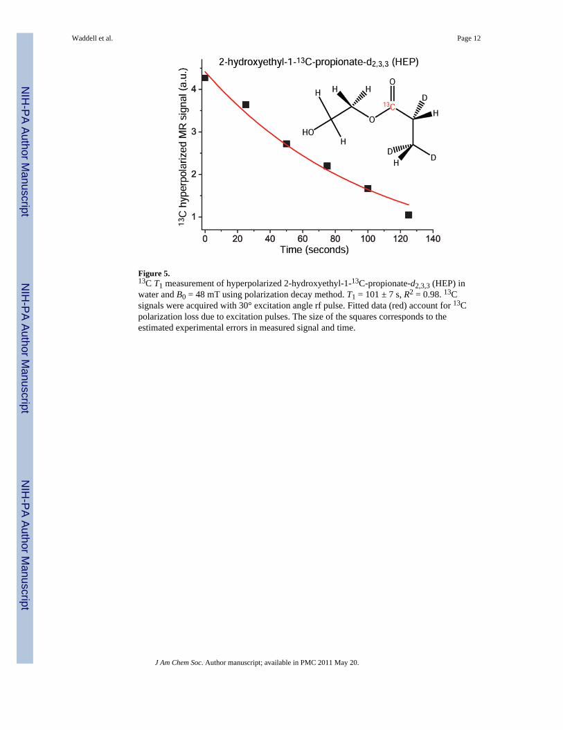
Figure 5.13C T1 measurement of hyperpolarized 2-hydroxyethyl-1-13C-propionate-d2,3,3 (HEP) inwater and B0 = 48 mT using polarization decay method. T1 = 101 ± 7 s, R2 = 0.98. 13Csignals were acquired with 30° excitation angle rf pulse. Fitted data (red) account for 13Cpolarization loss due to excitation pulses. The size of the squares corresponds to theestimated experimental errors in measured signal and time.
Waddell et al. Page 12
J Am Chem Soc. Author manuscript; available in PMC 2011 May 20.
NIH
-PA Author Manuscript
NIH
-PA Author Manuscript
NIH
-PA Author Manuscript
Related Documents


