Language switching and fMRI 1 In search of the language switch: An fMRI Study of Picture Naming In Spanish-English Bilinguals Arturo E. Hernandez 1 , Antigona Martinez 2 , Kathryn Kohnert 2,3 University of California, Santa Barbara 1 University of California, San Diego 2 & San Diego State University 3 Running Head: Language switching and fMRI Support for this research was provided by a McDonnell-Pew Postdoctoral fellowship, by the grant entitled "Cross-linguistic studies in aphasia" (NIH/NIDCD Grant #2-RO1-DC00216-10) and by the grant entitled "Aging and Bilingualism" (NIA Grant # NIA Grant # 5-R01-AG13474-02). I would like to thank Ronald Figueroa, Robert Buffington, Larry Juarez, and Cecelia Kemper for technical support. Finally, I would like to thank Elizabeth Bates for comments on earlier drafts of this manuscript. Please address correspondence to Arturo E. Hernandez, Department of Psychology 9660, University of California, Santa Barbara, California, 93106-9660. Email can be sent to: [email protected].

Welcome message from author
This document is posted to help you gain knowledge. Please leave a comment to let me know what you think about it! Share it to your friends and learn new things together.
Transcript
Language switching and fMRI
1
In search of the language switch: An fMRI Study of Picture Naming In
Spanish-English Bilinguals
Arturo E. Hernandez1, Antigona Martinez2, Kathryn Kohnert2,3
University of California, Santa Barbara1
University of California, San Diego2
&
San Diego State University3
Running Head: Language switching and fMRI
Support for this research was provided by a McDonnell-Pew Postdoctoralfellowship, by the grant entitled "Cross-linguistic studies in aphasia"(NIH/NIDCD Grant #2-RO1-DC00216-10) and by the grant entitled "Agingand Bilingualism" (NIA Grant # NIA Grant # 5-R01-AG13474-02). Iwould like to thank Ronald Figueroa, Robert Buffington, Larry Juarez, andCecelia Kemper for technical support. Finally, I would like to thankElizabeth Bates for comments on earlier drafts of this manuscript. Pleaseaddress correspondence to Arturo E. Hernandez, Department of Psychology9660, University of California, Santa Barbara, California, 93106-9660.Email can be sent to: [email protected].
Language switching and fMRI
2
Abstract
For many years, researchers investigating the brain bases of bilingualism have concentrated
on two basic questions. The first concerns the nature of language representation. That is,
are a bilinguals' two languages represented in distinct or overlapping areas of the brain.
The second basic question in the neuropsychology of bilingualism, concerns the neural
correlates of language switching, that is the areas that are active when bilinguals switch
from one language to the other. Performance between single-language and dual-language
picture naming was compared in a group of 6 Spanish-English bilinguals using behavioral
measures and functional Magnetic Resonance Imaging (fMRI). Participants showed
slower RTs and increased activation in the dorsolateral prefrontal cortex in the mixed
language condition relative to single language condition. There was no evidence that each
language was represented in different areas of the brain. Results are consistent with the
view that language switching is a part of a general executive attentional system and that
languages are represented in overlapping areas of the brain in early bilinguals
Language switching and fMRI
3
Introduction
How are a bilngual’s two languages represented in the brain? This question was
originally postulated by Pitres (1983) after observing the recovery patterns of bilingual and
polyglot aphasics. Specifically, Pitres observed that some patients seemed to
simultaneously recover both languages whereas others recovered one language sooner or
better than the other language. Since then a number of cases of bilingual and polyglot
aphasia have been reported (Paradis, 1977; Paradis, 1983; Paradis, 1987; Paradis, 1995a;
Paradis, 1995b). In looking across studies, no clear rule has emerged that accounts for the
patterns of recovery. Potential factors affecting recovery patterns include the first language
learned, language use before onset of aphasia, which languages were known (i.e.
Chinese/Japanese bilinguals vs. English/Japanese bilinguals) and language dominance.
Despite these findings, there have been a number of case studies in which selective
impairment of one language has been observed. Brain lesions that affect one language and
not the other would lead (or so it has been argued by some) to the conclusion that
languages are represented in different areas of the brain. Indeed, there is evidence of
different degrees of recovery after a stroke in each language (Junque, Vendrell, & Vendrell,
1995; Paradis, 1977). Extreme cases have shown impairment in only a single language
postoperatively with spontaneous recovery after 8 months (Paradis & Goldblum, 1989). A
more recent case has been used to suggest that there is a clear neuroanatomical dissociation
between the languages (Gomez-Tortosa, Martin, Gaviria, Charbel, & Ausman, 1995) In
this case, a patient who had surgery to remove areas of the left temperoparietal area
suffered from a more severe impairment of L1 relative to L2 (see Green, 1986; Hines,
1996; Paradis, 1996 for alternative interpretations). Finally, results from other methods
such as cortical stimulation have found that a bilingual’s two languages are represented on
partially non-overlapping areas of the brain (Ojemann, 1994; Ojemann & Whitaker, 1978).
These latter findings have fueled a consistent search for areas that are responsible for
regulating processing of each language.
Language switching and fMRI
4
More recently, researchers have turned to neuroimaing techniques to investigate
the nature of the brain bases of bilingualism. Klein, Milner, Zatorre, Meyer & Evans
(1994) using Positron Emission Tomography (PET) found that naming pictures in a
second language vs. naming pictures in a first language resulted in activation in the
putamen, a subcortical area which has been associated with phonological processing.
Other studies have found that bilinguals show activity in left frontal areas of the brain for
semantic and phonological analyses of words in both their languages (Klein, Milner,
Zatorre, Meyer, & Evans, 1995; Wagner et al., 1996). Similarly, Kim, Relkin, Lee and
Hirsh (1997) using functional Magnetic Resonance Imaging (fMRI) found overlapping
areas of activation for each language in early learners of a second language in both Broca’s
and Wernicke’s area when participants covertly told themselves what they were doing the
day before . For late learners, however, they found distinct foci of activation in Broca’s
area but not Wernicke’s area. Kim et al. suggest that these results are consistent with the
view that languages are represented in non-overlapping areas of the brain but only for late
second language learners. However, for early learners both languages resulted in
activation in overlapping areas of the brain.
The finding that the two language systems of bilingual speakers utilize the same or
adjacent pieces of neural tissue brings up another interesting point. Namely, how is it that
bilinguals keep information from one language from constantly interfering with processing
of the other language. The potential for interference is (at least in theory) massive,
particularly in view of the overlap in neural tissue (presumably, interference would be less
likely if each language occupied a spatially distinct region of cortex). To account for the
lack of interference by postulating the existence of a language switch at the
neurophysiological level (Penfield & Roberts, 1959). The search for a language switch has
proceeded at both behavioral and the neuroanatomical level. At the behavioral level, there
is mixed evidence for effects of language switching on behavioral performance in healthy
young adult bilinguals. Some have found no evidence of language switching effects
Language switching and fMRI
5
(Dalrymple-Alford & Aamiry, 1969; Kolers, 1966) while others have found significant
effects of language switching particularly in terms of speed of processing (MacNamara &
Kushnir, 1971; MacNamra, Krauthammer, & Bolgar, 1968; Soares & Grosjean, 1984).
At the neuroanatomical level, researchers have presented cases which suggest that the
language switch is localized in the supramarginal gyrus (Herschmann & Potzl, 1983;
Kauders, 1983; Potzl, 1983). However, others have found evidence of patients with
lesions in the supramarginal gyrus area, the posterior part of the sylvian fissure and
adjoining areas of the parietal lobe in which there was no switching difficulty (Gloning &
Gloning, 1983; Minkowski, 1983; Stengel & Zelmanowicz, 1933). One of the cases
presented by Stengel and Zelmanowicz is of particular interest given that it was one of the
few cases in which language mixing appears both in spontaneous conversation and in
picture naming for a person with a motor aphasia which was thought to be due to a frontal
lesion. Hence, there is evidence of lesions to both frontal, temporal, and parietal areas
which lead to problems with language mixing. Given these findings there appears to be no
specific area of the brain that is dedicated exclusively to language switching.
The findings in the neuropsychological literature leave open the question of which
neuronal circuit is involved in the process of language switching. Two candidates are the
supramarginal gyrus and adjacent areas as well as areas of the frontal lobe. To date,
however, no one has suggested particular areas of the frontal lobe that might be involved in
language switching. What areas might be involved in switching languages? The idea of
problems with set-switching has been attributed to the frontal lobes. For example, non-
aphasic frontal patients are often impaired at tasks in which one has to switch from one
strategy to another (Gershberg, 1997; Mangels, 1997). Furthermore, a recent study using
PET has found that switching between monitoring for color or shape of a stimulus relative
to a single task results in activation in the dorsolateral prefrontal cortex and in the inferior
parietal lobe including the supramarginal gyrus (Meyer et al., 1997). Hence, there is
Language switching and fMRI
6
evidence that switching between two different sets can result in activation in precisely the
areas that were proposed by Potzl, Kauders, and others.
To investigate the question of language switching and the localization of languages
in a bilingual, the current study used functional Magnetic Resonance Imaging to observe
brain activity during single and dual-language picture naming. In this “cued” picture-
naming paradigm, a group of six early Spanish-English bilinguals were told to name a
picture in the same language as a cue. The cue, the word “say” in English and the word
“diga” which is say in Spanish, instructed the subject about the language of response. Two
basic conditions were compared. In the blocked condition, participants were presented
with cues in one language (either Spanish or English) during a particular run. In the mixed
condition, the cue alternated from English to Spanish on successive pictures. Hence,
participants would name one picture in English followed by the next in Spanish, etc.
Previous behavioral studies have found that reaction times are faster for English pictures
than Spanish pictures and faster for the mixed design compared to the blocked design
(Hernandez & Kohnert, 1998). We predict that a similar pattern of reaction times will be
observed for the participants in the current study. Furthermore, based on previous
neuroimaging studies we predict that there will be very small differences in the activation
pattern associated with naming of pictures in each language. Furthermore, we predict that
conditions of language switching will involve additional activation both in terms of
intensity and extent of activation. However, it remains to be seen if this increase is in the
dorsolateral prefrontal cortex, the supramarginal gyrus or both.
Methods
Six participants from the San Diego community with a mean age of 23.5 (sd= 3.21)
participated in the current experiment. All six were Spanish-English bilinguals who learned
both languages before the age of five. The average years of formal study was significantly
higher in English (16) than in Spanish (3) (F = 65.56, p < 0.001). The Boston Naming
Language switching and fMRI
7
Test was also administered. Participants scored significantly higher on the Boston Naming
Test in English (54) than in Spanish (40) (F = 15.98, p < 0.01). Subjects were deemed to
be stronger in English than in Spanish. All were strongly right handed and reported no left
handed members in their immediate family.
A set of 180 pictures were chosen from the Snodgrass and Vanderwert (1980) and
the Pictures Please catalog (Abbate, 1984) and were presented on a Macintosh Performa
6100. Two slightly different versions of the experiments were employed. In the
behavioral version, each trial began when the participant pressed the space key on the
keyboard in front of them. Then an auditory cue (the word ‘say’ or ‘diga’ indicating the
language of response) and a visual picture were presented simultaneously. Voice response
latencies were collected by a microphone which was connected to a Carnegie-Mellon
University button box. Participants were given 3000 ms to respond. The picture
disappeared after the 3000 ms or when the participant responded. In the fMRI version, the
timing of presentation was controlled by the computer. Participants were shown the cue
(say or diga) visually for 200 ms followed immediately by the picture which was presented
for 400 ms. There was 1400 ms delay prior to the next stimulus presentation. The design
was run in blocks of 40 seconds of experimental task (Spanish, English or alternating) and
40 seconds of a control task (staring at a set of 4 XXXX’s followed by a non-object).
Each run consisted of 4 experimental cycles and 5 control cycles and lasted 6 minutes. The
pictures were presented in an alternating condition such that the cue switched between
English and Spanish on successive trials or in a blocked condition in which the cue stayed
constant across a block of trials.
The images were generated by a GE 1.5T SIGNA MRI fitted with a high
performance local head gradient and RF coils of our own design. The functional images
were T2*-weighted, echo-planar single shot pulse sequence with a matrix size of 64 X 64,
echo time (TE) of 40 ms, flip angle of 90° an in-plane resolution of 5X5. A total of 74
Language switching and fMRI
8
images were acquired for adjacent 5 mm thick axial slices in an interleaved mode with a
repetition time of 5 seconds. 20 consecutive axial images (5 mm thick) were acquired
covering the extent of the brain. The first two images of each slice were discarded to
assure that the MR signal had reached equilibrium on each slice. Artifactual signal change
caused by rigid head motion was detected by viewing the entire temporal sequence as a
movie loop. No subjects were discarded because of excessive movement. For anatomical
localization a standard whole-brain T1 weighted 3D SPGR sequence (TE= 30 ms, TE = 5
ms, flip angle 45°, 256 x 192 x 60 matrix) was acquired for each subject and the
echoplanar activation maps were overlaid on corresponding structural slices.
Data from three slices spanning the dorsolateral prefrontal cortex and the
supramarginal gyrus were collected. Average Inferior-Superior locations in Talairach
coordinates of the slices were z= 28, 24 ,20, for the dorsolateral prefrontal cortex z = 32,
24, 28 for the supramarginal gyrus, z= 20, 16,12 for the inferior frontal gyrus (areas 44
and 45), and z= 28, 24 20 for the superior temporal gyrus (area 22). Homologous regions
of interest were defined anatomically for each slice. The dorsolateral prefrontal cortex was
bound on the anterior portion by the superior frontal gyrus and on the posterior portion by
the inferior frontal gyurs. The supramarginal gyrus was bound on the anterior portion by
the postcentral gyrus and the posterior portion by the angular gyrus. The inferior frontal
gyrus was bounded by the precentral gyrus and the middle frontal and superior frontal
gyrus. The superior temporal gyrus was bounded on the posterior portion by the angular
gyrus and in the anterior portion by the post-central gyrus.
The functional data from each trial were analyzed by correlating the time course for
each particular voxel with an ideal trapezoidal reference waveform with a cycle of 4.5 s
according to the method described by Bandettini, Jesmanowicz, Wong, and Hyde (1993).
The fractional signal change and correlation coefficient for each voxel were calculated.
Activation of single voxels was discarded. Voxels that correlated positively at r=0.3 (p <
Language switching and fMRI
9
0.05 bonferroni corrected) were considered significantly activated in the picture naming
task. Any voxel below this correlation threshold was not included in subsequent statistical
analyses.
Anatomical localization of significantly activated voxels was determined with the
assistance of Analysis of Functional Neuroimages (AFNI) Software (Cox, 1996). Whole
brain three-dimensional structural reconstructions were generated and activation maps were
superimposed on to the individual subject brains scaled proportionally to the atlas of
Talairach and Tournoux (1988).
Results
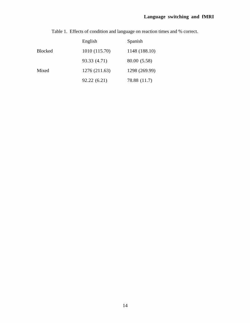
Results from the behavioral experiment revealed a main effect of language (F (1,5)
= 12.35, p < 0.05) with English response times being faster than Spanish response times
across both the mixed and blocked conditions. There was also a main effect of mixing (F
(1,5) = 13.64, p < 0.05) with the blocked condition revealing faster reaction times than the
mixed condition. There was no interaction between the two conditions. The results from
the behavioral portion of the experiment can be seen in Table 1.
_________________________________
Insert Table 1 about here
_________________________________
Results from the neuroimaging experiment revealed activation in the dorsolateral
prefrontal cortex in the left hemisphere for all participants and activation in the homologous
area in the right hemisphere for four of the six participants. Three of the six subjects
showed activation in the left supramarginal gyrus. One subject revealed activation in the
superior temporal gyrus in the left hemisphere. In the inferior frontal gyrus, one of the six
participants showed activation in the left hemisphere and the right hemisphere.
For each ROI, the number of pixels correlated above a threshold (r = 0.30.
Bonferroni corrected for p < 0.05) were selected. Both the area of activation and the
average intensity of the pixels above threshold were entered into a 2 (hemisphere) x 3
Language switching and fMRI
10
(condition; English, Spanish, Mixed) Analysis of Variance. The results for the
supramarginal gyrus, inferior temporal gyrus and the superior temporal gyrus did not yield
any significant results across hemispheres or conditions. The results for the DLPFC did
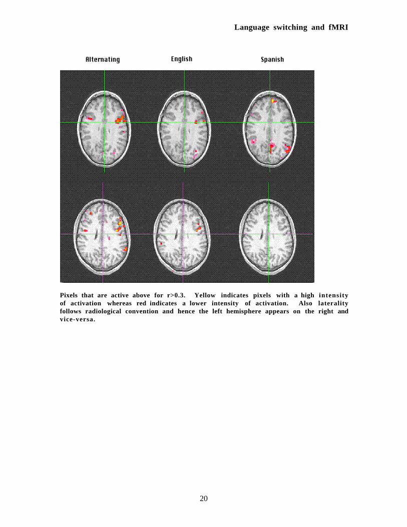
yield significant effects. An example of the activation maps for two representative subjects
can be seen in Figure 1.
_________________________________
Insert Figure 1 about here
_________________________________
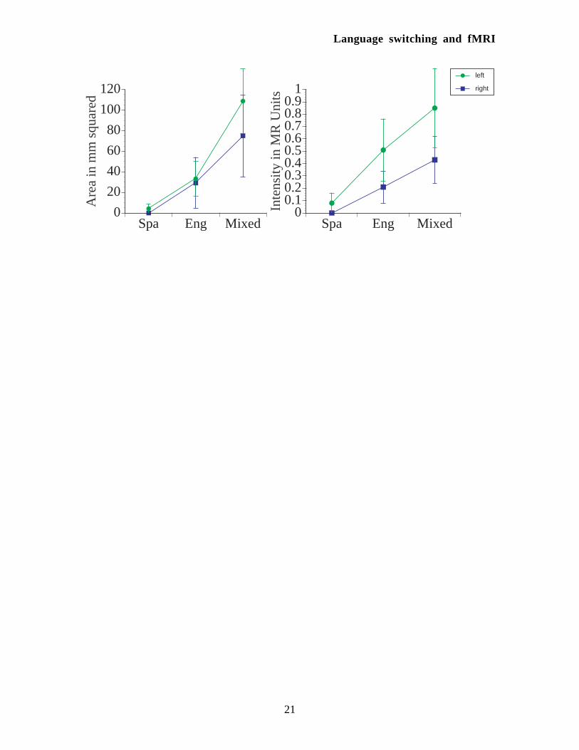
For the DLPFC, there main effect of condition for area of activation (F (2,10) =
8.94, p < 0.006). Planned comparisons for area of activation revealed a significant
difference for the mixed condition relative to the English condition (F (1,5) = 7.71, p <
0.01) and the Spanish condition (F (1,5) = 16.96, p < 0.001). For intensity of activation
there was a main effect of hemisphere- activation was stronger in the left hemisphere- (F
(1,5) = 10.95, p < 0.021) and a main effect of condition (F (1,5) = 4.07, p < 0.005).
Planned comparisons for intensity of activation across conditions revealed significant
differences for the mixed condition relative to the Spanish condition only (F (1,5) = 8.36, p
< 0.007). The results from the two ANOVA’s can be seen in Figure 2.
_________________________________
Insert Figure 2 about here
_________________________________
Discussion
The current study was designed to investigate two separate questions. First, the
study intended to address differences in the neurological areas that are active for each
language. The results revealed no differences between the two languages in our particular
Language switching and fMRI
11
regions of interest which included the dorsolateral prefrontal cortex (area 46 and 9), the
supramarginal gyrus (area 40), the inferior frontal gyrus (areas 44 and 45), and the
superior temporal gyrus (area 22). This is consistent with previous studies which have
found that bilinguals who learn a second language very early in life show few differences
in the pattern of activation for each language (Kim et al., 1997; Klein et al., 1995; Klein et
al., 1994). Hence, it appears that for early bilinguals processing of their two languages
occurs in overlapping pieces of neural tissue.
Second, the study was designed to investigate the areas that are involved in
switching between two languages. The areas involved in language switching have received
some attention in the neuropsychological literature. Initial case studies of bilingual aphasics
suggested that the supramarginal gyrus was involved in language switching (Herschmann
& Potzl, 1983; Kauders, 1983; Potzl, 1983). Others suggested that language mixing
occurs without supramarginal gyrus involvement (Gloning & Gloning, 1983; Minkowski,
1983; Stengel & Zelmanowicz, 1933). Finally, one study has suggested that frontal
lesions result in excessive language mixing (Stengel & Zelmanowicz, 1933).. It was
predicted that two areas might be involved in language switching. One candidate was the
supramarginal gyrus. The other candidate area was the dorsolateral prefrontal cortex which
is known to be involved in general executive function and has been found to be active for
switching between different tasks (Meyer et al., 1997). The only area that revealed
increased activity for language switching relative to single-language processing was the
dorsolateral prefrontal cortex.
The finding that the dorsolateral prefrontal cortex is involved in language switching
brings up two important points. First of all, it is not clear why earlier observations of
aphasics had not revealed such a candidate area. Lesions from strokes can affect brain
activity in a number of ways. They may disrupt a neurological ‘circuit’ by interrupting the
flow of information across multiple areas. Furthermore, it has been found that the
functional lesion as evidenced by reduced blood flow can be considerably larger than the
Language switching and fMRI
12
structural damage that is caused by a physical lesion (Metter, Jackson, Kempler, &
Hanson, 1992; Wong, Buxton, Love, Hickok, & Swinney, 1998). Hence, it is most
likely the case that the area affected by the damage was larger than that which is observed
by the eye. By using neuroimaging techniques in the current study we have been able to
eliminate some of these problems.
Second, it is important to note that there may not be an area that is exclusively
dedicated to language switching. That is there is no anatomically defined language switch.
Rather there is an area or areas that are involved in general executive function. One of
these functions is set or task switching and the area that is most implicated involves the
dorsolateral prefrontal cortex. In this sense, we feel that the current findings are consistent
with the view that language switching is part of a general cognitive process and as such
uses areas that are involved in general executive function.
Where does this all leave us? In some sense, it leaves us where we began. We are
still searching for the language switch, an area that is devoted exclusively to language
switching. It is our view that switching is a phenomenon that occurs across a variety of
domains. In the case of bilingualism switching occurs across languages. Rather than
develop a new area which is devoted to language switching the bilingual brain takes
advantage of their general cognitive processing mechanisms to achieve the task at hand. In
this light, the current data are consistent with the view that this language switch requires
general executive function and as such uses areas that are devoted to these processes. In
addition, we are still searching for evidence that two languages are represented in distinct
cortical areas, even in early bilinguals. We have found that when naming pictures in either
language bilinguals show no difference in the area or intensity of activation. Our results
support those from other researchers which have found that processing of a bilingual’s two
languages occurs in mostly overlapping areas of the brain for other paradigms (Kim et al.,
1997; Klein et al., 1995; Klein et al., 1994; Wagner et al., 1996).
Language switching and fMRI
13
In spite of our findings, it must be the case that each language is coded differently at
some level in the brain. In this sense, there are two possible limitations of neuroimaging
methods such as PET and fMRI. The first is noted by Steven Pinker when he says that
studying language using neuroimaging is like using “satellite photos to understand how
telecommunication lines are interconnected.” That is, perhaps we simply do not have the
spatial resolution to detect the subtle differences that occur in language processing. In
terms of bilingualism, improved resolution might uncover the smaller areas in brain regions
that are devoted exclusively to language switching or to one language but not the other. A
second limitation of fMRI for tapping language function in bilinguals may be with the
nature of the problem itself. Perhaps the differences that we are pursuing are not coded in
space but rather in time. Hence, switching between languages or processing only a single
language by bilinguals may be the result of fine timing distinctions which cannot be
detected by neuroimaging methods such as fMRI. Future studies combining techniques
such as Event-related potentials, fMRI or PET and behavioral techniques might be able to
uncover further differences between languages beyond those that have been uncovered so
far. Through the combination of methodologies we may begin to understand more clearly
the nature of language switching and language representation in the bilingual brain.
Language switching and fMRI
14
Table 1. Effects of condition and language on reaction times and % correct.
English Spanish
Blocked 1010 (115.70)
93.33 (4.71)
1148 (188.10)
80.00 (5.58)
Mixed 1276 (211.63)
92.22 (6.21)
1298 (269.99)
78.88 (11.7)
Language switching and fMRI
15
References
Bandettini, P. A., Jesmanowicz, A., Wong, E. C., & Hyde, J. S. (1993).
Processing strategies for time-course data sets in functional MRI of the human brain.
Magnetic Resonance in Medicine, 30 , 161-173.
Cox, R. W. (1996). AFNI: software for analysis and visualization of functional
magnetic resonance neuroimages. Computers and Biomedical Research, 29 (3), 162-173.
Dalrymple-Alford, E. C., & Aamiry, A. (1969). Language and category clustering
in bilingual free recall. Journal of Verbal Learning and Verbal Behavior, 8 , 762-768.
Gloning, I., & Gloning, K. (1983). Aphasia in polyglots contribution to the
dynamics of language disintegration as well as to the question of the localization of these
impairments. In M. Paradis (Ed.), Readings on aphasia in bilinguals and polyglots (pp.
681-716). Montreal: Marcel Didier.
Gomez-Tortosa, E., Martin, E., Gaviria, M., Charbel, F., & Ausman, J. (1995).
Selective deficit of one language in a bilingual patient following surgery in the left
perisylvian area. Brain and Language, 48 , 320-325.
Green, D. W. (1986). Control, activation, and resource: A framework and a model
for the control of speech in bilinguals. Brain and Language, 27 , 210-223.
Hernandez, A. E., & Kohnert, K. (1998). Aging and language switching in
bilinguals. Manuscript submitted for publication .
Herschmann, H., & Potzl, O. (1983). Observations on aphasia in polyglots. In M.
Paradis (Ed.), Readings on aphasia in bilinguals and polyglots (pp. 148-154). Montreal:
Marcel Didier.
Hines, T. M. (1996). Failure to demonstrate selective deficit in the native language
following surgery in the left perisylvian area. Brain and Language, 54 , 168-169.
Language switching and fMRI
16
Junque, C., Vendrell, P., & Vendrell, J. (1995). Differential impairments and
specific phenomena in 50 Catalan-Spanish bilingual aphasic patients. In M. Paradis (Ed.),
Aspects of Bilingual Aphasia . Oxford: Pergamon.
Kauders, O. (1983). On polyglot responses in a sensory aphasia. In M. Paradis
(Ed.), Readings on Aphasia in Bilinguals and Polyglots (pp. 286-300). Montreal: Marcel
Didier.
Kim, K. H. S., Relkin, N. R., Lee, K.-M., & Hirsch, J. (1997). Distinct cortical
areas associated with native and second languages. Nature, 388 (6638), 171-174.
Klein, D., Milner, B., Zatorre, R. J., Meyer, E., & Evans, A. C. (1995). The
neural substrates underlying word generation: a bilingual functional-imaging study.
Proceedingsof the National Academy of Sciences of the United States of America, 92 ,
2899-2903.
Klein, D., Zatorre, R. J., Milner, B., Meyer, E., & Evans, A. C. (1994). Left
putanimal activation when speaking a second language: Evidence from PET. Neuroreport:
An international journal for the rapid communication of research in Neuroscience, 5 , 2295-
2297.
Kolers, P. A. (1966). Interlingual facilitation of short-term memory. Journal of
Verbal Learning and Verbal Behavior, 5 , 314-319.
MacNamara, J., & Kushnir, S. (1971). Linguistic independence of bilinguals: The
input switch. Journal of Verbal Learning and Verbal Behavior, 10 , 480-487.
MacNamra, J., Krauthammer, M., & Bolgar, M. (1968). Language switching in
bilinguals as a function of stimulus and response uncertainty. Journal of Experimental
Psychology, 78 , 208-215.
Metter, E. J., Jackson, C. A., Kempler, D., & Hanson, W. R. (1992).
Temporoparietal cortex and the recovery of language comprehension in aphasia.
Aphasiology , 6 , 349-358.
Language switching and fMRI
17
Meyer, D. E., Evans, J. F., Lauber, E. J., Rubinstein, J., Gmeindi, L., Junck, L.,
& Koeppe, R. A. (1997). Activation of brain mechanisms for executive mental processes
in cognitive task switching . Poster presented at the fourth annual meeting of the Cognitive
Neuroscience Society.
Minkowski, M. (1983). A clinical contribution to the study of polyglot aphasia
especially with respect to swiss-german. In M. Paradis (Ed.), Readings on aphasia in
bilinguals and polyglots (pp. 205-232). Montreal: Marcel Didier.
Ojemann, G. A. (1994). Cortical stimulation and recording in language. In E.
Andrew Kertesz (Ed.), Localization and neuroimaging in neuropsychology . Foundations
of neuropsychology . (pp. 35-55): Academic Press, Inc, San Diego, CA, US.
Ojemann, G. A., & Whitaker, H. A. (1978). The bilingual brain. Archives of
Neurology, 35 , 409-412.
Paradis, M. (1977). Bilingualism and aphasia. In H. Whitaker & H. A. Whitaker
(Eds.), Studies in Neurolinguistics (Vol. 3, pp. 65-121). New York: Academic Press.
Paradis, M. (Ed.). (1983). Readings on aphasia in Bilinguals and Polyglots .
Quebec: Didier.
Paradis, M. (1987). The assessment of bilingual aphasia . Hillsdale, NJ: Erlbaum.
Paradis, M. (Ed.). (1995). Bilingual aphasia 100 years later: Consensus and
controversies . Oxford: Pergamon.
Paradis, M. (1996). Selective deficit in one language is not a demonstration of
different anatomical representation: Comments on Gomez-Tortosa et al. (1995). Brain and
Language, 54 , 170-173.
Paradis, M., & Goldblum, M. C. (1989). Selected crossed aphasia in a trilingual
aphasic patient followed by reciprocal antagonism. Brain and Language, 36 , 62-75.
Paradis, M., & International Association of Logopedics and Phoniatrics. (1995).
Aspects of bilingual aphasia . (1st ed.). Oxford, OX, UK ; Tarrytown, N.Y., U.S.A.:
Pergamon.
Language switching and fMRI
18
Penfield, W., & Roberts, L. (1959). Speech and brain mechanisms . Princeton, N.
J.: Princeton University Press.
Pitres, A. (1983). Aphasia in polyglots. In M. Paradis (Ed.), Readings on aphasia
in bilinguals and polyglots (pp. 26-49). Montreal: Marcel Didier.
Potzl, O. (1983). Aphasia and mutilingualism. In M. Paradis (Ed.), Readings on
aphasia in bilinguals and polyglots (pp. 301-316). Montreal: Marcel Didier.
Snodgrass, J. G., & Vanderwart, M. (1980). A standardized set of 260 pictures:
Norms fo name agreement, image agreement, familiarity, and visual complexity. Journal of
Experimental Psychology: Human Learning and Memory, 6 , 174-215.
Soares, C., & Grosjean, F. (1984). Bilinguals in a monolingual and a bilingual
speech mode: The effect on lexical access. Memory and Cognition, 12 , 380-386.
Stengel, E., & Zelmanowicz, J. (1933). On polyglot motor aphasia. In M. Paradis
(Ed.), Readings on aphasia in bilinguals and polyglots (pp. 356-375). Montreal: Marcel
Didier.
Talairach, J., & Tournoux, P. (1988). Co-planar stereotaxic atlas of the human
brain : a 3-dimensional proportional system, an approach to cerebral imaging . Stuttgart ;
New York: Thieme Medical Publishers.
Wagner, A. D., Illes, J., Desmond, J. E., Lee, C. J., Glover, G. H., & Gabrieli,
J. D. E. (1996). A functional MRI study of semantic processing in bilinguals.
NeuroImage , 3 , S465.
Wong, E., Buxton, R., Love, T., Hickok, G., & Swinney, D. (1998, ). Perfusion
MRI and functional behavioral deficits. Paper presented at the 1998 Annual Meeting of the
Cognitive Neuroscience Society, San Francisco, Ca.
Language switching and fMRI
19
Figure Caption.
Figure 1. Activation maps for two representative subjects.
Figure 2. Effects of condition and hemisphere for area and intensity of activation.
Language switching and fMRI
20
Pixels that are active above for r>0.3. Yellow indicates pixels with a high intensityof activation whereas red indicates a lower intensity of activation. Also lateralityfollows radiological convention and hence the left hemisphere appears on the right andvice-versa.
Language switching and fMRI
21
Spa Eng Mixed0
20
40
60
80
100
120A
rea
in m
m s
quar
ed
Spa Eng Mixed0
0.10.20.30.40.50.60.70.80.9
1
Inte
nsit
y in
MR
Uni
ts
right
left
Related Documents