Improving the Tolerance of a Protein A Analogue to Repeated Alkaline Exposures Using a Bypass Mutagenesis Approach Martin Linhult, 1 Susanne Gu ¨ lich, 3 Torbjo ¨ rn Gra ¨ slund, 1 Annelie Simon, 1 Martin Karlsson, 4 Anna Sjo ¨ berg, 2 Karin Nord, 2 and Sophia Hober 1 * 1 Department of Biotechnology, Royal Institute of Technology (KTH), Stockholm, Sweden 2 Affibody AB, Bromma, Sweden 3 Department of Molecular Biophysics and Biochemistry, Yale University, New Haven, Connecticut 4 IFM-Department of Chemistry, Linko ¨ ping University, Linko ¨ ping, Sweden ABSTRACT Staphylococcal protein A (SPA) is a cell surface protein expressed by Staphylococcus aureus. It consists of five repetitive domains. The five SPA-domains show individual interaction to the Fc-fragment as well as certain Fab-fragments of immunoglobulin G (IgG) from most mammalian spe- cies. Due to the high affinity and selectivity of SPA, it has a widespread use as an affinity ligand for capture and purification of antibodies. One of the problems with proteinaceous affinity ligands in large-scale purification is their sensitivity to alka- line conditions. SPA however, is considered rela- tively stable to alkaline treatment. Nevertheless, it is desirable to further improve the stability in order to enable an SPA-based affinity medium to with- stand even longer exposure to the harsh conditions associated with cleaning-in-place (CIP) procedures. For this purpose, a protein engineering strategy, which was used earlier for stabilization and con- sists of replacing the asparagine residues, is em- ployed. Since Z in its “nonengineered” form already has a significant tolerance to alkaline treatment, small changes in stability due to the mutations are difficult to assess. Hence, in order to enable detec- tion of improvements regarding the alkaline resis- tance of the Z domain, we chose to use a bypass mutagenesis strategy using a mutated variant Z(F30A) as a surrogate framework. Z(F30A) has earlier been shown to possess an affinity to IgG that is similar to the wild-type but also demonstrates decreased structural stability. Since the contribu- tion of the different asparagine residues to the deactivation rate of a ligand is dependent on the environment and also the structural flexibility of the particular region, it is important to consider all sensitive amino acids one by one. The parental Z-domain contains eight asparagine residues, each with a different impact on the alkaline stability of the domain. By exchanging asparagine 23 for a threonine, we were able to increase the stability of the Z(F30A) domain in alkaline conditions. Also, when grafting the N23T mutation to the Z scaffold, we were able to detect an increased tolerance to alkaline treatment compared to the native Z mole- cule. Proteins 2004;55:407– 416. © 2004 Wiley-Liss, Inc. Key words: affinity chromatography; deamidation; protein A; purification; stabilization; Z domain INTRODUCTION Staphylococcal protein A (SPA) is a cell surface protein expressed by Staphylococcus aureus 1 and consists of five highly homologous domains (EDABC) [Fig. 1(a)]. 2 The five SPA domains are about 58 residues each, arranged in an antiparallel three-helix bundle with two interconnecting loops (Fig. 1). The helices are in close contact with each other, forming a hydrophobic core containing most of the hydrophobic residues of the domain, hence, contributing to the stability. 3 The five SPA domains show individual affinity for the Fc-fragment [11 residues of helices 1 and 2 (domain B)], 4 as well as certain Fab-fragments of immuno- globulin G (IgG) from most mammalian species. The bacterial receptor also interacts with IgA and IgM. 5–8 SPA has a widespread use in the field of biotechnology for affinity chromatography purification, as well as detection of antibodies. 9,10 SPA is well suited for those purposes due to its high affinity and selectivity. In addition to the high selectivity, SPA-based affinity purification medium also shows rather high tolerance to both low pH and high concentrations of denaturants such as urea and guanidine hydrocloride (GdnHCl). These features have contributed to the fact that SPA-based affinity medium probably is the most widely used medium for isolation of monoclonal antibodies and their fragments. Hence, this chromatogra- phy technique is also commonly used in large-scale purifi- cation of monoclonal antibodies for therapeutic use. For these types of applications, extreme attention has to be maintained to minimize contamination. In order to remove contaminants such as nucleic acids, lipids, proteins, and microbes, a cleaning-in-place (CIP) step is often integrated into the purification protocol. Sodium hydroxide (NaOH) is probably the most extensively used cleaning agent for this Grant sponsor: Amersham Biosciences. *Correspondence to: Sophia Hober, Department of Biotechnology, KTH, Royal Institute of Technology, AlbaNova, SE-106 91 Stockholm, Sweden. E-mail: [email protected] Received 23 January 2003; Accepted 18 August 2003 Published online 27 February 2004 in Wiley InterScience (www.interscience.wiley.com). DOI: 10.1002/prot.10616 PROTEINS: Structure, Function, and Bioinformatics 55:407– 416 (2004) © 2004 WILEY-LISS, INC.

Welcome message from author
This document is posted to help you gain knowledge. Please leave a comment to let me know what you think about it! Share it to your friends and learn new things together.
Transcript

Improving the Tolerance of a Protein A Analogue to RepeatedAlkaline Exposures Using a Bypass Mutagenesis ApproachMartin Linhult,1 Susanne Gulich,3 Torbjorn Graslund,1 Annelie Simon,1 Martin Karlsson,4 Anna Sjoberg,2
Karin Nord,2 and Sophia Hober1*1Department of Biotechnology, Royal Institute of Technology (KTH), Stockholm, Sweden2Affibody AB, Bromma, Sweden3Department of Molecular Biophysics and Biochemistry, Yale University, New Haven, Connecticut4IFM-Department of Chemistry, Linkoping University, Linkoping, Sweden
ABSTRACT Staphylococcal protein A (SPA) isa cell surface protein expressed by Staphylococcusaureus. It consists of five repetitive domains. Thefive SPA-domains show individual interaction to theFc-fragment as well as certain Fab-fragments ofimmunoglobulin G (IgG) from most mammalian spe-cies. Due to the high affinity and selectivity of SPA,it has a widespread use as an affinity ligand forcapture and purification of antibodies. One of theproblems with proteinaceous affinity ligands inlarge-scale purification is their sensitivity to alka-line conditions. SPA however, is considered rela-tively stable to alkaline treatment. Nevertheless, itis desirable to further improve the stability in orderto enable an SPA-based affinity medium to with-stand even longer exposure to the harsh conditionsassociated with cleaning-in-place (CIP) procedures.For this purpose, a protein engineering strategy,which was used earlier for stabilization and con-sists of replacing the asparagine residues, is em-ployed. Since Z in its “nonengineered” form alreadyhas a significant tolerance to alkaline treatment,small changes in stability due to the mutations aredifficult to assess. Hence, in order to enable detec-tion of improvements regarding the alkaline resis-tance of the Z domain, we chose to use a bypassmutagenesis strategy using a mutated variantZ(F30A) as a surrogate framework. Z(F30A) hasearlier been shown to possess an affinity to IgG thatis similar to the wild-type but also demonstratesdecreased structural stability. Since the contribu-tion of the different asparagine residues to thedeactivation rate of a ligand is dependent on theenvironment and also the structural flexibility ofthe particular region, it is important to consider allsensitive amino acids one by one. The parentalZ-domain contains eight asparagine residues, eachwith a different impact on the alkaline stability ofthe domain. By exchanging asparagine 23 for athreonine, we were able to increase the stability ofthe Z(F30A) domain in alkaline conditions. Also,when grafting the N23T mutation to the Z scaffold,we were able to detect an increased tolerance toalkaline treatment compared to the native Z mole-cule. Proteins 2004;55:407–416. © 2004 Wiley-Liss, Inc.
Key words: affinity chromatography; deamidation;protein A; purification; stabilization; Zdomain
INTRODUCTION
Staphylococcal protein A (SPA) is a cell surface proteinexpressed by Staphylococcus aureus1 and consists of fivehighly homologous domains (EDABC) [Fig. 1(a)].2 The fiveSPA domains are about 58 residues each, arranged in anantiparallel three-helix bundle with two interconnectingloops (Fig. 1). The helices are in close contact with eachother, forming a hydrophobic core containing most of thehydrophobic residues of the domain, hence, contributing tothe stability.3 The five SPA domains show individualaffinity for the Fc-fragment [11 residues of helices 1 and 2(domain B)],4 as well as certain Fab-fragments of immuno-globulin G (IgG) from most mammalian species. Thebacterial receptor also interacts with IgA and IgM.5–8 SPAhas a widespread use in the field of biotechnology foraffinity chromatography purification, as well as detectionof antibodies.9,10 SPA is well suited for those purposes dueto its high affinity and selectivity. In addition to the highselectivity, SPA-based affinity purification medium alsoshows rather high tolerance to both low pH and highconcentrations of denaturants such as urea and guanidinehydrocloride (GdnHCl). These features have contributedto the fact that SPA-based affinity medium probably is themost widely used medium for isolation of monoclonalantibodies and their fragments. Hence, this chromatogra-phy technique is also commonly used in large-scale purifi-cation of monoclonal antibodies for therapeutic use. Forthese types of applications, extreme attention has to bemaintained to minimize contamination. In order to removecontaminants such as nucleic acids, lipids, proteins, andmicrobes, a cleaning-in-place (CIP) step is often integratedinto the purification protocol. Sodium hydroxide (NaOH) isprobably the most extensively used cleaning agent for this
Grant sponsor: Amersham Biosciences.*Correspondence to: Sophia Hober, Department of Biotechnology,
KTH,
Royal Institute of Technology, AlbaNova, SE-106 91 Stockholm,Sweden. E-mail: [email protected]
Received 23 January 2003; Accepted 18 August 2003
Published online 27 February 2004 in Wiley InterScience(www.interscience.wiley.com). DOI: 10.1002/prot.10616
PROTEINS: Structure, Function, and Bioinformatics 55:407–416 (2004)
© 2004 WILEY-LISS, INC.
purpose.11 Unfortunately, protein-based affinity mediashow high fragility in this extremely harsh environment,making them less attractive in industrial-scale proteinpurification. SPA, however, is considered relatively stablein alkaline conditions. Hale and coworkers12 report a lossof about 1% of the binding capacity when SPA is exposed to0.5 M NaOH for 15 min. Nevertheless, it is desirable tofurther improve the stability in order to enable an SPA-based affinity medium to withstand even longer exposureto the harsh conditions associated with CIP procedures.For this purpose, a protein engineering strategy, earlierused for stabilization of domains from streptococcal pro-tein G and involving replacement of the asparagine resi-dues, is employed.13
Asparagine is known to be the most susceptible residueto high pH due to covalent modifications such as deamida-tion or backbone cleavage. Glutamine is also susceptible,even though it is modified to a lesser extent. Thesereactions are spontaneous and may also occur at physiolog-ical solvent conditions, often resulting in loss of activity ofthe protein or peptide.14 The extent of modification of thedifferent residues is highly sequence and conformationdependent.15–17 The deamidation reaction involves themain-chain peptide nitrogen succeeding the asparagine.The nitrogen functions as the nucleophile and attacks the
side-chain carbonyl of asparagine, resulting in a succinim-ide intermediate. This succinimide intermediate may openat either of the two CON bonds to form aspartic acid orisoaspartic acid, resulting in the addition of a negativecharge. These two isomers may occur in their L- or D-forms.
To further increase the alkaline tolerance of SPA, wechose to work with Z, which is a small protein derived fromthe B domain of SPA.18 The B domain has been mutated inorder to achieve a purification domain resistant to cleav-age by hydroxylamine. An exchange of glycine 29 for analanine has been made in order to avoid the amino acidcombination asparagine–glycine, which is a cleavage sitefor hydroxylamine.19,20 Asparagine with a succeedingglycine has also been found to be the most sensitive aminoacid sequence to alkaline conditions.15–17 Protein Z is wellcharacterized and extensively used as both ligand andfusion partner in a variety of affinity chromatographysystems.21 The domain is also used as a scaffold for thegeneration of Affibody� molecules.22,23 The positive fea-tures of the domain include the high solubility and proteo-lytic stability, the lack of cysteines, the rapid foldingkinetics, and the well-characterized compact and robuststructure. Since Z in its original form already has asignificant stability toward alkaline treatment, smallchanges in stability due to the mutations are difficult to
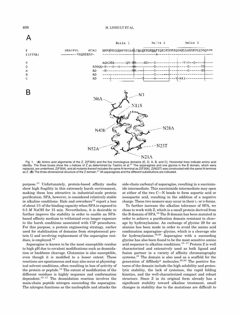
Fig. 1. (A) Amino acid alignments of the Z, Z(F30A) and the five homologous domains (E, D, A, B, and C). Horizontal lines indicate amino acididentity. The three boxes show the �-helices of Z as determined by Tashiro et al.47 The asparagines and one glycine in the B domain, which werereplaced, are underlined. Z(F30A), and all mutants thereof includes the same N-terminal as Z(F30A). Z(N23T) was constructed with the same N-terminalas Z. (B) The three-dimensional structure of the Z domain.47 All asparagines and the different substitutions are indicated.
408 M. LINHULT ET AL.
assess. Therefore, a bypass mutation method has beenused.24,25 According to this strategy, a destabilized variantof Z, Z(F30A), characterized by Cedergren et al.,26 waschosen as scaffold. The binding properties of this variantare similar to native Z, since F30 is not involved in theFc-binding. Instead, F30 is sandwiched between L44 andL51 of helix 3 in the hydrophobic core of the domain.Hence, the F30A substitution has a dramatic effect on theconformational stability. From GdnHCl experiments, adestabilization of 3.5 kcal/mol is reported.26 Single mu-tants with Z(F30A) as scaffold were designed, in which theasparagines were substituted for other amino acids notsusceptible to deamidation. Some replacements have beenbased on comparison with the homologous sequences of theother domains, EDABC [Fig. 1(a)].13 When the choice withthe aid of sequence homology was not obvious, the asparag-ines were substituted for alanines.
MATERIALS AND METHODSDNA Constructions and Bacterial Strains
Site-directed mutagenesis was performed using a two-step polymerase chain reaction (PCR)-technique.27 Plas-mid pDHZF30A26 was used as template. Oligonucleotidescoding for the different asparagine replacements weresynthesized by Interactiva (Interactiva BiotechnologieGmbH, Ulm, Germany). The restriction enzymes XbaI andHindIII (MBI Fermentas, Inc., Amherst, NY) were usedfor cloning into the vector pDHZ,28 performed according toSambrook et al.29 To create pTrpZ, the Z domain wasamplified by PCR, using plasmid pKN1 as template.22 Thefragment was restricted with XbaI and PstI, and ligatedinto the vector pTrpABDT1T230 that had been restrictedwith the same enzymes. A MegaBACE™ 1000 DNA Se-quencing System (Amersham Biosciences, Uppsala, Swe-den) was used to verify correct sequence of insertedfragments. MegaBACE™ terminator chemistry (Amer-sham Biosciences) was utilized according to the supplier’srecommendations in a cycle sequencing protocol based onthe dideoxy method.31 During cloning procedures, Esche-richia coli strain RR1�M15 (American Type Culture Col-lection, Rockville, MA) was used. For expression of Z(F30A)and different mutants thereof, strain O1732 was used. Forexpression of Z and Z(N23T), RRI�M15 was used.
Production and Purification
Production and purification of Z(F30A), and the differ-ent mutants thereof were performed according to theprotocol outlined by Gulich et al.33 The production of Z andZ(N23T) was performed as described by Kraulis et al.30
Relevant fractions were lyophilized. The amount of proteinwas estimated by absorbance measurements at 280 nmusing the specific absorbance coefficients,34 a (l g�1 cm�1),Z 0.156; Z(N23T) 0.169; Z(F30A), Z(F30A,N43E),Z(F30A,N23T,N43E) 0.157; Z(F30A,N6A), Z(F30A,N11S),Z(F30A,N21A), Z(F30A,N23T), Z(F30A,N28A), Z(F30A,N52A),Z(F30A,N6A,N23T),Z(F30A,N11S,N23T)0.158.Theconcentra-tion was confirmed by amino acid analysis (BMC, Uppsala,Sweden). The homogeneity was analyzed by sodium dode-cyl sulfate polyacrylamide gel electrophoresis (SDS-
PAGE)35 using the Phast™ system. Lyophilized proteinswere loaded on high-density gels (Amersham Biosciences)under reducing conditions and stained with Coomassiebrilliant blue according to the supplier’s recommenda-tions. The homogeneity and the molecular weights werefurther confirmed by using a mass spectrometer equippedwith an electrospray unit (Micromass, Manchester, UK).
Circular Dichroism Spectroscopy
To analyze the secondary structure content of Z(F30A)and mutants thereof, a quartz cell of path length 0.1 cmwas used. Protein samples were dissolved in 10 mM K2PO4
(pH 7.6) to a concentration of 10 �M. Spectra wererecorded using a J-720 spectropolarimeter (JASCO, Tokyo,Japan) in the far UV region from 250 to 190 nm at roomtemperature, with a scan speed of 10 nm min�1. Eachspectrum is the mean of five accumulated scans, and thefinal spectra were converted into mean residue ellipticity(MRE) (deg cm2 dmol�1). Also, spectra from the buffersused were recorded and subtracted from the protein spec-tra to correct for instrumental drift.
To analyze the secondary structure content in differentpH, the protein variants were dissolved in 0.5 M NaOH(pH 13.7) or 10 mM K2PO4 (pH 7.6), giving a final proteinconcentration of 1 mM. Spectra were collected from 260 to180 nm at room temperature using a quartz cell with apath length of 0.001 cm. Each spectrum is the mean of fiveaccumulated scans and the final spectra were convertedinto MRE (deg cm2 dmol�1). To correct for instrumentaldrift, spectra from the buffers used were subtracted fromthe protein spectra, and before calculating mean value,each scan was compared to exclude signal drift during theexperiment. Accurate protein concentrations were deter-mined by quantitative amino acid analysis in triplicate.The spectropolarimeter was calibrated according to themanufacturer’s recommendation.
Biospecific Interaction Analysis
Differences in affinity and kinetic constants of theassociation and dissociation rates were detected on aBiacore™ 2000 instrument (Biacore, Uppsala, Sweden).Human polyclonal IgG and human serum albumin (HSA)(negative reference) were immobilized by amine couplingon the carboxylated dextran layer of a CM5 sensor chip(Biacore) according to the supplier’s recommendations.The immobilization of IgG resulted in approximately 2000RU. Z, ZF30A, and the different mutants were prepared inHEPES buffered saline (HBS) (10 mM N-2-hydroxyeth-ylpiperazine-N�-2-ethanesulfonic acid (HEPES), 0.15 MNaCl, 3.4 mM ethylene diaminetetraacetic acid (EDTA),0.005% surfactant P20, pH 7.4) at 10 different concentra-tions (100–550 nM). The samples were injected over thesurfaces as duplicates in random order at a flow rate of 30�L min�1. 10 mM HCl was used to regenerate the surface.The data was analyzed using the BIA evaluation 3.0.2bsoftware (Biacore). The signals from a control surfaceimmobilized with HSA were subtracted from signals fromthe IgG surface. A 1:1 Langmuir model was assumed andapparent kinetic constants and affinity constants were
ENGINEERING STAPHYLOCOCCAL PROTEIN A 409

calculated. Also, the change in binding free energy (��G ��RTln Kaff, mutant/Kaff, native) in relation to the nativemolecule was calculated.
Analysis of Stability Toward Alkaline Conditions
The behavior of the variants of domain Z as affinityligands was analyzed by immobilization to a standardaffinity matrix. Z, Z(F30A), and mutated variants werecovalently coupled to HiTrap™ affinity columns (Amer-sham Biosciences) using the N-hydroxysuccinimide (NHS)chemistry according to the manufacturer’s recommenda-tions. The columns were pulsed with TST (25 mM Tris-HClpH 7.5, 150 mM NaCl, 1.25 mM EDTA, 0.05% Tween 20)and 0.2 M HAc, pH 3.1. Human polyclonal IgG in TST wasprepared and injected onto the columns in excess. Astandard affinity chromatography protocol was followedfor 16 cycles on the AKTA™ Explorer 10 (AmershamBiosciences). Between each cycle, a CIP-step was inte-grated. The cleaning agent was 0.5 M NaOH and thecontact time for each pulse was 30 min, resulting in a totalexposure time of 7.5 h for Z(F30A) and mutants thereof.The total time of exposure for Z and Z(N23T) was 23 h.Eluted material was detected at 280 nm.
RESULTSGeneral Strategy
A mutational analysis was performed to analyze whichasparagines in the Z domain are responsible for thedeactivation in alkaline conditions. The Z-domain alreadypossesses a significant tolerance to alkaline conditions.Therefore, we chose to use a structurally destabilizedvariant, Z(F30A), as scaffold to facilitate detection of theimprovements. Z(F30A) has earlier been shown to exhibitan affinity to IgG that is similar to the wild-type, but also aremarkably decreased structural stability due to the muta-tion of an amino acid that normally takes part in thehydrophobic core.26,36 The Z domain includes 8 asparag-ines (N3, N6, N11, N21, N23, N28, N43, and N52; Fig. 1).18
To evaluate the effect of the different asparagines on thedeactivation rate in alkaline conditions, 7 of these residueswere exchanged for other amino acids. Since N3 is locatedin the flexible N-terminal of the domain, it was excludedfrom the study [Fig. 1(b)]. A degradation of this amino acidwould probably not affect the activity, and changes aretherefore not detectable in our assay, which measures theretained activity. Moreover, since the amino acid is locatedoutside the structured part of the domain, it will mostlikely be easily replaceable during a multimerization of thedomain to achieve a protein A–like molecule. To facilitatethe protein design, a comparison with the homologoussequences from the other domains of protein A was made(Fig. 1).13 From the alignment, we could decide that Asn11should be exchanged for a serine, Asn23 for threonine, andfinally, Asn43 for a glutamic acid. Asparagine 6 wasexchanged for alanine, since the alternative when lookingon the homologous sequences was aspartic acid, which alsohas been reported to be prone to degradation. All 5domains of protein A have asparagines in positions 21, 28,and 52. Hence, they were exchanged for alanines.
Expression and Purification of Z Variants
All Z variants were successfully produced intracellularlyin E. coli and showed the same expression levels, approxi-mately 50 mg/L as estimated from SDS-PAGE. The pro-teins were all purified by IgG affinity chromatography.After the purification, samples were analyzed with SDS-PAGE (data not shown), lyophilized, and stored for furtheranalyses. The molecular mass of protein Z and the differ-ent mutants thereof was also confirmed by mass spectrom-etry, and the data confirmed correct amino acid content forall mutants (data not shown). Also, structural analyseswere performed on CD equipment, since it previously hasbeen shown to be suitable for detecting structuralchanges in �-helical proteins.22,37 All spectra show aminimum at 208 nm and 222 nm in combination with amaximum around 195 nm, indicating a similar structurefor the mutants and the parental molecule. However,Z(F30A,N52A) seems to have a somewhat lower �-helic-ity than the wild-type Z and the other mutants thereof(data not shown).
Biospecific Interaction Analysis
To determine if the mutations had caused changes in theaffinity of the Z variants for IgG, surface plasmon reso-nance (SPR), using a Biacore™ instrument, was carriedout. The aim was to compare the affinity for the differentmutated Z variants with the parental molecule. First, itwas important to confirm that the affinity between Z(F30A)and IgG was retained despite the mutation. As can be seenin Table I, the affinity (KA) of Z(F30A) for IgG is notsignificantly affected. The very small change in affinitygives a slightly higher stability to the complex of Z(F30A)and IgG compared to the parental molecule Z and IgG.This is in accordance with results reported earlier.26,36 Allmutants constructed from Z(F30A) were analyzed andcompared with their parental molecule Z(F30A). The re-sults show that the total affinity is rather unaffected bythe mutations, indicating that none of the mutations arevery important for the binding to IgG (Table I). Surpris-ingly, in the case of the N28A mutation, the decrease inaffinity (KA) is very small, which is in contradiction withearlier reported results where the N28 has been suggestedto be involved in the interaction with IgG.4,26,36 Thisbehavior might be due to the F30A mutation that issituated in the vicinity of the N28 and is included in allconstructs. However, all constructs including the N28Amutation have an increased dissociation rate constant(kd). Also, the N6A-mutation gives a higher dissociationrate constant (kd), but the affinity constant (KA) is notaffected because of the increased association rate constant(ka) that also follows the mutation.
Affinity Chromatography With an Integrated CIPStep
Z, Z(F30A), and mutants thereof were covalently at-tached to HiTrap™ columns using NHS-chemistry. IgG inexcess was loaded and the amount of eluted IgG wasmeasured after each cycle to determine the total capacityof the column. Between each cycle the columns were
410 M. LINHULT ET AL.

exposed to CIP treatment consisting of 0.5 M NaOH. After16 cycles, giving a total exposure time of 7.5 h, the columnwith the Z(F30A)-matrix shows a 70% decrease in capac-ity. The degradation data in Figure 2 suggest that 4 of theexchanged asparagines (N6, N11, N43, and N52) are notvery important for the sensitivity to alkaline conditions.Their degradation patterns are very similar to the paren-tal molecule’s Z(F30A). In contrast, N23 seems to be veryimportant for the functional stability after alkaline treat-ment of Z(F30A). Z(F30A,N23T) shows only a 28% de-crease in capacity despite the destabilizing F30A-muta-
tion. Hence, the Z(F30A,N23T) is almost as tolerant as Zand is thereby the most improved variant with Z(F30A) asscaffold (Figs. 2 and 3). Also the Z(F30A) domain with twoadditional mutations Z(F30A,N23T,N43E) shows the samepattern of degradation as Z(F30A,N23T). This is probablydue to the N23T-mutation, since the N43E-mutation alonedoes not show any improving effect. An exchange of N28 toan alanine also improves the tolerance of Z(F30A) toalkaline treatment. Interestingly, the column withZ(F30A,N21A) as affinity ligand reveals a dramatic loss ofcapacity when exposed to NaOH compared to Z(F30A).
TABLE I. An Overview of the Kinetic Study on the Different Z Domains Using the Biacore™
Mutant ka [105 M�1 s�1] kd [10�3 s�1] KA [107 M�1]��G (vs Z)[kcal/mol]
��G [vs Z(F30A)][kcal/mol]
Z 1.5 3.7 4.0 0Z(N23T) 2.7 3.9 7.0 �0.3Z(F30A) 1.9 4.2 4.5 �0.1 0Z(F30A,N6A) 7.0 21 3.3 0.1 0.2Z(F30A,N11S) 1.6 4.9 3.2 0.1 0.2Z(F30A,N21A) 1.0 3.8 2.6 0.3 0.3Z(F30A,N23T) 2.1 3.8 5.6 �0.2 �0.1Z(F30A,N28A) 3.1 9.9 3.2 0.1 0.2Z(F30A,N43E) 1.3 5.1 2.6 0.3 0.3Z(F30A,N52A) 1.5 4.9 3.0 0.2 0.2Z(F30A,N23T,N43E) 0.8 3.8 2.0 0.4 0.5
Z was used as an internal standard during the different measurements. The differences in free binding energy are calculated relative to Z andZ(F30A), respectively.
Fig. 2. A comparison of the capacity after repeated CIP treatment following an ordinary affinity chromatography scheme. 0.5 M NaOH was used ascleaning agent. The protocol was run 16 times, and the duration for the alkaline exposure was 30 min in each round. The inactivation patterns for Z(F30A)and variants thereof are shown. The total time of exposure to 0.5 M NaOH was 7.5 h.
ENGINEERING STAPHYLOCOCCAL PROTEIN A 411
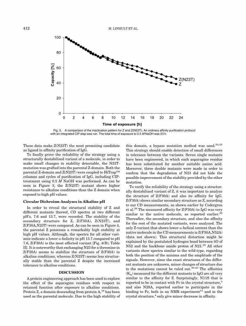
These data make Z(N23T) the most promising candidateas ligand in affinity purification of IgG.
To finally prove the reliability of the strategy using astructurally destabilized variant of a molecule, in order tomake small changes in stability detectable, the N23T-mutation was grafted into the parental Z-domain. Both theparental Z-domain and Z(N23T) were coupled to HiTrap™columns and cycles of purification of IgG, including CIP-treatment using 0.5 M NaOH was performed. As can beseen in Figure 3, the Z(N23T) mutant shows higherresistance to alkaline conditions than the Z domain whenexposed to high pH values.
Circular Dichroism Analyses in Alkaline pH
In order to reveal the structural stability of Z anddifferent mutants thereof, CD spectra at two differentpH’s, 7.6 and 13.7, were recorded. The stability of thesecondary structure for Z, Z(F30A), Z(N23T), andZ(F30A,N23T) were compared. As can be seen in Figure 4,the parental Z possesses a remarkably high stability athigh pH values. Although, the spectra for all other vari-ants indicate a lower �-helicity in pH 13.7 compared to pH7.6, Z(F30A) is the most affected variant [Fig. 4(B); TableII]. It is noteworthy that exchanging N23 for a threonine inZ(F30A) seems to stabilize the structure of Z(F30A) inalkaline conditions, whereas Z(N23T) seems less structur-ally stable than the parental Z despite the increasedtolerance to alkaline conditions.
DISCUSSION
A protein engineering approach has been used to explorethe effect of the asparagine residues with respect toretained function after exposure to alkaline conditions.Protein Z, a domain descending from protein A,18 has beenused as the parental molecule. Due to the high stability of
this domain, a bypass mutation method was used.24,25
This strategy should enable detection of small differencesin tolerance between the variants. Seven single mutantshave been engineered, in which each asparagine residuehas been substituted for another suitable amino acid.Moreover, three double mutants were made in order toconfirm that the degradation of N23 did not hide thepossible improvement of the stability provided by the othermutation.
To verify the reliability of the strategy using a structur-ally destabilized variant of Z, it was important to analyzethe structure of Z(F30A) and also its affinity for IgG.Z(F30A) shows similar secondary structure as Z, accordingto our CD measurements, as shown earlier by Cedergrenet al.26 The measured affinity for Z(F30A) to IgG was verysimilar to the native molecule, as reported earlier.36
Thereafter, the secondary structure, and also the affinityfor the rest of the mutated variants, were analyzed. Theonly Z-variant that shows lower �-helical content than thenative molecule in the CD measurements is Z(F30A,N52A)(data not shown). This structural distortion might beexplained by the postulated hydrogen bond between �O ofN52 and the backbone amide proton of N21.38 All othervariants show spectra similar to the wild-type, regardingboth the position of the minima and the amplitude of thesignals. However, since the exact structures of the differ-ent mutants are unknown, minor changes of structure dueto the mutations cannot be ruled out.39,40 The affinities(KA) measured for the different mutants to IgG are all verysimilar to the affinity for Z. Surprisingly, N11S that isreported to be in contact with Fc in the crystal structure,4
and also N28A, reported earlier to participate in thebinding to Fc, both in an NMR experiment41 and in thecrystal structure,4 only give minor decrease in affinity.
Fig. 3. A comparison of the inactivation pattern for Z and Z(N23T). An ordinary affinity purification protocolwith an integrated CIP step was run. The total time of exposure to 0.5 M NaOH was 23 h.
412 M. LINHULT ET AL.

When analyzing the capacity of a column by a purifica-tion scheme with an integrated CIP step, the parentalZ-domain was shown to possess a very high tolerancetoward alkaline treatment. As expected, the tolerance ofZ(F30A) was shown to be lower than that for the wild-type.Comparing the different Z(F30A) variants, we concluded
that most of the asparagines have a somewhat smallimpact on the deactivation process. Almost all asparagineslocated in the helices seem to be protected against degrada-tion, since the mutants, in which helix-located asparaginesare exchanged, do not show significantly improved toler-ance. A similar behavior was detected when analyzing the
Fig. 4. CD spectra visualizing the secondary structure for four of the variants (A) Z, (B) Z(F30A), (C)Z(N23T), and (D) Z(F30A,N23T) at pH 7.6 and pH 13.7.
ENGINEERING STAPHYLOCOCCAL PROTEIN A 413

alkaline resistance of an �-� protein.42 An exception isN28, since Z(F30A,N28A) shows an increased resistance toalkaline treatment. Interestingly, the N21A-mutant showslower resistance to alkaline conditions than all other Zvariants. This destabilization might have a structuralexplanation, since the side-chain of N21 is postulated tohydrogen-bond to the carbonyl oxygen of N52. Also, the �Oof N52 is postulated to interact with the backbone amideproton of N21.38 Hence, breaking this interaction couldhave a negative impact on the structural stability of thedomain and thereby might increase the deactivation ratein alkaline pH. The N23 is positioned in the loop betweenhelix 1 and 2. Z(F30A,N23T) shows increased tolerance tohigh pH-values when compared to Z(F30A). Asparagineslocated in loop regions have earlier been shown to be moresusceptible to covalent modifications than those located instructurally more inflexible regions.15,17,42,43 Conse-quently, a substitution to a more stable residue in thisposition may have a beneficial effect on the molecule.Moreover, the substitution N23T does not affect the affin-ity to IgG. To be able to verify whether the bypassmutation strategy is valid, the deactivation rate of Z andZ(N23T) was analyzed in the same purification protocol asthat for the other mutants. As can be seen in Figure 3,Z(N23T) showed higher resistance to alkaline conditions
than Z. Since both ligands, Z and Z(N23T), are attached tothe matrix with the same chemistry, the differences indeactivation rate should be significant. When analyzingthe secondary structure of four of the Z variants [Z,Z(F30A), Z(N23T,F30A), Z(N23T)], we could conclude thatthe structural stability of the parental molecule Z isremarkably high (Fig. 4; Table II). However, when exchang-ing phenylalanine (F30) for an alanine, the stabilitydecreases (Fig. 2).36 Also, the CD spectra imply that theN23T mutation stabilizes the secondary structure ofZ(F30A). Although the structural stability of the parentalZ domain does not increase by the N23T mutation, we areable to detect an increased functional stability after alka-line treatment, which is the essential characteristic for anaffinity ligand in industrial purification. This increasedresistance to alkaline conditions could be explained by theelimination of a sensitive amino acid. However, an in-creased propensity for the threonine, compared to theasparagine, to form a turn could not be excluded, althoughthe literature indicates the opposite.44–46
Here, we have shown that it is possible to enhance thefunctional stability of a protein ligand to treatment withalkaline solutions. Since the contribution of the differentasparagine residues to the deactivation rate of a ligand isdependent on the environment and also the structuralflexibility of the particular region, it is important toconsider all sensitive amino acids one by one. In order tofacilitate the evaluation of the alkaline resistance, we useda structurally destabilized Z variant as scaffold for thedifferent protein mutants. The parental Z domain contains8 asparagines, all with a different effect on the alkalinetolerance. By exchanging asparagine 23 for a threonine,the resistance of the Z(F30A) domain to alkaline condi-tions was remarkably increased. Also, when grafting theN23T mutation to the Z scaffold, an increased tolerance toalkaline treatment was detected. In conclusion, by stabiliz-ing the Z domain according to this strategy, we believe thatSPA-based affinity chromatography media can be even
TABLE II. An Overview of the Mean Residue Ellipticity at222 nm, Measured for the Different Variants of Z
in Neutral and Alkaline pH
MutantpH 7.6 MRE
[deg*cm2*dmol�1]pH 13.7 MRE
[deg*cm2*dmol�1]
Z �18600 �19100Z(N23T) �22900 �21800Z(F30A) �19000 �15200Z(F30A,N23T) �20300 �18600
The spectra are recorded at room temperature using a 0.001 cm quartzcell to minimize the absorbance of the alkaline buffer (0.5 M NaOH).
Figure 4. (Continued)
414 M. LINHULT ET AL.
more useful for cost-efficient, industrial-scale purificationof monoclonal antibodies.
ACKNOWLEDGMENTS
We are grateful to Dr. Mats Wikstrom, Biovitrum andalso to Prof. Torleif Hard, KTH, for giving us access to theirCD spectropolarimeters, and to Ulf Hellberg, AmershamBiosciences, for running the mass spectrometry analyses.Our thanks also to Dr. Lena Jendeberg, Biovitrum, forproviding us with the plasmid pDHZ(F30A). Finally, wewould like to thank Prof. Per-Åke Nygren, KTH, and Dr.Elin Gunneriusson, Affibody, for fruitful discussions.
REFERENCES
1. Forsgren A, Sjoquist J. “Protein A” from S. aureus: I. Pseudo-immune reaction with human gamma- globulin. J Immunol1966;97:822–827.
2. Uhlen M, Guss B, Nilsson B, Gatenbeck S, Philipson L, LindbergM. Complete sequence of the staphylococcal gene encoding pro-tein. Am J Biol Chem 1984;259:1695–1702.
3. Gouda H, Torigoe H, Saito A, Sato M, Arata Y, Shimada I.Three-dimensional solution structure of the B domain of staphylo-coccal protein A: comparisons of the solution and crystal struc-tures. Biochemistry 1992;31:9665–9672.
4. Deisenhofer J. Crystallographic refinement and atomic models ofa human Fc fragment and its complex with fragment B of proteinA from Staphylococcus aureus at 2.9- and 2.8-Å resolution. Bio-chemistry 1981;20:2361–2370.
5. Jansson B, Uhlen M, Nygren P-Å. All individual domains ofstaphylococcal protein A show Fab binding. FEMS Immunol MedMicrobiol 1998;20:69–78.
6. Graille M, Stura EA, Corper AL, Sutton BJ, Taussig MJ, Charbon-nier J-B, Silverman GJ. Crystal stucture of a Staphylococcusaureus protein A domain complexed with the Fab fragment of ahuman IgM antibody: structural basis for recognition of B-cellreceptors and superantigen activity. Proc Natl Acad Sci 2000;97:5399–5404.
7. Richman DD, Cleveland PH, Oxman MN, Johnson KM. Thebinding of staphylococcal protein A by the sera of different animalspecies. J Immunol 1982;128:2300–2305.
8. Erntell M, Myhre EB, Kronvall G. Two separate non-immuneinteractions between staphylococcal protein A and immunoglobu-lins are mediated by structures on gamma chains. Acta PatholMicrobiol Immunol Scand B 1986;94:69–73.
9. Goding JW. Use of staphylococcal protein A as an immunologicalreagent. J Immunol Methods 1978;20:241–253.
10. Langone JJ. Applications of immobilized protein A in immuno-chemical techniques. J Immunol Methods 1982;55:277–296.
11. Asplund M, Ramberg M, Johansson B-L. Development of acleaning in place protocol and repetitive application of Escherichiacoli homogenate on STREAMLINE™ Q XL. Process Biochem2000;35:1111–1118.
12. Hale G, Drumm A, Harrison P, Phillips J. Repeated cleaning ofprotein A affinity column with sodium hydroxide. J ImmunolMethods 1994;171:15–21.
13. Gulich S, Linhult M, Nygren P, Uhlen M, Hober S. Stabilitytowards alkaline conditions can be engineered into a proteinligand. J Biotechnol 2000;80:169–178.
14. Geiger T, Clarke S. Deamidation, isomerization, and racemizationat asparaginyl and aspartyl residues in peptides. J Biol Chem1987;262:785–794.
15. Kossiakoff AA. Tertiary structure is a principal determinant toprotein deamidation. Science 1988;240:191–194.
16. Lura R, Schirch V. Role of peptide conformation in the rate andmechanism of deamidation of asparaginyl residues. Biochemistry1988;27:7671–7677.
17. Kosky AA, Razzzaq UO, Treuheit MJ, Brems DN. The effects ofalpha-helix on the stability of Asn residues: deamidation rates inpeptides of varying helicity. Protein Sci 1999;8:2519–2523.
18. Nilsson B, Moks T, Jansson B, Abrahmsen L, Elmblad A, HolmgrenE, Henrichson C, Jones TA, Uhlen M. A synthetic IgG-binding
domain based on staphylococcal protein A. Protein Eng 1987;1:107–113.
19. Carter P. Site-specific proteolysis of fusions proteins. In ACSSymposium series No 427, Protein purification: from molecularmechanisms to large-scale processes. Ladisch MR, Willson RC,Painton CC, Builder SS, Eds. American Chemical Society; 1990. p181–193.
20. Forsberg G, Baastrup B, Brobjer M, Lake M, Jornvall H, Hartma-nis M. Comparision of two chemical clevage methods for prepara-tion of a truncated form of recombinant human insulin-likegrowth factor I from secreted fusion protein. BioFactors 1989;2:105–112.
21. Ståhl S, Nilsson J, Hober S, Uhlen M, Nygren P-Å, editors.Affinity fusions in biotechnology: focus on protein A and protein G.New York: Wiley; 1999. p 8–22.
22. Nord K, Nilsson J, Nilsson B, Uhlen M, Nygren P-Å. A combinato-rial library of an �-helical bacterial receptor domain. Protein Eng1995;8:601–608.
23. Nord K, Gunneriusson E, Ringdahl J, Ståhl S, Uhlen M, NygrenPÅ. Binding proteins selected from combinatorial libraries of analpha-helical bacterial receptor domain. Nat Biotechnol 1997;15:772–777.
24. Kotsuka T, Akanuma S, Tomuro M, Yamagishi A, Oshima T.Further stabilization of 3-isopropylmalate dehydrogenase of anextreme thermophile, Thermus thermophilus, by a suppressormutation method. J Bacteriol 1996;178:723–727.
25. Sieber V, Pluckthun A, Schmidt FX. Selecting proteins withimproved stability by a phage-based method. Nat Biotechnol1998;16:955–960.
26. Cedergren L, Andersson R, Jansson B, Uhlen M, Nilsson B.Mutational analysis of the intreaction between staphylococcalprotein A and human IgG1. Protein Eng 1993;6:441–448.
27. Higuchi R, Krummel B, Saiki RK. A general method of in vitropreparation and specific mutagenesis of DNA fragments: study ofprotein and DNA interactions. Nucleic Acids Res 1988;16:7351–7367.
28. Jansson M, Li Y-C, Jendeberg L, Andersson S, Montelione GT,Nilsson B. High-level production of uniformly 15N- and 13C-enriched fusion proteins in Escherichia coli. J Biomolec NMR1996;7:131–141.
29. Sambrook J, Fritsch EF, Maniatis T. Molecular cloning, a labora-tory manual. Cold Spring Harbor, NY: Cold Spring HarborLaboratory Press; 1987.
30. Kraulis PJ, Jonasson P, Nygren P-Å, Uhlen M, Jendeberg L,Nilsson B, Kordel J. The serum albumin-binding domain ofstreptococcal protein G is a three-helix bundle: a heteronuclearNMR study. FEBS Lett 1996;378:190–194.
31. Sanger F, Nicklen S, Coulson AR. DNA sequencing with chain-terminating inhibitors. Proc Natl Acad Sci USA 1977;74:5463–5467.
32. Olsson MO, Isaksson LA. Analysis of rpsD mutations in Esche-richia coli: I. Comparison of mutants with various alterations inribosomal protein S4. Molec Gen Genet 1979;169:251–257.
33. Gulich S, Uhlen M, Hober S. Protein engineering of an IgG-binding domain allows milder elution conditions during affinitychromatography [In Process Citation]. J Biotechnol 2000;76:233–244.
34. Gill SC, von Hippel PH. Calculation of protein extinction coeffi-cients from amino acid sequence data. Anal Biochem 1989;182:319–326.
35. Laemmli UK. Cleavage of structural proteins during the assemblyof the head of bacteriophage T4. Nature 1970;227:680–685.
36. Jendeberg L, Persson B, Andersson R, Karlsson R, Uhlen M,Nilsson B. Kinetic analysis of the interaction between protein Adomain variants and human Fc using plasmon resonance detec-tion. J Molec Recogn 1995;8:270–278.
37. Johnson CW Jr. Protein secondary structure and circular dichro-ism: a practical guide. Proteins 1990;7:205–214.
38. Starovasnik MA, Skelton NJ, O’Connell MP, Kelley RF, Reilly D,Fairbrother WJ. Solution structure of the E-domain of staphylococ-cal protein A. Biochemistry 1996;35:15558–15569.
39. Clackson T, Ultsch MH, Wells JA, de Vos AM. Structural andfunctional analysis of the 1:1 growth hormone:receptor complexreveals the molecular basis for receptor affinity. J Mol Biol1998;277:1111–1128.
ENGINEERING STAPHYLOCOCCAL PROTEIN A 415

40. Vaughan CK, Buckle AM, Fersht AR. Structural response tomutation at a protein–protein interface. J Mol Biol 1999;286:1487–1506.
41. Gouda H, Shiraishi M, Takahashi H, Kato K, Torigoe H, Arata Y,Shimada I. NMR Study of the interaction between the B domain ofstaphylococcal protein A and the Fc portion of immunoglobulin G.Biochemistry 1997;37:129–136.
42. Gulich S, Linhult, M, Ståhl S, Hober S. Engineering streptococcalprotein G for increased alkaline stability. Protein Eng 2002;15:835–842.
43. Xie M, Schowen RL. Secondary structure and protein deamida-tion. J Pharm Sci 1999;88:8–13.
44. Williams RW, Chang A, Juretic D, Loughran S. Secondary struc-
ture predictions and medium range interactions. Biochim BiophysActa 1987;916:200–204.
45. Crasto CJ, Feng J. Sequence codes for extended conformation: aneighbor-dependent sequence analysis of loops in proteins. Pro-teins 2001;42:399–413.
46. Wojcik J, Mornon JP, Chomilier J. New efficient statisticalsequence-dependent structure prediction of short to medium-sizedprotein loops based on an exhaustive loop classification. J Mol Biol1999;289:1469–1490.
47. Tashiro M, Tejero R, Zimmerman DE, Celda B, Nilsson B,Montelione G. High-resolution solution NMR structure of the Zdomain of staphylococcal protein Am J Mol Biol 1997;272:573–590.
416 M. LINHULT ET AL.
Related Documents


