1 IMPROVING THE INHIBITORY POTENCY OF PAPAYA CYSTATIN, USING SITE-DIRECTED MUTAGENESIS by STEFAN GEORGE VAN WYK Dissertation submitted in partial fulfillment of the requirements for the degree MAGISTER SCIENTIAE In the Faculty of Natural and Agricultural Sciences UNIVERSITY OF PRETORIA Pretoria SUPERVISORS: PROF. K.J. KUNERT DR. B.J. VORSTER DR. U. SCHLÜTER April 2011 © University of Pretoria

Welcome message from author
This document is posted to help you gain knowledge. Please leave a comment to let me know what you think about it! Share it to your friends and learn new things together.
Transcript

1
IMPROVING THE INHIBITORY POTENCY OF PAPAYA
CYSTATIN, USING SITE-DIRECTED MUTAGENESIS
by
STEFAN GEORGE VAN WYK
Dissertation submitted in partial fulfillment of the requirements for the degree
MAGISTER SCIENTIAE
In the Faculty of Natural and Agricultural Sciences
UNIVERSITY OF PRETORIA
Pretoria
SUPERVISORS: PROF. K.J. KUNERT
DR. B.J. VORSTER
DR. U. SCHLÜTER
April 2011
©© UUnniivveerrssiittyy ooff PPrreettoorriiaa

2
I, Stefan George van Wyk, declare that the dissertation, which I hereby submit for the degree
Magister Scientiae at the University of Pretoria, is my own work and has not previously been
submitted by me for a degree at this or any other tertiary institution.

3
TABLE OF CONTENTS Page no
TITLE PAGE 1
TABLE OF CONTENTS 3
ABSTRACT 6
COMPOSITION OF DISSERTATION 7
LIST OF FIGURES 8
LIST OF TABLES 10
ABBREVIATIONS AND SYMBOLS 11
1. INTRODUCTION 15
1.1 Cystatin super-family 15
1.2 Cystatin structure 18
1.3 Plant cystatin function 23
1.3.1 Expression during developmental processes 23
1.3.2 Expression under abiotic and biotic stress 24
1.3.3 Expression of exogenous cystatins in biotechnological applications 25
1.4 Cystatin mutagenesis 27
1.4.1 Mutations in the N-terminal region 28
1.4.2 Mutations in the first inhibitory loop 32

4
1.4.3 Mutations in the second inhibitory loop 37
1.4.4 Cysteine proteases 38
1.5 Previous work on the project 40
1.5.1 Phytocystatin sequence analysis 40
1.5.2 OCI and PC mutagenesis 40
1.5.3 Cystatin mutation and cloning 47
1.6 Research aim and objectives 54
2. MATERIALS AND METHODS 56
2.1 DNA work 56
2.1.1 Preparation of E. coli competent cells 56
2.1.2 Cloning into expression vector pGEX 56
2.1.3 DNA sequencing 59
2.2 Protein work 59
2.2.1 Protein expression 59
2.2.2 Protein purification 60
2.2.3 Protein determination 61
2.2.4 Determination of Ki values 63
2.2.5 Measurement of PC and OCI activity 64
2.3 Bioinformatics work 65

5
2.4 Statistical methods 65
3. RESULTS 66
3.1 DNA work 66
3.1.1 Cloning into vector pGEX 66
3.2 Protein work 69
3.2.1 Protein expression 69
3.2.2 Fusion protein purification 72
3.2.3.1 Determination of relative inhibitory potency 75
3.2.3.2 Determination of Ki values 78
3.2.3.3 Measurement of PC and OCI activity against plant and insect extracts
80
3.3 Bioinformatics work 86
4. DISCUSSION 93
5. LITERATURE CITED 104
6
ABSTRACT
Novel conserved amino acid variations of papaya cystatin (PC) were investigated by amino
acid substitutions using oryzacystatin-I (OCI) as a model plant cystatin for comparison.
These amino acid residues in the conserved motifs are involved in binding with cysteine
proteases, these include the GG (Gly-Gly) in the N-terminal region for both OCI and PC, the
(Q)QVVAG (Gln-Val-Val-Ala-Gly) motif for OCI and (Q)AVVEG (Ala-Val-Val-Glu-Gly)
motif for PC in the first inhibitory loop, and the PW (Pro-Trp) motif for OCI and LW (Leu-
Trp) motif for PC in the second inhibitory loop. Recombinant OCI and PC mutant proteins
were expressed in Escherichia coli and were tested for altered inhibitory activity against
commercial cysteine proteases (papain and cathepsin L) and extracts from Colorado potato
beetle (Leptinotarsa decemlineata) larvae, from banana weevil larvae (Cosmopolites
sordidus) and tobacco leaf extracts (Nicotiana benthamiana). In all tests higher amounts of
PC had to be used to obtain similar inhibition levels as OCI. Changing the amino acid Q at
position 52 to E in OCI in the first inhibitory loop, had lowered the Ki value of the mutant
against the commercial proteases. Concurrently the same amino acid string (EQ) in PC had
resulted in a significantly decreased Ki value compared to PC wild-type and other mutants.
All other OCI mutants were less efficient than the wild-type OCI, whereas all PC first
inhibitory loop mutants had improved inhibitory activity against protease activity with the
highest improvement against the protease extracts was found for the substitution of E with A
at position 55. This study has shown the importance of the three conserved motifs and that it
is possible to improve the binding capacity of a plant cystatins to cysteine protease activity by
amino acid substitution using site-directed mutagenesis. By mutating individual amino acid
residues in the first binding loop of the relatively “weak” papaya cystatin to amino acid
residues found in OCI caused a significant improvement in inhibitory potency of PC.
7
COMPOSITION OF DISSERTATION
Chapter 1 of this dissertation outlines the cystatin superfamily, with specific focus on their
characteristics and biological functions in plants. It also outlines the mechanism of interaction
between cysteine proteases and cysteine protease inhibitors, as well as work done on altering
this interaction mechanism. The aim and objectives of this study are then provided. At the
end of this chapter previous work contributing to this study is described, which also includes
the site-directed mutagenesis experiments to introduce the desired mutations. Chapter 2
outlines the materials and methods used in this study. This includes the molecular cloning of
the wild-type and mutant gene sequences, the expression thereof in a bacterial expression
system and the purification and testing of these proteins against cysteine proteases. The
various molecular biology techniques used are described, the cloning technique, the protein
expression and purification techniques, the sodium dodecyl sulphate polyacrylamide gel
electrophoresis (SDS PAGE) technique and the fluorometric techniques. The simulation of
the interaction between the models of the individual cystatins and a model of a cysteine
protease are described. Chapter 3 outlines the results obtained from the protein expression
and purification experiments and further reports the observed changes in inhibitory potency.
Finally, the simulated interaction data is reported. In Chapter 4 the results obtained are
discussed and results which have contributed to a better understanding of the cysteine
protease cystatin interaction mechanism are highlighted. In Chapter 5 the literature cited in
this dissertation is listed.
8
LIST OF FIGURES Page no
Figure 1.1: Amino acid sequence alignment of the cystatin superfamily 18
Figure 1.2: The typical structure of plant cystatins 20
Figure 1.3: The conserved motifs of plant cystatins 21
Figure 1.4: The interaction of cysteine protease and cystatin 22
Figure 1.5: A depiction of the different functions of plant cystatins 27
Figure 1.6: Amino acid residues in the conserved motifs investigated in other studies 36
Figure 1.7: The amino acid residues targeted for site-direct mutagenesis in initial study 45
Figure 1.8: Vector map of pGEM®
-T Easy 48
Figure 1.9: Vector map of pGEX-3X®
49
Figure 1.10: A schematic representation of the OCI and PC gene constructs 50
Figure 2.1: Expected protein standard curve 62
Figure 3.1: Digest of pGEM to remove gene sequences 67
Figure 3.2: Digest of pGEX to confirm correct orientation 68
Figure 3.3: Protein expression in E. coli DH5α to confirm expression 70
Figure 3.4: Protein expression in E. coli BL21 for purification 71
Figure 3.5: Binding fusion proteins during purification 73
Figure 3.6: Protein purification from fusion tag 74
9
Figure 3.7: Relative inhibitory activities of OCIs against papain and cathepsin-L 77
Figure 3.8: Relative inhibitory activities of PCs against papain and cathepsin-L 77
Figure 3.9: Determining 40-60% inhibition of OCI against CPB extract 80
Figure 3.10: Relative inhibitory activities of OCIs against CPB extract, BW extract and
tobacco leaf extract 84
Figure 3.11: Relative inhibitory activities of PCs against CPB extract, BW extract and
tobacco leaf extract 85
Figure 3.12: Cystatin models indicating mutation sites 87
Figure 3.13: OCI model showing interacting amino acid residues 92
10
LIST OF TABLES Page no
Table 1.1: List of mutations which were used in other studies 34
Table 1.2: Amino acid frequency scoring comparison of plant cystatins 44
Table 1.3: Mutant cystatins used during the project 46
Table 1.4: Primer sequences used for generating the mutant cystatins 53
Table 2.1: Protein standard preparation procedure 61
Table 3.1: The summarized Ki values determined against the commercial enzymes 79
Table 3.2: The summarized % inhibitions determined against the protein extracts 83
Table 3.3: The total interaction energies predicted for papain and the cystatins 90
Table 3.4: The interaction energies contributed by the individual amino acid residues 91

11
ABBREVEATIONS AND SYMBOLS
°C Degree Celsius
% Percentage
A Alanine amino acid residue
N Asparagine amino acid residue
D Aspartate amino acid residue
BW Banana weevil (Cosmopolites sordidus)
CPB Colorado potato beetle (Leptinotarsa decemlineata)
C Cysteine amino acid residue
dH2O Distilled water
DMSO Dimethyl sulfoxide
E-64 trans-epoxysuccinyl-L-leucylamido-(4-guanidino) butane
EDTA Ethylenediaminetetraacetic acid
FU/min Fluorescence units produced per minute
Q Glutamine amino acid residue
E Glutamic amino acid residue
G Glycine amino acid residue
GSH Glutathione
12
GST Glutathione S-transferase
H Histidine amino acid residue
I Isoleucine amino acid residue
IPTG Isopropyl-ß-D-thiogalactoside
kcal Kilocalorie
kDa Kilodalton
LB Luria-Bertani
LBA LB agar
L Leucine amino acid residue
K Lysine amino acid residue
Ki Dissociation constant for inhibitor
Ki(app) Apparent Ki
Km Michaelis constant
M Molar
mg Milligrams
µg Micrograms
mL Millilitres
µL Microlitres
mol Mole
13
mM Millimolar
ng Nanograms
nmol Nanomole
OCI Oryzacystatin-I
OD Optical density
O/N Overnight
F Phenylalanine amino acid residue
P Proline amino acid residue
PC Papaya cystatin
PCD Programmed cell death
Pfu Pyrococcus furiosus
Rpm Revolutions per minute
RT Room temperature
S Serine amino acid residue
SD Standard deviation
SDS Sodium dodecyl sulphate
SDS-PAGE SDS polyacrylamide gel electrophoresis
TAE Tris-acetate EDTA
Taq Thermus aquaticus
14
T Threonine
U Enzyme units
W Tryptophan amino acid residue
Y Tyrosine amino acid residue
V Valine amino acid residue
v/v Volume per volume
w/v Weight per volume
x Any amino acid residue
xg Times the force of gravity
Z-Phe-Arg-MCA Z-phenylalanine-arginine-7-amido-4-methylcoumarin

15
1. INTRODUCTION
The importance of cystatins in plant metabolism has been reiterated by several studies
confirming their involvement in controlling the activity of cysteine proteases in
plants. Cystatin, initially considered to only control the turnover of storage proteins
coinciding with seed germination and development, these proteins have been
identified in a range of physiological processes were the activity of cysteine proteases
needs to be regulated. These include regulating the plant’s endogenous proteolytic
activity during development, in the vegetative and reproductive organs, to inhibiting
exogenous cysteine proteases used by insects, nematodes and microbial pathogens for
digestive functions. Although major progress has been made to identify and
characterize plant cystatins, knowledge on their in vivo regulation and regulatory
functioning is still limited. Several studies have also looked at the potential cystatin in
biotechnology applications, such as crop improvement and recombinant protein
production. Understanding the role of individual amino acids involved in the binding
with proteases will aid in the molecular engineering of improved cystatin variants,
which are useful for various biotechnological applications (Benchabane et al., 2010).
1.1 Cystatin super-family
The members of the cystatin superfamily are protein inhibitors which form tight,
reversible bonds with papain-like cysteine proteases to prevent the enzyme’s
hydrolytic activity (Turk and Bode, 1991). The cystatin superfamily consists of four
different subfamilies (Oliveira et al., 2003), cystatins are subdivided into one of these
based on characteristics specific to each family: Type 1 = the stefin family, which has
16
a molecular size of about 11 kDa, consists of a single domain, lacking sugar
secondary groups and does not contain any disulphide bonds (Colella et al., 1989;
Turk and Bode, 1991); Type 2 = the cystatins family, which has a molecular size of
about 13 kDa, consists of a single interacting domain with glycosylated groups and
forms two disulphide bonds to stabilize the protein backbone (Colella et al., 1989;
Turk and Bode, 1991); Type 3 = the kininogen family, mainly consists of multiple
stefin-like domains in tandem (similar to multi-domain plant cystatins) (Oliveira et
al., 2003). In comparison, plant cystatins or phytocystatins are independently grouped
as they contain characteristics of the different animal cystatin families, but cannot be
grouped into one specific group (Kondo et al., 1991). For example, plant cystatins
contain type 1 stefin characteristics, i.e. they do not contain any glycan moiety on the
protein’s structures and do not contain any disulfide bonds, but also have primary
sequence similarities to type 2 cystatins (Kondo et al., 1991; Margis et al., 1998).
Similar to stefin-like cystatins, the most plant cystatins do not contain any cysteine
amino acid residues, with the exception of the papaya cystatin (Song et al., 1995).
Phylogenetic analysis of animal and plant cystatins, groups plant cystatins into a
separate evolutionary clade from animal cystatins, based on the leucine (L)-alanine
(A)-arginine (R)-phenylalanine (F)-alanine (A)-valine (V)-aspartate (D)-glutamine
(E)-histidine (H)-asparagine (N) motif (LARFAVDEHN), unique to the α-helix of
plant cystatins (Margis et al., 1998). Another differentiating characteristic of plant
cystatins includes a unique and complex gene structure organization, e.g. defined size
and positions of introns, and carboxy-terminal extension, which are not found for
animal cystatin genes (Margis et al., 1998). This possibly indicates an early
divergence between plant and animal cystatins in their independent, parallel
evolutionary course from a common eukaryotic cystatin ancestor (Kondo et al., 1991;
17
Margis et al., 1998). Additional evolutionary processes, such as gene duplication,
alternative splicing and adaptive evolution, could also have caused several changes
and further divergence between animal and plant cystatins (Benchabane et al., 2010).
The gene sequences of more than 200 plant cystatins, and even more from the whole
cystatin superfamily, are available on the NCBI database (Benchabane et al., 2010).
Structural comparisons have shown that plant cystatins range between 11 to 16 kDa in
size (but can also be 23 kDa and even up to 87 kDa has been found for multi-domain
plant cystatins) (Oliveira et al, 2003; Mosolov and Valueva, 2005). Structural
similarities of plant cystatins to some animal cystatins (e.g. human stefin A, stefin B
and chicken egg white cystatin) confirms an evolutionary relationship among the
cystatin superfamily, these structures include the one or two glycine amino acid
residues located on the N-terminal region, the Q (glutamine)-x-V (valine)-x-G
(glycine) motif (QxVxG, where x represents any amino acid) in the first inhibitory
loop and the P (proline)- W (tryptophan) (PW) amino acid residues in the second
inhibitory loop (Kondo et al., 1991; Margis et al., 1998). As cystatins perform crucial
roles in both plants and animals, regulating cysteine protease activity, a strong
purifying selection pressure might have caused these domains to be conserved in both
animal and plant cystatins (Kondo et al., 1991; Margis et al., 1998). The N-terminal
region is suggested to be essential for animal cystatins, but dispensable in some plant
cystatins (Abe et al., 1988; Zhao et al., 1996). Figure 1.1 shows the graphical
representation of the conserved amino acid sequences typically found for each
cystatin subfamily, the conserved motifs and intron positions are numbered and
disulfide bonds are underlined. The position of the unique LARFAVDEHN motif in
the α-helix of plant cystatins in also indicated.

18
Figure 1.1: Amino acid sequence alignment of different members of the respective families
in the cystatin superfamily. The amino acid residues conserved in the different motifs and
junction sites are indicted and numbered, the | symbol represents intron positions and the
underlined cysteine amino acid residues (C to C), represent the formation of disulfide bonds
(figure adapted for illustrative purposes from Kondo et al., 1991; Turk and Bode 1991; Križaj
et al., 1992; Margis et al., 1998; Oliveira et al, 2003).
1.2 Cystatin structure
The gene organization of plant cystatins typically consists of 3 exons and 2 introns, and the
TATA and CAAT sequence domains in the 5’-region of the transcription initiation point
(Abe et al., 1996; Kondo et al., 1991). The first exon contains the sequence data for the first
19
43 amino acid residues of the cystatin (Kondo et al., 1991). The first intron (approximate
length 408bp) separates exon 1 and 2, in animal cystatins this intron is mainly located in the
coding region of the gene and disrupts the conserved QxVxG motif, while in plant cystatins
the first intron is located upstream from the coding region for the QxVxG motif (Kondo et
al., 1991). The second exon contains the sequence data for the rest of the cystatin protein, the
stop codon (TAA) and part of the 3’- non-coding region (Kondo et al., 1991; Abe et al.,
1996). The second intron (approximate length 132 bp), separates exons 2 and 3, the third
exon only contains a 3’- non-coding region (the extended C-terminal structure of plant
cystatins), the intron is located in the non-coding region of the gene, which differs from
animal cystatins that also contain the second intron in the coding region (Kondo et al., 1991;
Abe et al., 1996). Plant cystatins, such as OCI, soya cystatin or tomato cystatin (SlCYS9),
have a signal sequence at the N-terminal to direct the cystatin to the targeted area of effect
(Doi-Kawano et al., 1998; Girard et al., 2006). Furthermore, the majority of plant cystatins
are translated as either a prepro-protein or a pre-protein, which requires subsequent
modifications to become active (Dubin, 2005; Fan and Wu 2005). Some unusual structures
found in plant cystatins, include the occurrence of multi-domain cystatins, e.g. potato or
tomato multi-cystatins which consist of eight, stefin-like cystatin domains that differ in
sequence and can even be evolutionary distinct (Brown and Dziegielewska, 1997; Girard et
al., 2006; Nissen et al., 2009).
Plant cystatins typically consist of a central α-helix (containing the conserved
LARFAVDEHN sequence, unique to plant cystatins), 5 beta-sheets in anti-parallel (Figure
1.2) and a conserved mechanism of inhibition found for most of cystatins, whereby the two
inhibitory loops determine the active site inhibition and interference (Turk and Bode, 1991;
Reis and Margis, 2001; Benchabane et al., 2010).
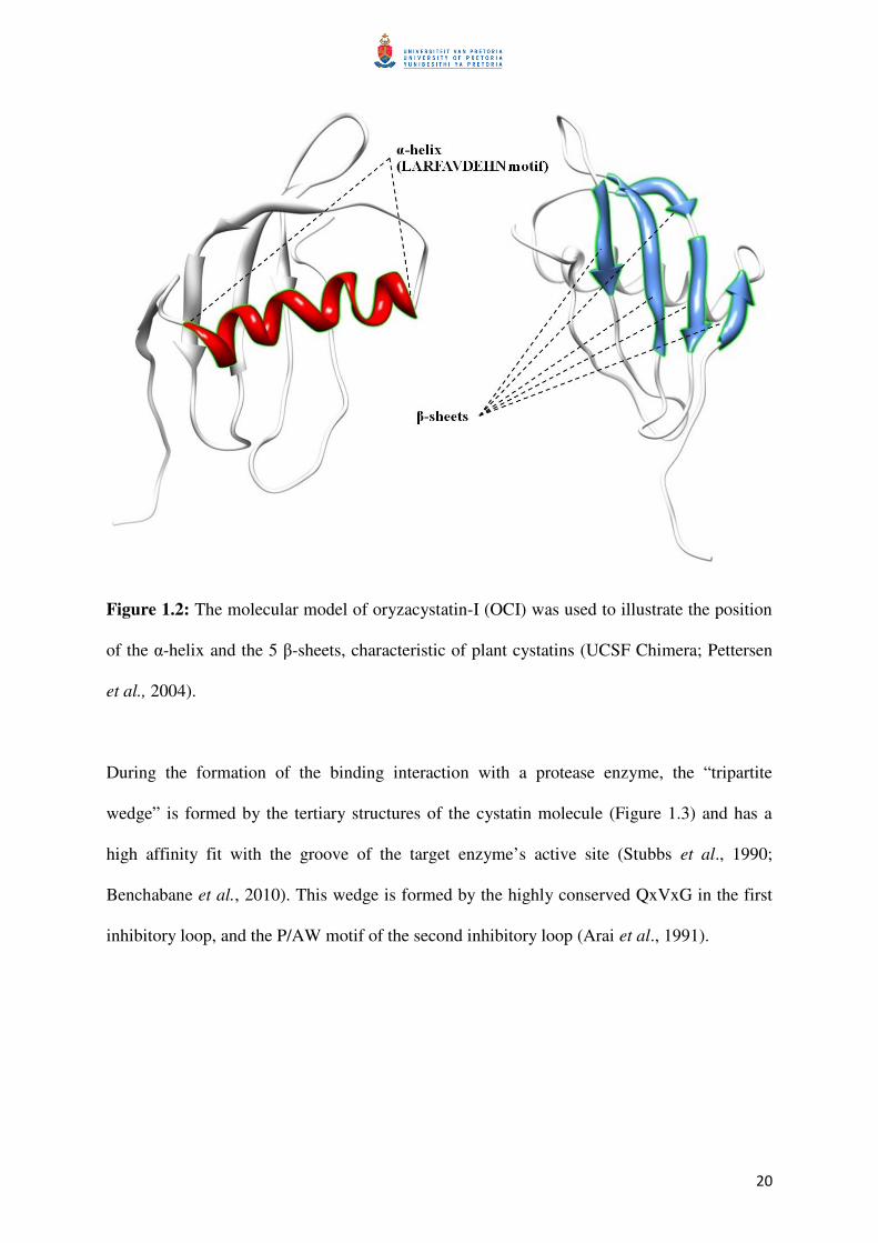
20
Figure 1.2: The molecular model of oryzacystatin-I (OCI) was used to illustrate the position
of the α-helix and the 5 β-sheets, characteristic of plant cystatins (UCSF Chimera; Pettersen
et al., 2004).
During the formation of the binding interaction with a protease enzyme, the “tripartite
wedge” is formed by the tertiary structures of the cystatin molecule (Figure 1.3) and has a
high affinity fit with the groove of the target enzyme’s active site (Stubbs et al., 1990;
Benchabane et al., 2010). This wedge is formed by the highly conserved QxVxG in the first
inhibitory loop, and the P/AW motif of the second inhibitory loop (Arai et al., 1991).
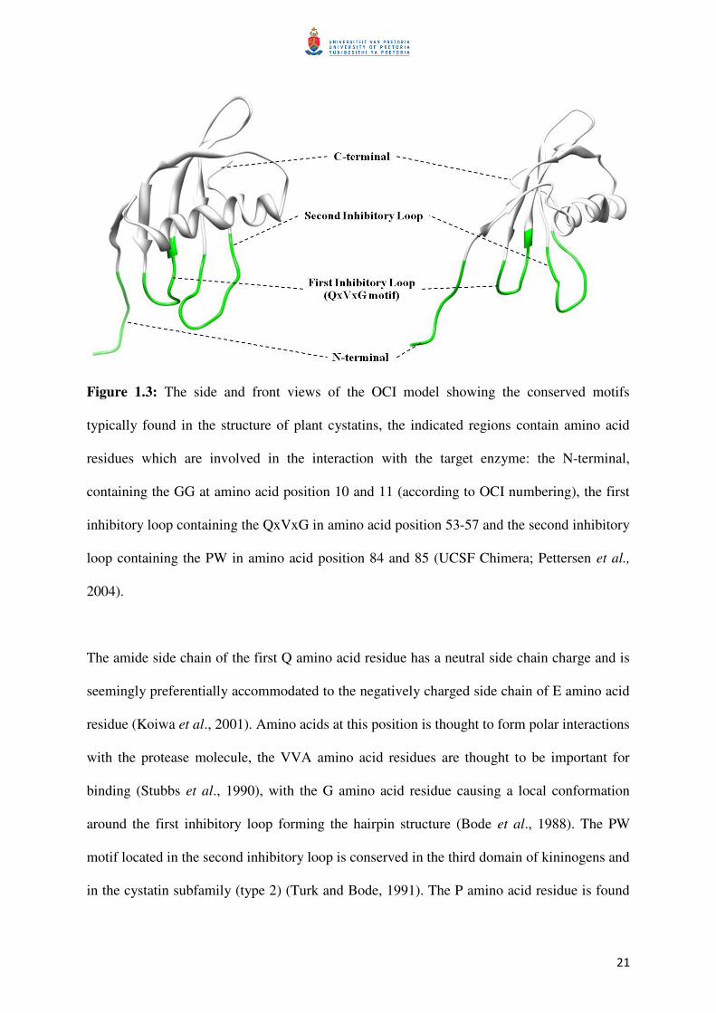
21
Figure 1.3: The side and front views of the OCI model showing the conserved motifs
typically found in the structure of plant cystatins, the indicated regions contain amino acid
residues which are involved in the interaction with the target enzyme: the N-terminal,
containing the GG at amino acid position 10 and 11 (according to OCI numbering), the first
inhibitory loop containing the QxVxG in amino acid position 53-57 and the second inhibitory
loop containing the PW in amino acid position 84 and 85 (UCSF Chimera; Pettersen et al.,
2004).
The amide side chain of the first Q amino acid residue has a neutral side chain charge and is
seemingly preferentially accommodated to the negatively charged side chain of E amino acid
residue (Koiwa et al., 2001). Amino acids at this position is thought to form polar interactions
with the protease molecule, the VVA amino acid residues are thought to be important for
binding (Stubbs et al., 1990), with the G amino acid residue causing a local conformation
around the first inhibitory loop forming the hairpin structure (Bode et al., 1988). The PW
motif located in the second inhibitory loop is conserved in the third domain of kininogens and
in the cystatin subfamily (type 2) (Turk and Bode, 1991). The P amino acid residue is found
22
in most plant cystatins, but can also be other amino acids (either an A/ Q/ L/ S, Table 1.2),
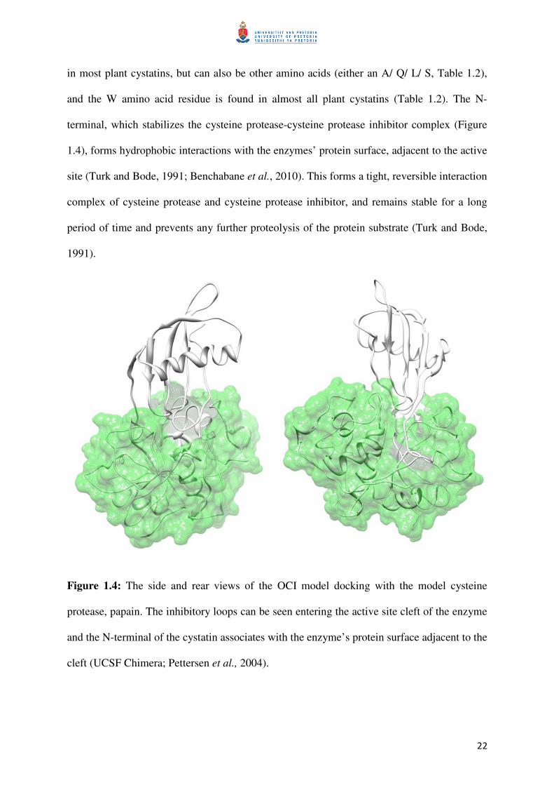
and the W amino acid residue is found in almost all plant cystatins (Table 1.2). The N-
terminal, which stabilizes the cysteine protease-cysteine protease inhibitor complex (Figure
1.4), forms hydrophobic interactions with the enzymes’ protein surface, adjacent to the active
site (Turk and Bode, 1991; Benchabane et al., 2010). This forms a tight, reversible interaction
complex of cysteine protease and cysteine protease inhibitor, and remains stable for a long
period of time and prevents any further proteolysis of the protein substrate (Turk and Bode,
1991).
Figure 1.4: The side and rear views of the OCI model docking with the model cysteine
protease, papain. The inhibitory loops can be seen entering the active site cleft of the enzyme
and the N-terminal of the cystatin associates with the enzyme’s protein surface adjacent to the
cleft (UCSF Chimera; Pettersen et al., 2004).
23
1.3 Plant cystatin function
The uncontrolled activity of proteases can have an extraordinarily destructive influence on
normal biological functions and the coordinated activity of cysteine proteases is essential.
Cystatins are natural inhibitors and are found in almost every form of life (Bode et al., 1988;
Turk and Bode, 1991; Oliveira et al., 2003). Four major classes of proteases exist, namely
cysteine, serine, aspartyl and metallo-proteases, a corresponding class of protease inhibitors
exists for each (Oliveira et al., 2003; Fan and Wan, 2005). Each protease inhibitor class has
distinguishing characteristics, such as an optimum pH range and specific amino acid residues
in their structures, which are required to efficiently bind to target proteases and prevent their
activity (Fan and Wan, 2005). Plant cystatins are involved in various defence and
developmental processes, the roles of cystatins have been inferred based on the expression
profiles of cystatins during these different processes.
1.3.1 Expression during developmental processes
Cysteine proteases are one of the main enzyme classes active in plants and are responsible for
the mobilization of storage proteins during germination (Grudkowska and Zagdańska, 2004).
The processes of storage protein deposition, cystatin expression and protease down-
regulation have been investigated in expression studies, e.g. during seed development,
cystatin mRNA transcripts start to accumulate to eliminate protease activity and allow storage
protein deposition and protecting these storage proteins until germination (Martinez et al.
2009; Benchabane et al., 2010). Cysteine proteases have been found to be up-regulated in
protein turnover during senescence, while cysteine and serine protease inhibitor genes are
down-regulated (Martinez et al. 2009; Benchabane et al., 2010). Cysteine proteases are also
24
involved in the different processes of programmed cell death (PCD) (e.g. process of tissue-
differentiation, such as xylogenesis or different stages of senescence), the expression of
cystatins was found to coincide with the activation of this signal transduction cascades, e.g.
regulating the protease enzymes involved in PCD during the hypersensitive response
(Solomon et al., 1999; Belenghi et al., 2003).
1.3.2 Expression under abiotic and biotic stress
Cysteine proteases have also been implicated in the response to abiotic (such as drought,
temperature shock and salinity) and biotic stress (such as mechanical wounding, insect
herbivory, fungal elicitors, abscisic acid and jasmonic acid) (Benchabane et al., 2010).
Cystatins used for housekeeping purposes and physiological regulation are thought to have
broad range of expression patterns, whereas cystatins involved in developmental cues and in
stress responses are thought to have a restricted expression pattern (Massonneau et al., 2005).
Five different cystatins from maize (Zea mays) were found to be down-regulated during
drought conditions, while two maize cystatins were found to be induced by cold stress
(Massonneau et al., 2005).
The expression of the soybean cystatins N1 and R1 were found to be induced, both locally
and systemically, by methyl-jasmonate or mechanical wounding, and it is suggested that
these cystatins are involved in plant defence responses (e.g. hypersensitive response against
pathogens, insects and nematodes) (Botella et al., 1996). Fungicidal characteristics were
observed for barley cystatin proteins which were produced recombinantly, the proteins were
capable of inhibiting the growth of phytopathogenic fungi Botrytis cinerea and Fusarium
oxysporum in vitro (Martinez et al. 2005).
25
1.3.3 Expression of exogenous cystatins in biotechnological applications
Plant cystatins are being investigated for several biotechnological applications, specifically
where the regulation of cysteine protease activity is desired. One such application is in food
processing, e.g. proteolytic enzymes are used to modify the proteins to be used in a food
product, for added value or improved properties. The activity of these protease enzymes is
regulated by protease inhibitors, such as cystatins, to obtain the required degree of hydrolysis
(hydrolyzed peptide bonds) (García-Carreño et al., 2000).
Plant cystatins have been successfully used to engineer crop plants for resistance against
different types of pests, such as insects, nematodes and pathogens (Benchabane et al., 2010;
Schlüter et al., 2010). Recombinant cystatins have been tested in in vitro feeding trials to
illustrate the deleterious effects of cystatins on insect larvae, specifically tested were the
oryzacystatin-I (OCI) and the papaya cystatin (PC) (Kiggundu et al., 2010). The cowpea
cystatin, found in cowpea (Vigna unguiculata) seeds, was found to inhibit the digestive
proteases of targeted insect pests (the bean weevil, Acanthoscelides obtectus, and the
Mexican bean weevil, Zabrotes subfasciatus) in in vitro assays, the protein was found to be
effective evenat levels as low as 0.025% of total seed protein content. Molecular modelling
studies of this cowpea cystatin revealed that five amino acid residues located on the N-
terminal are involved in the interaction with the proteases, which form hydrophobic
interactions to stabilize the enzyme-inhibitor complex (Aguiar et al., 2006). The functional
modification of the eighth cystatin domain of the multi-cystatin from Solanum lycopersicum
(SlCYS8) had shown some variants to have enhanced ability to inhibit target digestive
proteases from Leptinotarsa decemlineata (Colorado potato beetle) while at the same time

26
exhibiting lowered activity against non-targeted cysteine proteases (Goulet et al., 2008). The
interaction between inhibitors and target proteases is dose-dependent, therefore enhancing the
inhibitory potency of a cystatin would assist to decrease the dosage of cystatin required to
successfully provide anti-nutritive/ pesticidal effects (Goulet et al., 2008).
The ectopic expression of OCI in transgenic tobacco resulted in enhanced fitness and
tolerance to chilling stress along with an altered physiology, such as retarded stem elongation
and leaf expansion under low light intensities (Van der Vyver et al., 2003; Demirevska et al.,
2009). Expression of cystatins in the cytosol of a transgenic non-host plant have been found
to alter the phenotype with potentially beneficial traits, such as improved tolerance against
abiotic stress (Munger et al., 2009). The ectopic expression of maize cystatin-II in potato
leaves led to the constitutive expression of PR-proteins and other protease inhibitor proteins,
normally inducible during plant defence or stress response (Munger et al., 2009). Constitutive
over expression of the AtCYS1 gene in Arabidopsis led to the activation on signal
transduction cascades. This was found to coincide with the regulating the protease enzymes
involved in PCD during the hypersensitive response in the leaves and had a repressing effect
of the overall degree of the response (Belenghi et al., 2003).
A further recent biotechnological application of cystatin expression is the protection of
recombinant proteins expressed in transgenic plants. When a protease inhibitor, such as the
tomato cathepsin D inhibitor or bovine aprotinin, was co-expressed along with the protein of
interest, higher amounts of the protein could be harvested compared to the control plants
(Rivard et al., 2006; Benchabane et al., 2009; Goulet et al., 2010).

27
Figure 1.5: Various endogenous functions that cystatins are involved in and the exogenous
applications that cystatins have been used for. ↑ indicates an increase in transcription and ↓
indicates a decrease in transcription, while CYS represents cystatins, CYP represents cysteine
proteases.
1.4 Cystatin mutagenesis
Several research groups have attempted to improve the potency of cystatins against cysteine
proteases by substituting amino acids in the cystatin sequence. However, there are contrasting
ideas about the role of certain conserved amino acids in a specific position of a cystatin and
their role in determining inhibitory potency. Structure/function models have been used to
understand the role of specific amino acids or strings of amino acids in inhibitor potency
against specific proteases. Many of these studies have been based on a structural model using
the human stefin B-papain complex (Stubbs et al., 1990), which has been instrumental in
28
identifying particular target sites for the improvement of cystatins against cysteine proteases.
Molecular phage displays combined with random mutagenesis have also been used in
protease inhibitor sequence regions to identify biologically active cystatin variants. Site-
directed mutagenesis of selected amino acids has been applied as a technique to investigate
the structure/function relationships in combination with computer algorithms to predict
stronger binding interactions and has indeed resulted in more potent protease inhibitors to be
engineered (Laboissiere et al., 2002; Ceci et al., 2003; Stoop and Craik, 2003). Several
research groups have previously used these two techniques to investigate conserved amino
acid residues located in the N-terminal, the first inhibitory loop (the QxVxG motif in the first
inhibitory loop being the most conserved of these motives), as well as motives in the second
inhibitory loop.
1.4.1 Mutations in the N-terminal region
Human cystatin C has been one of the first cystatins to be used as target for site-directed
mutagenesis, to substitute particular amino acid residues in this cystatin. The conserved
amino acid residue G (glycine) amino acid residue in position 11 was investigated by Hall et
al. (1993) by replacing it with either a positively charged R (arginine), a negatively charged E
(glutamic acid), a bulky hydrophobic tryptophan, or by an amino acid containing small side-
chains, such as S (serine) or A (alanine). They found that G, which lacks a side-chain, at
amino acid position 11 was important to allow the local confirmation in the N-terminal
region, allowing for both high-affinity binding and efficient inhibition. Equilibrium constants
for dissociation (Ki) of complexes with papain and human cathepsin B, had showed that
human cystatin C variants, with serine (S) or alanine (A) in position 11, had Ki values about
20-fold higher than those of the wild-type cystatin C and variants with W, R or E in this
29
position had Ki values with a factor of about 2000-fold higher, implying less efficient
inhibition of the target proteases (Hall et al., 1993). In another study, the importance of the
N-terminal region in cystatin C was investigated by a method of random-centroid
optimization. This method was used to introduce double-site mutations and had shown to
improve the inhibitory activity of cystatin C, without any prior knowledge of the cystatin’s
molecular structure (Ogawa et al., 2002). The substitution mutations of G in position 12 to
W, and of H (histidine) in position 86 to V (valine), had increased inhibitory activity 5-fold
over wild-type cystatin C. Improved papain inhibition was also obtained by substitution of P
(proline) in position 13 with F (phenylalanine), while mutations decreasing beta-sheet content
resulted in reduced polymerization of cystatin C and had increased papain inhibition.
Furthermore, when three N-terminal amino acid residues preceding the conserved G of
cystatin A were replaced with a 10-amino acid residue long cystatin C segment, inhibitor
affinity to cathepsin B had increased approximately 15-fold (Pavlova and Björk, 2003). This
was predominantly due to a higher association rate constant. Furthermore, substituting G in
position 75 with either W or H in the second inhibitory loop of cystatin A, a 10-fold higher
papain affinity was observed due to both a higher association rate as well as a lower
dissociation rate constant. By changing the individual residues in the N-terminal region of
cystatin C, L in position 9 to W, or V in position 10 to either W, F or R as well as changing
W in position 106 to G in the C-terminal region, the binding ability to cathepsins B and H
was reduced (Mason et al., 1998). This indicates that both cathepsins are repulsed by large
aromatic residues in their S2 and S3 pockets, in contrast to cathepsin L, which preferentially
accommodates larger aromatic residues in the S2 pocket. This hypothetical binding pocket is
formed by amino acids in the enzyme’s active site, which interact with each other and
contribute to form hydrophobic interactions, hydrogen- and van der Waals bonds with the
inhibitor (or substrate). The inhibitor’s overall shape and hydrophobicity that would be
30
tolerated in this binding pocket is dependent on these amino acids present in the enzyme’s
active site and their contribution towards binding. Mason et al. (1998) further found that
introducing a charged residue into the S2 pocket resulted in an inhibitor that showed
selectivity towards cathepsin L and S compared with cathepsin B and H. Other mutants were
shown to be capable of distinguishing cathepsin H from cathepsin B, or cathepsin S from
cathepsin L, dependent on the amino acid residues at specific tertiary positions.
Several research groups have investigated the importance of the N-terminal region of plant
cystatins, but despite several studies, the function of the N-terminal region is still rather
unclear. In an early investigation, Abe et al. (1995) found distinct dissimilarity in the amino
acid sequence of corn cystatin-I (CCI) and corn cystatin-II (CCII) around the N-terminal
region, with CCII being a stronger inhibitor of cathepsin L compared to CCI. When OCI was
studied, it was found that the first 21 residues of the N-terminal region are seemingly not
essential for papain-inhibitory activity (Abe et al. 1988; Arai et al., 1991; Urwin et al.,
1995a). In contrast, OCI truncated of 38 N-terminal residues was almost completely inactive
(Abe et al., 1988). Further studies have also shown the importance of individual amino acid
residues for cystatin potency, when the conserved G in positions 5, 6, 10 or 11 in the N-
terminal region was studied by deletion and mutation and observed inhibitory characteristics
were compared to wild-type OCI. Only substitutions of the amino acid at position 10,
changing G to either R, C (cysteine), E, Q, H, L (leucine), K (lysine), F, P, S, or Y (tyrosine)
significantly changed the inhibitory potency of OCI, but not at any of the other amino acid
sites (Urwin et al., 1995a). This demonstrated the functional importance of the highly
conserved G residue at position 10, for effective inhibition of papain. In another study, using
N-terminal deletion mutants in the N-terminal of the sunflower cystatin, the amino acids
isoleucine (I) and P at position 1 and 2 as well as the N-terminal G residues at position 3
31
and/or 4, were shown to play a functionally important role in papain inhibition (Doi-Kawano
et al., 1998). The N-terminal regions of plant cystatins have also been associated with anti-
feedant and antifungal activity. Modification of either C, E/ D (aspartate), or R residues in the
N-terminal sequence of a pearl millet cystatin resulted in the loss of the antifungal activity
(Joshi et al., 1999).
Proteins that are under direct selection pressure, such as defence proteins, have been shown to
undergo adaptive evolution, whereby mutations that confer a selective advantage are
maintained under selection. The non-synonymous substitutions contribute to the
diversification and maintenance of cystatin variability at these interacting amino acids
residues of cystatins that have been shown to undergo positive selection in response to the
target proteases from the pathogen (Kiggundu et al. 2006). Single mutations at these
positively selected amino acid sites have been extensively investigated in several studies to
improve the anti-nutritional activity of plant cystatins, a possible link was established
between hyper-variable amino acid sites in the plant cystatin amino acid sequence and
inhibitory potency against cysteine proteases of the papain-like family (C1) (Kiggundu et al.
2006). In this study, a maximum likelihood approach was applied to assess plant cystatins for
positive selection during evolution by comparing the rates of synonymous to non-
synonymous mutations. This comparison had ultimately provided an indication to which
amino acid sites have potential for improving the inhibitory activity of cystatins. Hyper-
variable sites were located on each side of the conserved G residues in the N-terminal region,
also within the first and second inhibitory loops and areas surrounding the conserved
LARFAVDEHN motif in the α-helix of plant cystatins. Based on the study of Goulet et al.
(2008), 29 mutants of the tomato multi-cystatin 9 subunit were produced with single
substitutions at three positions shown to be under positive selection. Substituting the original
32
P at position 2, T (threonine) at position 6, or E at position 31, had resulted in mutants
showing a range of increased or lowered inhibitory potency against cysteine proteases. In
particular the P at position 2, the amino acid residue adjacent to the conserved GG motif in
the N-terminal region of the cystatin, showed an improved inhibitory potency against the
cysteine proteases of the Colorado potato beetle extract. Furthermore, the substitutions of P to
F, I, L or Y resulted either in lowered or unchanged inhibitory potency against target
proteases, potato leaf cysteine protease(s) or protease I, a cysteine protease utilized by the
predatory insect Perillus bioculatus.
1.4.2 Mutations in the first inhibitory loop
The motif QxVxG is found in the first inhibitory loop, this motif is conserved among most
members of the cystatin super-family and substitutions of amino acid residues in this motif
can greatly change binding affinity to cysteine proteases. When the amino acid residues L
and S in the QLVSG motif of chicken cystatin were changed to V and A, respectively, the
binding affinity with the cysteine protease actinidin (papain-like enzyme) significantly
increased more than 10-fold, the other cysteine proteases tested did not show the same degree
of change (Auerswald et al., 1996). Several researchers have investigated this conserved
motif in plant cystatins, in the first study by Abe et al. (1988), it was found that an N-
truncated OCI retaining the QVVAG motif inhibited papain as efficiently as a non-truncated
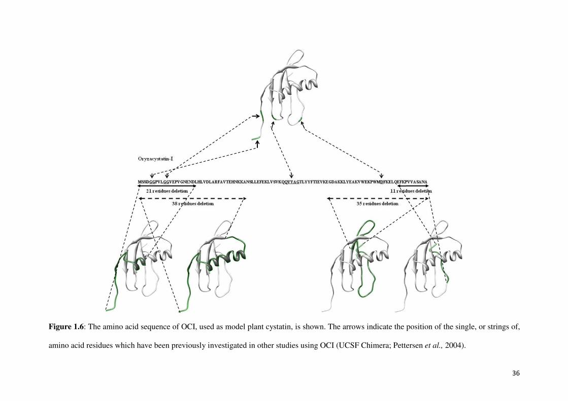
OCI, demonstrating the importance of this motif for activity. Arai et al. (1991) provided more
detailed information of the role of particular amino acid residues in the QVVAG motif of
OCI (Figure 1.6). These results clearly indicate that the QVVAG sequence of the cystatin
molecule is the primary region of interaction with the cysteine protease and is thus
responsible for the inhibitory activity. In general, substituting Q at position 53 with L caused

33
significantly lowered inhibition of papain and a Ki value approximately 150-times higher than
wild-type OCI. Furthermore, when substituting the same Q with P resulted in a completely
inactive cystatin, this completely inactive mutant was also unable to bind to a papain column,
indicating that the affinity site of OCI is also its reactive site. In a cystatin isolated from
papaya, the papaya cystatin (PC), the Q of the QVVAG motif is substituted by A (Song et al.,
1995, Figure 1.7). In this study, the function of this natural A substitution in PC was
primarily investigated. Furthermore, substituting the central V residue only resulted in a
moderate effect on activity, substituting V with G resulted in a protein which was as active as
the wild-type OCI, while changing the V to D resulted in a 40-times higher Ki value than the
wild-type OCI (Arai et al., 1991, Figure 1.6). Nikawa et al., (1989) claimed that the QVVAG
motif was not essential for inhibitory activity, but the study had only looked at two mutants
of human cystatin A. Changing the glutamine amino acid at position 46 to lysine (KVVAG)
had lowered inhibitory activity against cathepsin B (0.6-times) and H (4.0-times), but
improved against papain and cathepsin L (0.4-times) and changing the valine amino acid at
position 48 to threonine (QVTAG) had performed close to that of the wild-type against the
different enzymes assayed papain, cathepsin B and H, but had a 3.5-times reduction against
cathepsin L. In another study, Koiwa et al., (2001) confirmed the importance of the first
inhibitory loop in the structure of soyacystatin for inhibition of the cysteine protease papain.
By using a combination of phage display libraries and random mutations, targeting the
QVVAG motif of the first hairpin loop, all functional soyacystatin variants that were studied
had this motif conserved. This indicates the functional importance of this motif in the activity
of soyacystatin.
34
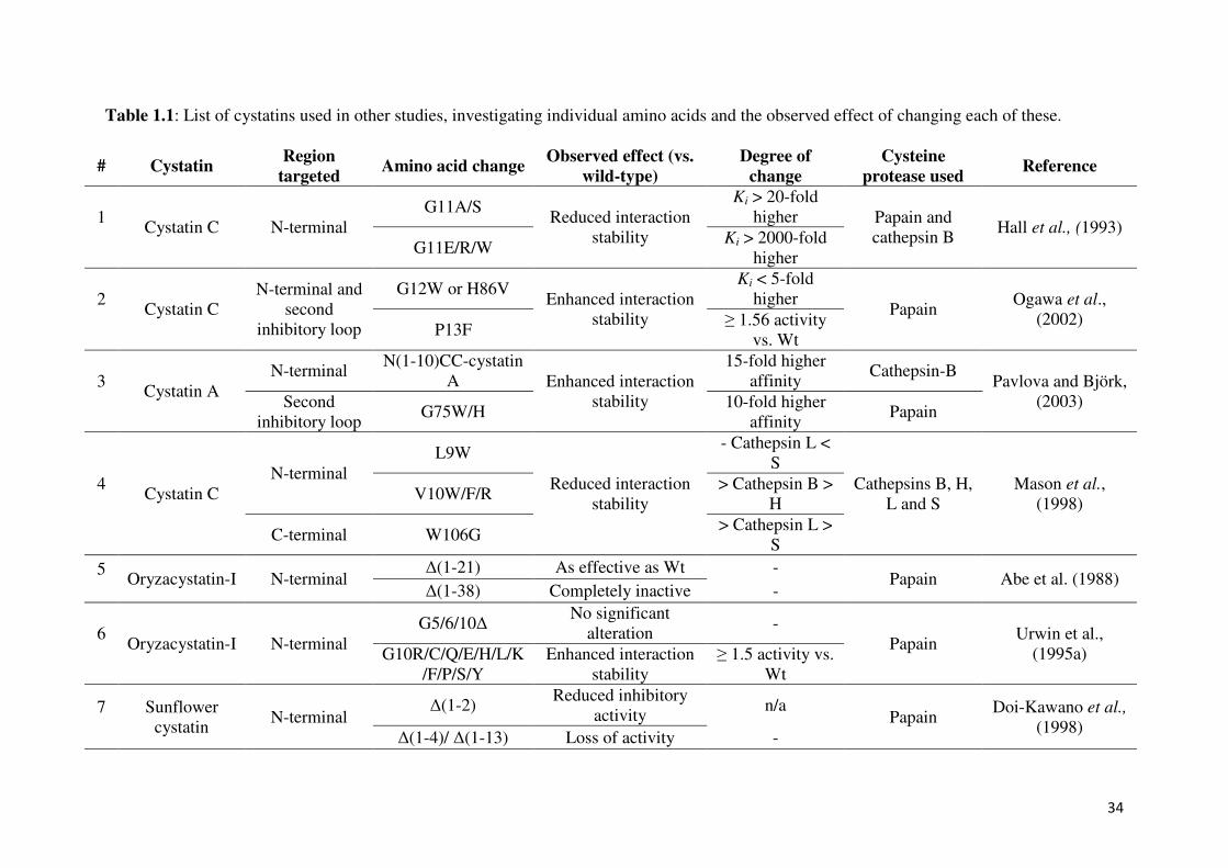
Table 1.1: List of cystatins used in other studies, investigating individual amino acids and the observed effect of changing each of these.
# Cystatin Region
targeted Amino acid change
Observed effect (vs. wild-type)
Degree of change
Cysteine protease used
Reference
1
Cystatin C N-terminal
G11A/S Reduced interaction
stability
Ki > 20-fold
higher Papain and
cathepsin B Hall et al., (1993)
G11E/R/W Ki > 2000-fold
higher
2
Cystatin C
N-terminal and
second
inhibitory loop
G12W or H86V Enhanced interaction
stability
Ki < 5-fold
higher Papain
Ogawa et al.,
(2002) P13F
≥ 1.56 activity
vs. Wt
3
Cystatin A
N-terminal N(1-10)CC-cystatin
A Enhanced interaction
stability
15-fold higher
affinity Cathepsin-B
Pavlova and Björk,
(2003) Second
inhibitory loop G75W/H
10-fold higher
affinity Papain
4
Cystatin C
N-terminal
L9W
Reduced interaction
stability
- Cathepsin L <
S
Cathepsins B, H,
L and S
Mason et al.,
(1998) V10W/F/R
> Cathepsin B >
H
C-terminal W106G > Cathepsin L >
S
5
Oryzacystatin-I N-terminal
∆(1-21) As effective as Wt - Papain Abe et al. (1988)
∆(1-38) Completely inactive -
6
Oryzacystatin-I N-terminal
G5/6/10∆ No significant
alteration -
Papain Urwin et al.,
(1995a) G10R/C/Q/E/H/L/K
/F/P/S/Y
Enhanced interaction
stability
≥ 1.5 activity vs.
Wt
7
Sunflower
cystatin N-terminal
∆(1-2) Reduced inhibitory
activity n/a
Papain Doi-Kawano et al.,
(1998) ∆(1-4)/ ∆(1-13) Loss of activity -
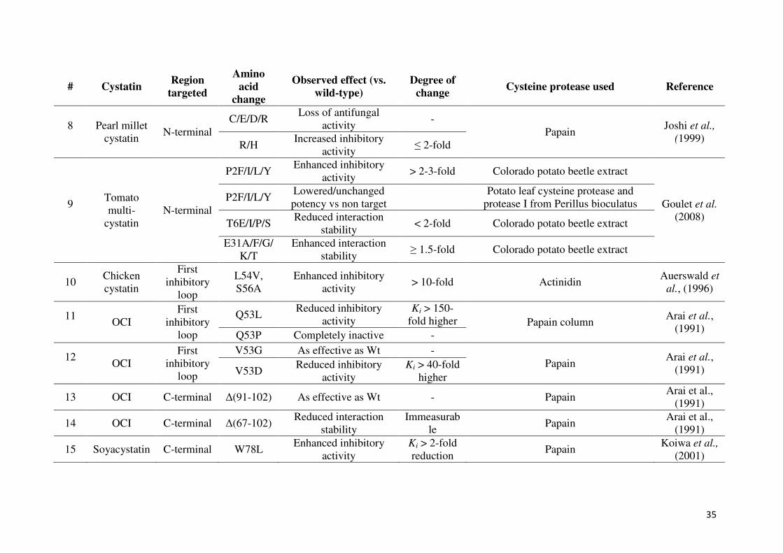
35
# Cystatin Region
targeted
Amino acid
change
Observed effect (vs. wild-type)
Degree of change
Cysteine protease used Reference
8
Pearl millet
cystatin N-terminal
C/E/D/R Loss of antifungal
activity -
Papain Joshi et al.,
(1999) R/H
Increased inhibitory
activity ≤ 2-fold
9
Tomato
multi-
cystatin
N-terminal
P2F/I/L/Y Enhanced inhibitory
activity > 2-3-fold Colorado potato beetle extract
Goulet et al.
(2008)
P2F/I/L/Y Lowered/unchanged
potency vs non target
Potato leaf cysteine protease and
protease I from Perillus bioculatus
T6E/I/P/S Reduced interaction
stability < 2-fold Colorado potato beetle extract
E31A/F/G/
K/T
Enhanced interaction
stability ≥ 1.5-fold Colorado potato beetle extract
10 Chicken
cystatin
First
inhibitory
loop
L54V,
S56A
Enhanced inhibitory
activity > 10-fold Actinidin
Auerswald et
al., (1996)
11
OCI
First
inhibitory
loop
Q53L Reduced inhibitory
activity
Ki > 150-
fold higher Papain column Arai et al.,
(1991) Q53P Completely inactive -
12
OCI
First
inhibitory
loop
V53G As effective as Wt -
Papain Arai et al.,
(1991) V53D Reduced inhibitory
activity
Ki > 40-fold
higher
13 OCI C-terminal ∆(91-102) As effective as Wt - Papain Arai et al.,
(1991)
14 OCI C-terminal ∆(67-102) Reduced interaction
stability
Immeasurab
le Papain
Arai et al.,
(1991)
15 Soyacystatin C-terminal W78L Enhanced inhibitory
activity
Ki > 2-fold
reduction Papain
Koiwa et al.,
(2001)
36
Figure 1.6: The amino acid sequence of OCI, used as model plant cystatin, is shown. The arrows indicate the position of the single, or strings of,
amino acid residues which have been previously investigated in other studies using OCI (UCSF Chimera; Pettersen et al., 2004).
37
1.4.3 Mutations in the second inhibitory loop
Research to date has also shown that the C-terminal region of cystatins plays an important
role in inhibitory activity and this region can affect the target specificity of cystatins
(Auerswald et al., 1996). C-terminal truncated oryzacystatin-I, lacking the 11 C-terminal
residues still exhibited potent papain-inhibitory activity (Abe et al., 1988; Arai et al., 1991;
Figure 1.6). The possible role of the C-terminal region was also demonstrated by
investigating biochemical interactions between digestive proteases of the Coleopteran pest,
the black vine weevil (Otiorynchus sulcatus) and two plant cysteine protease inhibitors,
oryzacystatin-I and oryzacystatin-II, when a C-terminal truncated form of oryzacystatin-II
was detected (Michaud et al., 1995). This truncated form was as active against papain and
human cathepsin H as the non-truncated oryzacystatin-II. However, when the C-terminal of
oryzacystatin-I was truncated by 35 residues, the truncated cystatin showed a considerably
reduced inhibitory activity (Abe et al., 1988; Arai et al., 1991; Figure 1.6).
By investigating the second inhibitory loop in greater detail, a variant of oryzacystatin-I with
improved activity against nematodes was produced by deleting the aspartate (D) amino acid
residue at position 86 of the wild-type’s sequence (Urwin et al., 1995b; Figure 1.6). Koiwa et
al., (2001) had isolated several soyacystatin variants with improved inhibitory potency, from
combining phage display libraries and after inducing random mutations, all the selected
variants had tryptophan (W) in position79 in the second hairpin-loop motif. However, there
was diversity in the original amino acid sequence in position 78, with preferentially
accommodate hydrophobic and basic amino acids in this position. Changing the glutamic
acid (E) in position 78 in the wild-type sequence to leucine (L) resulted in higher papain
affinity than the wild-type cystatin and a two-fold lower Ki than wild-type soyacystatin.
38
Changes in this first residue may be more involved in determining target specificity during
the association process, this was suggested by an increased association-rate constant of
leucine (L)-tryptophan (W), without affecting basal level affinity. The LW motif in the
second hairpin loop of soyacystatin, with greater affinity to papain, was also identified in the
papaya cystatin. The occurrence of these amino acids in the papaya cystatin could be
attributed to the co-evolution of the papaya cystatin and the cysteine protease papain in
Carica papaya (Song et al., 1995).
1.4.4 Cysteine proteases
The commercial enzymes selected to be used in this study included papain and cathepsin-L.
Furthermore, gut extracts from insect larvae (Colorado potato beetle and banana weevil), as
well as tobacco leaf extracts, were used since they are known to contain cysteine protease
activity that can be inhibited by plant cystatins. Papain and cathepsin L belong to the same
family (C1 proteases) and are characterized by two distinct domains, the N-terminal, mainly
α-helixes and the C-terminal, mainly β-sheets. The active site is formed by a cysteine (C)
amino acid residue at position 25, a histidine (H) amino acid residue at position 159 and a
asparagine (N) amino acid at position 175 (according to papain numbering) (Oliveira et al.,
2003).
Papain exhibits broad specificity, cleaving peptide bonds of basic amino acids, e.g. leucine
(L) or glycine (G), and also hydrolyzes esters and amides. Papain preferentially
accommodates an amino acid bearing a large hydrophobic side chain at the P2 position, but
does not accept valine (V) at the P1' position (IUBMB Enzyme Nomenclature). Papain
preferentially cleaves at the site indicated by ↓. Hydrophobic amino acids include: alanine

39
(A), phenylalanine (F), isoleucine (I), leucine (L), valine (V), tryptophan (W), and tyrosine
(Y).
P3 P2 P1 ↓ P1’ P2’
Xaa Hydrophobic Arg ↓ not Val Xaa
Xaa Hydrophobic Lys ↓ not Val Xaa
Cathepsin L is a lysosomal enzyme and exhibits higher specific activity than cathepsin B or
cathepsin H in the degradation of a variety of physiological protein substrates, both cellular
and endocytosed macromolecules (Barrett and Kirschke, 1981; Mason et al., 1985).
Cathepsin L will preferentially cleave at the site indicated by ↓, with the hydrophobic amino
acids listed as well as aromatic amino acids, phenylalanine (F), histidine (H), tryptophan (W)
and tyrosine (Y), denoting cleaving specificity.
P4 P3 P2 P1 ↓ P1’
Xaa Hydrophobic Phe Arg ↓ Xaa
Xaa Aromatic Phe Arg ↓ Xaa
Xaa Hydrophobic Arg Arg ↓ Xaa
Xaa Aromatic Arg Arg ↓ Xaa

40
1.5 Previous work on the project
1.5.1 Plant cystatin sequence analysis
At the commencement of this study, conserved amino acids in the N-terminal region, first and
second inhibitory loops had been identified from 153 gene sequences of plant cystatins from
62 different plant species, including OCI and papaya cystatin, and several OCI and PC
variants sequences were produced in an initial study by Dr Schlüter (U Schlüter, unpublished
data; Table 1.2). Amino acid substitutions in the conserved amino acids of PC and OCI have
been designed based on determined frequency of amino acid residues present in analyzed
sequences with the specific objective to understand the particular function of residues in PC
potency since this cystatin had some unique characteristics in comparison to OCI specifically
in the QVVAG motif. Particular focus on these two plant cystatins was based on results of a
previous study where Kiggundu et al. (2010) found better inhibition by OCI inhibition of
cysteine protease in banana weevil gut extracts when compared to PC and the reason for the
difference in inhibitory activity between the two cystatins is still unclear.
1.5.2 OCI and PC mutagenesis
The majority of analyzed cystatin sequences had been isolated by sequence homology, but
only 27% of them had been analysed for their binding characteristics against cysteine
proteases (mainly papain) and most of these sequences contain all the conserved motifs. The
highest conservation was found for G in the GG motif (position 10 and 11 in OCI and 8 and 9
in PC) present in the N-terminal end of all analyzed plant cystatins. Some sequence
41
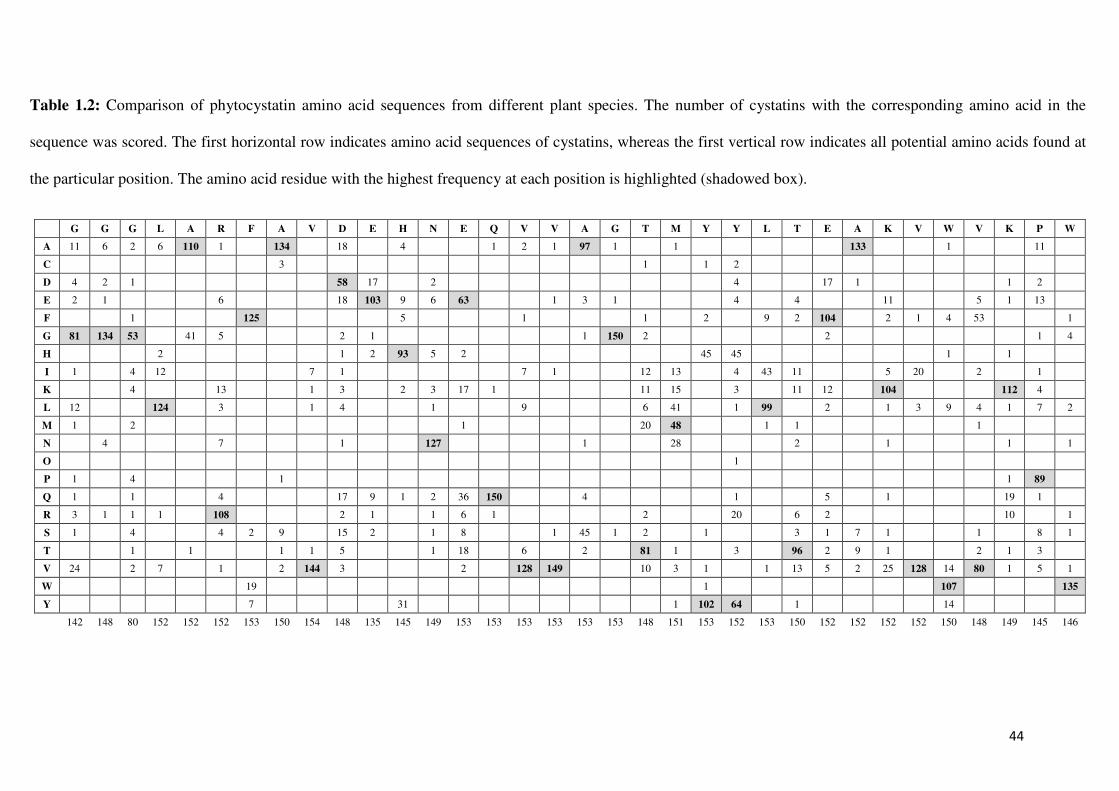
differences were identified with CLUSTAL W in the alignment of the GG motif, but shifts
were limited to +/- one amino acid or two amino acid residues as found for PC when
compared to OCI (Figure 1.7). In the plant cystatins analysed in the initial study, 100% of the
sequences had at least one conserved G in the N-terminal region (Table 1.2). The importance
of G at these positions is still unclear. N-terminal truncated OCI variants lacking both G have
previously been found to be as active as non-truncated OCI (Arai et al., 1991), whereas
Urwin et al. (1995a) reported that substitutions of G in position 10 changed the inhibitory
potency of OCI significantly. The GG motif was therefore mutated in the initial study to AA
(GG to AA). These two residues, which are non-polar with a neutral side chain charge and
without an extensive side chain, were similar to G and had been selected as substitution to
provide information about any importance of the GG motive in the N-terminal end.
Furthermore, A is naturally found in some cystatins in this position, having either AA or just
one A in combination with a G in this position (Table 1.2).
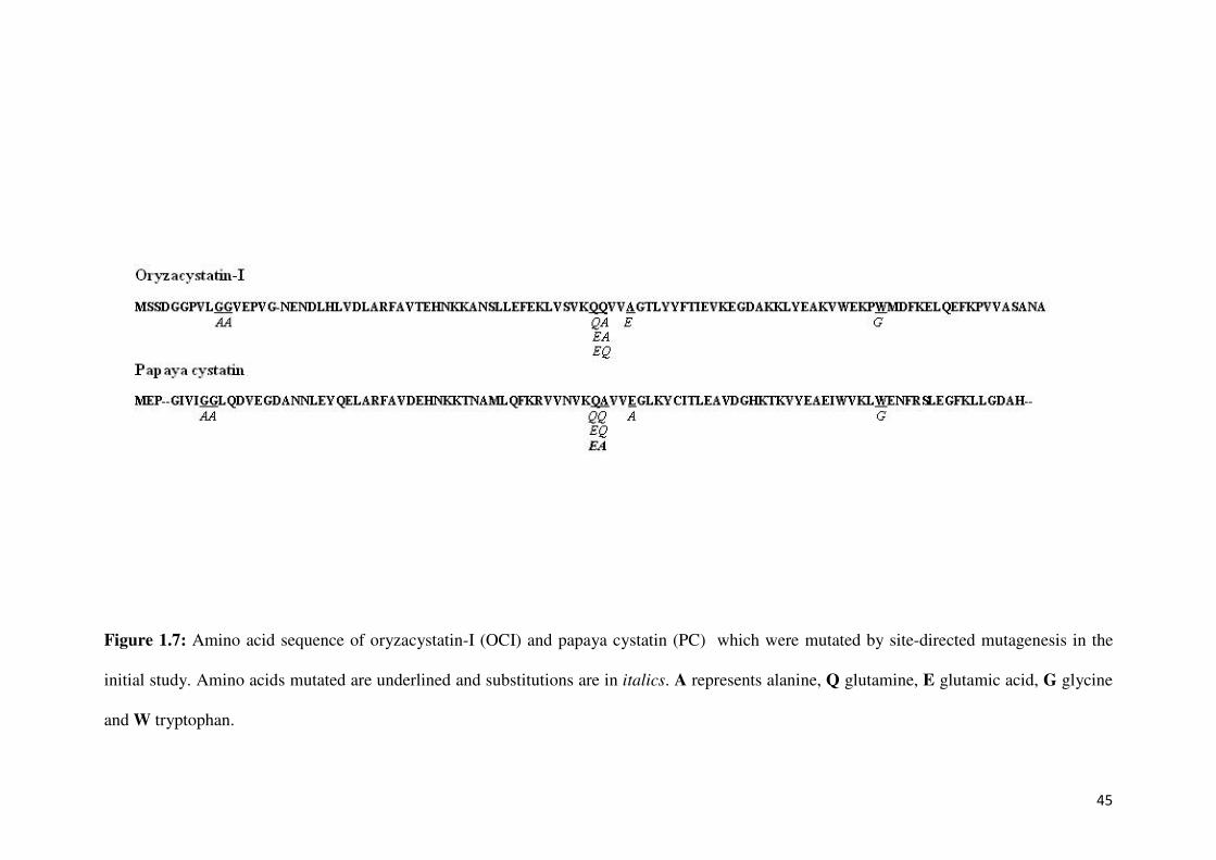
The first inhibitory loop, containing the conserved motif, QVVA/SG, was found to be highly
conserved. Q in position 53 in OCI occurs in 98% of sequences. V in position 54 in OCI and
in position 52 in PC was found in 84%, with V in position 55 in OCI and 54 in PC found in
97%. At position 56 in OCI (position 55 in PC), A was found in 63% or S in 29%, and G in
position 57 in OCI and 56 in PC was found in 98% of cystatins. The conserved residue Q was
also present in OCI. The PC sequence contains two unique characteristics, an A in position
52 of PC, which is only found in PC sequence, and an E in position 55 of PC. This variation
is only found in three other cystatin sequences. Most plant cystatins have an A in this
position, although S is also very common (A and S together occur in 92% of all analysed
sequences). This E amino acid residue is situated in the middle of the first inhibitory loop,
which is proposed to be inserted into the active site cleft of the cysteine protease, and might
42
therefore have a significant effect on the binding capacity of the cystatin, as this amino acid is
electrically charged compared to the hydrophobic amino acid A, normally found in this
position. Since PC shows the greatest discrepancy to the majority of cystatin sequences in the
conserved motif of the first inhibitory loop, mutations, shown in Figure 1.7, have been done
in the initial study. This allowed the investigation of whether if substitution of these amino
acid residues in the PC sequence will influence the activity or specificity of the mutant
cystatins towards different cysteine proteases.
Furthermore, the conserved residue Q in OCI or A in PC is preceded in both sequences by Q
(position 52 in OCI and 51 in PC). This amino acid residue is present in only 23% of plant
cystatin sequences analyzed. Since this amino acid is directly preceding the first amino acid
residue of the conserved motif in plant cystatins, which is still in the beta-sheet and not part
of the first inhibitory loop, this might have a limited effect on cystatin binding. This might be
especially relevant in PC, where the conserved Q is substituted by A. The preceding Q
residue in both sequences was therefore substituted by E in the initial study by site-directed
mutagenesis. E is found with the highest frequency (63 sequences) at this position in other
plant cystatins (Table 1.2). Furthermore, double mutations were created in the study changing
the amino acids (positions 52 and 53 in OCI and 51 and 52 in PC) simultaneously to obtain
all four combinations of amino acids: QQVVAG as in wild-type OCI changed to QAVVAG,
EAVVAG and EQVVAG, and in wild-type PC the QAVVEG was changed to QQVVEG,
EQVVEG and EAVVEG (Figure 1.7).
In the second inhibitory loop, a conserved W was found in 88% of the sequences including
OCI in position 84 and PC in position 83. So far only one plant cystatin (HvCPI7) not
containing this amino acid residue (F instead of W) has been experimentally tested for altered
43
binding capacity of the cystatin and no inhibitor activity was found against papain, cathepsin
B and cathepsin H (Abraham et al., 2006). AlthouYgh W is the most conserved amino acid in
the second inhibitory loop of the plant cystatins, in the preliminary study it was found that
variability in this position was higher than in the other conserved sites. A variety of other
amino acids residues could be found at this position (Table 1.2). To determine the influence
of this residue on OCI and PC activity, W was substituted with G in both sequences. G was
found in 4 of the 146 sequences analysed and is found substituting for W more often than
other amino acid residues.
44
Table 1.2: Comparison of phytocystatin amino acid sequences from different plant species. The number of cystatins with the corresponding amino acid in the
sequence was scored. The first horizontal row indicates amino acid sequences of cystatins, whereas the first vertical row indicates all potential amino acids found at
the particular position. The amino acid residue with the highest frequency at each position is highlighted (shadowed box).
G G G L A R F A V D E H N E Q V V A G T M Y Y L T E A K V W V K P W
A 11 6 2 6 110 1 134 18 4 1 2 1 97 1 1 133 1 11
C 3 1 1 2
D 4 2 1 58 17 2 4 17 1 1 2
E 2 1 6 18 103 9 6 63 1 3 1 4 4 11 5 1 13
F 1 125 5 1 1 2 9 2 104 2 1 4 53 1
G 81 134 53 41 5 2 1 1 150 2 2 1 4
H 2 1 2 93 5 2 45 45 1 1
I 1 4 12 7 1 7 1 12 13 4 43 11 5 20 2 1
K 4 13 1 3 2 3 17 1 11 15 3 11 12 104 112 4
L 12 124 3 1 4 1 9 6 41 1 99 2 1 3 9 4 1 7 2
M 1 2 1 20 48 1 1 1
N 4 7 1 127 1 28 2 1 1 1
O 1
P 1 4 1 1 89
Q 1 1 4 17 9 1 2 36 150 4 1 5 1 19 1
R 3 1 1 1 108 2 1 1 6 1 2 20 6 2 10 1
S 1 4 4 2 9 15 2 1 8 1 45 1 2 1 3 1 7 1 1 8 1
T 1 1 1 1 5 1 18 6 2 81 1 3 96 2 9 1 2 1 3
V 24 2 7 1 2 144 3 2 128 149 10 3 1 1 13 5 2 25 128 14 80 1 5 1
W 19 1 107 135
Y 7 31 1 102 64 1 14
142 148 80 152 152 152 153 150 154 148 135 145 149 153 153 153 153 153 153 148 151 153 152 153 150 152 152 152 152 150 148 149 145 146
45
Figure 1.7: Amino acid sequence of oryzacystatin-I (OCI) and papaya cystatin (PC) which were mutated by site-directed mutagenesis in the
initial study. Amino acids mutated are underlined and substitutions are in italics. A represents alanine, Q glutamine, E glutamic acid, G glycine
and W tryptophan.
46
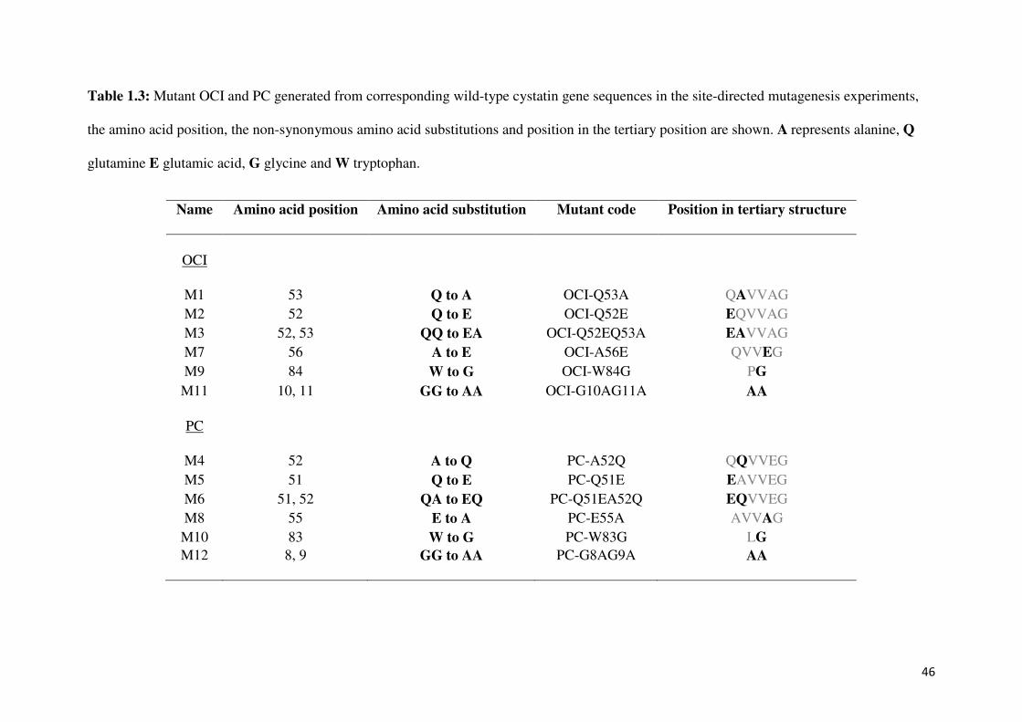
Table 1.3: Mutant OCI and PC generated from corresponding wild-type cystatin gene sequences in the site-directed mutagenesis experiments,
the amino acid position, the non-synonymous amino acid substitutions and position in the tertiary position are shown. A represents alanine, Q
glutamine E glutamic acid, G glycine and W tryptophan.
Name Amino acid position Amino acid substitution Mutant code Position in tertiary structure
OCI
M1 53 Q to A OCI-Q53A QAVVAG
M2 52 Q to E OCI-Q52E EQVVAG M3 52, 53 QQ to EA OCI-Q52EQ53A EAVVAG M7 56 A to E OCI-A56E QVVEG
M9 84 W to G OCI-W84G PG
M11 10, 11 GG to AA OCI-G10AG11A AA
PC
M4 52 A to Q PC-A52Q QQVVEG
M5 51 Q to E PC-Q51E EAVVEG M6 51, 52 QA to EQ PC-Q51EA52Q EQVVEG M8 55 E to A PC-E55A AVVAG
M10 83 W to G PC-W83G LG
M12 8, 9 GG to AA PC-G8AG9A AA
47
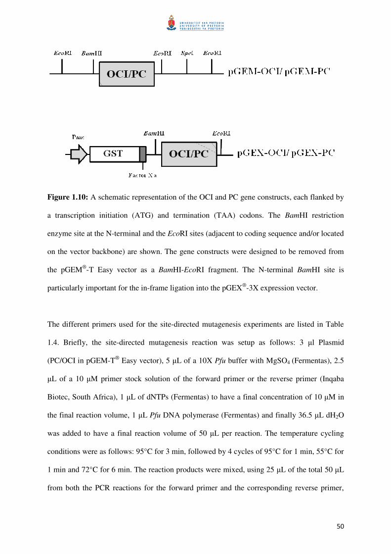
1.5.3 Cystatin mutation and cloning
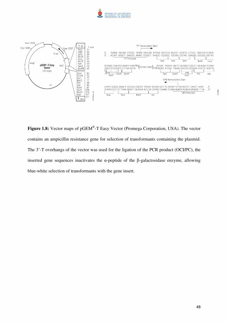
The wild-type gene sequences of OCI and PC cloned into pGEM®
-T Easy vector (Promega
Corporation), the vector map is shown in Figure 1.8, served as the template for the site-
directed mutagenesis PCR reactions. The gene sequences were amplified with
oligonucleotides to allow integration of restriction enzyme recognition sites for BamHI at the
N-terminal and an EcoRI at the C-terminal of the gene sequences, which serves for in-frame
cloning into the expression vector pGEX-3X®
(vector map shown in Figure 1.9). Figure 1.10
shows a schematic representation of the gene constructs containing the gene sequences of
oryzacystatin-I (OCI) and papaya cystatin (PC) in the pGEM®
-T Easy vector. The constructs
are collectively named pGEM-PC for PC and mutant gene sequences and pGEM-OCI for
OCI and mutant gene sequences and the individual constructs were named as pGEMPWT,
pGEMOWT, pGEM-M1, pGEM-M2, etc. Once the gene sequences were cloned into the
pGEX-3X vector, the constructs were collectively named as pGEX-PC and pGEX-OCI and
the individual constructs were named as pGEXPWT, pGEXOWT, pGEXOM1, pGEXOM2,
etc.
48
Figure 1.8: Vector maps of pGEM®
-T Easy Vector (Promega Corporation, USA). The vector
contains an ampicillin resistance gene for selection of transformants containing the plasmid.
The 3’-T overhangs of the vector was used for the ligation of the PCR product (OCI/PC), the
inserted gene sequences inactivates the α-peptide of the β-galactosidase enzyme, allowing
blue-white selection of transformants with the gene insert.
49
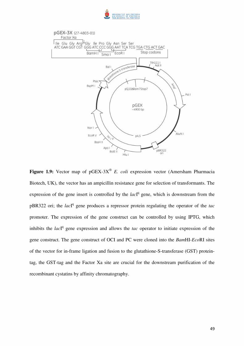
Figure 1.9: Vector map of pGEX-3X®
E. coli expression vector (Amersham Pharmacia
Biotech, UK), the vector has an ampicillin resistance gene for selection of transformants. The
expression of the gene insert is controlled by the lacIq gene, which is downstream from the
pBR322 ori; the lacIq gene produces a repressor protein regulating the operator of the tac
promoter. The expression of the gene construct can be controlled by using IPTG, which
inhibits the lacIq gene expression and allows the tac operator to initiate expression of the
gene construct. The gene construct of OCI and PC were cloned into the BamHI-EcoRI sites
of the vector for in-frame ligation and fusion to the glutathione-S-transferase (GST) protein-
tag, the GST-tag and the Factor Xa site are crucial for the downstream purification of the
recombinant cystatins by affinity chromatography.
50
Figure 1.10: A schematic representation of the OCI and PC gene constructs, each flanked by
a transcription initiation (ATG) and termination (TAA) codons. The BamHI restriction
enzyme site at the N-terminal and the EcoRI sites (adjacent to coding sequence and/or located
on the vector backbone) are shown. The gene constructs were designed to be removed from
the pGEM®
-T Easy vector as a BamHI-EcoRI fragment. The N-terminal BamHI site is
particularly important for the in-frame ligation into the pGEX®
-3X expression vector.
The different primers used for the site-directed mutagenesis experiments are listed in Table
1.4. Briefly, the site-directed mutagenesis reaction was setup as follows: 3 µl Plasmid
(PC/OCI in pGEM-T®
Easy vector), 5 µL of a 10X Pfu buffer with MgSO4 (Fermentas), 2.5
µL of a 10 µM primer stock solution of the forward primer or the reverse primer (Inqaba
Biotec, South Africa), 1 µL of dNTPs (Fermentas) to have a final concentration of 10 µM in
the final reaction volume, 1 µL Pfu DNA polymerase (Fermentas) and finally 36.5 µL dH2O
was added to have a final reaction volume of 50 µL per reaction. The temperature cycling
conditions were as follows: 95°C for 3 min, followed by 4 cycles of 95°C for 1 min, 55°C for
1 min and 72°C for 6 min. The reaction products were mixed, using 25 µL of the total 50 µL
from both the PCR reactions for the forward primer and the corresponding reverse primer,
51
and an additional 1 µL of Pfu enzyme was added. The following temperature cycling
conditions were then used: 3 min at 95°C, followed by 20 cycles of 1 min at 95°C, 1 min at
55°C and 6 min at 72°C, the reaction was cooled down to 37°C and 1 µl of DpnI (Fermentas)
was added and the reaction was incubation for 90 min. From the reaction volume 10 µL was
used to transformation 50 µL of E. coli JM109 competent cells, using the heat shock
transformation method. One hundred and fifty microlitres from the transformation reactions
were plated onto LB plates containing ampicillin at 100 mg/L. Plasmids containing mutated
PC or OCI sequences were sequenced using a T7 or M13 primer for sequencing.
52
Table 1.4: Primer sequences used to generate the mutant cystatins by site directed mutagenesis. The regions in each primer sequence that are
bolded indicate the position of the introduced nucleotides, corresponding to the desired amino acid change.
Name Forward primer Reverse primer
OCI
M1 (QQ to QA) 5' GTGAGTGTGAAGCAGGCAGTTGTCGCTGGCAC 3' 5' GTGCCAGCGACAACTGCCTGCTTCACACTCAC 3'
M2 (QQ to EQ) 5' GAGAAGCTTGTGAGTGTGAAGGAACAAGTTGTCGCTGGCACTTTG 3' 5'CAAAGTGCCAGCGACAACTTGTTCCTTCACACTCACAAGCTTCTC 3'
M3 (QQ to EA) 5' GAAGCTTGTGAGTGTGAAGGAGGCAGTTGTCGCTGGCACTTTG 3' 5' CAAAGTGCCAGCGACAACTGCCTCCTTCACACTCACAAGCTTC 3'
M7 (VAG to VEG) 5' GAAGCAGCAAGTTGTCGAAGGCACTTTGTACTATTTC 3' 5' GAAATAGTACAAAGTGCCTTCGACAACTTGCTGCTTC 3'
M9 (PW to PG) 5' CTGGGAGAAACCAGGGATGGACTTCAAG 3' 5' CTTGAAGTCCATCCCTGGTTTCTCCCAG 3'
M11 (GG to AA) 5' GCCGGTGCTTGCAGCCGTCGAGCCGG 3' 5' CCGGCTCGACGGCTGCAAGCACCGGC 3'
PC
M4 (QA to QQ) 5' GGTTGTGAATGTAAAGCAGCAGGTGGTTGAAGGCTTAAAG 3' 5' CTTTAAGCCTTCAACCACCTGCTGCTTTACATTCACAACC 3'
M5 (QA to EA) 5' GAGGGTTGTGAATGTAAAGGAAGCAGTGGTTGAAGGC 3' 5' GCCTTCAACCACTGCTTCCTTTACATTCACAACCCTC 3'
M6 (QA to EQ) 5' GAGGGTTGTGAATGTAAAGGAACAGGTGGTTGAAGGCTTAAAGTAC 3' 5' GTACTTTAAGCCTTCAACCACCTGTTCCTTTACATTCACAACCCTC 3'
M8 (VEG to VAG) 5' GCAGGCAGTGGTTGCAGGCTTAAAGTAC 3' 5' GTACTTTAAGCCTGCAACCACTGCCTGC 3'
M10 (LW to LG) 5' CTGGTTGAAGCTCGGGGAGAATTTCAGG 3' 5' CCTGAAATTCTCCCCGAGCTTCAACCAG 3'
M12 (GG to AA) 5' GAATTGTGATCGCAGCTTTGCAGGACG 3' 5' CGTCCTGCAAAGCTGCGATCACAATTC 3'

53
1.6 Research aim and objectives
Despite emerging evidence about certain amino acids in the cystatin sequence playing an
important role in inhibitory activity, there is still a lack of detailed information about the
possible function of individual amino acids, particularly in the conserved regions of the
cystatin amino acid sequence. Therefore, in this study, mutant cystatins derived from rice
(oryzacystatin-I) and papaya (papaya cystatin) were used to determine the importance of
individual amino acids in the N-terminal and first and second inhibitory loops of these two
cystatins for inhibitory activity against cysteine proteases and cysteine protease activity
contained in insect and plant extracts. The two cystatins were selected due to the unique
sequence characteristics of the papaya cystatin in the conserve motif and also due to their
significantly different activity when a gut extract from banana weevils was used to test their
inhibitory efficiency (Kiggundu et al., 2010). In this study, 12 mutant cystatins were
produced and purified and the results of their inhibitory activity against papain, cathepsin L
and extracts of Colorado potato beetle larvae, gut extracts of banana weevil larvae and a
Nicotiana benthamiana leaf extract are reported. To achieve the aim, the following three
objectives were set:
1. Produce and purify wild-type and mutant cystatins by applying the GST-fusion
protein technique and the GSH affinity chromatography technique to produce
cystatins for in vitro testing against cysteine proteases and cysteine protease-
containing insect and plant extracts.
54
2. Determine the inhibitory activity of wild-type and mutated cystatins using
fluorometric assays with specific fluorescent substrates to evaluate the importance of
individual amino acids in the conserved regions of cystatins.
3. Computer-simulate the interaction between mutant cystatins with the model cysteine
protease papain to determine changed interactions of mutated amino acids in
comparison to native amino acids.
55
2. MATERIALS AND METHODS
2.1 DNA work
2.1.1 Preparation of E. coli competent cells
Escherichia coli competent cells were prepared for strains DH5α and BL21 according to the
publication of Inoue et al. (1990) and the transformation procedure that followed was the
heat-shock transformation method also described in the article. The E. coli strain DH5α with
the genotype: F-, φ80dlacZ∆M15 ∆(lacZYA-argF)U169, deoR, recA1, endA1, hsdR17(rk-
mk+ phoA, supE44, λ
-, thi-1, gyrA96, relA1) is deficient in DNases due to the endA1
mutation, allowing high plasmid yields to be obtained and has an enhanced insert stability
due to the recA1 mutation. The strain can be used for blue/white screening and can accept
large plasmids due to the deoR mutation. The E. coli BL21 strain with the genotype: F-,
ompT, hsdS(r–
B,m–B), gal, dcm, Ion) was selected for recombinant protein expression, as the
strain is compatible with the tac promoter on the pGEX-3X®
expression vector, and lacks the
Ion and ompT proteases, which could cause the degradation of heterologous proteins
expressed in the strain (Sigma-Aldrich, 2006).
2.1.2 Cloning into expression vector pGEX
The pGEM-OCI variants (OCI-WT, M1, M2, M3, M7, M9 and M11) were maintained in E.
coli JM109 cells which were stored at -80°C in a glycerol stock until used. The cells were
streaked out onto LBA plates (10 g/L tryptone powder [Merck], 10 g/L NaCl [Merck], 5 g/L

56
yeast extract [Merck] and 15 g/L bacteriological agar [Merck] containing 100 µg/mL
ampicillin [Sigma Aldrich]). The plates were incubated overnight (O/N) at 37°C, the
following day a single colony was selected from the plates and used to inoculate 5ml of liquid
LB (10 g/L tryptone powder, 10 g/L NaCl and 5 g/L yeast extract) containing 100 µg/mL
ampicillin, these cultures were incubated O/N at 37°C with shaking at 200 rpm. The
following day, the cells were harvested by centrifugation at 13200 rpm for 2 min at room
temperature (RT). The cell pellet was used for a plasmid miniprep using the Fermentas –
GeneJET™ Plasmid Miniprep Kit, according to the manufacturer’s instructions with
variations. The elution buffer (10 mM Tris-HCl, pH 8.5) supplied with the kit was not used,
as the additional salt concentration might interfere with enzyme reactions still to follow.
Distilled water was used instead and was pre-heated to ±60°C before used, while still
following the recommended volumes and incubation times. The eluted plasmid was stored at
-20°C until used.
The eluted plasmid for each of the pGEM-PC and pGEM-OCI variants were used for a
restriction enzyme digest to remove the gene insert, which was cloned into the pGEX®
-3X
expression vector. All restriction enzymes and related buffers used were from Fermentas.
Briefly, 20 µL of plasmid DNA was incubated in 4 µL of 10X Tango™ buffer, 0.5 µL of
EcoRI (10 U/µL) and 1 µL of BamHI (10 U/µL). The reaction compositions were followed as
per manufacturer’s instructions for a double digest in a single reaction containing 2X
Tango™ buffer, EcoRI at 1 U/µL and BamHI at 2 U/µL (2-fold excess of enzyme to
compensate for lowered activity in the 2x Tango™ buffer). The reaction was incubated at
37°C for 1 h, before separation on a 1% agarose (Lonza, USA) gel at 100V for 30 min.

57
The agarose slice containing the DNA insert (±300 bp) was cut from the gel using a scalpel
and the agarose was dissolved to release the BamHI-EcoRI DNA fragment. The GFX Gel
purification kit (GE Healthcare) was used according to the manufacturer’s instructions to
purify DNA from the agarose, again with the same variation from the manufacturer’s
instructions. The elution buffer (10 mM Tris-HCl, pH 8.5) supplied with the kit was not used,
distilled water was used instead and pre-heated to ±60°C before used, the recommended
volumes and incubation times were as per manufacturer’s instructions. The eluted insert was
stored at -20°C until used.
The purified insert for PC and OCI and each mutant were then used in a ligation reaction with
a dephosphorylated pGEX-3X plasmid, pre-cut with BamHI and EcoRI. The ligation reaction
for each PC and OCI was prepared to contain a 3:1 ratio of insert to vector, 1 µL (5 U/ µL)
T4 DNA Ligase (Fermentas), 2 µL of 10X ligation buffer (Fermentas), 7 µL of
dephosphorylated pGEX-3X plasmid (BamHI-EcoRI fragment removed) (±7.2 ng/µL) and 10
µL of insert (±15.0 ng/µL). The ligation reaction was incubated at RT for 1 h, prior to heat
inactivation of the T4 DNA Ligase at 65°C for 10 min.
Five microlitres of the ligation mix was used to transform 50 µL of E. coli DH5α competent
cells by heat shock transformation, the remaining ligation mix was stored at -20°C, until
required for further use. The 100 µL of the transformation mixture was streaked out on LBA
plates, containing 100 µg/mL ampicillin, these plates were incubated O/N at 37°C. A single
colony from each was selected from the transformation plates, to inoculate 5 mL LB media
containing 100 µg/mL ampicillin. The culture was incubated O/N at 37°C with 200 rpm of
shaking. All the cells from the cultures were harvested by centrifugation at 13200 rpm for 2
min, the cell pellets were used for plasmid preparations, again using the Fermentas -
58
GeneJET™ Plasmid Miniprep Kit, performed as described above. The extracted plasmid for
each of the PC and OCI variants was used in a BamHI-EcoRI digestion reaction to confirm
the insertion of the BamHI-EcoRI gene fragment.
2.1.3 DNA sequencing
All obtained pGEX-PC and pGEX-OCI constructs were sequenced at FABI, using pGEX
specific sequencing primers (Amersham Pharmacia Biotech) to confirm the correct mutations
in PC and OCI coding sequence. The experimental procedure followed was according to the
BigDye® Terminator v3.1 Cycle Sequencing Kit from Applied Biosystems, US, and is
available from www.appliedbiosystems.com and https://bi.fabi.up.ac.za/seqserve websites.
2.2 Protein work
2.2.1 Protein expression
Plasmid DNA (5 µL) for gene sequences cloned was used to transform 50 µL of competent
DH5α cells to carry out the protein expression studies. From bacterial plates for each of the
variants in E. coli DH5α cells (pGEX-OCI: OCI-WT, M1, M2, M3, M7, M9 and M11 and
pGEX-PC: PC-WT, M4, M5, M6, M8, M10 and M12), a single colony was selected for each
of the variants and used to inoculate a 5 ml LB culture, containing 100 µg/mL ampicillin. The
culture was incubated O/N at 37°C with shaking at 200 rpm, from the culture 0.1 mL was
used to inoculate 10 mL of LB media, containing 100 µg/mL ampicillin. The culture was
incubated at 37°C with 200 rpm of shaking, until an OD600nm of 0.6 was reached. The non-

59
induced sample was taken prior to adding IPTG to a concentration of 0.2 mM, the culture was
incubated for a further 16 h under the same conditions. The induced sample was taken after
IPTG treatment and was diluted to an OD600nm of 2.0. The samples were diluted in a 5X SDS-
PAGE sample buffer containing 62.5 mM Tris-HCl (Merck) at pH 6.8, 20% glycerol
(Merck), 2% SDS (Merck), 5% β-mercaptoethanol (Merck) and 0.5% (w/v) bromophenol
blue (Bio-Rad) in a 4:1 ratio of sample to buffer, the samples were heated to 95°C for 4 min
before storing at -20°C until analysis on SDS-PAGE. Analysis of all samples was done on a
12% SDS-PAGE, which was setup according to the instructional leaflet “Reagent and Gel
Preparation for Laemmli SDS Polyacrylamide Gel Electrophoresis” from Bio-Rad
Laboratories. After confirming GST-tagged proteins based on the molecular marker, another
5 µL of pGEX-OCI/PC plasmid was used to transform 50 µL of competent E. coli BL21
cells, the expression procedure was followed as above. All of the cells from the 250 mL
cultures were harvested by centrifugation at 8000 xg and 4°C for 5 min, the pellet for each
was stored at -20°C until the purification of recombinant PC and OCI proteins.
2.2.2 Protein purification
The cells harvested from the 250 mL culture after IPTG treatment (or without IPTG treatment
was used as a control) were freeze-thawed 3-times, before re-suspending the pellet in 3 mL of
lysis buffer containing 50 mM Tris-HCl at pH 8.0, 5% (w/v) sucrose, 50 mM EDTA (Sigma-
Aldrich) and 5% (v/v) Triton X-100 (Merck). The suspension was incubated on ice for 5 min,
before the samples were centrifuged at 10000 xg at 4°C for 10 min. The supernatant was
transferred to a 15 mL Falcon tube, the lysis supernatant sample was taken, and 250 µL of
Glutathione Sepharose™ 4B (GE Healthcare) was added. The binding between the GST-tag
and the GSH-Sepharose was allowed to take place for 30 min at RT under 35 rpm shaking,
60
followed by 90 min at 4°C with 35 rpm of shaking. The Sepharose was washed 3-times with
2.5 mL of 50 mM Tris at pH 8.0. The GSH-Sepharose was then incubated O/N at RT in 250
µL of 1x cleaving buffer and 3 µL of Factor Xa enzyme (Novagen, Merck), with slight
agitation at 35 rpm on a shaker. After incubation, the reaction was centrifuged at 8000 xg for
10 min. The supernatant (±250 µL) was then transferred to a 1.5 mL Eppendorff tube. The
Sepharose was washed again with 250 µL of 50 mM Tris at pH 8.0 to recover any remaining
protein and the sample was centrifuged again and the two supernatant samples were pooled.
2.2.3 Protein determination
Protein concentration of all protein samples was determined using the commercially available
Bradford protein determination assay reagent (Bio-Rad) and using a BSA standard (Bio-Rad).
The OD595nm measurements were done in a clear, flat-bottom, 96-well plate in a total volume
of 200 µL. Triplicate measurements of all standards and samples were taken and mean values
were used. A BSA stock solution of 10 µg/µL was used for preparation of the BSA standards,
which were prepared as shown in Table 2.1.
Table 2.1: The BSA standards which were prepared to be used as comparison when
determining the quantity of purified cystatin obtained from the purification procedure.
Standard concentration (µg/µL) 0 1 2 3 4 5 6 7 8 9
BSA stock (µL) 0 10 20 30 40 50 60 70 80 90
dH2O (µL) 100 90 80 80 60 50 40 30 20 10
Only 2 µL of each of the standards were loaded in triplicate onto the plate and 5 µL of the
cystatin samples were added in triplicates. The Bradford protein determination assay reagent
was added to a final concentration of 20% (v/v) to the total of 200 µL. The OD595nm
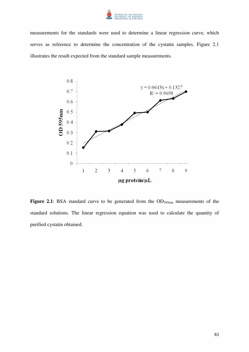
61
measurements for the standards were used to determine a linear regression curve, which
serves as reference to determine the concentration of the cystatin samples. Figure 2.1
illustrates the result expected from the standard sample measurements.
Figure 2.1: BSA standard curve to be generated from the OD595nm measurements of the
standard solutions. The linear regression equation was used to calculate the quantity of
purified cystatin obtained.

62
2.2.4 Determination of Ki values
A papain (cysteine protease) stock was prepared from a commercially available latex
preparation of C. papaya (Sigma-Aldrich) which contains a minimum of 3.2 U/mg of
enzyme. Human cathepsin-L, another commercially available protease, was also purchased
from Sigma-Aldrich. The cysteine protease activity was determined and the Ki values for the
wild-type PC and OCI, and the mutant variants were determined. The measurements were
performed using black, flat-bottom, polysorp, 96-well plates (Nunc). Each well contained a
total reaction volume of 100 µL, the fluorogenic substrate Z-Phe-Arg-MCA (cathepsin L-like
substrate from Sigma-Aldrich) was used at a final concentration of 8 µM from a 100 µM
stock dissolved in dimethyl sulfoxide (Sigma-Aldrich).
Dissociation (inhibition) constants (Ki) for the interaction between the different PC and OCI
variants, with the model cysteine proteases, papain and human cathepsin L, were determined
according to the method outlined by Goulet et al. (2008). Substrate hydrolysis progress
curves were monitored as described by Salvesen and Nagase (1989) and the linear equation
was determined as described by Henderson (1972). Papain activity was measured in 100 mM
sodium phosphate buffer, pH 7.0, and cathepsin L activity was measured in 100 mM citrate
phosphate buffer, pH 5.5. The hydrolysis reactions were allowed to proceed at 25°C, under
reducing conditions, by adding 10 mM L-cysteine to the reaction buffer. Cysteine protease
activities were determined using a Fluostar Galaxy fluorimeter (BMG, Offenburg, Germany),
using a 360 nm excitation filter and a 450 nm emission filter. Ki values were calculated, after
firstly empirically estimating the Ki(app) and Km values, and then using the equation Ki =
Ki(app)/(1 + [S]/Km). The Km values which were used were 13.6 µM and 1.0 µM, for papain,
63
and cathepsin L, respectively, which were determined with their test substrates (Goulet et al.,
2008).
2.2.5 Measurement of PC and OCI activity
Total protein extracts from plant material were used as sources for cysteine proteases in
assays to measure PCs and OCIs inhibitory activity. Extracts were prepared from leaf
material from Nicotiana benthamiana by crushing in liquid nitrogen, weighed and a 100mM
sodium phosphate buffer at pH 6.5, was added in a ratio of 1:2 (100 µg : 200 µL; sample :
buffer). The solution was incubated for 30 min on ice before centrifuging at 15000 xg for 15
min at 4°C to remove debris. The supernatant was removed and total protein concentration
determined. Total protein extracts from larvae of banana weevils (Cosmopolites sordidus)
and Colorado potato beetles (Leptinotarsa decemlineata) were used as another source of
cysteine proteases and were provided by Prof. K.J. Kunert. The total protein content of the
extracts was determined using the same method for the quantification of the purified
cystatins.
The concentration of inhibitor required to reduce the proteolytic activity by 40-60% was
determined using OCI and PC for each of the different extracts, the determined concentration
for the wild-type inhibitor was used for the respective mutants and allowed for comparative
analysis of OCI against PC and against the mutant inhibitors. Cysteine protease inhibitor
activities were assayed as before, by the monitoring the progression curves of substrate
hydrolysis, using the synthetic fluorogenic substrate Z-Phe-Arg-MCA. Substrate hydrolysis
by the plant and insect digestive proteases were allowed to proceed in a 100 mM phosphate
buffer, pH 6.5, at 25°C. The inhibitory activities were monitored using a Fluostar Galaxy
64
fluorimeter (BMG), using the same excitation and emission filters as mentioned before. As a
maximal inhibitory control, E-64 (from Sigma-Aldrich) was used as a broadspectrum
inhibitor for cysteine proteases (Michaud et al., 1993).
2.3 Bioinformatics work
Discovery Studio v2.5 (Accelrys Software Inc., USA) software package was used to produce
the different cystatin models and to simulate the interaction energies between the cysteine
protease, papain, and the PC and OCI variants generated in the project. The electrostatic
interaction energies and the van der Waals interaction energies were calculated between the
protease model and each of the cystatins. The binding interfaces of the models were refined
and re-scored using the RDOCK protocol and the energy minimization was done using the
CHARMm Polar H forcefield. The binding interface residues were grouped and the binding
energies were calculated.
The UCSF Chimera software package (http://www.cgl.ucsf.edu/chimera) was used for
manipulation of the individual models of OCI and PC for illustrative purposes in Figures 1.2-
6 and 3.12 (Molecular graphics images were produced using the UCSF Chimera package
from the Resource for Biocomputing, Visualization, and Informatics at the University of
California, San Francisco).
2.4 Statistical methods
The statistical significance of variations in mean values of both in vitro and in silico was
determined using the Student’s two-tailed t test at stringency level of P ≤ 0.05.
65
3. RESULTS
3.1 DNA work
3.1.1 Cloning into vector pGEX
All OCI and PC wild type and mutant clones were sequenced to confirm the correctness of
mutations introduced before a restriction enzyme digest was performed to obtain an OCI or
PC fragment for further sub-cloning into the pGEX®
-3X vector. Figure 3.1 shows the
digestion of plasmid pGEMPWT containing the PC wild-type and the different pGEMPM
vectors containing PC mutant coding sequences with restriction enzymes BamHI and EcoRI.
A DNA fragment with the predicted size of 300 bp for the coding sequence was detected after
restriction enzyme digest, confirming correct cloning of coding sequences into vector
pGEM®
-T Easy. An identical result was obtained when either plasmid pGEMOWT
containing the OCI wild-type or the different pGEMOM vectors containing OCI mutant
coding sequences were digested with Bam.
After isolation of wild-type and mutant OCI and PC sequences from an agarose gel, purified
BamHI/EcoRI fragments were cloned into plasmid pGEX®
-3X to create vectors pGEXOWT
and pGEXOM vectors for OCI and pGEXPWT and different pGEXPM vectors for PC.
Cloning into the plasmid pGEX®
-3X to created an in-frame gene fusion with the GST coding
sequence at the 5’-end of the OCI or PC coding sequence (Figure 2.1). To confirm that a
correct BamHI/EcoRI fragment was cloned into pGEX®
-3X, all pGEX plasmids carrying
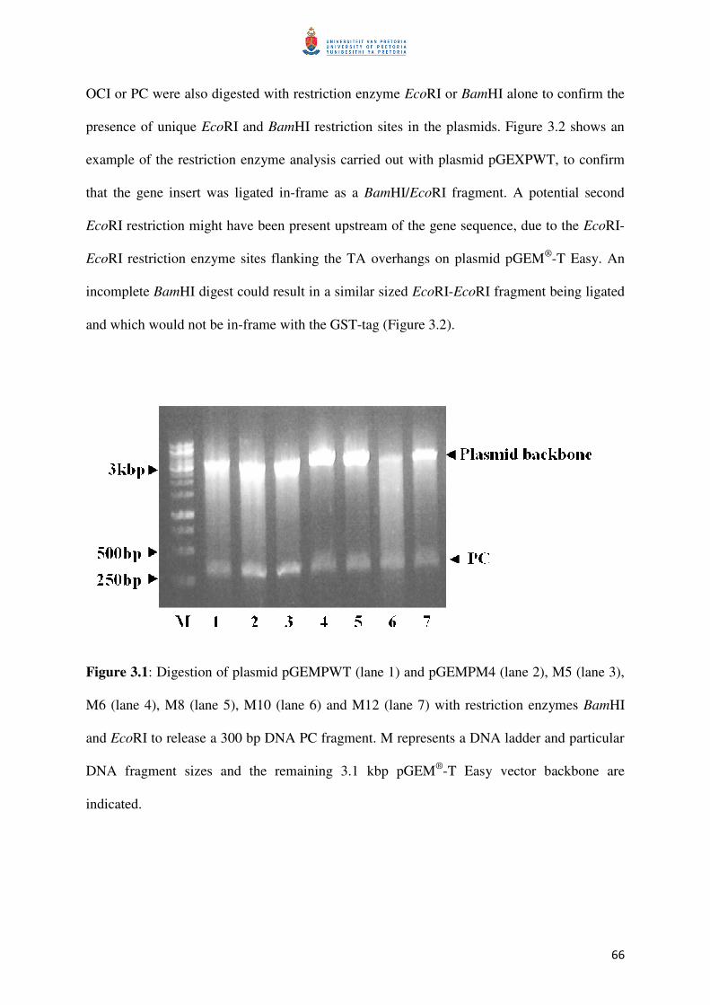
66
OCI or PC were also digested with restriction enzyme EcoRI or BamHI alone to confirm the
presence of unique EcoRI and BamHI restriction sites in the plasmids. Figure 3.2 shows an
example of the restriction enzyme analysis carried out with plasmid pGEXPWT, to confirm
that the gene insert was ligated in-frame as a BamHI/EcoRI fragment. A potential second
EcoRI restriction might have been present upstream of the gene sequence, due to the EcoRI-
EcoRI restriction enzyme sites flanking the TA overhangs on plasmid pGEM®
-T Easy. An
incomplete BamHI digest could result in a similar sized EcoRI-EcoRI fragment being ligated
and which would not be in-frame with the GST-tag (Figure 3.2).
Figure 3.1: Digestion of plasmid pGEMPWT (lane 1) and pGEMPM4 (lane 2), M5 (lane 3),
M6 (lane 4), M8 (lane 5), M10 (lane 6) and M12 (lane 7) with restriction enzymes BamHI
and EcoRI to release a 300 bp DNA PC fragment. M represents a DNA ladder and particular
DNA fragment sizes and the remaining 3.1 kbp pGEM®
-T Easy vector backbone are
indicated.

67
Figure 3.2: Digestion of plasmid pGEXPWT with BamHI (lane 1), EcoRI (lane 2) and
BamHI/EcoRI combined (lane 3) to release a 300 bp PC DNA fragment. The digestion of
plasmid pGEMPM4 with BamHI (lane 4), EcoRI (lane 5) and BamHI/EcoRI combined (lane
6) is also shown to release the expected 300 bp PC DNA fragment. M represents the DNA
ladder and particular DNA fragment sizes and the remaining 4.9 kbp pGEX®
-3X vector
backbone are indicated.
All pGEX-OCI and pGEX-PC expression plasmids were re-sequenced to confirm the
correctness of the junction site of the GST and either OCI or PC coding sequence for in frame
expression of a fusion protein. All plasmids were finally maintained in E. coli DH5α, and
stored at -80°C until protein expression studies.

68
3.2 Protein work
3.2.1 Protein expression
Expression of GST-PC fusion proteins in E. coli DH5α cells, which were used as a pre-screen
to confirm correctness of plasmids allowing fusion protein production, is shown in figure 3.3.
GST fusion proteins were produced after induction of expression by IPTG when the cell
suspension had an OD600nm of 0.6. Fusion protein samples were prepared from cells grown
for 12 hrs in the presence of IPTG by first diluting the suspension to an OD600nm of 1.0 with
distilled water. Cells were then boiled in the presence of 1% SDS and proteins analyzed by
SDS-PAGE on a 12% gel. The expected 37 kDa GST fusion protein (GSTF) was detected to
be expressed in all induced cells (Figure 3.3) but no significant fusion protein production was
found in non-IPTG-induced cells. An identical protein expression experiment was carried out
for GST-OCI fusions and for all OCI proteins (wild-type and mutants) a GST fusion protein
was detected (data not shown).
To optimize fusion protein expression, all plasmids were transferred into cells of E. coil strain
BL21 by heat shock treatment. The E. coli strain BL21 is widely used for expression of
recombinant proteins. Figure 3.4 shows the analysis of expressed proteins in non-induced and
IPTG-induced E. coli BL21 cells (6 hrs protein expression induction) expressing the different
pGEX-PC plasmids by a SDS-PAGE on a 12% gel. The expected 37 kDa GST fusion protein
(GSTF) was detected to be expressed in all induced E. coli BL21 cells (Figure 3.4) but no
significant fusion protein production was found in non-IPTG-induced cells. An identical
protein expression experiment was carried out for GST-OCI fusions and for all OCI proteins
(wild-type and mutants) a GST fusion protein was detected (data not shown).
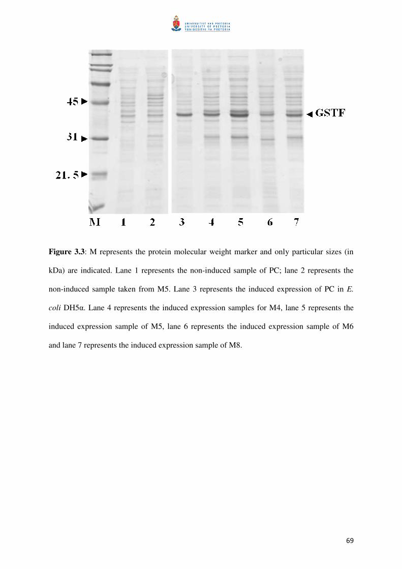
69
Figure 3.3: M represents the protein molecular weight marker and only particular sizes (in
kDa) are indicated. Lane 1 represents the non-induced sample of PC; lane 2 represents the
non-induced sample taken from M5. Lane 3 represents the induced expression of PC in E.
coli DH5α. Lane 4 represents the induced expression samples for M4, lane 5 represents the
induced expression sample of M5, lane 6 represents the induced expression sample of M6
and lane 7 represents the induced expression sample of M8.
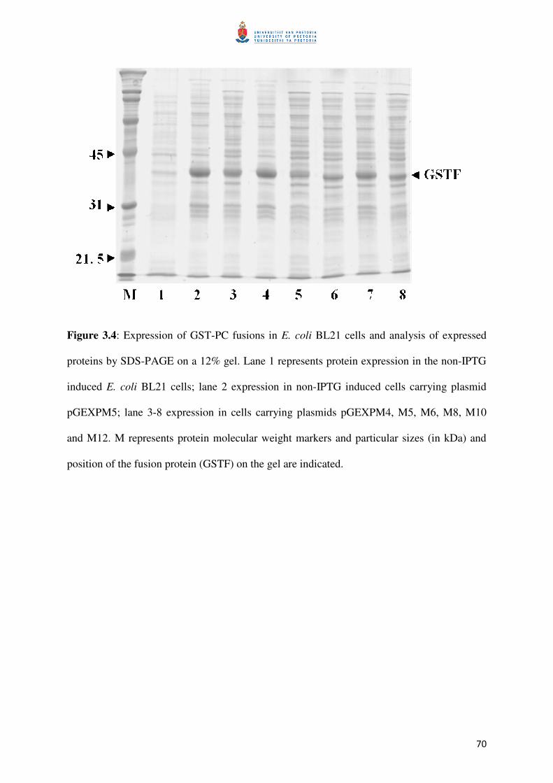
70
Figure 3.4: Expression of GST-PC fusions in E. coli BL21 cells and analysis of expressed
proteins by SDS-PAGE on a 12% gel. Lane 1 represents protein expression in the non-IPTG
induced E. coli BL21 cells; lane 2 expression in non-IPTG induced cells carrying plasmid
pGEXPM5; lane 3-8 expression in cells carrying plasmids pGEXPM4, M5, M6, M8, M10
and M12. M represents protein molecular weight markers and particular sizes (in kDa) and
position of the fusion protein (GSTF) on the gel are indicated.

71
3.2.2 Fusion protein purification
For fusion protein purification, expressed PC and OCI wild-type and mutant proteins fused to
GST were released from IPTG-induced E. coli BL21 cells by applying a freeze-thawing
procedure. After removing cell debris by centrifugation, the supernatant containing the
soluble fusion protein was mixed with GSH-Sepharose 4B (Amersham Pharmacia Biotech) to
bind the GST fusion protein. Figure 3.5 shows the different steps of the purification
procedure including the step of binding the GST fusion protein to GSH-Sepharose 4B. All
protein samples were analyzed by SDS-PAGE on a 12% gel and the expected 37 kDa GST
fusion proteins bound to GSH-Sepharose 4B was detected (Figure 3.5). An identical
procedure was carried out with OCI and mutant OCI and an identical result as found for PC
was obtained (data not shown).
The GST fusion protein bound to the GSH-Sepharose was collected by centrifugation.
Treatment of the different GST-fusions with protease Factor Xa released OCI and PC from
the GST fusion protein and the released OCI and PC protein could be detected in the
supernatant. Figure 3.6 shows the protein samples analyzed by SDS-PAGE on a 15% gel at
the different purification steps for PC and OCI and the released purified PC (or OCI) was
found with the predicted size of about 11kDa.
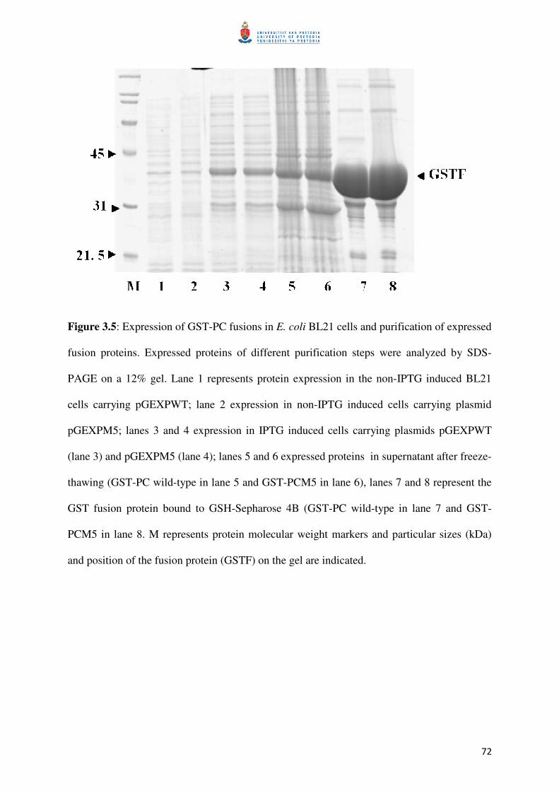
72
Figure 3.5: Expression of GST-PC fusions in E. coli BL21 cells and purification of expressed
fusion proteins. Expressed proteins of different purification steps were analyzed by SDS-
PAGE on a 12% gel. Lane 1 represents protein expression in the non-IPTG induced BL21
cells carrying pGEXPWT; lane 2 expression in non-IPTG induced cells carrying plasmid
pGEXPM5; lanes 3 and 4 expression in IPTG induced cells carrying plasmids pGEXPWT
(lane 3) and pGEXPM5 (lane 4); lanes 5 and 6 expressed proteins in supernatant after freeze-
thawing (GST-PC wild-type in lane 5 and GST-PCM5 in lane 6), lanes 7 and 8 represent the
GST fusion protein bound to GSH-Sepharose 4B (GST-PC wild-type in lane 7 and GST-
PCM5 in lane 8. M represents protein molecular weight markers and particular sizes (kDa)
and position of the fusion protein (GSTF) on the gel are indicated.
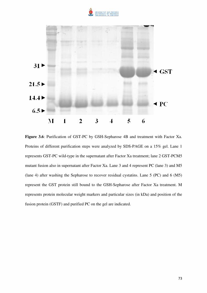
73
Figure 3.6: Purification of GST-PC by GSH-Sepharose 4B and treatment with Factor Xa.
Proteins of different purification steps were analyzed by SDS-PAGE on a 15% gel. Lane 1
represents GST-PC wild-type in the supernatant after Factor Xa treatment; lane 2 GST-PCM5
mutant fusion also in supernatant after Factor Xa. Lane 3 and 4 represent PC (lane 3) and M5
(lane 4) after washing the Sepharose to recover residual cystatins. Lane 5 (PC) and 6 (M5)
represent the GST protein still bound to the GSH-Sepharose after Factor Xa treatment. M
represents protein molecular weight markers and particular sizes (in kDa) and position of the
fusion protein (GSTF) and purified PC on the gel are indicated.
74
3.2.3.1 Determination of relative inhibitory potency
Inhibitory activity of OCI and PC mutants relative to wild-type OCI and PC were determined
to show changes in the potency of individual mutated inhibitors to modulate activity against
the two model cysteine proteases papain and cathepsin-L. Figure 3.7 shows the relative
inhibitory activity of OCI wild-type and OCI mutants against (A) papain with 17.5 ng of
papain per 100 µl of assay mixture and (B) with 2.81 ng cathepsin-L per 100 µl of assay
mixture. Assays were carried out in 100 mM sodium phosphate buffer, pH 6.5. For papain all
inhibitors tested were used at 0.625 ng per test and for cathepsin-L at 0.312 ng per test.
Papain activity without inhibitor addition was 64.0± 4.3 FU/min (fluorescence units produced
per minute) and cathepsin-L activity was 39.5± 0.7 FU/min. Inhibitory activity of each
mutant OCI is shown relative inhibitory activity of wild-type OCI set at 100%. Relative
activities greater than 100% correspond to improved and below 100% to decreased inhibitory
activity of OCI mutants when compared to wild-type OCI. Data shown for mutants are the
mean ± SD from 3 different individual measurements of protease activity in the presence of
the inhibitor. Only OCIM1 tested against papain did not show statistically significantly (P ≤
0.05) altered the inhibitory potency compared to the wild-type, all other cystatins in all other
tests had statistically significant change in inhibitory potency. The OCIM2 was the most
active mutant for both papain and cathepsin-L inhibition improving potency by 80% and
50%, respectively, all other OCI mutants had lower inhibitory activity against papain and
cathepsin-L when compared to wild-type OCI.
Figure 3.8 shows the relative inhibitory activity of PC wild-type and PC mutants against (A)
papain with 17.5 ng of papain per 100 µl of assay mixture and (B) with 2.81 ng of cathepsin-
L per 100 µl of assay mixture. Assay was carried out in 100 mM sodium phosphate buffer,

75
pH 6.5. All inhibitors tested for inhibition of papain activity were used at 1.25 ng per test and
at 19 ng per test for cathepsin-L. Papain activity without addition of inhibitor was 64.0± 4.3
FU/min and cathepsin-L activity was 39.7 ± 0.7 FU/min. Inhibitory activity of each mutant
PC is shown relative to the activity of wild-type PC, which was set at 100%. Relative
inhibitory activities greater than 100% correspond to improved and below 100% to decreased
inhibitory activity of mutants when compared to wild-type PC. Data shown for mutants are
the mean ± SD from 3 different individual measurements of protease activity in the presence
of the inhibitor. The inhibitory activity relative to wild-type PC increased in all PC mutants
when papain was used as a cysteine protease activity source with the highest increase found
for PCM6 (85%) followed by PCM5 (69%), PCM4 (33%) and PCM8 (23%). Both PCM10
(5%) and PCM12 (7%) showed only slightly improved inhibitory potency compared to the
wild-type PC activity. When cathepsin-L was used as a cysteine protease source, PCM6
again, as already found for papain, was the most active mutant improving wild-type PC
inhibition by 90% which was followed by PCM4 (83%), and PCM8 (63%). PCM5 only
increased wild-type PC inhibitory potency by 14% and again PCM10 and PCM12 did not
improve potency, which was also found when papain was used as a cysteine protease source.
Only PCM10 and PCM12 cystatins tested against papain did not show statistically significant
change in inhibitory potency (P ≤ 0.05) compared to the wild-type, all other cystatins had
statistically significant change in inhibitory potency.
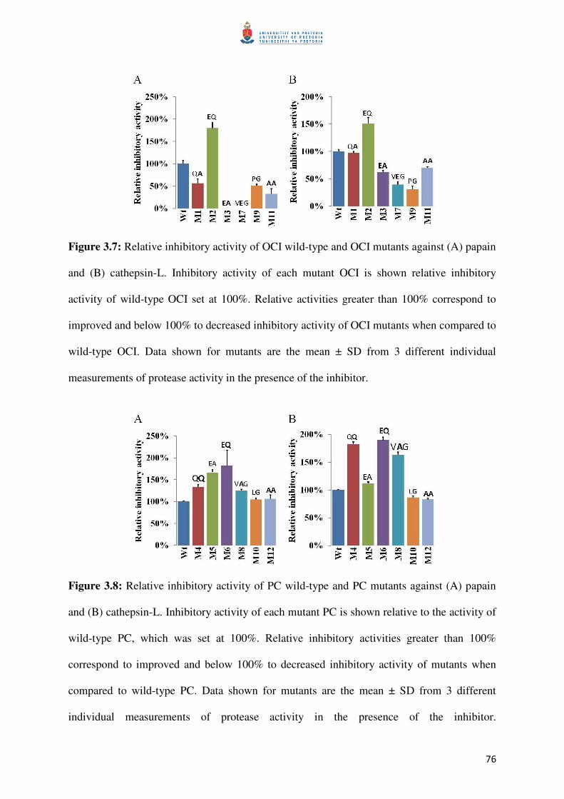
76
Figure 3.7: Relative inhibitory activity of OCI wild-type and OCI mutants against (A) papain
and (B) cathepsin-L. Inhibitory activity of each mutant OCI is shown relative inhibitory
activity of wild-type OCI set at 100%. Relative activities greater than 100% correspond to
improved and below 100% to decreased inhibitory activity of OCI mutants when compared to
wild-type OCI. Data shown for mutants are the mean ± SD from 3 different individual
measurements of protease activity in the presence of the inhibitor.
Figure 3.8: Relative inhibitory activity of PC wild-type and PC mutants against (A) papain
and (B) cathepsin-L. Inhibitory activity of each mutant PC is shown relative to the activity of
wild-type PC, which was set at 100%. Relative inhibitory activities greater than 100%
correspond to improved and below 100% to decreased inhibitory activity of mutants when
compared to wild-type PC. Data shown for mutants are the mean ± SD from 3 different
individual measurements of protease activity in the presence of the inhibitor.

77
3.2.3.2 Determination of Ki values
Table 3.1 shows the Ki values for wild-type PC and OCI and PC and OCI mutants using
papain and cathepsin-L for cysteine protease activity and Z-Phe-Arg-MCA as a cysteine
protease substrate. When papain was used, only OCIM2 (Ki = 0.04 nM) had a greatly lowered
Ki value compared to wild-type OCI (Ki = 0.30 nM) and all other OCI mutants did not lower
the Ki value of wild-type OCI. All mutant PCs had significantly lowered Ki values compared
to wild-type PC (Ki = 5.58 nM) with PCM5 (Ki = 0.47 nM) and PCM6 (Ki = 0.21 nM) the
most active and PCM10 (Ki = 4.38 nM) and PCM12 (Ki = 4.04 nM) the least active PC
mutant.
When cathepsin-L was used, only OCIM2 (Ki = 0.14 nM) had a significantly lower Ki value
compared to wild-type OCI (Ki = 0.49 nM) (Table 3.1). All other OCI mutants had a higher
Ki values compared to wild-type OCI with OCIM9 showing no inhibition of cathepsin–L
activity. Except for PCM10 and PCM12, which both showed no inhibition of cathepsin-L all
other PC mutants had showed lower Ki values compared to wild-type PC (Ki = 152 nM).
However, PCM4 (Ki = 3.58 nM) and PCM6 (Ki = 1.95 nM) had the lowest Ki values and
therefore the most improved potency against cathepsin-L.
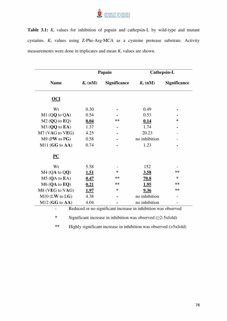
78
Table 3.1: Ki values for inhibition of papain and cathepsin-L by wild-type and mutant
cystatins. Ki values using Z-Phe-Arg-MCA as a cysteine protease substrate. Activity
measurements were done in triplicates and mean Ki values are shown.
Papain Cathepsin-L
Name Ki (nM) Significance Ki (nM) Significance
OCI
Wt
0.30
-
0.49
-
M1 (QQ to QA) 0.54 - 0.53 - M2 (QQ to EQ) 0.04 ** 0.14 * M3 (QQ to EA) 1.37 - 1.74 -
M7 (VAG to VEG) 4.25 - 20.23 -
M9 (PW to PG) 0.58 - no inhibition -
M11 (GG to AA) 0.74 - 1.23 -
PC
Wt
5.58
-
152
-
M4 (QA to QQ) 1.51 * 3.58 ** M5 (QA to EA) 0.47 ** 70.8 * M6 (QA to EQ) 0.21 ** 1.95 **
M8 (VEG to VAG) 1.97 * 9.36 ** M10 (LW to LG) 4.38 - no inhibition -
M12 (GG to AA) 4.04 - no inhibition -
- Reduced or no significant increase in inhibition was observed
* Significant increase in inhibition was observed (≥2-5xfold)
** Highly significant increase in inhibition was observed (>5xfold)
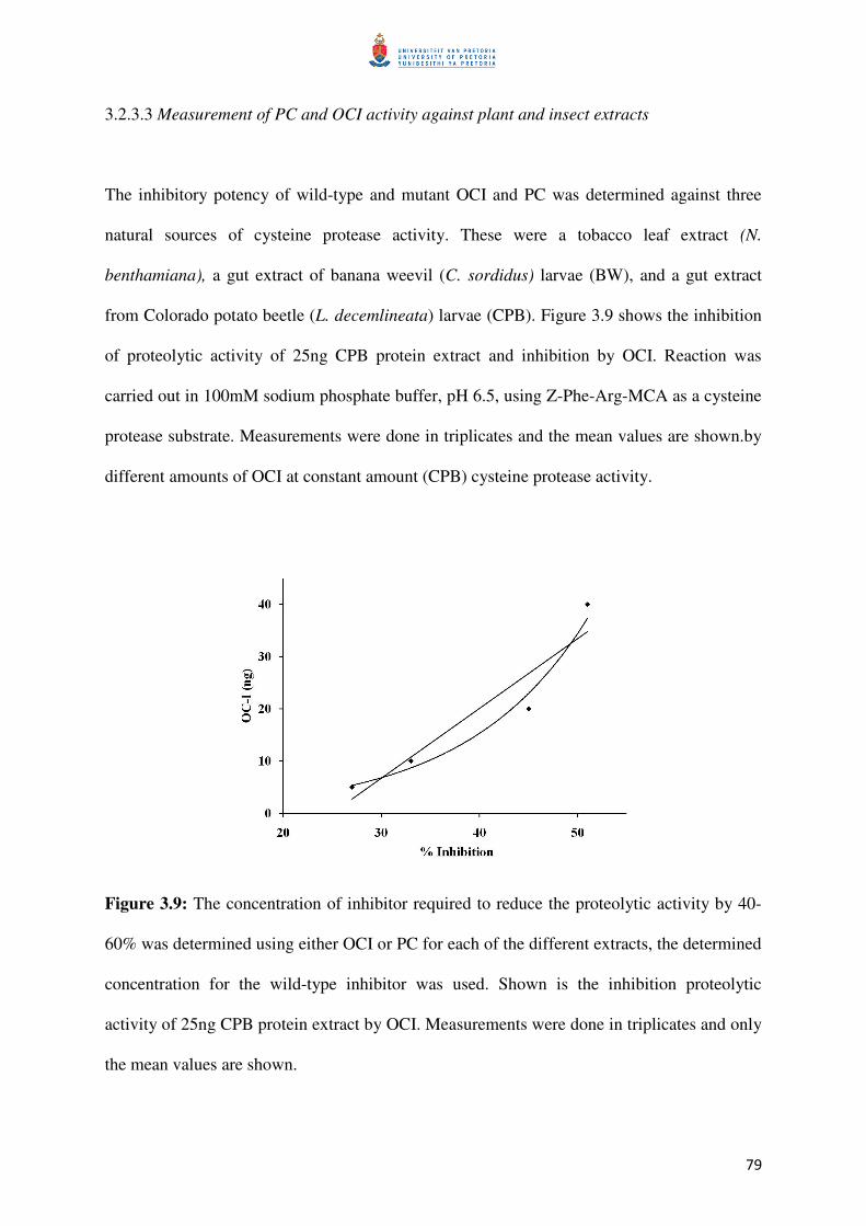
79
3.2.3.3 Measurement of PC and OCI activity against plant and insect extracts
The inhibitory potency of wild-type and mutant OCI and PC was determined against three
natural sources of cysteine protease activity. These were a tobacco leaf extract (N.
benthamiana), a gut extract of banana weevil (C. sordidus) larvae (BW), and a gut extract
from Colorado potato beetle (L. decemlineata) larvae (CPB). Figure 3.9 shows the inhibition
of proteolytic activity of 25ng CPB protein extract and inhibition by OCI. Reaction was
carried out in 100mM sodium phosphate buffer, pH 6.5, using Z-Phe-Arg-MCA as a cysteine
protease substrate. Measurements were done in triplicates and the mean values are shown.by
different amounts of OCI at constant amount (CPB) cysteine protease activity.
Figure 3.9: The concentration of inhibitor required to reduce the proteolytic activity by 40-
60% was determined using either OCI or PC for each of the different extracts, the determined
concentration for the wild-type inhibitor was used. Shown is the inhibition proteolytic
activity of 25ng CPB protein extract by OCI. Measurements were done in triplicates and only
the mean values are shown.

80
When the different inhibitory potency for wild-type and mutant OCI and PC against cysteine
protease activity of different protein extracts (CPB, BW, tobacco leaf) was determined,
higher amounts of wild-type or mutant PC had to be used than wild-type or mutant OCI to
obtain comparable inhibition of cysteine protease activity (Figures 3.10 and 3.11). Relative
inhibitory activity of the wild-type and mutants were tested against (A) 125 ng of CPB
protein extract, (B) 5 µg of BW protein extract protein extract and (C) 4.6 µg of tobacco leaf
protein extract. Assay mixture (100 µL) contained 100 mM sodium phosphate buffer, pH 6.5.
For inhibition of cysteine protease activity of CPB protein extract, 10 ng of wild-type OCI or
mutant OCI was used and 100 ng of wild-type PC or mutant PC. For BW protein extract, 400
ng of wild-type OCI or mutant OCI was used and 500 ng of wild-type PC or mutant PC. For
tobacco leaf protein extract, 10 ng of wild-type OCI or mutant OCI was used and 50ng of
wild-type PC or mutant PC. The cysteine protease activity of CPB protein extract activity
without inhibitor addition was measured to be 50.1 ± 1.9 FU/min, for BW protein extract
21.1 ± 0.7 FU/min and for tobacco leaf protein extract 38.8 ± 1.9 FU/min. Inhibitory activity
of each mutant OCI is shown relative to the inhibitory activity of wild-type OCI set at 100%.
Relative activities greater than 100% correspond to improved and below 100% to decreased
inhibitory activity of mutant OCI when compared to wild-type OCI. Data shown for mutants
are the mean ± SD from 3 different individual measurements of protease activity in the
presence of the inhibitor (Table 3.2; Figures 3.10 and 3.11). Only OCIM1, OCIM2, OCIM3
and OCIM11, tested against the tobacco leaf extract, did not show statistically significantly
(P ≤ 0.05) altered inhibitory potency compared to the wild-type, all other mutant cystatins in
other tests had statistically significant change in inhibitory potency. All other OCI mutants
did not increase potency of the inhibitor and were less effective than wild-type OCI (Table
3.2; Figure 3.10), only OCIM2 had a slight (6%) increase in inhibition when a CPB extract
was used (Table 3.2; Figure 3.10A).
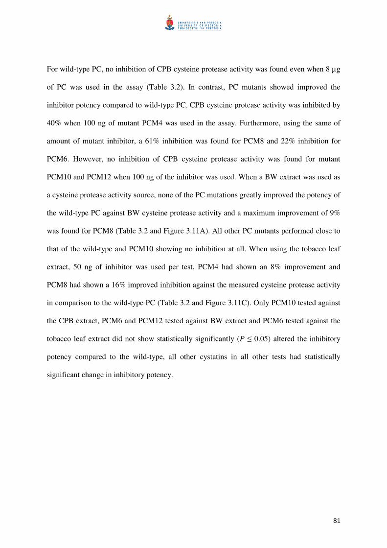
81
For wild-type PC, no inhibition of CPB cysteine protease activity was found even when 8 µg
of PC was used in the assay (Table 3.2). In contrast, PC mutants showed improved the
inhibitor potency compared to wild-type PC. CPB cysteine protease activity was inhibited by
40% when 100 ng of mutant PCM4 was used in the assay. Furthermore, using the same of
amount of mutant inhibitor, a 61% inhibition was found for PCM8 and 22% inhibition for
PCM6. However, no inhibition of CPB cysteine protease activity was found for mutant
PCM10 and PCM12 when 100 ng of the inhibitor was used. When a BW extract was used as
a cysteine protease activity source, none of the PC mutations greatly improved the potency of
the wild-type PC against BW cysteine protease activity and a maximum improvement of 9%
was found for PCM8 (Table 3.2 and Figure 3.11A). All other PC mutants performed close to
that of the wild-type and PCM10 showing no inhibition at all. When using the tobacco leaf
extract, 50 ng of inhibitor was used per test, PCM4 had shown an 8% improvement and
PCM8 had shown a 16% improved inhibition against the measured cysteine protease activity
in comparison to the wild-type PC (Table 3.2 and Figure 3.11C). Only PCM10 tested against
the CPB extract, PCM6 and PCM12 tested against BW extract and PCM6 tested against the
tobacco leaf extract did not show statistically significantly (P ≤ 0.05) altered the inhibitory
potency compared to the wild-type, all other cystatins in all other tests had statistically
significant change in inhibitory potency.

82
Table 3.2: Inhibitory potencies of the wild-types OCI and PC and mutants, tested against the cysteine protease of CPB, BW or tobacco leaf. (-
indicates that reduced or no significant increase in inhibition was observed (P ≤ 0.05); * indicates that a significant increase in inhibition was
observed [≥2-5xfold])
Colorado potato beetle (CPB) Banana weevil (BW) Tobacco
Inhibition (%) Significance Inhibition (%) Significance Inhibition (%) Significance
OCI
Wt
38
-
37
-
80
-
M1 (QQ to QA) 15 - 11 - 76 - M2 (QQ to EQ) 44 - 27 - 81 - M3 (QQ to EA) 4 - 13 - 77 -
M7 (VAG to VEG) 12 - 15 - 48 - M9 (PW to PG) 14 - 20 - 54 -
M11 (GG to AA) 16 - 25 - 78 -
PC
Wt
no inhibition
-
24
-
72
-
M4 (QA to QQ) 40 - 30 - 80 - M5 (QA to EA) 6 - 21 - 30 - M6 (QA to EQ) 22 - 22 - 74 -
M8 (VEG to VAG) 61 * 33 - 90 * M10 (LW to LG) no inhibition - no inhibition - 8 - M12 (GG to AA) no inhibition - 21 - 23 -
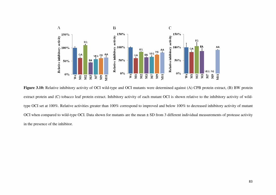
83
Figure 3.10: Relative inhibitory activity of OCI wild-type and OCI mutants were determined against (A) CPB protein extract, (B) BW protein
extract protein and (C) tobacco leaf protein extract. Inhibitory activity of each mutant OCI is shown relative to the inhibitory activity of wild-
type OCI set at 100%. Relative activities greater than 100% correspond to improved and below 100% to decreased inhibitory activity of mutant
OCI when compared to wild-type OCI. Data shown for mutants are the mean ± SD from 3 different individual measurements of protease activity
in the presence of the inhibitor.
84
Figure 3.11: Relative inhibitory activity of PC wild-type and PC mutants were determined against (A) CPB protein extract, (B) BW protein
extract protein and (C) tobacco leaf protein extract. Inhibitory activity of each mutant PC is shown relative to the inhibitory activity of wild-type
PC set at 100%. Relative activities greater than 100% correspond to improved and below 100% to decreased inhibitory activity of mutant PC
when compared to wild-type PC. Data shown for mutants are the mean ± SD from 3 different individual measurements of protease activity in the
presence of the inhibitor.
85
3.3 Bioinformatics work
The Discovery Studio v2.5 (Accelrys Software Inc., USA) software package was used to
simulate the interaction energies between the model cysteine protease model papain and both
wild-type and mutant OCI and PC generated in the study. Figure 3.12 shows the models
generated and the positions of the individual mutations are shown in the structural model of
both OCI and PC. The amino acid changes in the OCI model include Q53A, Q52E, A56E,
W84G, G10A and G11A. The amino acid changes indicated on the PC model include A52Q,
Q51E, A55E, W83G, G8A and G9A. The interaction energies were first simulated based on
the interaction between the entire models, which calculates the contribution all amino acids in
binding and taking into account the spacial arrangement of the protein’s structure in
secondary and tertiary folding, giving an overall interaction energy value (Table 3.3). The
interaction energies were then simulated based on the individual contributions that the
mutated amino acids contribute towards the interaction with the papain model and the
individual cystatin models, giving the exact value that each amino acid contributes to the
binding energy (Table 3.4). The simulations were done to determine similarities to the
biochemical data obtained in the study. The interaction energy between the enzyme-inhibitor
complex is expected to decrease when a more stable interaction complex (i.e. more
favourable) is formed and is expected to increase when a less stable interaction complex (i.e.
less favourable) is formed.

86
Figure 3.12: The wild-type cystatin models used to simulate the interactions energies are shown, the amino acid positions which were targeted
are indicated and the corresponding amino acid changes are shown (UCSF Chimera; Pettersen et al., 2004).
87
The predicted interaction energies (kcal/mol) calculated between the model cysteine protease
papain and the entire OCI or PC inhibitor is shown in Table 3.3. All OCI and PC mutants had
shown a decreased predicted total interaction energy and therefore predicts a less stable
association complexes being formed between the papain and the cystatins (4.5-11.8%) when
compared to wild-type OCI or wild-type PC. The highest decrease in interaction energy,
which relates to a reduced complex stability, was found for mutant OCIM7 (11.8%) and
PCM8 (11.3%), which contain the mutation A56E for OCI and E55A for PC within the same
tertiary position, indicating the importance of this site for binding. However, this reduced
complex stability observed for OCIM7 and PCM8, was not directly comparable to the
biochemical data obtained. The OCIM2 and PCM6 mutations were predicted to have a
destabilizing effect, but from the biochemical data was observed to have the most improved
inhibitory activity against papain.
Table 3.4 shows the predicted interaction energy (kcal/mol) based on the interaction between
papain and the individually mutated amino acid residues in the OCI and PC. For OCI,
increased interaction energies were only predicted for changing the Q (glutamine) at position
52 in the first inhibitory loop to E (glutamic acid) (OCIM2) and changing the conserved Q at
position 53 to A (OCIM1). The interaction energy at the site of mutation was predicted to
decrease from -15.74 kcal/mol for OCI wild-type to -23.8 kcal/mol for OCIM2, with a further
decrease to -27.77 kcal/mol in the double mutant Q52EQ53A (OCIM3). The lowered
interaction energy values indicate that the individual amino acids are cooperating to form a
more stable interaction complex. The overall change in interaction energy for OCIM3 was
determined by adding the individual contributions of the amino acid residues in the wild-type
(-44.13 kcal/mol) and compared to that of the mutant (-50.0 kcal/mol). This was directly
related to an increased binding ability of OCIM2 to papain, increasing the potency of the
88
mutant, having a much lower Ki value than wild-type OCI and the lowest Ki value of all OCI
mutants (Tables 3.1 and 3.4). All other OCI mutations had a decrease in predicted interaction
energy, having a destabilizing effect, also resulted in higher Ki values and lower binding
affinity to papain than wild-type OCI. The greatest change of all the OCI mutants, was
observed for OCIM7 with an A56E substitution, which had negative interaction energy (-
41.93 kcal/mol) and was changed to positive interaction energy with an overall change of
(+55.4 kcal/mol), this change coincides with the observed change in Ki. This indicates that
the introduced amino acid E is possibly competing during the interaction with an amino
acid(s) in the papain molecule, destabilizing the interaction complex.
Similar to OCI, when changing the Q at position 52 in the first inhibitory loop of OCI
(OCIM2) to an E and having a conserved Q at position 53 adjacent, had the same amino acid
sequence EQ in the motif as PCM6. These mutants had a predicted decrease in interaction
energy at the site of change, from -12.24 kcal/mol for PC wild-type to -61.87 kcal/mol for
PCM5 and a further increase for the double mutant Q51EA52Q (PCM6) to -65.03 kcal/mol.
Again, the overall change for PCM6 was determined by adding the individual contributions
of the amino acid residues in the wild-type (-39.44 kacl/mol) and comparing to that of the
mutant (-90.37 kcal/mol). These two mutants PCM5 and PCM6 had had shown largest
decreases in interaction energy at the site of the change of all PC mutants, indicating a
stabilizing effect on the interaction complex. The PCM6 and PCM5 had a lowered Ki value of
27-times and 12-times, respectively, compared to the wild-type PC. The predicted interaction
energy for the other mutants decreased in comparison to the wild-type PC, but these were
observed to have lower Ki values than wild-type PC against papain and cathepsin L.
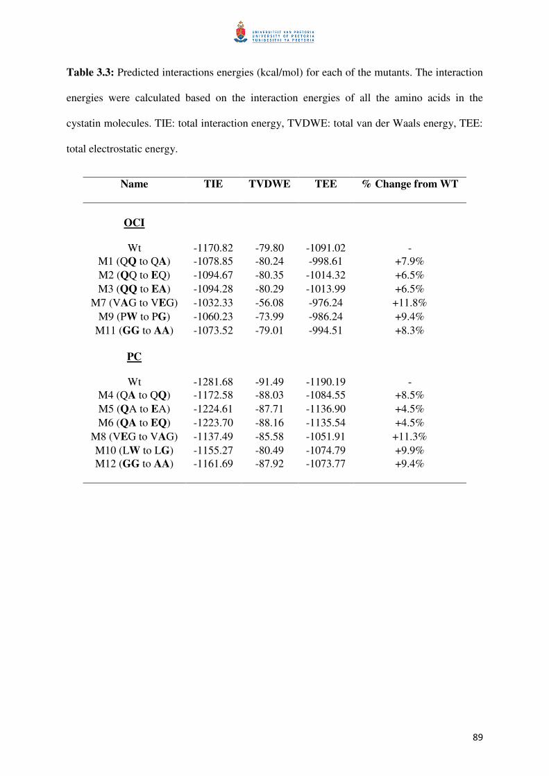
89
Table 3.3: Predicted interactions energies (kcal/mol) for each of the mutants. The interaction
energies were calculated based on the interaction energies of all the amino acids in the
cystatin molecules. TIE: total interaction energy, TVDWE: total van der Waals energy, TEE:
total electrostatic energy.
Name TIE TVDWE TEE % Change from WT
OCI
Wt
-1170.82
-79.80
-1091.02
-
M1 (QQ to QA) -1078.85 -80.24 -998.61 +7.9%
M2 (QQ to EQ) -1094.67 -80.35 -1014.32 +6.5%
M3 (QQ to EA) -1094.28 -80.29 -1013.99 +6.5%
M7 (VAG to VEG) -1032.33 -56.08 -976.24 +11.8%
M9 (PW to PG) -1060.23 -73.99 -986.24 +9.4%
M11 (GG to AA) -1073.52 -79.01 -994.51 +8.3%
PC
Wt
-1281.68
-91.49
-1190.19
-
M4 (QA to QQ) -1172.58 -88.03 -1084.55 +8.5%
M5 (QA to EA) -1224.61 -87.71 -1136.90 +4.5%
M6 (QA to EQ) -1223.70 -88.16 -1135.54 +4.5%
M8 (VEG to VAG) -1137.49 -85.58 -1051.91 +11.3%
M10 (LW to LG) -1155.27 -80.49 -1074.79 +9.9%
M12 (GG to AA) -1161.69 -87.92 -1073.77 +9.4%

90
Table 3.4: The table shows the predicted interactions energy (kcal/mol) of the individual amino acids that were targeted and changed, indicated
by the coloured blocks in the respective mutants. The other amino acid residues surrounding the coloured blocks had not shown considerable
change and were therefore not shown.
OCI Mutation Gly (10) Gly (11) Gln (52) Gln (53) Ala (56) Trp (84)
Wt
-27.05
-27.71
-15.74
-28.39
-41.93
-56.15
M1 Q53A -22.20
M2 Q52E -23.80
M3 Q52EQ53A -27.77 -22.23
M7 A56E +13.47
M9 W84G -29.01 M11 G10AG11A -14.76 -24.27
PC Mutation Gly (8) Gly (9) Gln (51) Ala (52) Glu (55) Trp (83)
Wt
-30.90
-15.17
-12.24
-27.20
-93.02
-59.90
M4 A52Q -26.29
M5 Q51E -61.87
M6 Q51EA52Q -65.03 -25.34
M8 E55A -43.85
M10 W83G -35.15 M12 G8AG9A -29.70 -0.71
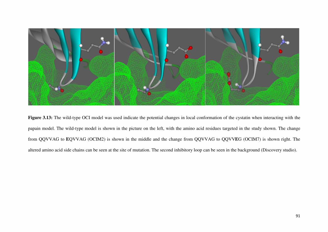
91
Figure 3.13: The wild-type OCI model was used indicate the potential changes in local conformation of the cystatin when interacting with the
papain model. The wild-type model is shown in the picture on the left, with the amino acid residues targeted in the study shown. The change
from QQVVAG to EQVVAG (OCIM2) is shown in the middle and the change from QQVVAG to QQVVEG (OCIM7) is shown right. The
altered amino acid side chains can be seen at the site of mutation. The second inhibitory loop can be seen in the background (Discovery studio).
92
4. DISCUSSION
During the interaction of a cysteine protease enzyme and cystatin, hydrophobic contacts are
established between the amino acid residues of the cystatin involved in binding and the
corresponding amino acid residues of the enzyme which form the binding pockets.
Knowledge of a given cysteine protease enzyme’s preferential selection toward amino acids
at given tertiary positions in a cystatin, such as cathepsins B/H which are repulsed by large
aromatic residues in their S2 and S3 pockets, in contrast to cathepsin L which preferentially
accommodates larger aromatic residues in the S2 pocket (Mason et al., 1998), would assist in
designing cystatins with enhanced inhibitory potency. Despite the similar mechanism of
action and structural homology among cystatins, they naturally exhibit distinct differences in
enzyme affinities. Looking at the frequency and occurrence of evolutionary conserved amino
acids at a specified tertiary position, such as the first inhibitory binding loop of cystatins, can
give an indication of what amino acids are preferentially accommodated and what might be
tolerated in the enzymes’ binding pockets. Using site-directed mutagenesis allows the relative
importance of amino acids at specific tertiary positions to be investigated, without
detrimentally altering the structural backbone of the cystatin. Although changing a single
amino acid residue alters the complementary shape, interacting charge and polarity of the
binding face, it also influences the neighboring amino acids which are dependent on the local
changes in amino acid side chains and the resulting functional charges. Previous studies have
followed a similar approach to investigate changes in conserved amino acid residues within
the N-terminal region and the first (QxVxG) and second (PW) inhibitory loops. However,
there is still no consensus based on the different observations found in previous studies on
which amino acids contribute to the inhibitory potency of cystatins. The importance of the N-
terminal region of cystatins is known to facilitate binding and stabilize the interaction
93
complex, but contrasting evidence from other studies obscures the importance and
involvement of the conserved glycine (G) amino acid residues in this region. The QxVxG
motif in the first binding loop of cystatins is conserved among most cystatins with a degree of
variance occurring at these variable amino acid sites. In the second inhibitory loop, the W
amino acid is known to be highly conserved and important in this position, yet no study has
looked at altering this amino acid residue using a mutagenesis approach. Based on the
hypervariable sites identified by Kiggundu et al. (2006), which could potentially improve the
inhibitory activity of cystatins and following a directed mutagenesis approach, the relative
importance of specific amino acid residues of cystatins may be elucidated and inhibitors with
improved inhibitory potency can be engineered.
This study was carried out to investigate the contribution of individual amino acid residues in
the N-terminal and the first and second inhibitory loops of the two plant cystatins, OCI and
PC, by using mutant OCI and PC inhibitors. These two cystatins were selected for this study
due to their recently identified differences in inhibitory activity against a gut extract from
banana weevils containing cysteine protease activity (Kiggundu et al., 2010) and also due to
unique sequence characteristics of PC in the conserved motif.
In a preceding study carried out by Dr Schlüter (unpublished data) the amino acid sequence
plant cystatins were compared, from this in silico analysis, PC was identified to have unique
amino acid residue characteristics in the conserved domain of the first inhibitory loop, a
unique A (alanine) in position 52, not found in any other cystatins, as well as an E (glutamic
acid) in position 55 (QAVVEG). The particular sequence variation (E in position 55) has
only been identified in three other characterized plant cystatin sequences: Celosia cristata
cystatin, a multi-cystatin unit of cowpea VuCYS2.3 and barley HvCYS7 cystatin. In this
94
study it was found that changing the unique A (alanine) in position 52 of PC to Q (glutamine)
(mutant PCM4), to obtain a QQ string in the conserved motif (QAVEG to QQVEG), which is
found naturally in many other plant cystatins, resulted in increased inhibitory activity of PC
against both papain and cathepsin-L without significantly changing the predicted interaction
energies. The change from a non-polar, hydrophobic amino acid residue A to a polar,
uncharged residue Q had possibly affected the shape of the binding loop and side-chain
polarity resulting in improved PC inhibitory potency. Removing the hydrophobic side chain
of A and replacing with the much larger, yet polar and uncharged side chain of Q, had
favourably altered the hydrophobicity of the binding face, however, why PC contains a
unique A residue and what role this residue has in PC, is still unclear. There is currently no
information available if this residue possibly plays a role in PC specificity against a particular
cysteine protease of a papaya pest or against a specific endogenous papaya cysteine protease.
In general, wild-type PC, isolated from mature papaya leaves, is active against chymopapain
and papaya protease IV, which is found in the latex of Carica papaya (Taylor et al., 1999;
Song et al., 1995). Papaya protease IV however, lacks activity against synthetic protease
substrates normally hydrolyzed by other papaya proteases (papain-like) and this protease is
also not inhibited by chicken cystatin (Buttle et al., 1989). Since PC potency was found in
this study to be much higher against papain (a papaya cysteine protease) than against human
cathepsin-L, PC might have a specific role in regulating endogenous papaya cysteine protease
activity, rather than a defensive role such as preventing cathepsin-L type cysteine protease
activity in the digestive system of herbivorous insects. Such interaction with an endogenous
papaya cysteine protease might require a unique amino acid residue(s) in the first binding
loop for optimal binding which might be investigated in more detail in a future study.

95
The importance of Q (a polar, uncharged amino acid) in the binding loop of the tertiary
structure for plant cystatin activity was confirmed in this study when Q53 in OCI was
mutated to a non-polar, hydrophobic A (mutant OCIM1 - QQVVAG to QAVVAG), likened
to the unique A (alanine) found in the first inhibitory loop of PC. This change had
significantly decreased OCI activity, as reflected by an increase in the Ki value against papain
and cathepsin-L. This was further associated with an increase of +6 kcal/mol in interaction
energy with papain from -28 to -22 kcal/mol as predicted in silico. This increase in energy
will result in a less stable complex. The importance of the Q residue in OCI activity has also
been reported by Arai et al. (1991) where substitution of Q in OCI with P (proline) (from
QQVVAG to QPVVAG) resulted in a completely inactive OCI. In addition, substituting Q
with L (leucine) (from QQVVAG to QLVVAG) resulted in a 150-fold increase in Ki, value
when compared to wild-type OCI causing a significant reduction in papain inhibition (Arai et
al., 1991).
A further new aspect of this study was the finding that substituting E with A in position 55 of
PC (PCM8) (from AVVEG to AVVAG) located in the first binding loop significantly
increased PC potency against both papain and cathepsin-L. Most other plant cystatins have
either this non-polar, neutral A or a polar, neutral S (serine) in this position (Table 1.2)
whereas E (glutamic acid) of PC in position 55 of PC is only found in three other plant
cystatins. However, the exact role of a polar, negatively charged E in this position is still
unknown. Since this amino acid residue is situated in the middle of the first inhibitory
binding loop inserting into the cleft of the target protease, mutations in this position might
significantly affect the binding capacity of PC. The alanine amino acid in this position is
considered to be a relatively small amino acid in comparison to the bulky E with a longer
side-chain. Also, changing the charge of the amino acid residue from a polar, negatively

96
charged E to a non-polar, neutral A will greatly influence the inhibitor’s binding ability. The
introduced amino acid change had affected the shape, size and charge of the resulting binding
loop and interaction with the given enzyme, which is dependent on the space available and
charge of the accepting binding cavity. The results observed within this study clearly
demonstrated that a change from E to A (PCM8) significantly increases PC potency against
the two model cysteine proteases papain and cathepsin-L by replacing the bulky E with the
smaller A. However, interaction energies were predicted to increase in silico by +49 kcal/mol
against the papain model (from -93 to -43 kcal/mol) resulting in a less stable interaction
complex being formed. However, this was done only using a papain model and will probably
have different observations when using a cathepsin-L model. This was further correlated to
an increase in predicted interaction energies of all amino acids in the inhibitor molecule. The
importance of A in this position in plant cystatin inhibitory activity against papain and
cathepsin-L protease activity was further confirmed by the dramatic decrease in OCI (OCM7)
activity against the two tested cysteine proteases (papain and cathepsin-L) and a large
increase in binding energy of +55 kcal/mol with papain from -42 to +13 kcal/mol as predicted
in silico. The introduction of the larger, bulky E in the place of the smaller A at this position
of OCI had interfered with the shape and charge of the binding loop, negatively affecting the
interaction with the target enzyme’s binding cavity resulting in a less stable interaction
complex being formed. The addition of an electron to the binding interface of OCI, where
there was none, may now be competing with an electron in the binding pocket of papain,
causing the electrons to repel each other. This excess energy destabilizes the interaction
complex.
A very interesting new result of this study was the superior functionality of the amino acid
residue string EQ in both PC positions 51 and 52 (QAVVEG to EQVVEG) and OCI
97
positions 52 and 53 (QQVVAG to EQVVAG), when compared to other mutated strings
tested at these positions. Mutating to the EQ string caused the greatest change in inhibitory
activity of the two inhibitors against both papain and cathepsin-L. It also greatly decreased
predicted interaction energies, a stronger overall change in PC of -51 kcal/mol, from -39 to -
90 kcal/mol, and smaller overall change in OCI with -6 kcal/mol from -44 to -50 kcal/mol.
The reduced predicted interaction energies indicate that a more stable interaction complex is
formed (i.e. more favourable). The Q (PC position 51; OCI position 52) is located on the
beta-sheet of the protein and is not part of the binding loop, but based on the modelling with
papain, bearing in mind that this could be different against a cathepsin-L model, this amino
acid is identified as it is not directly involved in the interaction, so it is possible that
substituting this amino acid had changed the local conformation of neighbouring amino acids
which are indeed involved in the binding interaction. Furthermore, this substitution with a
polar negatively charged E increases the polarity of the surrounding amino acids and also
increases the electrostatic strength of interaction with the target enzyme. Interestingly, the Q
residue located on the beta-sheet of the protein has also been previously identified as a
positively selected codon site (Kiggundu et al., 2006). Such sites possibly evolve under
selective pressure in response to challenges such as insect attack, implicating the
diversification of digestive proteases and providing a selective advantage to the host (Tiffin
and Gaut, 2001; Barbour et al., 2002; Lopes et al., 2004; Goulet et al., 2008). However, the
superiority of this EQ string in both plant cystatins could not be confirmed when either beetle
extracts or a tobacco plant extract was used as a source of cysteine proteases. This might be
because of the presence of various different forms of cysteine proteases in extracts, some of
which the inhibitory potency of the mutant inhibitor(s) might be increased coupled with
decreased activity against some of the other protease forms present.

98
In general, mutating PC in the first binding loop to obtain an amino acid sequence similar to
OCI had significantly improved the activity of PC when compared to wild-type PC. This was
particularly evident against cysteine protease activity from a Colorado potato beetle extract
and to a certain degree against cysteine protease activity from tobacco (N. benthamiana) leaf
extracts. Amongst all PC mutants tested, PCM8 with the E to A substitution at position 55
had the highest activity against the potato beetle cysteine protease activity. In contrast, most
OCI mutations had caused a decrease in inhibitory potency against protease activity in the
various extracts tested. Overall, this result has provided further evidence that cysteine
protease targets are seemingly different for OCI and PC. OCI very likely inhibits papain and
cathepsin-L type cysteine protease activities found in insect pests, such as the potato beetle,
as shown by the low amount of OCI needed, whereas to achieve a comparable inhibitory
activity similar to OCI, PC had to be used at a 10 times higher concentration. In all test
systems used, OCI was the better inhibitor compared to PC, however, by changing amino
acid residues in first inhibitory loop of the relatively “weak” PC to amino acid residues found
in OCI caused a significant improvement in inhibitory potency of PC against cysteine
protease activity in the potato beetle extract.
The observed in vitro and predicted in silico results when using the entire cystatin molecule
to calculate the interaction energy (Table 3.3) did not correlate. Although only a papain
model was used in the analysis, different predicted interaction energies would be expected
when used in conjunction with e.g. a cathepsin-L model as well. The interaction energies for
all mutant cystatins were predicted to increase, which would lead to a less stable enzyme-
inhibitor complex being formed (i.e. less favourable), yet several cystatins were shown to
have enhanced binding capabilities. The observed change was correctly predicted when only
looking at the contribution of the individual amino acids involved in the binding interaction

99
(Table 3.4). It is possible that additional algorithmic calculations to determine the spatial
interaction of all the amino acids in the whole cystatin molecule caused obscured interaction
energy to be predicted for all mutants.
Several amino acid substitutions were able to alter the binding capacities of the mutants by
just one amino acid substitution, e.g. OCIM2, PCM4, PCM5 and PCM8. In previous studies
and in this one, wild-type OCI is shown to have naturally high inhibitory activity against both
plant and insect cysteine proteases activity. Activity against endogenous proteases is
considered a “house-keeping” function and is considered to be conserved, as appose to
activity against exogenous proteases which is considered to be more divergent. A functional
interplay between one or several cystatins, with varying degree of activity against different
proteases (endogenous or exogenous) are responsible for the regulation of everyday plant
metabolism and are involved in elicited responses against attack. The OCI mutant M2
improved inhibitory potency, the introduced amino acid variation is frequently found in other
cystatins (42%) and is thought to allow better interaction with certain sub-set of cysteine
proteases (papain and cathepsin-L), the same observations were not seen against the protein
extracts. For the mutants of wild-type PC, changing its unique and unusual amino acid
variations within the first binding loop, improved the inhibitory potency dramatically. It is
speculated that these amino acid variations in PC are conserved due to their functional
significance in regulating an endogenous protease in papaya, or possibly an unknown
exogenous target and changing these to the more frequently occurring amino acid variations,
such as in OCI, changes the cystatin’s target specificity likened to OCI.
Finally, this study has also confirmed the importance of the amino acid residue W
(tryptophan), which is the most conserved amino acid in the second inhibitory loop and
100
present in the majority (93%) of plant cystatins (Table 1.2; Koiwa et al., 2001) and the G
(glycine) residue(s) in the N-terminal region of plant cystatins. Mutating the non-polar,
neutral amino acid residue W, containing a hydrophobic side chain to a non-polar, neutral G
had drastically decreased inhibitory activity against both papain and cathepsin-L, as well as
the cysteine protease-containing extracts. This substitution strongly influences the side-chain
polarity and hydrophobicity of a cystatin affecting both the overall secondary and tertiary
structure at this site. This influences the binding ability of the molecule which is then directly
related to decreases in both the in silico predicted interaction energies and inhibitory activity
against papain and cathepsin-L or even results in no inhibition at all. The N-terminal is
known to be critical in facilitating the binding interaction stabilizing the plant cystatin
interaction with the active site by associating with the surface of the protease (Bode et al.,
1988; Turk and Bode, 1991). All known plant cystatins contain at least one G in the N-
terminal region, but a previous study had demonstrated that substitutions of the conserved G
at position 10 could enhance the inhibitory activity of OCI and since G in position 11 lacks a
side-chain, it forms a local confirmation in the N-terminal region allowing for both high-
affinity binding and efficient inhibition (Urwin et al., 1995a).
In summary this study has provided new insight regarding the importance of single amino
acid residues in the different conserved regions of plant cystatins through amino acid
mutation. These changes did not greatly affect the overall structure of plant cystatins. This
study has further shown that it is possible to improve the binding capacity of plant cystatins
to papain and cathepsin-L, as well as to a potato beetle extract, by mutating individual amino
acid residues in the first binding loop of the relatively “weak” papaya cystatin. In particular,
mutation to the amino acid string EQ has been found to be superior for inhibitory potency in
both plant cystatins, with E located on the beta-sheet of the cystatin and not forming part of
101
the first binding loop, when papain and cathepsin-L were used as a source of cysteine
protease activity. In future studies, this potent loop mutation may be used in conjunction with
the approach followed by Goulet et al. (2008) to identify positively selected amino acid sites
in the N-terminal region of the two plant cystatins, to further improve plant cystatin
inhibitory potency. Also, future strategies may include completely changing the first binding
loop of PC to completely resemble the OCI binding loop and vice versa in order to determine
how much the first binding loop contributes to the overall plant cystatin inhibitory potency.
Future research may include determination of predicted binding energies for all natural amino
acid variations with papain to identify natural substitutions that significantly increase
predicted binding energies. In particular for PC, the function of the unique A in the first
binding loop should be investigated in more detail by comparing the inhibitory potency of PC
mutations against cysteine protease activities from various papaya extracts, such as latex or
mature leaves, in order to determine any superiority of PC compared to OCI. In addition,
more detailed knowledge about the type and composition of cysteine proteases in beetle and
plant extracts will be required to design more potent inhibitors tailored to improve inhibitor
potency against complex cysteine protease systems. Knowing the recombinant cystatin’s
inhibitory range, which proteases are inhibited and knowing what changes in amino acid
sequence would allow targeted specificity to certain proteases, optimizing and directing the
inhibitory potency of the cystatin, would not only allow the enhanced control of selective
proteases, but from an ecological point of view, would allow specific pests to be targeted
while minimizing the effects on complex multitrophic interactions within the ecosystem.
From a physiological point of view, improving the inhibitory potency of a cystatin and
directing its activity, would aid to minimize the interference of the cystatin in other metabolic
processes (e.g. defence protein induction, programmed cell death, response to abiotic/biotic

102
stress, etc.) when used in recombinant applications. Furthermore, the interaction between the
protease and inhibitor is dose-dependent, improved cystatin variants will aid to achieve the
desired protective effects in whatever biotechnological application, while minimizing the
input requirements. Finally, “synthetic” model plant cystatins could be engineered with
greatly improved inhibitory potency against a specific cysteine protease or cysteine protease
class. The same approach would be possible for other protease families.
103
5. LITERATURE CITED
Abe, M., Domoto, C., Watanabe, H., Abe, K., and Arai, S. (1996) Structural organization of
the gene encoding corn cystatin. Bioscience Biotechnology and Biochemistry. 60: 1173-
1175.
Abe M., Abe K., Domoto C., Arai S. (1995) Two distinct species of corn cystatin in corn
kernels. Bioscience Biotechnology and Biochemistry. 59: 756-758
Abe, K., Emori, Y., Kondo, H., Arai, S., and Suzuki K. (1988). The NH2-terminal 21 amino
acid residues are not essential for the papain-inhibitory activity of oryzacystatin, a member of
the cystatin superfamily. Journal of Biological Chemistry. 263: 7655-7659.
Abraham, Z., Martinez, M., Carbonero, P., Diaz, I. (2006). Structural and functional diversity
within the cystatin gene family of Hordeum vulgare. Journal of Experimental Botany. 15:
4245-4255.
Aguiar J.M., Franco O.L., Rigden D.J., Bloch Jr C., Monteiro A.C.S., Flores V.M.Q., Jacinto
T., Xavier-Filho J., Oliveira A.E.A., Grossi-de-Sa M.F., Fernandes K.V.S. (2006) Molecular
modelling and inhibitory activity of cowpea cystatin against bean bruchid pests. Proteins:
Structure, Function, and Bioinformatics. 63: 662–670.
Arai S., Watanabe, H., Kondo, H., Emori, Y., and Abe, K. (1991). Papain-inhibitory activity
of oryzacystatin, a rice seed proteinases inhibitor, depends on the central Gln-Val-Val-Ala-

104
Gly region conserved among cystatin superfamily members. Journal of Biochemistry. 109:
294-298.
Auerswald, E.A., Nägler, D.K., Gross, S., Assfai.G-Machleidt, I., Stubbs, M.T., Eckerskorn,
C., Machleidt, W., and Fritz, H. (1996). Hybrids of chicken cystatin with human kininogen
domain 2 sequences exhibit novel inhibition of calpain, improved inhibition of actinidin and
impaired inhibition of papain, cathepsin L and cathepsin B. European Journal of
Biochemistry. 23: 534-542.
Barbour, K.W., Goodwin, R.L., Guillonneau, F., Wang, Y., Baumann, H., and Berger, F.G.
(2002). Functional diversification during evolution of the murine a1-proteinase inhibitor
family: role of the hypervariable reactive center loop. Molecular Biology and Evolution. 19:
718–727.
Barrett, A.J., and Kirschke, H. (1981). Cathepsin B, Cathepsin H and Cathepsin L. Methods
in Enzymology. 80: 535-561.
Belenghi, B., Acconcia, F., Trovato, M., Perazzolli, M., Bocedi, S., Polticelli, F., Ascenzi, P.,
and Delledonne, M. (2003). AtCYS1, a cystatin from Arabidopsis thaliana, suppresses
hypersensitive cell death. European Journal of Biochemistry. 270: 2593-2604.
Benchabane, M., Schlüter, U., Vorster J., Goulet, M.C., and Michaud D. (2010). Plant
cystatins. Biochimie. 92: 1657-1666.
105
Benchabane, M., Rivard, D., Girard, C., Michaud, D. (2009). Companion protease inhibitors
to protect recombinant proteins in transgenic plant extracts. Methods in Molecular Biology.
483: 265-273.
Bode, W., Engh, R., Musil, D., Thiele, U., Huber, R., Karshikov, A., Brzin, J., Kos, J., and
Turk, V. (1988). The 2.0 A X-ray crystal structure of chicken egg white cystatin and its
possible mode of interaction with cysteine proteinases. The EMBO Journal. 7: 2593-2599.
Botella, M.A., Xu, Y., Prabha, T.N., Zhao, Y., Narasimhan, M.L., Wilson, K.A., Nielsen,
S.S, Bressan, R.A., and Hasegawa, P.M. (1996). Differential expression of soybean cysteine
proteinase inhibitor genes during development and in response to wounding and methyl
jasmonate. Plant Physiology. 112: 1201-1210.
Brown, W.M., and Dziegielewska K.M. (1997). Friends and relations of the cystatin
superfamily - new members and their evolution. Protein Science. 6: 5-12.
Buttle, D.J., Kembhavi, A.A., Sharp, S.L., Shute, R.E., Rich, D.H., and Barrett, A.J. (1989).
Affinity purification of the novel cysteine proteinase papaya proteinase IV, and papain from
papaya latex. Journal of Biochemistry. 261: 469–476.
Ceci, L.R., Volpicella, M., Rahbé, Y., Gallerani, R., Beekwilder, J., and Jongsma, M.A.
(2003). Selection by phage display of a variant mustard trypsin inhibitor toxic against aphids.
Plant Journal. 33: 557-566.
106
Colella, R., Sakaguchi, Y., Nagase, H., and Bird, J.W.C. (1989). Chicken egg white cystatin;
Molecular cloning, nucleotide sequence and tissue distribution. The Journal of Biological
Chemistry. 264: 17164-17169.
Demirevska, K., Simova-Stoilova, L., Fedina, I., Georgieva, K., and Kunert., K. (2009).
Response of Transformed with Oryzacystatin I Tobacco Plants to Drought, Heat and Light
Stress. Journal of Agronomy and Crop Science. 196: 90-99.
Doi-Kawano, K., Kouzuma, Y., Yamasaki, N., and Kimura, M. (1998). Molecular cloning,
functional expression, and mutagenesis of cDNA encoding a cysteine proteinase inhibitor
from sunflower seeds. Journal of Biochemistry. 124: 911-916.
Dubin, G. (2005). Proteinaceous cysteine protease inhibitors. Cellular and Molecular Life
Sciences. 62: 653-669.
Fan, S.G., and Wu, G.J. (2005). Characteristics of plant proteinases inhibitors and their
applications in combating phytophagous insects. Botanical Bulletin of Academia Sinica. 46:
273-292.
Fermentas Life Sciences, Canada. The Fermentas DoubleDigest™ Engine
(www.fermentas.com/doubledigest)
107
García-Carreño, F.L., An, H., Haard, N.F. (2000). Protease inhibitors in food processing. pg.
215-223 in: Michaud D. (Ed.), Recombinant Protease Inhibitors in Plants. Landes
Bioscience/Eurekah.com, Georgetown, TX, USA.
Girard, C., Rivard, D., Kiggundu, A., Kunert, K., Gleddie, S.C., Cloutier, C., and Michaud,
D. (2006). A multicomponent, elicitor-inducible cystatin complex in tomato, Solanum
lycopersicum. New Phytologist. 173: 841-851.
Goulet, C., Benchabane, M., Anguenot, R., Brunelle, F., Khalf, M., Michaud, D. (2010). A
companion protease inhibitor for the protection of cytosol-targeted recombinant proteins in
plants. Journal of Plant Biotechnology. 8: 142-154.
Goulet, M.C., Dallaire, C., Vaillancourt, L.P., Khalf, M., Badri, A.M., Preradov, A.,
Duceppe, M.O., Goulet, C., Cloutier, C., and Michaud, D. (2008). Tailoring the specificity of
a plant cystatin toward herbivorous insect digestive cysteine proteases by single mutations at
positively selected amino acid sites. Plant Physiology. 146: 1010–1019.
Grudkowska, M., and Zagdańska, B. (2004). Multifunctional role of plant cysteine
proteinases. Acta Biochimica Polonica. 51: 609-624.
108
Hall, A., Dalbøge, H., Grubb, A. and Abrahamson, M. (1993). Importance of the
evolutionarily conserved glycine residue in the N-terminal region of human cystatin C (Gly-
11) for cysteine endopeptidase inhibition. Journal of Biochemistry. 291: 123-129.
Henderson, P.J.F. (1972). A linear equation that describes the steady-state kinetics of
enzymes and subcellular particles interacting with tightly bound inhibitors. Journal of
Biochemistry. 127: 321–333.
Inoue, H., Nojima, H., and Okayama H. (1990). High efficiency transformation of
Escherichia coli with plasmids. Gene. 96, 23-28.
International Union of Biochemistry and Molecular Biology (IUBMB), Enzyme
Nomenclature (2010): www.chem.qmul.ac.uk/iubmb/enzyme
Joshi, B.N., Sainani, N., Bastawade, K.B., Deshpande, V.V., Gupta, V.S., and Ranjekar, P.K.
(1999). Pearl millet cysteine proteinase inhibitor. Evidence for the presence of two distinct
sites responsible for antifungal and anti-feedent activities. European Journal of Biochemistry.
265: 556-563.
Kiggundu, A., Muchwezi, J., Van der Vyver, C., Viljoen, A., Vorster, J., Schlüter, U.,
Kunert, K., and Michaud, D. (2010). Deleterious effects of plant cystatins against the banana
weevil Cosmopolites sordidus. Archives of Insect Biochemistry and Physiology. 73: 87–105.
109
Kiggundu, A., Goulet, M.C., Goulet, C., Dubé, J.F., Rivard, D., Benchabane, M., Pépin, G.,
Van der Vyver, C., Kunert, K., Michaud, D. (2006). Modulating the proteinase inhibitory
profile of a plant cystatin by single mutations at positively selected amino acid sites. The
Plant Journal. 48: 403-413.
Koiwa, H., D'Urzo, M.P., Assfalg-Machleidt, I., Zhu-Salzman, K., Shade, R.E., An, H.J.,
Murdock, L.L., Machleidt, W., Bressan, R.A., and Hasegawa, PM. (2001). Phage display
selection of hairpin loop soyacystatin variants that mediate high affinity inhibition of a
cysteine proteinase. The Plant Journal. 27: 383-391.
Kondo, H., Abe, K., Emori, Y., and Arai, S. (1991). Gene organization of oryzacystatin-II, a
new cystatin superfamily member of plant origin, is closely related to that of oryzacystatin-I
but different from those of animal cystatins. FEBS Letters. 278: 87-90.
Križaj, I., Turk, B., and Turk, V. (1992). The complete primary structure of bovine stefin B.
FEBS Letters. 2: 237 – 239.
Laboissiere, M.C., Young, M.M., Pinho, R.G., Todd, S., Fletterick, R.J., Kuntz, I., Craik,
C.S. (2002). Computer-assisted mutagenesis of ecotin to engineer its secondary binding site
for urokinase inhibition. Journal of Biological Chemistry. 277: 26623-26631.
Lopes, A.R., Juliano, M.A., Juliano, L., and Terra, W.R. (2004). Coevolution of insect
trypsins and inhibitors. Archives of Insect Biochemistry and Physiology: 55: 140–152.

110
Margis, R., Reis, E.M., Villeret, V. (1998). Structural and phylogenetic relationships among
plant and animal cystatins. Archives of Biochemistry and Biophysics. 359: 24-30.
Martinez, M., Cambra, I., Carrillo, L., Diaz-Mendoza, M., and Diaz, I. (2009).
Characterization of the entire cystatin gene family in barley and their target cathepsin L-like
cysteine-proteases, partners in the hordein mobilization during seed germination. Plant
Physiology. 151: 1531-1545.
Martinez, M., Abraham, Z., Gambardella, M., Echaide, M., Carbonero, P., Diaz, I. (2005).
The strawberry gene Cyf1 encodes a phytocystatin with antifungal properties. Journal of
Experimental Botany. 56: 1821-1829.
Mason, R.W., Sol-Church, K., and Abrahamson, M. (1998). Amino acid substitutions in the
N-terminal segment of cystatin C create selective protein inhibitors of lysosomal cysteine
proteinases. Journal of Biochemistry. 330: 833-838.
Mason, R.W., Green, G.D.J., and Barrett, A.J. (1985). Human liver cathepsin-L. Journal of
Biochemistry. 226: 233-241.
111
Massonneau, A., Condamine, P., Wisniewski, J.P., Zivy, M., and Rogowsky, P.M. (2005).
Maize cystatins respond to developmental cues, cold stress and drought. Biochimica et
Biophysica Acta. 1729:186-199.
Michaud, D., Cantin, L., and Vrain, T.C. (1995). Carboxy-terminal truncation of
oryzacysatin-II by oryzacytatin-insensitive insect digestive proteinases. Archives of
Biochemistry & Biophysics. 322: 469-474.
Michaud, D., Nguyen-Quoc, B., and Yelle, S. (1993). Selective inhibition of Colorado potato
beetle cathepsin H by oryzacystatins I and II. FEBS Letters. 331: 173-176.
Mosolov, V.V., and Valueva, T.A. (2005). Proteinase inhibitors and their function in plants: a
review. Applied Biochemistry and Microbiology. 41: 227-246.
Munger, A., Goulet, C., Vaillancourt, L.P., Schlüter, U., Kiggundu, A., Kunert, K., Goulet,
M.C., Michaud, D. (2011). Constitutive expression of endogenous defense proteins in
transgenic potato lines expressing the Cys protease inhibitor corn cystatin II. Aspects of
Applied Biology. 96 (in press).
National Centre for Biotechnology Information (NCBI). (2010): www.ncbi.nlm.nih.gov

112
Nikawa, T., Towatari, T., Ike, Y., and Karunuma N. (1989). Studies on the reactive site of the
cystatin superfamily using recombinant cystatin A mutants: Evidence that the QVVAG
region is not essential for cysteine proteinases inhibitory activities. FEBS Letters. 255: 309-
314.
Nissen, M.S., Kumar, M.G.N, Youn, B., Knowles, B.D., Lam, K.S. Ballinger, W.J., Knowles,
N.R., and Kang, C.H. (2009). Characterization of Solanum tuberosum multicystatin and its
structural comparison with other cystatins. The Plant Cell. 21: 861-875.
Ogawa, M., Nakamura, S., Scaman, C.H., Jing, H., Kitts, D.D., Dou, J., Nakai, S. (2002).
Enhancement of proteinase inhibitory activity of recombinant human cystatin C using
random-centroid optimization. Biochimica et Biophysica Acta. 1599: 115-124.
Oliveira, A.S., Xavier-Filho, J., and Sales, M.P. (2003). Cysteine proteinases and cystatins.
Brazilian Archives of Biology and Technology. 46: 91-104.
Pavlova, A., and Björk, I. (2003). Grafting of features of cystatins C or B into the N-terminal
region or second binding loop of cystatin A (stefin A) substantially enhances inhibition of
cysteine proteinases. Biochemistry. 42: 11326-11333.
113
Pettersen, E.F., Goddard, T.D., Huang, C.C., Couch, G.S., Greenblatt, D.M., Meng, E.C., and
Ferrin, T.E. (2004). UCSF Chimera--a visualization system for exploratory research and
analysis. Journal of Computational Chemistry. 25: 1605-1612.
Promega Corporation, USA. (2007). Technical Manual: pGEM®-T and pGEM®-T Easy
Vector Systems. Fig. 1 and Fig. 3, pg. 2, 4.
Reis, M.E., and Margis, R. (2001). Sugarcane phytocystatins: Identification, classification
and expression pattern analysis. Genetics and Molecular Biology. 24: 291-296.
Rivard, D., Anguenot, R., Brunelle, F., Le, V.Q., Vézina, L.P., Trépanier, S., and Michaud,
D. (2006). An in-built proteinase inhibitor system for the protection of recombinant proteins
recovered from transgenic plants. Journal of Plant Biotechnology. 4: 359-368.
Salvesen, G., and Nagase, H. (1989). Inhibition of proteolytic enzymes. In Bond, J.S., and
Beynon, R., eds., Proteolytic Enzymes. A Practical Approach. IRL Press, New York, pp 83–
104.
Schlüter, U., Benchabane, M., Munger, A., Kiggundu, A., Vorster, J., Goulet, M.C., Cloutier,
C., and Michaud, D. (2010). Recombinant protease inhibitors for herbivore pest control: a
multitrophic perspective. Journal of Experimental Botany. 61: 4169-4183.
114
Sigma Aldrich, USA. (2006). Competent Cell Compendium: Tools and Tips for Successful
Transformations: www.sigma-aldrich.com
Solomon, M., Belenghi, B., Delledonne, M., Menachem, E., and Levine, A. (1999). The
involvement of cysteine proteases and protease inhibitor genes in the regulation of
programmed cell death in plants. Plant Cell. 11: 431-444.
Song, I., Taylor, M., Baker, K., Bateman Jr., R.C. (1995). Inhibition of cysteine proteinases
by Carica papaya cystatin produced in Escherichia coli. Gene. 162: 221-224.
Stoop, A.A. and Craik, C.S. (2003). Engineering of a macromolecular scaffold to develop
specific protease inhibitors. Nature Biotechnology. 21: 1063-1068.
Stubbs, M.T., Laber, B., Bode, W., Huber, R., Jerala, R., Lenarcic, B., and Turk, V. (1990).
The refined 2.4A X-ray crystal structure of recombinant human stefin B in complex with the
cysteine proteinase papain: a novel type of proteinases inhibitor interaction. The EMBO
Journal. 9: 1939-1947.
Taylor, M.A.J., Al-sheikh, M., Revell, D.F., Sumner, I.G., and Connerton, I.F. (1999). cDNA
cloning and expression of Carica papaya prochymopapain isoforms in Escherichia coli. Plant
Science: 145: 41-47.
Tiffin, P., and Gaut, B.S. (2001). Molecular evolution of the wound induced serine protease
inhibitor wip1 in Zea and related genera. Molecular Biology and Evolution. 18: 2092–2101.

115
Turk, V., and Bode, W. (1991). The cystatins: protein inhibitors of cysteine proteinases.
FEBS Letters. 285: 213-219.
Urwin, P.E., Atkinson, H.J., and McPherson, M.J. (1995a). Involvement of the NH2-terminal
region of orzyacystatin-I in cysteine proteinase inhibition. Protein Engineering. 8: 1303-1307.
Urwin, P.E., Atkinson, H.J., Waller, D.A., and McPherson, M.J. (1995b). Engineered
oryzacystatin-expressed in transgenic hairy roots confers resistance to Globodera pallida.
The Plant Journal. 8: 121-131.
UCSF Chimera package (v1.4) from the Resource for Biocomputing, Visualization, and
Informatics at the University of California, San Francisco: http://www.cgl.ucsf.edu/chimera
Van der Vyver, C., Schneidereit, J., Driscoll, S., Turner, J., Kunert, K., Foyer, C.H. (2003).
Oryzacystatin I expression in transformed tobacco produces a conditional growth phenotype
and enhances chilling tolerance. Journal of Plant Biotechnology. 1: 101-112.
Zhao, Y., Botella, M.A., Subramanian, L., Niu, X., Nielsen, S.S., Bressan, R.A., and
Hasegawa, P.M. (1996). Two wound-inducible soybean cysteine proteinases inhibitors have
greater insect digestive proteinases inhibitory activities than a constitutive homolog. Plant
Physiology. 111: 1299-1306.
Related Documents