Improving Temporal Fidelity in k-t BLAST MRI Reconstruction Andreas Sigfridsson 1,2,3 , Mats Andersson 2,3 , Lars Wigstr¨ om 1,3 , John-Peder Escobar Kvitting 1,3 , and Hans Knutsson 2,3 1 Division of Clinical Physiology, Department of Medicine and Care 2 Department of Biomedical Engineering, 3 Center for Medical Image Science and Visualization (CMIV), Link¨ oping University, Sweden Abstract. Studies of myocardial motion using magnetic resonance imaging usually require multiple breath holds and several methods have been proposed in order to reduce the scan time. Rapid imaging using k-t BLAST has gained much attention with its high reduction factors and image quality. Temporal smoothing, however, may reduce the ac- curacy when assessing cardiac function. In the present work, a modi- fied reconstruction filter is proposed, that preserves more of the high temporal frequencies. Artificial decimation of a fully sampled data set was used to evaluate the reconstruction filter. Compared to the conven- tional k-t BLAST reconstruction, the modified filter produced images with sharper temporal delineation of the myocardial walls. Quantitative analysis by means of regional velocity estimation showed that the mod- ified reconstruction filter produced more accurate velocity estimations. 1 Introduction In the setting of myocardial ischemia, the assessment of regional wall motion is of great importance. Since the introduction of delayed enhancement magnetic resonance imaging (MRI) of late uptake of gadolinium in scarred tissue [1], this has become the method of choice to quantify viable myocardium. In combi- nation with this, MRI has become an important method to assess myocardial function. Its ability of arbitrary three-dimensional coverage is appealing, com- pared to echocardiography which is restricted to certain acoustic windows. A limitation of MRI compared to echocardiography is the longer imaging time. Imaging is usually carried out during several cardiac cycles in a gated fashion. In order to reduce artifacts caused by respiratory motion, this is usually done during a breath hold. Multiple slice coverage of the entire heart will not fit into a single breath hold without sacrificing spatial and/or temporal resolution. For patients, using multiple breath holds can be a burden, and the risk that the separate breath holds are not consistent is impending. Several methods for scan We acknowledge the financial support from the Swedish Research Council and the Swedish Heart-Lung Foundation. N. Ayache, S. Ourselin, A. Maeder (Eds.): MICCAI 2007, Part II, LNCS 4792, pp. 385–392, 2007. c Springer-Verlag Berlin Heidelberg 2007

Welcome message from author
This document is posted to help you gain knowledge. Please leave a comment to let me know what you think about it! Share it to your friends and learn new things together.
Transcript

Improving Temporal Fidelity in k-t BLASTMRI Reconstruction
Andreas Sigfridsson1,2,3, Mats Andersson2,3, Lars Wigstrom1,3,John-Peder Escobar Kvitting1,3, and Hans Knutsson2,3
1 Division of Clinical Physiology, Department of Medicine and Care2 Department of Biomedical Engineering,
3 Center for Medical Image Science and Visualization (CMIV),Linkoping University, Sweden�
Abstract. Studies of myocardial motion using magnetic resonanceimaging usually require multiple breath holds and several methods havebeen proposed in order to reduce the scan time. Rapid imaging usingk-t BLAST has gained much attention with its high reduction factorsand image quality. Temporal smoothing, however, may reduce the ac-curacy when assessing cardiac function. In the present work, a modi-fied reconstruction filter is proposed, that preserves more of the hightemporal frequencies. Artificial decimation of a fully sampled data setwas used to evaluate the reconstruction filter. Compared to the conven-tional k-t BLAST reconstruction, the modified filter produced imageswith sharper temporal delineation of the myocardial walls. Quantitativeanalysis by means of regional velocity estimation showed that the mod-ified reconstruction filter produced more accurate velocity estimations.
1 Introduction
In the setting of myocardial ischemia, the assessment of regional wall motion isof great importance. Since the introduction of delayed enhancement magneticresonance imaging (MRI) of late uptake of gadolinium in scarred tissue [1], thishas become the method of choice to quantify viable myocardium. In combi-nation with this, MRI has become an important method to assess myocardialfunction. Its ability of arbitrary three-dimensional coverage is appealing, com-pared to echocardiography which is restricted to certain acoustic windows. Alimitation of MRI compared to echocardiography is the longer imaging time.Imaging is usually carried out during several cardiac cycles in a gated fashion.In order to reduce artifacts caused by respiratory motion, this is usually doneduring a breath hold. Multiple slice coverage of the entire heart will not fit intoa single breath hold without sacrificing spatial and/or temporal resolution. Forpatients, using multiple breath holds can be a burden, and the risk that theseparate breath holds are not consistent is impending. Several methods for scan
� We acknowledge the financial support from the Swedish Research Council and theSwedish Heart-Lung Foundation.
N. Ayache, S. Ourselin, A. Maeder (Eds.): MICCAI 2007, Part II, LNCS 4792, pp. 385–392, 2007.c© Springer-Verlag Berlin Heidelberg 2007

386 A. Sigfridsson et al.
time reductions have been applied to limit this problem or to increase resolu-tion or coverage. These methods include parallel signal reception from multiplecoils with different signal encoding sensitivities [2,3], efficient k-space trajecto-ries [4], variable sampling density with subsequent interpolation [5,6,7] and aliassuppressing reconstruction from lattice subsampled data [8,9].
Of the aforementioned methods, the k-t BLAST (Broad-use Linear Acquisi-tion Speed-up Technique) approach [9] has shown impressive reductions of scantime by a factor 5 or 8 with little perceived loss of image quality. k-t BLASTworks by subsampling the k-t space (spatial frequency and time) sparsely ona lattice grid. By using a sheared lattice, the resulting signal aliasing will beshifted in the reciprocal x-f space, i.e. low temporal frequencies of one spatialposition will be aliased to higher temporal frequencies at a different spatial posi-tion. By also acquiring so-called training data, an estimate of the distribution ofthe signal and aliased signal in x-f space is obtained. This estimate can be usedto separate the true signal from the aliased copies. The rationale is that largeparts of the imaging field of view will only contain low temporal frequencies, andthe signals will not interfere. The separation is accomplished by a Wiener filterapproach in x-f space, using a filter R:
R =M2
M2 +∑
M2alias + Ψ2 (1)
where M2 is the signal distribution estimate,∑
M2alias is the estimated aliased
energy and Ψ2 is the measurement noise variance. For wide-sense stationarysources, the Wiener filter is the optimal linear reconstructor in the least squaressense [10]. However, the least squares error norm is not necessarily the best normfor motion analysis.
The result after k-t BLAST reconstruction is visually appealing, even withhigh subsampling, in single time frames. When considering temporal variations,however, it becomes apparent that temporal fidelity is suffering from the regu-larization. When the aliased signal or noise dominates over the true signal, thereconstruction filter R will attenuate the output in order to suppress the alias-ing signal and noise. The attenuation is more pronounced for signal with hightemporal frequency, because it is more easily dominated by noise or aliased sig-nal with low temporal frequency. The attenuation of high temporal frequenciestranslates into temporal smoothing and loss of rapid motion.
The aim of this work was to investigate if the conventional k-t BLAST re-construction filter could be improved to preserve more of the high temporalfrequency content by reducing the amount of regularization of noise and aliasedsignal.
2 Method
To evaluate the conventional k-t BLAST reconstruction and an alternative re-construction, a fully sampled reference data set was artificially decimated andreconstructed using different reconstruction filters.

Improving Temporal Fidelity in k-t BLAST MRI Reconstruction 387
Fig. 1. A Two-chamber delayed enhancement image (left) and an end diastolic timeframe from the dynamic image sequence (right). Note the scarred tissue in the antero-apical region of the left ventricle, indicated by the arrow. The box indicates the regionshown in subsequent images.
Data were used from a clinical follow-up of a patient who recently had suf-fered an ST-elevation myocardial infarction treated with percutaneous coronaryintervention. Image acquisition was done on a Philips Achieva 1.5T MRI scan-ner (Philips Medical Systems, Best, The Netherlands). Using delayed enhance-ment imaging the extension of the infarction in the anterior-apical region wasdemonstrated, as shown in a two-chamber view in Fig. 1. A time resolved slicein the same two-chamber orientation was acquired using a retrospectively gatedbalanced steady-state free precession pulse sequence with the following param-eters; slice thickness 8 mm, field of view 320 mm, repetition time 3.2 ms, echotime 1.6 ms, flip angle 60◦, k-space segmentation factor 11, acquisition matrix192 × 187 and reconstruction matrix 256 × 256. The SENSE-Cardiac coil wasused for signal reception, but SENSE was not utilized for image acceleration.An end diastolic time frame from this image sequence is also shown in Fig. 1.
The time resolved slice was reconstructed by the scanner into 30 time frames.Since the actual temporal resolution was 35 ms and the heart rate was 71 beatsper minute, the data were subsequently temporally interpolated into 24 timeframes using linear interpolation. This also allowed a k-t BLAST reduction factorof 8, since the number of time frames must be divisible by the reduction factor.These 24 time frames were considered to be the reference image data.
The reference data were Fourier transformed along the spatial dimensionsto obtain data in k-t space. The central 16 k-space lines were kept in all timeframes as training data to be used for the signal estimate M2. The referencedata were then artificially decimated using the lattice shown in Fig. 2. Thelattice was obtained by maximizing the shortest distance between signal aliasesin x-f space [11]. After decimation, the data were Fourier transformed in both

388 A. Sigfridsson et al.
k
t
Fig. 2. The lattice in k-t space that was used for artificial decimation. Open circlesrepresent the data points that were discarded and filled circles represent the data pointsthat were retained. The 8 × 8 tile was repeated to cover the full k-t space of 256 × 24.The central 16 lines in k-space were fully sampled and used for estimation of M2.
spatial and temporal dimensions, yielding data in x-f space. The reconstructionfilter (described below) was applied, and after Fourier transformation in thetemporal dimension, resulting images were obtained.
The terms in the conventional k-t BLAST reconstruction filter as describedin Eq. 1 were obtained as follows. The central 16 k-space lines acquired for thetraining data were windowed using a Hamming window and zero-filled in thephase-encoding direction to a size of 256 × 256 × 24 and then Fourier trans-formed into x-f space. The squared magnitude of the result is used as M2 in thereconstruction filter. In order to preserve high temporal frequencies, temporalfrequency windowing as described in the original k-t BLAST paper [9] was notperformed in this work. The variance of a non-moving area close to the heartwas used as the noise variance estimate Ψ2.
For the alternative reconstruction approach, a modified version of the conven-tional k-t BLAST filter was considered:
Ralt =M2
M2 + α(∑
M2alias)γ + βΨ2 (2)
In this work, β was empirically set to 0.1 to reduce the noise regularization.The exponent γ was set to 2, with α used as a normalization factor to keep themaximum value of
∑M2
alias constant. This was used to reduce suppression ofweak aliased signal while reverting to full suppression where the aliased signalis very strong.
To study the temporal fidelity of the data, the temporal evolution of a linethrough the ventricle was displayed. Analogous to M-mode ultrasound, the tem-poral dimension was combined with one spatial dimension to create a two-dimensional image.
In order to evaluate the different reconstruction filters quantitatively, the ve-locity of the wall was estimated in five regions in the left ventricle. The regionsare shown in Fig. 3. The velocity estimation was based on quadrature phase op-tical flow [12,13] and performed as follows. All images were filtered using three

Improving Temporal Fidelity in k-t BLAST MRI Reconstruction 389
a d ecb
3
21
4 5
Fig. 3. Region placement (a), M-mode line position (b) and M-mode projections overtime for the reference data set (c), conventional k-t BLAST data set (d) and alternativek-t BLAST data set (e). Note the induced temporal smoothing in both k-t BLASTreconstructions, and the better preservation of high temporal frequency content in thealternative reconstruction.
quadrature lognorm filters in the directions 0◦, 60◦ and 120◦, with cos2 shapedangle envelop and a lognorm radial response with center frequency of π/6 and 3octaves relative bandwidth.
Phase differences (ϕ) and corresponding certainties (Q) in the x, y and tdirections were formed from conjugate products according to
Qxeiϕx = fyt ∗(q(x) q′(x + Δx)
)
Qyeiϕy = fxt ∗
(q(x) q′(x + Δy)
)
Qteiϕt = fxy ∗
(q(x) q′(x + Δt)
) (3)
with ′ denoting complex conjugate, Δx, Δy and Δt are one pixel in the x, y,and t directions, respectively, and fyt, fxt and fxy are convolution kernels tocenter the phase differences around a common pixel.
The speed of the local region was estimated as a weighted sum of the phasedifference ratio weighted by the local certainty of the speed estimate
s =∑
Ω
√
Qt
√Q2
x + Q2y
|ϕt|√
ϕ2x + ϕ2
y
(4)
The sum was performed over all pixels in the region and all filter directions (Ω).Using similar energy weighted sums, a vector v in the direction of motion was
obtained as
v = −
⎛
⎝
∑
Ω
√QxQt
ϕx
ϕt
∑
Ω
√QyQt
ϕy
ϕt
⎞
⎠ (5)
The certainty weighting coefficients were normalized in all sums in Eqs. 4 and 5.The speed and direction were combined to a velocity vector as s v
|v| , that wasprojected onto the direction of the edge. The edge orientation was obtained from
390 A. Sigfridsson et al.
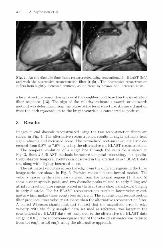
Fig. 4. An end diastolic time frame reconstructed using conventional k-t BLAST (left)and with the alternative reconstruction filter (right). The alternative reconstructionsuffers from slightly increased artifacts, as indicated by arrows, and increased noise.
a local structure tensor description of the neighborhood based on the quadraturefilter responses [13]. The sign of the velocity estimate (inwards or outwardsmotion) was determined from the phase of the local structure. An inward motionfrom the dark myocardium to the bright ventricle is considered as positive.
3 Results
Images in end diastole reconstructed using the two reconstruction filters areshown in Fig. 4. The alternative reconstruction results in slight artifacts fromsignal aliasing and increased noise. The normalized root-mean-square error de-creased from 9.8% to 7.9% by using the alternative k-t BLAST reconstruction.
The temporal evolution of a single line through the ventricle is shown inFig. 3. Both k-t BLAST methods introduce temporal smoothing, but qualita-tively sharper temporal evolution is observed in the alternative k-t BLAST dataset, along with slightly increased noise.
The estimated velocities across the edge from the different regions in the threeimage series are shown in Fig. 5. Positive values indicate inward motion. Thevelocity traces in the reference data set from the normal regions (1, 4 and 5)show a clear systolic peak, and two diastolic peaks related to early filling andatrial contraction. The regions placed in the scar tissue show paradoxical bulgingin early diastole. The k-t BLAST reconstructions result in lower velocity esti-mates which makes these events less apparent. The conventional reconstructionfilter produces lower velocity estimates than the alternative reconstruction filter.A paired Wilcoxon signed rank test showed that the magnitude error in edgevelocity, with the fully sampled data set used as reference, was larger in theconventional k-t BLAST data set compared to the alternative k-t BLAST dataset (p < 0.01). The root-mean-square error of the velocity estimates was reducedfrom 1.4 cm/s to 1.0 cm/s using the alternative approach.

Improving Temporal Fidelity in k-t BLAST MRI Reconstruction 391
1 4 8 12 16 20 24−2
0
2
1 4 8 12 16 20 24−2
0
2
4
1 4 8 12 16 20 24−6
−4
−2
0
2
4
1 4 8 12 16 20 24−8
−6
−4
−2
0
2
4
1 4 8 12 16 20 24
−4
−2
0
2
4
5
41
2
3
Fig. 5. Edge velocities [cm/s] in the different regions over time in the reference im-ages (blue), conventional k-t BLAST reconstructed images (green) and the alternativek-t BLAST reconstructed images (red). Regions 2 and 3 are located in the infarctedarea. The direction of positive velocities is inwards.
4 Discussion
An alternative reconstruction filter for k-t BLAST subsampled data has beenproposed and compared to a conventional filter. Velocities estimated in five re-gions in an infracted left ventricle were less underestimated using the alternativefilter. The alternative reconstruction filter preserves more of the high frequencycontent, at the expense of more artifacts from aliased signal and increased noise.Since the velocity analysis used in this work is based on regional image infor-mation, it is not highly sensitive to a slightly increased aliasing artifact or noiselevel, which do not manifest coherently in adjacent time frames.
Although regional analysis methods are in principle robust to the types ofartifacts introduced, optimal filter parameters have to be determined for specificanalysis strategies. The improved reconstruction filter suggested was designedwith few simple parameters, and optimization of these for other analysis toolsand acquisition parameters such as reduction factor and temporal resolutionmight yield even better results. In the present setting, the reduced noise regu-larization seemed to have largest impact on the temporal fidelity. Other typesof reconstruction filters may also be considered. The choice of reconstructionfilter does not affect the MRI data acquisition, allowing multiple reconstructionstargeted for different analysis tools to be performed from the same raw data.
Optimizing the filter kernel with respect to temporal support [14] was at-tempted, but did not produce significantly improved results in this setting. In

392 A. Sigfridsson et al.
cases with lower reduction factors, however, applying such a filter optimizationmight have a larger effect since the relaxed settings allow shorter filters in thetemporal domain.
In conclusion, the conventional k-t BLAST filter suppresses the high frequencycontent more than necessary for certain applications and better temporal fidelitycan be achieved by merely changing the reconstruction filter. Velocity estimationhas been shown to be significantly improved using a modified reconstructionfilter.
References
1. Kim, R., Fieno, D., Parrish, T., Harris, K., Chen, E., Simonetti, O., Bundy, J.,Finn, J., Klocke, F., Judd, R.: Relationship of MRI delayed contrast enhancementto irreversible injury, infarct age, and contractile function. Circulation 100(19),1992–2002 (1999)
2. Pruessman, K.P., Weiger, M., Scheidegger, M.B., Boesiger, P.: SENSE: sensitivityencoding for fast MRI. Magn. Reson Med. 42, 952–962 (1999)
3. Griswold, M.A., Jakob, P.M., Heidemann, R.M., Nittka, M., Jellus, V., Wang, J.,Kiefer, B., Haase, A.: Generalized autocalibrating partially parallel acquisitions(GRAPPA). Magn. Reson Med. 47, 1202–1210 (2002)
4. Nayak, K.S., Pauly, J.M., Yang, P.C., Hu, B.S., Meyer, C.H., Nishimura, D.G.:Real-time interactive coronary MRA. Magn. Reson Med. 46, 430–435 (2001)
5. van Vaals, J.J., Brummer, M.E., Dixon, W.T., Tuithof, H.H., Engels, H., Nelson, RC., Gerety, B.M., Chezmar, J.L., den Boer, J.A: ”Keyhole” method for acceleratingimaging of contrast agent uptake. J. Magn. Reson. Imaging 3(4), 671–675 (1993)
6. Doyle, M., Walsh, E., Blackwell, G., Pohost, G.: Block regional interpolationscheme for k-space (BRISK): a rapid cardiac imaging technique. Magn. Reson.Med. 33, 163–170 (1995)
7. Korosec, F.R., Frayne, R., Grist, T.M., Mistretta, C.A.: Time-resolved contrast-enhanced 3D MR angiography. Magn. Reson. Med. 36, 345–351 (1996)
8. Madore, B., Glover, G.H., Pelc, N.J.: Unaliasing by Fourier-encoding the overlapsusing the temporal dimension (UNFOLD), applied to cardiac imaging and fMRI.Magn. Reson. Med. 42, 813–828 (1999)
9. Tsao, J., Boesiger, P., Pruessman, K.P.: k-t BLAST and k-t SENSE: DynamicMRI with high frame rate exploiting spatiotemporal correlations. Magn. Reson.Med. 50, 1031–1042 (2003)
10. Mallat, S.: A Wavelet Tour of Signal Processing. Academic Press, San Diego (1999)11. Tsao, J., Kozerke, S., Boesiger, P., Pruessmann, K.P.: Optimizing spatiotemporal
sampling for k−t BLAST and k−t SENSE: Application to high-resolution real-timecardiac steady-state free precession. Magn. Reson. Med. 53, 1372–1382 (2005)
12. Andersson, K., Johansson, P., Forcheimer, R., Knutsson, H.: Backward-forwardmotion compensated prediction. In: Advanced Concepts for Intelligent Vision Sys-tems (ACIVS 2002), Ghent, Belgium, pp. 260–267 (2002)
13. Granlund, G.H., Knutsson, H.: Signal Processing for Computer Vision. KluwerAcademic Publishers, Dordrecht (1995)
14. Knutsson, H., Andersson, M., Wiklund, J.: Advanced filter design. In: SCIA. Pro-ceedings of the 11th Scandinavian Conference on Image Analysis, Greenland (1999)
Related Documents