Improving neurological outcomes post-cardiac arrest in a rat model: immediate hypothermia and quantitative EEG monitoring Xiaofeng Jia, MD, PhD a,* , Matthew A. Koenig, MD b,c , Hyun-Chool Shin, PhD a,e , Gehua Zhen, MD c , Carlos A. Pardo, MD b,d , Daniel F. Hanley, MD b , Nitish V. Thakor, PhD a , and Romergryko G. Geocadin, MD b,c aDepartment of Biomedical Engineering, Johns Hopkins University School of Medicine, Baltimore, MD 21205, USA bDepartment of Neurology, Johns Hopkins University School of Medicine, Baltimore, MD 21205, USA cDepartment of Anesthesiology-Critical Care Medicine, Johns Hopkins University School of Medicine, Baltimore, MD 21205, USA dDepartment of Pathology, Johns Hopkins University School of Medicine, Baltimore, MD 21205, USA eDepartment of Electronic Engineering, Soongsil University, Seoul, Rep. of Korea Summary Objectives—Therapeutic hypothermia (TH) after cardiac arrest (CA) improves outcomes in a fraction of patients. To enhance the administration of TH, we studied brain electrophysiological monitoring in determining the benefit of early initiation of TH compared to conventional administration in a rat model. Methods—Using an asphyxial CA model, we compared the benefit of immediate hypothermia (IH, T=33°C, immediately post-resuscitation, maintained 6 hours) to conventional hypothermia (CH, T=33°C, starting 1 hour post-resuscitation, maintained 12 hours) via surface cooling. We tracked quantitative EEG using relative entropy (qEEG) with outcome verification by serial Neurological Deficit Score (NDS) and quantitative brain histopathological damage scoring (HDS). Thirty-two rats were divided into 4 groups based on CH/IH and 7/9-minute duration of asphyxial CA. Four sham rats were included for evaluation of the effect of hypothermia on qEEG. Results—The 72-hour NDS of the IH group was significantly better than the CH group for both 7- minute (74/63; Median, IH/CH, p<0.001) and 9-minute (54/47, p=0.022) groups. qEEG showed greater recovery with IH (p<0.001) and significantly less neuronal cortical injury by HDS (IH: 18.9 ±2.5% versus CH: 33.2±4.4%, p=0.006). The 1-hour post-resuscitation qEEG correlated well with 72-hour NDS (p<0.05) and 72-hour behavioral subgroup of NDS (p<0.01). No differences in qEEG were noted in the sham group. *Corresponding author: Xiaofeng JIA MD, PhD, CRB II Building 3M-South, 1550 Orleans Street, Johns Hopkins University School of Medicine, Baltimore, MD 21231, USA, Telephone number: +1-410-502-2820, Fax: +1-410-502-7869, E-mail address: [email protected]. Publisher's Disclaimer: This is a PDF file of an unedited manuscript that has been accepted for publication. As a service to our customers we are providing this early version of the manuscript. The manuscript will undergo copyediting, typesetting, and review of the resulting proof before it is published in its final citable form. Please note that during the production process errors may be discovered which could affect the content, and all legal disclaimers that apply to the journal pertain. Portions of this work were previously presented in 4 th Annual Meeting of the Neurocritical Care Society in Baltimore, MD (November 2006) and at the 36 th Annual Meeting of the Society for Neuroscience at Atlanta, Georgia (October 2006) where it was selected for the lay language summary press book. Conflict of interest statement There are no conflicts of interest in this study. NIH Public Access Author Manuscript Resuscitation. Author manuscript; available in PMC 2009 March 1. Published in final edited form as: Resuscitation. 2008 March ; 76(3): 431–442. doi:10.1016/j.resuscitation.2007.08.014. NIH-PA Author Manuscript NIH-PA Author Manuscript NIH-PA Author Manuscript

Welcome message from author
This document is posted to help you gain knowledge. Please leave a comment to let me know what you think about it! Share it to your friends and learn new things together.
Transcript
Improving neurological outcomes post-cardiac arrest in a ratmodel: immediate hypothermia and quantitative EEG monitoring
Xiaofeng Jia, MD, PhDa,*, Matthew A. Koenig, MDb,c, Hyun-Chool Shin, PhDa,e, Gehua Zhen,MDc, Carlos A. Pardo, MDb,d, Daniel F. Hanley, MDb, Nitish V. Thakor, PhDa, and RomergrykoG. Geocadin, MDb,c
aDepartment of Biomedical Engineering, Johns Hopkins University School of Medicine, Baltimore, MD21205, USA
bDepartment of Neurology, Johns Hopkins University School of Medicine, Baltimore, MD 21205, USA
cDepartment of Anesthesiology-Critical Care Medicine, Johns Hopkins University School of Medicine,Baltimore, MD 21205, USA
dDepartment of Pathology, Johns Hopkins University School of Medicine, Baltimore, MD 21205, USA
eDepartment of Electronic Engineering, Soongsil University, Seoul, Rep. of Korea
SummaryObjectives—Therapeutic hypothermia (TH) after cardiac arrest (CA) improves outcomes in afraction of patients. To enhance the administration of TH, we studied brain electrophysiologicalmonitoring in determining the benefit of early initiation of TH compared to conventionaladministration in a rat model.
Methods—Using an asphyxial CA model, we compared the benefit of immediate hypothermia (IH,T=33°C, immediately post-resuscitation, maintained 6 hours) to conventional hypothermia (CH,T=33°C, starting 1 hour post-resuscitation, maintained 12 hours) via surface cooling. We trackedquantitative EEG using relative entropy (qEEG) with outcome verification by serial NeurologicalDeficit Score (NDS) and quantitative brain histopathological damage scoring (HDS). Thirty-two ratswere divided into 4 groups based on CH/IH and 7/9-minute duration of asphyxial CA. Four shamrats were included for evaluation of the effect of hypothermia on qEEG.
Results—The 72-hour NDS of the IH group was significantly better than the CH group for both 7-minute (74/63; Median, IH/CH, p<0.001) and 9-minute (54/47, p=0.022) groups. qEEG showedgreater recovery with IH (p<0.001) and significantly less neuronal cortical injury by HDS (IH: 18.9±2.5% versus CH: 33.2±4.4%, p=0.006). The 1-hour post-resuscitation qEEG correlated well with72-hour NDS (p<0.05) and 72-hour behavioral subgroup of NDS (p<0.01). No differences in qEEGwere noted in the sham group.
*Corresponding author: Xiaofeng JIA MD, PhD, CRB II Building 3M-South, 1550 Orleans Street, Johns Hopkins University School ofMedicine, Baltimore, MD 21231, USA, Telephone number: +1-410-502-2820, Fax: +1-410-502-7869, E-mail address: [email protected]'s Disclaimer: This is a PDF file of an unedited manuscript that has been accepted for publication. As a service to our customerswe are providing this early version of the manuscript. The manuscript will undergo copyediting, typesetting, and review of the resultingproof before it is published in its final citable form. Please note that during the production process errors may be discovered which couldaffect the content, and all legal disclaimers that apply to the journal pertain.Portions of this work were previously presented in 4th Annual Meeting of the Neurocritical Care Society in Baltimore, MD (November2006) and at the 36th Annual Meeting of the Society for Neuroscience at Atlanta, Georgia (October 2006) where it was selected for thelay language summary press book.Conflict of interest statement There are no conflicts of interest in this study.
NIH Public AccessAuthor ManuscriptResuscitation. Author manuscript; available in PMC 2009 March 1.
Published in final edited form as:Resuscitation. 2008 March ; 76(3): 431–442. doi:10.1016/j.resuscitation.2007.08.014.
NIH
-PA Author Manuscript
NIH
-PA Author Manuscript
NIH
-PA Author Manuscript
Conclusions—Immediate but shorter hypothermia compared to CH leads to better functionaloutcome in rats after 7- and 9- minute CA. The beneficial effect of IH was readily detected by neuro-electrophysiological monitoring and histological changes supported the value of this observation.
KeywordsCardiac arrest; Electroencephalography; Hypothermia; Functional outcome; Brain ischemia
IntroductionApproximately 400,000 out-of-hospital cardiac arrests (CA) occur in the United States eachyear1. Despite advances in cardiopulmonary resuscitation (CPR) and critical care, overallsurvival from out-of-hospital CA remains poor, averaging only 5–8% in most centers2. Amongsurvivors, neurological complications represent the leading cause of morbidity anddisability3, 4.
Two recent clinical trials demonstrated that induced hypothermia to 32–34°C improvessurvival and functional outcomes in comatose survivors of CA55,6. While a breakthroughtreatment for brain injury after cardiac arrest, its full beneficial therapeutic effect has not yetbeen fully realized. A meta-analysis showed that the number-needed-to-treat to allow oneadditional patient to leave the hospital with favorable neurological recovery was 4–137. Theoptimal initiation time, duration of therapy, and depth of hypothermia have not been defined.Early initiation of cooling has been suggested but it appears to be successful even if delayedby 4–6 hours8. Most clinical studies delay the initiation of hypothermia by 2 or more hoursafter resuscitation5, 6, 9, 10.
Recent animal studies have shown that mild to moderate hypothermia (33–34°C) mitigatesbrain injury when induced before11, during11, 12, or after resuscitation12–14. Wedemonstrated previously that initiation of hypothermia 1 hour after CA significantly improvedneurological outcomes compared to normothermic controls15, 16. Despite the use ofhypothermia to ameliorate brain injury, no direct monitoring is undertaken to assess the brain’sresponse to therapy. We have previously validated quantitative EEG (qEEG) monitoring in arodent model after resuscitation from CA 17, 18. This entropy-based qEEG provides real-time,objective tracking of neurological injury, recovery and early prognostication after CA17–23.This method is also sensitive to temperature-related modulation of brain injury, with rapid andsustained improvement of qEEG measures in those animals treated with hypothermia comparedto normothermic controls15, 16, 24.
In this study, we tested the hypothesis that neuromonitoring techniques track brain recoveryand response to brain-directed therapy in real time. Clinical translation of these techniques mayoptimize the delivery of hypothermia to a neuro-electrophysiological endpoint. We examinedthe impact of immediate initiation of 6-hour hypothermia (IH) compared to 12-hourconventional hypothermia (CH) initiated at 1 hour post-resuscitation with real-time qEEGtracking followed by functional outcome assessment and histological assessment of the braincortical injury.
Material and MethodsThe experimental protocol was approved by the Johns Hopkins Animal Care and UseCommittee and all procedures were compliant with NIH guidelines.
Jia et al. Page 2
Resuscitation. Author manuscript; available in PMC 2009 March 1.
NIH
-PA Author Manuscript
NIH
-PA Author Manuscript
NIH
-PA Author Manuscript
Experimental asphyxia-CA modelThirty-six adult male Wistar rats (360±20g) were assigned at random to 4 groups based on CH/IH and 7/9-minute duration of asphyxial CA (7CH, 7IH, 9CH, 9IH, n=8). Four rats wereincluded as a sham control group for evaluation of the effect of hypothermia on qEEG in theabsence of CA injury.
Our group and others have used this rodent model to study calibrated brain injury afterasphyxial CA 15, 18, 25. In brief (detail described by Jia15), rats were mechanically ventilatedwith 1.0% halothane in N2/O2 (50%/50%). Five minutes of baseline recording was followedby 5-minute washout to ensure no significant residual effect of halothane on qEEG17. CA wasinitiated via asphyxia with cessation of mechanical ventilation for periods of 7 or 9 minutes.Cardiopulmonary resuscitation (CPR) was performed with sternal chest compressions (200/min) until return of spontaneous circulation (ROSC). Sedative and anesthetic agents wereavoided to minimize confounding effects on EEG26.
CH was induced 1 hour after ROSC by surface cooling with cold mist to achieve the targetcore temperature of 33°C monitored by an intraperitoneal temperature sensor within 15 minutesand was maintained between 32–34°C for 12 hours15. For the IH groups, cooling was initiatedimmediately (within 15 minutes) after ROSC and was maintained at 32–34°C for 6 hours. The6-hour duration was chosen based on a pilot group of rats showing rapid recovery fromunresponsiveness to exploring behavior in IH animals. Then rats were gradually re-warmedfrom 33.0 to 37.0°C over 2 hours using a warming blanket. To ensure no post-resuscitationspontaneous hypothermia27, all animals were then kept inside a neonatal incubator (Isoletteinfant incubator, Air-shields Inc, Pennsylvania) for the first 24 hours post-ROSC.
Continuous EEG was undertaken in four anesthetized sham rats which underwent identicalsurgical preparation and subjected to normothermic baseline for 30 minutes, and then inducedhypothermia for 1 hour.
EEG recording and qEEG analysisWe have used qEEG analysis previously to track brain recovery after CA15, 16, 20–24. EEGswere recorded from baseline through re-warming periods using DI700 Windaq system15.Serial 30-minute EEG recordings were then performed at 24, 48, and 72 hours after ROSC ineach group. In this study we employed entropy to analyze nonstationary EEG signals. Theinformation quantity (IQ) is calculated using a sliding temporal window technique from awindow block of EEG signal for the entire data set15, 16. Sub-band IQ (SIQ, referred belowas qEEG) is the average value of IQ within different frequencies bands. The details of thisanalysis method are provided in the appendix. We chose 9 segments from EEG data in eachrat and calculated qEEG: baseline, CA, 30-min, 1-hour, 4-hour, 6-hour, 24-hour, 48-hour, and72-hour.
Neurological evaluationThe Neurological Deficit Scale (NDS) was patterned after the neurological examination inhumans10 and functional outcome scales for global cerebral ischemia in animals11, 13, 25.The previously validated15, 18 NDS ranges from 80 indicating a functionally normal rat to ascore of 0 for brain or cardiac death prior to conclusion of the study. Behavioral tests includinggait coordination, balance beam walking, righting reflex, negative geotaxis, visual placing andturning alley tests were analyzed as a subgroup of NDS17, 18 (see appendix for details). NDSwas determined by a trained examiner blinded to temperature groups after the re-warmingperiod at 2 hours post-hypothermia on the first day, and then repeated at 24, 48, and 72 hoursafter ROSC. The primary outcome measure was defined as the 72-hour NDS score.
Jia et al. Page 3
Resuscitation. Author manuscript; available in PMC 2009 March 1.
NIH
-PA Author Manuscript
NIH
-PA Author Manuscript
NIH
-PA Author Manuscript
Quantitative brain histopathological damage scoring (HDS)Ten µm paraffin-embedded sections were stained with Cresyl violet28, 29 which was used toquantify ischemic changes using a standard rat brain atlas30 in each hemisphere under ×400magnification with an Olympus BX51 microscope (Olympus, Center Valley, PA). Cresyl violetstaining was chosen based on its reliability and consistency in the evaluation of cytoplasmicand nuclear morphological changes as compared with H&E, and its better suitability forstereological techniques to identify nuclear and nucleolar structures that are required as partof the assessment of cell viability31–34. Ischemic neurons were identified using standardcriteria:29, 35–37 pyknosis, karyorrhexis, karyolysis and cytoplasmic changes in form andcolor.
After de-identifying the histologic slides of clinical data, two investigators, supervised by aneuropathologist, quantified ischemic neurons in a standardized region of temporal cortexlayers 4 and 5 anterior to the rhinal fissure and adjacent to the hippocampus, approximatingthe temporal cortex areas Te1 and Te330, along with CA1 of the hippocampus. These areaswere chosen because CA-1 is selectively vulnerable to global cerebral ischemia in animals andhumans studies and the cerebral cortex is closely related to functional outcome and EEGchanges11, 18, 26, 38, 39. Stereologic technique was performed in the predefined set ofrandom, non-overlapping microscope fields using Adreas Stereo Investigator software 5.05.4(MicroBrightField, Williston, VT) to estimate the degree of neuronal injury. In addition toischemic neurons, normal-appearing neurons were counted in each field. These data were usedto calculate a percentage of injury ([injured cells / total number of counted cells] × 100)representing the degree of necrotic cell death per region35.
StatisticsUnivariate analysis was performed for parametric data with the use of the Student’s t-test forcontinuous variables and the chi-square test for categorical variables. Non-parametric analysisof variance was used to test for differences in rank order NDS as a repeated measure.Multivariate General Linear Model was used for advanced comparison of aggregate data tocontrol for influencing factors such as hypothermia method and asphyxia time. The mortalityrate was analyzed by Fisher’s exact test (crosstabs) and survival was analyzed by a Kaplan-Meier test. Pearson correlation of bivariate analysis was used to analyze the correlation between72-hour NDS score with serial qEEG and HDS. Statistical significance was set at p<0.05.Statistical analysis was performed with SPSS 14.0 (SPSS Inc., Chicago, IL).
ResultsBaseline characteristics, temperature monitoring and ABG Data
There are no significant difference between groups of the baseline characteristics includingbody weights, anesthesia duration for preparation, heart rates and blood pressure (p>0.05). Thetemperature recording for 7- and 9-minute groups during the hypothermia experiment is shownin Figure 1. Cooling to target temperature was achieved in 11.1±4.6 (Mean±SEM) minutes andre-warming in 116.8±21.4 minutes to reach the target range of 36.5–37.5 °C. All animals weremaintained at 37±0.5 °C after re-warming during the first 24 hours.
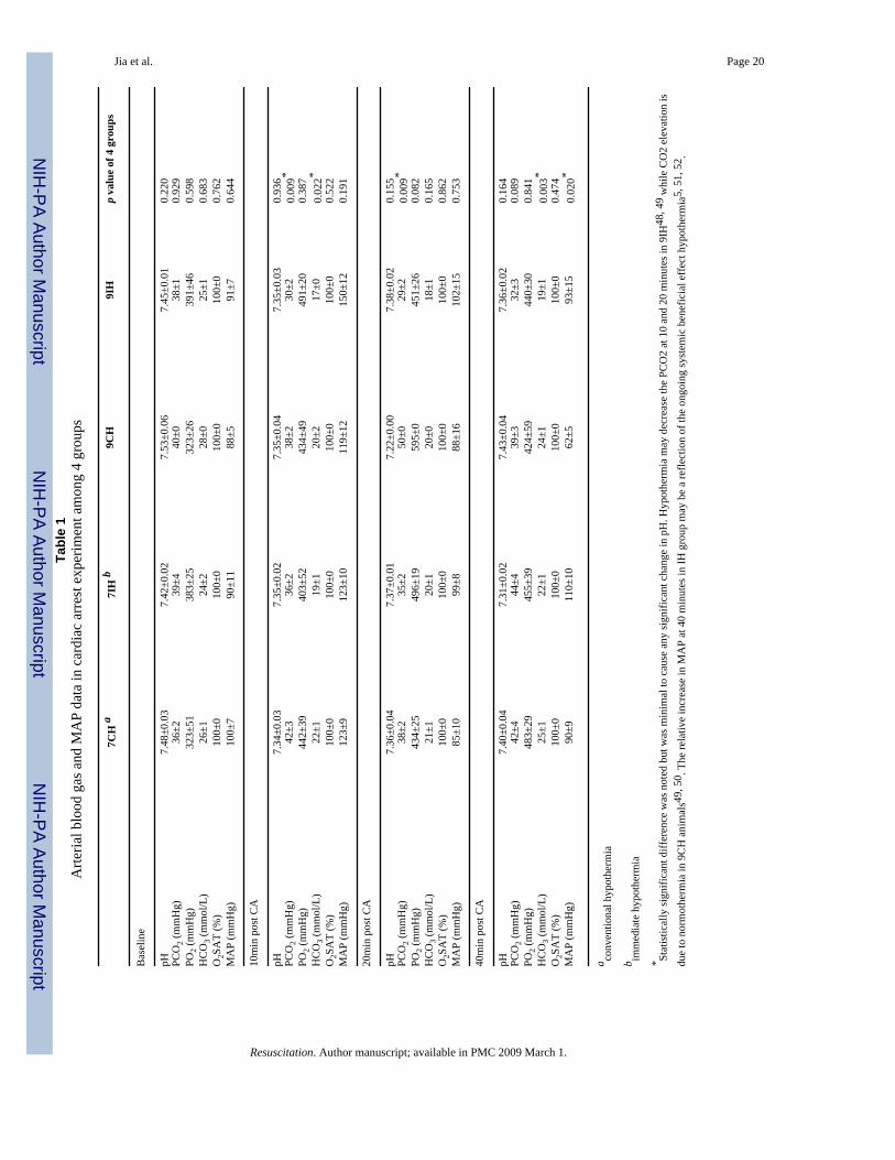
ABG data including arterial pH, HCO3−, PO2 and O2 saturation, were similar between groups
at all time points (baseline, 10, 20, and 40 minutes after ROSC). There are significantdifferences of PCO2 at 10 minutes (p<0.01) and 20 minutes (p<0.01) and of blood pressure at40 minutes (p<0.05) existed between groups (Data shown in table 1).
Jia et al. Page 4
Resuscitation. Author manuscript; available in PMC 2009 March 1.
NIH
-PA Author Manuscript
NIH
-PA Author Manuscript
NIH
-PA Author Manuscript
EEG-bursting analysis and qEEG analysisWithin seconds of CA, the normal EEG signal was reduced to an isoelectric tracing, whichwas followed by periodic activity resembling burst-suppression of variable duration. Theinterval between CA and the first burst in the 4 groups was 16.3±0.7 (7CH), 20.3±1.7 (9CH),16.3±1.2 (7IH) and 19.6±2.8 (9IH) minutes respectively, where CA refers to the starting periodof asphyxia and the interval from CA to the first burst is the duration of isoelectric EEG.
There was a statistically significant difference between 7-minute CA (16.3±0.7) and 9-minuteCA groups (20.0±0.9) (p=0.004) while no significant difference existed between IH and CHrats. No significant difference was observed in qEEG of sham rats between the periods ofhypothermia (0.59±0.02) and normothermia (0.60±0.02) (p=0.921).
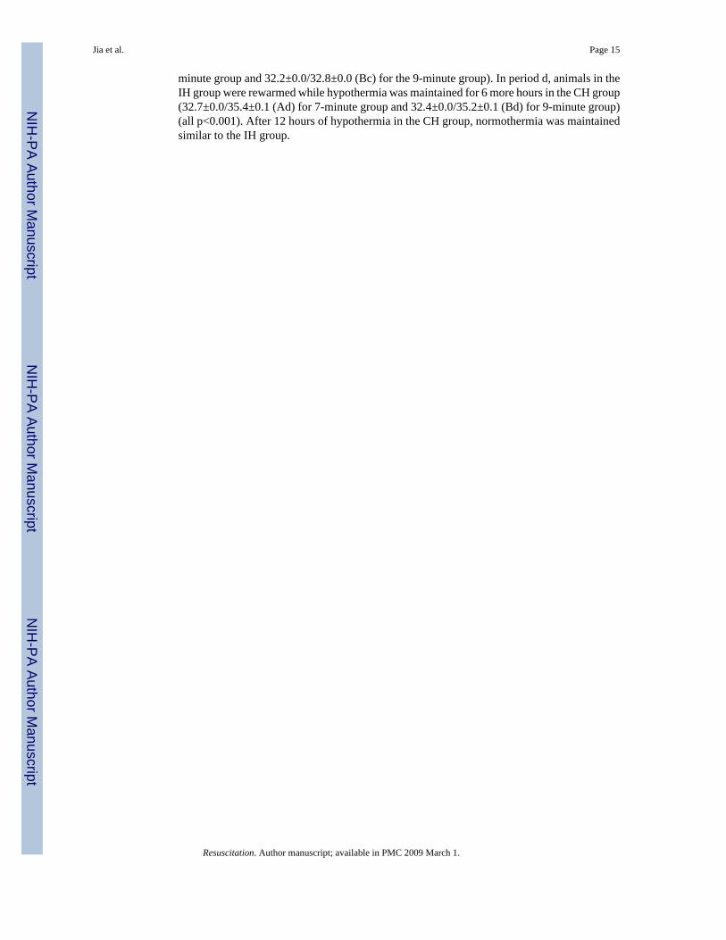
Using visual assessment by comparing the evolution of burst frequency and amplitude frombaseline of the raw EEG signal alone, qualitative differences between the groups were notevident (Figure 2 A, B). Subtle differences in EEG, however, were readily discerned usingqEEG analysis (Figure 2 C). Similar to the raw EEG data, the aggregate qEEG decreased frombaseline (qEEG=1) to the lowest point rapidly after CA, then gradually recovered close tobaseline.
Mean qEEG relative entropy values over the study period were not different (p=0.08) between7IH rats (0.78±0.03) and 7CH rats (0.75±0.04); while 9IH rats (0.73±0.03) had significantlygreater mean qEEG entropy than 9CH rats (0.63±0.04) (p<0.001) (Figure 3 A, B). Aggregateanalysis showed mean qEEG entropy was greater in the IH rats (0.76±0.02) compared to CHcontrols (0.70±0.03) over the 72-hour study period (p<0.001).
The predictive capacity of early qEEG for 72-hour NDS by bivariate analyses, using aggregateqEEG and NDS data (n=28), revealed significant correlations between 72-hour NDS and qEEGvalues showing statistical significance at 1, 4, 6 and 24 hours (p<0.05). The value of earlyqEEG to predict recovery of behavioral and cognitive function, evaluated by comparing qEEGagainst the behavioral subgroup of NDS, showed a significant correlation at 1 and 4 hours( p<0.05).
Functional Recovery by NDSSurvival NDS analysis results are shown in Figure 4. IH animals had persistently betterfunctional recovery by NDS scores at all time periods with statistical significance in the 7-minute groups (IH/CH (Median, 25th–75th percentile): 74, 60.75–74/63, 50.25–70; p=0.001)and 9-minute groups (IH/CH: 54, 49–61.75/47, 39–60; p=0.022). Aggregate analysis showedstatistically significant differences (p=0.001) between CH group (54, 42–66) and IH group(61.5, 51.25–74).
Mortality and qEEGThe mortality rates were 0% (0/8) in 7IH, 12.5% (1/8) in 7CH, 12.5% (1/8) in 9IH, and 25%(2/8) in 9CH group, with an odds ratio (OR) of mortality in the CH group of 3.667 (95%CI:0.3–42.9). No statistically significant differences in mean duration of survival hours existedbetween the 7IH (72.0 hours) and 7CH (63.8 hours) groups or between the 9IH (67.5 hours)and 9CH (59.0 hours) groups. Compared to survivors, rats that prematurely died within 72hours after ROSC had lower qEEG values at 6 hours (dead/survivors: 0.55±0.10/0.78±0.02,p=0.002) and 24 hours (dead/survivor: 0.71±0.06/0.86±0.02, p<0.05).
Quantitative brain histopathological damage scoring (HDS)There were more ischemic neurons in cortex in the CH group compared to the IH group after9-minute CA (p=0.04) while this difference was not noted between the 7-minute CA groups.
Jia et al. Page 5
Resuscitation. Author manuscript; available in PMC 2009 March 1.
NIH
-PA Author Manuscript
NIH
-PA Author Manuscript
NIH
-PA Author Manuscript
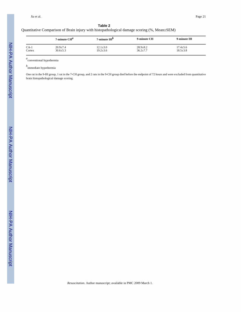
While significant neuronal injury was noted in CA-1 of the hippocampus as expected with thisinjury model, the HDS was not different in IH and CH groups subjected to 7- and 9-minuteCA (Table 2 and Figure 5). Aggregate analysis showed HDS was significantly higher in thecortex in the CH group (33.2±4.4%, Mean±SEM) compared to the IH group (18.9±2.5%)(p=0.006). No statistically significant of injury in CA1 in IH animals (14.4±2.9%) than CHcontrols (24.6±5.4%) (p=0.112).
Bivariate analyses revealed significant correlations between 72-hour NDS and HDS of cortex(p<0.05) as well as CA1 (p<0.01). HDS of cortex correlated well with HDS of CA1 (p<0.01).
DiscussionOur study shows that qEEG analysis methods readily tracked and differentiated the enhancedbrain recovery provided by the IH over the CH treatment groups. We also noted that the earlieradministration of therapeutic hypothermia after CA not only leads to better functional outcomecompared to conventional hypothermia administration, but allowed for reduction of treatmentduration by half (6 hours vs. 12 hours). We observed that the better qEEG recovery of the IHgroup, which was established during 30 minutes to 4 hours after ROSC, was validated by bothNDS and HDS (cortex) at 72 hours. These findings showed that the beneficial effects ofhypothermia optimization on the brain can be tracked using an advanced but easily interpretablemonitoring technique.
While the beneficial effect of therapeutic hypothermia has been a breakthrough in the care ofCA patients, its real time effect on brain recovery remains unclear. With the InternationalLiaison Committee on Resuscitation (ILCOR)8 and the American Heart Association40recommendation for the use of hypothermia in appropriate patients, the absolute mortalitybenefit was 16% in both studies, with a number needed to treat of 6 in order to save 1 life5,6. This therapy therefore has the potential to benefit more patients if better understood anddelivered appropriately. The findings of this study seek to address 2 issues: a) to respond tothe call of the National Heart Lung and Blood Institute (NHLBI)-Post-resuscitative and initialUtility in Life Saving Efforts (PULSE) initiative to prioritize efforts to improve and monitorneurological recovery after CA41 and b) the challenge to improve outcomes further with earlieryet shorter hypothermia delivery.
Previous experiments of delayed induction of hypothermia (by 1 hour post-ischemia) after CAhave demonstrated improvements in functional outcome and histological markers of injury inrats treated with hypothermia compared to normothermic controls14, 42, 43, which wasconsistent with our previous findings15. Separate work by Hicks et al. 44 comparedhistological and functional outcomes between immediate hypothermia and hypothermiadelayed by an hour using an experimental design similar to the present manuscript. Somedifferences in technique, methods and statistical powering may account for the difference infindings between the Hicks study and ours. While the paper by Hicks et al. did not specificallycompare the duration of hypothermia as we did, the potential advantage of earlieradministration was suggested with the immediate hypothermia group showing a trend towardbetter outcomes in the NDS, histological score, and heat shock protein levels. Our experimentlends further support to the theory that cooling should begin as soon as possible after ROSC,similar to other animal models12, 13, 45.
With electrophysiological markers, this study suggests that earlier initiation of hypothermiamay have a greater impact during the early period of recovery and injured neurons that areimmediately treated have a better chance of recovering. These observations, as supported byqEEG and histology, preclude the need for longer treatment duration and suggest the need tore-evaluate the timing of hypothermia initiation in human subjects. Given the risks of
Jia et al. Page 6
Resuscitation. Author manuscript; available in PMC 2009 March 1.
NIH
-PA Author Manuscript
NIH
-PA Author Manuscript
NIH
-PA Author Manuscript
coagulopathy and immune suppression, need for sedation and paralysis, expense, and highresource use associated with hypothermia5, 6, 9, 10, these results are potentially important.
As human studies have shown, with this degree of hypothermia (33±1°C), the detrimentaleffects have not been significantly different than normothermia. Given the identicaltemperature range in IH and CH, we did not expect significant differences in detrimentaleffects, so we did not look at the detrimental effects in much detail. However, considering thecommonly reported detrimental side effects, we observed no significant hemorrhage, seizureactivity, or arrhythmias. We did not look specifically for pneumonia or renal failure. Theenhanced recovery of shorter IH over longer CH is most likely the result of rapid onset ratherthan a decrement in adverse effects.
Translational research verification may justify changing resuscitation strategies such thatparamedics begin cooling in the field or ambulance en route to the hospital. The cost of longerperiods of hypothermia centers on need for intensive nursing care. And the cost of rapidinductions may be dependent on technologies; however chilled saline infusion is definitely aneconomical and effective way to achieve rapid hypothermia induction46.
While functional outcome by NDS was our primary outcome measure and qEEG was a trackingtool, we also employed histology for additional verification of outcomes. We acknowledgethat previous consensus47 showed that histological outcomes do not readily translate intoclinical outcomes, as reflected in the 7-minute CA group. The significant neuronal injuryobserved in CA-1 of the hippocampus reflects the high susceptibility of this area to globalischemia, but its lack of direct influence on arousal and cortical activity may account for thelack of concurrence with EEG findings. Histological injury in the cortex, a significant site forEEG generation and modulation, demonstrated stronger correlation with qEEG differences.The minor difference between qEEG in the 7IH and 7CH groups despite NDS differences maybe due to the fact that 7 minutes is a minimal injury for cortex (represented by EEG), despitesignificant subcortical injury (reflected in NDS).
As part of our protocol15, we attempted to normalize the ABG variables, especially pH to limitthe injury primarily to CA. In order to achieve this, we adjusted ventilator rates as needed17.At 10 minutes post-CA, all rats showed a decline in pH from the asphyxial cardiac arrest. Thesevalues however, are a result of the 10-minute post-ROSC hyperventilation period (tidalvolumes 10 ml/kg, respiration rate 65/min and positive expiratory end pressure 6 cm H2O) thatwe employed for all animals in this model15. Although, at some point, 9CH group appears tohave a larger metabolic acidosis compared to 9IH, there were no statistically significantdifferences noted in pH. Hypothermia has not been started for the 9CH group at 20 minuteswhile hypothermia has been reached in 9IH group (average cooling time to target is 11.1minutes). Hypothermia may decrease the PCO2 in 9IH while CO2 elevation is due tonormothermia in 9CH animals, which is consistent with other recent publications48–50. By40 minutes, hypothermia has been ongoing in the IH group for ~30 minutes while it has notbeen started in CH group. The relative increase in MAP is probably a reflection of the ongoingsystemic beneficial effects of hypothermia and may be due to an increase in systemic vascularresistence5, 51, 52.
From a neuromonitoring perspective, this study highlights the importance of the immediatepost-resuscitation period when brain injury may be most amendable to therapeuticinterventions. We have shown previously that qEEG analysis detected the therapeutic benefitof conventional hypothermia compared to normothermic controls15, 16. This observation iscarried over into the present study. Additionally, we also showed that it is not the temperature(32–34°C) itself that causes the change in EEG but the response of the injured brain tohypothermia as manifested in the qEEG.
Jia et al. Page 7
Resuscitation. Author manuscript; available in PMC 2009 March 1.
NIH
-PA Author Manuscript
NIH
-PA Author Manuscript
NIH
-PA Author Manuscript
As in our previous studies, we observed that animals that proceed more quickly from a flatEEG to a continuous EEG pattern and have higher qEEG values achieved better functionaloutcomes. We have reported previously the rapid EEG response within 10 to 90 minutes18,19, 38 and this reflects the ability of EEG to be a sensitive real-time measure of injury andrecovery. The qEEG as a reflection of brain injury was also further crystallized by the abilityof our algorithms to prognosticate functional outcomes and mortality within the first few hoursafter resuscitation. Using aggregate data that included the entire animal population, the qEEGmeasure was able to accurately predict neurological outcome defined by the NDS andcognitive-behavioral tests as early as 1–4 hours after ROSC, a time in which the animal remainscomatose. As a measure of coma recovery, the NDS is weighted toward brain stem function,which is highly preserved in all but the most severe global ischemic injury53. The principleimprovement in animals with higher qEEG, however, was seen in more advanced behavioraland coordination tests, such as balance beam walking, gait coordination, and righting reflexes.These functions, especially the neuro-behavioral assessment, may require more focused studyto fully document the long-term effects of hypothermia on these animals.
While the manual interpretation of continuous raw EEG is laborious, subjective, and requiresspecialized experience, entropy-based qEEG can be readily used to track brain recovery. Ourresults suggest that early qEEG monitoring may assist clinicians in tracking recovery after CAand the therapeutic response to hypothermia. The development of accurate neuromonitoringtechniques during hypothermia is particularly important for the evaluation of therapeuticresponse, because clinical neurological assessment is obscured by sedative and paralyticmedications. As a continuous, real-time, and non-invasive methodology, qEEG monitors theresponse to potential neuroprotective strategies by translating complicated and subjectivewaveform analysis into an objective measure. Similar use of qEEG analysis has beensuccessfully incorporated in hypothermia treatment in neonates with global brainischemia54.
ConclusionsIn conclusion, our qEEG analysis method is able to detect the brain’s response to therapeuticbenefits of hypothermia, and it is able to predict recovery of arousal, functional outcome, andsurvival. The neurological recovery appears to be better under immediate induction, but shorterduration, of hypothermia after resuscitation. These experiments have the potential to developbrain monitoring and guide the optimum effect of therapeutic hypothermia.
Supplementary MaterialRefer to Web version on PubMed Central for supplementary material.
AcknowledgmentsThis research was supported by NIH Grants R01 HL071568 and R21 NS054146.
Appendix
The NDS and its components can be found in Table 1Table 1
NEURODEFICIT SCORING FOR RATS (Normal = 80; Brain dead = 0)A)General Behavioral deficit Total Score : 19Consciousness Normal 10/ Stuporous 5 / Comatose or unresponsive 0Arousal: Eyes open spontaneously 3/ Eyes open to pain 1/ No Eye Opening 0Respiration: Normal 6/ Abnormal (hypo or hyperventilation) 3/ Absent 0B) Brain-stem Function: Total Score : 21
Jia et al. Page 8
Resuscitation. Author manuscript; available in PMC 2009 March 1.
NIH
-PA Author Manuscript
NIH
-PA Author Manuscript
NIH
-PA Author Manuscript
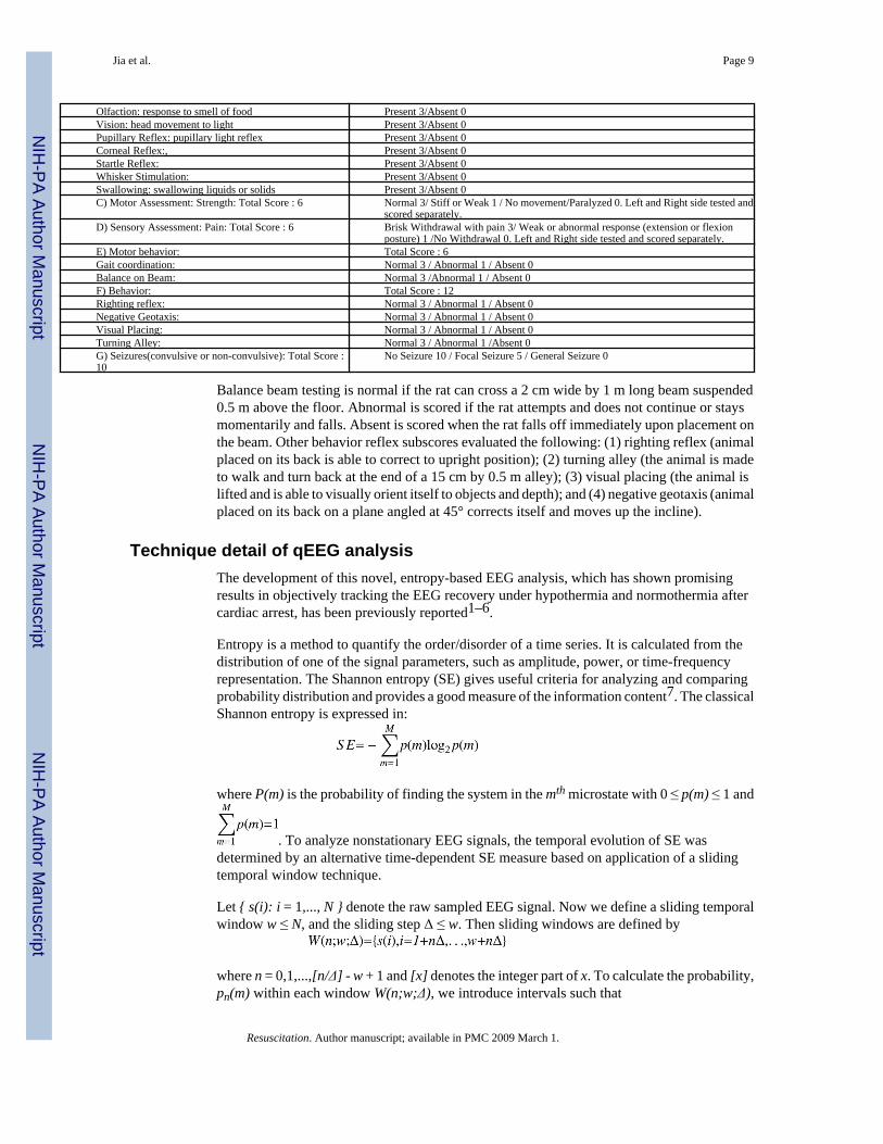
Olfaction: response to smell of food Present 3/Absent 0Vision: head movement to light Present 3/Absent 0Pupillary Reflex: pupillary light reflex Present 3/Absent 0Corneal Reflex:, Present 3/Absent 0Startle Reflex: Present 3/Absent 0Whisker Stimulation: Present 3/Absent 0Swallowing: swallowing liquids or solids Present 3/Absent 0C) Motor Assessment: Strength: Total Score : 6 Normal 3/ Stiff or Weak 1 / No movement/Paralyzed 0. Left and Right side tested and
scored separately.D) Sensory Assessment: Pain: Total Score : 6 Brisk Withdrawal with pain 3/ Weak or abnormal response (extension or flexion
posture) 1 /No Withdrawal 0. Left and Right side tested and scored separately.E) Motor behavior: Total Score : 6Gait coordination: Normal 3 / Abnormal 1 / Absent 0Balance on Beam: Normal 3 /Abnormal 1 / Absent 0F) Behavior: Total Score : 12Righting reflex: Normal 3 / Abnormal 1 / Absent 0Negative Geotaxis: Normal 3 / Abnormal 1 / Absent 0Visual Placing: Normal 3 / Abnormal 1 / Absent 0Turning Alley: Normal 3 / Abnormal 1 /Absent 0G) Seizures(convulsive or non-convulsive): Total Score :10
No Seizure 10 / Focal Seizure 5 / General Seizure 0
Balance beam testing is normal if the rat can cross a 2 cm wide by 1 m long beam suspended0.5 m above the floor. Abnormal is scored if the rat attempts and does not continue or staysmomentarily and falls. Absent is scored when the rat falls off immediately upon placement onthe beam. Other behavior reflex subscores evaluated the following: (1) righting reflex (animalplaced on its back is able to correct to upright position); (2) turning alley (the animal is madeto walk and turn back at the end of a 15 cm by 0.5 m alley); (3) visual placing (the animal islifted and is able to visually orient itself to objects and depth); and (4) negative geotaxis (animalplaced on its back on a plane angled at 45° corrects itself and moves up the incline).
Technique detail of qEEG analysisThe development of this novel, entropy-based EEG analysis, which has shown promisingresults in objectively tracking the EEG recovery under hypothermia and normothermia aftercardiac arrest, has been previously reported1–6.
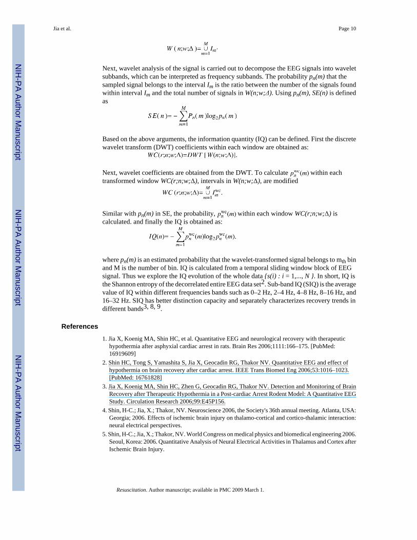
Entropy is a method to quantify the order/disorder of a time series. It is calculated from thedistribution of one of the signal parameters, such as amplitude, power, or time-frequencyrepresentation. The Shannon entropy (SE) gives useful criteria for analyzing and comparingprobability distribution and provides a good measure of the information content7. The classicalShannon entropy is expressed in:
where P(m) is the probability of finding the system in the mth microstate with 0 ≤ p(m) ≤ 1 and
. To analyze nonstationary EEG signals, the temporal evolution of SE wasdetermined by an alternative time-dependent SE measure based on application of a slidingtemporal window technique.
Let { s(i): i = 1,..., N } denote the raw sampled EEG signal. Now we define a sliding temporalwindow w ≤ N, and the sliding step Δ ≤ w. Then sliding windows are defined by
where n = 0,1,...,[n/Δ] - w + 1 and [x] denotes the integer part of x. To calculate the probability,pn(m) within each window W(n;w;Δ), we introduce intervals such that
Jia et al. Page 9
Resuscitation. Author manuscript; available in PMC 2009 March 1.
NIH
-PA Author Manuscript
NIH
-PA Author Manuscript
NIH
-PA Author Manuscript
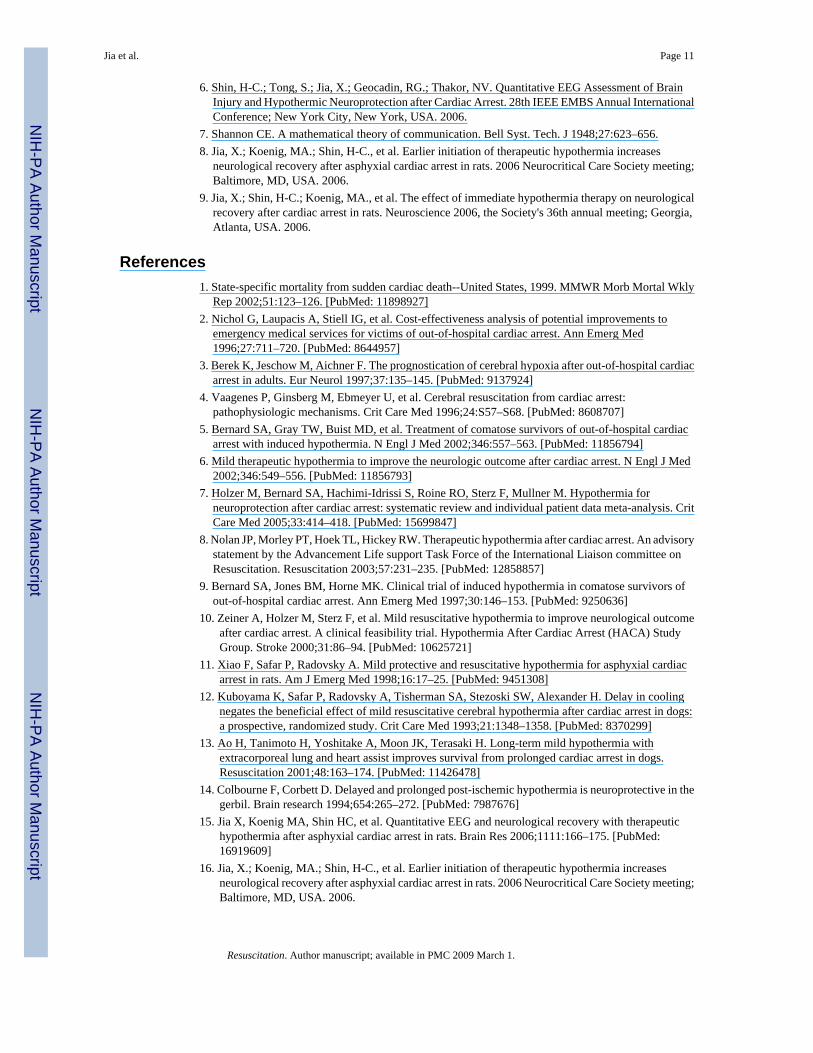
Next, wavelet analysis of the signal is carried out to decompose the EEG signals into waveletsubbands, which can be interpreted as frequency subbands. The probability pn(m) that thesampled signal belongs to the interval Im is the ratio between the number of the signals foundwithin interval Im and the total number of signals in W(n;w;Δ). Using pn(m), SE(n) is definedas
Based on the above arguments, the information quantity (IQ) can be defined. First the discretewavelet transform (DWT) coefficients within each window are obtained as:
Next, wavelet coefficients are obtained from the DWT. To calculate within eachtransformed window WC(r;n;w;Δ), intervals in W(n;w;Δ), are modified
Similar with pn(m) in SE, the probability, within each window WC(r;n;w;Δ) iscalculated. and finally the IQ is obtained as:
where pn(m) is an estimated probability that the wavelet-transformed signal belongs to mth binand M is the number of bin. IQ is calculated from a temporal sliding window block of EEGsignal. Thus we explore the IQ evolution of the whole data {s(i) : i = 1,..., N }. In short, IQ isthe Shannon entropy of the decorrelated entire EEG data set2. Sub-band IQ (SIQ) is the averagevalue of IQ within different frequencies bands such as 0–2 Hz, 2–4 Hz, 4–8 Hz, 8–16 Hz, and16–32 Hz. SIQ has better distinction capacity and separately characterizes recovery trends indifferent bands3, 8, 9.
References1. Jia X, Koenig MA, Shin HC, et al. Quantitative EEG and neurological recovery with therapeutic
hypothermia after asphyxial cardiac arrest in rats. Brain Res 2006;1111:166–175. [PubMed:16919609]
2. Shin HC, Tong S, Yamashita S, Jia X, Geocadin RG, Thakor NV. Quantitative EEG and effect ofhypothermia on brain recovery after cardiac arrest. IEEE Trans Biomed Eng 2006;53:1016–1023.[PubMed: 16761828]
3. Jia X, Koenig MA, Shin HC, Zhen G, Geocadin RG, Thakor NV. Detection and Monitoring of BrainRecovery after Therapeutic Hypothermia in a Post-cardiac Arrest Rodent Model: A Quantitative EEGStudy. Circulation Research 2006;99:E45P156.
4. Shin, H-C.; Jia, X.; Thakor, NV. Neuroscience 2006, the Society's 36th annual meeting. Atlanta, USA:Georgia; 2006. Effects of ischemic brain injury on thalamo-cortical and cortico-thalamic interaction:neural electrical perspectives.
5. Shin, H-C.; Jia, X.; Thakor, NV. World Congress on medical physics and biomedical engineering 2006.Seoul, Korea: 2006. Quantitative Analysis of Neural Electrical Activities in Thalamus and Cortex afterIschemic Brain Injury.
Jia et al. Page 10
Resuscitation. Author manuscript; available in PMC 2009 March 1.
NIH
-PA Author Manuscript
NIH
-PA Author Manuscript
NIH
-PA Author Manuscript
6. Shin, H-C.; Tong, S.; Jia, X.; Geocadin, RG.; Thakor, NV. Quantitative EEG Assessment of BrainInjury and Hypothermic Neuroprotection after Cardiac Arrest. 28th IEEE EMBS Annual InternationalConference; New York City, New York, USA. 2006.
7. Shannon CE. A mathematical theory of communication. Bell Syst. Tech. J 1948;27:623–656.8. Jia, X.; Koenig, MA.; Shin, H-C., et al. Earlier initiation of therapeutic hypothermia increases
neurological recovery after asphyxial cardiac arrest in rats. 2006 Neurocritical Care Society meeting;Baltimore, MD, USA. 2006.
9. Jia, X.; Shin, H-C.; Koenig, MA., et al. The effect of immediate hypothermia therapy on neurologicalrecovery after cardiac arrest in rats. Neuroscience 2006, the Society's 36th annual meeting; Georgia,Atlanta, USA. 2006.
References1. State-specific mortality from sudden cardiac death--United States, 1999. MMWR Morb Mortal Wkly
Rep 2002;51:123–126. [PubMed: 11898927]2. Nichol G, Laupacis A, Stiell IG, et al. Cost-effectiveness analysis of potential improvements to
emergency medical services for victims of out-of-hospital cardiac arrest. Ann Emerg Med1996;27:711–720. [PubMed: 8644957]
3. Berek K, Jeschow M, Aichner F. The prognostication of cerebral hypoxia after out-of-hospital cardiacarrest in adults. Eur Neurol 1997;37:135–145. [PubMed: 9137924]
4. Vaagenes P, Ginsberg M, Ebmeyer U, et al. Cerebral resuscitation from cardiac arrest:pathophysiologic mechanisms. Crit Care Med 1996;24:S57–S68. [PubMed: 8608707]
5. Bernard SA, Gray TW, Buist MD, et al. Treatment of comatose survivors of out-of-hospital cardiacarrest with induced hypothermia. N Engl J Med 2002;346:557–563. [PubMed: 11856794]
6. Mild therapeutic hypothermia to improve the neurologic outcome after cardiac arrest. N Engl J Med2002;346:549–556. [PubMed: 11856793]
7. Holzer M, Bernard SA, Hachimi-Idrissi S, Roine RO, Sterz F, Mullner M. Hypothermia forneuroprotection after cardiac arrest: systematic review and individual patient data meta-analysis. CritCare Med 2005;33:414–418. [PubMed: 15699847]
8. Nolan JP, Morley PT, Hoek TL, Hickey RW. Therapeutic hypothermia after cardiac arrest. An advisorystatement by the Advancement Life support Task Force of the International Liaison committee onResuscitation. Resuscitation 2003;57:231–235. [PubMed: 12858857]
9. Bernard SA, Jones BM, Horne MK. Clinical trial of induced hypothermia in comatose survivors ofout-of-hospital cardiac arrest. Ann Emerg Med 1997;30:146–153. [PubMed: 9250636]
10. Zeiner A, Holzer M, Sterz F, et al. Mild resuscitative hypothermia to improve neurological outcomeafter cardiac arrest. A clinical feasibility trial. Hypothermia After Cardiac Arrest (HACA) StudyGroup. Stroke 2000;31:86–94. [PubMed: 10625721]
11. Xiao F, Safar P, Radovsky A. Mild protective and resuscitative hypothermia for asphyxial cardiacarrest in rats. Am J Emerg Med 1998;16:17–25. [PubMed: 9451308]
12. Kuboyama K, Safar P, Radovsky A, Tisherman SA, Stezoski SW, Alexander H. Delay in coolingnegates the beneficial effect of mild resuscitative cerebral hypothermia after cardiac arrest in dogs:a prospective, randomized study. Crit Care Med 1993;21:1348–1358. [PubMed: 8370299]
13. Ao H, Tanimoto H, Yoshitake A, Moon JK, Terasaki H. Long-term mild hypothermia withextracorporeal lung and heart assist improves survival from prolonged cardiac arrest in dogs.Resuscitation 2001;48:163–174. [PubMed: 11426478]
14. Colbourne F, Corbett D. Delayed and prolonged post-ischemic hypothermia is neuroprotective in thegerbil. Brain research 1994;654:265–272. [PubMed: 7987676]
15. Jia X, Koenig MA, Shin HC, et al. Quantitative EEG and neurological recovery with therapeutichypothermia after asphyxial cardiac arrest in rats. Brain Res 2006;1111:166–175. [PubMed:16919609]
16. Jia, X.; Koenig, MA.; Shin, H-C., et al. Earlier initiation of therapeutic hypothermia increasesneurological recovery after asphyxial cardiac arrest in rats. 2006 Neurocritical Care Society meeting;Baltimore, MD, USA. 2006.
Jia et al. Page 11
Resuscitation. Author manuscript; available in PMC 2009 March 1.
NIH
-PA Author Manuscript
NIH
-PA Author Manuscript
NIH
-PA Author Manuscript
17. Geocadin RG, Ghodadra R, Kimura T, et al. A novel quantitative EEG injury measure of globalcerebral ischemia. Clin Neurophysiol 2000;111:1779–1787. [PubMed: 11018492]
18. Geocadin RG, Muthuswamy J, Sherman DL, Thakor NV, Hanley DF. Early electrophysiological andhistologic changes after global cerebral ischemia in rats. Mov Disord 2000;15:14–21. [PubMed:10755267]
19. Geocadin RG, Sherman DL, Christian Hansen H, et al. Neurological recovery by EEG bursting afterresuscitation from cardiac arrest in rats. Resuscitation 2002;55:193–200. [PubMed: 12413758]
20. Bezerianos A, Tong S, Thakor N. Time-dependent entropy estimation of EEG rhythm changesfollowing brain ischemia. Ann Biomed Eng 2003;31:221–232. [PubMed: 12627829]
21. Thakor NV, Tong S. Advances in quantitative electroencephalogram analysis methods. Annu RevBiomed Eng 2004;6:453–495. [PubMed: 15255777]
22. Tong S, Bezerianos A, Malhotra A, Zhu Y, Thakor NV. Parameterized entropy analysis of EEGfollowing hypoxic–ischemic brain injury. Physics Letters A 2003;314:354–361.
23. Tong S, Bezerianos A, Paul J, Zhu Y, Thakor NV. Nonextensive entropy measure of EEG followingbrain injury from cardiac arrest. Physica A: Statistical Mechanics and its Applications 2002;305:619–628.
24. Jia X, Koenig MA, Shin HC, Zhen G, Geocadin RG, Thakor NV. Detection and Monitoring of BrainRecovery after Therapeutic Hypothermia in a Post-cardiac Arrest Rodent Model: A Quantitative EEGStudy. Circulation Research 2006;99:E45P156.
25. Katz L, Ebmeyer U, Safar P, Radovsky A, Neumar R. Outcome model of asphyxial cardiac arrest inrats. J Cereb Blood Flow Metab 1995;15:1032–1039. [PubMed: 7593335]
26. Luft AR, Buitrago MM, Paul JS, et al. Early restitution of electrocorticogram predicts subsequentbehavioral recovery from cardiac arrest. J Clin Neurophysiol 2002;19:540–546. [PubMed:12488785]
27. Hickey RW, Kochanek PM, Ferimer H, Alexander HI, Garman RH, Graham SH. Inducedhyperthermia exacerbates neurologic neuronal histologic damage after asphyxial cardiac arrest inrats. Crit Care Med 2003;31:531–535. [PubMed: 12576962]
28. Derugin N, Wendland M, Muramatsu K, et al. Evolution of brain injury after transient middle cerebralartery occlusion in neonatal rats. Stroke 2000;31:1752–1761. [PubMed: 10884483]
29. Garcia JH, Liu KF, Ho KI. Neuronal necrosis after middle cerebral artery occlusion in Wistar ratsprogresses at different time intervals in the caudoputamen and the cortex. Stroke 1995;26:636–642.[PubMed: 7709411]discussion 643
30. Paxinos, G.; Watson, C. The rat brain in stereotaxic coordinates. Second ed.. San Diego, California:Academic Press; 1986.
31. Hattori K, Lee H, Hurn PD, Crain BJ, Traystman RJ, DeVries AC. Cognitive deficits after focalcerebral ischemia in mice. Stroke 2000;31:1939–1944. [PubMed: 10926961]
32. Schroeter M, Jander S, Stoll G. Non-invasive induction of focal cerebral ischemia in mice byphotothrombosis of cortical microvessels: characterization of inflammatory responses. J NeurosciMethods 2002;117:43–49. [PubMed: 12084563]
33. Kilic E, Kilic U, Bassetti CL, Hermann DM. Intravenously administered recombinant tissue-plasminogen activator attenuates neuronal injury after mild focal cerebral ischemia in mice.Neuroreport 2004;15:687–689. [PubMed: 15094476]
34. Mathews VP, Monsein LH, Pardo CA, Bryan RN. Histologic abnormalities associated withgadolinium enhancement on MR in the initial hours of experimental cerebral infarction. AJNR. AmJ Neuroradiol 1994;15:573–579. [PubMed: 8197960]
35. Buitrago MM, Luft AR, Thakor NV, Blue ME, Hanley DF. Effects of somatosensory electricalstimulation on neuronal injury after global hypoxia-ischemia. Exp Brain Res 2004;158:336–344.[PubMed: 15146305]
36. Farber JL, Chien KR, Mittnacht S Jr. Myocardial ischemia: the pathogenesis of irreversible cell injuryin ischemia. Am J Pathol 1981;102:271–281. [PubMed: 7008623]
37. Eke A, Conger KA, Anderson M, Garcia JH. Histologic assessment of neurons in rat models ofcerebral ischemia. Stroke 1990;21:299–304. [PubMed: 2305408]
38. Geocadin RG, Malhotra AD, Tong S, et al. Effect of acute hypoxic preconditioning on qEEG andfunctional recovery after cardiac arrest in rats. Brain Res 2005;1064:146–154. [PubMed: 16289119]
Jia et al. Page 12
Resuscitation. Author manuscript; available in PMC 2009 March 1.
NIH
-PA Author Manuscript
NIH
-PA Author Manuscript
NIH
-PA Author Manuscript
39. Rodriguez MJ, Ursu G, Bernal F, Cusi V, Mahy N. Perinatal human hypoxia-ischemia vulnerabilitycorrelates with brain calcification. Neurobiol Dis 2001;8:59–68. [PubMed: 11162240]
40. 2005 American Heart Association Guidelines for Cardiopulmonary Resuscitation and EmergencyCardiovascular Care. Circulation 2005;112:IV1-203. [PubMed: 16314375]
41. Becker LB, Weisfeldt ML, Weil MH, et al. The PULSE initiative: scientific priorities and strategicplanning for resuscitation research and life saving therapies. Circulation 2002;105:2562–2570.[PubMed: 12034666]
42. Colbourne F, Li H, Buchan AM. Indefatigable CA1 sector neuroprotection with mild hypothermiainduced 6 hours after severe forebrain ischemia in rats. J Cereb Blood Flow Metab 1999;19:742–749. [PubMed: 10413028]
43. Colbourne F, Corbett D. Delayed postischemic hypothermia: a six month survival study usingbehavioral and histological assessments of neuroprotection. J Neurosci 1995;15:7250–7260.[PubMed: 7472479]
44. Hicks SD, DeFranco DB, Callaway CW. Hypothermia during reperfusion after asphyxial cardiacarrest improves functional recovery and selectively alters stress-induced protein expression. J CerebBlood Flow Metab 2000;20:520–530. [PubMed: 10724117]
45. Carroll M, Beek O. Protection against hippocampal CA1 cell loss by post-ischemic hypothermia isdependent on delay of initiation and duration. Metab Brain Dis 1992;7:45–50. [PubMed: 1608365]
46. Kim F, Olsufka M, Longstreth WT Jr, et al. Pilot randomized clinical trial of prehospital inductionof mild hypothermia in out-of-hospital cardiac arrest patients with a rapid infusion of 4 degrees Cnormal saline. Circulation 2007;115:3064–3070. [PubMed: 17548731]
47. Gladstone DJ, Black SE, Hakim AM. Toward wisdom from failure: lessons from neuroprotectivestroke trials and new therapeutic directions. Stroke 2002;33:2123–2136. [PubMed: 12154275]
48. Jia J, Lin YQ, Liu WF, et al. Study of the effects of mild hypothermia on cerebral PO2, PCO2 andpH and body temperature in patients with acute severe head injury. Chin J Traumatol 2005;8:138–141. [PubMed: 15896268]
49. Palmer C, Vannucci RC, Christensen MA, Brucklacher RM. Regional cerebral blood flow and glucoseutilization during hypothermia in newborn dogs. Anesthesiology 1989;71:730–737. [PubMed:2817467]
50. Hawkins JA, Paape KL, Adkins TP, Shaddy RE, Gay WA Jr. Extracorporeal circulation in the fetallamb. Effects of hypothermia and perfusion rate. J Cardiovasc Surg (Torino) 1991;32:295–300.
51. Kanstrup IL, Poulsen TD, Hansen JM, et al. Blood pressure and plasma catecholamines in acute andprolonged hypoxia: effects of local hypothermia. J Appl Physiol 1999;87:2053–2058. [PubMed:10601149]
52. Shankaran S, Laptook A, Wright LL, et al. Whole-body hypothermia for neonatal encephalopathy:animal observations as a basis for a randomized, controlled pilot study in term infants. Pediatrics2002;110:377–385. [PubMed: 12165594]
53. Cerchiari EL, Sclabassi RJ, Safar P, Hoel TM. Effects of combined superoxide dismutase anddeferoxamine on recovery of brainstem auditory evoked potentials and EEG after asphyxial cardiacarrest in dogs. Resuscitation 1990;19:25–40. [PubMed: 2154022]
54. Higgins RD, Raju TN, Perlman J, et al. Hypothermia and perinatal asphyxia: executive summary ofthe National Institute of Child Health and Human Development workshop. J Pediatr 2006;148:170–175. [PubMed: 16492424]
Jia et al. Page 13
Resuscitation. Author manuscript; available in PMC 2009 March 1.
NIH
-PA Author Manuscript
NIH
-PA Author Manuscript
NIH
-PA Author Manuscript
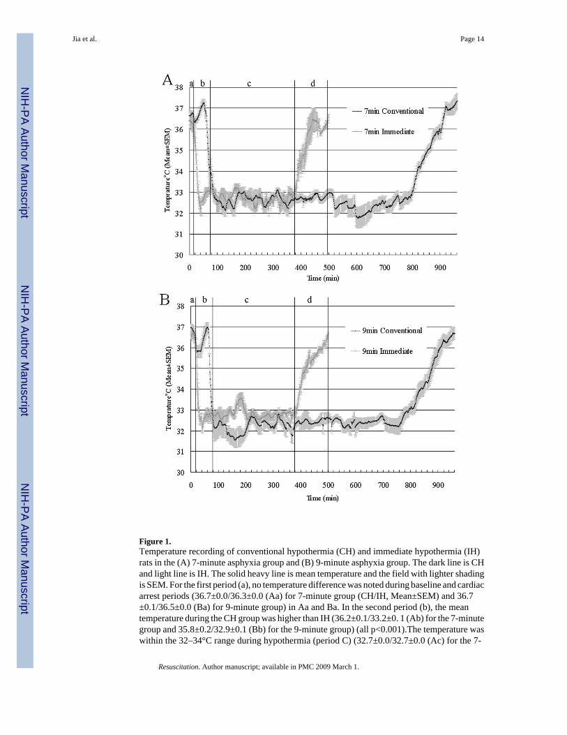
Figure 1.Temperature recording of conventional hypothermia (CH) and immediate hypothermia (IH)rats in the (A) 7-minute asphyxia group and (B) 9-minute asphyxia group. The dark line is CHand light line is IH. The solid heavy line is mean temperature and the field with lighter shadingis SEM. For the first period (a), no temperature difference was noted during baseline and cardiacarrest periods (36.7±0.0/36.3±0.0 (Aa) for 7-minute group (CH/IH, Mean±SEM) and 36.7±0.1/36.5±0.0 (Ba) for 9-minute group) in Aa and Ba. In the second period (b), the meantemperature during the CH group was higher than IH (36.2±0.1/33.2±0. 1 (Ab) for the 7-minutegroup and 35.8±0.2/32.9±0.1 (Bb) for the 9-minute group) (all p<0.001).The temperature waswithin the 32–34°C range during hypothermia (period C) (32.7±0.0/32.7±0.0 (Ac) for the 7-
Jia et al. Page 14
Resuscitation. Author manuscript; available in PMC 2009 March 1.
NIH
-PA Author Manuscript
NIH
-PA Author Manuscript
NIH
-PA Author Manuscript
minute group and 32.2±0.0/32.8±0.0 (Bc) for the 9-minute group). In period d, animals in theIH group were rewarmed while hypothermia was maintained for 6 more hours in the CH group(32.7±0.0/35.4±0.1 (Ad) for 7-minute group and 32.4±0.0/35.2±0.1 (Bd) for 9-minute group)(all p<0.001). After 12 hours of hypothermia in the CH group, normothermia was maintainedsimilar to the IH group.
Jia et al. Page 15
Resuscitation. Author manuscript; available in PMC 2009 March 1.
NIH
-PA Author Manuscript
NIH
-PA Author Manuscript
NIH
-PA Author Manuscript
Figure 2.Raw EEG data and qEEG of representative 9-minute asphyxia animals in the first 4 hoursA: 9-minute conventional hypothermia (9CH), B: 9-minute immediate hypothermia (9IH): (I)Baseline (0 minutes), (II) Cardiac arrest (CA) period (19 minutes), (III) 1 hour after CA, (IV)4 hours after CA - Hypothermia maintenance period; C: comparison of qEEG in CH and IH,CH started at 1 hour and IH started immediately after ROSC.
Jia et al. Page 16
Resuscitation. Author manuscript; available in PMC 2009 March 1.
NIH
-PA Author Manuscript
NIH
-PA Author Manuscript
NIH
-PA Author Manuscript
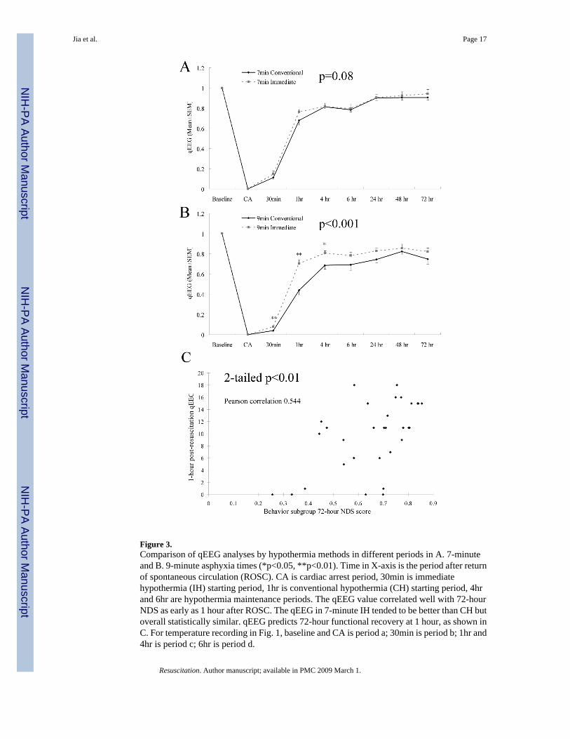
Figure 3.Comparison of qEEG analyses by hypothermia methods in different periods in A. 7-minuteand B. 9-minute asphyxia times (*p<0.05, **p<0.01). Time in X-axis is the period after returnof spontaneous circulation (ROSC). CA is cardiac arrest period, 30min is immediatehypothermia (IH) starting period, 1hr is conventional hypothermia (CH) starting period, 4hrand 6hr are hypothermia maintenance periods. The qEEG value correlated well with 72-hourNDS as early as 1 hour after ROSC. The qEEG in 7-minute IH tended to be better than CH butoverall statistically similar. qEEG predicts 72-hour functional recovery at 1 hour, as shown inC. For temperature recording in Fig. 1, baseline and CA is period a; 30min is period b; 1hr and4hr is period c; 6hr is period d.
Jia et al. Page 17
Resuscitation. Author manuscript; available in PMC 2009 March 1.
NIH
-PA Author Manuscript
NIH
-PA Author Manuscript
NIH
-PA Author Manuscript
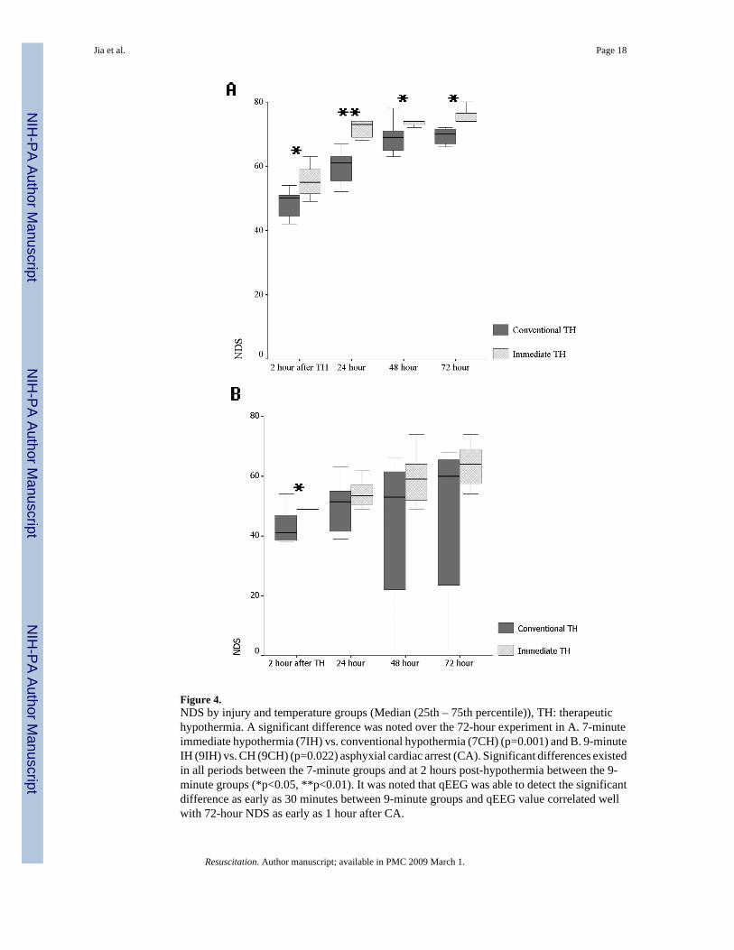
Figure 4.NDS by injury and temperature groups (Median (25th – 75th percentile)), TH: therapeutichypothermia. A significant difference was noted over the 72-hour experiment in A. 7-minuteimmediate hypothermia (7IH) vs. conventional hypothermia (7CH) (p=0.001) and B. 9-minuteIH (9IH) vs. CH (9CH) (p=0.022) asphyxial cardiac arrest (CA). Significant differences existedin all periods between the 7-minute groups and at 2 hours post-hypothermia between the 9-minute groups (*p<0.05, **p<0.01). It was noted that qEEG was able to detect the significantdifference as early as 30 minutes between 9-minute groups and qEEG value correlated wellwith 72-hour NDS as early as 1 hour after CA.
Jia et al. Page 18
Resuscitation. Author manuscript; available in PMC 2009 March 1.
NIH
-PA Author Manuscript
NIH
-PA Author Manuscript
NIH
-PA Author Manuscript
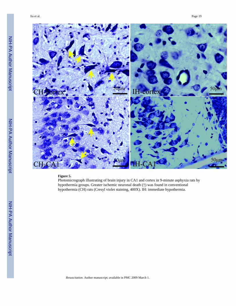
Figure 5.Photomicrograph illustrating of brain injury in CA1 and cortex in 9-minute asphyxia rats byhypothermia groups. Greater ischemic neuronal death (↑) was found in conventionalhypothermia (CH) rats (Cresyl violet staining, 400X). IH: immediate hypothermia.
Jia et al. Page 19
Resuscitation. Author manuscript; available in PMC 2009 March 1.
NIH
-PA Author Manuscript
NIH
-PA Author Manuscript
NIH
-PA Author Manuscript
NIH
-PA Author Manuscript
NIH
-PA Author Manuscript
NIH
-PA Author Manuscript
Jia et al. Page 20Ta
ble
1A
rteria
l blo
od g
as a
nd M
AP
data
in c
ardi
ac a
rres
t exp
erim
ent a
mon
g 4
grou
ps
7CH
a7I
H b
9CH
9IH
p va
lue
of 4
gro
ups
Bas
elin
e
pH7.
48±0
.03
7.42
±0.0
27.
53±0
.06
7.45
±0.0
10.
220
PCO
2 (m
mH
g)36
±239
±440
±038
±10.
929
PO2 (
mm
Hg)
323±
5138
3±25
323±
2639
1±46
0.59
8H
CO
3 (m
mol
/L)
26±1
24±2
28±0
25±1
0.68
3O
2SA
T (%
)10
0±0
100±
010
0±0
100±
00.
762
MA
P (m
mH
g)10
0±7
90±1
188
±591
±70.
644
10m
in p
ost C
A
pH7.
34±0
.03
7.35
±0.0
27.
35±0
.04
7.35
±0.0
30.
936
PCO
2 (m
mH
g)42
±336
±238
±230
±20.
009*
PO2 (
mm
Hg)
442±
3940
3±52
434±
4949
1±20
0.38
7H
CO
3 (m
mol
/L)
22±1
19±1
20±2
17±0
0.02
2*O
2SA
T (%
)10
0±0
100±
010
0±0
100±
00.
522
MA
P (m
mH
g)12
3±9
123±
1011
9±12
150±
120.
191
20m
in p
ost C
A
pH7.
36±0
.04
7.37
±0.0
17.
22±0
.00
7.38
±0.0
20.
155
PCO
2 (m
mH
g)38
±235
±250
±029
±20.
009*
PO2 (
mm
Hg)
434±
2549
6±19
595±
045
1±26
0.08
2H
CO
3 (m
mol
/L)
21±1
20±1
20±0
18±1
0.16
5O
2SA
T (%
)10
0±0
100±
010
0±0
100±
00.
862
MA
P (m
mH
g)85
±10
99±8
88±1
610
2±15
0.75
3
40m
in p
ost C
A
pH7.
40±0
.04
7.31
±0.0
27.
43±0
.04
7.36
±0.0
20.
164
PCO
2 (m
mH
g)42
±444
±439
±332
±30.
089
PO2 (
mm
Hg)
483±
2945
5±39
424±
5944
0±30
0.84
1H
CO
3 (m
mol
/L)
25±1
22±1
24±1
19±1
0.00
3*O
2SA
T (%
)10
0±0
100±
010
0±0
100±
00.
474
MA
P (m
mH
g)90
±911
0±10
62±5
93±1
50.
020*
a conv
entio
nal h
ypot
herm
ia
b imm
edia
te h
ypot
herm
ia
* Stat
istic
ally
sign
ifica
nt d
iffer
ence
was
not
ed b
ut w
as m
inim
al to
cau
se a
ny si
gnifi
cant
cha
nge
in p
H. H
ypot
herm
ia m
ay d
ecre
ase
the
PCO
2 at
10
and
20 m
inut
es in
9IH
48, 4
9 w
hile
CO
2 el
evat
ion
is
due
to n
orm
othe
rmia
in 9
CH
ani
mal
s49,
50 .
The
rela
tive
incr
ease
in M
AP
at 4
0 m
inut
es in
IH g
roup
may
be
a re
flect
ion
of th
e on
goin
g sy
stem
ic b
enef
icia
l eff
ect h
ypot
herm
ia5,
51,
52 .
Resuscitation. Author manuscript; available in PMC 2009 March 1.
NIH
-PA Author Manuscript
NIH
-PA Author Manuscript
NIH
-PA Author Manuscript
Jia et al. Page 21
Table 2Quantitative Comparison of Brain injury with histopathological damage scoring (%, Mean±SEM)
7-minute CHa 7-minute IHb 9-minute CH 9-minute IH
CA-1 20.9±7.4 12.1±3.0 28.9±8.2 17.4±5.6Cortex 30.6±5.3 19.2±3.6 36.2±7.7 18.5±3.8
aconventional hypothermia
bimmediate hypothermia
One rat in the 9-IH group, 1 rat in the 7-CH group, and 2 rats in the 9-CH group died before the endpoint of 72 hours and were excluded from quantitativebrain histopathological damage scoring.
Resuscitation. Author manuscript; available in PMC 2009 March 1.
Related Documents


