A California Toolkit to Transform Maternity Care Improving Health Care Response to Obstetric Hemorrhage THIS COLLABORATIVE PROJECT WAS DEVELOPED BY: THE OBSTETRIC HEMORRHAGE TASK FORCE THE MATERNAL QUALITY IMPROVEMENT PANEL CALIFORNIA MATERNAL QUALITY CARE COLLABORATIVE MATERNAL, CHILD AND ADOLESCENT HEALTH DIVISION; CENTER FOR FAMILY HEALTH CALIFORNIA DEPARTMENT OF PUBLIC HEALTH

Welcome message from author
This document is posted to help you gain knowledge. Please leave a comment to let me know what you think about it! Share it to your friends and learn new things together.
Transcript

A California Toolkit to Transform Maternity Care
Improving Health Care Response to Obstetric Hemorrhage THIS COLLABORATIVE PROJECT WAS DEVELOPED BY: THE OBSTETRIC HEMORRHAGE TASK FORCE THE MATERNAL QUALITY IMPROVEMENT PANEL CALIFORNIA MATERNAL QUALITY CARE COLLABORATIVE MATERNAL, CHILD AND ADOLESCENT HEALTH DIVISION; CENTER FOR FAMILY HEALTH CALIFORNIA DEPARTMENT OF PUBLIC HEALTH

OB Hemorrhage Toolkit
ii
Improving Health Care Response to Obstetric Hemorrhage Audrey Lyndon RNC, CNS, PhDa; David Lagrew, MDb; Larry Shields, MDc; Kathryn Melsop, MSd,e; Debra Bingham, RN, DrPHd,e; Elliott Maind,f, MD; Editors. University of California, San Franciscoa; Saddleback Memorial Hospitalb; Central Coast Maternal Fetal Medicinec; California Maternal Quality Care Collaboratived; Stanford University School of Medicinee; California Pacific Medical Centerf Suggested citation: Lyndon A, Lagrew D, Shields L, Melsop K, Bingham B, Main E (Eds). Improving Health Care Response to Obstetric Hemorrhage. (California Maternal Quality Care Collaborative Toolkit to Transform Maternity Care) Developed under contract #08-85012 with the California Department of Public Health; Maternal, Child and Adolescent Health Division; Published by the California Maternal Quality Care Collaborative, July 2010. Funding for the development of this toolkit was provided by: Federal Title V block grant funding from the California Department of Public Health; Maternal, Child and Adolescent Health Division and Stanford University. The California Toolkit to Transform Maternity Care called “Improving the Health Care Response to Obstetric Hemorrhage” was reviewed by the California Department of Public Health; Maternal, Child and Adolescent Health Division. This toolkit is considered a resource, but does not define the standard of care in California. Readers are advised to adapt the guidelines and resources based on their local facility’s level of care and patient populations served and are also advised to not rely solely on the guidelines presented here. Copyright Information © California Department of Public Health. The material in this toolkit may be reproduced and disseminated in any media in its original format, without modification, for informational, educational and non-commercial purposes only. A nominal sum to cover costs of reproduction and distribution can be assessed. Any modification or use of the materials in any derivative work is prohibited without prior permission of the California Department of Public Health. For correspondence, please contact:CMQCC Kathryn Melsop, MS Managing Editor, Transforming Maternity Care Series Medical School Office Building, Stanford University 251 Campus Drive Stanford, CA 93405 Phone: (650) 725-6108 FAX: (650) 721-5751 email: [email protected] Website: http://www.cmqcc.org/
CA Department of Public Health Maternal, Child and Adolescent Health Division Connie Mitchell, MD, MPH 1615 Capitol Avenue PO Box 997420, MS 8306 Sacramento, CA 95899-7420 Phone: (916) 650-0327 email: [email protected] Website: http://www.cdph.ca.gov

OB Hemorrhage Toolkit
iii
ACKNOWLEDGEMENTS
CMQCC would like to thank the California Department of Public Health; Maternal Child Health Division leaders Shabbir Ahmad, DVM, MS, PhD and Connie Mitchell, MD, MPH for their leadership to improve maternal health in California. CMQCC would like to thank volunteer members of the Hemorrhage Task Force (HTF), the Task Force Co-Chairs, David Lagrew, MD, and Audrey Lyndon, PhD, RND, CNS and the Maternal Quality Improvement Panel (MQIP) for their contributions to the Obstetric Hemorrhage Care Guidelines, Compendium of Best Practices and Hospital Level Implementation Guide. HEMORRHAGE TASK FORCE Co-Chairs: • Audrey Lyndon PhD, RNC, CNS – University of California, San Francisco • David Lagrew, MD – Saddleback Memorial Hospital
* Leslie Casper, MD – Kaiser, Southern CA, San Diego Medical Center * Nancy Corbett, BSN, RN – Kaiser, Northern CA * Maurice Druzin, MD – Stanford University * Sue Faron, MN, RNC, CNS – Sharp Mary Birch Hospital for Women * Kim Gregory, MD, MPH – Cedars-Sinai Medical Center * Andrew Hull, MD, FRCOG, FACOG – University of California, San Diego * Valerie Huwe, BSN, RNC – Stanford Hospital, El Camino Hospital * Richard Lee, MD – USC, Womenʼs and Childrenʼs Hospital * Holli Mason, MD – Harbor UCLA Medical Center * Elliott Main, MD – California Pacific Medical Center * Jennifer McNulty, MD – Long Beach Memorial Medical Center, UC Irvine * Suellen Miller, PhD, MHA, CNM - University of California, San Francisco * Connie Mitchell, MD – MCAH, CA Department of Public Health * Mark Rosen, MD – University of California, San Francisco * Diana Ramos, MD, MPH – MCAH, County of Los Angeles Public Health * Larry Shields, MD – Central Coast Maternal Fetal Medicine * Jean-Claude Veille, MD – Sutter Health, Sacramento
Conflict of Interest: The contributing authors and reviewers do not have any affiliations or financial
involvement that conflict with the material or recommendations presented in this toolkit. MATERNAL QUALITY IMPROVEMENT PANEL * Karyn Almryde, MSN, RN – Sharp Mary Birch Hospital for Women * James Byrne, MD – Santa Clara Valley Medical Center * Mary Campbell Bliss, MS, RN, CNS – Sutter Health Sacramento Sierra Region * Brenda Chagolla, MSN, RNC, CNS– Catholic Healthcare West * Holly Kennedy, PhD, CNM, FACNM – University of California, San Francisco * David Lagrew, MD – Saddleback Memorial Medical Center * Audrey Lyndon, PhD, RNC, CNS – University of California, San Francisco * Connie Mitchell, MD, MPH – MCAH, CA Department of Public Health * Barbara Murphy, MSN, RNC – CPQCC * Karen Ramstrom, DO, MSPH – MCAH, CA Department of Public Health * Linda Walsh, PhD, CNM, FACNM – University of San Francisco
OB Hemorrhage Toolkit
iv
iv
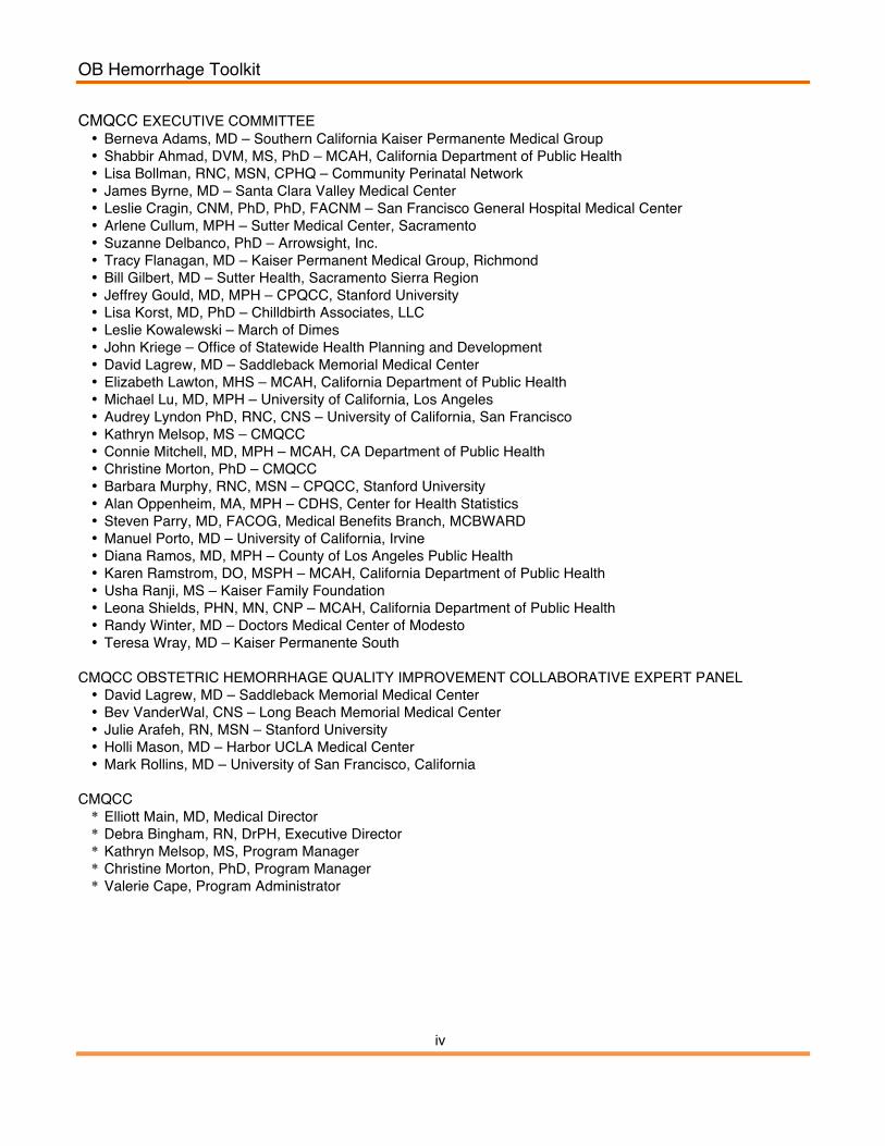
CMQCC EXECUTIVE COMMITTEE • Berneva Adams, MD – Southern California Kaiser Permanente Medical Group • Shabbir Ahmad, DVM, MS, PhD – MCAH, California Department of Public Health • Lisa Bollman, RNC, MSN, CPHQ – Community Perinatal Network • James Byrne, MD – Santa Clara Valley Medical Center • Leslie Cragin, CNM, PhD, PhD, FACNM – San Francisco General Hospital Medical Center • Arlene Cullum, MPH – Sutter Medical Center, Sacramento • Suzanne Delbanco, PhD – Arrowsight, Inc. • Tracy Flanagan, MD – Kaiser Permanent Medical Group, Richmond • Bill Gilbert, MD – Sutter Health, Sacramento Sierra Region • Jeffrey Gould, MD, MPH – CPQCC, Stanford University • Lisa Korst, MD, PhD – Chilldbirth Associates, LLC • Leslie Kowalewski – March of Dimes • John Kriege – Office of Statewide Health Planning and Development • David Lagrew, MD – Saddleback Memorial Medical Center • Elizabeth Lawton, MHS – MCAH, California Department of Public Health • Michael Lu, MD, MPH – University of California, Los Angeles • Audrey Lyndon PhD, RNC, CNS – University of California, San Francisco • Kathryn Melsop, MS – CMQCC • Connie Mitchell, MD, MPH – MCAH, CA Department of Public Health • Christine Morton, PhD – CMQCC • Barbara Murphy, RNC, MSN – CPQCC, Stanford University • Alan Oppenheim, MA, MPH – CDHS, Center for Health Statistics • Steven Parry, MD, FACOG, Medical Benefits Branch, MCBWARD • Manuel Porto, MD – University of California, Irvine • Diana Ramos, MD, MPH – County of Los Angeles Public Health • Karen Ramstrom, DO, MSPH – MCAH, California Department of Public Health • Usha Ranji, MS – Kaiser Family Foundation • Leona Shields, PHN, MN, CNP – MCAH, California Department of Public Health • Randy Winter, MD – Doctors Medical Center of Modesto • Teresa Wray, MD – Kaiser Permanente South
CMQCC OBSTETRIC HEMORRHAGE QUALITY IMPROVEMENT COLLABORATIVE EXPERT PANEL • David Lagrew, MD – Saddleback Memorial Medical Center • Bev VanderWal, CNS – Long Beach Memorial Medical Center • Julie Arafeh, RN, MSN – Stanford University • Holli Mason, MD – Harbor UCLA Medical Center • Mark Rollins, MD – University of San Francisco, California
CMQCC * Elliott Main, MD, Medical Director * Debra Bingham, RN, DrPH, Executive Director * Kathryn Melsop, MS, Program Manager * Christine Morton, PhD, Program Manager * Valerie Cape, Program Administrator
OB Hemorrhage Toolkit
v
v
EXECUTIVE SUMMARY Between 1996 and 2006, the rate of maternal deaths in California nearly tripled from 6 per 100,000 to 17 per 100,000. In African-American women, that increase in rate was far higher: 28.7 to 54.9 per 100,000 live births 1. Postpartum hemorrhage (PPH) is a leading cause of maternal mortality2, 3 and is increasing in incidence. Two recent studies confirm nationwide increases in PPH over approximately 10-year periods: 26% increase between 1994 and 20064 and 27.5% increase between 1995 and 20045. Concurrently, blood transfusions increased 92% during delivery hospitalizations nationwide between 1997 and 20056. In a review of California administrative data, Lu et al. found that 2.4% of all live births in California were complicated by PPH7. Even this appears to represent significant underreporting as hospital level review usually find rates of 5% or more than twice as high. The California Pregnancy Related Maternal Mortality Review (CA-PAMR) found that obstetric (OB) hemorrhage was one of the leading causes for maternal death and a major contributor to maternal morbidity (publication pending). Tragically, deaths from hemorrhage consistently rank at the top of the most preventable list: with 70-92% of deaths judged preventable2, 8. In 2009, California Maternal Quality Care Collaborative (CMQCC) performed a survey of California maternity services and found a number of quality improvement (QI) opportunities. Most hospitals were lacking in updated hemorrhage treatment methods such as less-invasive uterine compression balloons and B-Lynch uterine compression sutures. Few hospitals had massive transfusion protocols and most lacked an updated obstetric hemorrhage protocol, if they had a protocol in place at all (Internal Survey, CMQCC, 2008). In response to alarming trends in PPH and a paucity of state-of-the-art methods and guidelines for responding to hemorrhage, CMQCC and the Hemorrhage Task Force are pleased to provide the toolkit, “Improving Health Care Response to Obstetric Hemorrhage.” The toolkit authors represent a multi-disciplinary team of experts from every corner of the state and from both large and low volume OB units. The editorial process in developing the toolkit was extensive and included peer review and consensus among experts from around the state. The tools and best practices outlined in the toolkit have been used in several large health care quality improvement collaboratives in California with benefits extending beyond PPH events. One of the most important findings to date is that hospitals performing regular drills and debriefs for PPH events report behavioral changes among staff that improve the care of all ill pregnant women, not just those with hemorrhage (Internal Survey, CMQCC, 2010). The toolkit provides a series of articles on best practices for obstetric hemorrhage that range in topic from identifying hemorrhage to treating women who decline blood transfusions or treating
OB Hemorrhage Toolkit
vi
vi
women with high-risk pregnancies to supporting families of women who have undergone hemorrhage. In addition, the toolkit provides care guideline summaries (in checklist, flowchart and table chart formats) and a hospital-level guide to implementing quality improvement around obstetric hemorrhage. It is organized into the following sections:
• Compendium of Best Practices: fifteen articles on multiple topics around obstetric hemorrhage
• Care Guidelines: three summaries of best practices for obstetric hemorrhage, including checklist, flowchart and table chart formats
• Hospital-level Implementation Guide: A step-by-step guide to assist hospital leaders with implementation efforts
• Appendices: Sample forms for policy and procedure, risk assessment, quantitative measurement of blood loss and QI Implementation model tools
• Slide set for Professional Education: slides that summarize the problem of and the best practices for obstetric hemorrhage to be used for local education and training
CMQCC and the California Department of Public Health (CDPH), Maternal, Child and Adolescent Health (MCAH) Division collaborated to develop and disseminate this toolkit using Title V funds provided by CDPH-MCAH. The goal of this toolkit is to guide and support obstetrical providers, clinical staff, hospitals and healthcare organizations to develop methods within their facilities for timely recognition and organized, swift response to obstetric hemorrhage and to implement successful quality improvement programs for obstetric hemorrhage that will decrease short- and long-term PPH-related morbidity in women who give birth in California. REFERENCES 1. California Department of Public Health; Maternal C, and Adolescent Health Division. Statistical Master Files.
1991-2006. 2. Clark SL, Belfort MA, Dildy GA, Herbst MA, Meyers JA, Hankins GD. Maternal death in the 21st century:
causes, prevention, and relationship to cesarean delivery. Am J Obstet Gynecol 2008 Jul;199(1):36 e1-5; discussion 91-2 e7-11.
3. Baker DW, Asch SM, Keesey JW, Brown JA, Chan KS, Joyce G, et al. Differences in education, knowledge, self-management activities, and health outcomes for patients with heart failure cared for under the chronic disease model: the improving chronic illness care evaluation. J Card Fail 2005 Aug;11(6):405-13.
4. Callaghan WM, Kuklina EV, Berg CJ. Trends in postpartum hemorrhage: United States, 1994-2006. Am J Obstet Gynecol 2010 Apr;202(4):353 e1-6.
5. Bateman BT, Berman MF, Riley LE, Leffert LR. The epidemiology of postpartum hemorrhage in a large, nationwide sample of deliveries. Anesth Analg 2010 May 1;110(5):1368-73.
6. Kuklina E, Meikle, S., Jamieson, D., Whiteman, M., Barfield, W., Hillis, S., Posner, S. Severe Obstetric Morbidity in the US, 1998-2005. Obstetrics and Gynecology 2009;113:293-9.
7. Lu MC, Fridman M, Korst LM, Gregory KD, Reyes C, Hobel CJ, et al. Variations in the incidence of postpartum hemorrhage across hospitals in California. Maternal Child Health Journal 2005 September;9(3):297-306.
8. Berg CJ, Harper MA, Atkinson SM, Bell EA, Brown HL, Hage ML, et al. Preventability of pregnancy-related deaths - Results of a state-wide review. Obstetrics and Gynecology 2005 Dec;106(6):1228-34.

OB Hemorrhage Toolkit
vii
vii
TABLE OF CONTENTS ACKNOWLEDGEMENTS..............................................................................................................iii
EXECUTIVE SUMMARY ............................................................................................................... v
HOW TO USE THIS TOOLKIT ......................................................................................................1
OBSTETRIC HEMORRHAGE: COMPENDIUM OF BEST PRACTICES ..........................................2DEFINITION, EARLY RECOGNITION AND RAPID RESPONSE USING TRIGGERS .........3INHERITED COAGULATION DISORDERS IN PREGNANCY ..............................................7OBSTETRIC CARE FOR WOMEN WHO DECLINE TRANSFUSIONS...............................14
JEHOVAHʼS WITNESS BLOOD PRODUCT AND TECHNIQUE INFORMED CONSENT/DECLINE CHECKLIST.................................................................................17SPECIFIC CHECKLIST FOR MANAGEMENT OF PREGNANT WOMEN WHO DECLINE TRANSFUSIONS ...........................................................................................19IRON SUCROSE PROTOCOL.......................................................................................20
PLACENTA ACCRETA AND PERCRETA: INCIDENCE, RISKS, DIAGNOSIS, COUNSELING AND PREPARATION FOR DELIVERY.......................................................22OB HEMORRHAGE: CARTS, KITS AND TRAYS ...............................................................26
CHECKLIST: CARTS, KITS, TRAYS..............................................................................29SIMULATIONS AND DRILLS...............................................................................................32
SIMULATIONS AND DRILLS: EDUCATIONAL TOOL #1. .............................................34SIMULATIONS AND DRILLS: EDUCATIONAL TOOL #2. .............................................36SIMULATIONS AND DRILLS: EDUCATIONAL TOOL #3. .............................................38SIMULATIONS AND DRILLS: EDUCATIONAL TOOL #4. .............................................40SIMULATIONS AND DRILLS: EDUCATIONAL TOOL #5. .............................................43SIMULATIONS AND DRILLS: EDUCATIONAL TOOL #6. .............................................46
POSTPARTUM HEMORRHAGE: LESSONS LEARNED FROM OTHER STATES ............48ACTIVE MANAGEMENT OF THIRD STAGE LABOR .........................................................50BLOOD LOSS: CLINICAL TECHNIQUES FOR ONGOING QUANTITATIVE MEASUREMENT .................................................................................................................52
NHS OBSTETRIC EARLY WARNING CHART ..............................................................56OB HEMORRHAGE REPORT TEMPLATE ...................................................................57OB HEMORRHAGE REPORT-SAMPLE........................................................................58
BLOOD PRODUCT REPLACEMENT: OBSTETRIC HEMORRHAGE ................................60APPENDIX A: USE OF FACTOR VIIA ...........................................................................64APPENDIX B: ADVERSE REACTIONS TO TRANSFUSIONS ......................................65
UTERINE ARTERY OCCLUSION AND EMBOLIZATION ...................................................70UTERINE HEMOSTATIC SUTURES...................................................................................72
OB Hemorrhage Toolkit
vii
i viii
UTEROTONIC AGENTS FACT SHEET ..............................................................................74ANTI-SHOCK GARMENTS: NON-PNEUMATIC ANTI-SHOCK GARMENT (NASG) AND PNEUMATIC ANTI-SHOCK GARMENT (PASG)........................................................76FAMILY SUPPORT..............................................................................................................83
OBSTETRIC HEMORRHAGE CARE GUIDELINES 86CHECKLIST....................................................................................................................87TABLECHART................................................................................................................91FLOWCHART.................................................................................................................92
HOSPITAL LEVEL IMPLEMENTATION GUIDE 95MODEL FOR IMPROVEMENT............................................................................................97
RAPID CYCLE QI METHODOLOGY..............................................................................97MOBILIZE, ASSESS, PLAN, IMPLEMENT, TRACK (MAP-IT).......................................97
APPENDICES....................................................................................................................110APPENDIX A. SAMPLE HEMORRHAGE POLICY AND PROCEDURE......................110APPENDIX B: CMQCC OB HEMORRHAGE CARE GUIDELINES CHECKLIST.........117APPENDIX C. CMQCC OBHEMORRHAGE CARE GUIDELINES FLOW CHART......121APPENDIX D. CMQCC OB HEMORRHAGE CARE GUIDELINES TABLE CHART....122APPENDIX E.1. CMQCC IN HOSPITAL AUDIT TOOL: RISK ASSESSMENT FOR OB HEMORRHAGE .....................................................................................................124APPENDIX E.2. CMQCC IN HOSPITAL AUDIT TOOL: ACTIVE MANAGEMENT OF THIRD STAGE LABOR................................................................125APPENDIX E.3. METHODS FOR DEVELOPING TRAINING AND TOOLS FOR QUANTITATIVE MEASUREMENT OF BLOOD LOSS.................................................126APPENDIX E.4. CMQCC IN HOSPITAL AUDIT TOOL: CUMULATIVE BLOOD LOSS AND QUANTITATIVE MEASUREMENT METHODS...........................127APPENDIX E.5. CMQCC OBSTETRIC HEMORRHAGE TEAM DE-BRIEFING FORM...................................................................................................128APPENDIX F. CMQCC MAP-IT PLANNING WORKSHEET, SAMPLE WORKSHEET...............................................................................................129APPENDIX G. CMQCC QUALITY IMPROVEMENT COLLABORATIVE OBSTETRIC HEMORRHAGE MEASUREMENT GRID ...............................................131
SLIDE SET FOR PROFESSIONAL EDUCATION .......................................................................142

OB Hemorrhage Toolkit
1
HOW TO USE THIS TOOLKIT COMPENDIUM OF BEST PRACTICES
The Compendium of Best Practices consists of fifteen (15) articles presented by authors from the CMQCC Obstetric Hemorrhage Task Force. The articles highlight the current practices and recommendations for optimal care during an obstetric hemorrhage. Most articles provide topic-specific tools for use at your facility. For example, the article titled, “Obstetric Care for Women who Decline Transfusions” provides tools including an informed consent form and management checklist for use with Jehovahʼs Witness patients. Each article provides topic-specific recommendations for optimal care, evidence grading for the literature reviewed and full references.
OBSTETRIC HEMORRHAGE CARE GUIDELINES
The Obstetric Hemorrhage Care Guidelines provide summary information to assist with preparing for and responding to obstetric hemorrhage. The three formats—checklist, table chart, flowchart—are presented in order from the most comprehensive (checklist format) to the most streamlined (flowchart). One goal of the Hemorrhage Task Force was to create guideline documents that would outline best practices for each stage of hemorrhage while addressing the different levels of clinical and staff involvement in care and their various learning styles. The checklist format is the most comprehensive summary guideline and provides detailed information for identifying stages of hemorrhage and for preparing appropriate clinical strategies at each stage of hemorrhage. In addition, it is intended to guide all clinicians and staff involved in maternal care during a hemorrhage event. The table chart is a summary of the checklist; it provides an intermediate level of detail and is sufficiently simple to be contained on one page and to act as a cognitive aid. Finally, the flowchart is intended to be a simpler summary and cognitive aid and provides treatment decision points for each stage of hemorrhage.
HOSPITAL LEVEL IMPLEMENTATION GUIDE
This toolkit contains a separate stand-alone guide for hospital level implementation of best practices and care guidelines. Quality improvement implementation strategies are presented.
SLIDE SET FOR PROFESSIONAL EDUCATION A comprehensive slide set is included in this toolkit for use by clinicians, educators and hospital administration. The slide set includes background information about the problem of obstetric hemorrhage and outlines the elements of the toolkit including best practices, care guidelines and guidance for implementation of policies and procedures in hospital to improve readiness, recognition, response and reporting of obstetric hemorrhage.

OB Hemorrhage Toolkit
2
2
OBSTETRIC HEMORRHAGE: COMPENDIUM OF BEST PRACTICES BACKGROUND, PREPARATION AND MANAGEMENT The Best Practice articles that follow are topic specific and include background information, current literature review and recommendations for clinicians who are preparing for and responding to obstetric hemorrhage. In each article, a “grade” is provided for the level of evidence found in the literature to support the topic recommendations made by the Task Force. Many articles include specific tools—topic specific forms, tables or appendices—that are included at the end of each article. The first section of Best Practice articles is intended to provide background information and guidance for preparation. Articles include information about definitions of hemorrhage, preparing for women who decline blood products, and for women at higher risk for hemorrhage—including women with placenta previa, accreta or percreta. The last section of Best Practice articles focuses on management issues including blood replacement, methods for quantifying blood loss and support considerations for families of women who have experienced a hemorrhage. The Compendium of Best Practices provides a broad range of background information for obstetric hemorrhage and provides the current literature and expertise from which the summary care guidelines were developed.
OBSTETRIC HEMORRHAGE CARE GUIDELINES AND COMPENDIUM OF BEST PRACTICES CMQCC OBSTETRIC HEMORRHAGE TOOLKIT REVIEWED BY CADPH-MCAH: 12/1/09
3
DEFINITION, EARLY RECOGNITION AND RAPID RESPONSE USING TRIGGERS Kim Gregory, MD, Cedars-Sinai Medical Center; Elliott Main, MD, California Pacific Medical Center; Audrey Lyndon, PhD, CNS, RNC, Department of Family Health Care Nursing, University of California, San Francisco BACKGROUND AND LITERATURE REVIEW Postpartum hemorrhage (PPH) affects 1-3% of pregnancies in the first 24 hours after birth and is a leading cause of pregnancy-related mortality in developing and developed countries. Deaths due to PPH have declined in developed countries because hospitals have easier access to blood products, but PPH-related morbidities have remained constant and include massive transfusions, secondary surgical procedures, ICU admissions and fertility loss. (1) The risk of hemorrhage is always present at birth, but early identification creates the potential to intervene and prevent major blood loss. Early intervention requires the following: 1) recognition of risk factors leading to heightened surveillance; 2) standardized approach to estimating blood loss; and 3) the use of clinical evaluative thresholds—typically vital signs—as triggers or alerts. While efforts to standardize treatment abound, relatively few institutions have created a systematic PPH protocol for early recognition and rapid response. This deficit is due in part to the broad range of clinical risk factors involved in PPH, lack of standardized methods for estimating blood loss and lack of a “gold standard” for defining PPH. (2-6) This document focuses on: 1) providing a consensus definition of early or primary PPH occurring within the first 24 hours after delivery; 2) outlining clinical cues or triggers to quickly identify and respond to prevent progression of heavy bleeding to massive hemorrhage and its potential sequelae: shock, disseminated intravascular coagulation, multi-system organ dysfunction, and death. Quantified Blood Loss as a Trigger Whether PPH occurs early (within first 24 hours) or late (≥12 weeks postpartum) no single definition of PPH exist that undermines the true incidence of PPH. Various definitions include: ≥500 ml of estimated blood loss (EBL) after completion of the third stage; 900 ml of EBL which typically corresponds to a 15% volume deficit; 10% change in hematocrit or need for blood transfusion; and any blood loss from the genital tract >500 ml. (3-6) The ICD-9-CM: 666.x or ICD-10-CM: 072 give little guidance, leaving the definition of “Postpartum Hemorrhage” to the healthcare provider. (7, 8) In addition, incidence is ambiguous because it is related not only to estimated blood loss but also to the motherʼs initial total blood volume and the rapidity of blood loss. Pritchard et al., in a quantitative study of actual blood loss, noted that approximately 5% of women lost >1000 ml during vaginal delivery. (9) A European study using the 500/1000 ml limit, reported incidence rates of 19% (vaginal) and 4.2% (cesarean). (10) When carefully

DEFINITION, EARLY RECOGNITION AND RAPID RESPONSE USING TRIGGERS CMQCC OBSTETRIC HEMORRHAGE TOOLKIT GREGORY, K. ET AL. REVIEWED BY CADPH-MCAH: 12/01/09
4
measured, the average blood loss during delivery is approximately 500 ml for a vaginal delivery and 1000 ml for a cesarean delivery. (2, 3) Findings are difficult to compare across studies due to differing threshold definitions for hemorrhage. Although >500 ml for vaginal birth and >1000 ml for cesarean birth may be the most common clinical definition in the U.S., it is somewhat arbitrary and may not necessarily take into account a womanʼs initial volume status and may be clinically irrelevant to hemodynamic compromise. Clinicians typically underestimate true blood loss; therefore, blood loss at and following birth should be quantified. (3, 11-13) Quantitative measures of 500 ml EBL are appropriate “triggers” for heightened surveillance and/or more aggressive treatment in the face of ongoing bleeding; 1000 ml is an appropriate “trigger” for movement toward more emergent efforts. (2, 3) Similarly, 1000 ml can be used a “safety indicator” for hospital and statewide surveillance. This definition captures clinically relevant “near miss” morbidity, is consistently associated with hemodynamic instability and advances coders, clinicians and hospitals toward consistency and standardization. (14, 15) Vital Signs as Triggers Clinical triggers can include heart rate, blood pressure and oxygen saturation, among others. “Alert lines” are designed to heighten a clinicianʼs awareness of the patientʼs changing clinical status that could indicate an impending adverse event, and should prompt consideration of possible underlying causes. “Action lines” are designed to stimulate specific clinical activity and appropriate treatment interventions. (2) The National Health System of the United Kingdom has published a detailed “Obstetric Early Warning Chart” that provides a colored checklist for vital status and a guide for intervention when a patient “triggers” in one red or two yellow scores at any one time and makes use of both numeric and visual clues for care providers (See Best Practice article “Blood Loss: Clinical Techniques for Ongoing Quantitative Measurement”). (16) RECOMMENDATIONS: Aggressive treatment of women at clinical trigger points has the potential to prevent the development of serious PPH. To address this CMQCC recommends the following:
1. Use the following as a standard clinical definition of PPH: a) Estimated blood loss greater than 500 ml or hemodynamic instability as a
“trigger” for heightened surveillance and/or more aggressive treatment in the face of ongoing bleeding.
2. Use the following standard definition for safety and quality monitoring: a) Blood loss of 1000 ml as a “trigger” for monitoring safety related to maternal
health care quality. 3. Birthing facilities adopt and maintain protocols addressing:
a) Quantification of blood loss at all births (See Best Practice article “Blood Loss: Clinical Techniques for Ongoing Quantitative Measurement”)
b) Management of all women with cumulative blood loss ≥500 ml (Refer to Hemorrhage Care Guidelines-Checklist Format)

DEFINITION, EARLY RECOGNITION AND RAPID RESPONSE USING TRIGGERS CMQCC OBSTETRIC HEMORRHAGE TOOLKIT GREGORY, K. ET AL. REVIEWED BY CADPH-MCAH: 12/01/09
5
i. Nursing personnel should notify the attending physician and proceed with administration of Methergine 0.2 mg IM (if no contraindications) and fundal massage.
ii. Clinical Triggers: surveillance and intervention: 1. Heart Rate ≥110 2. Blood Pressure ≤85/45 (>15% drop) 3. Oxygen Saturation <95%
c) It is the responsibility and authority of all licensed health care team members, including RNs, to call for help and activate maternal hemorrhage response as clinically indicated.
4. Hospitals and other health care organizations internally monitor and report all cases with EBL >500 ml for internal site-specific quality monitoring to ensure adherence to institutional guideline.
5. Hospitals and other health care organizations internally monitor and report rates and associated outcomes for all women with cumulative blood loss >1000 ml.
EVIDENCE GRADING Level of Evidence: II.2. One prospective cohort study; expert consensus opinion (WHO, NHS) REFERENCES 1. Rizvi F, Mackey R, T B, McKenna P, Geary M. Successful reduction of massive
postpartum haemorrhage by use of guidelines and staff education. BJOG 2004;111:495-8.
2. Coker A, Oliver R. Definitions and Classifications in B Lynch ed. 3. Cunningham F, Leveno K, Bloom S, Hauth J, Gilstrap L, Wenstrom K. Williams
Textbook of Obstetrics: 22nd Edition. 2005. 4. Gabbe S, Simpson J, Niebyl J, Galan H, Goetzl L, Jauniaux E, et al. Obstetrics: Normal
and Problem Pregnancies 5th edition. 2007. 5. ACOG. Postpartum Hemorrhage. Obstet and Gynecol 2006;108(4). 6. World Health Organization. Report of a Technical Working Group: The prevention and
management of postpartum haemorrhage. Unpublished document WHO, 1990 1989 July 3-6, 1989 Geneva.
7. International Statistical Classification of Diseases and Related Health Problems. 9th Revision Clinical Modification 6th edition 2009.
8. Hart A, Ford B, Stegman M. International Statistical Classification of Diseases and Related Health Problems, 10th revision. 2007.
9. Pritchard J, et al. Blood volume changes in pregnancy and the peurperium. Am J Obstet Gynecol 1962;84:1271-72.
DEFINITION, EARLY RECOGNITION AND RAPID RESPONSE USING TRIGGERS CMQCC OBSTETRIC HEMORRHAGE TOOLKIT GREGORY, K. ET AL. REVIEWED BY CADPH-MCAH: 12/01/09
6
10. Bais J, Eskes M, Pel M, Bonsel G, Bleker O. Postpartum haemorrhage in nulliparous
women: incidence and risk factors in low and high risk women. A Dutch population-based cohort study on standard (> or + 500ml) and severe (> or = 1000ml) postpartum haemorrhage. Eur J Obstet Gynecol Reprod Biol 2004.
11. Dildy G, Paine A, George N, Velasco C. Estimating Blood Loss: Can Teaching Significantly Improve Visual Estimation? Obstet Gynecol
2004;104(3):601-6. 12. Patel A, Goudar SS, Geller SE, Kodkany BS, Edlavitch SA, Wagh K, et al. Drape
estimation vs. visual assessment for estimating postpartum hemorrhage. Int J Gynaecol Obstet 2006 Jun;93(3):220-4.
13. Patel A, et al. Blood loss: accuacy of visual estimation in A Textbook of PostPartum Hemorrhage, Lynch CB, ed. et al. 2006.
14. World Health Organization. Technical Working Group Care in Normal Birth: a Practical guide. 1996.
15. World Health Organization. Technical Working Group Department of Making Pregnancy Safer; recommendations for the prevention of postpartum haemorrhage. 2007.
16. Harrison P, et al. Early warning scoring in obstetrics. Intern J Obstetric Anesthesia 2005;15(Supplement):S1-S43.
OBSTETRIC HEMORRHAGE CARE GUIDELINES AND COMPENDIUM OF BEST PRACTICES CMQCC OBSTETRIC HEMORRHAGE TOOLKIT REVIEWED BY CADPH-MCAH: 11/24/09
7
INHERITED COAGULATION DISORDERS IN PREGNANCY David Lagrew, MD, Saddleback Memorial Medical Center BACKGROUND AND LITERATURE REVIEW The coagulation process is a complex biochemical chain reaction involving several pathways and proteins. Genetic abnormalities in any of these proteins can lead to serious coagulation problems. Although relatively rare in pregnancy, such abnormalities can lead to maternal hemorrhage events during antepartum, birth or postpartum and can have deleterious effects on the motherʼs and babyʼs health. Identifying patients with inherited coagulation disorders and carefully planning their care is crucial for optimal outcomes. Although postpartum hemorrhage can occur in these patients, coagulation defects are sufficiently rare that routine screening in patients with postpartum hemorrhage will not identify a large number of these patients. (1, 2) Though incidence is low, this is an important group of individuals to identify and prepare for. (3-7) The most commonly identified coagulation disorders are von Willebrandʼs Disease (Factor VIII platelet adhesion and coagulant deficiency), Hemophilia A (Factor VIII coagulant deficiency), Hemophilia B (Factor IX deficiency) and Hemophilia C (Factor XI deficiency). Basic knowledge of these disorders will help to better understand the management recommendations below. von Willebrand Disease (vWD) is the most common hereditary coagulation abnormality described in humans with a prevalence of 1% in the general population. (3, 8, 9) It occurs less frequently as an acquired disorder (acquired von Willebrand Syndrome) manifested by the presence of auto-antibodies. Von Willebrand Disease is caused by a deficiency of the plasma protein that controls platelet adhesion (VIII:vWF) and decreased activity of the protein that stabilizes blood coagulation (VIII:C). The disorder can cause mucous membrane and skin bleeding symptoms, bleeding with vaginal birth, surgical events or other hemostatic challenges. Women of child-bearing age may be disproportionately symptomatic compared with other age groups. Several types of vWD have been described. (10) Type 1 individuals make up 60-80% of all vWD cases and have a quantitative defect (heterozygous for the defective gene) but may not have clearly impaired clotting function. Decreased levels of vWF are detected in these patients (10-45% of normal, i.e., 10-45 IU). Most patients lead nearly normal lives without significant bleeding episodes. Patients may experience bleeding following surgery (including dental procedures), noticeable easy bruising or menorrhagia (heavy periods). Type 2 vWD patients (20-30% of all vWD cases) have a qualitative defect and the tendency to bleed varies between
INHERITED COAGULATIONS DISORDERS IN PREGNANCY CMQCC OBSTETRIC HEMORRHAGE TOOLKIT LAGREW, D. REVIEWED BY CADPH-MCAH: 11/24/09
8
individuals. Individuals with Types I and II are usually mildly affected by the disorder and pass on the trait in an autosomal dominant fashion. Type III vWD is the most severe form; it is autosomal recessive and severely affected individuals are homozygous for the defective gene. Patients have severe mucosal bleeding, no detectable vWF antigen, and may have sufficiently low factor VIII. They can have occasional hemarthoses (joint bleeding) as in cases of mild hemophilia. Most vWD diagnoses are in women with a positive family history or menorrhagia. Blood testing for vWF activity provides confirmation of diagnosis.
Hemophilia A (Factor VIII coagulant deficiency) is a blood clotting disorder caused by a mutation of the factor VIII gene, which leads to Factor VIII deficiency. Inheritance is X-linked recessive; hence, males are affected while females are carriers or very rarely display a mild phenotype. It is the most common hemophilia, occurring in 1 in 5000 males. Women can, on rare occasion, exhibit a homozygous state if both parents carry the disorder. More frequently, carriers show atypical performance of “Lyonization” of the X chromosome (random inactivation of the X chromosome). Usually women have 50% activity but if inactivation of the “normal” gene occurs in greater frequency, lower levels can be seen. (11) Of note, Factor VIII activity usually increases during pregnancy. (12) Hemophilia B (Factor IX deficiency) is a blood clotting disorder caused by a mutation of the Factor IX gene, also carried on the X-chromosome. It is the least common form of hemophilia (sometimes called “Christmas Disease,” after the first afflicted patient), occurring in about 1:30,000 males and very rarely in females. Diagnosis can be made by measuring levels of IX activity in the blood, which does not usually change during pregnancy. Hemophilia C (Factor XI deficiency) is a rare condition in the general population (less than 1:100,000) but more common in Ashkenazi Jewish patients, and it can occur in both males and females. (13) Up to 8% of these individuals are carriers (autosomal recessive) of the gene, which is located on Chromosome 4. Treatment is usually not necessary because patients have approximately 20-60% factor XI activity; however, they should be closely followed since the postpartum hemorrhage rate is 20%. Diagnosis in pregnancy of any of these coagulation disorders may be difficult due to the variability of clotting factor activity caused by hormonal changes of pregnancy. (14) When a patient with an inherited coagulation disorder delivers, one must be concerned about extra-uterine bleeding and hematomas and the effect of the disorder on the fetus. Cesarean section is rarely recommended. (15) Autoimmune acquisition of these disorders has been described and therefore may occur despite the lack of familial history.

INHERITED COAGULATIONS DISORDERS IN PREGNANCY CMQCC OBSTETRIC HEMORRHAGE TOOLKIT LAGREW, D. REVIEWED BY CADPH-MCAH: 11/24/09
9
RECOMMENDATIONS 1. Review family, surgical and pregnancy history for possible clinical symptoms of
excessive bleeding following surgery (including dental procedures), noticeable easy bruising, joint hemorrhage or menorrhagia (heavy periods).
2. Request the following laboratory screening tests for patients with suspected disorders: (10, 11)
• von Willebrand Disorder: Measurement of Ristocetin Co-Factor Activity and von Willebrand Antigen (VIII:Ag) activity
• Hemophilia A: Measurement of Factor VIII activity (Factor VIII:C assay) • Hemophilia B: Measurement of Factor IX activity (If Factor VIII:C is normal) • Hemophilia C: Measurement of Factor XI activity • Other tests performed for patients with bleeding problems: complete blood
count (especially platelet counts), APTT (activated partial thromboplastin time), prothrombin time, thrombin time and fibrinogen level. Note that patients with von Willebrand disease typically display normal prothrombin time and variable prolongation of partial thromboplastin.
3. Affected patients or carriers, or patients with suspected history should consult with a hematologist who has specific interest and knowledge of coagulation disorders.
4. Obtain perinatal consultation for planning and coordination of antepartum and intrapartum management.
5. Refer patients for genetic counseling regarding possible testing and evaluation of the fetus and newborn.
6. Develop intrapartum and postpartum management plans well in advance of the anticipated date of birth so specific medications and blood components are available at the time of delivery and given in consultation with a hemotologist:
• von Willebrand Disorder: Mild forms can be treated with desmopressin acetate (DDAVP) but more severe forms require vWF and VIII factor replacement. (7) DDAVP challenge testing can identify whether patients will respond to this medication.
• Hemophilia A/B: Concentrates of clotting factor VIII (for hemophilia A) or clotting factor IX (for hemophilia B) are slowly dripped in or injected into a vein. Consider DDAVP adjunctive therapy.
• Hemophilia C: FFP is the first product used to treat patients with hemophilia C. The main advantage of FFP is its availability. Disadvantages of its use include the large volumes required, the potential for transmission of infective agents and the possibility of allergic reactions.
• Factor XI activity: Factor XI concentrates provide the best source for factor XI replacement.
INHERITED COAGULATIONS DISORDERS IN PREGNANCY CMQCC OBSTETRIC HEMORRHAGE TOOLKIT LAGREW, D. REVIEWED BY CADPH-MCAH: 11/24/09
10
EVIDENCE GRADING Level of Evidence: III C. Opinions of respected authorities, based on clinical experience, descriptive studies, or reports of expert committees. Recommendations based primarily on consensus and expert opinion. REFERENCES 1. Clark P, Greer I. Practical Obstetric Hematology. 2006 Taylor and Francis Abington,
Oxon 2006. 2. Kadir R, Kingma C, Chi C, Lee C, Economides D. Is primary postpartum haemorrhage a
good predictor of inherited bleeding disorders? Haemophilia 2007;13(2):178-81. 3. Demers C, Derzko C, David M, Douglas J. Gynaecological and obstetric management of
women with inherited bleeding disorders. Society of Obstetricians and Gynaecologissts of Canada 2006;95(1):75-87.
4. Gojnic M, Fazlagic A, Likie I, Stefanovic A, Vidakovic S, Pervulov M, et al. New approach of the treatment of von Willebrand's disease during pregnancy. Arch Gynecol Obstet 2005;273(1):35-8.
5. Nichols WL, Hultin MB, James AH, Manco-Johnson MJ, Montgomery RR, Ortel TL, et al. von Willebrand disease (VWD): evidence-based diagnosis and management guidelines, the National Heart, Lung, and Blood Institute (NHLBI) Expert Panel report (USA). Haemophilia 2008 Mar;14(2):171-232.
6. Dhar P, Abramovitz S, DiMichele D, Gibb C, F G. Management of pregnancy in a patient with severe haemophilia. Anaesthesia 2003;91(3):432-5.
7. Michiels J, van Vliet H, Berneman Z, Gadisseur A, van der Planken M, Schroyens W, et al. Intravenous DDAVP and factor VII-von Willebrand factor concentrate for the treatment and prophylaxis of bleeding in patients with von Willebrand disease type 1, 2 and 3. Clin Appl Thromb Hemost 2007;13(1):14-34.
8. James AH. Von Willebrand disease. Obstet Gynecol Surv 2006 Feb;61(2):136-45. 9. Kouides P. Current understanding of Von Willebrand's disease in women - some
answers, more questions. Haemophilia 2006;12(Supplement 3):143-51. 10. Sadler JE. A revised classification of von Willebrand disease. For the Subcommittee on
von Willebrand Factor of the Scientific and Standardization Committee of the International Society on Thrombosis and Haemostasis. Thromb Haemost 1994 Apr;71(4):520-5.
11. Verbruggen B, Meijer P, Novakova I, Van Heerde W. Diagnosis of factor VIII deficiency. Haemophilia 2008 Jul;14 Suppl 3:76-82.
12. Chi C, Lee CA, Shiltagh N, Khan A, Pollard D, Kadir RA. Pregnancy in carriers of haemophilia. Haemophilia 2008 Jan;14(1):56-64.
13. Kadir RA, Kingman CE, Chi C, O'Connell N M, Riddell A, Lee CA, et al. Screening for factor XI deficiency amongst pregnant women of Ashkenazi Jewish origin. Haemophilia 2006 Nov;12(6):625-8.
14. Franchini M. Haemostasis and pregnancy. Thromb Haemost 2006 Mar;95(3):401-13.

INHERITED COAGULATIONS DISORDERS IN PREGNANCY CMQCC OBSTETRIC HEMORRHAGE TOOLKIT LAGREW, D. REVIEWED BY CADPH-MCAH: 11/24/09
11
15. Tarantino MD, Gupta SL, Brusky RM. The incidence and outcome of intracranial haemorrhage in newborns with haemophilia: analysis of the Nationwide Inpatient Sample database. Haemophilia 2007 Jul;13(4):380-2.

OBSTETRIC HEMORRHAGE CARE GUIDELINES AND COMPENDIUM OF BEST PRACTICES CMQCC OBSTETRIC HEMORRHAGE TOOLKIT REVIEWED BY CADPH-MCAH: 11/24/09
14
OBSTETRIC CARE FOR WOMEN WHO DECLINE TRANSFUSIONS (Jehovahʼs Witnesses and others) Elliott Main, MD, Department of Obstetrics and Gynecology, California Pacific Medical Center, Sutter Health BACKGROUND AND LITERATURE REVIEW Given the known rate of obstetric hemorrhage, it is very unsettling to many obstetricians and anesthesiologists to have a patient decline a potentially life-saving treatment. Fortunately, discussions regarding limits to intervention generally occur in advance of emergencies in pregnant women whose belief systems preclude blood transfusion. The goals of the interaction with the woman who is declining transfusion are the following: 1) to find common ground to manage the birth as safely as possible; 2) to build trust or if not possible, to transfer to a program amenable with the plans; and 3) to develop a well thought out delivery plan to minimize blood loss and maximize decisive decisions. A large study in New York of 391 live births among Jehovahʼs Witness found 2 maternal deaths from hemorrhage (512 maternal deaths per 100,000 births). (1) With regard to goal #3, there is a broad movement in the United States to develop skills and promote the concepts of “Bloodless Surgery.” While this may sound a bit utopian, there are case series of open-heart surgeries and liver transplants without transfusions. The principles of this approach are listed below: (2) General Principles of Bloodless Medicine Management
• Employ a multidisciplinary treatment approach to blood conservation • Formulate a plan of care for avoiding/controlling blood loss • Consult promptly with senior specialist experienced in blood conservation • Promptly investigate and treat anemia • Decisive intervention, including surgery • Be prepared to modify routine practice when appropriate • Restrict blood drawing for laboratory tests • Decrease or avoid the use of anticoagulants and antiplatelet agents • Stimulate erythropoiesis • Transfer a stabilized patient, if necessary, to a major center before the patientʼs
condition deteriorates
OBSTETRIC CARE FOR WOMEN WHO DECLINE TRANSFUSIONS (JEHOVAH’S WITNESS) CMQCC OBSTETRIC HEMORRHAGE MAIN, E. TOOLKITREVIEWED BY CADPH-MCAH: 11/24/09
15
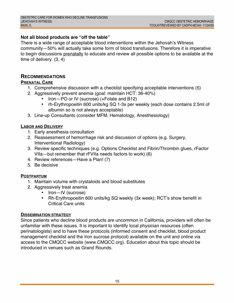
Not all blood products are “off the table” There is a wide range of acceptable blood interventions within the Jehovahʼs Witness community—50% will actually take some form of blood transfusions. Therefore it is imperative to begin discussions prenatally to educate and review all possible options to be available at the time of delivery. (3, 4) RECOMMENDATIONS PRENATAL CARE
1. Comprehensive discussion with a checklist specifying acceptable interventions (5) 2. Aggressively prevent anemia (goal: maintain HCT: 36-40%)
• Iron—PO or IV (sucrose) (+Folate and B12) • rh-Erythropoeitin 600 units/kg SQ 1-3x per weekly (each dose contains 2.5ml of
albumin so is not always acceptable) 3. Line-up Consultants (consider MFM, Hematology, Anesthesiology)
LABOR AND DELIVERY
1. Early anesthesia consultation 2. Reassessment of hemorrhage risk and discussion of options (e.g. Surgery,
Interventional Radiology) 3. Review specific techniques (e.g. Options Checklist and Fibrin/Thrombin glues, rFactor
VIIa—but remember that rFVIIa needs factors to work) (6) 4. Review references—Have a Plan! (7) 5. Be decisive
POSTPARTUM
1. Maintain volume with crystaloids and blood substitutes 2. Aggressively treat anemia
• Iron—IV (sucrose) • Rh-Erythropoeitin 600 units/kg SQ weekly (3x week); RCTʼs show benefit in
Critical Care units DISSEMINATION STRATEGY Since patients who decline blood products are uncommon in California, providers will often be unfamiliar with these issues. It is important to identify local physician resources (often perinatologists) and to have these protocols (informed consent and checklist, blood product management checklist and the Iron sucrose protocol) available on the unit and online via access to the CMQCC website (www.CMQCC.org). Education about this topic should be introduced in venues such as Grand Rounds.

OBSTETRIC CARE FOR WOMEN WHO DECLINE TRANSFUSIONS (JEHOVAH’S WITNESS) CMQCC OBSTETRIC HEMORRHAGE MAIN, E. TOOLKITREVIEWED BY CADPH-MCAH: 11/24/09
16
EDUCATIONAL TOOLS, SAMPLE DOCUMENTS 1. Jehovahʼs Witness Consent Form and Management Checklist (8) 2. Specific Checklist for Management of Gravidas Who Decline Transfusions 3. IV Iron Sucrose Protocol
EVIDENCE GRADING Level of Evidence: III. Recommendations based primarily on consensus and expert opinion.

OBSTETRIC CARE FOR WOMEN WHO DECLINE TRANSFUSIONS (JEHOVAH’S WITNESS) CMQCC OBSTETRIC HEMORRHAGE MAIN, E. TOOLKITREVIEWED BY CADPH-MCAH: 11/24/09
17
JEHOVAHʼS WITNESS BLOOD PRODUCT AND TECHNIQUE INFORMED CONSENT/DECLINE CHECKLIST My signature below indicates that I request no blood derivatives other than the ones which I have designated in this consent be administered to me during this hospitalization. My attending physician,_____________________M.D. has reviewed and fully explained to me, the risks and benefits of the following blood products and methods for alternative non-blood medical management and blood conservation available to me. My attending physician_____________________M.D. has also fully explained to me the potential risks associated by not authorizing blood and / or non-blood management during this hospitalization. ACCEPT DO NOT ACCEPT COMPONENTS OF HUMAN BLOOD Red Blood Cells ________ ________ Fresh Frozen Plasma ________ ________ Platelets ________ ________ Cryoprecipitate ________ ________ Albumin ________ ________ Plasma Protein Fraction ________ ________ INTRAVENOUS FLUIDS WHICH ARE NOT COMPONENTS OF HUMAN BLOOD Hetastarch ________ ________ Balanced Salt Solutions ________ ________ MEDICATIONS WHICH CONTAIN A FRACTION OF HUMAN BLOOD Rhogam ________ ________ Erythropoeitin ________ ________ Human Immunoglobulin ________ ________ Tisseel ________ ________ TECHNIQUES FOR BLOOD CONSERVATION / PROCESSING Hemodilution ________ ________ Cell Saver ________ ________ Autologous Banked Blood ________ ________ Cardiopulmonary Bypass ________ ________ Chest Drainage Autotransfusion ________ ________ Plasmapheresis ________ ________ Hemodialysis ________ ________ Other_____ ________ ________

OBSTETRIC CARE FOR WOMEN WHO DECLINE TRANSFUSIONS (JEHOVAH’S WITNESS) CMQCC OBSTETRIC HEMORRHAGE MAIN, E. TOOLKITREVIEWED BY CADPH-MCAH: 11/24/09
18
PLEASE CIRCLE WHICH ONE APPLIES I do (do not) have a durable power of attorney. I accept (do not accept) this consent as an addendum to my durable power of attorney.
I fully understand the options available to me and hereby release the hospital, its personnel, the attending physician and any other person participating in my care from any responsibility whatsoever for unfavorable reactions or any untoward results due to my decision not to permit the use of blood or its derivatives. The possible risks and consequences of such refusal on my part have been fully explained to me by my attending physician. I fully understand such risks and consequences may occur as a result of my decision. DATE:______________ TIME:_______________ SIGNATURE:__________________________________ (patient/parent/guardian/conservator) RELATIONSHIP:_______________________________ WITNESS:_____________________________________

OBSTETRIC CARE FOR WOMEN WHO DECLINE TRANSFUSIONS (JEHOVAH’S WITNESS) CMQCC OBSTETRIC HEMORRHAGE MAIN, E. TOOLKITREVIEWED BY CADPH-MCAH: 11/24/09
19
SPECIFIC CHECKLIST FOR MANAGEMENT OF PREGNANT WOMEN WHO DECLINE TRANSFUSIONS
Prenatal Care Comprehensive discussion with a checklist specifying acceptable interventions Aggressively prevent anemia (goal: HCT: 36-40%)
Iron—PO or IV (sucrose) with Folate and B12 as needed rh-Erythropoeitin 600units/kg SQ 1-3x per weekly as needed
(most preparations have 2.5ml of albumin so may be refused by some Jehovahʼs Witnesses)
Line-up Consultants (consider MFM, Hematology, Anesthesiology)
Labor and Delivery Anesthesia consultation early Reassessment of hemorrhage risk and discussion of options
(e.g. Surgery, Interventional Radiology) Review specific techniques (e.g. Options Checklist and Fibrin/Thrombin glues,
rFactor VIIa—but remember that rFVIIa needs factors to work) Review references—Have a Plan!! Be decisive
Postpartum Maintain volume with Crystaloids and Blood substitutes Aggressively treat anemia
Iron—IV (sucrose) Rh-Erythropoeitin 600units/kg SQ weekly (3x week)
RCTʼs show benefit in Critical Care units For more information, please review: www.CMQCC.org section on “OB Hemorrhage/Jehovahʼs Witness”

OBSTETRIC CARE FOR WOMEN WHO DECLINE TRANSFUSIONS (JEHOVAH’S WITNESS) CMQCC OBSTETRIC HEMORRHAGE MAIN, E. TOOLKITREVIEWED BY CADPH-MCAH: 11/24/09
20
IRON SUCROSE PROTOCOL Elliott Main, MD Iron Sucrose (Venofer ®) is a safe intravenous preparation of iron for those who need iron and do not respond or cannot take oral iron. Side Effects
Iron sucrose has not been associated with anaphylaxis which makes it the preferred drug for parenteral iron supplementation. No serious adverse effects have been seen, including no hypotension. Occasionally, patients (5-10%) may have a transient metallic taste and hot flashes. (9, 10)
Indications
Selected patients with the following: 1. Severe antepartum iron deficient anemia non-responsive (or intolerant) to oral iron replacement 2. Anemia in a high-risk setting requiring quick replacement of iron stores: a) placenta previa/accreta b) Jehovahʼs Witness or other decliners of blood transfusions 3. Severe anemia from obstetric hemorrhage 4. Post autologous donation with need for rapid replenishment In indications 2-4, there is additional consideration for recombinant human erythropoietin (EPO) (300 units/kg SQ, once), which combined with iron sucrose gives the most rapid response.
Administration
Option 1: 500 mg Iron Sucrose in NS 250 ml administered over three (3) hours; repeat in 3-7 days to reach 1 gm. Option 2: 200 mg in NS 100 ml administered over 20-30 minutes; may repeat every other day to reach target. Fe need; see below.
Calculate Fe (Iron sucrose) need:
Fe need = wt (kg) x 0.24 x Hgb (gm/L) + 500mg = target - current
Example: 70 kg woman with Hgb of 7.0 gm/dL and a target of 11 gm/L
= 70 kg x 0.24 x (target: 110 gm/L — actual: 70 gm/L) + 500 mg Remember: 7 gm/dL = 70 gm/L Remember: Use pre-pregnancy weight (kg)
= 672 mg + 500 mg = 1172 mg (This is usually rounded off to 100 or 200 mg increments)

OBSTETRIC CARE FOR WOMEN WHO DECLINE TRANSFUSIONS (JEHOVAH’S WITNESS) CMQCC OBSTETRIC HEMORRHAGE MAIN, E. TOOLKITREVIEWED BY CADPH-MCAH: 11/24/09
21
REFERENCES 1. Singla A, Lapinski R, Berkowitz R, CJ S. Are women who are Jehovah's Witnesses at
risk of maternal death. Am J Obstet Gynecol 2001;185:893-5. 2. Remmers PA, Speer AJ. Clinical strategies in the medical care of Jehovah's Witnesses.
Am J Med 2006 Dec;119(12):1013-8. 3. Gyamfi C, Berkowitz RL. Responses by pregnant Jehovah's Witnesses on health care
proxies. Obstet Gynecol 2004 Sep;104(3):541-4. 4. Gyamfi C, Berkowitz RL. Management of pregnancy in a Jehovah's Witness. Obstet
Gynecol Clin North Am 2007 Sep;34(3):357-65, ix. 5. Gyamfi C, Gyamfi MM, Berkowitz RL. Ethical and medicolegal considerations in the
obstetric care of a Jehovah's Witness. Obstet Gynecol 2003 Jul;102(1):173-80. 6. Laird R, Carabine U. Recombinant factor VIIa for major obstetric haemorrhage in a
Jehovah's Witness. Int J Obstet Anesth 2008 Apr;17(2):193-4. 7. Massiah N, Athimulam S, Loo C, Okolo S, Yoong W. Obstetric care of Jehovah's
Witnesses: a 14-year observational study. Arch Gynecol Obstet 2007 Oct;276(4):339-43.
8. Marsh J, Elebute M, DH B. Special circumstances: Jehovah's Witnesses, those who refuse blood transfusion and/or consent in A Textbook of Postpartum Hemorrhage. 2006;(ed C. B-Lynch et al.) Sapiens Publishing
9. Breymann C, Visca E, Huch R, A H. Efficacy and safety of intravenously administered iron sucrose with and without adjuvant recombinant human erythropoietin for the treatment of resistant iron-deficiency anemia during pregnancy. Am J Obstet Gynecol 2001;184(4):662-7.
10. Al RA, Unlubilgin E, Kandemir O, Yalvac S, Cakir L, Haberal A. Intravenous versus oral iron for treatment of anemia in pregnancy: a randomized trial. Obstet Gynecol 2005 Dec;106(6):1335-40.

CMQCC OBSTETRIC HEMORRHAGE TOOLKIT OBSTETRIC HEMORRHAGE CARE GUIDELINES AND COMPENDIUM OF BEST PRACTICES REVIEWED BY CADPH-MCAH: 2/2/10
22
PLACENTA ACCRETA AND PERCRETA: INCIDENCE, RISKS, DIAGNOSIS, COUNSELING AND PREPARATION FOR DELIVERY Richard Lee, MD, Los Angeles County and University of Southern California Medical Center BACKGROUND AND LITERATURE REVIEW The rising incidence of placenta accreta is due to the rapidly rising numbers of primary and repeat cesarean births. The most recent data in California shows that 31% of all births are by cesarean section. (1) One study at The University of Chicago showed that between 1982 and 2002 (before the greatest rise in cesarean births) the overall incidence of placenta accreta was 1 in every 533 deliveries. (2) There are four types of placenta previa: 1) a complete previa occurs when the placenta completely covers the internal os; 2) a partial previa occurs when the placenta partially covers the internal os; 3) a marginal previa occurs when the placenta is located next to the internal os; 4) a low lying placenta occurs when the placental margin is within two centimeters of the internal os, but not next to the internal os. A placenta accreta occurs when there is abnormally firm attachment of placental villi to the uterine wall with the absence of the normal intervening deciduas basalis and Nitabuchʼs layer. There are three variants of this condition: 1) accreta: the placenta is attached to the myometrium; 2) increta: the placenta extends into the myometrium; and 3) percreta: the placenta extends through the entire myometrial layer and uterine serosa. Risk The risk of placenta accreta is highest in patients with both prior cesarean birth and placenta previa (placenta previa also increases with prior cesarean births). Silver, et al. reported proportionaly increased risk of placenta accreta with higher numbers of prior cesareans in women with or without placenta previa (See Table). (3)
Placenta Previa and Placenta Accreta by Number of Cesarean Deliveries
Cesarean Delivery
Previa
Previa*: Accreta† N (%)
No Previa‡: Accreta† N (%)
First§ 398 13 (3.3%) 2 (0.03%) Second 211 23 (11%) 26 (0.2%) Third 72 29 (40%) 7 (0.1%) Fourth 33 20 (61%) 11 (0.8%) Fifth 6 4 (67%) 2 (0.8%) ≥6 3 2 (67%) 4 (4.7%)
*Percentage of accreta in women with placenta previa †Increased risk with increasing number of cesarean deliveries; P < .001 ‡Percentage of accreta in women without placenta previa §Primary cesarean
PLACENTA ACCRETA & PERCRETA CMQCC OBSTETRIC HEMORRHAGE TOOLKIT LEE, R. REVIEWED BY CADPH-MCAH: 2/2/10
23
Diagnosis: A diagnosis of accreta can be confirmed with tissue histology; however, medical imaging can be an effective diagnostic tool. Ultrasound can detect the presence of accreta (80% sensitivity) and absence of accreta (95% specificity). (4-7) Warshak et al. reported that in cases with suspicious or inconclusive ultrasonography results, MRI accurately predicted placenta accreta with 88% sensitivity and 100% specificity. (6) While MRIʼs specificity is enhanced when gadolinium is used, its effects on the fetus remain uncertain; many researchers believe benefits of its use outweigh risks associated with mis- or undiagnosed placenta accreta. (6) A recent Stanford study suggests that high-resolution sonography and MRI give similar results but are complimentary when one modality is inconclusive. (7)
Second trimester Maternal Serum Alpha-Fetoprotein (MSAFP) may also be helpful. In two recent studies of patients with placenta previa, MSAFP was elevated in 45% of those with accreta, and not in those without accreta. (8) Counseling: Providers caring for patients with prenatally suspected placenta accreta should counsel patients extensively about potential risks and complications well in advance of their estimated due date. Patients with accreta are at increased risk for hemorrhage, blood transfusion, bladder/ureteral damage, infection, need for intubation, prolonged hospitalization, ICU admission, need for reoperation, thromboembolic events and death. (3, 7-9) Discussions should involve relative likelihood for hysterectomy and subsequent infertility. Delivery Timing: In patients with strong suspicion for placenta accreta, it is strongly advised to perform the delivery before labor begins or hemorrhaging occurs. (8) Therefore, consideration should be given to performing the cesarean birth electively and prematurely, either after corticosteroids for fetal lung maturation or after documentation of fetal lung maturity. The committee could not reach consensus on the recommended gestational age for elective delivery; some tertiary referral centers recommended 32-34 weeks and others 35-36 weeks. All agreed that patients with repeated bleeding episodes or deeper invasion (e.g. placenta percreta) should be delivered early. Delivery Preparations: Advance planning with anesthesia, blood bank, nursing (OB and OR) and advanced surgeons is an essential first step. Advanced surgeons are gynecology oncologists or experienced pelvic surgeons familiar with the operative management of complex pelvic surgeries. A Massive Transfusion Pack with 4-6 units PRBCs, FFP and Platelets should be available (see OB Hemorrhage Care Guidelines: Checklist Format and Blood Product Replacement Best Practice Article). At the time of cesarean, the hysterotomy should be made away from the location of the placenta. In all but those with focal accretas, a hysterotomy —without disturbance of the placenta—is strongly advised. (8) Blood salvage equipment should also be considered where available. (10) The results of conservative surgery have been recently reviewed with many complications noted (e.g. infection, delayed hemorrhage, re-operation requiring hysterectomy, disseminated intravascular coagulation) and should only be considered in the most select situations. (11) Consultation with experienced surgeons (e.g. gynecologic oncologist) or referral to appropriate facilities is required when a provider lacks
PLACENTA ACCRETA & PERCRETA CMQCC OBSTETRIC HEMORRHAGE TOOLKIT LEE, R. REVIEWED BY CADPH-MCAH: 2/2/10
24
appropriate support services or surgical experience with managing placenta accreta. The use of prophylactic intravascular balloon catheters for cesarean hysterectomy for placenta accreta is controversial as a recent large case control study (UC Irvine/Long Beach Memorial) showed no benefit. (12) If a focal placenta accreta is found (typically in the lower uterine segment at the delivery of a placenta previa) management options are broader and include over-sewing, fulguration and placement of an intrauterine compression balloon (with drainage through the cervix/vagina) for 24 hours. RECOMMENDATIONS SCREEN
1. Screen all women with prior cesarean birth for placenta previa with ultrasound. (3) (C) 2. Screen all women with placenta previa for accreta first with ultrasound, then with MRI if
ultrasound results are suspicious or inconclusive. (5) (B) COUNSEL
1. Counsel all patients with placenta accreta about delivery risks and complications and future infertility if hysterectomy is performed. (C)
PREPARE 1. Prepare a multi-disciplinary approach for delivery, including a plan for emergent surgery
prior to scheduled delivery. a. Planning should include primary OB surgeon, Blood Bank, perinatologist,
anesthesiologist, gynecologic oncologist/experienced pelvic surgeon, labor & delivery nursing, operating room personnel, nursery and pediatric teams. (C)
2. Consider early delivery (32-36 weeks) before labor and after pretreatment with betamethasone for fetal lung maturity. (C)
3. Perform the delivery surgery in main OR with a surgical scrub team. (C) 4. Actively involve surgeon(s) with advanced skills for controlling heavy pelvic bleeding
and repairing bladder or ureteral injury. (C) 5. Strongly consider hysterectomy (without removal of placenta) if no further children
desired. (C) 6. Notify blood blank for potential of massive hemorrhage and ensure immediate
availability of 4-6 units of PRBC, FFP, and platelets. (C) 7. The Committee was divided on the desirability for pre-placement of internal iliac artery
balloon catheters with a recent large case control study (UC Irvine/Long Beach Memorial) showing no benefit. (12) (B)
EVIDENCE GRADING Level of Evidence: B. Recommendations based on limited or inconsistent evidence Level of Evidence: C. Recommendations based primarily on consensus and expert opinion
PLACENTA ACCRETA & PERCRETA CMQCC OBSTETRIC HEMORRHAGE TOOLKIT LEE, R. REVIEWED BY CADPH-MCAH: 2/2/10
25
REFERENCES 1. Hamilton B, Martin J, Ventura S, Sutton P, Menacker F. Births: preliminary data for
2004. National Vital Statistics Report 2005 2005;54(8):1-17. 2. Wu S, Kocherginsky M, Hibbard JU. Abnormal placentation: twenty-year analysis. Am J
Obstet Gynecol 2005 May;192(5):1458-61. 3. Silver RM, Landon MB, Rouse DJ, Leveno KJ, Spong CY, Thom EA, et al. Maternal
morbidity associated with multiple repeat cesarean deliveries. Obstetrics and Gynecology 2006 Jun;107(6):1226-32.
4. Chou M, Ho E, Lee Y. Prenatal diagnosis of placenta previa accreta by transabdominal color Doppler ultrasound. Ultrasound Obstet Gynecol 2000;15(1):28-35.
5. Comstock CH, Love JJ, Jr., Bronsteen RA, Lee W, Vettraino IM, Huang RR, et al. Sonographic detection of placenta accreta in the second and third trimesters of pregnancy. Am J Obstet Gynecol 2004 Apr;190(4):1135-40.
6. Warshak CR, Eskander R, Hull AD, Scioscia AL, Mattrey RF, Benirschke K, et al. Accuracy of ultrasonography and magnetic resonance imaging in the diagnosis of placenta accreta. Obstet Gynecol 2006 Sep;108(3 Pt 1):573-81.
7. Dwyer BK, Belogolovkin V, Tran L, Rao A, Carroll I, Barth R, et al. Prenatal diagnosis of placenta accreta: sonography or magnetic resonance imaging? J Ultrasound Med 2008 Sep;27(9):1275-81.
8. Oyelese Y, Smulian JC. Placenta previa, placenta accreta, and vasa previa. Obstet Gynecol 2006 Apr;107(4):927-41.
9. O'Brien JM, Barton JR, Donaldson ES. The management of placenta percreta: conservative and operative strategies. Am J Obstet Gynecol 1996 Dec;175(6):1632-8.
10. ACOG. Clinical Management Guidelines for Obstetrician-Gynecologists. Obstetrics and Gynecology 2006;108(4):1039-47.
11. Timmermans S, van Hof AC, Duvekot JJ. Conservative management of abnormally invasive placentation. Obstet Gynecol Surv 2007 Aug;62(8):529-39.
12. Shrivastava V NM, Major C, Haydon M, Wing D. Case-control comparison of cesarean hysterectomy with and without prophylactic placement of intravascular balloon catheters for placenta accreta. Am J Obstet Gynecol 2007;197(4):402.e1-5.

CMQCC OBSTETRIC HEMORRHAGE TOOLKIT OBSTETRIC HEMORRHAGE CARE GUIDELINES AND COMPENDIUM OF BEST PRACTICES REVIEWED BY CADPH-MCAH: 12/1/09
26
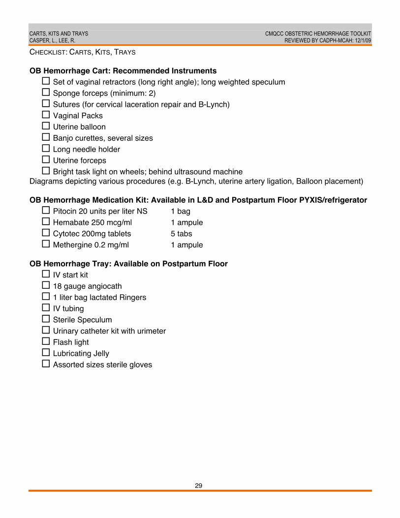
OB HEMORRHAGE: CARTS, KITS AND TRAYS Leslie Casper, MD, San Diego Medical Center, Southern California Permanente Medical Group; Richard Lee, MD, Los Angeles County and University of Southern California Medical Center BACKGROUND AND LITERATURE REVIEW Postpartum hemorrhage (PPH) is a commonly encountered obstetrical emergency on labor and delivery units throughout California. (1) Although medical management is often successful in treating PPH, the obstetrician may have to resort to surgical measures. For ideal response to the emergency, the obstetrician should have rapid access to surgical instruments and tools designed to treat PPH. Equipment and instruments compiled on an obstetrical hemorrhage “cart” is designed to treat vaginal/cervical lacerations and perform uterine tamponade or uterine/ovarian artery ligation. In short, the cart would have all the instruments necessary to treat PPH before hysterectomy is considered. The reader is referred to other guidelines in this toolkit that depict the use of these techniques. For more in-depth details about the hemorrhage cart, the reader is referred to articles by T.F. Baskett. (2, 3) RECOMMENDATION Labor and delivery units construct a sterile tray that provides rapid access to instruments used to surgically treat PPH. Hysterectomy trays are separately available. EDUCATIONAL TOOLS AND SUPPORT DOCUMENTS OB Hemorrhage Cart: Recommended Instruments • Set of vaginal retractors (long right angle); long weighted speculum • Sponge forceps (minimum: 2) • Sutures (for cervical laceration repair and B-Lynch) • Vaginal Packs • Uterine balloon • Banjo curettes, several sizes • Long needle holder • Uterine forceps • Bright task light on wheels; behind ultrasound machine Diagrams depicting various procedures (e.g. B-Lynch, uterine artery ligation, Balloon
placement)

CARTS, KITS AND TRAYS CMQCC OBSTETRIC HEMORRHAGE TOOLKIT CASPER, L., LEE, R. REVIEWED BY CADPH-MCAH: 12/1/09
27
OB Hemorrhage Medication Kit: Available in L&D and Postpartum Floor PYXIS/refrigerator • Pitocin 20 units per liter NS 1 bag • Hemabate 250 mcg/ml 1 ampule • Cytotec 200mg tablets 5 tabs • Methergine 0.2 mg/ml 1 ampule
OB Hemorrhage Tray: Available on Postpartum Floor • IV start kit • 18 gauge angiocath • 1 liter bag lactated Ringers • IV tubing • Sterile Speculum • Urinary catheter kit with urimeter • Flash light • Lubricating Jelly • Assorted sizes sterile gloves
Labor and Delivery Emergency Hysterectomy Tray: Available in L&D OR Suite • 4 Towel Clips, Backhaus (perforating) 5
1/4" • 4 Mosquito, Curved, 5" • 2 Clamp, Mixter 9" • 2 Clamp, tonsil • 2 Clamp, Allis, Extra long 10" • 2 Clamp, Allis 6" • 2 Clamp, Babcock 8" • 2 Clamp, Babcock 6 1/4" • 2 Clamp, Lahey 6" • 2 Clamp, Heaney-Rezak, Straight, 8"
• 8 Kelly, Curved 5 3/4" • 2 Kelly, Straight 5 3/4" • 8 Pean Curved, 6 1/4"
• 2 Forceps, Debakey, 9 1/2" • 1 Forceps, Tissue with teeth 9 3/4" • 1 Forceps, Russian 8" • 1 Forceps, Smooth 8"
• 1 Forceps, Ferris Smith • 2 Forceps with Teeth, 6 " • 1 Forceps, Russian 6" • 2 Forceps, Adson with Teeth • 1 Forceps, Tissue, Smooth, 7"
• 2 Kocher, Straight, 8" • 6 Forceps, Heaney, Curved, 8 1/4" • NH, Mayo Hegar, 8" • 4 Sponge Stick, 9 1/2" • 1 Scissor, Jorgensen, Curved, 9" • 1 Scissors, bandage 7" • 1 Scissors, curved dissecting,
Metzenbaum • 1 Scissors, Mayo, curved • 1 Scissors, sharp/blunt, Straight, 5 1/2' • 1 Scissors, Curved Metzenbaum 12" • 1 Scissors, Mayo Straight 11" • 1 Scissors, Mayo Curved 11"
• 1 Knife Handle #3 • 1 Knife Handle #4 • 1 Knife Handle #3, Long • 1 Retractor, Kelly, large • 1 Retractor, Deaver, Large, 3" x 12" • 1 Retractor, Deaver, Medium • 2 Retractor, Med/large Richardson • 1 Retractor, Balfour Blades • 2 Retractor, Goulet, 7 1/2" • 1 Suction, Yankauer Tip • 1 Suction, Pool Tip

CARTS, KITS AND TRAYS CMQCC OBSTETRIC HEMORRHAGE TOOLKIT CASPER, L., LEE, R. REVIEWED BY CADPH-MCAH: 12/1/09
28
EVIDENCE GRADING Level of Evidence: III. Opinions of respected authorities based on clinical experience, descriptive studies or reports of expert committees. Level of Evidence: II-3. Evidence obtained from multiple time series with or without intervention. Dramatic results in uncontrolled experiments also could be regarded as this type of evidence. Strong quality improvement data such as statistical process control, or other well-designed analysis.
CARTS, KITS AND TRAYS CMQCC OBSTETRIC HEMORRHAGE TOOLKIT CASPER, L., LEE, R. REVIEWED BY CADPH-MCAH: 12/1/09
29
CHECKLIST: CARTS, KITS, TRAYS OB Hemorrhage Cart: Recommended Instruments
Set of vaginal retractors (long right angle); long weighted speculum Sponge forceps (minimum: 2) Sutures (for cervical laceration repair and B-Lynch) Vaginal Packs Uterine balloon Banjo curettes, several sizes Long needle holder Uterine forceps Bright task light on wheels; behind ultrasound machine
Diagrams depicting various procedures (e.g. B-Lynch, uterine artery ligation, Balloon placement) OB Hemorrhage Medication Kit: Available in L&D and Postpartum Floor PYXIS/refrigerator
Pitocin 20 units per liter NS 1 bag Hemabate 250 mcg/ml 1 ampule Cytotec 200mg tablets 5 tabs Methergine 0.2 mg/ml 1 ampule
OB Hemorrhage Tray: Available on Postpartum Floor IV start kit 18 gauge angiocath 1 liter bag lactated Ringers IV tubing Sterile Speculum Urinary catheter kit with urimeter Flash light Lubricating Jelly Assorted sizes sterile gloves

CARTS, KITS AND TRAYS CMQCC OBSTETRIC HEMORRHAGE TOOLKIT CASPER, L., LEE, R. REVIEWED BY CADPH-MCAH: 12/1/09
30
Labor and Delivery Emergency Hysterectomy Tray: Available in L&D OR Suite 4 Towel Clips, Backhaus (perforating)
5 1/4" 4 Mosquito, Curved, 5" 2 Clamp, Mixter 9" 2 Clamp, tonsil 2 Clamp, Allis, Extra long 10" 2 Clamp, Allis 6" 2 Clamp, Babcock 8" 2 Clamp, Babcock 6 1/4" 2 Clamp, Lahey 6" 2 Clamp, Heaney-Rezak, Straight, 8" 8 Kelly, Curved 5 3/4" 2 Kelly, Straight 5 3/4" 8 Pean Curved, 6 1/4" 2 Forceps, Debakey, 9 1/2" 1 Forceps, Tissue with teeth 9 3/4" 1 Forceps, Russian 8" 1 Forceps, Smooth 8" 1 Forceps, Ferris Smith 2 Forceps with Teeth, 6 " 1 Forceps, Russian 6" 2 Forceps, Adson with Teeth 1 Forceps, Tissue, Smooth, 7"
2 Kocher, Straight, 8" 6 Forceps, Heaney, Curved, 8 1/4" NH, Mayo Hegar, 8" 4 Sponge Stick, 9 1/2" 1 Scissor, Jorgensen, Curved, 9" 1 Scissors, bandage 7" 1 Scissors, curved dissecting,
Metzenbaum 1 Scissors, Mayo, curved 1 Scissors, sharp/blunt, Straight, 5 1/2' 1 Scissors, Curved Metzenbaum 12" 1 Scissors, Mayo Straight 11" 1 Scissors, Mayo Curved 11" 1 Knife Handle #3 1 Knife Handle #4 1 Knife Handle #3, Long 1 Retractor, Kelly, large 1 Retractor, Deaver, Large, 3" x 12" 1 Retractor, Deaver, Medium 2 Retractor, Med/large Richardson 1 Retractor, Balfour Blades 2 Retractor, Goulet, 7 1/2" 1 Suction, Yankauer Tip 1 Suction, Pool Tip
CARTS, KITS AND TRAYS CMQCC OBSTETRIC HEMORRHAGE TOOLKIT CASPER, L., LEE, R. REVIEWED BY CADPH-MCAH: 12/1/09
31
REFERENCES 1. Lu MC, Fridman M, Korst LM, Gregory KD, Reyes C, Hobel CJ, et al. Variations in the
incidence of postpartum hemorrhage across hospitals in California. Maternal Child Health Journal 2005 September;9(3):297-306.
2. Baskett T. Equipment tray for postpartum hemorrhage in A Textbook of PostPartum Hemorrhage. (ed C B-Lynch et al) Sapiens Publishing 2006 (III) 2006.
3. Baskett TF. Surgical management of severe obstetric hemorrhage: experience with an obstetric hemorrhage equipment tray. J Obstet Gynaecol Can 2004 Sep;26(9):805-8.

CMQCC OBSTETRIC HEMORRHAGE TOOLKIT OBSTETRIC HEMORRHAGE CARE GUIDELINES AND COMPENDIUM OF BEST PRACTICES REVIEWED BY CADPH-MCAH: 1/6/10
32
SIMULATIONS AND DRILLS Leslie Casper, MD, San Diego Medical Center, Southern California Permanente Medical Group BACKGROUND AND LITERATURE REVIEW Medical simulation drills of obstetrical hemorrhage cases can assess system weaknesses and strengths, test policies and procedures for coping with hemorrhage and improve teamwork and communication skills of staff members. Drills that include all disciplines (obstetrics, anesthesia, pediatrics and nursing) can be especially effective in improving communication and coordination among team members. Drills are practice sessions of relatively uncommon but critical events, such as antenatal or postpartum hemorrhage and amniotic fluid embolism. Critical Event Training simulations for all physicians, midwives, anesthesiologists and nurses may improve neonatal outcomes. (1) Implementing a rapid response team and addressing systemsʼ issues for management of obstetrical hemorrhage has been shown to decrease maternal mortality and improve outcomes. (2) The Joint Commission recommends team training in their 2005 Executive Summary of Strategies to Improve the Medical Liability System and Prevent Patient Injury. (3) Human factors training can improve communications and teamwork. Such training includes briefings, handoffs, time-outs and situational awareness for the team, which is a shared understanding of what is happening now and what happens next. Explicit communication skills to be taught include: addressing team members by name, making eye contact, repeating back orders and confirming that you are responding to an order, and not speaking to the room and assuming that you were heard. In addition, the concept of “Just Culture” or a similar environment should be implemented in all health care settings so that all team members feel respected and comfortable with asserting observations, suggestions and opinions. Improving team communications skills is one of the Joint Commission 2009 National Patient Safety Goals. Scenarios for simulation should be designed for the needs of the learners (nurses, physicians, residents, respiratory therapy, etc.) and tailored to available resources. Interdisciplinary training should include all disciplines involved in the care of obstetric patients. Simulation can be low tech—using live models—or high tech, using complex computerized simulators or a combination of both. The objective of simulation is to create situations that are as similar to “real life” as possible. Simulation in situ may improve ability to address systems issues and provides practice in oneʼs own hospital setting with familiar resources. Simulation in a computerized simulation center offers high technology in an environment similar to real life, but without the distractions of the hospital. The choice of high or low fidelity simulation is institution dependent; both can work well for hemorrhage scenarios. For practicing complex events requiring a maternal cardiorespiratory arrest, high fidelity may be a better choice since chest compressions cannot be performed on a

SIMULATIONS AND DRILLS CMQCC OBSTETRIC HEMORRHAGE TOOLKIT CASPER, L. REVIEWED BY CADPH-MCAH: 1/6/10
33
live model, for example. Some institutions use a combination of both types. Debriefing is appropriate both for simulation drills and for live events. Video taken during simulation serves as a realistic debriefing tool to explore what went well and what needs improvement after a scenario is performed. Evaluation tools such as checklists for expectations of each participant in their role and for team and individual performances can provide an objective approach to debriefing. Similarly, follow-up evaluation ensures that specific goals and objectives for each level of participant are met. The Ottawa Crisis Resource Management Global Rating Scale and Mayo High Performance Teamwork Scale are examples. (4, 5) RECOMMENDATION All hospitals adopt regularly scheduled simulation drills for practicing response to obstetric hemorrhage. The choice of high or low fidelity drills is institution dependent; both can work well for hemorrhage scenarios. EDUCATIONAL TOOLS
1. Obstetric Hemorrhage Sample Scenario 1: Drill for Abruptio Placentae (attached) 2. Obstetric Hemorrhage Sample Scenario 2; Drill for Placenta Previa (attached) 3. Obstetric Hemorrhage Sample Scenario 3: Hemorrhage and Hypotension (attached) 4. Obstetric Hemorrhage Sample Scenario 4: Atonic Uterus (attached) 5. Kaiser Evaluation Form for Drills; Debriefing Tool: “Labor and Delivery/Family Centered
Care, Mock Obstetrical Hemorrhage, Roles and Responsibilities of Staff Skills Validation” (attached)
6. Dreyfus Model of Skill Acquisition (attached) (6) 7. Ottawa Crisis Resource Management Global Rating Scale (4) 8. Mayo High Performance Team Work Scale (5)
EVIDENCE GRADING Level of Evidence: B. Recommendations based on limited or inconsistent evidence.
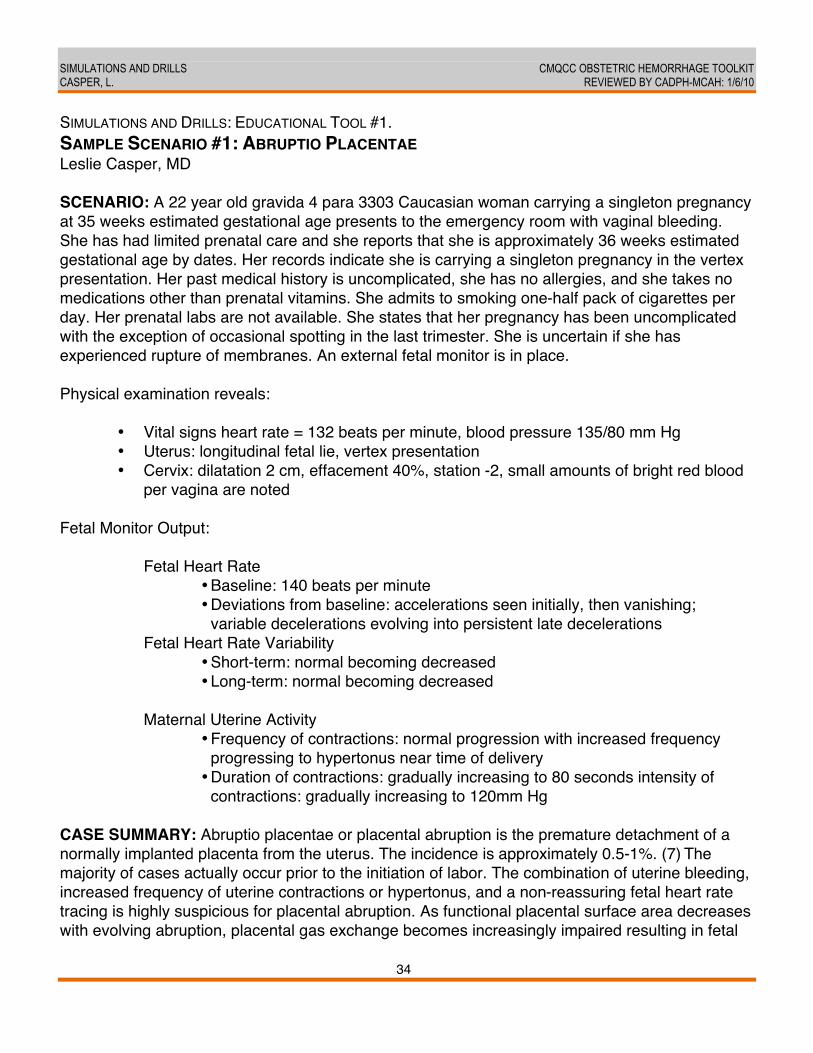
SIMULATIONS AND DRILLS CMQCC OBSTETRIC HEMORRHAGE TOOLKIT CASPER, L. REVIEWED BY CADPH-MCAH: 1/6/10
34
SIMULATIONS AND DRILLS: EDUCATIONAL TOOL #1. SAMPLE SCENARIO #1: ABRUPTIO PLACENTAE Leslie Casper, MD SCENARIO: A 22 year old gravida 4 para 3303 Caucasian woman carrying a singleton pregnancy at 35 weeks estimated gestational age presents to the emergency room with vaginal bleeding. She has had limited prenatal care and she reports that she is approximately 36 weeks estimated gestational age by dates. Her records indicate she is carrying a singleton pregnancy in the vertex presentation. Her past medical history is uncomplicated, she has no allergies, and she takes no medications other than prenatal vitamins. She admits to smoking one-half pack of cigarettes per day. Her prenatal labs are not available. She states that her pregnancy has been uncomplicated with the exception of occasional spotting in the last trimester. She is uncertain if she has experienced rupture of membranes. An external fetal monitor is in place. Physical examination reveals:
• Vital signs heart rate = 132 beats per minute, blood pressure 135/80 mm Hg • Uterus: longitudinal fetal lie, vertex presentation • Cervix: dilatation 2 cm, effacement 40%, station -2, small amounts of bright red blood
per vagina are noted
Fetal Monitor Output: Fetal Heart Rate
• Baseline: 140 beats per minute • Deviations from baseline: accelerations seen initially, then vanishing;
variable decelerations evolving into persistent late decelerations Fetal Heart Rate Variability
• Short-term: normal becoming decreased • Long-term: normal becoming decreased
Maternal Uterine Activity
• Frequency of contractions: normal progression with increased frequency progressing to hypertonus near time of delivery • Duration of contractions: gradually increasing to 80 seconds intensity of
contractions: gradually increasing to 120mm Hg CASE SUMMARY: Abruptio placentae or placental abruption is the premature detachment of a normally implanted placenta from the uterus. The incidence is approximately 0.5-1%. (7) The majority of cases actually occur prior to the initiation of labor. The combination of uterine bleeding, increased frequency of uterine contractions or hypertonus, and a non-reassuring fetal heart rate tracing is highly suspicious for placental abruption. As functional placental surface area decreases with evolving abruption, placental gas exchange becomes increasingly impaired resulting in fetal

SIMULATIONS AND DRILLS CMQCC OBSTETRIC HEMORRHAGE TOOLKIT CASPER, L. REVIEWED BY CADPH-MCAH: 1/6/10
35
hypoxemia and acidosis. A retroplacental clot is often found upon inspection of the placenta after delivery. Abruptio placentae is classified as follows: (8)
• Grade 1: mild vaginal bleeding and uterine irritability • Grade 2: moderate bleeding with increased uterine irritability or tetany; maternal
supine hypotension and tachycardia; evidence of fetal distress on fetal monitor tracings
• Grade 3: severe bleeding; uterine tetany; maternal hypotension and coagulopathy Ultrasound will help to differentiate placental abruption from placenta previa. Placental abruption has been associated with maternal hypertension, chorioamnionitis, advanced maternal age, advanced parity, maternal trauma, and ingestion of cocaine or tobacco. (8, 9) Although the incidence of placental abruption is <1%, it accounts for a significant percentage of perinatal mortality. (9, 10) Management of abruptio placentae can be difficult. While a grade 1 abruption in a woman carrying a full term fetus mandates close monitoring and active management of labor, more advanced degrees of abruption may require emergent operative delivery due to significant risk to both mother and fetus.
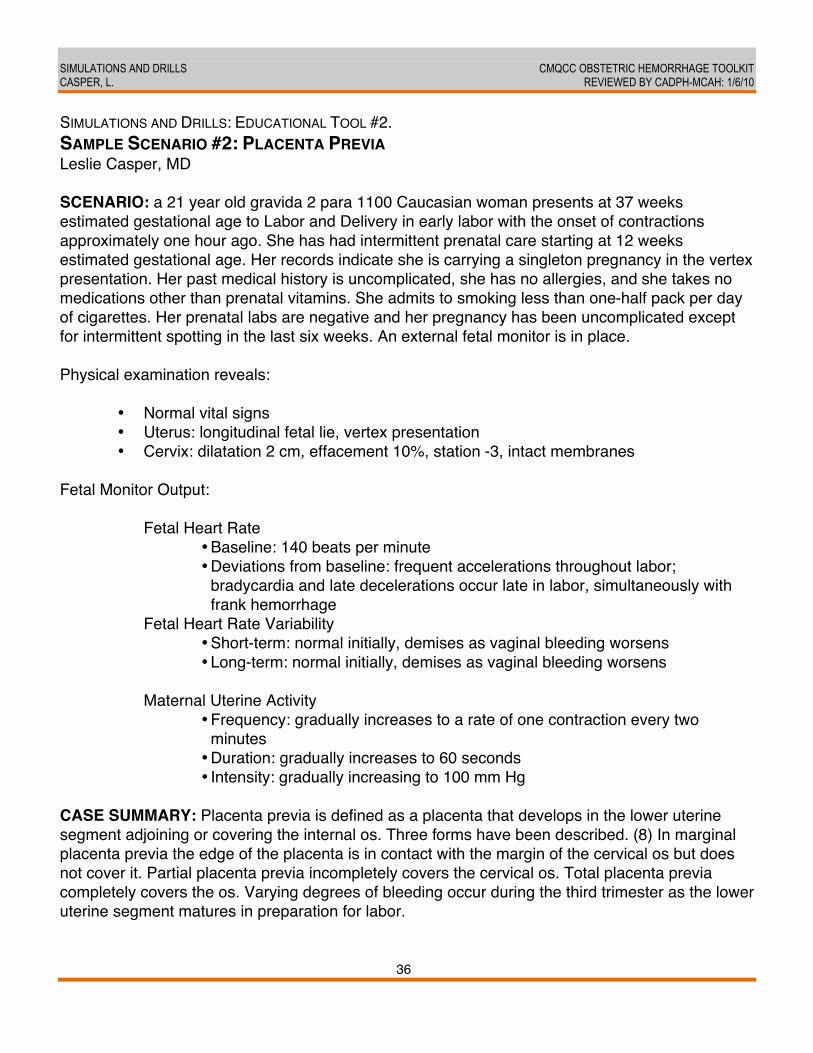
SIMULATIONS AND DRILLS CMQCC OBSTETRIC HEMORRHAGE TOOLKIT CASPER, L. REVIEWED BY CADPH-MCAH: 1/6/10
36
SIMULATIONS AND DRILLS: EDUCATIONAL TOOL #2. SAMPLE SCENARIO #2: PLACENTA PREVIA Leslie Casper, MD SCENARIO: a 21 year old gravida 2 para 1100 Caucasian woman presents at 37 weeks estimated gestational age to Labor and Delivery in early labor with the onset of contractions approximately one hour ago. She has had intermittent prenatal care starting at 12 weeks estimated gestational age. Her records indicate she is carrying a singleton pregnancy in the vertex presentation. Her past medical history is uncomplicated, she has no allergies, and she takes no medications other than prenatal vitamins. She admits to smoking less than one-half pack per day of cigarettes. Her prenatal labs are negative and her pregnancy has been uncomplicated except for intermittent spotting in the last six weeks. An external fetal monitor is in place. Physical examination reveals:
• Normal vital signs • Uterus: longitudinal fetal lie, vertex presentation • Cervix: dilatation 2 cm, effacement 10%, station -3, intact membranes
Fetal Monitor Output: Fetal Heart Rate
• Baseline: 140 beats per minute • Deviations from baseline: frequent accelerations throughout labor;
bradycardia and late decelerations occur late in labor, simultaneously with frank hemorrhage
Fetal Heart Rate Variability • Short-term: normal initially, demises as vaginal bleeding worsens • Long-term: normal initially, demises as vaginal bleeding worsens
Maternal Uterine Activity
• Frequency: gradually increases to a rate of one contraction every two minutes • Duration: gradually increases to 60 seconds • Intensity: gradually increasing to 100 mm Hg
CASE SUMMARY: Placenta previa is defined as a placenta that develops in the lower uterine segment adjoining or covering the internal os. Three forms have been described. (8) In marginal placenta previa the edge of the placenta is in contact with the margin of the cervical os but does not cover it. Partial placenta previa incompletely covers the cervical os. Total placenta previa completely covers the os. Varying degrees of bleeding occur during the third trimester as the lower uterine segment matures in preparation for labor.
SIMULATIONS AND DRILLS CMQCC OBSTETRIC HEMORRHAGE TOOLKIT CASPER, L. REVIEWED BY CADPH-MCAH: 1/6/10
37
The incidence of placenta previa is approximately 0.4%. Risk factors include previous cesarean section and tobacco use. (9, 11) The management of a pregnancy complicated by placenta previa is dependent on first recognition of the presence of the abnormally located placenta; this is usually done by ultrasound (digital examination may inadvertently lead to severe hemorrhage). A history of prior cesarean section or total placenta previa likely mandates operative delivery. In patients with marginal or partial placenta previa, as in this case, vaginal delivery may be attempted, provided an emergency cesarean section can be performed should uncontrollable hemorrhage result. The descending fetal head often places pressure on the edge of the placenta and may act to limit bleeding. In this case fetal bradycardia and late decelerations ensue after frank hemorrhage develops late in labor.
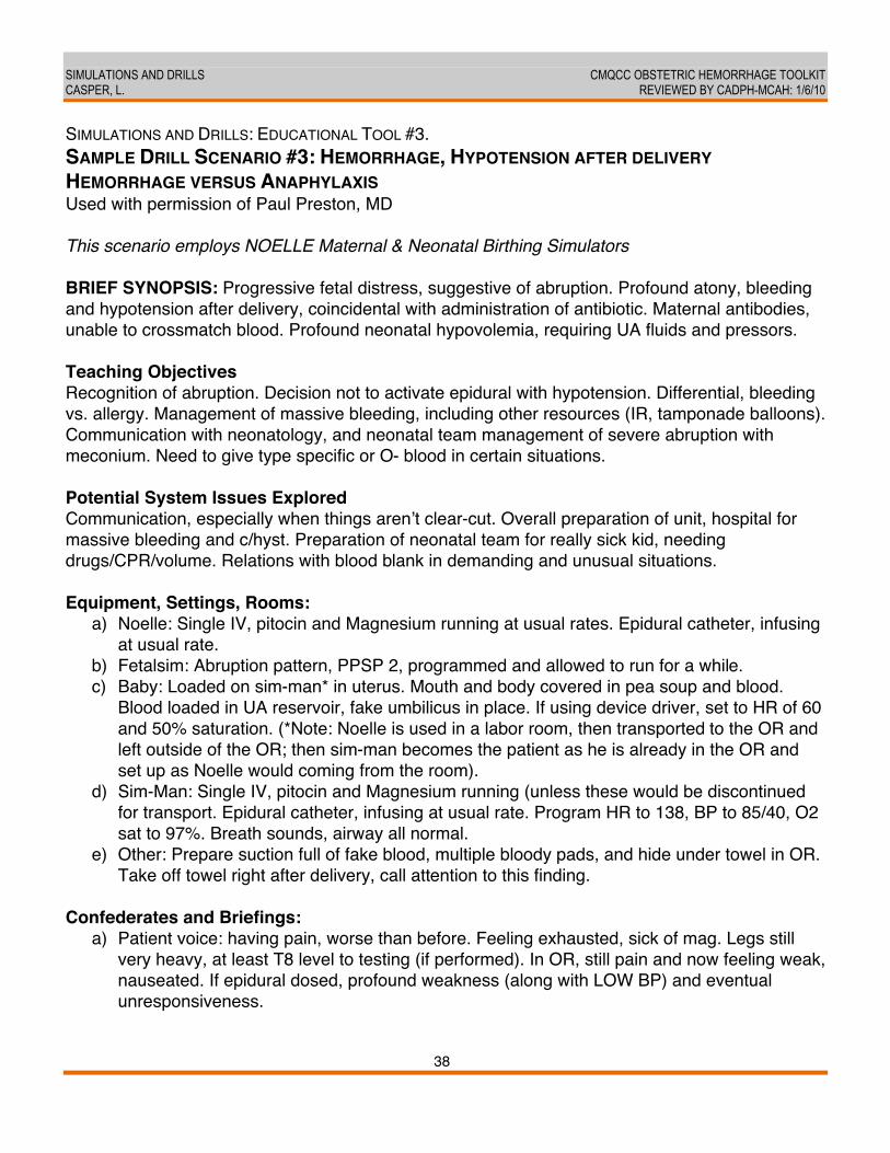
SIMULATIONS AND DRILLS CMQCC OBSTETRIC HEMORRHAGE TOOLKIT CASPER, L. REVIEWED BY CADPH-MCAH: 1/6/10
38
SIMULATIONS AND DRILLS: EDUCATIONAL TOOL #3. SAMPLE DRILL SCENARIO #3: HEMORRHAGE, HYPOTENSION AFTER DELIVERY HEMORRHAGE VERSUS ANAPHYLAXIS Used with permission of Paul Preston, MD This scenario employs NOELLE Maternal & Neonatal Birthing Simulators BRIEF SYNOPSIS: Progressive fetal distress, suggestive of abruption. Profound atony, bleeding and hypotension after delivery, coincidental with administration of antibiotic. Maternal antibodies, unable to crossmatch blood. Profound neonatal hypovolemia, requiring UA fluids and pressors. Teaching Objectives Recognition of abruption. Decision not to activate epidural with hypotension. Differential, bleeding vs. allergy. Management of massive bleeding, including other resources (IR, tamponade balloons). Communication with neonatology, and neonatal team management of severe abruption with meconium. Need to give type specific or O- blood in certain situations. Potential System Issues Explored Communication, especially when things arenʼt clear-cut. Overall preparation of unit, hospital for massive bleeding and c/hyst. Preparation of neonatal team for really sick kid, needing drugs/CPR/volume. Relations with blood blank in demanding and unusual situations. Equipment, Settings, Rooms:
a) Noelle: Single IV, pitocin and Magnesium running at usual rates. Epidural catheter, infusing at usual rate.
b) Fetalsim: Abruption pattern, PPSP 2, programmed and allowed to run for a while. c) Baby: Loaded on sim-man* in uterus. Mouth and body covered in pea soup and blood.
Blood loaded in UA reservoir, fake umbilicus in place. If using device driver, set to HR of 60 and 50% saturation. (*Note: Noelle is used in a labor room, then transported to the OR and left outside of the OR; then sim-man becomes the patient as he is already in the OR and set up as Noelle would coming from the room).
d) Sim-Man: Single IV, pitocin and Magnesium running (unless these would be discontinued for transport. Epidural catheter, infusing at usual rate. Program HR to 138, BP to 85/40, O2 sat to 97%. Breath sounds, airway all normal.
e) Other: Prepare suction full of fake blood, multiple bloody pads, and hide under towel in OR. Take off towel right after delivery, call attention to this finding.
Confederates and Briefings:
a) Patient voice: having pain, worse than before. Feeling exhausted, sick of mag. Legs still very heavy, at least T8 level to testing (if performed). In OR, still pain and now feeling weak, nauseated. If epidural dosed, profound weakness (along with LOW BP) and eventual unresponsiveness.

SIMULATIONS AND DRILLS CMQCC OBSTETRIC HEMORRHAGE TOOLKIT CASPER, L. REVIEWED BY CADPH-MCAH: 1/6/10
39
b) Partner: (optional) Nervous, wants to accompany patient, but reasonable—will stay behind if given good explanation.
Briefing for team Mrs. Jones is a wonderful lady—itʼs painful to see her having such an awful labor. 38YO, G2P1, induced for moderate pre-eclampsia. On mag and pit. Prior section for breech, really wants vag delivery. Slow labor all night, minimal sleep. CBC, labs—compatible with moderate pre-eclampsia, not HELLP. Finally got epidural 2 hours ago, now calling RN for something. If asked, no other major illness or known allergy, normal height and weight, last BP was 150/82, last HR 110, last cervical check 4 cm. How Scenario Runs
a) RN arrives, patient feels pain. Strip looks progressively more ominous, but without catastrophic decel. Maternal BP starts at 110/50, HR of 122, progressive deterioration. Good sensory level to epidural. On cervical exam, still 4 cm, thick mec with blood. Advance the Fetalsim appropriately to make sure we go to OR.
b) In OR, low BP noted and strip keeps looking worse. Class 2 airway if examined. If epidural activated, profound hypotension/near cardiac arrest. If GA induced, reward light doses of anesthetic. Airway not a problem. Does discussion of probably shock, uterine rupture, choice of anesthetic, advance planning for bleeding occur?
c) Baby delivered---does peds get briefed about blood and mec and probable abruption? How well are they able to do this resuscitation? NALS instructor—donʼt let them get baby back until they actually cannulate cord and give volume.
d) After delivery, while Kefzol being given, really drop the BP—70/30, add some PVCs. Show bloody suction, rags to team. Uterus is totally boggy, tone is terrible. Give some credit for pressors and lighter anesthesia, but not enough to feel safe—push them to order blood--her color looks very pale. Stat crit---14, if ordered. How well prepared is OR team to do a C/hyst? Start more IV access and give blood? Blood bank is reluctant to release blood (“Can you just give us another 30 minutes to work on this antibody?”) Requires clear communication to release uncrossmatched blood.
Key Times to be Alert
a) Recognition of rupture, communication, decision to go to CS. b) Briefings in OR-situation, shock, baby, choice of anesthesia? c) Management of bleeding d) Management of baby
Why This Scenario? Bleeding is commonly seen. Many of our facilities report systems issues in handling this problem, often related to blood bank. Overall, a good discussion of system strengths and weaknesses results. New information on uterine tamponade balloons, best practices for blood storage (coolers work if no fridge), clear planning for requesting emergency blood will often result.

SIMULATIONS AND DRILLS CMQCC OBSTETRIC HEMORRHAGE TOOLKIT CASPER, L. REVIEWED BY CADPH-MCAH: 1/6/10
40
SIMULATIONS AND DRILLS: EDUCATIONAL TOOL #4. SAMPLE SCENARIO #4: ATONIC UTERUS AND POSTPARTUM HEMORRHAGE (12) Used with permission from Martin P. Eason, MD, JD Participants: OB residents, FM residents. Learning Objectives At the end of the session the participants will be able to:
1) recognize the signs of uterine atony 2) appropriately treat uterine atony and hemorrhage
a. use correct medications b. order appropriate labs c. ensure resuscitation measures are instituted d. make decision to treat surgically
Simulation Overview This case involves the condition of uterine atony. It is the most common cause of significant obstetric bleeding. It may be associated with placenta previa, placental abruption, retained placenta, or occur alone. Factors associated with uterine atony include multiple gestation, macrosomia, polyhydramnios, high parity, prolonged labor, excessive use of oxytocin, and chorioamnionitis. An atonic uterus may contain up to 1L of blood. Although rarely life threatening, uterine atony can cause severe postpartum hemorrhage and hypotension. The problem should be recognized and treated quickly. Treatment should include administration of appropriate medication to stimulate uterine contractions (oxytocin, Hemabate---prostglandin F2 alpha, ergonivine) and management of hypovolemic shock. Postpartum hemorrhage should be treated as follows:
1. Initiate appropriate general resuscitation principals (ABCʼs) 2. Ask for assistance 3. Place large bore intravenous lines 4. Order blood tests and blood products 5. Begin volume replacement with crystalloid and/or colloid solutions 6. Consider invasive monitoring 7. Treat bleeding disorders if present 8. Monitor urine output 9. Consider use of vasopressors
Patient History Patient is a 32 year old G4P3 female with no prenatal care who presents in labor. She is crowning and ready for delivery. She is a stat transfer from the ED.
SIMULATIONS AND DRILLS CMQCC OBSTETRIC HEMORRHAGE TOOLKIT CASPER, L. REVIEWED BY CADPH-MCAH: 1/6/10
41
Patient History, continued PMH: Asthma PSH: None Meds: albuterol 2 puffs BID prn All: bee stings SHx: married; husband is rushing from work. Smokes ½ pack per day; no ETOH; she wants more children FHx: none ROS: labor started about 2 hours ago. Water broke 45 minutes ago; clear Physical examination (provided only if asked) Cardiac: tachycardic otherwise normal Lungs: clear bilaterally Neck: supple Labs: None initially available If ordered: CBC Hct 35% Platelets 235; WBC 8.2 Chem 7 WNL PT/PTT 9.5/26 seconds Simulation Parameters Initial Parameters BP: 135/78 Hr: 96 RR: 20
Sat: 98% Heart Sounds: Normal
Lung sounds: clear Scenario Run The patient will present with the fetal head crowning; she will be wanted to push. The baby will be delivered OA without complications. Immediately after the placenta is removed, blood will come from the vaginal opening. If palpated the uterus will soft. It will remain so despite medications. The blood pressure will drop from the initial readings over the next 10 minutes to 70/40. Oxytocics will not cause uterine contraction. If Hemabate is given, the patient will complain of shortness of breath and her sats will drop to 80%. If the lungs are auscultated, wheezing will be evident. Rise in blood pressure will depend on replacement of volume. If large bore IVs are placed rapidly and put on pressure bags, the pressure will initially improve to 80/45 but will slowly (over five minutes)
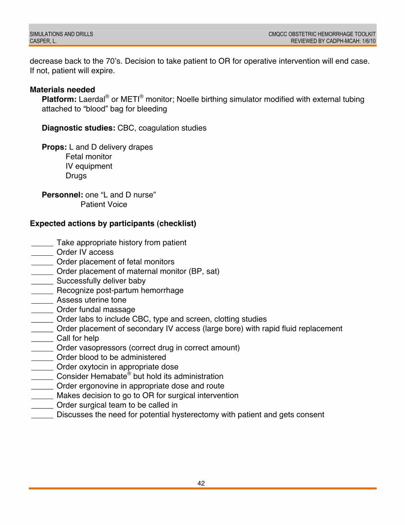
SIMULATIONS AND DRILLS CMQCC OBSTETRIC HEMORRHAGE TOOLKIT CASPER, L. REVIEWED BY CADPH-MCAH: 1/6/10
42
decrease back to the 70ʼs. Decision to take patient to OR for operative intervention will end case. If not, patient will expire. Materials needed
Platform: Laerdal® or METI® monitor; Noelle birthing simulator modified with external tubing attached to “blood” bag for bleeding Diagnostic studies: CBC, coagulation studies Props: L and D delivery drapes Fetal monitor IV equipment Drugs Personnel: one “L and D nurse” Patient Voice
Expected actions by participants (checklist) _____ Take appropriate history from patient _____ Order IV access _____ Order placement of fetal monitors _____ Order placement of maternal monitor (BP, sat) _____ Successfully deliver baby _____ Recognize post-partum hemorrhage _____ Assess uterine tone _____ Order fundal massage _____ Order labs to include CBC, type and screen, clotting studies _____ Order placement of secondary IV access (large bore) with rapid fluid replacement _____ Call for help _____ Order vasopressors (correct drug in correct amount) _____ Order blood to be administered _____ Order oxytocin in appropriate dose _____ Consider Hemabate® but hold its administration _____ Order ergonovine in appropriate dose and route _____ Makes decision to go to OR for surgical intervention _____ Order surgical team to be called in _____ Discusses the need for potential hysterectomy with patient and gets consent
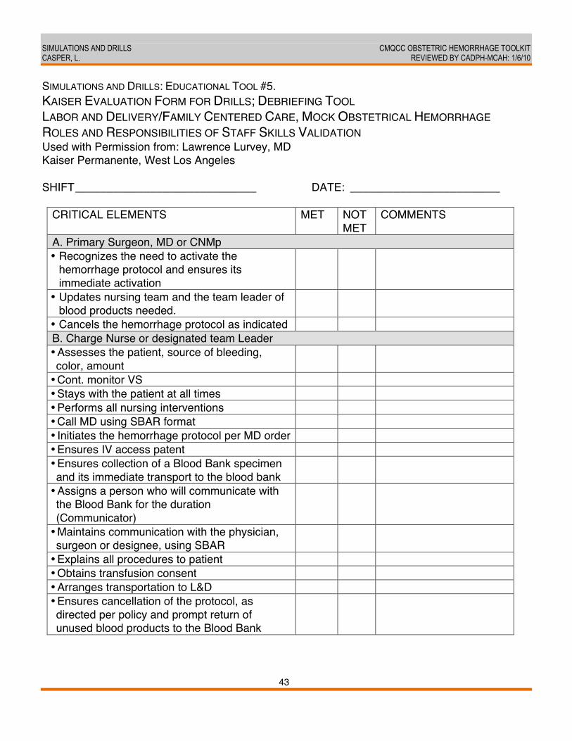
SIMULATIONS AND DRILLS CMQCC OBSTETRIC HEMORRHAGE TOOLKIT CASPER, L. REVIEWED BY CADPH-MCAH: 1/6/10
43
SIMULATIONS AND DRILLS: EDUCATIONAL TOOL #5. KAISER EVALUATION FORM FOR DRILLS; DEBRIEFING TOOL LABOR AND DELIVERY/FAMILY CENTERED CARE, MOCK OBSTETRICAL HEMORRHAGE ROLES AND RESPONSIBILITIES OF STAFF SKILLS VALIDATION Used with Permission from: Lawrence Lurvey, MD Kaiser Permanente, West Los Angeles SHIFT_____________________________ DATE: ________________________
CRITICAL ELEMENTS MET NOT MET
COMMENTS
A. Primary Surgeon, MD or CNMp • Recognizes the need to activate the
hemorrhage protocol and ensures its immediate activation
• Updates nursing team and the team leader of blood products needed.
• Cancels the hemorrhage protocol as indicated B. Charge Nurse or designated team Leader • Assesses the patient, source of bleeding, color, amount
• Cont. monitor VS • Stays with the patient at all times • Performs all nursing interventions • Call MD using SBAR format • Initiates the hemorrhage protocol per MD order • Ensures IV access patent • Ensures collection of a Blood Bank specimen and its immediate transport to the blood bank
• Assigns a person who will communicate with the Blood Bank for the duration (Communicator)
• Maintains communication with the physician, surgeon or designee, using SBAR
• Explains all procedures to patient • Obtains transfusion consent • Arranges transportation to L&D • Ensures cancellation of the protocol, as directed per policy and prompt return of unused blood products to the Blood Bank
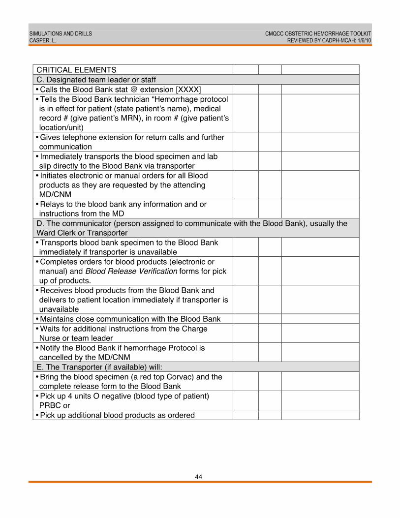
SIMULATIONS AND DRILLS CMQCC OBSTETRIC HEMORRHAGE TOOLKIT CASPER, L. REVIEWED BY CADPH-MCAH: 1/6/10
44
CRITICAL ELEMENTS C. Designated team leader or staff • Calls the Blood Bank stat @ extension [XXXX] • Tells the Blood Bank technician “Hemorrhage protocol is in effect for patient (state patientʼs name), medical record # (give patientʼs MRN), in room # (give patientʼs location/unit)
• Gives telephone extension for return calls and further communication
• Immediately transports the blood specimen and lab slip directly to the Blood Bank via transporter
• Initiates electronic or manual orders for all Blood products as they are requested by the attending MD/CNM
• Relays to the blood bank any information and or instructions from the MD
D. The communicator (person assigned to communicate with the Blood Bank), usually the Ward Clerk or Transporter • Transports blood bank specimen to the Blood Bank immediately if transporter is unavailable
• Completes orders for blood products (electronic or manual) and Blood Release Verification forms for pick up of products.
• Receives blood products from the Blood Bank and delivers to patient location immediately if transporter is unavailable
• Maintains close communication with the Blood Bank • Waits for additional instructions from the Charge Nurse or team leader
• Notify the Blood Bank if hemorrhage Protocol is cancelled by the MD/CNM
E. The Transporter (if available) will: • Bring the blood specimen (a red top Corvac) and the complete release form to the Blood Bank
• Pick up 4 units O negative (blood type of patient) PRBC or
• Pick up additional blood products as ordered
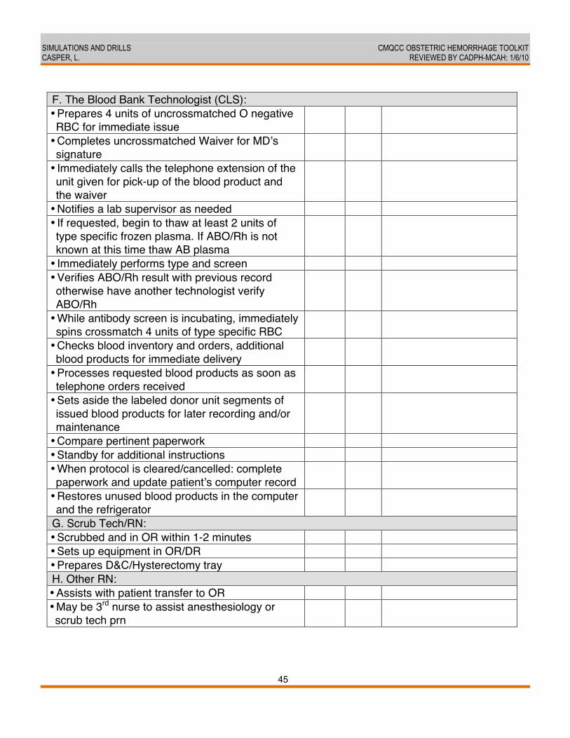
SIMULATIONS AND DRILLS CMQCC OBSTETRIC HEMORRHAGE TOOLKIT CASPER, L. REVIEWED BY CADPH-MCAH: 1/6/10
45
F. The Blood Bank Technologist (CLS): • Prepares 4 units of uncrossmatched O negative RBC for immediate issue
• Completes uncrossmatched Waiver for MDʼs signature
• Immediately calls the telephone extension of the unit given for pick-up of the blood product and the waiver
• Notifies a lab supervisor as needed • If requested, begin to thaw at least 2 units of type specific frozen plasma. If ABO/Rh is not known at this time thaw AB plasma
• Immediately performs type and screen • Verifies ABO/Rh result with previous record otherwise have another technologist verify ABO/Rh
• While antibody screen is incubating, immediately spins crossmatch 4 units of type specific RBC
• Checks blood inventory and orders, additional blood products for immediate delivery
• Processes requested blood products as soon as telephone orders received
• Sets aside the labeled donor unit segments of issued blood products for later recording and/or maintenance
• Compare pertinent paperwork • Standby for additional instructions • When protocol is cleared/cancelled: complete paperwork and update patientʼs computer record
• Restores unused blood products in the computer and the refrigerator
G. Scrub Tech/RN: • Scrubbed and in OR within 1-2 minutes • Sets up equipment in OR/DR • Prepares D&C/Hysterectomy tray H. Other RN: • Assists with patient transfer to OR • May be 3rd nurse to assist anesthesiology or scrub tech prn

SIMULATIONS AND DRILLS CMQCC OBSTETRIC HEMORRHAGE TOOLKIT CASPER, L. REVIEWED BY CADPH-MCAH: 1/6/10
46
SIMULATIONS AND DRILLS: EDUCATIONAL TOOL #6. DREYFUS MODEL OF SKILL ACQUISITION (6, 13)
REFERENCES 1. Draycott T, Sibanda T, Owen L, Akande V, Winter C, Reading S, et al. Does training in
obstetric emergencies improve neonatal outcome? BJOG 2005;113(2):177-82. 2. Skupski DW, Lowenwirt IP, Weinbaum FI, Brodsky D, Danek M, Eglinton GS. Improving
hospital systems for the care of women with major obstetric hemorrhage. Obstetrics and Gynecology 2006;107(5):977-83.
DREYFUS MODEL OF SKILL ACQUISITION
Level 1 Novice Rigid adherence to taught rules or plans Little situational perception No discretionary judgment
Observer Observes the procedure performed by a colleague
Level 2 Advanced Beginner Guidelines for action based on attributes or aspects Situational perception still limited All attributes and aspects are treated separately and given equal importance
Assistant Assists a colleague in performing the procedure
Level 3 Competent Coping with crowdedness Now sees actions at least partially in terms of longer-term goals Conscious deliberate planning
Directly supervised Performs the entire procedure under direct senior supervision
Level 4 Proficient See situations holistically rather than in terms of aspects See what is most important in a situation Perceives deviations from the normal pattern Decision-making less laboured Uses maxims for guidance, whose meaning varies according to the situation
Indirectly supervised Performs the entire procedure with indirect senior supervision
Level 5 Expert No longer relies on rules, guidelines, or maxims Intuitive grasp of situations based on deep tacit understanding Analytic approaches used only in novel situation or when problems occur Vision of what is possible
Independent Performs the entire activity without the need for supervision

SIMULATIONS AND DRILLS CMQCC OBSTETRIC HEMORRHAGE TOOLKIT CASPER, L. REVIEWED BY CADPH-MCAH: 1/6/10
47
3. Healthcare at the Crossroads: Strategies for Improving the Medical Liability System and Preventing Patient Injury. Joint Commission on Accreditation of Healthcare Organizations 2008.
4. Kim J, Neilipovitz D, Cardinal P, Chiu M, Clinch J. A pilot study using high-fidelity simulaiton to formally evaluate performance in the resuscitation of critically ill patients: The University Ottawa Critical Care Medicine, High-Fidelity Simulation and Crisis Resource Management I Study. Crit Care Med 2006;34:2167-74.
5. Malec J, Torsher L, Dunn W, Wiegmann D, Arnold J, Brown D, et al. The Mayo High Performance Teamwork Scale: Reliability and Validity for Evaluating Key Crew Resource Management Skills. Simulation in Healthcare 2007;2(1):4-10.
6. Dreyfus S, Dreyfus H, et al. A Five-Stage Model of the Mental Activities Involved in Directed Skill Acquisition. Available at http://www.stormingmedia.us/ (Search Term: Drefyfus), 1980.
7. Knab D. Abruptio placentae. An Assessment of the time and method of delivery. Obstet Gynecol Clin North Am 1979;52:625-9.
8. Benedetti T. Obstetric hemorrhage. In: Bragge SG, Niebyl JR, Simson JL, eds. Obstetrics: Normal and Problem Pregnancies 2E 1991:579-84.
9. Andres R. The association of cigarette smoking with placenta previa and abruptio placentae. Sem Perinatol 1996;20:154-9.
10. Acker D, BP S, Tracey K, Wise W. Abruptio placentae associated with cocaine use. Am J Obstet Gynecol 1983;146:220-1.
11. Handler A, Mason E, Rosenberg D, Davis F. The relationship between exposure during pregnancy to cigarette smoking and cocaine use and placenta previa. Am J Obstet Gynecol 1994;170:884-9.
12. Crochetiere C. Obstetric emergencies. Anesthesiology Clinics of North America 2003;21:11-125.
13. Smith C. Learning Experience of Senior House Officers in Obstetrics and Gynecology. 2005:66.
CMQCC OBSTETRIC HEMORRHAGE TOOLKIT OBSTETRIC HEMORRHAGE CARE GUIDELINES AND COMPENDIUM OF BEST PRACTICES REVIEWED BY CADPH-MCAH: 12/21/09
48
POSTPARTUM HEMORRHAGE: LESSONS LEARNED FROM OTHER STATES Larry Shields, MD, Central Coast Maternal Fetal Medicine; Jean Claude Veille, MD, Maternal-Fetal Medicine, Sutter Health BACKGROUND AND LITERATURE REVIEW We reviewed maternal hemorrhage protocols developed in the states of Washington and New York. Primary goals included improving patient care and providing guidelines that practitioners could use to respond seamlessly and consistently to life-threatening maternal hemorrhage. The protocols were not published. The Washington protocol was initially developed at a single Level III center, University of Washington, and then later expanded to other Level II and III centers in the Puget Sound Region. The protocol focused on blood product replacement and assumed that initial attempts to control bleeding had failed. Specific medical therapy management, uterine compression sutures and hysterectomy were not addressed. In contrast, the New York protocol was developed to address a more global approach to maternal hemorrhage and focused on issues related to early response by displaying educational posters in labor and delivery units. Both protocols accounted for the infrequent occurrence of life-threatening hemorrhage (incidence in California is approximately 2% of births) and addressed the occurrence of disseminated intravascular coagulation (DIC) outlining that its development significantly increases risks of maternal morbidity and death. (1, 2) Clinical knowledge and skills atrophy without regular practice; therefore, well-defined protocols improve coordination and management of obstetrical hemorrhage. The Washington protocol focused on availability of appropriate blood product replacement. The obstetrician or anesthesiologist activated the protocol when they recognized hemorrhage. Fixed quantities of packed red blood cells (RBCs), fibrinogen, fresh frozen plasma (FFP) and platelets were released from the blood bank to labor and delivery as soon as they were prepared, while blood products were administered in accordance with lab results. Additional personnel were assigned to patient care as needed; for example, a hematologist was usually available for immediate consultation. In New York State, maternal hemorrhage accounted for 52% of all maternal deaths with the vast majority occurring during hospitalization. Individual obstetrical care providers infrequently encounter major obstetrical hemorrhage and may not readily recall the type of medicines, the exact doses and the frequency of administration of these drugs. The focus of this state protocol was to decrease variability in management of acute hemorrhage among care providers. Key steps deemed necessary to manage such catastrophic acute events were printed on large posters and placed in strategic areas such as labor and delivery rooms, OR suites, antepartum and postpartum areas and in emergency departments. Space to write key phone numbers or providersʼ names was available (i.e. blood bank, anesthesia, invasive radiologist, gynecology
POSTPARTUM HEMORRHAGE: LESSONS LEARNED FROM OTHER STATES CMQCC OBSTETRIC HEMORRHAGE TOOLKIT SHIELDS, L., VEILLE, J.C. REVIEWED BY CADPH-MCAH: 12/21/09
49
oncologist or general surgeon, hematologist, interventionist and maternal fetal medicine specialists). Additionally, guidelines recommended that key medications be readily accessible for bedside response instead of requiring pharmacy requests. Hemorrhage “kits” compiled and stored in labor and delivery and OR suites added to successful response to hemorrhage. RECOMMENDATIONS
1. Every hospital has a scripted algorithm or protocol that outlines all steps for activation and response to maternal hemorrhage. Charge nurses triage and designate additional staff to respond to specific protocol steps; and all phone numbers of potentially needed personnel are included.
2. Both physician and nursing staff can activate an emergency maternal hemorrhage protocol. 3. Develop a collaborative policy/procedure with the blood bank for issue of a specified “OB
Emergency Hemorrhage Pack” that includes RBCs, FFP, cryoprecipitate and platelets. 4. Identify a local expert with experience in hemorrhage and DIC treatment for contact as
needed. 5. Post medicine doses and suture techniques in labor and delivery and OR suites for
reference. 6. Perform scheduled hemorrhage protocol drills and assessments for both physicians and
registered nurses. EVIDENCE GRADING Level of Evidence: II-3. Evidence obtained from multiple time series with or without interventions. Dramatic results in uncontrolled experiments also could be regarded as this type of evidence. Strong quality improvement data such as statistical process control or other well-designed analysis. Level of Evidence: III-C. Opinions of respected authorities, based on clinical experience, descriptive studies, or reports of expert committees (C: recommendations based primarily on consensus and expert opinion). REFERENCES 1. Lu MC, Fridman M, Korst LM, Gregory KD, Reyes C, Hobel CJ, et al. Variations in the
incidence of postpartum hemorrhage across hospitals in California. Maternal Child Health Journal 2005 September;9(3):297-306.
2. Lurie S, Feinstein M, Mamet Y. Disseminated intravascular coagulopathy in pregnancy: thorough comprehension of etiology and management reduces obstetricians' stress. Archives of gynecology and obstetrics 2000;263(3):126-30.
CMQCC OBSTETRIC HEMORRHAGE TOOLKIT OBSTETRIC HEMORRHAGE CARE GUIDELINES AND COMPENDIUM OF BEST PRACTICES REVIEWED BY CADPH-MCAH: 11/24/09
50
ACTIVE MANAGEMENT OF THIRD STAGE LABOR Andrew Hull, MD, Department of Reproductive Medicine, Division of Perinatal Medicine, University of California, San Diego; David Lagrew, MD, Maternal-Fetal Medicine, Saddleback Memorial Medical Center BACKGROUND AND LITERATURE REVIEW Active management of the third stage of labor describes techniques of expediting placental delivery rather than awaiting spontaneous completion. The most common protocol involves routine administration of oxytocin as the fetal shoulders are delivering, followed by cord clamping and cutting, and then controlled cord traction and uterine massage after delivery of the placenta. The Cochrane Review of four randomized controlled trials examining expectant versus active management concluded that active management reduced risks of the following: 1) maternal blood loss; 2) postpartum hemorrhage exceeding 500 mL; and 3) prolonged third stage labor. (1) However, actively managed patients experienced increased risk of unpleasant side effects like nausea and vomiting compared with expectant management. In studies where ergometrine was used, there was an increased incidence of hypertension. Given the benefits, this review recommends active management as the routine practice for women delivering a baby by vaginal delivery. As with all labor techniques and routine procedures, staff should be well trained on this technique. The risks and benefits should be understood by staff and explained clearly to women and their families. Active management may be controversial with some women who are medication-averse or desire spontaneous cessation of cord pulsation prior to cutting the umbilical cord. As with other components of individual birth plans, the risks and benefits of the procedure should be discussed prior to the acute setting. (2-4) Further studies are needed to identify the components of “Active Management” that result in improved outcomes, including which uterotonic medications are the most beneficial. RECOMMENDATIONS
1. Active management of third stage of labor for all vaginal births including routine administration of oxytocin with shoulder delivery, cord traction and uterine massage.
2. Inclusion of active management of third stage of labor techniques in standard policies and procedures by institutions and education of all providers on proper administration of techniques, including appropriate protocols for timing and method of oxytocin administration.

ACTIVE MANAGEMENT OF THIRD STAGE LABOR CMQCC OBSTETRIC HEMORRHAGE TOOLKIT HULL, A., LAGREW, D. REVIEWED BY CADPH-MCAH: 11/24/09
51
3. Case-reviews of excessive maternal hemorrhage should incorporate assessments of timing, documentation and performance of active management of the third stage compared to institutional protocols.
EVIDENCE GRADING Level of Evidence: IA. Evidence obtained from at least one properly designed randomized controlled trial; recommendations based on high quality and consistent evidence. REFERENCES 1. Prendiville W, Elbourne D, McDonald S. Active versus expectant management in the third
stage of labour. Cochrane Database Syste Rev 3: CD000007. 2. Lalonde A, Daviss BA, Acosta A, Herschderfer K. Postpartum hemorrhage today:
ICM/FIGO initiative 2004-2006. Int J Gynaecol Obstet 2006 Sep;94(3):243-53. 3. McDonald S. Management of the third stage of labor. J Midwifery Women's Health
2007;52:254-61. 4. Leduc D, Senikas V, Lalonde AB, Ballerman C, Biringer A, Delaney M, et al. Active
management of the third stage of labour: prevention and treatment of postpartum hemorrhage. J Obstet Gynaecol Can 2009 Oct;31(10):980-93.
CMQCC OBSTETRIC HEMORRHAGE TOOLKIT OBSTETRIC HEMORRHAGE CARE GUIDELINES AND COMPENDIUM OF BEST PRACTICES REVIEWED BY CADPH-MCAH: 1/6/10
52
BLOOD LOSS: CLINICAL TECHNIQUES FOR ONGOING QUANTITATIVE MEASUREMENT Audrey Lyndon, PhD, CNS, RNC, Department of Family Health Care Nursing, University of California, San Francisco; Suellen Miller, PhD, CNM, MHA, Department of Obstetrics, Gynecology and Reproductive Sciences, University of California, San Francisco; Valerie Huwe, RN, BSN, El Camino Hospital; Mark Rosen, MD, Department of Anesthesia, University of California, San Francisco; David Lagrew, MD, Saddleback Memorial Medical Center; Patricia Dailey, MD; Anesthesiology, Mills Peninsula Health Services; Elliott Main, MD, Department of OB/GYN, California Pacific Medical Center, Sutter Health BACKGROUND AND LITERATURE REVIEW Accurate measurement of blood loss is essential for 1) recognizing potentially life-threatening hemorrhage and 2) managing blood product replacement. While multiple methods for estimating blood loss are available, most are either impractical (e.g., acid hematin; chromium tagged RBCs) or inaccurate (e.g., visual estimation). (1-5) Visual estimation of blood loss volume is inaccurate and can underestimate postpartum blood loss by 33%-50% when compared to the gold standard for quantifying blood loss, which is photospectrometry or colorimetric measurement of alkaline hematin in blood. (3) Visual estimation of blood loss may also be complicated by the presence of a large volume of amniotic fluid, stool or sponges. Several studies demonstrate that while visual estimation of blood loss is inaccurate, especially for larger volumes, it can be improved with training and by quantification of blood loss using calibrated under-buttocks drapes to collect blood. (2-5) Average amniotic fluid volumes have been described across gestational ages from 8-43 weeks and can be approximated using a published nomogram when necessary. (6) The specific materials used to collect blood and the presence of clots may also affect accuracy of blood loss measurement. (7) Measurement of blood loss by weight is the most accurate and practical method for determining the volume of blood not captured in graduated containers. This can be accomplished by subtracting the dry weight of absorbing materials (pads, sponges, etc) from the weight of blood-containing materials and using the conversion 1 gm weight = 1 mL to quantify the blood volume contained in the materials. RECOMMENDATIONS
1. All facilities provide chart tools and regularly scheduled standardized training in formal quantitative measurement of blood loss, which is critical for early recognition of and response to maternal hemorrhage. (Level I B) Examples are provided in attachments.
BLOOD LOSS: CLINICAL TECHNIQUES FOR ONGOING QUANTITATIVE MEASUREMENT CMQCC OBSTETRIC HEMORRHAGE TOOLKIT LYNDON, A., ET AL REVIEWED BY CADPH-MCAH: 1/6/10
53
2. Quantitative measurement of blood loss should be a collaborative effort that includes nurses, anesthesia and obstetric providers.
3. For vaginal birth: a. Use under-buttock drapes, preferably with graduated markers, to collect blood
with vaginal birth. (Level I B) b. Immediately after the birth of the baby, stop to assess the amount of fluid in the
under-buttock calibrated drape. This value becomes the ʻbaselineʼ and all subsequent fluid represents blood loss.
c. When clinicians first note excessive bleeding (more than 500 ml of quantified blood loss), weigh all blood soaked materials to determine cumulative volume and evaluate sizes of blood clots. (Examples of chart tools to assist this process are provided).
4. For cesarean birth: a. After birth of the baby, suction all amniotic fluid and stop to assess the amount of
collected fluid before delivery of the placenta. This value is the ʻbaselineʼ. All subsequent fluid represents blood loss (except use of measured irrigation fluid volume).
b. In addition to counting lap sponges, the circulating nurse should assess volume of blood loss by weight or saturation assessment techniques (see tools).
5. For birth without prior rupture of membranes, the following volumes can be used to estimate the contribution of amniotic fluid at term: Bragg et al found normal fluid volume 700 mL; oligohydramnios 300 mL; polyhydramnios 1400 mL. (Level III A) (6)
6. Unusual visual and auditory cues to excessive bleeding should be urgently investigated. Such cues include blood on the floor, walls, or ceiling, blood dripping off of the bed, table, or stretcher, continuously vibrating suction tubing or continuous full suction. (Level III C)
7. For all cases of ongoing hemorrhage, intake and output measurements should be documented, tallied, and reported to the team at frequent intervals. (Level III C) This data provides important direction to the team.
8. Trigger tools such as the NHS obstetric early warning chart (attached; printed with permission Fiona McIlveney, PhD) should be used for all women to assist staff in recognizing and responding to concealed hemorrhage. (Level III C)
EDUCATIONAL TOOLS, SAMPLE DOCUMENTS
1. Posters with volumes collected on materials commonly used in Labor and Delivery (L&D) (2)
2. Gram scales readily available in US L&D settings: a. Blood soaked materials should be placed in precautionary container system, such as
red-bagging, but kept accessible during an acute bleed to allow a visual cue to blood

BLOOD LOSS: CLINICAL TECHNIQUES FOR ONGOING QUANTITATIVE MEASUREMENT CMQCC OBSTETRIC HEMORRHAGE TOOLKIT LYNDON, A., ET AL REVIEWED BY CADPH-MCAH: 1/6/10
54
volume loss and to facilitate resolution of any discrepancies in blood volume loss assessment. (Level III C)
b. Dry weight of materials must be subtracted from weight of blood soaked materials.
The best technique for accounting for dry weight may depend on the circumstances and volume of material. Strategies include:
i. Zeroing the scale with comparable dry material ii. Subtracting known weight of dry materials from the total weight
c. Facilities should keep an updated list of standard dry weights for materials available in patient care areas.
3. Under-buttocks calibrated drapes with measurement marks on collection pouches
United States Manufacturer: Medline www.medline.com Product .pdf: surgical gowns and drapes: http://www.medline.com/international/lit/european%20catalog/english/proxima_english.pdf
International Manufacturer: Excellent Fixable Drapes Plot No. 4, Thai Moogambigai Nagar K. Pudur Madurai 625 007 TN India [email protected] +91 (452) 256 8495 Name of Drape: BRASSS-V Drape
4. Skills Stations: Measuring Blood, Use of Simulated Blood: Use of Powdered Blood
(from Simulaids: http://www.simulaids.com; phone: 800-431-4310; item #225) is recommended for use in training skills station and in some drill or simulation settings. Recipes for simulated blood made from household-based ingredients are listed below; however, note that sugar-based simulated blood is a source of bacterial growth, is difficult to clean-up and should only be used in a skills station setting, not in a L&D room drill or simulation or with a mannequin. Blue dishwashing soap instead of food coloring can be used to thicken the simulated blood or give it a darker color. Because the mannequins are not stained, clean up is easier. Simulated Blood Clots: clumps of corn starch can be used to simulate blood clots; or parts of gauze sponges or anything to give the appearance of a clot. Some groups have used jello or “jello-jigglers”; however, bacterial growth and staining should be considered with these options.
BLOOD LOSS: CLINICAL TECHNIQUES FOR ONGOING QUANTITATIVE MEASUREMENT CMQCC OBSTETRIC HEMORRHAGE TOOLKIT LYNDON, A., ET AL REVIEWED BY CADPH-MCAH: 1/6/10
55
a. Imitation Blood Recipe #1:
• 1 cup Karo Syrup • 1 tablespoon Water • 2 tablespoons Red Food Coloring • 1 teaspoon Yellow Food Coloring
b. Imitation Blood Recipe #2: • 2 cups corn syrup • 1 cup water • 10 tablespoons maize flour • 10 teaspoons red food coloring • 10 drops blue food coloring
5. Simulations and Drills of OB Hemorrhage: Blood-red-colored cloth (challis fabric or synthetic silk works well; 3 yards) used during drills and simulations in lieu of imitation blood is recommended; it works effectively as a visual cue, is easy to transport and requires no clean-up. Tuck the fabric into the mannequinʼs pelvis with one corner hanging out onto the bedsheets/chux; an actor in the simulation then pushes/pulls the rest of the cloth out of the pelvis as the hemorrhage continues.
6. Template with approximate volumes for blood product replacements 7. Template for trigger tool such as NHS Obstetric Early Warning Chart (attached) (8, 9) 8. OB Hemorrhage Report Template (attached) 9. Example “SBAR” (Situation-Background-Assessment-Recommendations) communications
regarding QBL EVIDENCE GRADING Level of Evidence: I B. Evidence obtained from at least one properly designed randomized controlled trial. Recommendations based on limited or inconsistent evidence. Level of Evidence: III A. Opinions of respected authorities, based on clinical experience, descriptive studies, or reports of expert committees. Recommendations based on high quality and consistent evidence. Level of Evidence: III C. Opinions of respected authorities, based on clinical experience, descriptive studies, or reports of expert committees. Recommendations based primarily on consensus and expert opinion.
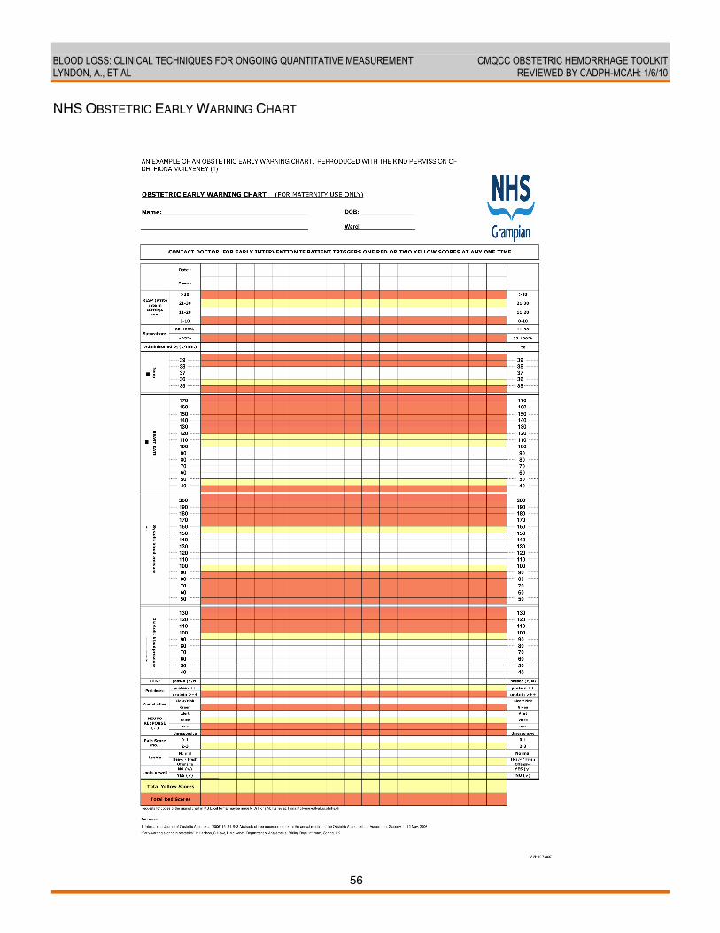
BLOOD LOSS: CLINICAL TECHNIQUES FOR ONGOING QUANTITATIVE MEASUREMENT CMQCC OBSTETRIC HEMORRHAGE TOOLKIT LYNDON, A., ET AL REVIEWED BY CADPH-MCAH: 1/6/10
56
NHS OBSTETRIC EARLY WARNING CHART
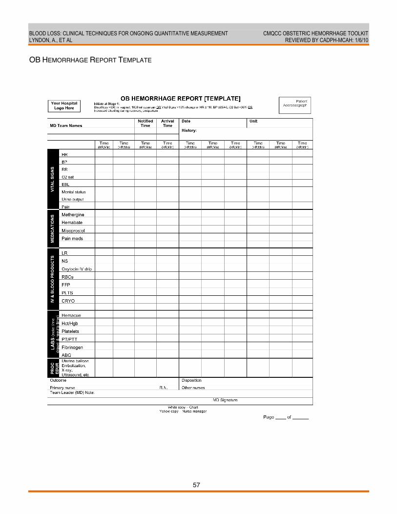
BLOOD LOSS: CLINICAL TECHNIQUES FOR ONGOING QUANTITATIVE MEASUREMENT CMQCC OBSTETRIC HEMORRHAGE TOOLKIT LYNDON, A., ET AL REVIEWED BY CADPH-MCAH: 1/6/10
57
OB HEMORRHAGE REPORT TEMPLATE
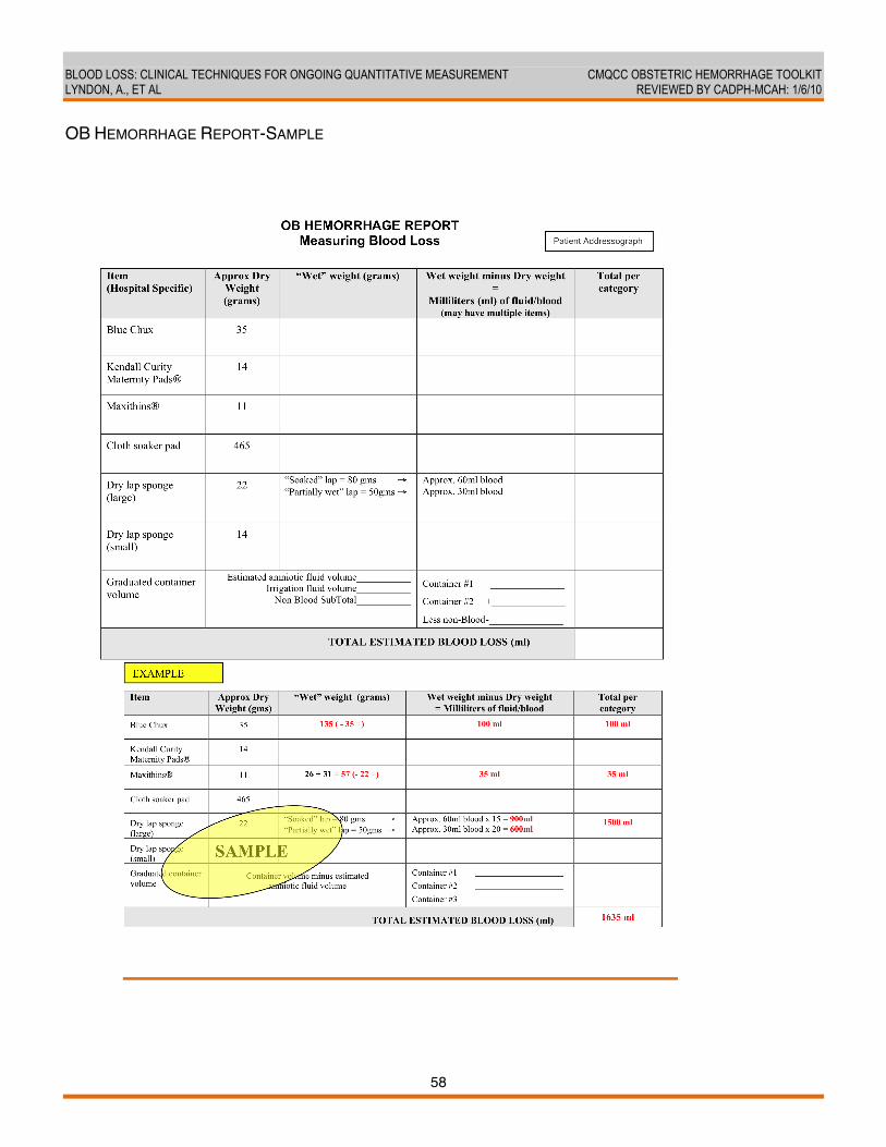
BLOOD LOSS: CLINICAL TECHNIQUES FOR ONGOING QUANTITATIVE MEASUREMENT CMQCC OBSTETRIC HEMORRHAGE TOOLKIT LYNDON, A., ET AL REVIEWED BY CADPH-MCAH: 1/6/10
58
OB HEMORRHAGE REPORT-SAMPLE
BLOOD LOSS: CLINICAL TECHNIQUES FOR ONGOING QUANTITATIVE MEASUREMENT CMQCC OBSTETRIC HEMORRHAGE TOOLKIT LYNDON, A., ET AL REVIEWED BY CADPH-MCAH: 1/6/10
59
REFERENCES 1. Kodkany B, Derman R. Pitfalls in assessing blood loss and decision to transfer in A
Textbook of PostPartum Hemorrhage. (ed C B-Lynch et al) Sapiens Publishing 2006. 2. Bose P, Regan F, Paterson-Brown S. Improving the accuracy of estimated blood loss at
obstetric haemorrhage using clinical reconstructions. BJOG 2006;113:919-24. 3. Patel A, Goudar SS, Geller SE, Kodkany BS, Edlavitch SA, Wagh K, et al. Drape estimation
vs. visual assessment for estimating postpartum hemorrhage. Int J Gynaecol Obstet 2006 Jun;93(3):220-4.
4. Dildy G, Paine A, George N, Velasco C. Estimating Blood Loss: Can Teaching Significantly Improve Visual Estimation? Obstet Gynecol 2004;104(3):601-6.
5. Toledo P, McCarthy R, Hewlett B, Fitzgerald P, Wong C. The accuracy of blood loss estimation after simulated vaginal delivery. Anesth Analg 2007;105:1736-40.
6. Brace RA, Wolf EJ. Normal amniotic fluid volume changes throughout pregnancy. Am J Obstet Gynecol 1989 Aug;161(2):382-8.
7. Patel A, et al. Blood loss: accuacy of visual estimation in A Textbook of PostPartum Hemorrhage. (ed C B-Lynch et al) Sapiens Publishing 2006 2006.
8. Harrison P, et al. Early warning scoring in obstetrics. Intern J Obstetric Anesthesia Abstracts of free papers presented at the annual meeting of the Obstetric Anaesthetists Association Glasgow, May 11-12, 2008 2005;15(Supplement):S1-S43.
9. Swanton R, Al-Rawi S, MYK W. A national survey of obstetric early warning systems in the United Kingdom. Intern J Obstetric Anesthesia 2009;18(3):253-57.
CMQCC OBSTETRIC HEMORRHAGE TOOLKIT OBSTETRIC HEMORRHAGE CARE GUIDELINES AND COMPENDIUM OF BEST PRACTICES REVIEWED BY CADPH-MCAH: 11/24/09
60
BLOOD PRODUCT REPLACEMENT: OBSTETRIC HEMORRHAGE Larry Shields, MD, Central Coast Maternal Fetal Medicine; Richard Lee, MD, Los Angeles County and University of Southern California Medical Center; Maurice Druzin, MD, Division of Maternal Fetal Medicine, Stanford University School of Medicine; Jennifer McNulty, MD, Division of Maternal Fetal Medicine, Long Beach Memorial Medical Center; Holli Mason, MD, Transfusion Medicine and Serology, Harbor UCLA Medical Center BACKGROUND AND LITERATURE REVIEW After the first several units of packed red blood cells (PRBCs) and in the face of continuing or worsening hemorrhage, aggressive transfusion therapy becomes critical. This report covers the new science of massive transfusion protocols. Lessons from military trauma units in Iraq as well as civilian experience with motor vehicle accidents and massive obstetric hemorrhage have identified new principles such as prominent use of fresh frozen plasma (FFP) and resuscitation transfusion without waiting for laboratory results. Life-threatening maternal hemorrhage occurs in approximately 1-2% of deliveries and is a leading cause of maternal death in both industrial and developing countries. (1, 2) Delays in recognizing and treating hemorrhage frequently lead to inadequate blood product replacement and concomitant development of disseminated intravascular coagulation (DIC). Both of these factors significantly contribute to maternal morbidity and mortality. Furthermore, delayed treatment increases the likelihood that the patient will require multiple units of blood products and, if available, activation of “massive hemorrhage protocols.” This section reviews blood component replacement therapy in the context of significant maternal hemorrhage. Nine massive hemorrhage protocols tailored specifically to obstetrics were evaluated. (1-9) No formal clinical trials were available and all of the protocols were developed in consultation with obstetric and hematology “experts.” Only one has published a case series of their results. (7) The salient feature of each protocol was an attempt to address three primary problems: 1) delayed diagnosis; 2) underestimated blood loss; and 3) treatment and prevention of fulminate disseminated intravascular coagulation (DIC). (9) To address these problems, the use of “obstetrical hemorrhage packs,” which included all needed blood components (i.e. PRBCs, FFP, cryoprecipitate, platelets) was recommended. None of the protocols recommended routine use of recombinant factor VIIa as part of initial therapy. The American College of Obstetrics and Gynecology has no specific recommendation for the use of blood components for treating postpartum hemorrhage. (10) Recommendations by the American Society of Anesthesiologists Task Force on Perioperative Blood Loss are consistent with the protocols reviewed here and with recommendations outlined below.
BLOOD PRODUCT REPLACEMENT CMQCC OBSTETRIC HEMORRHAGE TOOLKIT SHIELDS, L., ET AL REVIEWED BY CADPH-MCAH: 11/24/09
61
BLOOD PRODUCTS PACKED RED BLOOD CELLS (PRBCS) The majority of protocols recommended six units of PRBCs be prepared and available and hematocrit be maintained minimally at 21-24%. (1, 3, 4, 6, 8) These recommendations are consistent with a recent survey of obstetricians and practice guidelines from the American Society of Anesthesiologists. (11) Ideally, the use of a single unit of PRBCs should increase the hematocrit by approximately 3-4% in a 70 kg patient. (12) However, the expected increase in hematocrit may be slightly less due to expanded blood volume during pregnancy. As noted elsewhere in this toolkit, any patient with continued bleeding after initial measures have failed (Stage 2) should have two units of PRBCs released from the blood bank. If these are not readily available, consideration should be given for the use of uncross-matched O negative blood while the blood bank is completing the patientʼs type and crossmatch. Consistent with recommendations from this toolkit, for any patient that reaches Stage 3, a massive OB hemorrhage pack should be prepared, which includes an additional 4 or 6 units of PRBCs as pre-arranged with the blood bank for massive transfusion protocol. Good communication with the laboratory regarding the urgency of the situation is essential. FRESH FROZEN PLASMA (FFP) Fresh frozen plasma contains nearly all coagulation factors and can be used up to 24 hours after thawing and up to 5 days if relabeled as “thawed plasma.” Concomitant use of FFP and PRBCs is recommended during massive hemorrhage. Using a high ratio of PRBCs to FFP (1.5:1 or 1:1) has been shown to significantly improve survival from hemorrhage after trauma. (13) Similar recommendations have been established at centers with existing massive OB hemorrhage protocols with the goal of maintaining the INR at <1.5-1.7. (1, 3, 8, 14) (15) If diffuse bleeding is noted, or there is laboratory evidence of DIC and the patient has not been crossmatched, initial requests for 4 units of AB-FFP are recommended. AB plasma is usually in short supply and reserved for infants. However, 2-3 units of mismatched plasma can be transfused to adults while type and crossing is completed. FFP usually requires 30 minutes to thaw and will not be available immediately. Pre-arrangement with the blood bank for an inventory of thawed plasma for immediate issue may be considered if plasma usage volume is sufficient. If the patient enters into Stage 3 status, there should be no delays in preparation of FFP while waiting for laboratory results. PLATELETS All protocols in our review recommended transfusion of a single donor apheresis unit when platelet levels varied between 50,000-100,000 u/L. (3, 5, 7, 8) Platelet pheresis units are the standard equivalent of 6 units whole blood-derived pooled platelets and may increase the platelet count in a 70 kg patient by approximately 40-50,000/uL. (12) In the face of massive maternal hemorrhage, platelet transfusions should maintain platelet count between 50,000-100,000/uL. However, platelet counts should be used only as a guide and should be interpreted in conjunction with the patientʼs clinical condition. These recommendations are consistent with those of the American Society of Anesthesiologists Task Force on Perioperative Blood Loss. (16) Some protocols have suggested higher platelet counts for initiating transfusion and maintaining
BLOOD PRODUCT REPLACEMENT CMQCC OBSTETRIC HEMORRHAGE TOOLKIT SHIELDS, L., ET AL REVIEWED BY CADPH-MCAH: 11/24/09
62
appropriate platelet levels. These suggestions are based on the assumption that unless bleeding and DIC have been controlled, the patient will experience ongoing platelet loss. (2, 3) Platelets do not require crossmatching and are generally not type specific. Rh negative platelets are given to patients with an Rh negative blood type because of the slight risk of sensitization to the D-antigen. However, a dose of Rh-Immune Globulin can be given and is protective if Rh negative platelets are unavailable. CRYOPRECIPITATE AND FIBRINOGEN In the face of hypofibrinoginemia (fibrinogen levels <100-125 mg/dL and ongoing bleeding), fibrinogen should be used in addition to FFP. Transfusion recommendations were based on maintaining a fibrinogen concentration above 100 mg/dL. Cryoprecipitate release from the Blood Bank is usually in groups of 6-10 units. Each unit provides ≥150 mg of fibrinogen for a total of at least 1500 mg in a pool of 10 units in a total volume of approximately 80-100 cc. A pooled “ten-unit” pack would be expected to increase the fibrinogen level of a 70 kg patient by approximately 75mg/dL. It is worth noting that a 10-unit pool represents 10 separate donor exposures. If continued bleeding and hypofibrinoginemia is present, additional units of cryoprecipitate should be used. RECOMBINANT FACTOR VIIA Factor VII is a vitamin K-dependent serine protease with a pivotal role in coagulation. After reconstitution with sterile water, each vial contains approximately 0.6 mg/mL (600 μg/mL). It is marketed for use in patients with hemophilia A and B. The role of rVII in primary postpartum hemorrhage is controversial. (17, 18) It has been reported to significantly improve hemostasis in hemorrhaging obstetrical patients, but may also result in life-threatening thrombosis. (19) When available, its use should be reserved for rescue therapy when conventional therapy has failed (i.e., after 10-12 units of PRBC, 6-10 units of FFP and 2-3 units of platelets). Dosing recommendations in obstetrical hemorrhage patients has not been uniform. See Appendix A for additional information. SUMMARY During obstetrical hemorrhage, the primary goals are to provide adequate blood product replacement and to either prevent or correct DIC. The literature and protocols reviewed provided remarkable consensus related to therapy in the setting of massive obstetrical hemorrhage. RECOMMENDATIONS For transfusion in the setting of massive obstetrical hemorrhage, use a ratio of PRBCs to FFP to platelets that is 6 units PRBC: 4 units FFP: 1 unit pheresis platelets. If bleeding continues after initial treatment, strong consideration should be given to increasing the amount of FFP to a ratio of 4 units PRBC: 4 units of FFP: 1 unit of pheresis platelets.

BLOOD PRODUCT REPLACEMENT CMQCC OBSTETRIC HEMORRHAGE TOOLKIT SHIELDS, L., ET AL REVIEWED BY CADPH-MCAH: 11/24/09
63
STAT LABS If bleeding exceeds expected volume for routine delivery and there is no response to initial therapy, request stat laboratory analysis for the following:
1) CBC with platelets 2) PT/PTT 3) Fibrinogen
Repeat labs 1-3 every 30 minutes until patient is stable. A glass red-top tube without additives should be collected and taped to the wall and checked after 10 minutes; if the red-top blood is not clotted at 10 minutes, assume patient has DIC until laboratory test(s) show otherwise. Note that per OSHA regulations, many hospitals are now using plastic red top tubes, which contain an additive to induce clotting; in glass tubes, clotting was induced by the negative surface charge of glass. This simple test is reliable with the use of glass tubes, but not with plastic. PBRCS
• Initial request: 4-6 units of RBCs • O-negative or type-specific blood initially until cross match units are released
FFP • RBCs to FFP ratio not to exceed 3:2 • Infuse FFP to maintain INR <1.5
PLATELETS • Single donor apheresis platelet pack • Infuse to maintain platelet count >50,000-100,000/uL in the face of ongoing hemorrhage
CRYOPRECIPITATE • Initial request: 10 units cryoprecipitate if fibrinogen is less than 100mg/dL • Additional units to maintain fibrinogen concentration ≥100-125mg/dL
RECOMBINANT ACTIVATED FACTOR VII (rVII) If available, use when there is continued hemorrhage AND all other blood replacement therapies have failed (ie., after the use of 10-12 units PRBC, 6-12 units FFP and 2-3 units platelets). EDUCATIONAL TOOLS, SUPPORT DOCUMENTS APPENDIX A: Use of Factor VIIA APPENDIX B: Adverse Reactions to Transfusions EVIDENCE GRADING Level of Evidence: II-3C: Evidence obtained from multiple time series with or without intervention. Dramatic results in uncontrolled experiments also could be regarded as this type of evidence. Strong quality improvement data, such as statistical process control or other well-designed analysis.
BLOOD PRODUCT REPLACEMENT CMQCC OBSTETRIC HEMORRHAGE TOOLKIT SHIELDS, L., ET AL REVIEWED BY CADPH-MCAH: 11/24/09
64
BLOOD PRODUCT REPLACEMENT: OBSTETRIC HEMORRHAGE APPENDIX A: USE OF FACTOR VIIA The use of Recombinant factor VIIa has been shown in a number of case series to reduce ongoing massive obstetrical hemorrhage. (19-22) The use of recombinant factor VIIa for obstetrical or trauma hemorrhage would be considered “off label” use. Dosing for recombinant factor VIIa in trauma and obstetrical patients has varied (60-90 mcg/kg) and no studies have attempted to identify the ideal dose in the setting of maternal hemorrhage. (21) Anecdotal experience from members of this committee suggests that lower dosages have also been effective. It should be noted that most members of this committee who have experience using recombinant VIIa have reported anecdotal cases of maternal thrombosis; unfortunately, none of these have been reported in the literature. The committee recognizes that recombinant factor VIIa may not be available in smaller centers and/or non-trauma centers. If available, its use should be limited to patients after reasonable attempts for correction of ongoing bleeding with conventional therapy have failed (i.e., after the use of 10-12 units PRBC, 6-9 units FFP, 2-3 apheresis platelet units and cryoprecipitate). In addition, prior to treatment the patientʼs platelet count should be ≥50,000/uL. If the patientʼs platelet count is not ≥50,000/uL, platelets should be given concurrently. Due to their negative impact on all coagulation factors, correction acidosis and/or hypothermia is essential for successful use of recombinant factor VIIa. Based on available data, initial dosing of recombinant factor VIIa should be between 30-90 mcg/kg and repeated in 20-30 minutes if <90 mcg/kg was used and there was no clinical response. Additional dosing may be helpful if there was no initial clinical response and if hypothermia and/or acidosis have been corrected. Adoption of a massive obstetrical hemorrhage policy that includes recombinant factor VIIa should be reviewed and approved in conjunction with laboratory medicine, pharmacy and the local blood bank depending on who supplies and distributes this agent. If there is continued coagulopathy, and an initial response was seen, additional dosing may be used in 2-3 hours due to the relatively short half-life of recombinant factor VIIa. Further treatment should be provided in consultation with a local and/or regional expert in the area of maternal coagulopathy/massive obstetrical hemorrhage. It should also be emphasized that the use of conventional therapy (PRBCs, platelets, FFP, and cryoprecipitate) should also continue.
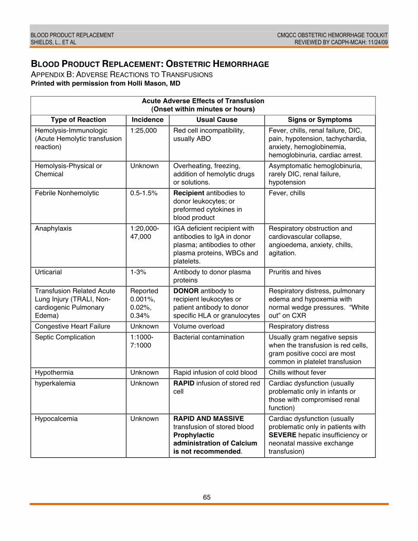
BLOOD PRODUCT REPLACEMENT CMQCC OBSTETRIC HEMORRHAGE TOOLKIT SHIELDS, L., ET AL REVIEWED BY CADPH-MCAH: 11/24/09
65
BLOOD PRODUCT REPLACEMENT: OBSTETRIC HEMORRHAGE APPENDIX B: ADVERSE REACTIONS TO TRANSFUSIONS Printed with permission from Holli Mason, MD
Acute Adverse Effects of Transfusion (Onset within minutes or hours)
Type of Reaction Incidence Usual Cause Signs or Symptoms Hemolysis-Immunologic (Acute Hemolytic transfusion reaction)
1:25,000 Red cell incompatibility, usually ABO
Fever, chills, renal failure, DIC, pain, hypotension, tachychardia, anxiety, hemoglobinemia, hemoglobinuria, cardiac arrest.
Hemolysis-Physical or Chemical
Unknown Overheating, freezing, addition of hemolytic drugs or solutions.
Asymptomatic hemoglobinuria, rarely DIC, renal failure, hypotension
Febrile Nonhemolytic 0.5-1.5% Recipient antibodies to donor leukocytes; or preformed cytokines in blood product
Fever, chills
Anaphylaxis 1:20,000-47,000
IGA deficient recipient with antibodies to IgA in donor plasma; antibodies to other plasma proteins, WBCs and platelets.
Respiratory obstruction and cardiovascular collapse, angioedema, anxiety, chills, agitation.
Urticarial 1-3% Antibody to donor plasma proteins
Pruritis and hives
Transfusion Related Acute Lung Injury (TRALI, Non-cardiogenic Pulmonary Edema)
Reported 0.001%, 0.02%, 0.34%
DONOR antibody to recipient leukocytes or patient antibody to donor specific HLA or granulocytes
Respiratory distress, pulmonary edema and hypoxemia with normal wedge pressures. “White out” on CXR
Congestive Heart Failure Unknown Volume overload Respiratory distress Septic Complication 1:1000-
7:1000 Bacterial contamination Usually gram negative sepsis
when the transfusion is red cells, gram positive cocci are most common in platelet transfusion
Hypothermia Unknown Rapid infusion of cold blood Chills without fever hyperkalemia Unknown RAPID infusion of stored red
cell Cardiac dysfunction (usually problematic only in infants or those with compromised renal function)
Hypocalcemia Unknown RAPID AND MASSIVE transfusion of stored blood Prophylactic administration of Calcium is not recommended.
Cardiac dysfunction (usually problematic only in patients with SEVERE hepatic insufficiency or neonatal massive exchange transfusion)
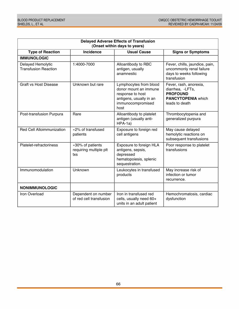
BLOOD PRODUCT REPLACEMENT CMQCC OBSTETRIC HEMORRHAGE TOOLKIT SHIELDS, L., ET AL REVIEWED BY CADPH-MCAH: 11/24/09
66
Delayed Adverse Effects of Transfusion
(Onset within days to years) Type of Reaction Incidence Usual Cause Signs or Symptoms
IMMUNOLOGIC Delayed Hemolytic Transfusion Reaction
1:4000-7000 Alloantibody to RBC antigen, usually anamnestic
Fever, chills, jaundice, pain, uncommonly renal failure days to weeks following transfusion
Graft vs Host Disease Unknown but rare Lymphocytes from blood donor mount an immune response to host antigens, usually in an immunocompromised host
Fever, rash, anorexia, diarrhea, LFTs, PROFOUND PANCYTOPENIA which leads to death
Post-transfusion Purpura Rare Alloantibody to platelet antigen (usually anti-HPA-1a)
Thrombocytopenia and generalized purpura
Red Cell Alloimmunization »2% of transfused patients
Exposure to foreign red cell antigens
May cause delayed hemolytic reactions on subsequent transfusions
Platelet-refractoriness »30% of patients requiring multiple plt txs
Exposure to foreign HLA antigens, sepsis, depressed hematopoiesis, splenic sequestration.
Poor response to platelet transfusions
Immunomodulation Unknown Leukocytes in transfused products
May increase risk of infection or tumor recurrence.
NONIMMUNOLOGIC
Iron Overload Dependent on number of red cell transfusion
Iron in transfused red cells, usually need 60+ units in an adult patient
Hemochromatosis, cardiac dysfunction
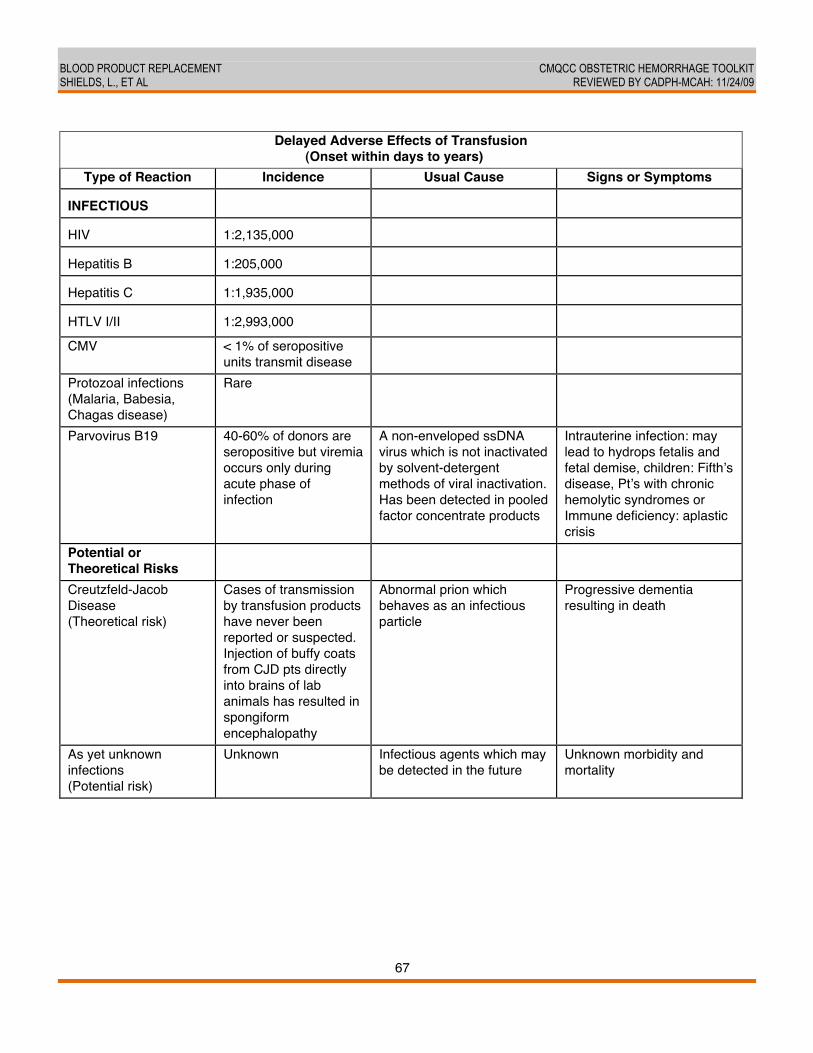
BLOOD PRODUCT REPLACEMENT CMQCC OBSTETRIC HEMORRHAGE TOOLKIT SHIELDS, L., ET AL REVIEWED BY CADPH-MCAH: 11/24/09
67
Delayed Adverse Effects of Transfusion
(Onset within days to years) Type of Reaction Incidence Usual Cause Signs or Symptoms
INFECTIOUS
HIV
1:2,135,000
Hepatitis B
1:205,000
Hepatitis C
1:1,935,000
HTLV I/II
1:2,993,000
CMV < 1% of seropositive units transmit disease
Protozoal infections (Malaria, Babesia, Chagas disease)
Rare
Parvovirus B19 40-60% of donors are seropositive but viremia occurs only during acute phase of infection
A non-enveloped ssDNA virus which is not inactivated by solvent-detergent methods of viral inactivation. Has been detected in pooled factor concentrate products
Intrauterine infection: may lead to hydrops fetalis and fetal demise, children: Fifthʼs disease, Ptʼs with chronic hemolytic syndromes or Immune deficiency: aplastic crisis
Potential or Theoretical Risks
Creutzfeld-Jacob Disease (Theoretical risk)
Cases of transmission by transfusion products have never been reported or suspected. Injection of buffy coats from CJD pts directly into brains of lab animals has resulted in spongiform encephalopathy
Abnormal prion which behaves as an infectious particle
Progressive dementia resulting in death
As yet unknown infections (Potential risk)
Unknown Infectious agents which may be detected in the future
Unknown morbidity and mortality

BLOOD PRODUCT REPLACEMENT CMQCC OBSTETRIC HEMORRHAGE TOOLKIT SHIELDS, L., ET AL REVIEWED BY CADPH-MCAH: 11/24/09
68
IMMEDIATE STEPS FOR ALL REACTIONS:
1. Stop transfusion. 2. Keep IV open with 0.9% NaCl. 3. Notify Attending Physician and Blood Bank.
If transfusion is terminated:
1. Send freshly collected blood and any necessary urine samples to Blood Bank. 2. Send blood unit and administration set to Blood Bank. 3. Fill out COMPLETELY and send to Blood Bank the Transfusion Reaction section of the blood tag.
Source: Harbor-UCLA Medical Center Appendix to Hospital Policy for Informed Consent for Blood and Blood Products, initially developed by Priscilla Figueroa, MD 8/1998 and most recently revised by Holli M. Mason, MD 1/2010; based on information from the American Association of Blood Banks REFERENCES 1. Bonnar J. Best Practice & Research. Bailliere's Clinical Obstet Gynaecol 2000;14:1-18. 2. Santoso J, Saunders B, Grosshart K. Massive blood loss and transfusion in obstetrics and
gynecology. Obstet Gynecol Surv 2005;60:827-37. 3. University of Washington. Obstetric Emergency Hemorrhage Protocol. Not Published. 4. Stanford University. Obstetrical Emergency Hemorrhage Protocol. Not Published. 5. California Pacific Medical Center. Obstetrical Hemorrhage Protocol. Not Published. 6. Barbeiri R. Lessons from Iraq reach the US Labor and Delivery Suite. OBG Management
20007;19(7):8-11. 7. Burtelow M, Riley E, Druzin M, Fontaine M, Viele M, Goodnough LT. How we treat:
management of life-threatening primary postpartum hemorrhage with a standardized massive transfusion protocol. Transfusion 2007 Sep;47(9):1564-72.
8. University of Southern California. Emergency Obstetrical Hemorrhage Protocol. Approved and in use in Department of Obstetrics, Women's and Children's Hospital.
9. Lurie S, Feinstein M, Mamet Y. Disseminated intravascular coagulopathy in pregnancy: thorough comprehension of etiology and management reduces obstetricians' stress. Archives of gynecology and obstetrics 2000;263(3):126-30.
10. ACOG. Clinical Management Guidelines for Obstetrician-Gynecologists. Obstetrics and Gynecology 2006;108(4):1039-47.
11. Matot I, Einav S, Goodman S, Zeldin A, Weissman C, U E. A Survey of physicians' attitudes toward blood transfusion in patients undergoing cesarean section. American Journal of Obstetrics and Gynecology 2004;190(2):462-7.
12. Puget Sound Blood Center. Available at http://www.psbc.org/therapy/index.htm. 13. Borgman M, Spinella P, Perkins J, Grathwohl K, Repine T, Beekley A, et al. The Ratio of
Blood Products Transfused Affects Motality in Patients Receiving Massive Transfusions at a Combat Support Hospital. The Journal of Trauma 2007;63(4):805-13.
14. Stinger H, Spinella P, Perkins J, Grathwohl K, Salinas J, Martini W, et al. The Ratio of Fibrinogen to Red Cells Transfused Affects Survival in Casualties Receiving Massive

BLOOD PRODUCT REPLACEMENT CMQCC OBSTETRIC HEMORRHAGE TOOLKIT SHIELDS, L., ET AL REVIEWED BY CADPH-MCAH: 11/24/09
69
Transfusions at an Army Combat Support Hospital. The Journal of Trauma 2008;64(2):S79-S85.
15. Riskin D, et al. Reduced Mortality After Implementation of a Massive Transfusion Protocol: A Single Trauma Center Experience. American College of Surgeons 2008(October):13-6.
16. Matot I, Einav S, Goodman S, Zeldin A, Weissman C, Elchalal U. A survey of physicians' attitudes toward blood transfusion in patients undergoing cesarean section. Am J Obstet Gynecol 2004 Feb;190(2):462-7.
17. Ahonen J, Jokela R, Korttila K. An open non-randomized study of recombinant activated factor VII in major postpartum haemorrhage. Acta Anaesthesiol Scand 2007 Aug;51(7):929-36.
18. Bhuskute N, Kritzinger S, Dakin M. Recombinant factor VIIa in massive obstetric haemorrhage. Eur J Anaesthesiol 2008 Mar;25(3):250-1.
19. Alfirevic Z, Elbourne D, Pavord S, Bolte A, Van Geijn H, Mercier F, et al. Use of recombinant activated factor VII in primary postpartum hemorrhage: the Northern European registry 2000-2004. Obstet Gynecol 2007 Dec;110(6):1270-8.
20. Bouma L, et al. Use of recombinant activated factor VII in massive postpartum haemorrhage. Eu J Obstet Gynecol 2008;137:172-7.
21. Franchini M, et al. A critical review on the use of recombinant factor VIIa in life threatening obstetric postpartum hemorrhage. Semin Thromb Hemostasis 2008;34:104-12.
22. Welsh A, et al. Guidelines for the use of recombinant activated factor VII in massive obstetric haemorrhage. Australian NZ J Obstet Gynaecol 2008;48:12-6.
CMQCC OBSTETRIC HEMORRHAGE TOOLKIT OBSTETRIC HEMORRHAGE CARE GUIDELINES AND COMPENDIUM OF BEST PRACTICES REVIEWED BY CADPH-MCAH: 12/22/09
70
UTERINE ARTERY OCCLUSION AND EMBOLIZATION David Lagrew, MD, Saddleback Memorial Medical Center; Andrew Hull, MD, University of California, San Diego BACKGROUND AND LITERATURE REVIEW Many authors have written on their experience with arterial balloon occlusion and embolization as an alternative to other conservative measures or hysterectomy for controlling postpartum hemorrhage. (1-3) Temporary balloon occlusion is typically used as a prophylactic measure when conditions such as placenta accreta are diagnosed in the antenatal period. The occlusive balloons are placed preoperatively while the patient is stable. Embolization is typically used in patients with persistent postpartum postoperative bleeding who are hemodynamically stable enough to tolerate transport to the interventional radiology suite. These procedures are to be performed by experienced interventional radiologists given the critical state of postpartum hemorrhage patients and the potential for complications. The literature describing the efficacy and safety of these techniques is limited to several case reports and small series. A review article of 46 studies of conservative measures found that they were effective but fell behind balloon catheters and hemostatic uterine sutures in efficacy (4). The success rates for controlling obstetrical hemorrhage were as follows: 90.7% (95% confidence interval [CI], 85.7%-94.0%) for arterial embolization, 84.0% (95% CI, 77.5%-88.8%) for balloon tamponade, 91.7% (95% CI, 84.9%-95.5%) for uterine compression sutures, and 84.6% (81.2%-87.5%) for iliac artery ligation or uterine devascularization. (4) The major limitation in these studies was the difficulty in assessing operator experience across various studies, and, unfortunately, the results have not always demonstrated clear-cut efficacy. (5) There is the possibility of severe complications from arterial balloon occlusion and embolization. One complication is uterine necrosis. (6) In one case-control study the authors found that 3 out of 19 subjects (15.8%) had complications from catheter placement and two required stent placement and/or arterial bypass. (7) Other serious complications such as thromboembolic events or fistulae have been reported. (8) Given the severity of these reports, one should use these techniques only when sufficient expertise is available and after full review of the risks and benefits with the patient or surrogate decision maker. RECOMMENDATIONS
1. Vaso-occlusive balloons and embolization techniques appear to be another option in centers with adequate interventional radiology expertise.
2. The indications, potential complications and effectiveness of these techniques are not well established and therefore must be approached with caution.

UTERINE ARTERY OCCLUSION AND EMBOLIZATION CMQCC OBSTETRIC HEMORRHAGE TOOLKIT LAGREW, D., HULL, A. REVIEWED BY CADPH-MCAH: 12/22/09
71
3. If utilized the patient must be in stable condition for transport to the interventional radiology suite and should be accompanied by a nurse skilled in the assessment and treatment of obstetrical hemorrhage should the patientʼs status suddenly decline.
4. Obstetrical staff should keep abreast of further research developments as to the most effective technique and indications for these procedures.
EVIDENCE GRADING Level of Evidence: II-2. Evidence obtained from well-designed cohort or case-control analytic studies, preferably from more than one center or research group. REFERENCES 1. Hansch E, Chitkara U, McAlpine J, El-Sayed Y, Dake MD, Razavi MK. Pelvic arterial
embolization for control of obstetric hemorrhage: a five-year experience. Am J Obstet Gynecol 1999 Jun;180(6 Pt 1):1454-60.
2. Vendantham S, Goodwin SC, McLucas B, Mohr G. Uterine artery embolization: an underused method of controlling pelvic hemorrhage. Am J Obstet Gynecol 1997 Apr;176(4):938-48.
3. Dubois J, Garel L, Grignon A, Lemay M, Leduc L. Placenta percreta: balloon occlusion and embolization of the internal iliac arteries to reduce intraoperative blood losses. Am J Obstet Gynecol 1997 Mar;176(3):723-6.
4. Doumouchtsis S, Papageorghiou A, S A. Systematic review of conservative management of postpartum hemorrhage: what to do when medical treatment fails. Obstet Gynecol Surv 2007;62(8):540-7.
5. Bodner LJ, Nosher JL, Gribbin C, Siegel RL, Beale S, Scorza W. Balloon-assisted occlusion of the internal iliac arteries in patients with placenta accreta/percreta. Cardiovasc Intervent Radiol 2006 May-Jun;29(3):354-61.
6. Coulange L, Butori N, Loffroy R, Filipuzzi L, Cercueil J, Douvier S, et al. Uterine necross following selective embolization for postpartum hemorrhage using absorbable material. Acta Obstet Gynecol Scand 2009;88(2):238-40.
7. Shrivastava V, Nageotte M, Major C, Haydon M, Wing D. Case-control comparison of cesarean hysterectomy with and without prophylactic placement of intravascular balloon catheters for placenta accreta. Am J Obstet Gynecol 2007;197(4):402.e1-5.
8. Maassen MS, Lambers MD, Tutein Nolthenius RP, van der Valk PH, Elgersma OE. Complications and failure of uterine artery embolisation for intractable postpartum haemorrhage. BJOG 2009 Jan;116(1):55-61.
CMQCC OBSTETRIC HEMORRHAGE TOOLKIT OBSTETRIC HEMORRHAGE CARE GUIDELINES AND COMPENDIUM OF BEST PRACTICES REVIEWED BY CADPH-MCAH: 12/22/09
72
UTERINE HEMOSTATIC SUTURES David Lagrew, MD, Saddleback Memorial Medical Center; Andrew Hull, MD, University of California, San Diego BACKGROUND AND LITERATURE REVIEW As an alternative to hysterectomy in the face of severe postpartum hemorrhage, several techniques of uterine compression suturing have been described. Suturing is a mechanical method of compressing the uterine musculature and closing the arterial bed, which leads to reduced bleeding. The primary and most frequent indication for these procedures is the patient with an atonic uterus who is hemodynamically-challenged but stable and who desires the possibility of future reproductive capability. Various authors have described several techniques. The most common is the B-Lynch technique where thick absorbable sutures such as #1 Chromic are anchored in the lower segment with a large loop going over the fundocorneal region and down posterior to just above the cardinal ligaments, across to the opposite side, back up over the fundocorneal region, and anchoring and tying in the lower uterine segment. (1) Hayman described a simpler but similar procedure using two separate transfundal sutures. (2) Other techniques of multiple square sutures through the uterine body have been described, but reports of adhesions and abscesses have been published and have dampened support. (3, 4) The reported efficacy must be estimated by using the case reports and series published to date. A Canadian series by Baskett reported that during a 7-year period, 28 uterine compression sutures were performed (1 per 1,126 deliveries). Of note, all were used after cesarean delivery and use was more frequent in non-elective cesarean (1 in 221) than in elective cesarean deliveries (1 in 637). The procedure avoided hysterectomy in 82% of cases. (5) Success rates are similar to smaller reports. (6, 7) Based on these types of experiences, one can recommend usage of the technique in clinical situations and that preparations, equipment and training are available in all obstetrical units. (8, 9) Published reports show use of this technique in the following cases: 1) atony unresponsive to standard treatments at cesarean section; 2) atony unresponsive to standard treatments after vaginal delivery; 3) placenta previa; 4) localized placenta accreta; 5) prophylactic use in “high risk” patients (high multiples, suspected accreta); and 6) with other adjunct treatments for PPH such as intrauterine balloons. Several questions require further clinical research to clarify the role of compression suturing in clinical practice such as: 1) what is the best suture material; 2) what are complication rates for short and long term sequelae and what are the appropriate techniques; and 3) what is the efficacy for various types of hemorrhage?
UTERINE HEMOSTATIC SUTURES CMQCC OBSTETRIC HEMORRHAGE TOOLKIT LAGREW, D., HULL, A. REVIEWED BY CADPH-MCAH: 12/22/09
73
RECOMMENDATIONS 1. Uterine hemostatic suturing should be available in all obstetrical units. 2. Staff Education: all delivery providers at the institution should be made aware of the steps
included in the technique; appropriate protocols for the timing and method of usage should be added to institutional policies and procedures. Diagrams with the technique and indications for usage clearly posted may be helpful.
3. Appropriate supplies for the technique should be immediately available to surgeons. 4. The obstetrical staff should keep abreast of further research developments as to the most
effective technique and indications for hemostatic suturing. EVIDENCE GRADING Level of Evidence: II-3 A. Evidence obtained from multiple time series with or without intervention. Dramatic results in uncontrolled experiments also could be regarded as this type of evidence. Strong quality improvement data, such as statistical process control or other well-designed analysis. Recommendations based on high quality and consistent evidence. REFERENCES 1. Allam MS, B-Lynch C. The B-Lynch and other uterine compression suture techniques. Int J
Gynaecol Obstet 2005 Jun;89(3):236-41. 2. Hayman RG, Arulkumaran S, Steer PJ. Uterine compression sutures: surgical management
of postpartum hemorrhage. Obstet Gynecol 2002 Mar;99(3):502-6. 3. Wu HH, Yeh GP. Uterine cavity synechiae after hemostatic square suturing technique.
Obstet Gynecol 2005 May;105(5 Pt 2):1176-8. 4. Ochoa M, Allaire AD, Stitely ML. Pyometria after hemostatic square suture technique.
Obstet Gynecol 2002 Mar;99(3):506-9. 5. Baskett TF. Uterine compression sutures for postpartum hemorrhage: efficacy, morbidity,
and subsequent pregnancy. Obstet Gynecol 2007 Jul;110(1):68-71. 6. Smith K, Baskett T. Uterine compression sutures as an alternative to hysterectomy for
severe postpartum hemorrhage. J Obstet Gynaecol Can 2003;25(3):197-200. 7. Sentilhes L, Gromez A, Razzouk K, Resch B, Verspyck E, Marpeau L. Uterine compression
sutures for postpartum bleeding with uterine atony. Acta Obstet Gynecol Scand 2008;87(10):1020-6.
8. El-Hamamy E, B-Lynch C. A Worldwide review of the uses of the uterine compression suture techniques as alternative to hysterectomy in the management of severe post-partum haemorrhage. J Obstet Gynaecol 2005;25:143-9.
9. Ouahba J, Piketty M, Huel C, Azarian M, Feraud O, Luton D, et al. Uterine compression sutures for postpartum bleeding with uterine atony. BJOG 2007 May;114(5):619-22.

CMQCC OBSTETRIC HEMORRHAGE TOOLKIT OBSTETRIC HEMORRHAGE CARE GUIDELINES AND COMPENDIUM OF BEST PRACTICES REVIEWED BY CADPH-MCAH: 12/22/09
74
UTEROTONIC AGENTS FACT SHEET Larry Shields, MD, Central Coast Maternal Fetal Medicine Pitocin® (oxytocin): Oxytocin is a synthetic version of the natural nonapeptide produced in the posterior pituitary. The drug comes in solution at a concentration of 10 U/ml. For postpartum use, including third stage of labor, oxytocin is dosed at 10-40 U per liter of IV fluid and given as an IV infusion. The rate of infusion should be sufficient to maintain uterine contractility. The plasma half-life of oxytocin is 1-6 minutes and the clinical response is rapid after IV infusion. Alternatively, the agent may be given as an IM injection (10 units). Intramuscular response to the drug occurs within 3-5 minutes, with a clinical response lasting about 2-3 hours. The drug may be stored at room temperature.
o Side Effects: Side effects are rare in the absence of prolonged use. Nausea and vomiting have been reported. The most serious side effect from prolonged use of IV oxytocin is water intoxication with subsequent dilutional hyponatremia. Rapid IV infusion is associated with hypotension and tachycardia. The drug should not to be given as an IV bolus.
o Contraindications: The only postpartum contraindication to use of oxytocin would be hypersensitivity to the drug.
Hemabate® (carboprost or 15 methyl PGF2 alpha): Hemabate is FDA-approved for the treatment of postpartum hemorrhage secondary to uterine atony not responsive to conventional treatment (massage and oxytocin). The drug is supplied in 1 ml ampoules containing 250 mcg of the drug. The dose is one ampoule given as an IM injection. The peak plasma level of the drug is reached about 30 minutes after injection. A successful clinical response is expected after a single injection in about 75% of cases. In refractory cases, additional dosing at 15-90 minute intervals may be beneficial. The total amount of drug given should not exceed 2 mg (8 doses). The clinical response may be enhanced with concomitant use of oxytocin. It may be less effective when used in the setting of chorioamnionitis. It should be noted that other uterotonic agents are also less effective in the setting of chorioamnionitis. The drug must be refrigerated when stored.
o Side Effects: Recognized side effects include nausea, vomiting, diarrhea, fever (up to 1 degree Celsius), bronchospasm, and hypertension.
o Contraindications: It is recommended that the drug be given with caution to patients with active hepatic or cardiovascular disease, asthma, or hypersensitivity to the drug.

UTEROTONIC AGENT FACT SHEET CMQCC OBSTETRIC HEMORRHAGE TOOLKIT SHIELDS, L. REVIEWED BY CADPH-MCAH: 12/22/09
75
Methergine® (methylergonovine maleate): Methergine is a semi-synthetic ergot alkaloid that is FDA-approved for routine management of the third stage of labor and postpartum atony. It is supplied in ampoules containing 0.2 mg of active drug in a volume of 1 mL or as a single tablet of 0.2 mg of active drug. The drug is given either as an intramuscular injection (1 ampoule) or orally (single tablet). When given as an oral agent, the onset of action is within 5-10 minutes with a bioavailability of 60%. When given as an intramuscular injection, the onset of action is 2-5 minutes and the bioavailability is 78% (about 25% greater than when given orally). The plasma half-life is about 3.4 hours. The agent should not be given by intravascular injection. The frequency of administration is 2-4 hours for IM administration and 6-8 hours when given orally. The drug must be refrigerated when stored.
o Side Effects: Side effects are rare in the absence of prolonged use. Most common side effects are nausea and vomiting. Chest pain, arterial spasm, myocardial infarction, and hallucination have been reported in cases of toxicity.
o Contraindications: Methergine should be used with extreme caution in the setting of hypertension or preeclampsia. Care should be exercised when there has been recent administration of other vasoconstrictive agents (i.e. ephedrine). In these settings, there may be an exaggerated blood pressure response to the use of this agent. Care should also be taken when CYP 3A3 inhibiting agents, such as macrolide antibiotics, protease inhibitors, or azole antifungals, have recently been used.
Cytotec® (misoprostol): This agent is a synthetic prostaglandin E1 analog. This agent is FDA-approved for reducing the risk of NSAID-induced gastric ulcers. It comes in either 100 or 200 mcg tablets. This agent is not FDA-approved for uterine atony or obstetrical hemorrhage, although its effectiveness has been clearly demonstrated in the obstetrical literature. The drug is water-soluble and is quickly absorbed after sublingual, oral, vaginal, and rectal use. The most common method of administering misoprostol for postpartum hemorrhage is rectally, although in a conscious patient sublingual use would also be reasonable. The dose usually ranges between 800-1000 mcg. The time to peak plasma concentration is shortest for sublingual administration and the plasma concentration is higher than when given rectally. However, after rectal administration, plasma concentrations are maintained for a longer period. The drug undergoes a series of chemical reactions after ingestion, converting the agent to a prostaglandin F analog, making the drug very similar to hemabate (15 methyl PGF2 alpha). Therefore, it is unlikely that misoprostol would be effective if hemabate has failed, or vice versa. Unlike hemabate, misoprostol does not appear to exacerbate bronchoconstriction in patients with asthma. One of the major advantages of this agent is that the drug does not need to be refrigerated and may be easily stored on labor and delivery hospital units.
o Side Effects: Diarrhea, shivering, pyrexia and headaches are the most common side effects. o Contraindications: Hypersensitivity to the drug.
REFERENCE
1. http://www.accessdata.fda.gov/scripts/cder/drugsatfda/index.cfm?fuseaction=Search.Label_ApprovalHistory (Accessed 4/2009).
CMQCC OBSTETRIC HEMORRHAGE TOOLKIT OBSTETRIC HEMORRHAGE CARE GUIDELINES AND COMPENDIUM OF BEST PRACTICES REVIEWED BY CADPH-MCAH: 11/24/09
76
ANTI-SHOCK GARMENTS: NON-PNEUMATIC ANTI-SHOCK GARMENT (NASG) AND PNEUMATIC ANTI-SHOCK GARMENT (PASG) Suellen Miller, PhD, CNM, MHA, Department of Obstetrics, Gynecology and Reproductive Sciences, University of California, San Francisco BACKGROUND AND LITERATURE REVIEW In 2006, the Joint Statement of the International Confederation of Midwives (ICM) and the Federation International of Gynecology and Obstetrics (FIGO) recommended research on anti-shock garments to reduce mortality among women suffering postpartum hemorrhage.(1) The non-pneumatic anti-shock garment (NASG) is a first-aid device that reverses hypovolemic shock and decreases obstetric hemorrhage. It consists of articulated segments of neoprene, which close tightly with Velcro, shunting blood from the lower body to the core organs, elevating blood pressure and increasing preload and cardiac output. The NASG is not an FDA-approved device, but is a 510(k) equivalent device, which did receive FDA clearance based on its similarity to the Pneumatic Anti-Shock Garment (PASG, aka, MAST suit). Theoretically, all anti-shock garments work on the same principle: a compression suit which upon placement to a hypovolemic person, restores blood pressures—particularly to the core—by returning blood from the lower body. PASG The PASG was omnipresent in emergency pre-hospital trauma treatment until results of a randomized control trial were published in the late 1980s-1990s. All trauma victims transported to Ben Taub General Hospital, Houston with entry systolic blood pressure ≤ 90 mmHg were admitted to the study; patients were randomized into control and PASG-intervention groups by alternate-day methodology. There were no significant differences in standard paramedic management or group demographics as a whole or when split into population subsets by injury type. Two key analyses came out of this research – Pepe (1986) and Bickell (1987).(2, 3) A third analysis was done by Mattox (1989) after continuing an additional year of data collection.(4) A study similar in both methodology and protocols to the above was carried out by Chang (1995).(5) None of the studies (Table 1) demonstrated that the PASG could reduce morbidity or mortality for pre-hospital trauma treatment in urban settings.
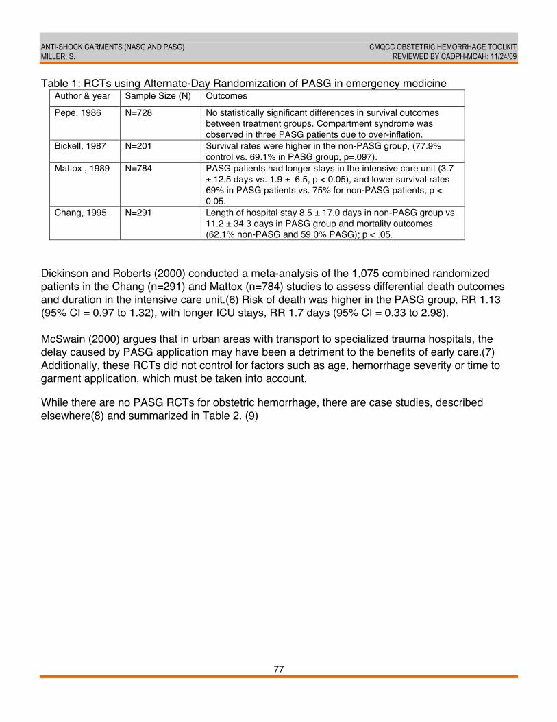
ANTI-SHOCK GARMENTS (NASG AND PASG) CMQCC OBSTETRIC HEMORRHAGE TOOLKIT MILLER, S. REVIEWED BY CADPH-MCAH: 11/24/09
77 77
Table 1: RCTs using Alternate-Day Randomization of PASG in emergency medicine Author & year Sample Size (N) Outcomes
Pepe, 1986 N=728 No statistically significant differences in survival outcomes between treatment groups. Compartment syndrome was observed in three PASG patients due to over-inflation.
Bickell, 1987 N=201 Survival rates were higher in the non-PASG group, (77.9% control vs. 69.1% in PASG group, p=.097).
Mattox , 1989 N=784 PASG patients had longer stays in the intensive care unit (3.7 ± 12.5 days vs. 1.9 ± 6.5, p < 0.05), and lower survival rates 69% in PASG patients vs. 75% for non-PASG patients, p < 0.05.
Chang, 1995 N=291 Length of hospital stay 8.5 ± 17.0 days in non-PASG group vs. 11.2 ± 34.3 days in PASG group and mortality outcomes (62.1% non-PASG and 59.0% PASG); p < .05.
Dickinson and Roberts (2000) conducted a meta-analysis of the 1,075 combined randomized patients in the Chang (n=291) and Mattox (n=784) studies to assess differential death outcomes and duration in the intensive care unit.(6) Risk of death was higher in the PASG group, RR 1.13 (95% CI = 0.97 to 1.32), with longer ICU stays, RR 1.7 days (95% CI = 0.33 to 2.98). McSwain (2000) argues that in urban areas with transport to specialized trauma hospitals, the delay caused by PASG application may have been a detriment to the benefits of early care.(7) Additionally, these RCTs did not control for factors such as age, hemorrhage severity or time to garment application, which must be taken into account. While there are no PASG RCTs for obstetric hemorrhage, there are case studies, described elsewhere(8) and summarized in Table 2. (9)
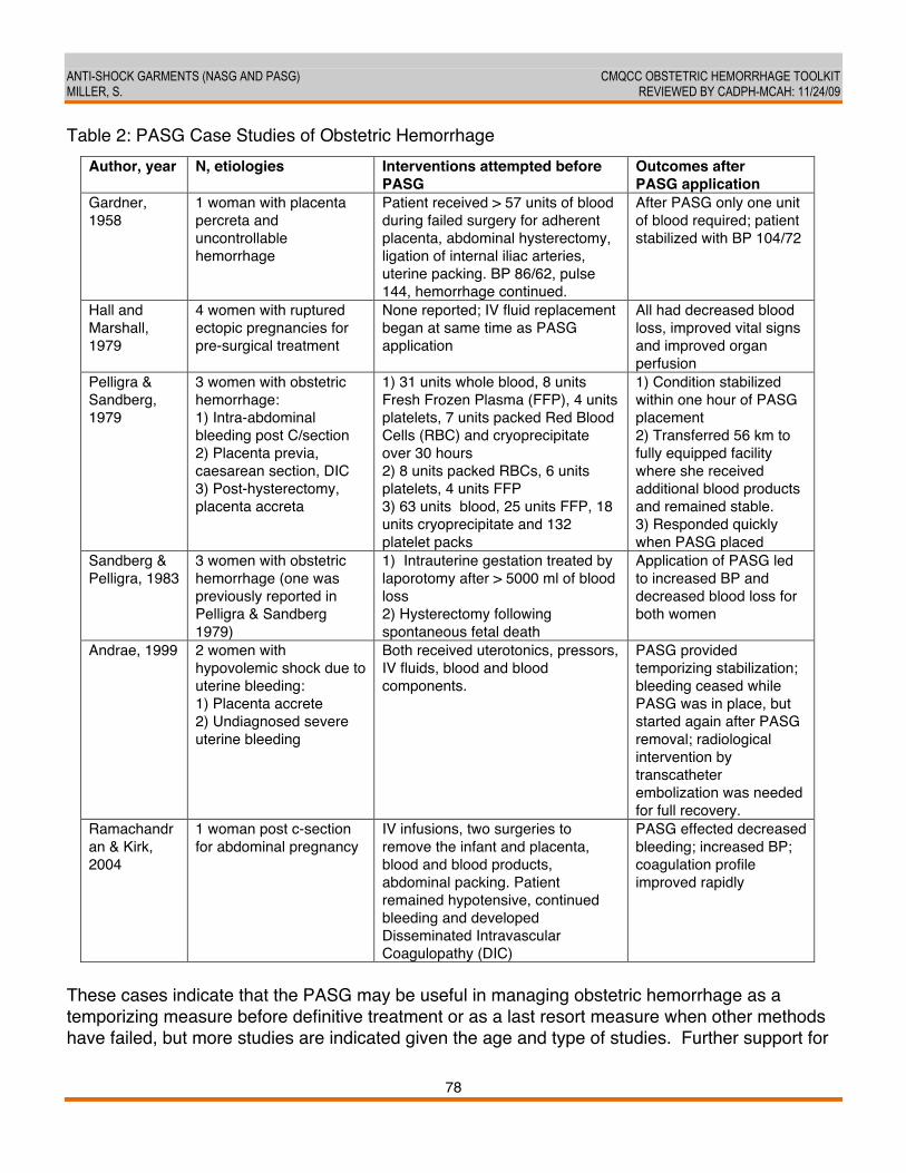
ANTI-SHOCK GARMENTS (NASG AND PASG) CMQCC OBSTETRIC HEMORRHAGE TOOLKIT MILLER, S. REVIEWED BY CADPH-MCAH: 11/24/09
78 78
Table 2: PASG Case Studies of Obstetric Hemorrhage Author, year N, etiologies Interventions attempted before
PASG Outcomes after PASG application
Gardner, 1958
1 woman with placenta percreta and uncontrollable hemorrhage
Patient received > 57 units of blood during failed surgery for adherent placenta, abdominal hysterectomy, ligation of internal iliac arteries, uterine packing. BP 86/62, pulse 144, hemorrhage continued.
After PASG only one unit of blood required; patient stabilized with BP 104/72
Hall and Marshall, 1979
4 women with ruptured ectopic pregnancies for pre-surgical treatment
None reported; IV fluid replacement began at same time as PASG application
All had decreased blood loss, improved vital signs and improved organ perfusion
Pelligra & Sandberg, 1979
3 women with obstetric hemorrhage: 1) Intra-abdominal bleeding post C/section 2) Placenta previa, caesarean section, DIC 3) Post-hysterectomy, placenta accreta
1) 31 units whole blood, 8 units Fresh Frozen Plasma (FFP), 4 units platelets, 7 units packed Red Blood Cells (RBC) and cryoprecipitate over 30 hours 2) 8 units packed RBCs, 6 units platelets, 4 units FFP 3) 63 units blood, 25 units FFP, 18 units cryoprecipitate and 132 platelet packs
1) Condition stabilized within one hour of PASG placement 2) Transferred 56 km to fully equipped facility where she received additional blood products and remained stable. 3) Responded quickly when PASG placed
Sandberg & Pelligra, 1983
3 women with obstetric hemorrhage (one was previously reported in Pelligra & Sandberg 1979)
1) Intrauterine gestation treated by laporotomy after > 5000 ml of blood loss 2) Hysterectomy following spontaneous fetal death
Application of PASG led to increased BP and decreased blood loss for both women
Andrae, 1999 2 women with hypovolemic shock due to uterine bleeding: 1) Placenta accrete 2) Undiagnosed severe uterine bleeding
Both received uterotonics, pressors, IV fluids, blood and blood components.
PASG provided temporizing stabilization; bleeding ceased while PASG was in place, but started again after PASG removal; radiological intervention by transcatheter embolization was needed for full recovery.
Ramachandran & Kirk, 2004
1 woman post c-section for abdominal pregnancy
IV infusions, two surgeries to remove the infant and placenta, blood and blood products, abdominal packing. Patient remained hypotensive, continued bleeding and developed Disseminated Intravascular Coagulopathy (DIC)
PASG effected decreased bleeding; increased BP; coagulation profile improved rapidly
These cases indicate that the PASG may be useful in managing obstetric hemorrhage as a temporizing measure before definitive treatment or as a last resort measure when other methods have failed, but more studies are indicated given the age and type of studies. Further support for
ANTI-SHOCK GARMENTS (NASG AND PASG) CMQCC OBSTETRIC HEMORRHAGE TOOLKIT MILLER, S. REVIEWED BY CADPH-MCAH: 11/24/09
79 79
PASG use for obstetric hemorrhage is a Doppler study of regional blood flow on ten healthy adults.(10) PASG inflation resulted in an immediate decrease in aortic blood flow below and proximal to the renal arteries; the vessels more distal from the renal pelvis showed a lower response. NASG The PASG—bulky, heavy, and difficult to use—has had no place in emergency obstetrics in low-resource settings. The NASG, developed in 1971 by teams associated with the National Aeronautics and Space Administration/Ames Research Centre (NASA/Ames), may overcome some of the deficiencies of the PASG.(11) In 1991, the NASG (Zoex Corporation, Ashland, OR, USA) was granted a US Food and Drug Administration 510(k) medical device regulations number. Based on the same principles as the PASG, circumferential counter pressure, but without air bladders, manometers, stop cocks, foot pump and tubing, and the associated risks of over-inflation and excessive pressures, the NASG is a promising first-aid treatment for hemorrhagic shock.(8, 12-19) Comparative NASG Studies: Obstetric Hemorrhage NASG use for obstetric hemorrhage in low-resource settings was first explored in two case series at a tertiary-level maternity hospital in Sialkot, Pakistan.(12, 14) The first comparative NASG study was a pre-post pilot of severe obstetric hemorrhage in four Egyptian tertiary hospitals.(17) All 364 women (158 pre-intervention phase, 206 post-intervention/NASG phase) had ≥750 ml EBL with signs of shock (pulse >100 BPM, SBP <100 mmHg) at study entry. All were treated with a standardized protocol, including crystalloid fluids, uterotonics, blood transfusions, and vaginal procedures or abdominal surgeries as needed. Post-intervention women also received the NASG. Blood loss after study entry, the main outcome variable, was measured with a graduated, closed-end blood collection device. NASG-phase women entered the study in worse condition with statistically significant greater EBL (975 ml vs. 750 ml median blood loss, p<0.001) and more severe signs of shock (mean SBP 97.5mmHg vs. 88.7mmHg, p<0.0005). In spite of this discrepancy at time of study entry, the NASG-treated women had better outcomes, with a statistically significant lower median measured blood loss (500 ml pre-intervention vs. 250 ml post-intervention, median difference -200, 95% CI -250 to -120, p<0.001) and a non-statistically significant 69% decrease in extreme adverse outcomes (mortality and morbidity combined). Further analysis of this data found that NASG-treated women experienced decreased shock recovery times, indicated by return to normal shock index (SI). Median SI recovery time in 249 obstetric hemorrhage cases was significantly shorter in the NASG group (75 vs.120 minutes, p=0.003), independent of standard treatments, such as volume of IV fluids and/or waiting time for blood transfusions.(17) Miller and colleagues have completed data collection on a similar pre-post pilot trial in 12 tertiary hospitals in Nigeria.(18) These hospitals are often understaffed, under-equipped, and lack blood transfusions. Results on over 580 women treated with NASG are pending analysis.
ANTI-SHOCK GARMENTS (NASG AND PASG) CMQCC OBSTETRIC HEMORRHAGE TOOLKIT MILLER, S. REVIEWED BY CADPH-MCAH: 11/24/09
80 80
Case Report of NASG for PPH in high-resource settings While the NASG is being studied for efficacy in reducing maternal mortality and morbidity in low-resource settings, it also can be used in high-resource settings. El Sayed, et al. reported on an 18 year old woman with intractable PPH at the Lucile Packard Childrenʼs Hospital, Stanford University, California, US.(13) The woman, bleeding profusely after vaginal twin delivery, received multiple interventions, including Ringerʼs Lactate infusions, each with 35 units of oxytocin per liter; two doses of 0.2 mg methergine IM; three doses of 250 mcg hemabate IM; 800 mcg misoprostol per rectum; along with transfusions of packed RBCs, recombinant factor VII, uterine massage and uterine curettage. Having exhausted standard treatment measures, the surgeons packed the uterus and applied the NASG. Within minutes of NASG placement, bleeding subsided, pulse decreased and blood pressure rose. The patient remained hemodynamically stable with normal vaginal bleeding. The NASG was removed on postpartum day 1, without complications or recurrent bleeding. RCTs of NASG The NASG has not yet proven to significantly decrease morbidity or mortality. Furthermore, NASG studies have a) only been of less rigorous pre/post design and b) only been conducted in tertiary care centers. A cluster RCT, jointly funded by the National Institutes of Health, National Institutes for Child Health, and the Bill and Melinda Gates Foundation, has been initiated in Zimbabwe and Zambia to determine if early application of the NASG by midwives at the primary health care level, prior to transfer to a referral hospital, will decrease extreme adverse outcomes (mortality or severe-end organ failure). The study will also analyze potential side effects of NASG. Lead investigators include those from University of California, San Francisco; University of Zimbabwe; University Teaching Hospital, Zambia; the Reproductive Health Research Unit of the World Health Organization; and the Centro Rosario Institutio Estudios Perinatales. (http://clinicaltrials.gov/ct2/show/NCT00488462?cntry1=AF%3AZW&rank=12). Results will be available in 3-5 years depending on recruitment. RECOMMENDATIONS More and more rigorous research is needed before recommending the clinical use of NASG or PASG in the setting of acute obstetrical hemorrhage. Research recommendations include:
• Efficacy trials for morbidity and mortality reduction (RCT currently ongoing) • Hemodynamics:
o The mechanism of action in pregnancy/postpartum is not clear. How much blood flow is decreased to the uterus?
o Should the NASG, if introduced into California hospital protocols, be used on women with placenta previa and a viable fetus?
o What is the effect on cardiac output? SPR? Stroke Volume?
ANTI-SHOCK GARMENTS (NASG AND PASG) CMQCC OBSTETRIC HEMORRHAGE TOOLKIT MILLER, S. REVIEWED BY CADPH-MCAH: 11/24/09
81 81
o Best methods of cleaning, reuse, and storage: The NASG manufactured by ZOEX was designed for one time use and the manufacturers do not recommend the use of bleach; therefore, questions remain on how to clean, decontaminate, and reuse/whether to reuse.
EDUCATIONAL TOOLS, SAMPLE DOCUMENTS Note that the NASG is not FDA-approved, but does have FDA 510(k) certification, substantially similar to an FDA approved device, the Pneumatic Anti-Shock Garment (PASG) so that is can be marketed in the US and internationally. The US distributor of NASG is ZOEX Corporation, Ashland Oregon: [email protected] A training video, meant for use by physicians and midwives in low resource settings, can be accessed on the website, www.nasgexchange.org. A password can be obtained from [email protected] to enter the training page of the website. EVIDENCE GRADING Level of Evidence: B. Recommendations based on limited or inconsistent evidence. REFERENCES 1. ICM/FIGO. Joint Statement: Management of the Third Stage of Labour to Prevent Post-
partum Haemorrhage. 2003 [cited January 10, 2010]; Available from: http://www.pphprevention.org/files/ICM_FIGO_Joint_Statement.pdf
2. Bickell WH, Pepe PE, Bailey ML, Wyatt CH, Mattox KL. Randomized trial of pneumatic antishock garments in the prehospital management of penetrating abdominal injuries. Ann Emerg Med 1987 Jun;16(6):653-8.
3. Pepe PE, Bass RR, Mattox KL. Clinical trials of the pneumatic antishock garment in the urban prehospital setting. Ann Emerg Med 1986 Dec;15(12):1407-10.
4. Mattox KL, Bickell W, Pepe PE, Burch J, Feliciano D. Prospective MAST study in 911 patients. J Trauma 1989 Aug;29(8):1104-11; discussion 11-2.
5. Chang FC, Harrison PB, Beech RR, Helmer SD. PASG: does it help in the management of traumatic shock? J Trauma 1995 Sep;39(3):453-6.
6. Dickinson K, Roberts I. Medical anti-shock trousers (pneumatic anti-shock garments) for circulatory support in patients with trauma. Cochrane Database Syst Rev 2000(2):CD001856.
7. McSwain MJ, McSwain N.E. Pneumatic antishock garment: state of the art at the turn of the century. Trauma 2000 January;2(1):63-75.
8. Miller S, Ojengbede A, Turan JM, Ojengbede O, Butrick E, Hensleigh P. Anti-Shock Garments for Obstetric Hemorrhage. Current Women's Health Reviews 2007;3(1):3-11.
ANTI-SHOCK GARMENTS (NASG AND PASG) CMQCC OBSTETRIC HEMORRHAGE TOOLKIT MILLER, S. REVIEWED BY CADPH-MCAH: 11/24/09
82 82
9. Miller S, Ojengbede L, et al. Anti-Shock Garments for Obstetric Hemorrhage. Current Women's Health Reviews 2007;3(1):3-11.
10. Hauswald M, Greene ER. Regional blood flow after pneumatic anti-shock garment inflation. Prehosp Emerg Care 2003 Apr-Jun;7(2):225-8.
11. Haggerty J. Anti Shock Garment. 1996 [cited; Available from: http://www.sti.nasa.gov/tto/spinoff1996/28.html
12. Brees C, Hensleigh PA, Miller S, Pelligra R. A non-inflatable anti-shock garment for obstetric hemorrhage. Int J Gynaecol Obstet 2004 Nov;87(2):119-24.
13. El-Sayed Y, Brodzinsky L, Collins J, Munro I, Helmer A, Miller S. Incorporation of the Non-Pneumatic Anti-Shock Garment (NASG) in the Management of Postpartum Haemorrhage and Shock at a Tertiary Level Hospital. International Federation of Gynecology and Obstetrics (FIGO) World Congress; 2006 November 5-10; Kuala Lumpur; 2006.
14. Hensleigh PA. Anti-shock garment provides resuscitation and haemostasis for obstetric haemorrhage. BJOG 2002 Dec;109(12):1377-84.
15. Miller S, Hensleigh P. Non-pneumatic Anti-shock Garment for Obstetric Hemorrhage. Chapter 14 in: (eds) B-Lynch, C, Keith, L, LaLonde, A, Karoshi, M. An International Federation of Obstetrics and Gynecology (FIGO) Book Postpartum Hemorrhage: New Thoughts, New Approaches. London, UK: Sapiens Publications; 2006. p. 136-46.
16. Miller S, Lester F, Hensleigh P. Prevention and treatment of postpartum hemorrhage: new advances for low-resource settings. J Midwifery Womens Health 2004 Jul-Aug;49(4):283-92.
17. Miller S, Turan J, Dau K, Fathalla M, Mourad M, Sutherland T, et al. Use of the non-pneumatic anti-shock garment (NASG) to reduce blood loss and time to recovery from shock for women with obstetric haemorrhage in Egypt. Global Public Health 2007;2(2):110-24.
18. Miller S, Turan JM, Ojengbede A, Ojengbede O, Fathalla M, Morhason-Bello IO, et al. The pilot study of the non-pneumatic anti-shock garment (NASG) in women with severe obstetric hemorrhage: Combined results from Egypt and Nigeria. International Journal of Gynecology & Obstetrics 2006;94(Supplement 2):S154-S6.
19. Ojengbede O, Turan J, Galadanci H, Morhason-Bello I, Duro-Aina T, Fabamwo A, et al. Use of the Non-pneumatic Anti-Shock Garment for Treatment of Obstetric Haemorrhage. Society of Gynaecology and Obstetrics of Nigeria (SOGON) Annual Conference; 2007 November; Ibadan, Nigeria; 2007.

CMQCC OBSTETRIC HEMORRHAGE TOOLKIT OBSTETRIC HEMORRHAGE CARE GUIDELINES AND COMPENDIUM OF BEST PRACTICES REVIEWED BY CADPH-MCAH: 12/21/09
83
FAMILY SUPPORT Audrey Lyndon, PhD, CNS, RNC, Department of Family Health Care Nursing, University of California, San Francisco BACKGROUND AND LITERATURE REVIEW Families and other close support persons are an integral part of the birth process. They also generally serve as a primary source of strength and support for patients in managing unexpected health crises. During childbirth, health crises are especially unexpected and family members need information and support; they may wish to stay with their loved one throughout the course of care. Health care providers are often uncomfortable with family presence during procedures and resuscitation due to fears of distraction, interference by family members, psychological distress for family members and liability concerns. (1-3) Historically, family members were routinely excluded from invasive procedures and resuscitations in most settings due to these concerns. However, studies suggest these fears are unfounded, and family members, patients and clinicians benefit from family presence even during resuscitation. (3-10) In surveys, patients and families overwhelmingly stated that they wanted the option of family presence at resuscitations. (4) There is no evidence to support health care providersʼ perceptions that family members are disruptive during invasive procedures or resuscitations, nor that family presence increases malpractice risk. (4, 9) Support for family presence during invasive procedures and resuscitations is formally endorsed by the Society for Critical Care Medicine, American Academy of Pediatrics, American College of Emergency Physicians, American Association of Critical Care Nurses, and the Emergency Nurses Association. (4, 11-13) Patient and family expectations and desires for presence during urgent medical care and resuscitation should be ascertained and supported. It is generally appropriate for a family member or other close personal support person to remain with the patient during resuscitation if they wish to do so. Family members who witnessed resuscitations in patients who ultimately died report easier adjustment to death and grieving and felt their presence was beneficial to their loved one. Patients who have survived resuscitations witnessed by family members also report this was beneficial. (1, 2, 4, 10) RECOMMENDATIONS
1. Family members should be given the option to remain present with the mother (or infant) during invasive procedures and/or resuscitation. • Health care providers should expect and be prepared for the presence of at least one
support person during these procedures. • Patient preferences regarding family presence should be ascertained whenever
possible and followed when known.

FAMILY SUPPORT CMQCC OBSTETRIC HEMORRHAGE TOOLKIT LYNDON, A. REVIEWED BY CADPH-MCAH: 12/21/09
84 84
• Patient confidentiality needs to be maintained. This can be facilitated by asking the family/support persons to designate a single individual to manage communications (family liaison).
2. In urgent or emergent situations, a staff member should be assigned to the family support role. Ideally this staff member would be a medical social worker, but could also be a staff nurse, manager, supervisor, or clinical nurse specialist. • Specific supportive activities include remaining with the family during emergency care,
coordinating communications and obtaining regular updates on patient status, providing photographs of the baby to mothers who are medically separated from their infants, and initiating early pumping of breast milk for mothers who are separated unless there is a medical contraindication.
• The family should select one person to serve as a liaison for communications. Where a surrogate decision-maker is documented in the medical record, that person should typically serve as the family liaison to facilitate smooth communications with the health care team.
EVIDENCE GRADING Level of Evidence: I-B: Evidence obtained from at least one properly designed randomized
controlled trial; recommendations based on limited or inconsistent evidence. Level of Evidence: III-C: Opinions of respected authorities, based on clinical experience,
descriptive studies, or reports of expert committees; recommendations based primarily on consensus and expert opinion.
REFERENCES 1. Critchell C, Marik P. Should family members be present during cardiopulmonary
resuscitation? A review of the literature. Am J Hosp Palliat Care 2007;24(4):311-7. 2. Duran C, Oman K, Abel J, Koziel V, Szymanski D. Attitudes toward and beliefs about family
presence; a survey of healthcare providers, patients' families, and patients. Am J Crit Care 2007;16(3):270-9.
3. Moreland P. Family presence during invasive procedures and resuscitation in the enmergency department: a review of the literature. J Emerg Nurs 2005;31(1):58-72, quiz 119.
4. Davidson JE, Powers K, Hedayat KM, Tieszen M, Kon AA, Shepard E, et al. Clinical practice guidelines for support of the family in the patient-centered intensive care unit: American College of Critical Care Medicine Task Force 2004-2005. Crit Care Med 2007 Feb;35(2):605-22.
5. Eichhorn DJ, Meyers TA, Guzzetta CE, Clark AP, Klein JD, Taliaferro E, et al. During invasive procedures and resuscitation: hearing the voice of the patient. Am J Nurs 2001 May;101(5):48-55.

FAMILY SUPPORT CMQCC OBSTETRIC HEMORRHAGE TOOLKIT LYNDON, A. REVIEWED BY CADPH-MCAH: 12/21/09
85 85
6. MacLean SL, Guzzetta CE, White C, Fontaine D, Eichhorn DJ, Meyers TA, et al. Family presence during cardiopulmonary resuscitation and invasive procedures: practices of critical care and emergency nurses. Am J Crit Care 2003 May;12(3):246-57.
7. Merlevede E, Spooren D, Henderick H, Portzky G, Buylaert W, C J, et al. Perceptions, needs and mourning reactions of bereaved relatives confronted with a sudden unexpected death. Resuscitation 2004;61(3):341-8.
8. Meyers T, Eichhorn D, Guzzetta C. Do families want to be present during CPR? A retrospective survey. J AEmerg Nurs 1998;24(5):400-5.
9. Sacchetti A, Paston C, Carraccio C. Family members do not disrupt care when present during invasive procedures. Acad Emerg Med 2005;12(5):477-9.
10. Wagner JM. Lived experience of critically ill patients' family members during cardiopulmonary resuscitation. Am J Crit Care 2004 Sep;13(5):416-20.
11. AACN. Family presence during CR and invasive procedures. American Association of Critical-Care Practice Alert 2004 2004(cited August 31, 2008).
12. O'Malley P, Brown K, Mace SE. Patient- and family-centered care and the role of the emergency physician providing care to a child in the emergency department. Pediatrics 2006 Nov;118(5):2242-4.
13. Association EN. Family Presence at the bedside during invasive procedures and cardiopulmonary resuscitation. Emergency Nurses Association Position Statement 2005 2005 (cited 2008 August 31).
OB Hemorrhage Toolkit
86
86
OBSTETRIC HEMORRHAGE CARE GUIDELINES DIRECTIONS FOR USE The following care guidelines provide summary information to assist with preparing for and responding to obstetric hemorrhage. The three formats—checklist, table chart, flowchart—are presented in order from the most comprehensive (checklist format) to the most streamlined (flowchart). One goal of the Hemorrhage Task Force was to create guideline documents that would outline best practices for each stage of hemorrhage while addressing the different levels of clinical and staff involvement in care and their various learning styles. The checklist format is the most comprehensive summary guideline and provides detailed information for identifying stages of hemorrhage and for preparing appropriate clinical strategies at each stage of hemorrhage. In addition, it is intended to guide all clinicians and staff involved in maternal care during a hemorrhage event. The table chart is a summary of the checklist; it provides an intermediate level of detail and is sufficiently simple to be contained on one page and to act as a cognitive aid. Finally, the flowchart is intended to be a simpler summary and cognitive aid and provides treatment decision points for each stage of hemorrhage.
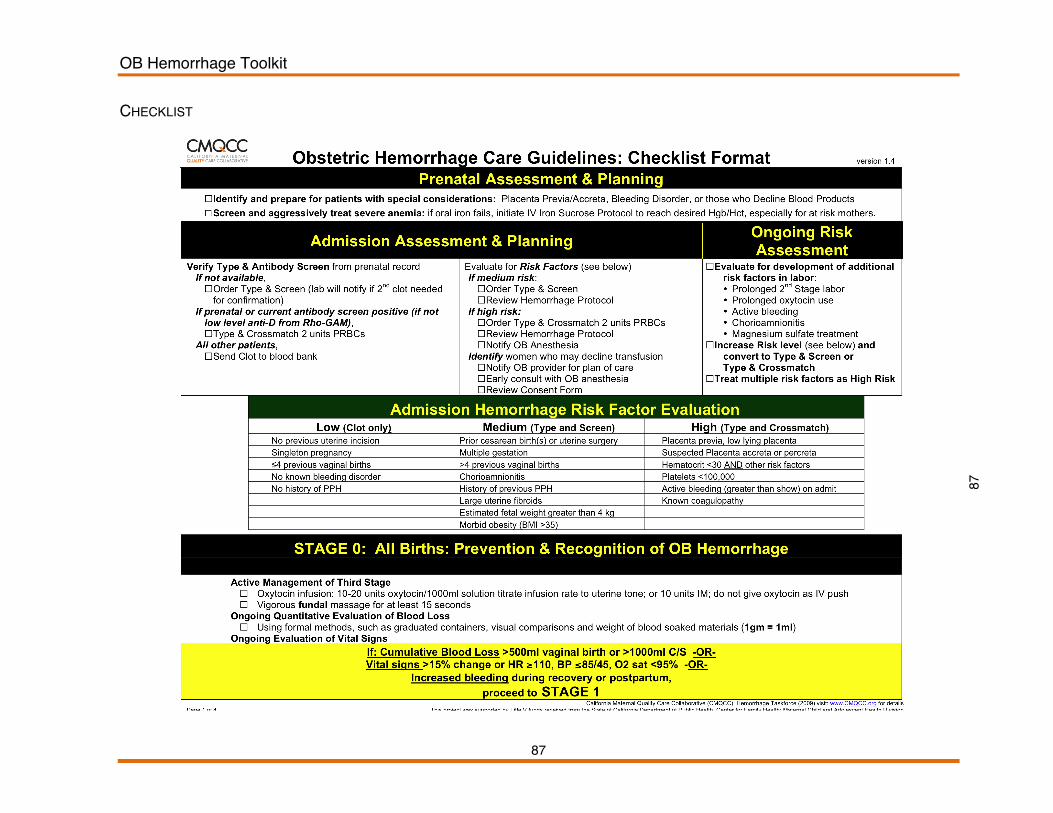
OB Hemorrhage Toolkit
87
87
CHECKLIST
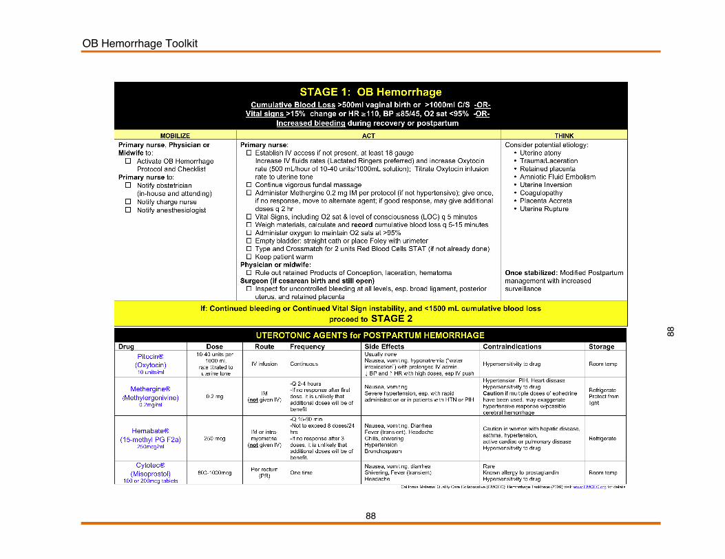
OB Hemorrhage Toolkit
88
88
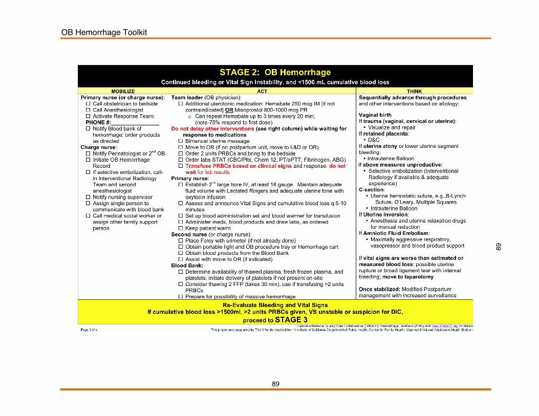
OB Hemorrhage Toolkit
89
89

OB Hemorrhage Toolkit
90
90

OB Hemorrhage Toolkit
91
TABLECHART
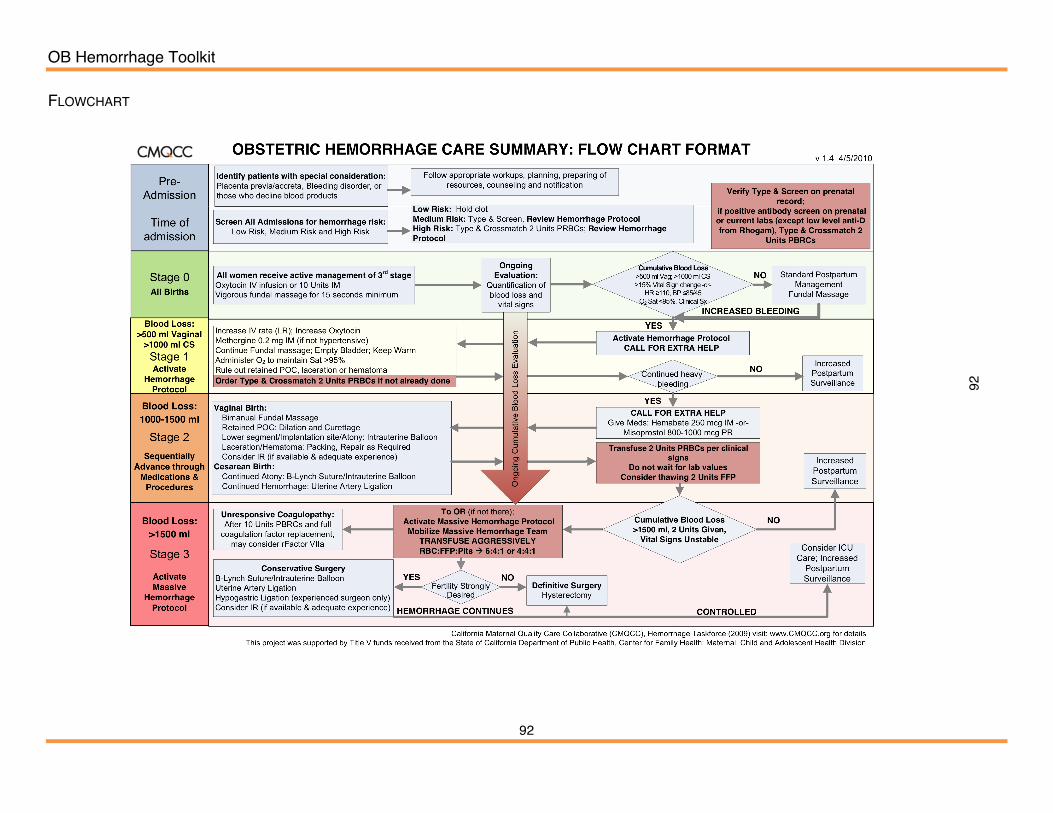
OB Hemorrhage Toolkit
92
92
FLOWCHART
OB HEMORRHAGE TOOLKIT
95 95
HOSPITAL LEVEL IMPLEMENTATION GUIDE
This implementation guide was developed to support local leadersʼ efforts to successfully implement best practices and care guidelines for obstetric hemorrhage and to create active quality improvement processes to drive future implementation plans. The implementation guide is organized to address four broad objectives as described by the Hemorrhage Task Force:
1. Improve readiness to respond to an obstetric hemorrhage by implementing standardized policies and procedures (general and massive) and developing obstetric hemorrhage rapid response teams.
2. Improve recognition of OB hemorrhage by performing on-going objective quantification of actual blood loss and triggers of maternal deterioration during and after all births.
3. Improve response to hemorrhage by performing regular on-site inter-professional hemorrhage drills.
4. Improve reporting of OB hemorrhage by standardizing definitions and consistency in coding and reporting.
The following elements are critical to affecting change to ensure that improvements are adopted and sustained over time:
• Leadership o Identifying Leader and Clinician behaviors, including “Champions” o Defining the problem and making the case for change o Setting goals o Allocating resources
• Policy & Procedure o Agreeing on a plan o Creating consistency between departments to improve teamwork and
cohesive quality of care; see Appendix for sample policy and protocol • Monitoring
o Creating audit tools that work; see Appendix for sample tools o Communicating progress toward goals

OB HEMORRHAGE TOOLKIT
96 96
COMPOSITE CASE The following is an outline of a composite case (combined elements from multiple cases with indentifying features, including all person identifiers, removed to ensure patient confidentiality) that demonstrates how a normal low-risk pregnancy and birth can at times quickly escalate to an emergent situation and death. An outline of learning points and opportunities for quality improvement (QI) opportunities based on the case follows. Composite Case Example: A 24yo woman, G2 P1 at 38 wks gestation was induced for “tired of being pregnant”:
• After 8hr active phase and 2hr 2nd stage, she gave birth (NSVD) to an 8lb 6oz infant. • After placental delivery, she had an episode of atony that firmed with massage. A second
episode of atony responded to IM methergine and the physician went home (now 1am). • The nurses called the physician 30min later to report more bleeding and further methergine was
ordered. • 60min after the call, the physician performed a D&C with minimal return of tissue. The woman
received more methergine. • 45min later a second D&C was performed, again with minimal returns. EBL at this point >2,000
ml. • Delays in blood transfusion occurred because of inability to find proper tubing. • Anesthesia was delayed, but a second IV started for more crystalloid. VS became markedly
abnormal, P=144, BP 80/30. • One further methergine given and patient taken for a 3rd D&C. She received 2u PRBCs by this
point. • After D&C is complete, she had a cardiac arrest from hypovolemia/hypoxia and was taken to the
ICU where she succumbed 3 hours later. QI Opportunities and Learning Points from the above composite case: How to reducing Mortality and Morbidity from OB Hemorrhage?
• Need a medical indication before performing an induction • No documentation of actual blood loss, e.g., what does “more bleeding” mean? • Only a few treatments tried, e.g., Methergine and D&C, and then repeated, even when they were
ineffective • Underestimation of blood loss • Delay in administration of blood • Lack of working equipment • Delay in response from other team members • Delays in adequate resuscitation • Lack of an organized standardized team approach
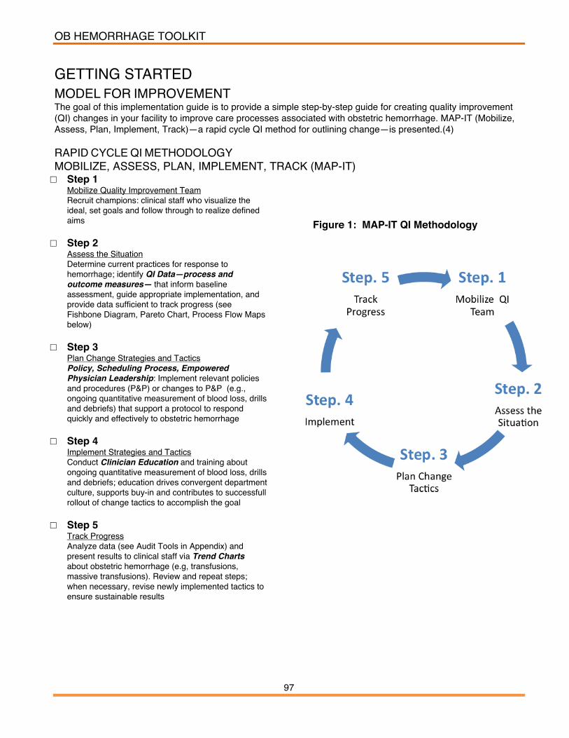
OB HEMORRHAGE TOOLKIT
97 97
GETTING STARTED
MODEL FOR IMPROVEMENT The goal of this implementation guide is to provide a simple step-by-step guide for creating quality improvement (QI) changes in your facility to improve care processes associated with obstetric hemorrhage. MAP-IT (Mobilize, Assess, Plan, Implement, Track)—a rapid cycle QI method for outlining change—is presented.(4) RAPID CYCLE QI METHODOLOGY MOBILIZE, ASSESS, PLAN, IMPLEMENT, TRACK (MAP-IT) □ Step 1
Mobilize Quality Improvement Team Recruit champions: clinical staff who visualize the ideal, set goals and follow through to realize defined aims
□ Step 2 Assess the Situation Determine current practices for response to hemorrhage; identify QI Data—process and outcome measures— that inform baseline assessment, guide appropriate implementation, and provide data sufficient to track progress (see Fishbone Diagram, Pareto Chart, Process Flow Maps below)
□ Step 3 Plan Change Strategies and Tactics Policy, Scheduling Process, Empowered Physician Leadership: Implement relevant policies and procedures (P&P) or changes to P&P (e.g., ongoing quantitative measurement of blood loss, drills and debriefs) that support a protocol to respond quickly and effectively to obstetric hemorrhage
□ Step 4 Implement Strategies and Tactics Conduct Clinician Education and training about ongoing quantitative measurement of blood loss, drills and debriefs; education drives convergent department culture, supports buy-in and contributes to successfull rollout of change tactics to accomplish the goal
□ Step 5 Track Progress Analyze data (see Audit Tools in Appendix) and present results to clinical staff via Trend Charts about obstetric hemorrhage (e.g, transfusions, massive transfusions). Review and repeat steps; when necessary, revise newly implemented tactics to ensure sustainable results
Figure 1: MAP-IT QI Methodology
OB HEMORRHAGE TOOLKIT
98 98
GETTING STARTED
STEP 1. MOBILIZE QUALITY IMPROVEMENT TEAM Essential team members include individuals with the following roles or skills:
• System Leadership • Technical Expertise • Day-to-Day Leadership
In obstetric service units, doctor and nurse leaders would fulfill these essential roles and skills. Utilizing the expertise and tacit knowledge of the front-line leaders and personnel, including nurses, general obstetricians, unit clerks, blood bank technicians is critical to successful implementation. A first step toward designating a team is identifying a system or administrative leader who leads MAP-IT cycles; e.g., meetings held, data collected, assignments completed, monitoring occurred. The following is a sample draft form to document your team: The Obstetric (OB) Hemorrhage System or Administrative Leader for our birthing facility is: _________________________________________________________ The nurse leader is:___________________________ The physician leader is:________________________ The members of the team that will work with the system/administrative, nurse, and physician leaders to improve readiness, recognition, response, and reporting are: NAME TITLE eMAIL ADDRESS
Mobilize
OB HEMORRHAGE TOOLKIT
99 99
STEP 2. ASSESS THE SITUATION In order to identify and prioritize quality improvement needs and next steps, teams can use these and other tools and methods:
• Fishbone Cause and Effect Diagrams: A team analysis of how individual process “problem areas” interact as a “whole”(5-7)
• Process Flowcharts: A team analysis of the numerous steps needed to complete a process. For example, the number of tasks a nurse must perform in order to obtain emergency medications(5, 7)
• Failure Modes and Effects Analysis (FMEA): A systematic analysis of the potential ways in which a failure, e.g., delay in recognition of excessive treatment and delay in adequate treatment of a woman bleeding will occur. The FMEA includes classification by severity or the “effects” of the failure based on the “mode” or type of failure that could occur.(5)
• Pareto Analysis and Diagrams: A method to quantify and rank which of the identified opportunities for improvement are the most problematic. The Pareto Principle is the 80/20 rule, e.g., 80% of the failures are caused by only 20% of the processes.(5)
Examples of the Fishbone and Pareto tools are described below. A. Fishbone Cause and Effect Diagrams: Quality improvement begins by looking at the “Big Picture” then narrowing down the problem to specific tasks that are reasonable and achievable starting points for change. The fishbone diagram is a common tool for seeing both elements—the whole and its parts.(5) The head of the fish points to the project goal and six (or more) bones extend from the side to capture the inter-relationships between PEOPLE, PROCEDURES, EQUIPMENT, MATERIALS, ENVIRONMENT AND MISCELLANEOUS factors that contribute to the problem. Fishbone diagrams are a good way to get group feedback and insights into the root cause of a problem. Example: Use a Fishbone Diagram (Figures 2a, 2b) to identify areas for Quality Improvement using OB Hemorrhage Care Guidelines Checklist, starting with “Pre-Admission” and working through Stages of Hemorrhage to identify problems. Diagrams can be as extensive and detailed as needed; alternately, diagrams can be drafted for each stage of hemorrhage; e.g., create one diagram that identifies processes of care surrounding Stage 1 Hemorrhage; create a second diagram that identifies processes of care surrounding Stage 2 Hemorrhage, etc. First drafts can be simple, hand-written versions that are edited, honed and vetted within leadership teams and front line staff to finalize plans for changes.
Assess

OB HEMORRHAGE TOOLKIT
100 10
0
Labor and Delivery leaders and staff identify processes of care in need of improvement by filling in the “fishbone diagram” based on a step-by-step review of the OB Hemorrhage Care Guidelines Checklist (Figures 2a, 2b). This approach is a useful framework to:
• Identify and define clinical roles and assignments of responsibility during each stage (People, Management)
• Identify medications—usage and dosage—during each stage of hemorrhage; assess that all Obstetric Services clinicians at all levels know and understand medications, usage and dosage (Materials, Process); assess availability and ease of access to critical medication supplies.
• Identify procedures, such as fundal massage, intrauterine balloon, B-Lynch suturing, quantification of blood loss during each stage of hemorrhage (Process, Materials, Equipment)
• Identify blood products and blood replacement during each stage of hemorrhage (Process, Equipment, Materials, Environment); identify availability and ease of access to these products and the supplies needed to administer them.
• Identify procedures for communication between all areas of obstetric care including, but not limited to, communications with the Blood Bank.
Figure 2a.

OB HEMORRHAGE TOOLKIT
101 10
1
Figure 2b.
. B. Pareto Analysis and Diagrams Pareto analysis, developed by Italian economist Vilfredo Pareto, is a statistical technique in decision-making to select limited tasks that will have a significant overall effect. The technique is based on the “Pareto Principle” or the “80/20” rule, which states, in quality improvement terms, that most (80%) of the problems are caused by a few (20%) key causes. There are many applications of the Pareto Principal in quality control including the Pareto Diagram, a key tool in Six Sigma (Motorola’s popular business management strategy).(5) Example: Use Pareto Analysis to create a Pareto Diagram (Figure 3) to identify areas for Quality Improvement using OB Hemorrhage Care Guidelines Checklist. Use the following steps (8) for completing a Pareto Analysis:
• Form a table (e.g., in a spreadsheet program) listing key causes of quality problems and their frequency as a percentage.
• Arrange the rows in the decreasing order of importance of the causes, i.e. the most important problems first.
• Add a cumulative percentage column to the table. • Plot with causes on x-axis and cumulative percentage on y-axis. • Join the above points to form a curve. • Plot (on the same graph) a bar graph with causes on x-axis and percent frequency
on y-axis.
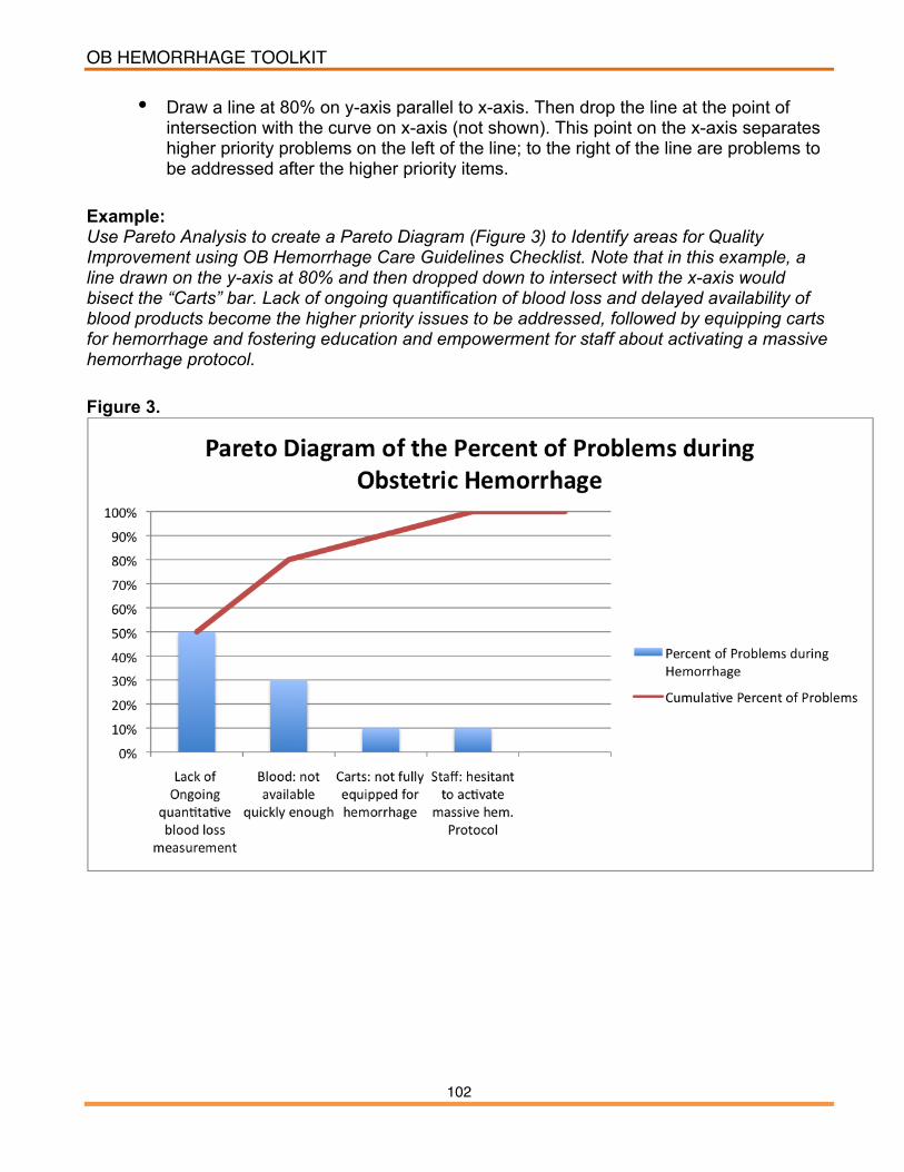
OB HEMORRHAGE TOOLKIT
102 10
2
• Draw a line at 80% on y-axis parallel to x-axis. Then drop the line at the point of intersection with the curve on x-axis (not shown). This point on the x-axis separates higher priority problems on the left of the line; to the right of the line are problems to be addressed after the higher priority items.
Example: Use Pareto Analysis to create a Pareto Diagram (Figure 3) to Identify areas for Quality Improvement using OB Hemorrhage Care Guidelines Checklist. Note that in this example, a line drawn on the y-axis at 80% and then dropped down to intersect with the x-axis would bisect the “Carts” bar. Lack of ongoing quantification of blood loss and delayed availability of blood products become the higher priority issues to be addressed, followed by equipping carts for hemorrhage and fostering education and empowerment for staff about activating a massive hemorrhage protocol. Figure 3.

OB HEMORRHAGE TOOLKIT
103 10
3
STEP 3. DEVELOP A PLAN TO MEET YOUR PROJECT AIMS An essential component of a plan is to develop aims or project objectives. The aims or project objectives include the use of specific terms, numerical goals and a time frame or deadline. Teams should work toward consensus agreement for the aims for your facility and have a willingness to refocus and modify the aims as needed. As an example, the following are aims of the first CMQCC Obstetric Hemorrhage Multi-Hospital Collaborative that began in October 2009 (Appendix E):
• Aim 1: Reduce the number of massive hemorrhages and the number of major complications from massive hemorrhage, including transfusions and hysterectomies, for all birthing women in participating hospitals by 75% by September 30, 2010.
• Aim 2: All collaborative participants develop and implement a multidisciplinary
team response to every massive obstetric hemorrhage by September 30, 2010.
Complete a MAP-IT planning worksheet for your project (See Appendix F). The plan should focus on specific objectives that will help you achieve the overall aims of the project. Developing a plan for each objective facilitates communication and tracking of the project. The MAP-IT plan needs to be regularly reviewed and re-adjusted based on feedback obtained during the rapid cycles of change and small tests of improvement. In Appendix F is a sample of a completed MAP-IT Plan for the objective to quantify blood loss in order to recognize an obstetric hemorrhage.
Plan
OB HEMORRHAGE TOOLKIT
104 10
4
STEP 4. IMPLEMENT STRATEGIES AND TACTICS TO MITIGATE BARRIERS A. Implementation Strategies and Tactics There are three broad types of implementation strategies the project team can utilize during implementation: 1) Education, 2) Data, and 3) Discourse or Communication Within each strategy there are multiple implementation tactics that leaders can choose from. Tables 1, 2 and 3 below outline some specific strategies and tactics for improving obstetric hemorrhage practices.(9, 10) Table 1. Examples of Implementation EDUCATION Tactics Educational Tactics Examples
Definition and Examples Specific to OB Hemorrhage
Grand Rounds Physician educational sessions that are often held once a week. • Hold a multi-disciplinary Obstetric Hemorrhage grand rounds
using the standard toolkit slide set • Make a copy of the slides and put them in a binder in L&D for all
staff to review Classes or Conferences Formal educational sessions developed on specific topics.
• Develop a class and skills station on quantification of blood loss – use hospital specific products
Simulation Training Simulation training is education that allows clinicians to practice skills and knowledge through a fabricated situation that mimics a complicated situation that they will face and need to practice how to respond. • Organize and run multi-disciplinary drills that help teams learn to
follow the CMQCC Obstetric Hemorrhage checklist so they have a more organized and systematic approach to hemorrhage
• This strategy also helps teams uncover hidden systems problems within their facility and get teams excited about finding solutions.
Competency Fairs, Tests, Learning Fairs, Return Demonstrations
Clinicians demonstrate their knowledge of a new concept or demonstrate their ability to perform a clinical skill. • Develop methods of measuring and tracking competency of team
members for the quantification of blood loss • Be creative and make learning fun
On-Line Learning The use of the internet for the transfer of information. • Provide clinicians with information on how to log-in to CMQCCʼs
website and have access to all of the CMQCC OB Hemorrhage resources: Table Chart, Check-list, Flow Chart, Compendium of Best Practices, and research articles
Implement

OB HEMORRHAGE TOOLKIT
105 10
5
Table 2. Examples of Implementation DATA Tactics Examples of Data Tactics Definition and Examples Specific to OB Hemorrhage Audit and feedback (group and individual)
An examination of clinical records in order to gather specific pre-determined information. The information gathered are summarized and shared with the relevant group or individuals. • Revise charting methods to track critical data • Develop a quality improvement data collection plan using audit
tools (refer to Appendices E1-E5) • Develop a quality improvement measurement grid (refer to
Appendix F) Public Release of Data Details of care patterns and outcomes are reported openly to the
community in such a way that anyone can access this information. • Improve how obstetric hemorrhage is defined and coded to make
it comparisons among hospitals more accurate *Adapted from Bingham D. Measuring and increasing the effectiveness of the quality improvement implementation change practices of front-line maternity physician and nurse leaders, The University of North Carolina at Chapel Hill, 2009, 316 pages; AAT 3352932. Table 3. Examples of Implementation COMMUNICATION Tactics Examples of Communication Tactics
Definition
Meetings Group discussions, e.g., staff meetings. • Regularly discuss the goals of the project and chart audit results
at staff meetings and at other gatherings • Present project data to senior leadership at hospital wide
meetings One-to-One Discussions A discussion between a change leader and someone else that they are
seeking to influence to change. • Team members can promote the change by explaining the
project goals during one-to-one discussions Debriefs A team discussion that takes place as soon as possible after an
emergency as possible. • Complete a debrief form for every stage 2 and 3 obstetric
hemorrhage emergency (refer to Appendix E.5) Academic Detailing A review of relevant academic research by one leader meeting with one
clinician at a time. A common tactic utilized by pharmaceutical representatives.
Policy and Procedure A document utilized by a hospital to set and communicate clinical standards. • Update policies and procedures
Reminders A method for helping someone remember to perform specific tasks, e.g., checklists, order set. • Have blood loss markings added to the pouch on the under
buttocks drape
OB HEMORRHAGE TOOLKIT
106 10
6
Table 3. Examples of Implementation COMMUNICATION Tactics, continued Examples of Communication Tactics
Definition
Newsletters A formal written update that is periodically distributed.
Posters and Bulletin Boards A collection of data and information that are organized for display on poster board or a bulletin board. • Develop posters and post on the unit.
Emails Electronic communications either one-to-one or by means of a “listserv” that is broadcast to many recipients.
Rewards Something given in compensation for reaching a pre-determined goal, e.g., professional recognition, a bonus. • Make learning fun – have some contests on assessing
quantification of blood loss Disciplinary Discussions A discussion that is held by someone in the position to give employee
feedback and a formal review of performance in order to outline how current behavior do not meet required expectations of job performance. • Follow-up with individuals who refuse to adopt the new practice
standards *Adapted from Bingham D. Measuring and increasing the effectiveness of the quality improvement implementation change practices of front-line maternity physician and nurse leaders, The University of North Carolina at Chapel Hill, 2009, 316 pages; AAT 3352932.

OB HEMORRHAGE TOOLKIT
107 10
7
B. Potential Implementation Barriers The most effective implementation plans include targetted strategies to mitigate potential and identified barriers. (10) Some types of barriers that have been identified are outlined in Table 4. Table 4. Examples of Potential Implementation Barriers*
Leader Barriers Design, plan, and implement Quality Improvement (QI) Perform QI data analysis
Lack of Leader Knowledge to:
Assess how to enhance their individual QI leadership abilities Topic selection QI topic goals Selection of QI implementation tactics
Leader Attitudes (Beliefs and Assumptions) that Affect: Definitions of success
Lack of clarity of QI project goals Backing down or stop trying Lack of time and other resources
Leader Practices
Inadequate practices to ensure sustainability, e.g., new hires, staff returning from vacation, leaves of absences
Clinician Barriers About their own practices (lack adequate feedback) New or inexperienced
Lack of Clinician Knowledge:
Lack knowledge of the QI project Not persuaded to change Clinician Attitudes
(Beliefs and Assumptions):
Want autonomy
Clinician Practices: Inertia – no motivation to change Forget Changes add more work or slow down usual work flow Characteristics of the QI Project Positive or negative effect(s) on clinician income or time Complexity of the QI project, e.g., how many groupsʼ work flow is affected by the QI
project changes Implementation Climate/Culture Type of hospital Amount of resources, e.g., workload, lack of organizational support for time or
supplies needed to implement change Type of community or population of patients History of previous unsuccessful change attempts or minimal previous history of
successful change attempts *Adapted from Bingham D. Measuring and increasing the effectiveness of the quality improvement implementation change practices of front-line maternity physician and nurse leaders, The University of North Carolina at Chapel Hill, 2009, 316 pages; AAT 3352932.

OB HEMORRHAGE TOOLKIT
108 10
8
STEP 5. TRACK PROGRESS USING ESTABLISHED MEASURES Essential components of measures include outcome, process and balance measures. When creating measures, teams should consider: What measures will be useful at your specific hospital based on your population of patients? Below are some definitions of different types of measures.
• Outcome Measures: These measures tell you whether changes are actually leading to improvement – that is, helping to achieve the overall aim of reducing major complications of OB hemorrhage. Outcome measures answer questions like, “How many women had injuries?” and “How many women had markers that indicate potential morbidities (e.g., transfusions?”). Outcome measures can sometimes be collected from administrative data.
• Process Measures: To affect the outcome measures of reducing major
complications of OB hemorrhage, changes will be made to improve many core processes in the care system, as well as changes to improve the culture as it relates to safety. We will want to know if the parts/steps in the system are performing as planned. Measuring the results of these process changes will tell you if the changes are leading to an improved, safer system.
• Balancing Measures: Measures to make sure that changes to improve one part
of the system are not causing new problems in other parts of the system. Balancing measures can also help teams to draw reasonable conclusions about the sustainability of the changes.
CMQCC developed Obstetric Hemorrhage quality improvement measurements for a multi-hospital collaborative. These measures are outlined in Appendix G. Hospital leaders can choose which of these measures are critical for tracking progress and whether they are achieving their aims and objectives. The tracking phase can also utilize the analysis tools outlined under the assessment phase.
Track
OB HEMORRHAGE TOOLKIT
109 10
9
Evaluating results early and often at each stage of implementation—before, during and after—will guide decision-making for small tests of change. Small tests of change done over a short time frame and reviewed frequently provide necessary feedback to the team leaders. The feedback will help leaders re-adjust the MAP-IT Plan appropriately.
Repeat the MAP-IT Process
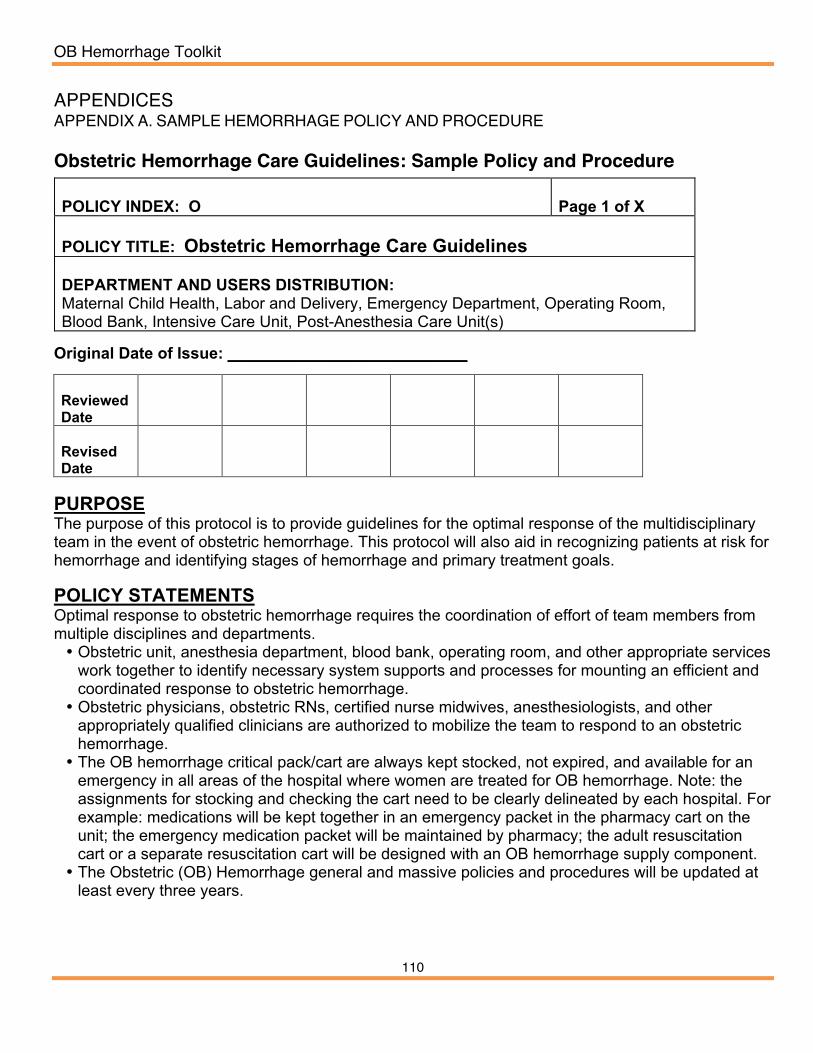
OB Hemorrhage Toolkit
110
APPENDICES APPENDIX A. SAMPLE HEMORRHAGE POLICY AND PROCEDURE Obstetric Hemorrhage Care Guidelines: Sample Policy and Procedure
POLICY INDEX: O
Page 1 of X
POLICY TITLE: Obstetric Hemorrhage Care Guidelines DEPARTMENT AND USERS DISTRIBUTION: Maternal Child Health, Labor and Delivery, Emergency Department, Operating Room, Blood Bank, Intensive Care Unit, Post-Anesthesia Care Unit(s)
Original Date of Issue: ___________________________ Reviewed Date
Revised Date
PURPOSE The purpose of this protocol is to provide guidelines for the optimal response of the multidisciplinary team in the event of obstetric hemorrhage. This protocol will also aid in recognizing patients at risk for hemorrhage and identifying stages of hemorrhage and primary treatment goals. POLICY STATEMENTS Optimal response to obstetric hemorrhage requires the coordination of effort of team members from multiple disciplines and departments. • Obstetric unit, anesthesia department, blood bank, operating room, and other appropriate services
work together to identify necessary system supports and processes for mounting an efficient and coordinated response to obstetric hemorrhage.
• Obstetric physicians, obstetric RNs, certified nurse midwives, anesthesiologists, and other appropriately qualified clinicians are authorized to mobilize the team to respond to an obstetric hemorrhage.
• The OB hemorrhage critical pack/cart are always kept stocked, not expired, and available for an emergency in all areas of the hospital where women are treated for OB hemorrhage. Note: the assignments for stocking and checking the cart need to be clearly delineated by each hospital. For example: medications will be kept together in an emergency packet in the pharmacy cart on the unit; the emergency medication packet will be maintained by pharmacy; the adult resuscitation cart or a separate resuscitation cart will be designed with an OB hemorrhage supply component.
• The Obstetric (OB) Hemorrhage general and massive policies and procedures will be updated at least every three years.
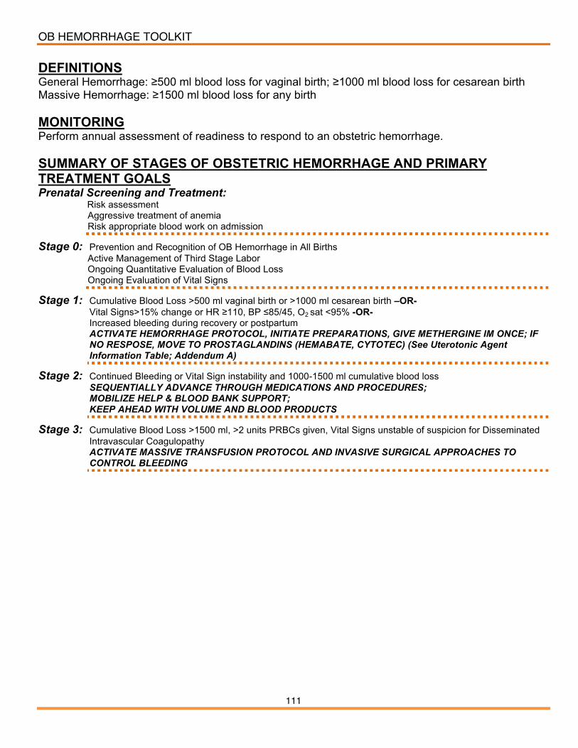
OB HEMORRHAGE TOOLKIT
111 11
1
DEFINITIONS General Hemorrhage: ≥500 ml blood loss for vaginal birth; ≥1000 ml blood loss for cesarean birth Massive Hemorrhage: ≥1500 ml blood loss for any birth MONITORING Perform annual assessment of readiness to respond to an obstetric hemorrhage. SUMMARY OF STAGES OF OBSTETRIC HEMORRHAGE AND PRIMARY TREATMENT GOALS Prenatal Screening and Treatment:
Risk assessment Aggressive treatment of anemia Risk appropriate blood work on admission
Stage 0: Prevention and Recognition of OB Hemorrhage in All Births Active Management of Third Stage Labor Ongoing Quantitative Evaluation of Blood Loss Ongoing Evaluation of Vital Signs
Stage 1: Cumulative Blood Loss >500 ml vaginal birth or >1000 ml cesarean birth –OR- Vital Signs>15% change or HR ≥110, BP ≤85/45, O2 sat <95% -OR- Increased bleeding during recovery or postpartum ACTIVATE HEMORRHAGE PROTOCOL, INITIATE PREPARATIONS, GIVE METHERGINE IM ONCE; IF NO RESPOSE, MOVE TO PROSTAGLANDINS (HEMABATE, CYTOTEC) (See Uterotonic Agent Information Table; Addendum A)
Stage 2: Continued Bleeding or Vital Sign instability and 1000-1500 ml cumulative blood loss SEQUENTIALLY ADVANCE THROUGH MEDICATIONS AND PROCEDURES; MOBILIZE HELP & BLOOD BANK SUPPORT; KEEP AHEAD WITH VOLUME AND BLOOD PRODUCTS
Stage 3: Cumulative Blood Loss >1500 ml, >2 units PRBCs given, Vital Signs unstable of suspicion for Disseminated Intravascular Coagulopathy ACTIVATE MASSIVE TRANSFUSION PROTOCOL AND INVASIVE SURGICAL APPROACHES TO CONTROL BLEEDING

OB HEMORRHAGE TOOLKIT
112 11
2
PROCEDURES Prenatal, Admission and Ongoing Risk Assessment • Identify and prepare for patients with special considerations: Placenta Preview/Accreta, Bleeding
Disorder, or those who decline blood products • Screen and aggressively treat severe anemia: if oral iron fails, initiate IV Iron Sucrose Protocol
(Best Practice: Iron Sucrose Protocol) to reach desired Hgb/Hct, especially for at-risk mothers
Admission Assessment & Planning Verify Type & Antibody Screen from prenatal record
If not available, Order Type & Screen (lab will notify if 2nd clot needed
for confirmation) If prenatal or current antibody screen positive (if not
low level anti-D from Rho-GAM), Type & Crossmatch 2 units PRBCs
All other patients, Send Clot to blood bank
Evaluate for Risk Factors (see below) If medium risk: Order Type & Screen Review Hemorrhage Protocol
If high risk: Order Type & Crossmatch 2 units PRBCs Review Hemorrhage Protocol Notify OB Anesthesia
Identify women who may decline transfusion Notify OB provider for plan of care Early consult with OB anesthesia Review Consent Form
*If admitted patients are started on magnesium sulfate they are at higher risk of postpartum hemorrhage.
Ongoing Risk Assessment Evaluate for development of additional risk factors in labor:
• Prolonged 2nd Stage labor • Prolonged oxytocin use • Active bleeding • Chorioamnionitis • Magnesium sulfate treatment
Increase Risk level (see below) and convert to Type & Screen or Type & Crossmatch
Treat multiple risk factors as High Risk
Admission Hemorrhage Risk Factor Evaluation Low (Clot only) Medium (Type and Screen) High (Type and Cross)
No previous uterine incision Prior cesarean birth(s) or uterine surgery Placenta previa, low lying placenta, Singleton pregnancy Multiple gestation Suspected placenta accreta or percreta ≤4 previous vaginal births >4 previous vaginal births Hematocrit <30 AND other risk factors No known bleeding disorder Chorioamnionitis Platelets <100,000 No history of PPH History of previous PPH Active bleeding (greater than show) on
admit Large uterine fibroids Known coagulopathy Estimated fetal weight greater than 4 kg Morbid obesity (BMI >35)

OB HEMORRHAGE TOOLKIT
113 11
3
PROCEDURES, CONTINUED STAGE 0 Prevention & Recognition of Hemorrhage during all births Active Management of Third Stage of Labor
1. Administer Oxytocin infusion: 10-20 Units/1000 ml solution for women with IV access. Note that the dosage and rates should be clearly specified by each hospital.
a. Titrate infusion rate to uterine tone. b. Use 10 units IM for women without IV access. c. Do not give oxytocin as IV push
2. Provide vigorous fundal massage for at least 15 seconds Ongoing Quantitative Measurement of Blood Loss at all Births
1. Assess blood loss at birth, prior to delivery of the placenta whenever possible. 2. Reassess cumulative blood loss after delivery of the placenta 3. Use formal methods to assess blood loss:
a. Use graduated under-buttock drapes b. Weigh blood soaked materials on gram scale (1 gm = 1ml)
i. Subtract known dry weight of materials ii. Use a hemorrhage report or Early Warning Chart (National Health Survey, NHS)
*NOTE: if a dry chux is used to protect scale from blood-soaked material, ZERO the scale after placing dry chux and prior to placing saturated item(s).
Ongoing Evaluation of Vital Signs and Clinical Triggers
STAGE 1 Cumulative Blood Loss >500 ml vaginal birth or >1000 ml C/S -OR- Vital Signs >15% change or HR ≥110, BP ≤85/45, O2 sat <95% -OR- Increased bleeding during recovery or postpartum Interventions: Follow Obstetric hemorrhage care guidelines checklist to mobilize response, act to mitigate bleeding, and move sequentially through treatment. Evaluate patient response to interventions:
1. If the patient is stable following Stage 1 interventions then perform increased postpartum surveillance.
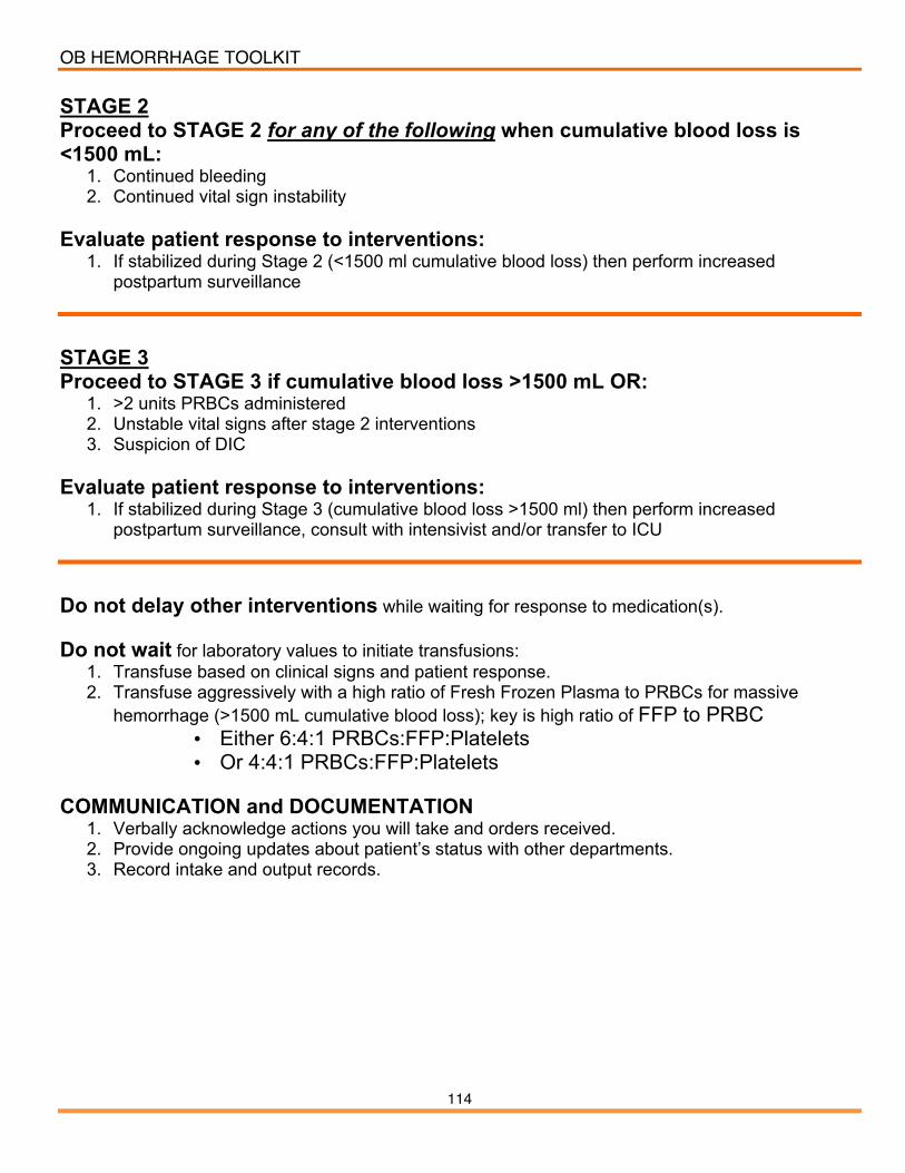
OB HEMORRHAGE TOOLKIT
114 11
4
STAGE 2 Proceed to STAGE 2 for any of the following when cumulative blood loss is <1500 mL:
1. Continued bleeding 2. Continued vital sign instability
Evaluate patient response to interventions:
1. If stabilized during Stage 2 (<1500 ml cumulative blood loss) then perform increased postpartum surveillance
STAGE 3 Proceed to STAGE 3 if cumulative blood loss >1500 mL OR:
1. >2 units PRBCs administered 2. Unstable vital signs after stage 2 interventions 3. Suspicion of DIC
Evaluate patient response to interventions: 1. If stabilized during Stage 3 (cumulative blood loss >1500 ml) then perform increased
postpartum surveillance, consult with intensivist and/or transfer to ICU
Do not delay other interventions while waiting for response to medication(s). Do not wait for laboratory values to initiate transfusions:
1. Transfuse based on clinical signs and patient response. 2. Transfuse aggressively with a high ratio of Fresh Frozen Plasma to PRBCs for massive
hemorrhage (>1500 mL cumulative blood loss); key is high ratio of FFP to PRBC • Either 6:4:1 PRBCs:FFP:Platelets • Or 4:4:1 PRBCs:FFP:Platelets
COMMUNICATION and DOCUMENTATION
1. Verbally acknowledge actions you will take and orders received. 2. Provide ongoing updates about patient’s status with other departments. 3. Record intake and output records.
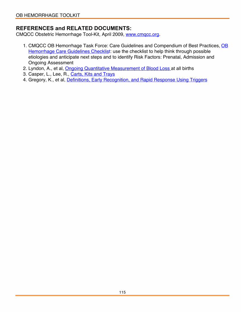
OB HEMORRHAGE TOOLKIT
115 11
5
REFERENCES and RELATED DOCUMENTS: CMQCC Obstetric Hemorrhage Tool-Kit, April 2009, www.cmqcc.org.
1. CMQCC OB Hemorrhage Task Force: Care Guidelines and Compendium of Best Practices, OB Hemorrhage Care Guidelines Checklist: use the checklist to help think through possible etiologies and anticipate next steps and to identify Risk Factors: Prenatal, Admission and Ongoing Assessment
2. Lyndon, A., et al, Ongoing Quantitative Measurement of Blood Loss at all births 3. Casper, L., Lee, R., Carts, Kits and Trays 4. Gregory, K., et al, Definitions, Early Recognition, and Rapid Response Using Triggers
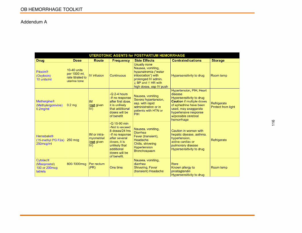
OB HEMORRHAGE TOOLKIT
116
Addendum A

OB HEMORRHAGE TOOLKIT
117
APPENDIX B: CMQCC OB HEMORRHAGE CARE GUIDELINES CHECKLIST
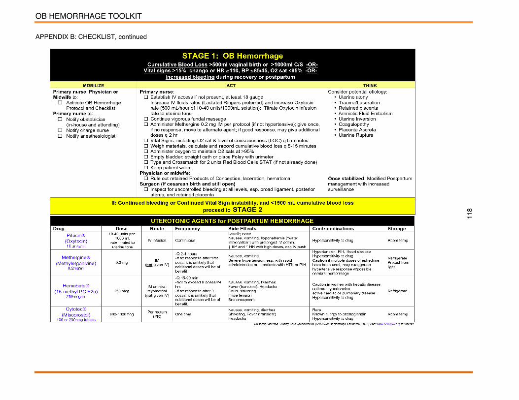
OB HEMORRHAGE TOOLKIT
118
APPENDIX B: CHECKLIST, continued

OB HEMORRHAGE TOOLKIT
119
APPENDIX B: CHECKLIST, continued

OB HEMORRHAGE TOOLKIT
120
APPENDIX B: CHECKLIST, continued

OB HEMORRHAGE TOOLKIT
121
APPENDIX C. CMQCC OBHEMORRHAGE CARE GUIDELINES FLOW CHART

OB HEMORRHAGE TOOLKIT
122
APPENDIX D. CMQCC OB HEMORRHAGE CARE GUIDELINES TABLE CHART
OB HEMORRHAGE TOOLKIT
123
APPENDIX E. CMQCC OBSTETRIC HEMORRHAGE QUALITY IMPROVEMENT
COLLABORATIVE
CMQCC initiated a statewide Obstetric Hemorrhage Quality Improvement (QI) Collaborative in October 2009. The OB Hemorrhage QI Collaborative is using the Institute for Healthcare Improvement (IHI) Breakthrough Series Model for Improvement, which includes intensive expert and peer mentoring. The Expert Panel, with input from the collaborating hospitals, developed specific goals or aims for the 12-month initiative. The aims guided the development of measures that assess improvement over the life of the collaborative and beyond. Aims and measures from this Collaborative are presented as a guide, but can be changed to meet facilitiesʼ needs. Refer to the appendix for checklists, flowcharts, audit tools and additional documents to use during implementation and change.

OB HEMORRHAGE TOOLKIT
124
APPENDIX E.1. CMQCC IN HOSPITAL AUDIT TOOL: RISK ASSESSMENT FOR OB HEMORRHAGE
Topic: Risk Assessment for obstetric hemorrhage is documented in the chart at admission. Goal: 100% of women are assessed for risk of obstetric hemorrhage on admission by [date]. Instructions: Audit 20 randomly selected charts per month (10 Vaginal, 10 cesarean).
MR# Vaginal Delivery Cesarean Delivery
MR# Vaginal Delivery Cesarean Delivery
MR# Vaginal Delivery Cesarean Delivery
MR# Vaginal Delivery Cesarean Delivery
MR# Vaginal Delivery Cesarean Delivery
Risk Assessment is documented in the chart Yes No Yes No Yes No Yes No Yes No
MR# Vaginal Delivery Cesarean Delivery
MR# Vaginal Delivery Cesarean Delivery
MR# Vaginal Delivery Cesarean Delivery
MR# Vaginal Delivery Cesarean Delivery
MR# Vaginal Delivery Cesarean Delivery
Risk Assessment is documented in the chart Yes No Yes No Yes No Yes No Yes No
MR# Vaginal Delivery Cesarean Delivery
MR# Vaginal Delivery Cesarean Delivery
MR# Vaginal Delivery Cesarean Delivery
MR# Vaginal Delivery Cesarean Delivery
MR# Vaginal Delivery Cesarean Delivery
Risk Assessment is documented in the chart Yes No Yes No Yes No Yes No Yes No
MR# Vaginal Delivery Cesarean Delivery
MR# Vaginal Delivery Cesarean Delivery
MR# Vaginal Delivery Cesarean Delivery
MR# Vaginal Delivery Cesarean Delivery
MR# Vaginal Delivery Cesarean Delivery
Risk Assessment is documented in the chart Yes No Yes No Yes No Yes No Yes No
Total number of audited charts with: Numerator: _______ Risk Assessment Documented in Chart Denominator: ________Total Number of Charts Audited
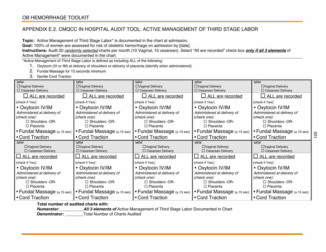
OB HEMORRHAGE TOOLKIT
125
APPENDIX E.2. CMQCC IN HOSPITAL AUDIT TOOL: ACTIVE MANAGEMENT OF THIRD STAGE LABOR Topic: Active Management of Third Stage Labor* is documented in the chart at admission. Goal: 100% of women are assessed for risk of obstetric hemorrhage on admission by [date]. Instructions: Audit 20 randomly selected charts per month (10 Vaginal, 10 cesarean). Select “All are recorded” check box only if all 3 elements of Active Management* were documented in the chart. *Active Management of Third Stage Labor is defined as including ALL of the following:
1. Oxytocin (IV or IM) at delivery of shoulders or delivery of placenta (identify when administered) 2. Fundal Massage for 15 seconds minimum 3. Gentle Cord Traction
MR# Vaginal Delivery Cesarean Delivery
MR# Vaginal Delivery Cesarean Delivery
MR# Vaginal Delivery Cesarean Delivery
MR# Vaginal Delivery Cesarean Delivery
MR# Vaginal Delivery Cesarean Delivery
ALL are recorded (check if Yes): • Oxytocin IV/IM Administered at delivery of (check one):
Shoulders -OR- Placenta
• Fundal Massage (≥ 15 sec) • Cord Traction
ALL are recorded (check if Yes): • Oxytocin IV/IM Administered at delivery of (check one):
Shoulders -OR- Placenta
• Fundal Massage (≥ 15 sec) • Cord Traction
ALL are recorded (check if Yes): • Oxytocin IV/IM Administered at delivery of (check one):
Shoulders -OR- Placenta
• Fundal Massage (≥ 15 sec) • Cord Traction
ALL are recorded (check if Yes): • Oxytocin IV/IM Administered at delivery of (check one):
Shoulders -OR- Placenta
• Fundal Massage (≥ 15 sec) • Cord Traction
ALL are recorded (check if Yes): • Oxytocin IV/IM Administered at delivery of (check one):
Shoulders -OR- Placenta
• Fundal Massage (≥ 15 sec) • Cord Traction
MR# Vaginal Delivery Cesarean Delivery
MR# Vaginal Delivery Cesarean Delivery
MR# Vaginal Delivery Cesarean Delivery
MR# Vaginal Delivery Cesarean Delivery
MR# Vaginal Delivery Cesarean Delivery
ALL are recorded (check if Yes): • Oxytocin IV/IM Administered at delivery of (check one):
Shoulders -OR- Placenta
• Fundal Massage (≥ 15 sec) • Cord Traction
ALL are recorded (check if Yes): • Oxytocin IV/IM Administered at delivery of (check one):
Shoulders -OR- Placenta
• Fundal Massage (≥ 15 sec) • Cord Traction
ALL are recorded (check if Yes): • Oxytocin IV/IM Administered at delivery of (check one):
Shoulders -OR- Placenta
• Fundal Massage (≥ 15 sec) • Cord Traction
ALL are recorded (check if Yes): • Oxytocin IV/IM Administered at delivery of (check one):
Shoulders -OR- Placenta
• Fundal Massage (≥ 15 sec) • Cord Traction
ALL are recorded (check if Yes): • Oxytocin IV/IM Administered at delivery of (check one):
Shoulders -OR- Placenta
• Fundal Massage (≥ 15 sec) • Cord Traction
Total number of audited charts with: Numerator: __________All 3 elements of Active Management of Third Stage Labor Documented in Chart Denominator: ________Total Number of Charts Audited
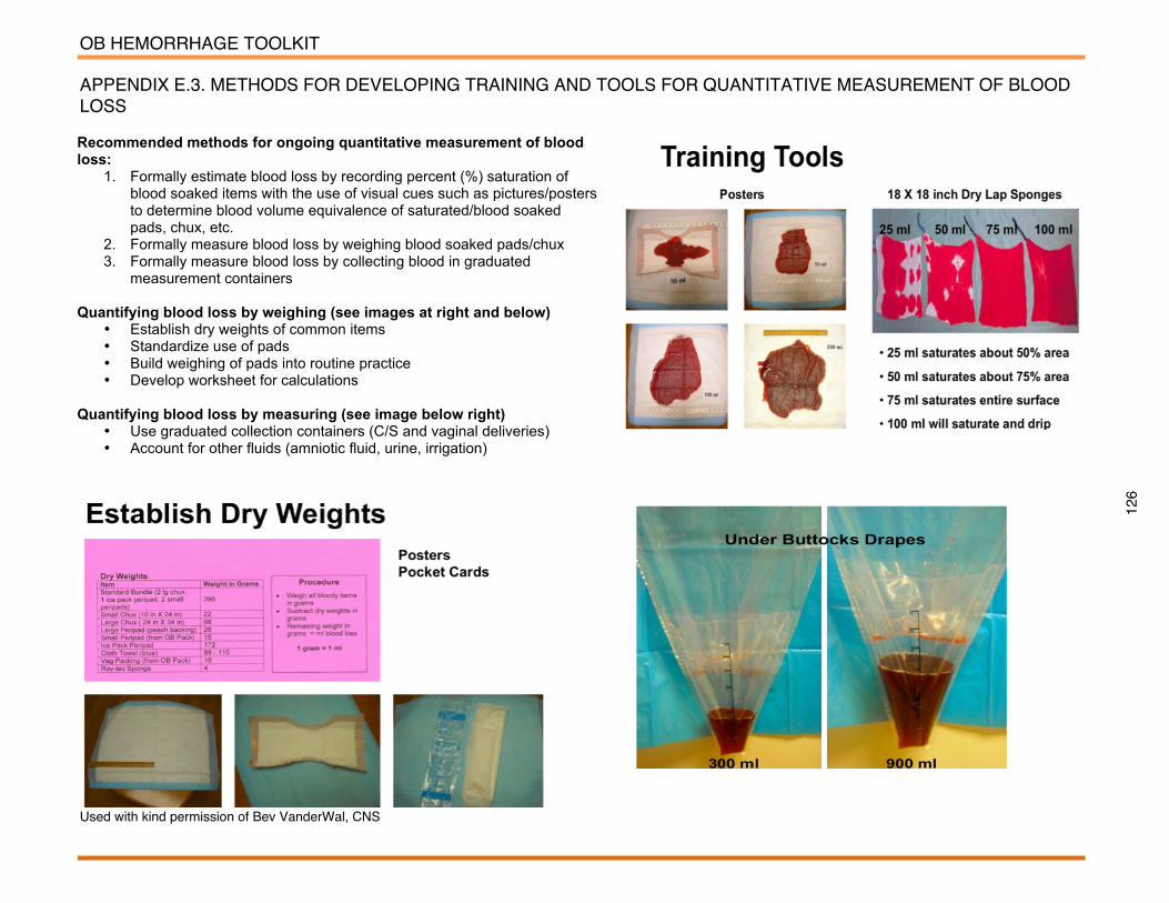
OB HEMORRHAGE TOOLKIT
126
APPENDIX E.3. METHODS FOR DEVELOPING TRAINING AND TOOLS FOR QUANTITATIVE MEASUREMENT OF BLOOD LOSS
Used with kind permission of Bev VanderWal, CNS
Recommended methods for ongoing quantitative measurement of blood loss:
1. Formally estimate blood loss by recording percent (%) saturation of blood soaked items with the use of visual cues such as pictures/posters to determine blood volume equivalence of saturated/blood soaked pads, chux, etc.
2. Formally measure blood loss by weighing blood soaked pads/chux 3. Formally measure blood loss by collecting blood in graduated
measurement containers Quantifying blood loss by weighing (see images at right and below)
• Establish dry weights of common items • Standardize use of pads • Build weighing of pads into routine practice • Develop worksheet for calculations
Quantifying blood loss by measuring (see image below right)
• Use graduated collection containers (C/S and vaginal deliveries) • Account for other fluids (amniotic fluid, urine, irrigation)
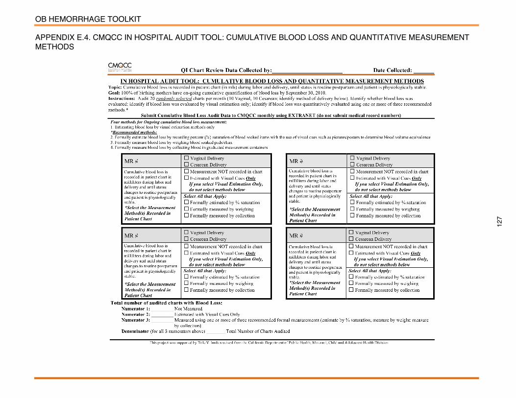
OB HEMORRHAGE TOOLKIT
127
APPENDIX E.4. CMQCC IN HOSPITAL AUDIT TOOL: CUMULATIVE BLOOD LOSS AND QUANTITATIVE MEASUREMENT METHODS

OB HEMORRHAGE TOOLKIT
128
APPENDIX E.5. CMQCC OBSTETRIC HEMORRHAGE TEAM DE-BRIEFING FORM Topic: The de-brief form provides an opportunity for maternity service teams to review then document sequence of events, successes and barriers to a swift and
coordinated response to obstetric hemorrhage. Goal: De-brief completed in 100% of all obstetric hemorrhages that progress to Stage 2 or 3. All de-briefs have at least Primary RN, and Primary MD who participates in the de-briefing session. Instructions: Complete as soon as possible, but no later than 24 hours after any Stage 2 or 3 hemorrhages. During de-brief, obtain input from participants (all or as many as possible). Stage 2 or 3 hemorrhages are defined as bleeding that continues after administration of IV or IM Oxytocin, vigorous fundal massage, emptied bladder and Methergine 0.2 mg IM. PRINT THIS FORM DOUBLE-SIDED; PATIENT STAMP ON BACK OF FORM WILL NOT BE FAXED WITH COMPLETED FORM. - Attach additional pages with notes as neededWere the following medications, procedures or blood products used? (Check if yes, check all that apply) Medications High dose misoprostol (800-1000 mcg) Carboprost tromethamine (Hemobate Blood Volume/Options Invasive hemodynamic monitoring Blood warmer Rapid fluid infuser (level one machine) Blood cell salvage machine (cell saver) Factor VIIa (non-standard treatment) Procedures Intrauterine balloons B-Lynch suture Uterine artery ligation Uterine artery embolization Non-pneumatic Anti-shock Garments (NASG; non-standard
treatment)
COMMENTS about medications, procedures, or blood products: Primary MD participated in de-brief? (Check if yes) Primary RN participated in de-brief? (Check if yes) Post-hemorrhage, the patient required… (Check if yes, check all that apply) Intubation Central Line Pressors Arterial Line Admission to ICU Admission to higher acuity unit (e.g., PACU)
Volume of blood lost: _____ mls Method of Blood Loss Measurement (Check all that
apply) Visually Estimated Only Formal Estimate using Posters/Pictures Formal Measure by weight Formal Measure by volume collection Blood Product Transfusion Ratios - Active Hemorrhage Treatment and Resuscitation Period (~the first 4-6 hours PP) Units of PRBCs: ______________ Units of FFP: ______________ Units of Platelets: _____________ Units of Cryo: _____________
Thinking about how the obstetric hemorrhage was managed… Identify what went well (Check if yes, describe) Communication went well Teamwork went well Leadership went well Decision-making went well Assessing the situation went well Other Briefly describe:
Identify opportunities for improvement: “human factors” (Check if yes, describe) Communication needed improvement Teamwork needed improvement Leadership needed improvement Decision-making needed improvement Assessing needed improvement Other Briefly describe:
Identify opportunities for improvement: “non-human factors” (Check if yes, describe) Delay in blood products availability Equipment issues Medications issues Inadequate support (in-unit or other areas of the hospital)
Delays in transporting the patient (within the hospital or to another facility) Other Briefly describe:

OB HEMORRHAGE TOOLKIT
129
APPENDIX F. CMQCC MAP-IT PLANNING WORKSHEET, SAMPLE WORKSHEET

OB HEMORRHAGE TOOLKIT
130
APPENDIX F. CMQCC MAP-IT PLANNING Worksheet, sample worksheet
OB HEMORRHAGE TOOLKIT
131
APPENDIX G. CMQCC QUALITY IMPROVEMENT COLLABORATIVE OBSTETRIC HEMORRHAGE MEASUREMENT GRID CMQCC Quality Improvement Collaborative Obstetric Hemorrhage Measurement Grid Purpose of the Measurement Grid: The measurement grid outlines the measures to be collected over the 12-month life of the OB Hemorrhage multi-hospital collaborative. The grid includes the specific parameters for each measure. Measurement for Improvement: CMQCC multi-hospital improvement collaboratives are about making hospital systems safer for patients. Measurements play an important role. Always remember that measurements should be designed to accelerate improvement, not slow it down. Timeline for Measurement: The measurement grid is broken down into three categories: Outcome, Process and Balancing Measures. Teams may also develop additional measures based on the issues that are of most interest and importance to their hospital and patient population needs. Only the measures in the grid below will be submitted to CMQCC. Outcome Measures: These measures tell you whether changes are actually leading to improvement – that is, helping to achieve the overall aim of reducing major complications of OB hemorrhage. Outcome measures answer questions like, “How many women had injuries?” and “How many women had markers that indicate potential morbidities (e.g., transfusions?”). Outcome measures can sometimes be collected from administrative data.
Process Measures: To affect the outcome measures of reducing major complications of OB hemorrhage, changes will be made to improve many core processes in the care system, as well as changes to improve the culture as it relates to safety. We will want to know if the parts/steps in the system are performing as planned. Measuring the results of these process changes will tell you if the changes are leading to an improved, safer system. Balancing Measures: We will use these measures to make sure that changes to improve one part of the system are not causing new problems in other parts of the system. Balancing measures can also help us to draw reasonable conclusions about the sustainability of the change
OB Hemorrhage Multi-Hospital Change Collaborative
Aim 1: Reduce the number of massive hemorrhages and the number of major complications from massive hemorrhage, including transfusions and hysterectomies, for all birthing women in participating hospitals by 75% by September 30, 2010.
Aim 2: All collaborative participants develop and implement a multidisciplinary team response to every massive obstetric hemorrhage by September 30, 2010.
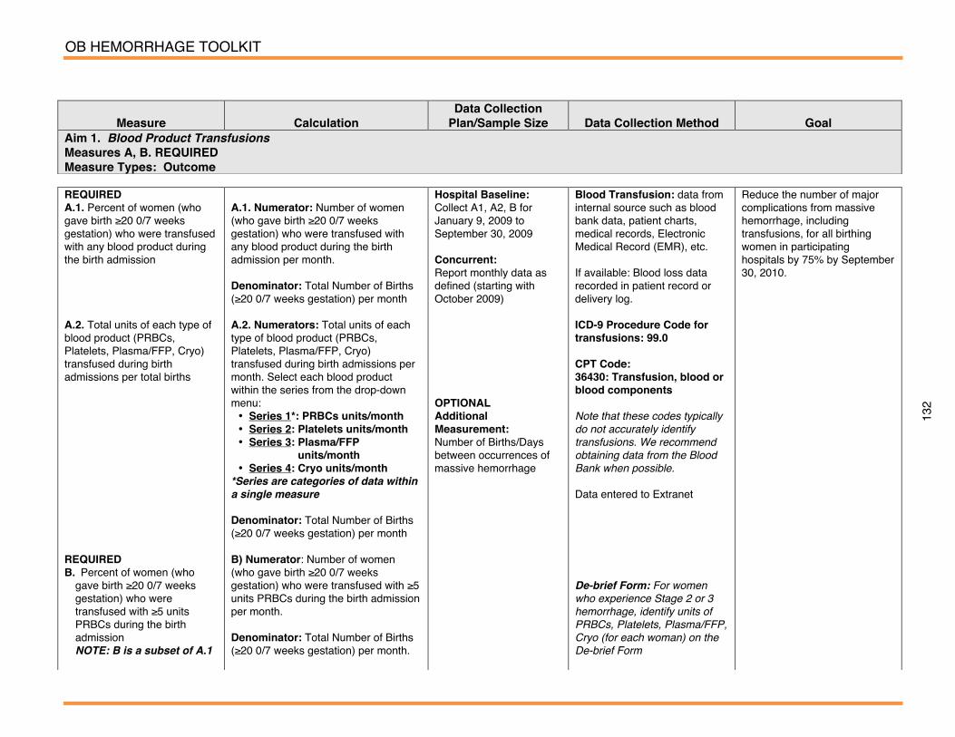
OB HEMORRHAGE TOOLKIT
132
Measure
Calculation
Data Collection Plan/Sample Size
Data Collection Method
Goal
Aim 1. Blood Product Transfusions Measures A, B. REQUIRED Measure Types: Outcome
REQUIRED A.1. Percent of women (who gave birth ≥20 0/7 weeks gestation) who were transfused with any blood product during the birth admission A.2. Total units of each type of blood product (PRBCs, Platelets, Plasma/FFP, Cryo) transfused during birth admissions per total births REQUIRED B. Percent of women (who
gave birth ≥20 0/7 weeks gestation) who were transfused with ≥5 units PRBCs during the birth admission NOTE: B is a subset of A.1
A.1. Numerator: Number of women (who gave birth ≥20 0/7 weeks gestation) who were transfused with any blood product during the birth admission per month. Denominator: Total Number of Births (≥20 0/7 weeks gestation) per month A.2. Numerators: Total units of each type of blood product (PRBCs, Platelets, Plasma/FFP, Cryo) transfused during birth admissions per month. Select each blood product within the series from the drop-down menu: • Series 1*: PRBCs units/month • Series 2: Platelets units/month • Series 3: Plasma/FFP
units/month • Series 4: Cryo units/month
*Series are categories of data within a single measure Denominator: Total Number of Births (≥20 0/7 weeks gestation) per month B) Numerator: Number of women (who gave birth ≥20 0/7 weeks gestation) who were transfused with ≥5 units PRBCs during the birth admission per month.
Denominator: Total Number of Births (≥20 0/7 weeks gestation) per month.
Hospital Baseline: Collect A1, A2, B for January 9, 2009 to September 30, 2009 Concurrent: Report monthly data as defined (starting with October 2009) OPTIONAL Additional Measurement: Number of Births/Days between occurrences of massive hemorrhage
Blood Transfusion: data from internal source such as blood bank data, patient charts, medical records, Electronic Medical Record (EMR), etc. If available: Blood loss data recorded in patient record or delivery log. ICD-9 Procedure Code for transfusions: 99.0 CPT Code: 36430: Transfusion, blood or blood components Note that these codes typically do not accurately identify transfusions. We recommend obtaining data from the Blood Bank when possible. Data entered to Extranet De-brief Form: For women who experience Stage 2 or 3 hemorrhage, identify units of PRBCs, Platelets, Plasma/FFP, Cryo (for each woman) on the De-brief Form
Reduce the number of major complications from massive hemorrhage, including transfusions, for all birthing women in participating hospitals by 75% by September 30, 2010.

OB HEMORRHAGE TOOLKIT
133
Measure
Calculation Data Collection
Plan/Sample Size
Data Collection Method
Goal Aim 1. Peripartum Hysterectomies Measure C. REQUIRED Measure Type: Outcome C. Rate of peripartum hysterectomies in women (who gave birth ≥20 0/7 weeks gestation) per 1000 births (hysterectomy performed during birth admission) stratified by risk of Placenta Previa and/or Placenta Accreta/percreta
C. Numerator: Number of peripartum hysterectomies (performed during birth admission) in women (who gave birth ≥20 0/7 weeks gestation) per month stratified by: • Series 1*: Women with Placenta
Previa and/or Placenta Accreta/Percreta
• Series 2: Women without Placenta Previa and/or Placenta Accreta/Percreta
*Series are categories of data within a single measure
Denominator: Total Number of Births (≥20 0/7 weeks gestation) per month Risk Stratification/Adjustment: Women who had a hysterectomy and placenta previa and/or accreta/percreta are reported separately from women who had a hysterectomy (and NO placenta previa/accreta/percreta)
Annotate** for each hysterectomy: a) Indication for hysterectomy b) Number of prior cesarean
sections c) Number of Days Post-
Delivery (Days = 0 if procedure done on day of delivery)
Hospital Baseline: Number of peripartum hysterectomies (performed during birth admission) in women who gave birth ≥20 0/7 weeks gestation Between 1/1/09 to 9/30/09 Concurrent: Report monthly data as defined
Peripartum Hysterectomy: Data Collection from internal source such as EMR, medical records, or other method determined by each site ICD-9 Procedure Codes 68.3 Subtotal abdominal hysterectomy 68.39 Other and unspecified subtotal abdominal hysterectomy 68.4 Total abdominal hysterectomy 68.49 Other and unspecified total abdominal hysterectomy CPT Codes 59525 Cesarean Hysterectomy 58150 Hysterectomy Total/Partial (Use Post-Partum or with Vaginal) 59160 D&C after delivery Data entered to Extranet **Annotation is available in the Data Entry fields for this measure; identify a), b), and c) (from Calculation column) for each patient in the Annotation Field
Reduce the number of major complications from massive hemorrhage, including peripartum hysterectomies, for all birthing women in participating hospitals by 75% by September 30, 2010.

OB HEMORRHAGE TOOLKIT
134
Measure
Calculation Data Collection
Plan/Sample Size
Data Collection Method
Goal Aim 1. Quantification of Blood Loss Measures D, E, F. REQUIRED Deliverables 1, 2, 3. REQUIRED Measure Types: Outcome and Process D. Percent of Audited Charts in which quantification and documentation of blood loss is performed (during and after all births until immediate recovery status changes to routine postpartum care and woman is physiologically stable) using one or more of three preferred methods: 1. Formally estimate blood loss
by recording percent (%) saturation of blood soaked items with the use of visual cues such as pictures/posters to determine blood volume equivalence of saturated/blood soaked pads, chux, etc.
2. Formally measure blood loss by weighing blood soaked pads/chux
3. Formally measure blood loss by collecting blood in graduated measurement containers
Process Measures: E. % Non-MD clinicians and staff
who are educated about Cumulative Blood Loss and Quantitative Measurement Methods
D. Numerator: Number of charts per month where on-going quantification is: • Series 1: NOT recorded • Series 2: Recorded using
visualization only • Series 3: Recorded using one
of three preferred formal methods (1, 2, or 3 in Measure Column)
Denominator: Number of charts audited per month E. Numerator: Number of non-MD clinicians, (e.g., RNs, midwives) and staff (e.g., clerks, aides) who are educated about Cumulative Blood Loss and Quantitative Measurement Methods Audit Tool per month Denominator: Number of Non-MD
Audit 20 randomly selected charts per month (10 vaginal births and 10 cesarean births)
Chart Review: Refer to sample audit tool entitled: “In Hospital Audit Tool: Cumulative Blood Loss and Quantitative Measurement Methods” Data entered to Extranet
100% of birthing women will have on-going cumulative quantification of blood loss by September 30, 2010.
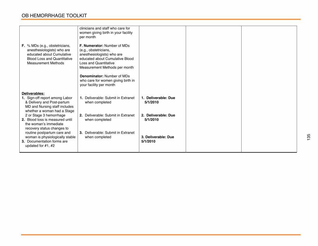
OB HEMORRHAGE TOOLKIT
135
F. % MDs (e.g., obstetricians,
anesthesiologists) who are educated about Cumulative Blood Loss and Quantitative Measurement Methods
Deliverables: 1. Sign-off report among Labor
& Delivery and Post-partum MD and Nursing staff includes whether a woman had a Stage 2 or Stage 3 hemorrhage
2. Blood loss is measured until the womanʼs immediate recovery status changes to routine postpartum care and woman is physiologically stable
3. Documentation forms are updated for #1, #2
clinicians and staff who care for women giving birth in your facility per month F. Numerator: Number of MDs (e.g., obstetricians, anesthesiologists) who are educated about Cumulative Blood Loss and Quantitative Measurement Methods per month Denominator: Number of MDs who care for women giving birth in your facility per month 1. Deliverable: Submit in Extranet
when completed 2. Deliverable: Submit in Extranet
when completed 3. Deliverable: Submit in Extranet
when completed
1. Deliverable: Due
5/1/2010 2. Deliverable: Due
5/1/2010 3. Deliverable: Due 5/1/2010

OB HEMORRHAGE TOOLKIT
136
Measure Calculation Data Collection Plan/Sample Size
Data Collection Method Goal
Aim 1. OB Hemorrhage Risk Assessment on Admission Measure G. RECOMMENDED Measure Type: Process Percent of women (from audited charts) who are assessed for risk of obstetric hemorrhage on admission
Numerator: Among the audited charts, the number of women admitted to Labor and Delivery whose risk of obstetric hemorrhage assessment is recorded in the medical record Denominator: Total number of admission charts audited per month
Suggestion: Audit 20 randomly selected charts per month (10 vaginal births and 10 cesarean births)
Chart Review Determined by individual hospital. Refer to Risk Assessment Audit Tool. Data entered to Extranet
100% of women are assessed for risk of obstetric hemorrhage on admission by September 30, 2010
Aim 1. Active Management of Third Stage Measure H. RECOMMENDED Measure Type: Process Percent of women (from audited charts) who receive Active Management of Third Stage Labor
Numerator: Total number of women who receive Active Management of Third Stage Labor including • Oxytocin (IV or IM) at delivery of shoulders or delivery of placenta (identify when administered) • Fundal Massage for 15
seconds minimum • Gentle Cord traction Note: Need all three to be considered Active Management Denominator: Total number of charts audited per month
Suggestion: Audit 20 randomly selected charts of women who gave birth vaginally per month
Chart review Determined by individual hospital. Refer to Active Management of Third Stage Labor Audit Tool. Data entered to Extranet
100% of women giving birth will receive active management of labor by September 30, 2010

OB HEMORRHAGE TOOLKIT
137
Measure
Calculation Data Collection
Plan/Sample Size
Data Collection Method
Goal Aim 2. Policies and Procedures and Drills Measure I, J, K, L. REQUIRED Deliverables 4, 5, 6, 7, 8. REQUIRED Measure Type: Process Deliverables: 4. General and massive
hemorrhage policies and procedures (P&P) were updated after April 30, 2009.
5. Identify roles and multi-
disciplinary team responders for stage 1, 2, and 3 hemorrhages
6. Determine and implement
the most desirable method for maintaining accessibility to the needed OB hemorrhage supplies
Measures I. % Non-MD clinicians and
staff who are educated to the hemorrhage P&Ps
4. Deliverable: Submit in
Extranet when completed; Date completed; date received by CMQCC
5. Deliverable: Roles defined
for stage 1, 2, or 3 hemorrhages: Submit in Extranet when completed; date completed; date received by CMQCC
6. Deliverable: Provide the
emergency supply maintenance plan to CMQCC: Submit in Extranet when completed; date completed; date received by CMQCC
I. Numerator: Number of non-MD clinicians, (e.g., RNs, midwives) and staff (e.g., clerks, aides) who receive education on the hemorrhage P&Ps per month Denominator: Number of Non-MD clinicians and staff who are in the pool of possible responders per month (defined
4. Determined by hospital, Due: 2/1/2010 5. Determined by hospital, Due: 3/1/2010 6. Determined by hospital, Due: 2/1/2010 I. Determined by hospital
Hospital records and forms 100% of the collaborative participating hospitals will meet 100% of the P&Ps and drills measurements by September 30, 2010

OB HEMORRHAGE TOOLKIT
138
J. % MDs (e.g., obstetricians,
anesthesiologists) who are educated to the hemorrhage P&Ps
Deliverables 7. Create drills tailored to your
hospital P&Ps and responder roles
8. After Deliverables 4-7 are
completed, run 1 multi-disciplinary drill per month for four consecutive months (two on night/evening shift and two on day shift) to identify system and process improvement opportunities. After each drill complete a drill de-brief form
Measure K. % Non-MD clinicians and staff who are involved drill de-
by hospital) J. Numerator: Number of MDs (e.g., obstetricians, anesthesiologists) who receive education on the hemorrhage P&Ps per month Denominator: Number of MDs who are in the pool of possible responders per month (defined by hospital) 7. Deliverable: Drill scenarios created: Submit in Extranet when completed; date completed; date received by CMQCC 8. Deliverable: Drills are performed: Submit in Extranet when completed; dates completed. Maintain a list of problems identified by the drills and document how and when the problem is resolved. Submit the list to CMQCC. K. Numerator: Number of non-MD clinicians (e.g., RNs, midwives) and staff (e.g.,
J. Determined by hospital 7. Determined by hospital, Due: 5/1/2010 8. Determined by hospital Due: 8/1/2010 K. Determined by hospital
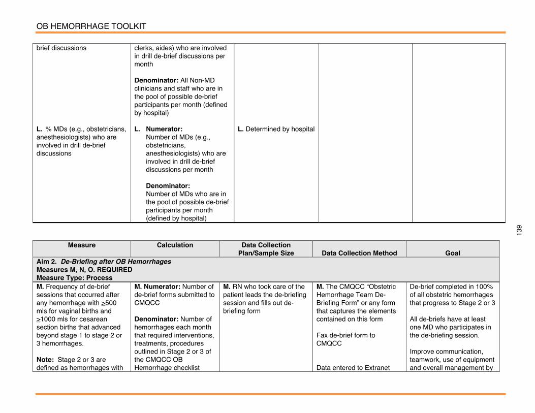
OB HEMORRHAGE TOOLKIT
139
brief discussions L. % MDs (e.g., obstetricians, anesthesiologists) who are involved in drill de-brief discussions
clerks, aides) who are involved in drill de-brief discussions per month Denominator: All Non-MD clinicians and staff who are in the pool of possible de-brief participants per month (defined by hospital) L. Numerator: Number of MDs (e.g.,
obstetricians, anesthesiologists) who are involved in drill de-brief discussions per month
Denominator: Number of MDs who are in
the pool of possible de-brief participants per month (defined by hospital)
L. Determined by hospital
M. Frequency of de-brief sessions that occurred after any hemorrhage with >500 mls for vaginal births and >1000 mls for cesarean section births that advanced beyond stage 1 to stage 2 or 3 hemorrhages. Note: Stage 2 or 3 are defined as hemorrhages with
M. Numerator: Number of de-brief forms submitted to CMQCC Denominator: Number of hemorrhages each month that required interventions, treatments, procedures outlined in Stage 2 or 3 of the CMQCC OB Hemorrhage checklist
M. RN who took care of the patient leads the de-briefing session and fills out de-briefing form
M. The CMQCC “Obstetric Hemorrhage Team De-Briefing Form” or any form that captures the elements contained on this form Fax de-brief form to CMQCC Data entered to Extranet
De-brief completed in 100% of all obstetric hemorrhages that progress to Stage 2 or 3 All de-briefs have at least one MD who participates in the de-briefing session. Improve communication, teamwork, use of equipment and overall management by
Measure Calculation Data Collection Plan/Sample Size
Data Collection Method
Goal
Aim 2. De-Briefing after OB Hemorrhages Measures M, N, O. REQUIRED Measure Type: Process
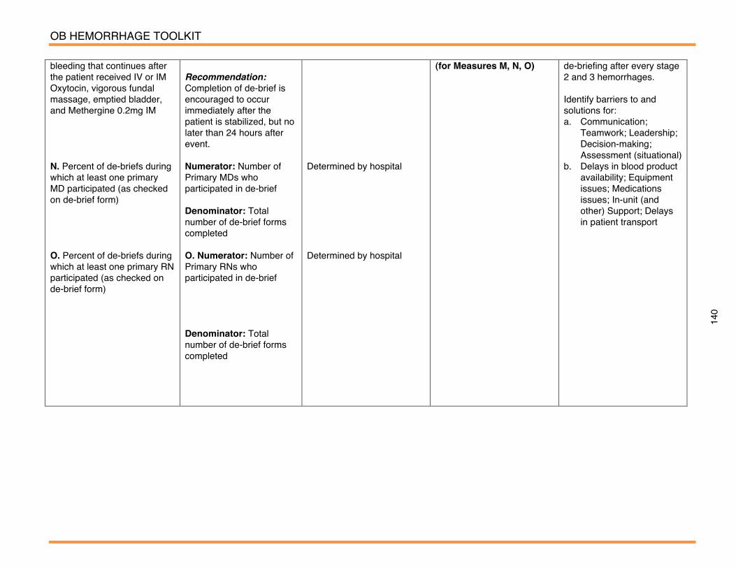
OB HEMORRHAGE TOOLKIT
140
bleeding that continues after the patient received IV or IM Oxytocin, vigorous fundal massage, emptied bladder, and Methergine 0.2mg IM N. Percent of de-briefs during which at least one primary MD participated (as checked on de-brief form) O. Percent of de-briefs during which at least one primary RN participated (as checked on de-brief form)
Recommendation: Completion of de-brief is encouraged to occur immediately after the patient is stabilized, but no later than 24 hours after event. Numerator: Number of Primary MDs who participated in de-brief Denominator: Total number of de-brief forms completed O. Numerator: Number of Primary RNs who participated in de-brief Denominator: Total number of de-brief forms completed
Determined by hospital Determined by hospital
(for Measures M, N, O)
de-briefing after every stage 2 and 3 hemorrhages. Identify barriers to and solutions for: a. Communication;
Teamwork; Leadership; Decision-making; Assessment (situational)
b. Delays in blood product availability; Equipment issues; Medications issues; In-unit (and other) Support; Delays in patient transport
141
REFERENCES 1. Clark S, Miller D, Belford M, Dildy G, Frye D, Meyers J. Neonatal and maternal outcomes
associated with elective term delivery. American Journal of Obstetrics and Gynecology. 2009;200:156.e1-.e4.
2. Lu MC, Fridman M, Korst LM, et al. Variations in the incidence of postpartum hemorrhage across hospitals in California. Maternal Child Health Journal. 2005 September;9(3):297-306.
3. Kuklina E, Meikle, S., Jamieson, D., Whiteman, M., Barfield, W., Hillis, S., Posner, S. Severe Obstetric Morbidity in the US, 1998-2005. Obstetrics and Gynecology. 2009;113:293-9.
4. Guidry M, Vischi T, Han R, Passons O. Healthy people in healthy communities: A community planning guide using healthy people 2010. US Department of Health and Human Services, The Office of Disease Prevention and Health Promotion http://wwwhealthypeoplegov/Publications/HealthyCommunities2001/defaulthtm. 2010.
5. Eckes G. Six Sigma for Everyone Hoboken, NJ: John Wiley & Sons. 2003. 6. Brussee W. Statistics for six sigma made easy! New York: McGraw-Hill; 2004. 7. Carey RG, Lloyd RC. Measuring quality improvement in healthcare: A guide to statistical
process control applications. Milwaukee, Wisconsin: American Society for Quality; 2001. 8. Haugehy D. Pareto Analysis Step by Step http://www.ginque.com/index.php/projmngmt/505-
pareto-analysis-step-by-step. Accessed March 31, 2010. 9. Bingham D. Measuring and increasing the effectiveness of the quality improvement
implementation change practices of front-line maternity physician and nurse leaders. The University of North Carolina at Chapel Hill, 2009, 316 pages; AAT 3352932; 2009. p. 316.
10. Bingham D, Main E. Effective implementation strategies and tactics for leading change on maternity units. Journal of Neonatal and Perinatal Nursing. 2010 Accepted for publication;24(1):32-42.

142
SLIDE SET FOR PROFESSIONAL EDUCATION

1
Instructions: Please use the CMQCC template for any slide
you use from the slide deck Please do not use the CMQCC logo if you
modify a slide – feel free to use your own logo in these situations
Slides with copyright material that we do not yet have copyright permission to use should not be shared with others (these slides are marked)
If you add slides to the slide set please do not use the CMQCC logo
Please provide us feedback and recommendations for improving the slide set
Generic Slide
Obstetric Hemorrhage: New Strategies,
New Protocol
This project was supported by Title V funds received from the California Department of Public Health; Maternal, Child and Adolescent Health Division
Objectives:
Describe the rise of maternal mortality in the state of California
Discuss the four objectives of the CMQCC OB Hemorrhage Task Force
Discuss implementation of the CMQCC OB Hemorrhage tools
Describe the CMQCC OB Hemorrhage Care Guidelines

2
CMQCC will transform maternity care in California to end preventable death and injury.
To achieve this we will: Define and implement best practices for public
health, communities, women with quality, safety, and social justice as the clear priorities of every decision and action. Best Practice Guidelines
Promote communication and collaboration between all maternity stakeholders.
Gather, review, and organize maternity data and statistics into actionable information. Refine Data Advocate Policy
Build the next generation of maternal health leaders to continue the growth and scope of CMQCC.
CMQCC Mission Statement Two Arm Approach… Right Arm: Data-- QI Measures,
Research, And Analysis “If you can’t measure it, you can’t improve it.”
Left Arm: Action “What good is measuring, if we don’t work on improving it?”
Our Philosophy: Quality Improvement using a systems approach
“Every change needs leaders.”
: Transforming Maternity Care
7
Every OB’s nightmare… REQUESTING
PERMISION FOR USE OF IMAGE
8
Hemorrhage remains a major cause of obstetric morbidity and mortality
The rate of maternal deaths has nearly tripled from 6 per 100,000 in 1996 to 17 per 100,000 annual births in 2006.(1)
Alarmingly, the rate for African American women has risen from 28.7 to 54.9 per 100,000 live births between 1999 and 2006.(1)
The California Pregnancy Related Maternal Mortality Review (CA-PAMR) found that obstetric hemorrhage is one of the leading causes for maternal death and is a major cause of maternal morbidity.
In 1997, 2.4% of all live births in California were complicated by postpartum hemorrhage.(2)
Nationwide, blood transfusions increased 92% during delivery hospitalizations between 1997 and 2005.(3)
1. Department of Public Health, Maternal, Child, and Adolescent Health Division Public Released Data available at: www.cmqcc.org
2. Lu MC, Fridman M, Korst LM, et al. Variations in the incidence of postpartum hemorrhage across hospitals in California. Maternal Child Health Journal. September 2005;9(3):297-306.
3. Kuklina E, Meikle, S., Jamieson, D., Whiteman, M., Barfield, W., Hillis, S., Posner, S. Severe Obstetric Morbidity in the US, 1998-2005. Obstetrics and Gynecology. 2009;113:293-299.
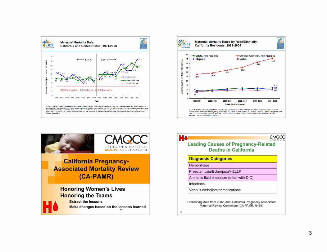
3
9
Maternal Mortality Rate of California Residents and United States: 1991-2006
SOURCE: State of California, Department of Public Health, California Birth and Death Statistical Master Files, 1991-2006. Maternal mortality for California (deaths ≤ 42 days postpartum) calculated using ICD-9 cause of death classification (codes 630-638, 640-648, 650-676 ) for 1991-1998 and ICD-10 cause of death classification (codes A34, O00-O95,O98-O99) for 1999-2006. United States data and HP2010 Objective were calculated using the same methods. United States data not are available for 2006 and 2007. The break in the trend line represents the change from ICD-9 to ICD-10. Produced by California Department of Public Health, Maternal, Child and Adolescent Health Program, April 2009.
ICD-10 ICD-9
HP 2010 Objective – 4.3 Deaths per 100,000 Live Births
Pregnancy-Related Mortality Rates by Race/Ethnicity, California Residents:
1999-2006
SOURCE: State of California, Department of Public Health, California Birth and Death Statistical Master Files, 1999-2006. Pregnancy-related mortality for California calculated beginning 1999 using ICD-10 cause of death codes A34, O00-O96, O98-O99. Maternal single race code used 1990-1999; multirace code used beginning 2000. Produced by California Department of Public Health, Maternal, Child and Adolescent Health Program, April 2009.
11
California Pregnancy-Associated Mortality Review
(CA-PAMR)
Extract the lessons Make changes based on the lessons learned
Honoring Women’s Lives Honoring the Teams
12
Diagnosis Categories Hemorrhage
Preeclampsia/Eclampsia/HELLP
Amniotic fluid embolism (often with DIC)
Infections
Venous embolism complications
Leading Causes of Pregnancy-Related Deaths in California
Preliminary data from 2002-2003 California Pregnancy Associated Maternal Review Committee (CA-PAMR, N=98)
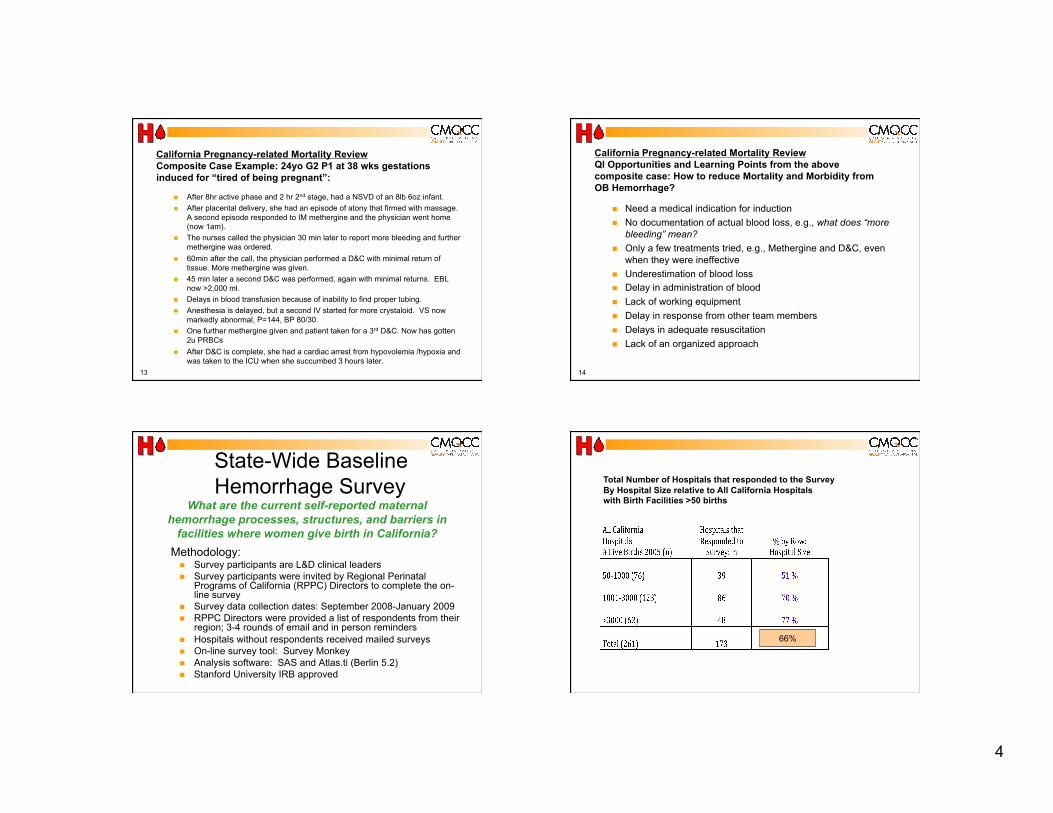
4
California Pregnancy-related Mortality Review Composite Case Example: 24yo G2 P1 at 38 wks gestations induced for “tired of being pregnant”:
After 8hr active phase and 2 hr 2nd stage, had a NSVD of an 8lb 6oz infant. After placental delivery, she had an episode of atony that firmed with massage.
A second episode responded to IM methergine and the physician went home (now 1am).
The nurses called the physician 30 min later to report more bleeding and further methergine was ordered.
60min after the call, the physician performed a D&C with minimal return of tissue. More methergine was given.
45 min later a second D&C was performed, again with minimal returns. EBL now >2,000 ml.
Delays in blood transfusion because of inability to find proper tubing. Anesthesia is delayed, but a second IV started for more crystaloid. VS now
markedly abnormal, P=144, BP 80/30. One further methergine given and patient taken for a 3rd D&C. Now has gotten
2u PRBCs After D&C is complete, she had a cardiac arrest from hypovolemia /hypoxia and
was taken to the ICU when she succumbed 3 hours later. 13
California Pregnancy-related Mortality Review QI Opportunities and Learning Points from the above composite case: How to reduce Mortality and Morbidity from OB Hemorrhage?
Need a medical indication for induction No documentation of actual blood loss, e.g., what does “more
bleeding” mean? Only a few treatments tried, e.g., Methergine and D&C, even
when they were ineffective Underestimation of blood loss Delay in administration of blood Lack of working equipment Delay in response from other team members Delays in adequate resuscitation Lack of an organized approach
14
What are the current self-reported maternal hemorrhage processes, structures, and barriers in
facilities where women give birth in California? Methodology:
Survey participants are L&D clinical leaders Survey participants were invited by Regional Perinatal
Programs of California (RPPC) Directors to complete the on-line survey
Survey data collection dates: September 2008-January 2009 RPPC Directors were provided a list of respondents from their
region; 3-4 rounds of email and in person reminders Hospitals without respondents received mailed surveys On-line survey tool: Survey Monkey Analysis software: SAS and Atlas.ti (Berlin 5.2) Stanford University IRB approved
State-Wide Baseline Hemorrhage Survey Total Number of Hospitals that responded to the Survey
By Hospital Size relative to All California Hospitals with Birth Facilities >50 births
66%

5
Summary: Key Survey Findings
40% of hospitals DO NOT have a hemorrhage protocol
Inconsistent definitions
70% of hospitals DO NOT perform drills (MDs are not regularly participating in drills)
Most have access to all 4 uterotonics (More specific data will be released after complete analysis)
Many hospital report they do not have access to alternative treatment methods, e.g., Balloons
(More specific data will be released after complete analysis)
18
Quality Improvement Opportunities for OB Hemorrhage
Reduce Risks of hemorrhage Perform admission risk assessments Reduce Denial, Delay… Quantify blood loss Follow a step-by-step plan Increase use of non-pharmacologic treatments Improve treatments with blood products
“Too little, too late”—Resuscitation v. Treatment “Old wine in new bottles”—“Whole blood” v. PRBCs
Enhance Teamwork and Communications!
CMQCC Hemorrhage Task Force: Co-Chairs: Audrey Lyndon PhD, RNC, CNS, University of California, San Francisco David Lagrew, MD –Saddleback Memorial Hospital
Leslie Casper, MD – Kaiser, Southern CA, San Diego Medical Center Nancy Corbett, BSN, RN – Kaiser, Northern CA Maurice Druzin, MD – Stanford University Sue Faron, MN, RNC, CNS – Sharp Mary Birch Hospital for Women Kim Gregory, MD, MPH – Cedars-Sinai Medical Center Andrew Hull, MD, FRCOG, FACOG – University of California, San Diego Valerie Huwe, BSN, RNC – Stanford Hospital, El Camino Hospital Richard Lee, MD – USC, Women’s and Children’s Hospital Holli Mason, MD – Harbor UCLA Medical Center
20
CMQCC Hemorrhage Task Force: Hemorrhage Task Force, continued
Elliott Main, MD – California Pacific Medical Center Jennifer McNulty, MD – Long Beach Memorial Medical Center, UC Irvine Suellen Miller, PhD, MHA, CNM - University of California, San Francisco Connie Mitchell, MD – MCAH, CA Department of Public Health Mark Rosen, MD – University of California, San Francisco Diana Ramos, MD, MPH – MCAH, County of Los Angeles Public Health Larry Shields, MD – Central Coast Maternal Fetal Medicine Jean-Claude Veille, MD – Sutter Health, Sacramento
: Transforming Maternity Care
6
21
5 meetings in 2008-2009 Developed a Tool Kit for OB services:
Care Guidelines: Checklist, Flowchart and Table Chart formats
Compendium of Best Practices that includes articles about background, preparation for and management of obstetric hemorrhage
Hospital level Implementation Guide that includes Quality Improvement tools and a sample Policy and Procedure
All resources on-line at: www.cmqcc.org/ob_hemorrhage
CMQCC is sponsoring an Obstetric Quality Care Collaborative following the IHI QI Model
CMQCC Hemorrhage Task Force: Four Major Recommendations for California Birth Facilities:
Improve readiness to hemorrhage by implementing standardized protocols (general and massive).
Improve recognition of OB hemorrhage by performing on-going objective quantification of actual blood loss during and after all births.
Improve response to hemorrhage by performing regular on-site multi-professional hemorrhage drills.
Improve reporting of OB hemorrhage by standardizing definitions and consistency in coding and reporting.
23
What’s New?
With kind permission of Bev VanderWal With kind permission of Bev VanderWal
Quantification of blood loss for all births Active management of the 3rd stage for all Vital sign triggers “Move along” on uterotonic medications Bakri intrauterine balloon / B-Lynch suture A new approach to blood products A role for rFactor VIIa? The value of a formal protocol
24
How can we improve the clinical measurement of blood loss during vaginal and cesarean birth?
How can communication of blood loss be improved among caregivers?
Formal Quantification of Blood Loss at Birth (QBL)
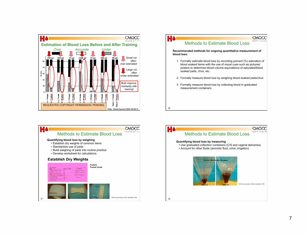
7
25
Over Accurate Under
350 ml 100 ml 25 ml 1200 ml 35 ml 400 ml 80 ml
Dildy: Obstet Gynecol 2004:104:601-6
Estimation of Blood Loss Before and After Training
Small vol. often
over estimated
Large vol. often
under estimated
Both improve markedly with
training!
REQUESTED COPYRIGHT PERMISSION: PENDING 26
Methods to Estimate Blood Loss Recommended methods for ongoing quantitative measurement of blood loss:
1. Formally estimate blood loss by recording percent (%) saturation of blood soaked items with the use of visual cues such as pictures/posters to determine blood volume equivalence of saturated/blood soaked pads, chux, etc.
2. Formally measure blood loss by weighing blood soaked pads/chux
3. Formally measure blood loss by collecting blood in graduated measurement containers
27
Methods to Estimate Blood Loss Quantifying blood loss by weighing
• Establish dry weights of common items • Standardize use of pads • Build weighing of pads into routine practice • Develop worksheet for calculations
With kind permission of Bev VanderWal, CNS 28
Methods to Estimate Blood Loss Quantifying blood loss by measuring
• Use graduated collection containers (C/S and vaginal deliveries) • Account for other fluids (amniotic fluid, urine, irrigation)
With kind permission of Bev VanderWal, CNS

8
29
Methods to Estimate Blood Loss Develop Training Tools: Visual aids displayed in Labor & Delivery and/or Postpartum areas are guides for more accurate visual estimation (visual aids can be displayed discreetly for clinicians)
With kind permission of Bev VanderWal, CNS
30
Recommendations Teach clot size using posters showing known blood
quantities on common materials or compared to common volumes (e.g a Coke can=350ml)
Weigh wet materials (with known dry weight); this can be done by gathering a group of pads and weighing them all together
Measure what can be suctioned at CS (less irrigation+AF) Use calibrated under-buttock drapes (at vaginal birth, note
the volume of amniotic fluid, urine and stool after birth but before the placenta)
What we don’t know: How to estimate the blood loss that we don’t see… (i.e intra- abdominal)
31
Recommendations Many centers will customize their approach to
quantification using a combination of approaches for different settings
Vaginal deliveries Cesarean sections Minimal loss Greater than usual loss Massive loss
The process is intentional—a formal effort! No more vague “Guesstimates” Continues and is cumulative
32
Who should determine QBL? Anesthesia is at the head of the table and often
does not see it all OB’s aren’t looking at the suction bottles or at the
collective sponges No one is doing it in a standardized manner—we
obstetricians need help! Collaboratively! We should be able to answer:
How much blood is in the suction bottle (after amniotic fluid)?
How much blood is on sponges? How much blood is on the floor/on the table? In a big case, hourly and cumulatively
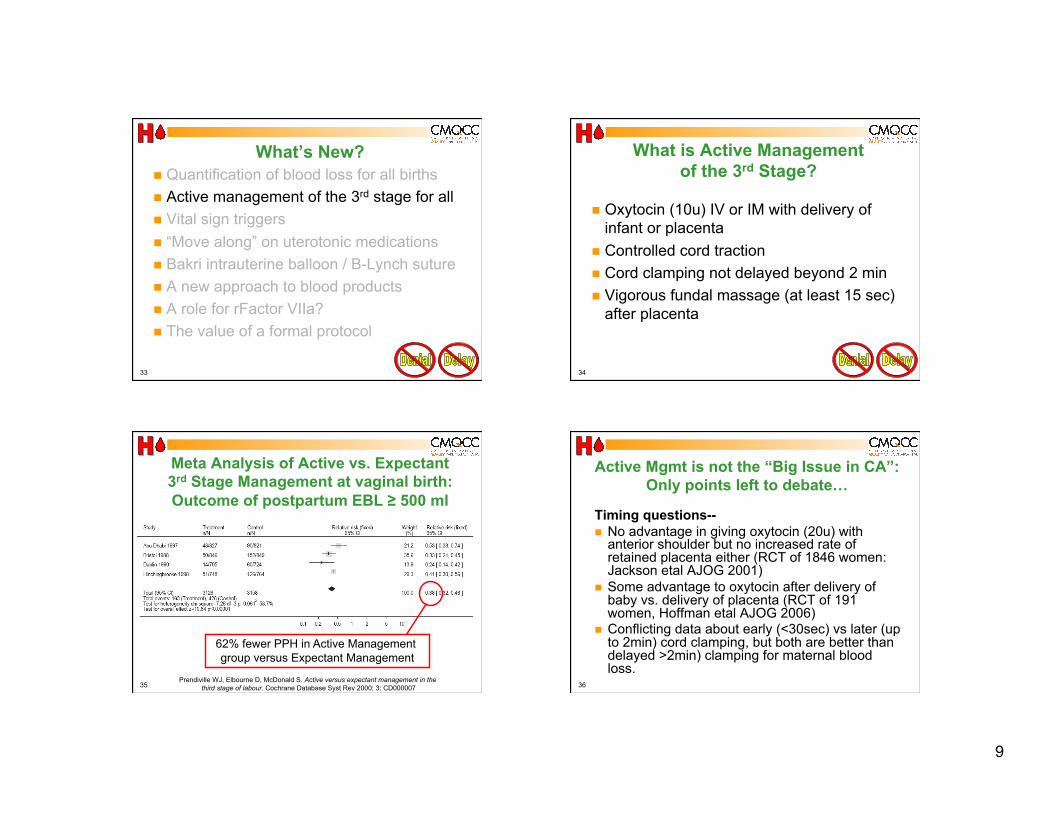
9
33
What’s New? Quantification of blood loss for all births Active management of the 3rd stage for all Vital sign triggers “Move along” on uterotonic medications Bakri intrauterine balloon / B-Lynch suture A new approach to blood products A role for rFactor VIIa? The value of a formal protocol
34
What is Active Management of the 3rd Stage?
Oxytocin (10u) IV or IM with delivery of infant or placenta
Controlled cord traction Cord clamping not delayed beyond 2 min Vigorous fundal massage (at least 15 sec)
after placenta
35
Meta Analysis of Active vs. Expectant 3rd Stage Management at vaginal birth: Outcome of postpartum EBL ≥ 500 ml
62% fewer PPH in Active Management group versus Expectant Management
Prendiville WJ, Elbourne D, McDonald S. Active versus expectant management in the third stage of labour. Cochrane Database Syst Rev 2000; 3: CD000007 36
Active Mgmt is not the “Big Issue in CA”: Only points left to debate…
Timing questions-- No advantage in giving oxytocin (20u) with
anterior shoulder but no increased rate of retained placenta either (RCT of 1846 women: Jackson etal AJOG 2001)
Some advantage to oxytocin after delivery of baby vs. delivery of placenta (RCT of 191 women, Hoffman etal AJOG 2006)
Conflicting data about early (<30sec) vs later (up to 2min) cord clamping, but both are better than delayed >2min) clamping for maternal blood loss.
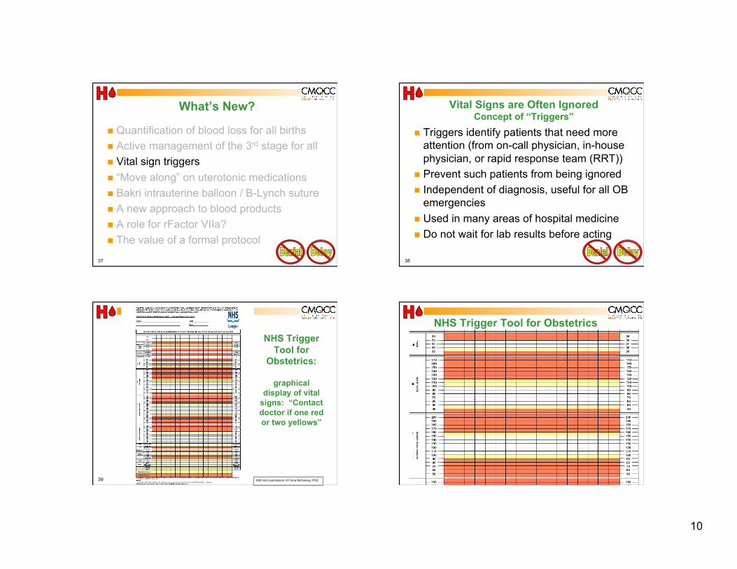
10
37
What’s New?
Quantification of blood loss for all births Active management of the 3rd stage for all Vital sign triggers “Move along” on uterotonic medications Bakri intrauterine balloon / B-Lynch suture A new approach to blood products A role for rFactor VIIa? The value of a formal protocol
38
Vital Signs are Often Ignored Concept of “Triggers”
Triggers identify patients that need more attention (from on-call physician, in-house physician, or rapid response team (RRT))
Prevent such patients from being ignored Independent of diagnosis, useful for all OB
emergencies Used in many areas of hospital medicine Do not wait for lab results before acting
39
NHS Trigger Tool for
Obstetrics:
graphical display of vital
signs: “Contact doctor if one red or two yellows”
With kind permission of Fiona McIlveney, PhD 40
NHS Trigger Tool for Obstetrics
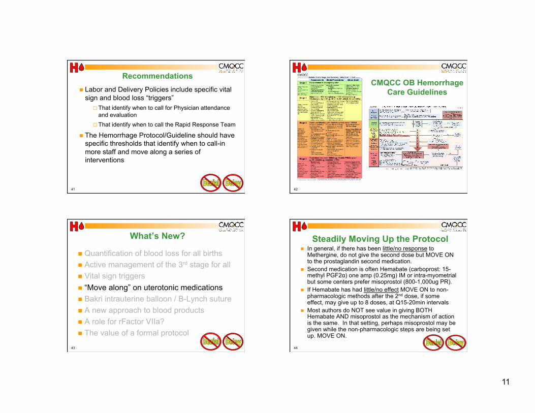
11
41
Recommendations Labor and Delivery Policies include specific vital
sign and blood loss “triggers” That identify when to call for Physician attendance
and evaluation That identify when to call the Rapid Response Team
The Hemorrhage Protocol/Guideline should have specific thresholds that identify when to call-in more staff and move along a series of interventions
42
CMQCC OB Hemorrhage Care Guidelines
43
What’s New?
Quantification of blood loss for all births Active management of the 3rd stage for all Vital sign triggers “Move along” on uterotonic medications Bakri intrauterine balloon / B-Lynch suture A new approach to blood products A role for rFactor VIIa? The value of a formal protocol
44
Steadily Moving Up the Protocol In general, if there has been little/no response to
Methergine, do not give the second dose but MOVE ON to the prostaglandin second medication.
Second medication is often Hemabate (carboprost: 15-methyl PGF2α) one amp (0.25mg) IM or intra-myometrial but some centers prefer misoprostol (800-1,000ug PR).
If Hemabate has had little/no effect MOVE ON to non-pharmacologic methods after the 2nd dose, if some effect, may give up to 8 doses, at Q15-20min intervals
Most authors do NOT see value in giving BOTH Hemabate AND misoprostol as the mechanism of action is the same. In that setting, perhaps misoprostol may be given while the non-pharmacologic steps are being set up. MOVE ON.
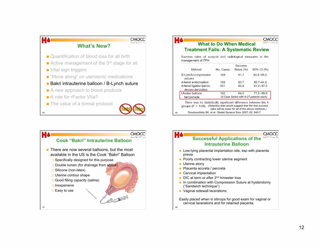
12
45
What’s New?
Quantification of blood loss for all birth Active management of the 3rd stage for all Vital sign triggers “Move along” on uterotonic medications Bakri intrauterine balloon / B-Lynch suture A new approach to blood products A role for rFactor VIIa? The value of a formal protocol
46
What to Do When Medical Treatment Fails: A Systematic Review
Doumouchtsis SK, et al Obstet Gynecol Surv 2007; 62: 540-7.
(9 Case Series with 6-27 patients each)
(Selection bias would suggest that the real success rates will be lower for all of the above methods.)
47
Cook “Bakri” Intrauterine Balloon
There are now several balloons, but the most available in the US is the Cook “Bakri” Balloon Specifically designed for this purpose Double lumen (for drainage from above) Silicone (non-latex) Uterine contour shape Good filling capacity (saline) Inexpensive Easy to use
48
Successful Applications of the Intrauterine Balloon
Low-lying placental implantation site, esp with placenta previa
Poorly contracting lower uterine segment Uterine atony Placenta accreta / percreta Cervical implantation DIC at term or after 2nd trimester loss In combination with Compression Suture at hysterotomy
(“Sandwich technique”) Vaginal sidewall lacerations
Easily placed when in stirrups for good exam for vaginal or cervical lacerations and for retained placenta.

13
49
“Intrauterine Balloon Should be First Step after Failure of Medical Therapy”
High success rate not different than other approaches
Low-tech, fast, inexpensive, easy to utilize on any L&D Unit
Least morbidity of any “next step” Can be used as “Tamponade Test” to temporize,
determine needs and mobilize other resources
Doumouchtsis SK, et al Obstet Gynecol Surv 2007; 62: 540-7. Dabelea V, Schultze PM, McDuffie RS Am J Perinatol 2007; 24: 359-64 50
Issues for Balloons There are several balloons, but the most
available in the US is the Cook “Bakri” Balloon It is the balloon specifically designed for this purpose Double lumen, silicone (non-latex), uterine contour
shape, good filling capacity, inexpensive There is some user learning—
How much to fill? (150-500ml is a big range). We recommend estimating the uterine volume bimanually --usually 250-300ml is sufficient unless the uterus is very “floppy”.
There can be “hour-glassing” of the balloon thru the cervix into the vagina. We recommend using vaginal packing if the cervix is more than 1-2cm dilated)
51
B-Lynch Compression Suture “Belt and Suspenders”
52
B-Lynch Suture completed
Photo courtesy of Elliott Main, MD-CPMC

14
53
B-Lynch Suture
Every Obstetrician should know how to do this (diagrams are in each OR)
Quick (<2 minutes) and easy! Ideal at time of Cesarean birth for atony Can be combined with an intrauterine
balloon for “Sandwich technique”
54
What’s New?
Quantification of blood loss for all births Active management of the 3rd stage for all Vital sign triggers “Move along” on uterotonic medications Bakri intrauterine balloon / B-Lynch suture A new approach to blood products A role for rFactor VIIa? The value of a formal protocol
55
Lessons from Combat in Iraq
Lowest losses ever from hemorrhage
Key: increased FFP:RBC ratio
Pending use of photos purchased
for CMQCC use
56
Iraq Theatre Experience
Borgman MA. J Trauma. 2007 (Oct);63:805– 813.
Retrospective review of all soldiers with massive transfusion (>10u RBC in 24 hr) Nov 2003-Sept 2005. N=246
Composition closer to whole blood more effective than1980’s ratios:
Not a RCT! Biased against
greatest EBL But results are
striking…
Pending copyright permission or describe in words
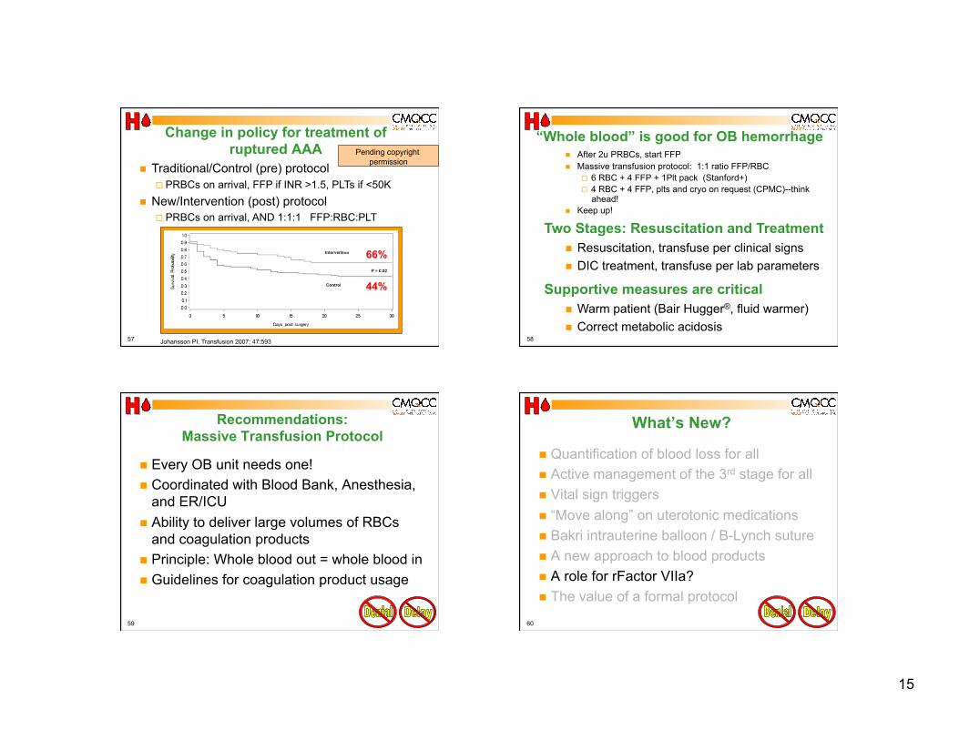
15
57
Change in policy for treatment of ruptured AAA
Johansson PI. Transfusion 2007; 47:593
Traditional/Control (pre) protocol PRBCs on arrival, FFP if INR >1.5, PLTs if <50K
New/Intervention (post) protocol PRBCs on arrival, AND 1:1:1 FFP:RBC:PLT
66%
44%
Pending copyright permission
58
“Whole blood” is good for OB hemorrhage After 2u PRBCs, start FFP Massive transfusion protocol: 1:1 ratio FFP/RBC
6 RBC + 4 FFP + 1Plt pack (Stanford+) 4 RBC + 4 FFP, plts and cryo on request (CPMC)--think
ahead! Keep up!
Two Stages: Resuscitation and Treatment Resuscitation, transfuse per clinical signs DIC treatment, transfuse per lab parameters
Supportive measures are critical Warm patient (Bair Hugger®, fluid warmer) Correct metabolic acidosis
59
Recommendations: Massive Transfusion Protocol
Every OB unit needs one! Coordinated with Blood Bank, Anesthesia,
and ER/ICU Ability to deliver large volumes of RBCs
and coagulation products Principle: Whole blood out = whole blood in Guidelines for coagulation product usage
60
What’s New?
Quantification of blood loss for all Active management of the 3rd stage for all Vital sign triggers “Move along” on uterotonic medications Bakri intrauterine balloon / B-Lynch suture A new approach to blood products A role for rFactor VIIa? The value of a formal protocol
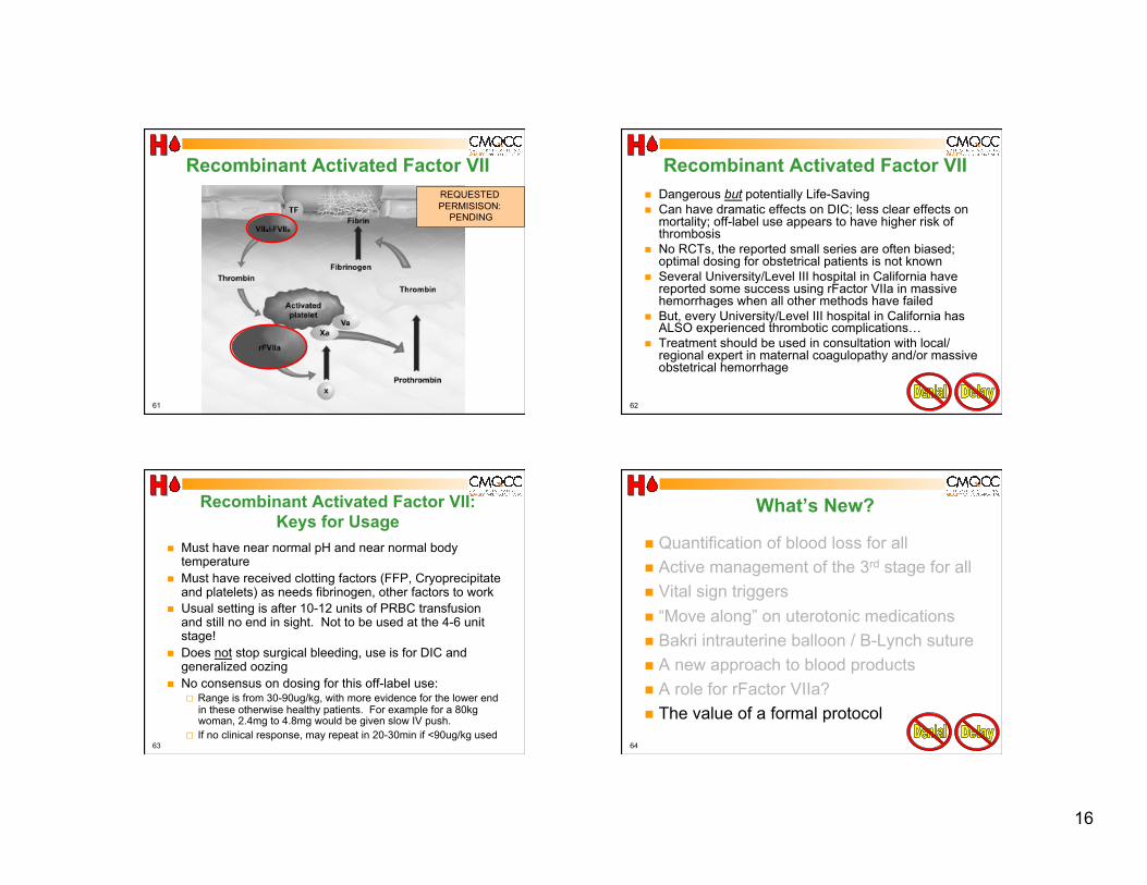
16
61
Recombinant Activated Factor VII REQUESTED PERMISISON:
PENDING
62
Recombinant Activated Factor VII Dangerous but potentially Life-Saving Can have dramatic effects on DIC; less clear effects on
mortality; off-label use appears to have higher risk of thrombosis
No RCTs, the reported small series are often biased; optimal dosing for obstetrical patients is not known
Several University/Level III hospital in California have reported some success using rFactor VIIa in massive hemorrhages when all other methods have failed
But, every University/Level III hospital in California has ALSO experienced thrombotic complications…
Treatment should be used in consultation with local/regional expert in maternal coagulopathy and/or massive obstetrical hemorrhage
63
Recombinant Activated Factor VII: Keys for Usage
Must have near normal pH and near normal body temperature
Must have received clotting factors (FFP, Cryoprecipitate and platelets) as needs fibrinogen, other factors to work
Usual setting is after 10-12 units of PRBC transfusion and still no end in sight. Not to be used at the 4-6 unit stage!
Does not stop surgical bleeding, use is for DIC and generalized oozing
No consensus on dosing for this off-label use: Range is from 30-90ug/kg, with more evidence for the lower end
in these otherwise healthy patients. For example for a 80kg woman, 2.4mg to 4.8mg would be given slow IV push.
If no clinical response, may repeat in 20-30min if <90ug/kg used 64
What’s New?
Quantification of blood loss for all Active management of the 3rd stage for all Vital sign triggers “Move along” on uterotonic medications Bakri intrauterine balloon / B-Lynch suture A new approach to blood products A role for rFactor VIIa? The value of a formal protocol

17
65
Why a Protocol for Obstetric Hemorrhage?
Now a complex series of steps that involve many staff members and departments
Communications! PPH seems to always happens at night or
weekends…(when people may be tired or there are less resources)
We can improve…
66
Systems Approach to Obstetric Hemorrhage
Organize your unit and your response Recognize Denial and Delay
Get help Get exposure to perform thorough exams and identify the source of bleeding Do not get behind
Process Is Most Important!
67
Doctor’s Company Review Closed Claim Review of Maternal Deaths from PPH
DENIAL: “Catch up” phenomenon: Initial manifestations of hemorrhage were VS Δ’s (hypotension and/or tachycardia) NOT frank vaginal bleeding.
DELAY: in delivery of products from the blood bank to the labor and delivery operating room.
DELAY: of administration to patient once products arrived at L+D.
DELAY: Mobilization of equipment. DELAY: Waiting for cross-matched blood instead of
utilizing O negative or type specific blood. “Underutilization”-- DELAY in administering additional
amounts and types of blood products (i.e. FFP, platelets, and cryoprecipitate)
68
Doctor’s Company Review (cont) COMMUNICATION BREAKDOWNS
Among Team Members…
For example, Obstetrician and anesthesiologist regarding
efficacy of intervention(s) and need to escalate care or change strategy.
Operating room and blood bank concerning urgency of situation.
Among support personnel concerning delivery of products and location of specialized equipment (I.e rapid infusion devices or specialized kits) Doctors Company Reviews Maternal Arrests Cases (Reprinted with permission from The Doctors Company); APSF NEWSLETTER Summer 2007; page 28; Ann Lofsky, MD.
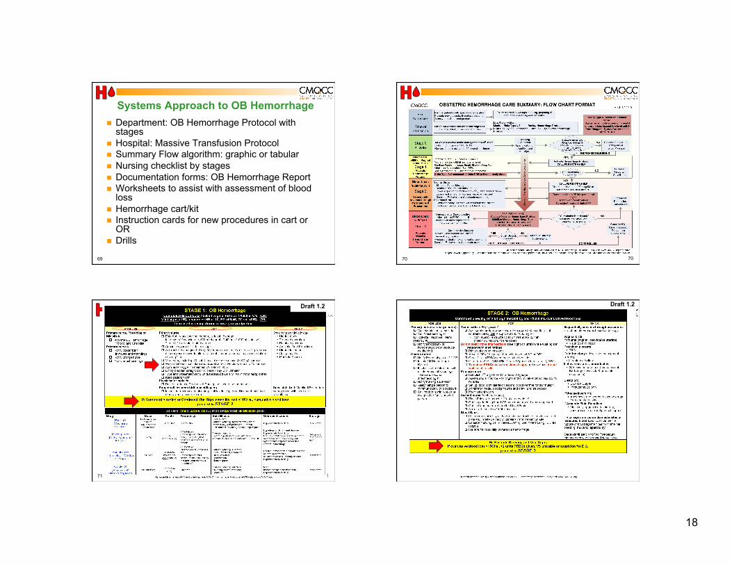
18
69
Systems Approach to OB Hemorrhage Department: OB Hemorrhage Protocol with
stages Hospital: Massive Transfusion Protocol Summary Flow algorithm: graphic or tabular Nursing checklist by stages Documentation forms: OB Hemorrhage Report Worksheets to assist with assessment of blood
loss Hemorrhage cart/kit Instruction cards for new procedures in cart or
OR Drills
70 70
71 71
Draft 1.2
72 72
Draft 1.2

19
73
Importance of Drills / Simulations Safety and QI Leader: Paul Preston, MD
“Medicine is the last high-risk industry that expects people to perform perfectly in complex, rare emergencies but does not support them with high-quality training and practice throughout their careers.”
“Certain individual and team skills require regular practice that cannot ethically occur in routine care.”
74
Can we lower the frequency and morbidity/mortality of OB Hemorrhage?
Lower the incidence: Reduce the cesarean birth rate (both primary and
repeat) Reduce chorioamnionitis Fewer multiple gestations Reduce long inductions of labor Reduce long second stages
Respond rapidly to OB hemorrhage: Use the new techniques and respond in an organized,
well-executed, timely fashion Keep a small hemorrhage from evolving into a massive
hemorrhage
75
Definitions, Early Recognition and Response Triggers Congenital Coagulation Disorders OB Care for Pregnant Women who Decline Transfusion
Checklist for OB Care for Jehovah’s Witness Informed Consent for Blood Products Jehovah’s Witness Protocol for IV Iron Sucrose
Placenta Accreta and Percreta: Risks, Dx and Tx Hemorrhage Kits, Carts and Trays Simulations and Drills-Scenarios and Worksheets Lessons Learned from New York and Washington State
Taskforces
CMQCC Hemorrhage Task Force Best Practice Documents:
Hemorrhage Background and Preparation
www.cmqcc.org/ob_hemorrhage 76
Active Management of 3rd Stage Labor Blood Loss: Clinical Techniques for Ongoing Quantitative
Measurement Blood Product Replacement
Massive Transfusion Protocol Intrauterine Balloons (coming Soon)
Surgery: B-Lynch Sutures, Uterine Artery Occlusion Utertonic Agent Summary Sheet Anti-Shock Garments Family Support
CMQCC Hemorrhage Task Force Best Practice Documents:
Hemorrhage Management
www.cmqcc.org/ob_hemorrhage

20
77
What’s New--Summary
Quantification of blood loss for all Active management of the 3rd stage for all Vital sign triggers “Move along” on uterotonic medications Bakri intrauterine balloon / B-Lynch suture A new approach to blood products A role for rFactor VIIa? The value of a formal protocol
Any Questions?
Become a CMQCC member and keep up-to-date: www.cmqcc.org
Related Documents


