1 Contemporary Percutaneous Coronary Intervention for Complex Lesions: the Treatment of Chronic Total Occlusions and Bifurcations in the Drug-eluting Stent Era Angela Hoye

Welcome message from author
This document is posted to help you gain knowledge. Please leave a comment to let me know what you think about it! Share it to your friends and learn new things together.
Transcript

1
Contemporary PercutaneousCoronary Intervention for
Complex Lesions:the Treatment of Chronic TotalOcclusions and Bifurcationsin the Drug-eluting Stent Era
Angela Hoye

2
ISBN 1-84426-364-9
Printed by Printondemand-worldwide.com
© Angela Hoye 2006

3
Contemporary PercutaneousCoronary Intervention for ComplexLesions: the Treatment of Chronic
Total Occlusions and Bifurcations inthe Drug-eluting Stent Era
Hedendaagse PercutaneRevascularisatie van Complexe
Coronaire Lesies: de Behandelingvan Chronische Totale Occlusies en
Bifurcatielesies Gebruik Makendevan Drug-eluting Stents
Thesis
to obtain the degree of Doctor from the
Erasmus University Rotterdam
by command of the
rector magnificus
Prof.dr. S.W.J. Lamberts
in accordance with the decision of the Doctorate Board
The public defense shall be held on
Wednesday, 22nd March 2006 at 13.45 hrs
By
Angela Hoye
born in Nottingham, England

4
Doctoral CommitteePromotors: Prof. dr. P.W.J. Serruys
Prof. dr. W.J. van der Giessen
Other members: Prof. dr. P.J. de Feyter
Prof. dr. A.F.W. van der Steen
Prof. dr. P.M.T. Pattynama
The generous sponsoring by Cordis, a Johnson & Johnson Company is
gratefully acknowledged

5
Table of Contents
Chapter 1: Introduction and overview
Part 1: Chronic total occlusions
Chapter 2: Chronic Total OcclusionsAngela Hoye Chapter in “A Colour Handbook of Adult Interventional Cardiology”, Manson publishing (in press)
Chapter 3: Percutaneous Coronary Intervention for Chronic Total Occlusions: theThoraxcenter Experience 1992 - 2002Angela Hoye, Ron T van Domburg, Karel Sonnenschein, Patrick W SerruysEuropean Heart Journal 2005 Dec;26(24):2630-6
Chapter 4: Predictors, Incidence and Prognosis of Coronary Occlusion followingIntracoronary Beta-radiation Therapy Angela Hoye, Georgios Sianos, Francesco Saia, Pedro A. Lemos, Willem van der Giessen, Pim J.de Feyter, Veronique L.M.A. Coen, Ron T. van Domburg, Peter C. Levendag, Patrick W. SerruysSubmitted for publication
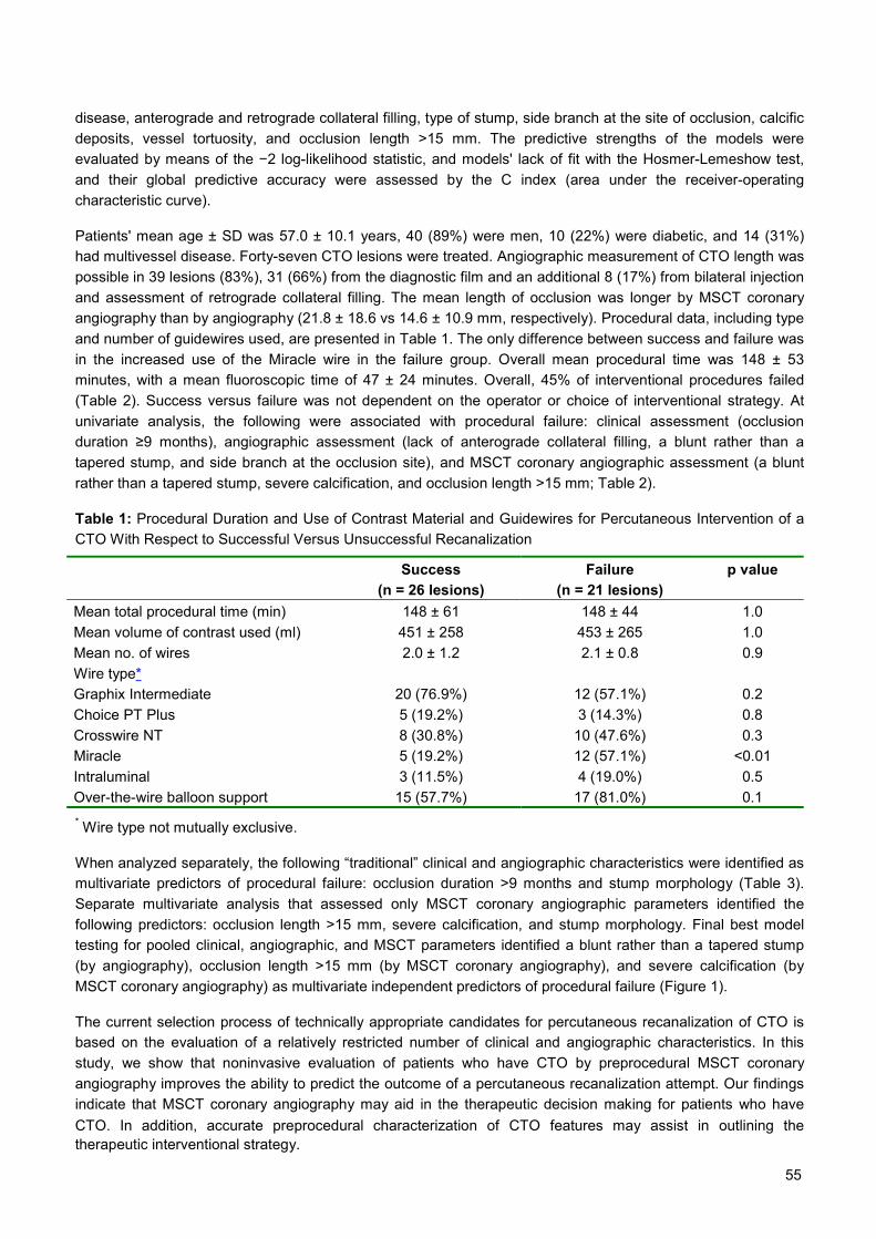
Chapter 5: Value of Pre-Procedure Multislice CT Coronary Angiography to PredictPercutaneous Recanalization of Chronic Total OcclusionsNico Mollet, Angela Hoye, Pedro Lemos, Filippo Cademartiri, Georgios Sianos, Eugene McFadden,Patrick W Serruys, Pim de FeyterAmerican Journal of Cardiology 2005 Jan 15;95(2):240-3
Chapter 6: Improved Recanalization of Chronic Total Coronary Occlusions Using an OpticalCoherence Reflectometry-Guided GuidewireAngela Hoye, Emile Onderwater, Paul Cummins, Georgios Sianos, Patrick W. SerruysCatheterization and Cardiovascular Interventions 2004 Oct;63(2):158-63
Chapter 7: Successful Use of a New Guidewire with Radiofrequency Ablation Capability forthe Treatment of Chronic Total Occlusion at the Ostium of the Left Anterior DescendingArtery Angela Hoye, Pedro A. Lemos, Emile Onderwater, Paul Cummins, Patrick W. Serruys Journal of Invasive Cardiology 2005 May;17(5):277-9
Chapter 8: Multimodality Plaque Ablation to Allow Successful Stent Implantation FollowingFailure of Conventional Wires and Balloons to Cross a Chronic Total OcclusionJose Ruiz-Cantador, Angela Hoye, Eugene McFadden Journal of Invasive Cardiology 2005;17(10):E7-E10
Chapter 9: Significant Reduction in Restenosis Following the Use of Sirolimus-Eluting Stentsin the Treatment of Chronic Total Occlusions Angela Hoye, Kengo Tanabe, Pedro Lemos, Jiro Aoki, Francesco Saia, Chourmouzios Arampatzis,Muzaffer Degertekin, Sjoerd Hofma, Georgios Sianos, Eugene McFadden, Willem van der Giessen,Peter Smits, Pim J. de Feyter, Ron van Domburg, Patrick W SerruysJournal of the American College of Cardiology 2004;43:1954-8
9
15
17
27
39
51
61
71
79
87
6
Chapter 10: Drug-Eluting Stent Implantation for Chronic Total Occlusions: Comparisonbetween the Sirolimus- and Paclitaxel-Eluting StentAngela Hoye, Andrew TL Ong, Jiro Aoki, Carlos AG van Mieghem, Gaston A. Rodriguez Granillo,Marco Valgimigli, Georgios Sianos, Eugene McFadden, Willem J. van der Giessen, Pim J. deFeyter, Ron T. van Domburg, Patrick W SerruysEurointervention 2005;1:193-197
Chapter 11: Sirolimus-Eluting Stent Implantation for Chronic Total Occlusion of the Left MainCoronary Artery Jiro Aoki, Angela Hoye, AV Staferov, BG Alekyan, Patrick W SerruysJournal of Interventional Cardiology 2005;18(1):65-9
Part 2: Bifurcations
Chapter 12: BifurcationsAngela Hoye Chapter in “A Colour Handbook of Adult Interventional Cardiology”, Manson publishing (in press)
Chapter 13: New approaches to ostial and bifurcation lesionsAngela Hoye, Willem van der GiessenJournal of Interventional Cardiology 2004;17(6):397-403
Chapter 14: Restenosis Rates Following Bifurcation Stenting with Sirolimus-Eluting Stentsfor De Novo NarrowingsTanabe K, Hoye A, Lemos PA, Aoki J, Arampatzis CA, Saia F, Lee CH, Degertekin M, Hofma SH,Sianos G, McFadden E, Smits PC, van der Giessen WJ, de Feyter P, van Domburg RT, Serruys PWAmerican Journal of Cardiology 2004;94:115-8
Chapter 15: Treatment of De Novo Bifurcation Lesions: Comparison of Sirolimus- andPaclitaxel-Eluting Stents Angela Hoye, Carlos AG van Mieghem, Andrew TL Ong, Jiro Aoki, Gaston A. Rodriguez Granillo,Marco Valgimigli, Georgios Sianos, Eugene McFadden, Willem J. van der Giessen, Pim J. deFeyter, Ron T. van Domburg, Patrick W. Serruys Eurointervention 2005;1:24-30
Chapter 16: Long-term Outcomes Following Stenting of Bifurcation Lesions Utilizing the“Crush” Technique: Predictors of an Adverse OutcomeAngela Hoye, Ioannis Iakovou, Lei Ge, Carlos AG van Mieghem, Andrew TL Ong, John Cosgrave,Giuseppe M Sangiorgi, Flavio Airoldi, Matteo Montorfano, Iassen Michev, Alaide Chieffo, MauroCarlino, Nicola Corvaja, Jiro Aoki, Gaston A Rodriguez Granillo, Marco Valgimigli, Georgios Sianos,Willem J van der Giessen, Pim J de Feyter, Ron T van Domburg, Patrick W Serruys, AntonioColomboJournal of the American College of Cardiology, In press
Chapter 17: Percutaneous Therapy of Bifurcation Lesions with Drug-Eluting StentImplantation: the Culotte Technique RevisitedAngela Hoye, Carlos AG van Mieghem, Andrew TL Ong, Jiro Aoki, Gaston A. Rodriguez Granillo,Marco Valgimigli, Keiichi Tsuchida, Georgios Sianos, Eugene P. McFadden, Willem J. van derGiessen, Pim J. de Feyter, Ron T. van Domburg, Patrick W. SerruysInternational Journal of Cardiovascular Interventions 2005;7(1):36-40
97
107
115
117
129
141
149
161
179

7
Part 3: The unrestricted use of drug-eluting stents: predictors of an adverseoutcome
Chapter 18: Thirty-day incidence and six-month clinical outcome of thrombotic stentocclusion after bare-metal, sirolimus, or paclitaxel stent implantationAndrew Ong, Angela Hoye, Jiro Aoki, Carlos AG van Mieghem, Gaston Rodríguez Granillo, KarenSonnenschein, Evelyn Regar, Eugene McFadden, Georgios Sianos, Willem J van der Giessen,Pieter de Jaegere, Pim J de Feyter, Ron T van Domburg, Patrick W SerruysJournal of the American College of Cardiology 2005; 45(6):947-53
Chapter 19: Clinical, angiographic, and procedural predictors of angiographic restenosisafter sirolimus-eluting stent implantation in complex patients: an evaluation from theRapamycin-Eluting Stent Evaluated At Rotterdam Cardiology Hospital (RESEARCH) studyLemos PA, Hoye A, Goedhart D, Arampatzis CA, Saia F, van der Giessen WJ, McFadden E, SianosG, Smits PC, Hofma SH, de Feyter PJ, van Domburg RT, Serruys PWCirculation. 2004 Mar 23;109(11):1366-70
Chapter 20: The unrestricted use of paclitaxel- versus sirolimus-eluting stents for coronaryartery disease in an unselected population: one-year results of the Taxus-Stent Evaluated AtRotterdam Cardiology Hospital (T-SEARCH) registryAndrew TL Ong Patrick W Serruys, Jiro Auki, Angela Hoye, Carlos AG van Mieghem, Gaston ARodriguez-Granillo, Marco Valgimigli, Karen Sonnenschein, Evelyn Regar, Martin van der Ent, PeterPT de Jaegere, Eugene McFadden, Georgios Sianos, Willem J van der Giessen, Pim J de Feyter,Ron T van Domburg Journal of the American College of Cardiology 2005; 45(7):1135-41
Part 4:
Summary and conclusions
Samenvatting en Conclusies
Acknowledgements
Curriculum Vitae
List of Publications
189
191
203
215
229
235
241
247
251

8

Chapter 1
Introduction and Overview
9

10

11
Percutaneous intervention of coronary stenoses has undergone dramatic evolution in the last 30 years. Theutilisation of stents has dramatically increased in the last 10-15 years with stenting becoming applicable in awide variety of lesion morphologies and clinical settings. Stents provide a scaffold which supports the arterialwall thereby sealing dissections and eliminating elastic recoil which reduces the rate of abrupt vessel closurecompared to balloon angioplasty alone. 1,2 Angiographically, stents provide a very pleasing immediate result;however the long-term success is hindered by the development of restenosis which has proven to be extremelydifficult to treat effectively. The struts of an expanding stent cause focal deep vascular trauma, and the severityof arterial injury has been shown to directly correlate with inflammation and the development of late neointimalgrowth and restenosis. 3
Drug-eluting stents, whilst maintaining the beneficial effect of scaffolding the vessel, have been shownto reduce the rate of subsequent restenosis. Large randomised studies evaluated outcomes in selectedpopulations, and demonstrated efficacy of drug-eluting stents when used to treat relatively simple lesions. 4-7
However, the short- and long-term efficacy of percutaneous coronary intervention is related to the baselinepatient and lesion characteristics, and the majority of coronary intervention carried out in current clinical practiceinvolves the therapy of such complex lesions, which were excluded from these studies.
The aim of this thesis was to evaluate contemporary coronary intervention of two of the most complexlesion subtypes: chronic total occlusions (CTOs) and bifurcations. In patients with significant coronary disease,both these lesion subtypes are commonly found on diagnostic angiography. However, both lesions areassociated with lower procedural success rates: CTOs because of the difficulty in crossing the lesion with a wireand / or balloon; and bifurcations because of a higher rate of procedural myocardial infarction commonly relatedto impairment of flow in the side branch. In addition, data with bare metal stents show that both these lesionsare subject to a relatively high rate of restenosis. We evaluated the impact of drug-eluting stent implantation onthe outcomes of patients treated in the “real world” of interventional cardiology, in particular looking at theimpact of these stents in patients treated for chronic total occlusions and bifurcation lesions. Part 1 of the thesis evaluates chronic total occlusions (CTOs), with an overview described in Chapter 2.In chapter 3, we review the outcomes and trends of all patients treated for a CTO between 1992 and 2002. Priorto drug-eluting stents, intracoronary brachytherapy was the treatment of choice for in-stent restenosis. However,recent data has suggested that this therapy is associated with late recurrence of restenosis including CTO. 8 Inchapter 4, we describe the predictors, incidence and prognosis of patients who develop a coronary occlusionfollowing intracoronary beta-radiation therapy.
Despite the development of improved technologies to facilitate CTO recanalization, the overall successrate remains <70% in most catheterization laboratories. In chapter 5 we evaluate the value of pre-proceduralmultislice CT scanning in order to predict a subsequent successful recanalization attempt. In chapters 6,7 and 8we evaluate the efficacy of a novel dedicated CTO recanalization technology. The system comprises of aguidewire that combines guidance from optical coherence reflectometry, with power provided by radiofrequencyablation to enable penetration through the occlusion.
The long-term outcomes of CTOs with respect to stent type are assessed in chapters 9 and 10.Consecutive patients treated with sirolimus-eluting, and paclitaxel-eluting stents are compared with an historicalcohort treated with bare metal stent implantation, in order to determine the efficacy of drug-eluting stents in thispatient subgroup.
In part 2, we assess the treatment of bifurcation lesions, with overviews presented in chapters 12 and13. The difficulty in this situation relates to the presence of a sizeable side branch (usually defined as ≥2.0mmdiameter). Even temporary loss of such a branch may be associated with a significant (≥2x upper limit ofnormal) release of creatine kinase. This is important as even minor elevations of CK-MB after successfulcoronary interventions identify a population with a worse long-term prognosis compared with patients with noenzyme release. 9
In addition, the side branch is at particular risk of subsequent restenosis and the most effective strategyfor stenting bifurcation lesions is currently undefined. Studies have evaluated a variety of techniques, howeverdata with bare metal stents demonstrated that stent implantation of both the main vessel and side branch is

12
associated with a trend towards a higher rate of adverse events compared with use of single stent implantationof the main vessel only. 10-13
The efficacy of drug-eluting stents for bifurcations is demonstrated in Chapters 14 and 15. Theintroduction of drug-eluting stents has led to a resurgence of techniques involving elective stent implantation inthe side branch. Strategies such as the “crush” technique and Culotte stenting ensure complete lesioncoverage, and the results are evaluated in Chapters 16 and 17.
In part 3, the treatment of both chronic total occlusions and bifurcations are put into the context ofresults of drug-eluting stent implantation in an unselected population. One of the concerns of drug-eluting stentsis that by impairing the process of re-endothelialization, these stents might be subject to higher rates of stentthrombosis despite prolongation of the duration of prescribed dual anti-platelet therapy. Such an event isassociated with a high rate of mortality and morbidity. 14 In chapter 18, we assess the incidence of stentthrombosis at 30 days following sirolimus- and paclitaxel-eluting stents as compared with previous patientstreated with bare metal stents, and evaluate the independent predictors of stent thrombosis.
In chapter 19, we evaluate the clinical, angiographic, and procedural predictors of angiographicrestenosis after sirolimus-eluting stent implantation in complex patients, including whether chronic totalocclusions and bifurcations are predictors of restenosis. Chapter 20 evaluates the clinical outcomes followingthe use of paclitaxel-eluting versus sirolimus-eluting stents in unselected populations. Multivariate analysis ofthe populations determines the independent predictors of both major adverse cardiac events and target vesselrevascularization.

13
References
1. Serruys PW, de Jaegere P, Kiemeneij F et al. A comparison of balloon-expandable-stent implantation with balloon angioplasty in
patients with coronary artery disease. Benestent Study Group. N Engl J Med. 1994;331:489-95.
2. Serruys PW, van Hout B, Bonnier H et al. Randomised comparison of implantation of heparin-coated stents with balloon
angioplasty in selected patients with coronary artery disease (Benestent II). Lancet. 1998;352:673-81.
3. Virmani R, Farb A. Pathology of in-stent restenosis. Curr Opin Lipidol. 1999;10:499-506.
4. Stone GW, Ellis SG, Cox DA et al. A polymer-based, paclitaxel-eluting stent in patients with coronary artery disease. N Engl J
Med. 2004;350:221-31.
5. Morice MC, Serruys PW, Sousa JE et al. A randomized comparison of a sirolimus-eluting stent with a standard stent for coronary
revascularization. N Engl J Med. 2002;346:1773-80.
6. Moses JW, Leon MB, Popma JJ et al. Sirolimus-eluting stents versus standard stents in patients with stenosis in a native
coronary artery. N Engl J Med. 2003;349:1315-23.
7. Grube E, Silber S, Hauptmann KE et al. TAXUS I: six- and twelve-month results from a randomized, double-blind trial on a slow-
release paclitaxel-eluting stent for de novo coronary lesions. Circulation. 2003;107:38-42.
8. Sianos G, Hoye A, Saia F et al. Long term outcome after intracoronary beta radiation therapy. Heart. 2005;91:942-7.
9. Abdelmeguid AE, Topol EJ, Whitlow PL et al. Significance of mild transient release of creatine kinase-MB fraction after
percutaneous coronary interventions. Circulation. 1996;94:1528-36.
10. Al Suwaidi J, Berger PB, Rihal CS et al. Immediate and long-term outcome of intracoronary stent implantation for true bifurcation
lesions. J Am Coll Cardiol. 2000;35:929-36.
11. Anzuini A, Briguori C, Rosanio S et al. Immediate and long-term clinical and angiographic results from Wiktor stent treatment for
true bifurcation narrowings. Am J Cardiol. 2001;88:1246-50.
12. Pan M, Suarez de Lezo J, Medina A et al. Simple and complex stent strategies for bifurcated coronary arterial stenosis involving
the side branch origin. Am J Cardiol. 1999;83:1320-5.
13. Yamashita T, Nishida T, Adamian MG et al. Bifurcation lesions: two stents versus one stent--immediate and follow-up results. J
Am Coll Cardiol. 2000;35:1145-51.
14. Cutlip DE, Baim DS, Ho KK et al. Stent thrombosis in the modern era: a pooled analysis of multicenter coronary stent clinical
trials. Circulation. 2001;103:1967-71.

14

Part 1
CHRONIC TOTAL OCCLUSIONS
15

16

Chapter 2
Angela Hoy
Chapter in Handbook InterventionCardiologyManson Pu
Chronic Total Occlusions
17
e
A Colour of Adult al
blishing (in press)

18

19
IntroductionA chronic total occlusion (CTO) remains a technical challenge to the interventional cardiologist. Proceduralsuccess rates vary in the literature and are very much dependant on patient selection, age of occlusion, andoperator experience. Despite technological advances, even in those patients selected to be suitable forpercutaneous therapy, published success rates are between 40-80%,1,2 considerably lower compared to non-occlusive lesions. However, there are several advantages to opening a CTO, with studies demonstrating areduction in long-term mortality, improvement in anginal symptoms and left ventricular function and a reductionin the need for subsequent coronary artery bypass surgery.3-8
IncidenceRecent data suggests that in patients found to have significant coronary disease (defined as ≥70% diameterstenosis) at least one CTO will be found in 52%.9 However, the presence of a CTO has a significant impact onchoice of therapy, with the majority of these patients managed with either medical therapy or referred directly forcoronary artery bypass surgery. In most centres, percutaneous intervention for CTO generally comprises ≈10%of angioplasty procedures.
DefinitionCTO is commonly defined as a complete occlusion within a coronary artery with TIMI 0 flow, though somestudies have also included lesions with TIMI I flow, so-called ‘functional occlusions’ where late antegradeopacification of the distal vessel is detected though without a discernible luminal continuity.
Determining the duration of occlusion can be difficult without angiographic data, and relies somewhatempirically on the clinical history of onset of angina pain or an episode of prolonged pain which may indicatevessel occlusion. The definition of what is ‘chronic’ is also variable, many studies have included lesions of morethan 15 days duration, though it is generally accepted that to be truly chronic, lesions are of more than 3 monthsduration.
PathophysiologyCTO’s are thought to either develop after an episode of acute occlusion with plaque rupture and subsequentthrombosis, or relate to progression of a flow-limiting atherosclerotic stenosis. Histology reveals variableamounts of atheroma and thrombus that are increasingly replaced by fibrous tissue and calcification (figure 1).10
Neointimal channels of 160-230µm in diameter form and are present in 85% lesions older than 1 year (figure2).10 There is debate as to whether these channels, which are too small to be visible on angiography, help inproviding a route for a guidewire, or hinder angioplasty success due to connection between the vasa vasorumand adventitia thus increasing the likelihood of extra-luminal wire passage.
Figure 1: Low power view (hematoxylin-eosin stain)of a hard or fibrocalcific chronic total occlusion withextensive calcification (arrow)Reproduced with permission from Srivatsa SS et alHistologic correlates of angiographic chronic totalcoronary artery occlusions: influence of occlusionduration on neovascular channel patterns and intimalplaque composition. J Am Coll Cardiol. 1997;29:955-63.
20
A B
Figure 2: A Low power view (hematoxylin-eosin stain) of a chronic total occlusion lumen recanalization by largecentral neovascular channels (arrow). B High power view (hematoxylin-eosin stain) demonstrating extensivesmall, medium and large intimal plaque neovascular channels (arrow). Reproduced with permission from Srivatsa SS et al Histologic correlates of angiographic chronic total coronary arteryocclusions: influence of occlusion duration on neovascular channel patterns and intimal plaque composition. J Am CollCardiol. 1997;29:955-63.
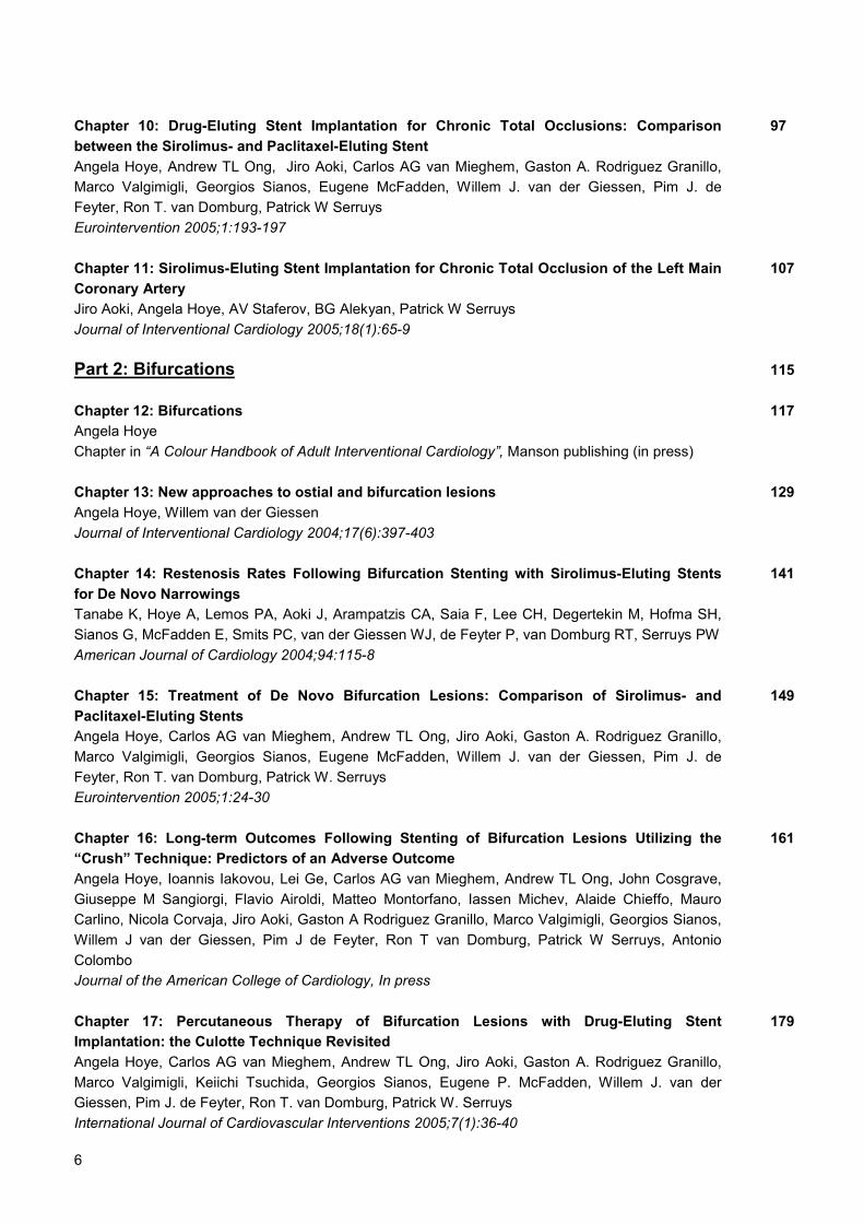
The ProcedureThe limitation of a successful outcome to angioplasty of a CTO is the inability to cross with a wire / balloon.Adverse predictors of successful recanalisation are documented in the list below, and demonstrated in figures 3and 4.
• Age of occlusion > 3 months• Length of occlusion > 15mm• Presence of calcification• An abrupt blunt stump as opposed to one which is tapered• Presence of a side branch at the site of occlusion• Tortuosity proximal to the occlusion• Presence of bridging collaterals• Multivessel disease• Occlusion in the circumflex
In general, the older the occlusion, the more likely it is to be composed of dense fibrous tissue and calcification,though age alone should not necessarily preclude an attempt to open the artery.
Figure 3: Chronic total occlusion with featuresconsistent of a high chance of recanalizationsuccess: short length of 4.2mm, tapered tip (arrow),and central entry point.
21
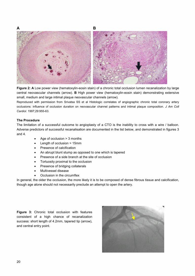
Figure 4: Chronic total occlusion with featuressuggesting it may be difficult to recanalise: longlength of occlusion of 18.6mm with bridgingcollaterals, a blunt / abrupt cut-off, and a side-branch at the site of the stump
The Equipment: choice of guiding catheter Whichever artery is involved, good support from the guiding catheter is essential. For the right coronary artery,a standard Judkins right curve catheter is usually the first choice, though a left Amplatz will provide additionalsupport and is particularly useful if the proximal part is a “Shepherds crook”. In the left anterior descendingartery, a standard Judkins left catheter can be used though a Voda or Extra Backup provides extra support. Forthe circumflex artery, a left Amplatz may be preferable.
Choice of wire For non-occlusive stenoses, wires with a floppy tip are used which avoid wall or plaque injury. However in aCTO, the wire needs to penetrate the proximal cap, which may be fibrous, thus one with a stiffer tip is likely to beneeded. Commonly an intermediate-strength wire is used initially, progressing, for safety reasons, to stiffer wiresin a step-wise fashion. For comparison, it is useful to know the tip load of individual wires. This is defined as theweight needed to be applied to bend / buckle the tip of the guidewire; for intermediate wires this is ≈3g. Somemanufacturers make specialized stiffer CTO wires with tip loads of >3g (eg the family of Miracle wires fromAsahi-Intecc (Japan) available with tip loads of 3g, 4.5g, 6g, or 12 g).
Hydrophilic wires have a coating, which when wet, makes them extremely slippery. This incurs improvedsteerability and trackability, though with the disadvantage of a relative lack of feeling resistance by the operator.Most of these wires have a very floppy tip, and unfortunately, if the lesion is particularly tough, have a tendencyto “follow the path of least resistance" and take a subintimal route leading to dissection. For CTOs, these wiresare most useful in the presence of some antegrade filling of the distal vessel.
Specialised tapered-tip wires are available which are more able to penetrate dense fibrous tissue.Examples include the family of Cross-it wires from Guidant which have a tip of 0.010”, and are available in arange of tip stiffness, and the Confianza / Conquest wire from Asahi-Intecc, Japan which has a very stiff tip thatis 0.009” diameter. The major disadvantage of these wires is the risk of perforation, and they must be advancedacross the lesion with care.
Specialised devices The Intraluminal Wire (figures 5 and 6):This system combines guidance from optical coherence reflectometry, with the power of radiofrequency ablationto penetrate and cross a CTO. The system emits near infrared light and analysis the backscatter in an A-scanmode, to determine the position of the wire tip in relation to the vessel wall compared with the true vessel lumen.It is therefore a truly forward-looking system, and scans a distance of approximately 5mm. The 0.014” wire isequipped with the capability of radiofrequency ablation with low frequency (250-500 kHz) short duration(100millisecond) pulses.

22
Figure 5: The Safe-Cross system display.
When the Intraluminal wire is within the lumen a green band appears on the monitor and the operator canablate forwards enabling the wire to advance (a). If the wire is directed outside the vessel, a red band appearson the monitor and the ability to ablate is disabled (b). Once the wire has been manoeuvred back towards thelumen, the band is again green and ablation is once again permitted (c)
Only when the wire is within the true lumen, is the operator allowed to ablate the tissue ahead advance the wire;when the wire is heading extra-luminal the system does not allow ablation. Preliminary experience suggeststhat the device facilitates recanalization in an additional 52% of lesions that have been unsuccessful usingconventional means.11 Importantly, use of the device was associated with no procedural major adverse events.
Figure 6: Case example using the Intraluminal™ wire.
Chronic total occlusion at the ostium of the left anterior descending (LAD), thought, on the clinical history, to betwo years old. A: By quantitative coronary angiography it was measured to be 21.7mm in length. B: The distalLAD is well filled via retrograde collaterals from the right coronary artery (red arrows). These are demonstratedutilizing a dual injection technique with a diagnostic catheter in the right coronary artery. C/D: The Intraluminal™wire, which has a 10mm distal radio-opaque tip, successfully ablates a path forwards (yellow arrow). E:Following successful wire passage, the Intraluminal™ wire was exchanged for a conventional floppy tip wireusing an over-the-wire balloon. The lesion was pre-dilated and then stented with a 3.5 x 28mm stent. F: Finalexcellent result.f
A
D E
B
F
C
a b c

23
The Frontrunner catheter:
The distal part of this device can be shaped to improve torquability and it has a hydrophilic coating to improvepenetrability. The tip itself is blunt and the ‘jaws’ open allowing controlled blunt dissection and advancement ofthe device. Preliminary data has shown the device to be successful in 53% lesions with a history of failure usingconventional wires.12 However, in this study of 50 patients, there was a relatively high rate of vessel perforation(18%), leading to tamponade in 2 (4%) patients. The rate of coronary perforation did decrease with time andmore experience, but emphasises that care is needed when using this catheter.
Figure 8: Case example using the Frontrunner catheter to open a chronic total occlusion of a long segment ofstented vessel
TechniqueIt is vital to make a detailed coronary angiogram at the start of the procedure to clearly delineate the site ofocclusion and stump. With a supportive guiding catheter in position, the tip of the guidewire is shaped in theusual manner with a 30-45˚ angle, though some operators also place a secondary 20-30˚ angle more proximalto the first. Stiff wires have a risk of traumatising the proximal vessel particularly if this is tortuous; this can beavoided by advancing such wires via a support catheter or over-the-wire balloon that has been positioned justproximal to the occlusion. In current practice, wiring technique is to gently rotate the tip (no more than 90˚clockwise / counter-clockwise) whilst maintaining gentle forward pressure, and aiming the tip towards the distalvessel lumen. It is important to ensure that the wire tip remains ‘on track’ and it must be visualised in at leasttwo projections. Though not mandatory, the advantage of using biplane is that co-axial views can be evaluatedsimultaneously, otherwise the operator should change projection at frequent intervals. Unfortunately at times,the “path of least resistance” may lead the wire into the subintima at which point the operator may detect thefeeling of some resistance. The wire may be withdrawn and an alternative path sought.Particularly when the stump is blunt, progression to a relatively stiffer wire may be necessary to penetrate thefibrous cap at the proximal edge. Because of the dangers of artery perforation, some operators may then switchback to a softer wire to traverse the middle part of the occlusion. The distal edge may also be difficult topenetrate due to fibrous tissue and may again require a stiffer wire. Bridging collaterals are relatively fragilevessels; when present, wires must be used with extreme caution because of the increased risk dissection orperforation.Once the occlusion has been crossed, the operator should have free movement of the tip of the wire, whichadvances easily. Confirmation of an intraluminal wire position must be made at least two co-axial projections. Itis particularly important with both the stiff and hydrophilic wires to ensure that once advanced distally, theyremain within the lumen of a large artery and are not at risk of perforating a small branch. After balloon

24
dilatation of the occlusion, a standard wire with a safer floppy tip may be advanced through to the distal vesseland the stiffer wire removed.ffffffffffff
Double injection technique (figure 9):The presence of collateral filling from the contralateral artery helps to preserve viable myocardium. Such alesion is liable to be associated with angina, as there remains an insufficient blood supply to meet the increasedmetabolic needs of physical stress. When antegrade flow is not seen beyond the occlusion, simultaneousinjection of contrast into the contralateral artery provides information on the true length of the occlusion andhelps direct safe positioning of the guidewire into the distal vessel. In this situation, it is preferable to obtain asecond arterial access and position a diagnostic catheter (5F or 6F) at the start of the procedure.
Figure 9: Double injection technique to help with guidance of the wire and facilitate recanalization
The RCA is occluded at the ostium (block arrow) with no antegrade flow. Contrast injection is made via aseparate guiding catheter in the left coronary artery ostium to visualise the distal collaterals from the distal LADto the RCA (open arrow) and help direct the wire.
Guide catheter support and deep engagementIt is important for guide catheters to have a soft and relatively atraumatic tip. However, the shaft of cathetersfrom different manufacturers provide differing degrees of “active” backup support – for a CTO, a moresupportive catheter may be an advantage. For this reason, some operators routinely use catheters of 7F or 8Ffor CTOs. Alternatively, if the lesion is tough and difficult to cross, additional support can be gained from a lesssupportive guiding catheter by deeply engaging it. A 5F guiding catheter can be particularly useful in thissituation, and may even be advanced down the vessel right up to the site of occlusion. In Japan, some 6Fguiding catheters have an inner lumen big enough to accommodate a 5F catheter, through which the procedurecan be attempted. The combination of both (mother-and-child) catheters provides excellent back-up support.
Balloon supportSupport catheters or over the wire (OTW) balloons significantly increase the ‘pushability’ (and thereby thepenetration ability) of guidewires. They can be advanced up to the occlusion (using a floppy wire particularly ifthere is proximal tortuosity), and used to maintain position whilst allowing change of guidewires. For particularlytough lesions which cannot be penetrated with a wire, the system can be stabilised to allow the wire to bepushed with greater penetration force, by inflating a balloon in the proximal vessel. This can either by using asecond wire and a balloon in a side branch, or a balloon within the proximal part of the main vessel. In thesesituations, a compliant balloon is used with the same diameter of the vessel, and inflated to nominal pressure.With the balloon inflated, the wire is advanced in the usual manner.
25
Multiple wire techniquesIt is common for several wires with different properties to be used to achieve a successful result in opening aCTO. However, it may also be useful to use several wires at the same time:
• If a wire takes a subintimal course when it is advanced, it is fixed within the false channel (thusoccluding it) and a second wire taken and progressed along a new path (double wire technique).
• If a side-branch originates at the site of occlusion, one wire may be positioned in the branch, sometimestogether with a small balloon, to try to block entry into the branch and deflect the tip of a second wiretowards the occlusion. Occasionally in this situation an IVUS catheter may be placed in such a sidebranch and used to image and guide the second wire penetrate and cross the CTO.
The dilation processOnce the lesion is crossed with the wire, most procedures will have a successful outcome with increasing sizesof balloon utilised until a good calibre lumen is achieved. Very occasionally however, it proves impossible tocross the lesion with even the smallest low-profile balloon and the best guide catheter support. If this happens,it can be useful to advance a second wire parallel to the first into the distal lumen. Alternatively, specializedtechnologies may be considered eg rotational atherectomy, and laser technology (Spectranetics Corporation,Colorado Springs). The Tornus device (Asahi Intecc, Japan) is a novel penetration catheter that is a corelessstainless steel coil consisting of 8 stranded stainless steel wires to cross a severe stenosis by manual rotation.The learning curve for use of the device is relatively short, and preliminary results are encouraging. It iscurrently under evaluation by the regulators for licensed use.
StentingEvidence from several randomised studies demonstrated that long-term results are superior with stentingcompared with balloon-only angioplasty. However, the long-term results following bare metal stent implantationsuggested a relatively high rate of restenosis of 32-55%. 13-17 However, recent excellent results have beendemonstrated following implantation of drug-eluting stents, with restenosis rates for the sirolimus- andpaclitaxel-eluting stents of 9% and 8% respectively. 18,19
ComplicationsIntervention in CTO’s is subject to the same complications as intervention in non-occlusive stenoses, withreported rates of serious complication (death, or myocardial infarction) in 1-2%. There are, however, severalcomplications more specific to the treatment of CTO’s:
• Impairment of collateral flow may occur through several mechanisms including distal embolisation ofdebris or extension of wire-induced dissection. This may be associated with a rise in cardiac enzymesconsistent with myocardial infarction.
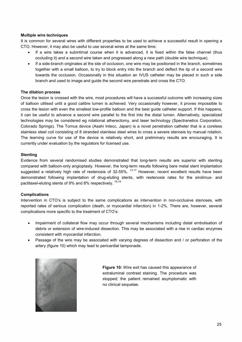
• Passage of the wire may be associated with varying degrees of dissection and / or perforation of theartery (figure 10) which may lead to pericardial tamponade.
Figure 10: Wire exit has caused this appearance ofextraluminal contrast staining. The procedure wasstopped; the patient remained asymptomatic withno clinical sequelae.

26
References1. Puma JA, Sketch MH, Jr., Tcheng JE et al. Percutaneous revascularization of chronic coronary occlusions: an overview. J Am
Coll Cardiol. 1995;26:1-11.
2. Serruys PW, Hamburger JN, Koolen JJ et al. Total occlusion trial with angioplasty by using laser guidewire. The TOTAL trial. Eur
Heart J. 2000;21:1797-805.
3. Finci L, Meier B, Favre J et al. Long-term results of successful and failed angioplasty for chronic total coronary arterial occlusion.
Am J Cardiol. 1990;66:660-2.
4. Ivanhoe RJ, Weintraub WS, Douglas JS, Jr et al. Percutaneous transluminal coronary angioplasty of chronic total occlusions.
Primary success, restenosis, and long-term clinical follow-up. Circulation. 1992;85:106-15.
5. Melchior JP, Doriot PA, Chatelain P et al. Improvement of left ventricular contraction and relaxation synchronism after
recanalization of chronic total coronary occlusion by angioplasty. J Am Coll Cardiol. 1987;9:763-8.
6. Rambaldi R, Hamburger JN, Geleijnse ML et al. Early recovery of wall motion abnormalities after recanalization of chronic totally
occluded coronary arteries: a dobutamine echocardiographic, prospective, single-center experience. Am Heart J. 1998;136:831-6.
7. Suero JA, Marso SP, Jones PG et al. Procedural outcomes and long-term survival among patients undergoing percutaneous
coronary intervention of a chronic total occlusion in native coronary arteries: a 20-year experience. J Am Coll Cardiol.
2001;38:409-14.
8. Hoye A, van Domburg RT, Sonnenschein K, Serruys PW. Percutaneous coronary intervention for chronic total occlusions: the
Thoraxcenter experience 1992-2002. Eur Heart J. 2005;26:2630-6.
9. Christofferson RD, Lehmann KG, Martin GV et al. Effect of chronic total coronary occlusion on treatment strategy. Am J Cardiol.
2005;95:1088-91.
10. Srivatsa SS, Edwards WD, Boos CM et al. Histologic correlates of angiographic chronic total coronary artery occlusions: influence
of occlusion duration on neovascular channel patterns and intimal plaque composition. J Am Coll Cardiol. 1997;29:955-63.
11. Hoye A, Onderwater E, Cummins P et al. Improved Recanalization of Chronic Total Coronary Occlusions Using an Optical
Coherence Reflectometry-Guided Guidewire. Cathet Cardiovasc Interv. 2004;63:158-63
12. Orlic D, Stankovic G, Sangiorgi G et al. Preliminary experience with the Frontrunner coronary catheter: novel device dedicated to
mechanical revascularization of chronic total occlusions. Catheter Cardiovasc Interv. 2005;64:146-52.
13. Buller CE, Dzavik V, Carere RG et al. Primary stenting versus balloon angioplasty in occluded coronary arteries: the Total
Occlusion Study of Canada (TOSCA). Circulation. 1999;100:236-42.
14. Rubartelli P, Niccoli L, Verna E et al. Stent implantation versus balloon angioplasty in chronic coronary occlusions: results from
the GISSOC trial. Gruppo Italiano di Studio sullo Stent nelle Occlusioni Coronariche. J Am Coll Cardiol. 1998;32:90-6.
15. Sirnes PA, Golf S, Myreng Y et al. Stenting in Chronic Coronary Occlusion (SICCO): a randomized, controlled trial of adding stent
implantation after successful angioplasty. J Am Coll Cardiol. 1996;28:1444-51.
16. Lotan C, Rozenman Y, Hendler A et al. Stents in total occlusion for restenosis prevention. The multicentre randomized STOP
study. The Israeli Working Group for Interventional Cardiology. Eur Heart J. 2000;21:1960-6.
17. Hoher M, Wohrle J, Grebe OC et al. A randomized trial of elective stenting after balloon recanalization of chronic total occlusions.
J Am Coll Cardiol. 1999;34:722-9.
18. Hoye A, Tanabe K, Lemos PA et al. Significant reduction in restenosis after the use of sirolimus-eluting stents in the treatment of
chronic total occlusions. J Am Coll Cardiol. 2004;43:1954-8.
19. Werner GS, Krack A, Schwarz G et al. Prevention of lesion recurrence in chronic total coronary occlusions by paclitaxel-eluting
stents. J Am Coll Cardiol. 2004;44:2301-6.

27
Chapter 3
Angela HoyeRon T van DomburgKarel SonnenscheinPatrick W Serruys
European Heart Journal2005;26(24):2630-6
Percutaneous Coronary Intervention forChronic Total Occlusions: the Thoraxcenter
Experience 1992 – 2002

28
29
AbstractBackground: Chronic total occlusions are commonly found on diagnostic angiography, andthere is some evidence from one study that successful percutaneous revascularization leadsto an improvement in long-term survival rates. However, this study included patients treatedfor unstable angina with short-duration occlusion, and stent implantation was utilized in only7%. We re-evaluated the long-term outcomes of a large consecutive series of patients with aCTO of >1 months’ duration treated at our centre, with stent implantation utilized in themajority.
Methods: All patients treated with PCI between 1992 and 2002 were retrospectivelyidentified from a dedicated database. A total of 874 consecutive patients were treated for 885CTO lesions. Mean follow-up time was 4.47 ± 2.69 years (median 4.10 years). Patients wereevaluated for the occurrence of major adverse events (MACE) comprising death, acutemyocardial infarction, and need for repeat revascularization with either CABG orpercutaneous coronary intervention (PCI).
Results: Successful revascularization was achieved in 576 lesions (65.1%), in which, stentimplantation was used in 81.0%. At 30 days, the overall MACE rate was significantly lower inthose patients with a successful recanalization (5.5% versus 14.8%, p<0.00001). At 5 years,patient survival was significantly higher in those with successful revascularization (93.5%versus 88.0%, p=0.02). In addition, there was a significantly higher survival-free of MACE(63.7% versus 41.7%, p<0.0001), with the majority of events reflecting the need for repeatintervention. Independent predictors for survival were successful revascularization, lowerage, and the absence of diabetes mellitus and multivessel disease.
Conclusions: Successful percutaneous revascularization of a CTO leads to a significantlyimproved survival rate, and a reduction in major adverse events at 5 years. Most eventsrelate to the need for repeat re-intervention, and the introduction of drug-eluting stents, withlow restenosis rates, encourages the development of technologies to improve recanalizationsuccess rates. However, failed recanalization may be associated acutely with an adverseevent, and new technologies must focus on a safe approach to successful recanalization.

30
IntroductionAt least one chronic total occlusion (CTO) is found on approximately one-third patients found to have significantcoronary disease on angiography. 1 Yet data suggest that percutaneous coronary intervention (PCI) for a CTOaccounts for approximately only 10-15% of angioplasty procedures, with the majority of patients treated witheither coronary artery bypass surgery (CABG) or medical therapy. Compared with non-occlusive lesions, PCIfor a CTO is associated with lower procedural success rates predominantly related to the inability to cross thelesion. However, technical advances in the design of angioplasty equipment, particularly of specialized wires,have improved recanalization rates. The choice of therapy for patients with a CTO (PCI versus CABG versusmedical therapy) is dependant on local policies, and outcomes of revascularization are dependent on operatorexperience. In the current study, we analysed the trends in revascularization and the treatment of CTOs at theThoraxcenter, Rotterdam between 1992 and 2002.
In addition, the long-term outcomes of patients with PCI for a CTO were analysed. Previously, a largesingle centre series of more than 2000 patients importantly demonstrated that successful percutaneousrevascularization of a CTO confers a significant 10-year survival rate compared with failed revascularization. 2
This study, analysed patients treated between 1980 and December 1999 in the Mid-America Heart Institute,and included all patients treated for an occluded vessel provided they had not had a myocardial infarction withinthe preceding 7 days. Therefore, those with relatively recent thrombotic occlusions and unstable angina wereincluded. Indeed, one of the multivariable predictors for long-term mortality was percutaneous interventionundertaken in patients with unstable angina. In addition, only 7% patients with successful revascularizationwere treated with stent implantation. Long-term outcomes of CTOs have been improved since the widespreadintroduction of stent utilization, which is associated with reduced rates of restenosis and re-occlusion comparedwith balloon-only angioplasty. 3-6 In the current study, we analysed whether the benefits demonstrated in theMAHI study are applicable to PCI carried out in chronic occlusions in another tertiary centre. In our study,chronic total occlusion was more strictly defined, those with occlusion related to unstable angina and recent (<1month) occlusion were excluded, and in addition, stent implantation was used in the majority.
MethodsDemographic and procedural data regarding all patients undergoing PCI at our centre are prospectively enteredinto a dedicated database. All procedures undertaken for an occluded vessel between 1st January 1992 and 31st
December 2002 were retrospectively identified (n=2131). Those treated in the setting of acute myocardialinfarction (AMI), and recent (<1 month) occlusion were excluded, leaving a total of 874 consecutive patientstreated for CTO.
Chronic total occlusion was defined as a lesion exhibiting Thrombolysis In Myocardial Infarction flowgrade 0-1. All patients included had at least one occlusion within a native vessel; occlusions within saphenousvein grafts were excluded. Duration of occlusion was estimated to be at least 1 month, on the basis of either ahistory of sudden chest pain, a previous AMI in the same target vessel territory, or the time between thediagnosis made on coronary angiography and PCI. Procedures were undertaken using standard techniques ofthe time. All patients were treated with heparin to maintain an ACT>250 seconds, and all were on long-termaspirin therapy. For those treated with stent implantation prior to 1996, additional anticoagulation was providedwith the use of warfarin given for 1 month. Subsequent to that time, a thienopyridine was used (ticlopidine orclopidogrel). Procedural success was defined as successful recanalization and dilatation of the vessel with orwithout stent implantation, with a final residual diameter stenosis <50%.
Median follow-up time was 4.48 years (quartiles 2.72, 6.64 years). All patients were assessed for theoccurrence of major adverse cardiac events (MACE) comprising death, non-fatal AMI, and repeatrevascularization (PCI and / or CABG). Long-term survival status was assessed by written inquires to theMunicipal Civil Registries. Follow-up clinical data were determined from electronic hospital archives and byquestionnaires sent to all living patients. The referring physician and institutions as well as the generalpractitioners were directly approached whenever necessary. Complete 30-day clinical follow-up was obtained inall patients, with complete long-term follow-up data obtained in 99% patients up until 1st April 2004. Thediagnosis of AMI required an elevation of creatine kinase to twice the upper limit of normal, together with a rise

31
in creatine kinase-MB fraction. If made following patient admission to another hospital, the diagnosis of AMI wasconfirmed through direct contact with the referring physician, using the same criteria.
Statistics: Discrete variables are presented as percentages and compared with Fisher exact test. Continuousvariables are expressed as mean ± standard deviation and compared with Student’s t test. Cumulative survival-free of major adverse events were calculated according to the Kaplan-Meier method. The log-rank test wasused to compare event-free survival between the groups. Multivariable analyses were performed usingbackward and forward stepwise Cox regression. Baseline characteristics were included if they were (i)associated with high incidence of cardiac events (p<0.1), or (ii) known risk factors from literature. Pre-selectedvariables were: age, gender, diabetes mellitus, hypertension, hypercholesterolaemia, presence of multivesseldisease, impaired left ventricular function, prior AMI, prior PCI, prior CABG, use of a glycoprotein IIb/IIIainhibitor, target vessel, successful procedure, and use of a stent. The proportional hazard assumptions wereinvestigated by testing the constancy over time of the log hazard ratio for each model. In addition, theproportional hazard assumption for all covariates was tested using Schoefeld residuals. According to thesetests, the proportional hazard assumption was not validated. Linearity was checked graphically and by inclusioncontinuous variables both as such according to quintiles. Absence effect of the grouped variable indicates thatthe effect is linear. Also assumptions of linearity were assessed and satisfied using a general linear model(GLM) univariate method. No deviation from linearity was found in any continuous variable. To investigateinteraction, an interaction model was performed using a likelihood ratio test in the multivariable Cox. Interactionwas performed on all selected variables. However, no interaction was found. Odds ratio with corresponding95% confidence intervals are reported. All tests were two-tailed; due to the large number of statistical tests, p-values should be interpreted with caution. While no specific level of significance is defined, a p-value of 0.01should be considered for strong evidence in support of a true effect.
Table 1: Baseline patient demographics and target vessel site with respect to a successful versus anunsuccessful chronic total occlusion revascularization procedure
CTO successn=567
CTO failuren=304
p value
Age (years) 59.6 ± 10.8 60.5 ± 10.4 0.2Male sex (%) 73.6 72.2 1.0Diabetes mellitus (%) 12.0 9.1 0.2Hypertension (%) 20.3 21.0 0.7Hypercholesterolaemia (%) 48.6 43.3 0.2Family history of coronary disease (%) 21.9 18.8 0.3Impaired LV function (%) 32.5 38.1 0.5Previous myocardial infarction (%) 55.7 49.2 0.2Previous PCI (%) 24.3 23.0 0.9Previous CABG (%) 8.7 10.4 0.4Vessel disease 0.03
Single-vessel (%) 46.0 32.62 vessel (%) 36.2 40.53 vessel (%) 17.8 27.0
Number of lesions 573 306Target vessel of the lesion 0.8
RCA (%) 42.2 52.6LAD (%) 33.2 26.5LCX (%) 24.4 20.6LMS (%) 0.2 0.3

32
ResultsBetween 1st January 1992 and 31st December 2002, a total of 874 patients underwent PCI for at least one CTO.Of these, 11 had attempted revascularization of 2 CTO’s, making a total of 885 attempted lesions. Overall,successful revascularization was achieved in 576 lesions (65.1%), with failure in the remaining 309 (34.9%). Ofthe 11 patients with attempted therapy of 2 CTOs, PCI outcome was the same in both lesions in 8 patients. Theremaining 3 patients with both one success and one unsuccessful PCI have been excluded from furtheranalysis. The baseline demographics for the remaining patients are presented in table 1.
There were no significant differences in characteristics, though a trend towards an increase in 2- and 3-vessel disease in those in whom PCI for occlusion was unsuccessful. Over time, the proportion of patients withcoronary disease who underwent revascularization with PCI as opposed to CABG surgery, increased with time(figure 1). Similarly, there was a trend to an increased proportion of PCI for CTO (figure 2). Percutaneous CTOtherapy was undertaken utilizing the contemporary techniques of the time including specialized hydrophilic,tapered tip, and stiff wires when available, with the laser wire used in 72 (8.1%). However, despite theintroduction of more specialised technologies, the success rate of recanalization did not improve (figure 2).Following successful recanalization, the overall use of stent implantation was 81.0%, with stent utilizationincreasing with time (figure 3).
Figure 1: Trends in the number of revascularization procedures with percutaneous coronary intervention (PCI)versus coronary artery bypass surgery (CABG) at the Thoraxcenter.
Figure 2: Trends in the increase in the proportion of percutaneous intervention (PCI) for a chronic totalocclusion (CTO), and success rates for PCI for CTO with respect to year of intervention.
0
200
400
600
800
1000
1200
1400
1600
1800
1992 1994 1996 1998 2000 2002
PCI CABG
1993 1995 1997 1999 2001
Num
ber o
f pro
cedu
res
0
80
CTO as a % of total PCI procedures
60
40
20
1800
1400
1000
600
200
1992 1993 1994 1995 1996 1997 1998 1999 2000 2001 2002
Total number of PCI procedures
CTO success as a % of CTO attempts
%

33
Figure 3: Utilization of stent implantation following successful recanalization of a chronic total occlusion withrespect to the year of intervention.
The 30-day MACE rates are presented in table 2. In addition, this table demonstrates the events directly relatedto the procedure and occurring within the first 48 hours. A failed recanalization procedure was associated with asignificantly higher rate of MACE in the immediate period following the procedure.
Table 2: Incidence of major adverse cardiac events at 2 and 30 daysCTO success
n=567CTO failure
n=304p value
2 days 2 (0.4) 3 (1.0) 0.2Death, n (%)30 days 4 (0.7) 6 (2.0) 0.092 days 3 (0.5) 5 (1.6) 0.1Death or acute myocardial infarction, n (%)30 days 7 (1.2) 7 (2.3) 0.22 days 7 (1.2) 13 (4.3) 0.004Death or CABG, n (%)30 days 10 (1.8) 30 (9.9) <0.000012 days 14 (2.5) 17 (5.6) 0.02MACE, n (%)30 days 31 (5.5) 45 (14.8) <0.00001
In the long-term, all outcomes were significantly worse following a failed attempt at revascularization. The 5-year survival was significantly lower than when revascularization was successful (figure 4), and the survival-freeof AMI, CABG, and MACE were also significantly lower (figures 5-7). By multivariable analysis, the independentpredictors for survival and MACE following PCI for CTO are presented in table 3. The presence of multivesseldisease was an independent predictor for both survival and MACE. The cumulative survival-free of MACE withrespect to the presence of single versus multivessel coronary disease is shown in table 4.
Table 3: Independent predictors of death and major adverse cardiac events (MACE) after attemptedpercutaneous coronary intervention of a chronic total occlusion
Hazard ratio 95% confidence intervals p valueDeathSuccessful revascularization 0.58 0.34 – 0.98 0.04Age 1.04 1.02 – 1.07 0.002Diabetes mellitus 2.49 1.33 – 4.66 0.005Multivessel disease 4.29 1.93 – 9.55 <0.001
Major adverse cardiac eventsSuccessful revascularization 0.55 0.44 – 0.70 <0.001Multivessel disease 1.43 1.14 – 1.79 0.002Use of a stent 0.69 0.54 – 0.88 0.002
20
40
60
80
1992 1993 1994 1995 1996 1997 1998 1999 2000 2001 2002
Sten
t use
(%) 100
0

34
Figure 4: Cumulative survival at 5 years withrespect to the outcome of attempted recanalizationof a chronic total occlusion.
Figure 6: Cumulative survival-free of coronaryartery bypass surgery (CABG) at 5 years withrespect to the outcome of attempted recanalizationof a chronic total occlusion.
Figure 5: Cumulative survival-free of acutemyocardial infarction (AMI) at 5 years with respectto the outcome of attempted recanalization of achronic total occlusion.
Figure 7: Cumulative survival-free of major adversecardiac events (death, acute myocardial infarction, orrepeat reintervention (percutaneous or bypasssurgery)) at 5 years with respect to the outcome ofattempted recanalization of a chronic total occlusion.
Follow-up543210
100
90
80
70
60
40
93.5%
88.0%
Log rank p=0.02Hazard ratio 1.86(95% CI 1.12-3.10)
Unsuccessful recanalizationSuccessful recanalization
Cum
ulat
ive
surv
ival
(%)
Follow-up43210
100
90
80
70
60
50
40
5
Log rank p=0.02Hazard ratio 1.62(95% CI 1.07-2.45)
Unsuccessful recanalizationSuccessful recanalization
Cum
ulat
ive
surv
ival
-free
of A
MI (
%)
Follow-up
543210
100
90
80
70
60
50
40
87.4%
61.5%
Log rank p<0.0001Hazard ratio 3.94(95% CI 2.88-5.39)
Unsuccessful recanalizationSuccessful recanalization
Cum
ulat
ive
surv
ival
-free
of C
AB
G (%
)
Follow-up543210
100
90
80
70
60
50
40
63.7%
41.7%Log rank p<0.0001Hazard ratio 2.14(95% CI 1.74-2.63)
Cum
ulat
ive
surv
ival
-free
of M
AC
E (%
) Unsuccessful recanalizationSuccessful recanalization

Table 4: Cumulative survival-free of major adverse cardiac events at 5 years with respect to the presence ofsingle versus multivessel coronary disease Single vessel Multivessel
CTO successn=261
CTO failuren=99
p value CTO successn=306
CTO failuren=205
p value
Death (%) 97.3 99.0 0.3 92.5 86.3 0.02Death / AMI (%) 94.6 96.0 0.6 88.6 82.0 0.03Death / CABG (%) 91.6 70.7
<0.0001
86.9 61.5<0.000
1MACE (%) 72.0 47.5
<0.0001
61.1 42.9<0.000
1
Figure 8: Cumulative survival at 5 years with respect to diabetic status
Figure 9: Cumulative survival-free of major adverse cardiac events (death, acute myocardial infarction, orrepeat reintervention (percutaneous or bypass surgery)) at 5 years with respect to diabetic status, and theoutcome of attempted recanalization of a chronic total occlusion
Follow-up (years)543210
100
80
60
40
20
0
92.7%
83.6%
Log rank p=0.001Hazard ratio 2.70(95% CI 1.46-5.02)
Diabetes mellitusNo diabetes mellitus
Cum
ulat
ive
surv
ival
(%)
100
80
60
40
20
0
Diabetes mellitusNo diabetes mellitus
Success
No success
Cum
ulat
ive
surv
ival
-free
of M
AC
E (%
)
543210 543210Follow-up (years) 35
36
Overall survival was significantly lower in those patients with diabetes mellitus (figure 8). Within the diabeticpopulation, 5-year survival was 84.9% in those with a successful recanalization versus 79.1% followingunsuccessful recanalization (p=0.4), suggesting that most of the benefit in terms of survival following successfulrecanalization is in the non-diabetic group. However, the beneficial effect of successful recanalization of a CTOon survival-free of MACE remains clearly apparent (figure 9), irrespective of diabetic status. Successfulrecanalization led to a 5 year MACE-free survival of 63.7% and 62.3% in those with and without diabetesmellitus respectively. Following unsuccessful recanalization, the 5 year MACE-free survival was 42.0% and41.5% for those with and without diabetes mellitus respectively, p<0.0001.
DiscussionThe present study evaluated only consecutive patients with chronic total occlusions (CTO) of at least onemonths’ duration, and for the first time confirms a 5-year survival benefit in successful recanalization of theselesions. In addition, there was a significant reduction in major adverse cardiac events, particularly the need forrevascularization with CABG. Independent predictors of survival were a successful recanalization, lower age,and the absence of diabetes mellitus and multivessel disease. Independent predictors of major adverse eventswere an unsuccessful recanalization, multivessel disease, and non-usage of stent implantation.
Although one large series of the long-term outcomes of patients following PCI for CTO has beenpublished, the authors acknowledged that their study was limited as they did not always know the duration ofocclusion. 2 Indeed, analysis of 100 consecutive patients who had been included in the study demonstrated that42% were <1 months’ duration. These patients are likely to have thrombotic occlusions, rather than the fibrotic /calcific lesion of a CTO. This difference in lesion pathophysiology may affect the long-term outcome, indeed oneof the independent predictors for survival in the MAHI study, was therapy for unstable angina. In the presentstudy, we have confirmed that a successful outcome following PCI for a truly chronic occlusion does confer asignificant benefit on survival, and reduces the rate of MACE with a marked reduction in the need for CABG.
The difference in the rate of MACE between those with a successful versus unsuccessful recanalizationwas apparent immediately (at 48 hours), predominantly related to the need for emergency CABG in the failedrecanalization group. An acute complication of CTO recanalization therefore confers serious adverseconsequences in the short-term, which would have been potentially avoided if an alternative treatment optionhad been undertaken. It is particularly important to note that the mortality rate of those with a failed procedurewas not insignificant at 1.9% at 30 days.
The present study demonstrated a 5-year survival benefit following successful CTO recanalization. Thepossible reasons for the improved survival are beyond the scope of the present study. There were differencesin the medical therapy received by the 2 groups which could potentially be a confounding factor. Unlike thosewith a failed recanalization, the group with a successful recanalization together with stent implantation weretreated with additional medical therapy with warfarin, ticlopidine, or clopidogrel. However, this was given for only1 month, and it is unlikely that such a short duration of therapy is the reason for the improved long-termoutcomes. More likely, improved survival may relate to the greater proportion of viable but inadequatelyperfused myocardium. The improvement in prognosis following successful revascularisation might relate to theassociated improvement in left ventricular function, 7 or a reduction in the risk of ischaemic-driven malignantarrhythmia. In addition, a successful procedure could potentially avoid the need for CABG with its’ associatedmortality risk.
In the present study, the survival benefit of successful CTO recanalization was most apparent in thosewith multivessel rather than single vessel disease. Indeed, the patients with single vessel disease and failedrecanalization had a very high rate of survival at 5-years of 98.9%. Approximately half of these patientsunderwent CABG or repeat re-intervention with PCI, with a survival-free of MACE at 5 years of 45.8%. Theremaining patients were treated with medical therapy alone, and although this constitutes only a relatively smallnumber of patients, the excellent survival rate suggests that from a prognostic point of view medical therapymay not be unreasonable. Little data is currently available on the outcomes of patients with a CTO who aremanaged with optimal medical therapy (aspirin, beta-blocker, statin, ACE-inhibitor etc). In particular, those withan excellent collateral circulation may have a very good prognosis. Our manuscript does not therefore provide

37
scientific proof to support a broad generalized recommendation to try to open all CTOs; a randomised studycomparing “best” medical therapy with a more aggressive strategy of attempted recanalization would berequired to assess this. Importantly, studies are needed with more detailed assessment of left ventricularfunction, degree of viability, and ischaemic burden both pre-procedure and at follow-up, to determine therelationship these have with long-term survival.
The Thoraxcenter in Rotterdam is a tertiary referral centre for PCI, taking referrals from 13 surroundinghospitals covering a large region. The majority of patients requiring repeat re-intervention, whether it bepercutaneous or surgical, come back to be re-treated in our centre. As in other centres, the number ofpercutaneous revascularizations increased over time, whilst that of CABG gradually decreased. In addition, therelative number of PCI procedures carried out for a chronic occlusion also increased with time. However, theoverall success rate of recanalization remained stable despite advances in the technology of specialised wiresand other equipment. The chances of successful recanalization are known to be dependent on lesionmorphology and it is possible that with time and the increase in PCI for CTO, relatively more complex lesionswere attempted.
It is well recognised from large scale studies that mortality is higher following percutaneous coronaryintervention procedures in those with diabetes compared to those without diabetes mellitus. 8,9 Our studyconcurs with these results, with a significantly lower 5 year survival in diabetics (83.6% versus 92.7%, p=0.001).However, the beneficial effect of successful recanalization of a CTO on overall survival-free of major adverseevents was clearly apparent to be irrespective of diabetic status (figure 9).
Of those patients with a successful revascularization, the majority of subsequent adverse events relateto a need for repeat reintervention. Long-term results have been shown to improve with the advent of stentimplantation, with reduced rates of restenosis and re-occlusion when compared with results of balloon-onlyangioplasty. However, the recent introduction of drug-eluting stents will further improve on these results. Datafrom our own centre have shown a significant higher cumulative survival-free of major adverse cardiac events at1 year with the sirolimus-eluting stent compared with bare metal stent implantation (96.4% versus 82.8%,p<0.05). 10 These results encourage the development of further technologies to facilitate safe and successfulCTO recanalization.
Limitations:The present study is limited by being a retrospective observational analysis of outcomes. However, it iscomprised of a large cohort of patients with complete clinical follow-up obtained in virtually all. The study isfurther limited by the lack of randomised comparison with a group of patients treated with medical therapyalone, or those treated directly with CABG. In addition, the possible reasons for improved survival in thesuccessful recanalization group have not been fully explored and require further study.
Conclusions:Successful percutaneous revascularization of a CTO leads to a significantly improved survival rate, and areduction in major adverse events at 5 years. Most events relate to the need for repeat re-intervention, and theintroduction of drug-eluting stents, with reduced restenosis rates, encourages the development of furthertechnologies to improve recanalization success rates. However, failed recanalization may be associated acutelywith a major adverse event, and new technologies must focus on a safe approach to successful recanalization.Additional studies are needed to evaluate the comparative prognostic value of CTO recanalization comparedwith optimal medical therapy, particularly in patients with single vessel disease.

38
References 1. Kahn JK. Angiographic suitability for catheter revascularization of total coronary occlusions in patients from a community hospital
setting. Am Heart J. 1993;126:561-4.
2. Suero JA, Marso SP, Jones PG et al. Procedural outcomes and long-term survival among patients undergoing percutaneous
coronary intervention of a chronic total occlusion in native coronary arteries: a 20-year experience. J Am Coll Cardiol.
2001;38:409-14.
3. Lotan C, Rozenman Y, Hendler A et al. Stents in total occlusion for restenosis prevention. The multicentre randomized STOP
study. The Israeli Working Group for Interventional Cardiology. Eur Heart J. 2000;21:1960-6.
4. Sirnes PA, Golf S, Myreng Y et al. Stenting in Chronic Coronary Occlusion (SICCO): a randomized, controlled trial of adding stent
implantation after successful angioplasty. J Am Coll Cardiol. 1996;28:1444-51.
5. Rubartelli P, Niccoli L, Verna E et al. Stent implantation versus balloon angioplasty in chronic coronary occlusions: results from
the GISSOC trial. Gruppo Italiano di Studio sullo Stent nelle Occlusioni Coronariche. J Am Coll Cardiol. 1998;32:90-6.
6. Buller CE, Dzavik V, Carere RG et al. Primary stenting versus balloon angioplasty in occluded coronary arteries: the Total
Occlusion Study of Canada (TOSCA). Circulation. 1999;100:236-42.
7. Rambaldi R, Hamburger JN, Geleijnse ML et al. Early recovery of wall motion abnormalities after recanalization of chronic totally
occluded coronary arteries: a dobutamine echocardiographic, prospective, single-center experience. Am Heart J. 1998;136:831-6.
8. Pell JP, Pell AC, Jeffrey RR et al. Comparison of survival following coronary artery bypass grafting vs. percutaneous coronary
intervention in diabetic and non-diabetic patients: retrospective cohort study of 6320 procedures. Diabet Med. 2004;21:790-2.
9. Seven-year outcome in the Bypass Angioplasty Revascularization Investigation (BARI) by treatment and diabetic status. J Am
Coll Cardiol. 2000;35:1122-9.
10. Hoye A, Tanabe K, Lemos PA et al. Significant reduction in restenosis after the use of sirolimus-eluting stents in the treatment of
chronic total occlusions. J Am Coll Cardiol. 2004;43:1954-8.

39
Chapter 4
Angela HoyeGeorgios SianosFrancesco SaiaPedro A LemosWillem J van der GiessenPim J de FeyterVeronique LMA CoenRon T van DomburgPeter C LevendagPatrick W Serruys
Submitted for publication
Predictors, Incidence and Prognosis ofCoronary Occlusion following Intracoronary
Beta-radiation Therapy
been
Markering
Med. Library
Notitie
EMBARGO paper available after publication

40
41
Abstract
Background: Intracoronary brachytherapy (IRT) has been associated with the development
of late vessel occlusion.
Objectives: To assess the incidence, predictors and prognosis of coronary occlusion in a
consecutive series of patients following beta-radiation therapy
Methods: Between April 1997 and December 2001, 301 consecutive patients were
successfully treated with IRT, and 37 patients (12.3%) were subsequently found to have an
occlusion of the treated vessel and form the present study population. Patient and procedural
data were retrospectively analysed from a dedicated database.
Results: One patient had subacute thrombosis on day 21, and over a mean follow-up of 40.3
months, target lesion occlusion was found in a further 36 patients at a mean time after IRT of
16.0 months (range 3.4-66.8 months). In 12 patients (32.4%), vessel closure caused an
acute myocardial infarction, and was associated with 3 (8.1%) cardiac-related deaths. At 4
years, the cumulative survival-free of target lesion closure was 85.4%.
By multivariate analysis, the factor predictive for development of occlusion was
treatment of a de novo lesion rather than in-stent restenosis (15.4% versus 7.9%, p=0.03
(HR=2, 95% CI: (1.1-5)). Occlusion was not related to the dosage administered, the source
length, the duration of dual anti-platelet agents, or the “learning curve” of therapy.
Conclusions: A high incidence of late vessel occlusion is observed after IRT. Prolongation
of dual anti-platelet therapy to 6 months duration is insufficient to protect against the
development of occlusion, which is associated with significant morbidity.
51
Chapter 5
Nico MolletAngela HoyePedro LemosFilippo CademartiriGeorgios SianosEugene McFaddenPatrick W SerruysPim J de Feyter
Americal JournalOf Cardiology2005;95(2):240-3
Value of Pre-Procedure MultisliceComputed Tomographic Coronary
Angiography to Predict PercutaneousRecanalization of Chronic Total Occlusions

52

53
Abstract
We performed multislice computed tomographic coronary angiography in 45 patients who
had chronic total occlusions and were scheduled for percutaneous recanalization.
Multivariate analysis identified a blunt stump (by conventional angiography), occlusion length
>15 mm, and severe calcification (by multislice computed tomographic coronary
angiography) as independent predictors of procedural failure.

54
Sixteen-row multislice spiral computed tomographic (MSCT) coronary angiography has recently been shown toallow reliable noninvasive evaluation of coronary morphology.1-3 In the present study, we analyzed the potentialof preprocedural MSCT coronary angiography to provide additional information and thus predict the proceduraloutcome in patients who had chronic total occlusion (CTO) and were referred for percutaneous coronaryrecanalization.
Forty-five patients referred for percutaneous recanalization of ≥1 CTO lesion underwent MSCT coronaryangiography before the coronary procedure (median interval 29 days, interquartile range 9 to 53). The diagnosisof CTO was made on diagnostic angiograms that demonstrated complete occlusion of a major epicardialcoronary artery, which was deemed to be of ≥3 months' duration from the date from the previous angiogram, aclinical history of myocardial infarction, or onset of or a severe episode of prolonged anginal chest pain. Inaddition, inclusion into the study required a serum creatinine level <120 mmol/L, presence of sinus rhythm, andthe ability to hold a breath for 20 seconds. The protocol was approved by the institutional review board, and allpatients gave written informed consent.
Conventional angiographic assessment was performed by observers who were unaware of the results of MSCTscans. Parameters previously reported to have prognostic importance for procedural failure were assessed:absence of anterograde flow through bridging collaterals, absence of a tapered stump, presence of severecalcification at the occluded segment, side branch at the occlusion site, and tortuosity of the vessel proximal tothe occlusion (defined as an angle >45° in any projection). Where possible, occlusion length was measuredfrom the view with the longest lesion on quantitative coronary angiography as the distance between a stumpand a distal vessel as visualized by anterograde filling through bridging collaterals. In addition, in some otherpatients, length was determined from the baseline angioplastic procedure film using a bilateral coronaryinjection.
Twenty-two patients who had a heart rate >65 beats/min before multislice spiral computed tomography receivedan oral dose of 100 mg of metoprolol 1 hour before scanning. All examinations were performed with a 16-rowMSCT scanner (Sensation 16, Siemens, Forehheim, Germany; collimation 16 × 0.75 mm, rotation time 420 ms,table feed 3.0 mm/rotation, tube voltage 120 kV, tube current 400 to 450 mA). After intravenous administrationof 120 ml of nonionic contrast material (Visipaque 320, Amersham Health, Little Chalfont, United Kingdom), anautomatic bolus-tracking technique triggered the start of MSCT scanning. Images were reconstructed withretrospective electrocardiographic gating during the mid- to end-diastolic phase to provide nearly motion-freeimage quality; additional reconstruction windows (e.g., early diastolic phase) were explored when necessary.
All MSCT scans were analyzed off-line by operators who were blinded to angiographic and procedural data.Parameters similar to those of conventional angiography were evaluated: a blunt rather than tapered stump,severe calcification, side branch at the occlusion site, proximal tortuosity, and occlusion length. Severecalcification was defined as the presence of high-density plaques (≥130 HU) involving >50% of the coronarywall on a cross-sectional image and localized within the occlusion stump or occluded segment.
All procedures were performed by operators who were highly experienced in the treatment of CTOs, with theinterventional strategy left to the discretion of the operator. Wires were used in a stepwise progression, startingwith a wire that had a relatively less traumatic tip (Graphix Intermediate, Boston Scientific Corporation, Miami,Florida) or a hydrophilic wire (Choice PT Plus, Boston Scientific Corporation, or Crosswire NT TerumoCorporation, Tokyo, Japan) and progressing to stiffer wires (Miracle, Asahi Intec, Nagoya, Japan) andspecialized technologies (Safe-Cross, Intraluminal Therapeutics, Carlsbad, New Mexico).4,5 Procedural failurewas defined as an inability to cross the occlusion with a guidewire.
Multivariate logistic regression analyses were performed to identify angiographic and MSCT parametersassociated with procedural failure (all univariate predictors with a p value ≤0.1 were tested for their multivariatepredictive value, and final models were built by backward stepwise selection). Angiographic parametersassessed were those identified in previous studies: 6 the occluded artery, duration of occlusion, multivessel
55
disease, anterograde and retrograde collateral filling, type of stump, side branch at the site of occlusion, calcificdeposits, vessel tortuosity, and occlusion length >15 mm. The predictive strengths of the models wereevaluated by means of the −2 log-likelihood statistic, and models' lack of fit with the Hosmer-Lemeshow test,and their global predictive accuracy were assessed by the C index (area under the receiver-operatingcharacteristic curve).
Patients' mean age ± SD was 57.0 ± 10.1 years, 40 (89%) were men, 10 (22%) were diabetic, and 14 (31%)had multivessel disease. Forty-seven CTO lesions were treated. Angiographic measurement of CTO length waspossible in 39 lesions (83%), 31 (66%) from the diagnostic film and an additional 8 (17%) from bilateral injectionand assessment of retrograde collateral filling. The mean length of occlusion was longer by MSCT coronaryangiography than by angiography (21.8 ± 18.6 vs 14.6 ± 10.9 mm, respectively). Procedural data, including typeand number of guidewires used, are presented in Table 1. The only difference between success and failure wasin the increased use of the Miracle wire in the failure group. Overall mean procedural time was 148 ± 53minutes, with a mean fluoroscopic time of 47 ± 24 minutes. Overall, 45% of interventional procedures failed(Table 2). Success versus failure was not dependent on the operator or choice of interventional strategy. Atunivariate analysis, the following were associated with procedural failure: clinical assessment (occlusionduration ≥9 months), angiographic assessment (lack of anterograde collateral filling, a blunt rather than atapered stump, and side branch at the occlusion site), and MSCT coronary angiographic assessment (a bluntrather than a tapered stump, severe calcification, and occlusion length >15 mm; Table 2).
Table 1: Procedural Duration and Use of Contrast Material and Guidewires for Percutaneous Intervention of aCTO With Respect to Successful Versus Unsuccessful Recanalization
Success(n = 26 lesions)
Failure(n = 21 lesions)
p value
Mean total procedural time (min) 148 ± 61 148 ± 44 1.0Mean volume of contrast used (ml) 451 ± 258 453 ± 265 1.0Mean no. of wires 2.0 ± 1.2 2.1 ± 0.8 0.9Wire type*Graphix Intermediate 20 (76.9%) 12 (57.1%) 0.2Choice PT Plus 5 (19.2%) 3 (14.3%) 0.8Crosswire NT 8 (30.8%) 10 (47.6%) 0.3Miracle 5 (19.2%) 12 (57.1%) <0.01Intraluminal 3 (11.5%) 4 (19.0%) 0.5Over-the-wire balloon support 15 (57.7%) 17 (81.0%) 0.1* Wire type not mutually exclusive.
When analyzed separately, the following “traditional” clinical and angiographic characteristics were identified asmultivariate predictors of procedural failure: occlusion duration >9 months and stump morphology (Table 3).Separate multivariate analysis that assessed only MSCT coronary angiographic parameters identified thefollowing predictors: occlusion length >15 mm, severe calcification, and stump morphology. Final best modeltesting for pooled clinical, angiographic, and MSCT parameters identified a blunt rather than a tapered stump(by angiography), occlusion length >15 mm (by MSCT coronary angiography), and severe calcification (byMSCT coronary angiography) as multivariate independent predictors of procedural failure (Figure 1).
The current selection process of technically appropriate candidates for percutaneous recanalization of CTO isbased on the evaluation of a relatively restricted number of clinical and angiographic characteristics. In thisstudy, we show that noninvasive evaluation of patients who have CTO by preprocedural MSCT coronaryangiography improves the ability to predict the outcome of a percutaneous recanalization attempt. Our findingsindicate that MSCT coronary angiography may aid in the therapeutic decision making for patients who haveCTO. In addition, accurate preprocedural characterization of CTO features may assist in outlining thetherapeutic interventional strategy.

56
Ove
rall
Occ
lusi
on le
ngth
not
det
erm
inab
le
Occ
lusi
on le
ngth
>15
mm
Vess
el to
rtuos
ity
Seve
re c
alci
ficat
ion
Side
bra
nch
at o
cclu
sion
site
Stum
p m
orph
olog
y no
t det
erm
inab
le
Tape
red
stum
p
Brid
ging
col
late
rals
Ret
rogr
ade
colla
tera
l fill
ing
Ante
rogr
ade
fillin
g
Mul
tives
sel d
isea
se
Occ
lusi
on d
urat
ion ≥9
mo
Left
circ
umfle
x ar
tery
Left
ante
rior d
esce
ndin
g ar
tery
Rig
ht c
oron
ary
arte
ry
100
17262334570603872603451154343
Freq
uenc
y( %
)
455067645059-253944323863434050
Failu
re ra
te( %
)
-
1.29
(0.2
8–5.
94)
3.39
(0.8
5–13
.48)
2.75
(0.6
8–11
.14)
1.39
(0.4
1–4.
65)
4.37
(1.2
3–15
.54)
-
0.12
(0.0
3–0.
45)
0.68
(0.2
1–2.
25)
0.92
(0.2
6–3.
32)
0.27
(0.0
8–0.
94)
0.64
(0.1
9-2.
20)
4.72
(1.3
6-16
.39)
0.92
(0.1
8-4.
64)
0.72
(0.2
2-2.
32)
1.45
(0.4
5-4.
66)
OR
(95%
CI)
-0.7
0.080.2
0.02-
<0.0
1
0.5
0.9
0.040.5
0.020.9
0.6
0.5p
valu
e
Ang
iogr
aphy
-0512138451560--------
Freq
uenc
y( %
)
--635067524332--------
Failu
re ra
te( %
)
--
4.72
(1.3
6-16
.39)
1.31
(0.3
2-5.
32)
4.44
(1.2
7-15
.61)
1.76
(0.5
5-5.
64)
0.25
(0.3
4-1.
82)
0.16
(0.0
3–0.
73)
--------
OR
(95%
CI)
--
0.020.7
0.020.3
0.2
0.02--------p
valu
e
MSC
T co
rona
ry a
ngio
grap
hy
Tabl
e 2:
Clin
ical
, ang
iogr
aphi
c an
d M
SCT
coro
nary
ang
iogr
aphy
lesi
on c
hara
cter
istic
s (n
=47)
CI =
con
fiden
ce in
terv
al; M
SC
T =
mul
tislic
e co
mpu
ted
tom
ogra
phic
; OR
= o
dds
ratio

57
C In
dex
0.80
0.84
0.85
p va
lue
0.66
0.99
0.60
Hos
mer
-Le
mes
how
tes
t
DF 2 6 5
-2 L
ogLi
kelih
ood
50.0
44.2
41.0
OR
(95%
CI)
3.56
(0.9
0-14
.02)
0.15
(0.0
4-0.
58)
-
6.39
(1.3
0-31
.41)
12.0
1 (1
.78-
81.1
)
- -
0.11
(0.0
2-0.
73)
0.07
(0.0
0-1.
15)
-
0.09
(0.0
2-0.
48)
8.77
(1.5
8-48
.76)
7.62
(1.3
3-43
.74)
p va
lue
0.07
<0.0
1
0.7
0.02
0.01
0.06 -
0.02
0.06 0.6
<0.0
1
0.01
0.02 0.4
DF 1 1 1 1 1 2 - 1 1 1 1 1 1 1
Wal
d’s
Chi
-sq
uare
3.30
7.46
0.13
5.21
6.51
5.63 -
5.23
3.46
0.26
7.98
6.16
5.18
0.74
Coe
ffici
ent
1.27
-1.9
3
0.24
1.86
2.49 -
1 (r
efer
ence
)
-2.1
9
-2.6
5
-0.4
5
-2.4
3
2.17
2.03
-0.6
7
Clin
ical
/ an
giog
raph
ic p
redi
ctor
s
Occ
lusi
on d
urat
ion
>9 m
o
Tape
red
stum
p
Con
stan
t
MSC
T co
rona
ry a
ngio
grap
hy p
redi
ctor
s
Occ
lusi
on le
ngth
>15
mm
Sev
ere
calc
ifica
tion
Stu
mp
mor
phol
ogy
Blu
nt
Tape
red
Not
det
erm
inab
le
Con
stan
t
Clin
ical
/ang
iogr
aphi
c +
MSC
T co
rona
ryan
giog
raph
ic p
redi
ctor
s
Tape
red
stum
p*
Occ
lusi
on le
ngth
>15
mm
Sev
ere
calc
ifica
tion
Con
stan
t
DF
= de
gree
s of
free
dom
; oth
er a
bbre
viat
ions
as
in T
able
2. * A
−2
log-
likel
ihoo
d ch
ange
in th
e gl
obal
mod
el if
1 v
aria
ble
is re
mov
ed: t
aper
ed s
tum
p−1
0.7
(p <
0.01
for c
hang
e), o
cclu
sion
leng
th −
8.2
(p <
0.01
), an
d ca
lcifi
catio
n −6
.6 (p
= 0
.01
for c
hang
e).

58
MSCT coronary angiography of CTOs adds important information compared with “conventional” coronaryangiography. The length of the occluded segment has long been identified as an important predictor of failedrecanalization. However, accurate measurement of lesion length using conventional angiography may bedifficult, mainly due to foreshortening, calibration limitations, and lack of visualization of the distal vessel in theabsence of collateral filling. In the present study, lesion length could be measured in only 66% of diagnosticfilms. Conversely, MSCT coronary angiography allowed reliable 3-dimensional length measurement of coronarysegments.7 In the present series, when angiographic and MSCT coronary angiographic occlusion lengths weremeasured, results of the MSCT coronary angiography were “longer,” perhaps reflecting the inaccuracy ofquantitative coronary angiography as previously described. Moreover, MSCT coronary angiography allowsevaluation of the morphology of the occlusion trajectory, including detailed delineation of coronary calcification.Long occlusions and severe calcifications on MSCT coronary angiograms were found to be importantpredictors of procedural failure, whereas neither feature was identified as an independent predictor onconventional angiograms.
The need to use contrast material may pose a limitation to preprocedural MSCT coronary angiography forinterventions scheduled to be performed shortly after scanning. However, the elective nature of CTOrecanalization angioplasty allows a safe time lag between these procedures. In our study, multislice computedtomography was performed 1 month before coronary intervention. The relatively high radiation exposureduring MSCT coronary angiography, reportedly between 6.7 and 13.0 mSv,8-10 remains a matter of concern.However, MSCT coronary angiography may optimize therapeutic strategy (e.g., calcifications may requireintraluminal techniques), resulting in shorter procedures. MSCT coronary angiography is currently feasible forselected patients, and further studies are needed to evaluate its value in a more general patient population.
Figure 1: (A, inset) CTO of the left anterior descending coronary artery with favorable invasive angiographicCTO characteristics, tapered stump, absence of calcifications, and occlusion length <15 mm (arrowheads) in apatient who had been referred for percutaneous recanalization. Volume-rendered MSCT image that provides a3-dimensional overview of the coronary arteries, and (B) maximum intensity projection of the same MSCTimage show a severely calcified occlusion stump (arrows). (C) Curved multiplanar reconstructed MSCT imageof the left anterior descending coronary artery shows a severely calcified stump (arrow) and an occludedsegment (arrowheads). Collateral filling is clearly visible distal to the occlusion. This percutaneous attempt atrecanalization of the CTO was unsuccessful.
59
References1. K. Nieman, F. Cademartiri, P.A. Lemos, R. Raaijmakers, P.M. Pattynama and P.J. de Feyter, Reliable noninvasive coronary
angiography with fast submillimeter multislice spiral computed tomography, Circulation 106 (2002), pp. 2051–2054.
2. D. Ropers, U. Baum, K. Pohle, K. Anders, S. Ulzheimer, B. Ohnesorge, C. Schlundt, W. Bautz, W.G. Daniel and S. Achenbach,
Detection of coronary artery stenoses with thin-slice multi-detector row spiral computed tomography and multiplanar reconstruction,
Circulation 107 (2003), pp. 664–666.
3. N.R. Mollet, F. Cademartiri, K. Nieman, F. Saia, P.A. Lemos, E.P. McFadden, P.M.T. Pattynama, P.W. Serruys, G.P. Krestin and P.J.
De Feyter, Multislice spiral CT coronary angiography in patients with stable angina pectoris, J Am Coll Cardiol 43 (2004), pp. 2265–
2270.
4. W. Ng, W.H. Chen, P.Y. Lee and C.P. Lau, Initial experience and safety in the treatment of chronic total coronary occlusions with a
new optical coherent reflectometry-guided radiofrequency ablation guidewire, Am J Cardiol 92 (2003), pp. 732–734.
5. Hoye A, Onderwater E, Cummins P, Sianos G, Serruys P. Improved recanalization of chronic total coronary occlusions using an
optical coherence reflectometry-guided guidewire. Catheter Cardiovasc Interv 2004;63:158–163.
6. J.A. Puma, M.H. Sketch Jr, J.E. Tcheng, R.A. Harrington, H.R. Phillips, R.S. Stack and R.M. Califf, Percutaneous revascularization of
chronic coronary occlusions: an overview, J Am Coll Cardiol 26 (1995), pp. 1–11.
7. M. Ferencik, F. Moselewski, D. Ropers, U. Hoffmann, U. Baum, K. Anders, E.V. Pomerantsev, S. Abbara, T.J. Brady and S.
Achenbach, Quantitative parameters of image quality in multidetector spiral computed tomographic coronary imaging with
submillimeter collimation, Am J Cardiol 92 (2003), pp. 1257–1262.
8. R.L. Morin, T.C. Gerber and C.H. McCollough, Radiation dose in computed tomography of the heart, Circulation 107 (2003), pp. 917–
922.
9. T. Trabold, M. Buchgeister, A. Kuttner, M. Heuschmid, A.F. Kopp, S. Schroder and C.D. Claussen, Estimation of radiation exposure in
16-detector row computed tomography of the heart with retrospective ECG-gating, Rofo Fortschr Geb Rontgenstr Neuen Bildgeb
Verfahr 175 (2003), pp. 1051–1055.
10. P. Hunold, F.M. Vogt, A. Schmermund, J.F. Debatin, G. Kerkhoff, T. Budde, R. Erbel, K. Ewen and J. Barkhausen, Radiation
exposure during cardiac CT: effective doses at multi-detector row CT and electron-beam CT, Radiology 226 (2003), pp. 145–152.

60

61
Chapter 6
Angela HoyeEmile OnderwaterPaul CumminsGeorgios SianosPatrick W Serruys
Catheterization andCardiovascularInterventions2004;63(2):158-63
Improved Recanalization of Chronic TotalCoronary Occlusions Using an Optical
Coherence Reflectometry-GuidedGuidewire

62
63
Abstract:
Successful percutaneous therapy of chronic total occlusions is limited predominantly by the
inability to cross the lesion. We report our experience of 29 chronic total occlusions (CTO)
that could not be crossed with conventional wires and subsequently underwent attempted
recanalization facilitated using a wire navigated with optical coherence reflectometry. Mean
length of occlusion was 22.1mm (range 4.5-88.7mm). Successful recanalization was
achieved in a further 15 (51.7%), with no complications of tamponade, myocardial infarction
or death. These results demonstrate that this wire can be a useful tool in addition to
conventional wires in the treatment of chronic total occlusions.

64
Introduction:Chronic total occlusions (CTO) are common and continue to present a challenge to the InterventionalCardiologist. In patients undergoing angiography who are found to have significant coronary disease, at leastone CTO will be found in approximately one third.1 Yet most of these patients are currently treated with eithermedical therapy or are referred for bypass surgery with percutaneous intervention of a CTO accounting forapproximately only 10% of all angioplasties. The major limitation of percutaneous intervention is the inability tocross the occlusion with a wire. Success rates in CTO`s vary, and are dependant on both patient selection andoperator experience; even in those patients selected to be suitable for percutaneous therapy, success rates arebetween 40 and 80%.2,3
The Safe-Cross system (Intraluminal Therapeutics, Carlsbad, CA):This is comprised of the IntraluminalTM guidewire which is plugged directly into a console. 4 The wire itself is0.014” in diameter and notably has a blunt tip (figure 1A); the distal 10mm is seen as opaque on fluoroscopy.The system uses optical coherence reflectometry to enable accurate guidance of the wire and a reduced risk ofwire perforation. Near-infrared light is emitted from the tip of the 0.014” IntraluminalTM guidewire (figure 1B) andthe system then measures the reflectivity of the beam. Near-infrared light is used because it has a muchshorter wavelength (1.3microns) compared to the sound waves used in conventional ultrasonography(100microns). This confers several advantages: firstly, light is less strongly reflected by calcified tissues so thatinformation can still be gathered even from behind calcified plaque. In addition, it enables the system to be`forward looking` with a very high resolution of up to 15 microns. Different tissue types such as plaque andintima can be accurately differentiated on the basis of variable absorption rates and scattering coefficients. 4,5
In addition to this, the current IntraluminalTM Guidewire has the capability of radiofrequency ablationwith short duration bursts (100 millisecond pulses) of low frequency energy (250-500 kHz) delivered at the tip toenhance forward wire passage.5 Histological examination has shown that the associated collateral damagearound the ablated hole is contained, and lies within a 75-100 micron zone. When the tip detects lumen orplaque, the systems` monitor demonstrates a green bar (figure 2) and radiofrequency ablation is enabled.However, when the tip is too close to the vessel wall, a red bar is demonstrated on the system monitor, ablationis disabled, and the operator is alerted with an audible sound. The operator then gently rotates the wire until thegreen bar returns to confirm the tip is heading in a proper direction, and the wire may then be advanced.
Previous reports have shown the efficacy of this wire in the therapy of long occlusive in-stentrestenosis. 6 We present our results utilising this wire in a consecutive series of patients with at least onechronic total occlusion who had had failed recanalization using conventional wire techniques.
Figure 1: A close-up view of the 0.014” IntraluminalTM Guidewire. The tip is relatively blunt (A), and emits abeam of near-infrared light which can be seen (arrow B).
A B
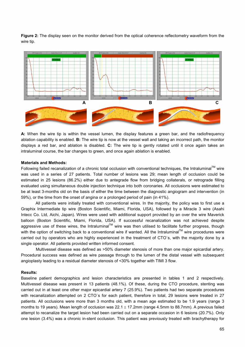
65
Figure 2: The display seen on the monitor derived from the optical coherence reflectometry waveform from thewire tip.
A: When the wire tip is within the vessel lumen, the display features a green bar, and the radiofrequencyablation capability is enabled. B: The wire tip is now at the vessel wall and taking an incorrect path, the monitordisplays a red bar, and ablation is disabled. C: The wire tip is gently rotated until it once again takes anintraluminal course, the bar changes to green, and once again ablation is enabled.
Materials and Methods:Following failed recanalization of a chronic total occlusion with conventional techniques, the IntraluminalTM wirewas used in a series of 27 patients. Total number of lesions was 29; mean length of occlusion could beestimated in 25 lesions (86.2%) either due to antegrade flow from bridging collaterals, or retrograde fillingevaluated using simultaneous double injection technique into both coronaries. All occlusions were estimated tobe at least 3-months old on the basis of either the time between the diagnostic angiogram and intervention (in59%), or the time from the onset of angina or a prolonged period of pain (in 41%).
All patients were initially treated with conventional wires. In the majority, the policy was to first use aGraphix Intermediate tip wire (Boston Scientific, Miami, Florida, USA), followed by a Miracle 3 wire (AsahiIntecc Co. Ltd, Aichi, Japan). Wires were used with additional support provided by an over the wire Maverickballoon (Boston Scientific, Miami, Florida, USA). If successful recanalization was not achieved despiteaggressive use of these wires, the IntraluminalTM wire was then utilised to facilitate further progress, thoughwith the option of switching back to a conventional wire if wanted. All the IntraluminalTM wire procedures werecarried out by operators who are highly experienced in the treatment of CTO`s, with the majority done by asingle operator. All patients provided written informed consent.
Multivessel disease was defined as >50% diameter stenosis of more than one major epicardial artery.Procedural success was defined as wire passage through to the lumen of the distal vessel with subsequentangioplasty leading to a residual diameter stenosis of <30% together with TIMI 3 flow.
Results:Baseline patient demographics and lesion characteristics are presented in tables 1 and 2 respectively.Multivessel disease was present in 13 patients (48.1%). Of these, during the CTO procedure, stenting wascarried out in at least one other major epicardial artery 7 (25.9%). Two patients had two separate procedureswith recanalization attempted on 2 CTO`s for each patient, therefore in total, 29 lesions were treated in 27patients. All occlusions were more than 3 months old, with a mean age estimated to be 1.9 years (range 3months to 19 years). Mean length of occlusion was 22.1 ± 17.2mm (range 4.5mm to 88.7mm). A previous failedattempt to recanalize the target lesion had been carried out on a separate occasion in 6 lesions (20.7%). Onlyone lesion (3.4%) was a chronic in-stent occlusion. This patient was previously treated with brachytherapy for
A B C
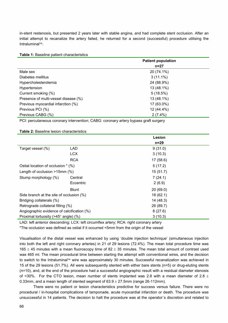
66
in-stent restenosis, but presented 2 years later with stable angina, and had complete stent occlusion. After aninitial attempt to recanalize the artery failed, he returned for a second (successful) procedure utilising theIntraluminal.
Table 1: Baseline patient characteristicsPatient population
n=27Male sex 20 (74.1%)Diabetes mellitus 3 (11.1%)Hypercholesterolemia 24 (88.9%)Hypertension 13 (48.1%)Current smoking (%) 5 (18.5%)Presence of multi-vessel disease (%) 13 (48.1%)Previous myocardial infarction (%) 17 (63.0%)Previous PCI (%) 12 (44.4%)Previous CABG (%) 2 (7.4%)PCI: percutaneous coronary intervention; CABG: coronary artery bypass graft surgery
Table 2: Baseline lesion characteristicsLesionn=29
LAD 9 (31.0)LCX 3 (10.3)
Target vessel (%)
RCA 17 (58.6)Ostial location of occlusion * (%) 5 (17.2)Length of occlusion >15mm (%) 15 (51.7)
Central 7 (24.1)Eccentric 2 (6.9)
Stump morphology (%)
Blunt 20 (69.0)Side branch at the site of occlusion (%) 18 (62.1)Bridging collaterals (%) 14 (48.3)Retrograde collateral filling (%) 26 (89.7)Angiographic evidence of calcification (%) 8 (27.6)Proximal tortuosity (>45° angle) (%) 3 (10.3)LAD: left anterior descending; LCX: left circumflex artery; RCA: right coronary artery*The occlusion was defined as ostial if it occurred <5mm from the origin of the vessel
Visualisation of the distal vessel was enhanced by using ‘double injection technique’ (simultaneous injectioninto both the left and right coronary arteries) in 21 of 29 lesions (72.4%). The mean total procedure time was165 ± 45 minutes with a mean fluoroscopy time of 82 ± 35 minutes. The mean total amount of contrast usedwas 465 ml. The mean procedural time between starting the attempt with conventional wires, and the decisionto switch to the Intraluminal wire was approximately 30 minutes. Successful recanalization was achieved in15 of the 29 lesions (51.7%). All were subsequently stented with either bare stents (n=5) or drug-eluting stents(n=10), and, at the end of the procedure had a successful angiographic result with a residual diameter stenosisof <30%. For the CTO lesion, mean number of stents implanted was 2.8 with a mean diameter of 2.8 ±0.33mm, and a mean length of stented segment of 63.9 ± 27.5mm (range 26-112mm).
There were no patient or lesion characteristics predictive for success versus failure. There were noprocedural / in-hospital complications of tamponade, acute myocardial infarction or death. The procedure wasunsuccessful in 14 patients. The decision to halt the procedure was at the operator`s discretion and related to

67
failure to cross the lesion in 8 (57.1%), and the occurence of dissection in the remaining 6 (42.9%). All episodesof dissection occurred whilst a conventional wire (rather than the Intraluminal wire) was in use, and all werewithout clinical sequelae. Of these 14 patients with unsuccessful recanalization, 3 (21.4%) were referred forcoronary artery bypass graft surgery, and the remaining 11 (78.6%) were managed with medical therapy.
Discussion:Previous studies have demonstrated the importance of the revascularization of chronic total occlusions withsignificant benefits on quality of life with improvement in both anginal symptoms and exercise capacity. 7 Inaddition, successful recanalization of a chronically occluded artery reduces the need for subsequent bypasssurgery and the rate of myocardial infarction. 8 There is also evidence that, provided the artery remains patent,there is an improvement in left ventricular function. 9 Importantly, long-term evaluation has shown a 10-yearsurvival advantage following successful PCI compared to those in whom PCI is unsuccessful (survival 73.5%versus 65.1%, p=0.001). 10
The inability to cross the lesion is the primary reason why a percutaneous strategy for revascularizationis unsuccessful. Results of other guidewire technologies specifically designed for treating CTO`s, including thehydrophilic coated wire, the ball-tipped wire, and laser wire, have been disappointing with success rates of 45-60%.3,11,12 Failure generally relates to either perforation with an extraluminal passage of the wire, or failure ofthe wire to advance forwards due to dense fibrous tissue or heavy calcification within the occlusion. Withregards to the laser wire, the TOTAL surveillance study 13 suggested that because of its ablation capacity, therewas an advantage conferred by the wire in improving successful recanalization rates. Following a failed attemptutilising a mechanical guidewire, 182 patients were treated with the laser wire, with success achieved in 105patients (58%). However, perforation occurred in 21%, with tamponade in 1% cases. The subsequent TOTALtrial 3 randomized patients to either the laser wire or “conventional” mechanical wires, with the possibility ofcrossover to the other modality for a second attempt, should the first fail. In all, 303 patients were treated, withno significant difference in overall success rates between the 2 groups. Failure with the laser wire was primarilyrelated to it taking a false route, whereas failure with mechanical wires was felt to be mainly due to absence ofwire progression. The Safe-Cross system provides the operator with the potential to overcome both of theselimitations. The technology of optical coherence reflectometry provides the means to precisely navigate theguidewire forward thereby minimising the risk of perforation, and the capability of radiofrequency ablationincreases the crossing potential of the wire. In-vitro studies have confirmed that the system is able to accuratelydifferentiate plaque from the media / adventitia boundary, 14 and in vivo evaluation has shown efficacy of theablation capacity even when used in heavily calcified vessels. 5 In the current study, failure of successfulrecanalization occurred in 48.3% lesions, related to dissection in six (42.9%). However, all these episodesoccurred whilst a conventional wire was in use and not the Intraluminal wire.
One of the most important drawbacks of percutaneous revascularization of a CTO is that the prolongedprocedural time is associated with increased radiation exposure to both the patient and operator. Excessiveradiation dosage can cause skin injury ranging from a temporary erythema, to deep ulceration and permanentscarring. 15 One further advantage of the Safe-Cross system is that the wire has the potential to be manipulatedwith guidance from the monitor display, without the need for continuous fluoroscopy. The fluoroscopy times inthe current study (82 minutes) are slightly lower than those seen during the pilot study of the laser guidewire(99 minutes). 16 Importantly, there were no complications utilizing this system, in particular no episodes oftamponade, and this now provides reassurance of the ability to use the system with less fluoroscopy in thefuture. In addition to this, although the mean amount of contrast used in the present study (465ml) iscomparable to that of the laser wire pilot study 16 (515ml), less fluoroscopy should correlate with a lower amountof contrast utilization.
Our population was a difficult one with all occlusions being older than 3 months duration (a typical caseexample is presented in figure 3). Other than age, other factors suggested to be predictive for unsuccessfulpercutaneous therapy include multivessel disease, a blunt stump, the presence of a side branch at the site ofocclusion, calcification, bridging collaterals, and lesion length >15mm. 2,8,17 The overall mean length ofocclusion in our patients was long at 22.1mm, and following successful recanalization, a relatively long total

68
length of stents (mean 63.9mm) was required to ensure coverage of the entire lesion. All the lesions treatedhad at least one of these additional characteristics, with the majority (69.0%) having at least 3 additionaladverse characteristics. With such a high prevalence of these characteristics in our group of patients, therewere no patient or lesion characteristics that appeared to be predictive for successful recanalization.
Figure 3: Case example
A 74-year old man with two previous coronary bypass surgery operations in 1983 and 1997. A) Left circumflexartery occlusion estimated to be at least 5 years old, 12mm in length, and with a blunt stump. B) Progressmade with a Miracle wire (Asahi Intecc Co. Ltd, Aichi, Japan) but unable to fully cross the lesion. C) Successfulrecanalization utilising the Intraluminal™ wire, the final 10mm of the wire is opaque and can be seen within thelumen of the distal vessel (arrow). D) Final results following pre-dilatation and stenting with a 2.5x33mm Cypherstent (Cordis, Johnson and Johnson).
The Safe-Cross system is easy to use, with a fairly short learning curve. The main difference compared toconventional wires is that wire manipulation / torquing needs to be done relatively slowly in order to give thesystem time to interpret the optical coherence reflectometry signal. In addition, as the wire has a blunt tip (andtherefore some potential for perforation), following successful recanalization, care must be taken to ensure safepositioning in the distal vessel. Once a lesion is crossed, the wire can be disconnected from the console and anouter sheath removed to maintain sterility; the wire can then be used like any other 0.014” wire.
In our patients in whom attempted recanalization with conventional wires had failed, the Safe-Crosssystem improved the rate of successful CTO recanalization with a further 51.7% vessels successfully opened.The major limitation at present is some difficulty in steering the wire, but it is hoped that this will improve withthe next generation of IntraluminalTM Guidewires, which have a tip that can be pre-shaped. Although this studyis limited by the fact that it is non-randomized and carried out in a single center on a small number of selectedpatients, our preliminary data suggests that this system is a useful adjunctive device in the therapy of chronictotal occlusions. Further evaluation in the context of a large randomized study is warranted to assess theimpact of this new device in the treatment of this technically challenging patient population.
A
C D
B

69
References:1. Kahn JK. Angiographic suitability for catheter revascularization of total coronary occlusions in patients from a community hospital
setting. Am Heart J. 1993;126:561-4.
2. Puma JA, Sketch MH, Jr., Tcheng JE, Harrington RA, Phillips HR, Stack RS, Califf RM. Percutaneous revascularization of
chronic coronary occlusions: an overview. J Am Coll Cardiol. 1995;26:1-11.
3. Serruys PW, Hamburger JN, Koolen JJ, Fajadet J, Haude M, Klues H, Seabra-Gomes R, Corcos T, Hamm C, Pizzuli L, Meier B,
Mathey D, Fleck E, Taeymans Y, Melkert R, Teunissen Y, Simon R. Total occlusion trial with angioplasty by using laser
guidewire. The TOTAL trial. Eur Heart J. 2000;21:1797-805.
4. Cordero H, Warburton KD, Underwood PL, Heuser RR. Initial experience and safety in the treatment of chronic total occlusions
with fiberoptic guidance technology: optical coherent reflectometry. Catheter Cardiovasc Interv. 2001;54:180-7.
5. Neet J, Winston T, Siglinger M, Janssen M, Balaster A. Optical coherence reflectometry (OCR) guided RF ablation guide wire for
total occlusions. SPIE. 2001;4244:419-427.
. Chen WH, Ng W, Lee PY, Lau CP. Recanalization of chronic and long occlusive in-stent restenosis using optical coherence
reflectometry-guided radiofrequency ablation guidewire. Catheter Cardiovasc Interv. 2003;59:223-9.
7. Finci L, Meier B, Favre J, Righetti A, Rutishauser W. Long-term results of successful and failed angioplasty for chronic total
coronary arterial occlusion. Am J Cardiol. 1990;66:660-2.
8. Ivanhoe RJ, Weintraub WS, Douglas JS, Jr., Lembo NJ, Furman M, Gershony G, Cohen CL, King SB, 3rd. Percutaneous
transluminal coronary angioplasty of chronic total occlusions. Primary success, restenosis, and long-term clinical follow-up.
Circulation. 1992;85:106-15.
9. Rambaldi R, Hamburger JN, Geleijnse ML, Poldermans D, Kimman GJ, Aiazian AA, Fioretti PM, Ten Cate FJ, Roelandt JR,
Serruys PW. Early recovery of wall motion abnormalities after recanalization of chronic totally occluded coronary arteries: a
dobutamine echocardiographic, prospective, single-center experience. Am Heart J. 1998;136:831-6.
10. Suero JA, Marso SP, Jones PG, Laster SB, Huber KC, Giorgi LV, Johnson WL, Rutherford BD. Procedural outcomes and long-
term survival among patients undergoing percutaneous coronary intervention of a chronic total occlusion in native coronary
arteries: a 20-year experience. J Am Coll Cardiol. 2001;38:409-14.
11. Meier B, Carlier M, Finci L, Nukta E, Urban P, Niederhauser W, Favre J. Magnum wire for balloon recanalization of chronic total
coronary occlusions. Am J Cardiol. 1989;64:148-54.
12. Freed M, Boatman JE, Siegel N, Safian RD, Grines CL, O'Neill WW. Glidewire treatment of resistant coronary occlusions. Cathet
Cardiovasc Diagn. 1993;30:201-4.
13. Hamburger JN, Serruys PW, Scabra-Gomes R, Simon R, Koolen JJ, Fleck E, Mathey D, Sievert H, Rutsch W, Buchwald A,
Marco J, Al-Kasab SM, Pizulli L, Hamm C, Corcos T, Reifart N, Hanrath P, Taeymans Y. Recanalization of total coronary
occlusions using a laser guidewire (the European TOTAL Surveillance Study). Am J Cardiol. 1997;80:1419-23.
14. Yamashita T, Kasaoka S, Son R, Gordon IL, Khan R, Neet J, Hedrick AD, Tobis JM. Optical coherent reflectometry: a new
technique to guide invasive procedures. Catheter Cardiovasc Interv. 2001;54:257-63.
15. den Boer A, de Feijter PJ, Serruys PW, Roelandt JR. Real-time quantification and display of skin radiation during coronary
angiography and intervention. Circulation. 2001;104:1779-84.
16. Hamburger JN, Serruys PW, Scabra-Gomes R, Simon R, Koolen JJ, Fleck E, Mathey D, Sievert H, Rutsch W, Buchwald A,
Marco J, Al-Kasab SM, Pizulli L, Hamm C, Corcos T, Reifart N, Hanrath P, Taeymans Y. Recanalization of total coronary
occlusions using a laser guidewire (the European TOTAL Surveillance Study). Am J Cardiol. 1997;80:1419-23.
17. Maiello L, Colombo A, Gianrossi R, Mutinelli MR, Bouzon R, Thomas J, Finci L. Coronary angioplasty of chronic occlusions:
factors predictive of procedural success. Am Heart J. 1992;124:581-4.

70

71
Chapter 7
Angela HoyePedro A LemosPatrick W Serruys
Journal of InvasiveCardiology2005;17(5):277-9
Successful Use of a New Guidewire withRadiofrequency Ablation Capability for the
Treatment of Chronic Total Occlusion at theOstium of the Left Anterior Descending
Artery

72
73
Summary
The major limitation of percutaneous therapy (PCI) for the treatment of chronic total
occlusions (CTOs) is the inability to cross with a wire. We report successful recanalization of
a CTO situated at the ostium of the left anterior descending artery. The lesion demonstrated
several anatomical features known to be associated with an unsuccessful outcome, and
attempts with conventional wires failed. However, recanalization was facilitated with the
Intraluminal wire™, (Intraluminal Therapeutics Inc, Carlsbad, California) a novel technology
that combines guidance of the wire tip with the capability of radiofrequency ablation.
The majority of patients with CTOs are currently managed medically or referred for
coronary bypass surgery. However, drug-eluting stent (DES) implantation for the treatment
of CTOs have been shown to reduce the subsequent rate of restenosis compared with bare
metal stents. If rates of successful recanalization can be increased with new technologies
such as demonstrated here, then the advent of DESs will lead to more widespread
applicability of PCI for this complex group.

74
Case Report: A 58-year old woman was referred with a 2-year history of chest pain. Risk factors for coronarydisease were smoking, hypertension, and a positive family history. Though the resting ECG was normal, anexercise test demonstrated ST-depression across the anterior leads during stage 2 of a Bruce protocol.Subsequent coronary angiography showed single vessel disease of the left anterior descending artery (LAD)which was completely occluded at the ostium, with a blunt stump (figure 1). Notably, there was clearly identifiedheavy calcification, and a ring of calcium can be seen in figure 1B (arrow). There was some antegrade filling viasmall bridging collaterals originating from the proximal part of the left circumflex artery (figure 1A). The distalvessel was also filled via retrograde collaterals from the right coronary artery (figure 1B). Left ventricularfunction was normal with no anterior wall movement abnormality.
Figure 1A: Diagnostic angiography of the left coronary artery demonstrating complete occlusion at the ostiumof the left anterior descending artery (LAD). The distal vessel is filled via small bridging collaterals originatingfrom the proximal left circumflex artery (arrow). B: Diagnostic angiography of the right coronary artery whichsupplies retrograde collateral filling of the LAD. A ring of heavy calcification at the occlusion site is readilyapparent (arrow).
The patient was taking long-term aspirin, and before the procedure was pre-loaded with 300mg clopidogrel.Heparin was administered to maintain the activated clotting time >250 seconds. A 7F left Amplatz 2 guidingcatheter was used to catheterize the left main coronary artery, and the procedure was carried out utilisingbiplane X-ray screening. By quantitative coronary angiography, the estimated length of the occlusion was 13.6mm. Initial attempts to open the LAD were made using an Intermediate tip 0.014” guidewire (Boston Scientific),and then a 0.014” Miracle 3g wire (Asahi Intecc Co. Ltd, Aichi, Japan) with over-the-wire balloon support.However, both these wires prolapsed into the circumflex artery with no antegrade progress into the LAD (figure2B). The Intraluminal wire was then advanced to the stump and radiofrequency ablation applied which wasable to penetrate the proximal cap of the occlusion (figure 2C). The Intraluminal wire™ was substituted for theMiracle wire, with successful wire passage into the distal vessel lumen (figure 2D). Following pre-dilatation, a3.0x18mm sirolimus-eluting stent (Cypher stent, Cordis Corporation, Miami, Florida) was implanted (figure 2E)and subsequently post-dilated with a 3.5mm balloon; the final result was excellent (Figure 2F). At 6-monthsfollow-up the patient was symptom-free and underwent a repeat exercise test. On this occasion she managed10 minutes of a Bruce protocol (maximum predicted heart rate attained) without chest discomfort or ECGchanges.
A B

75
Figure 2A: Baseline angiography of the occlusion at the time of angioplasty. B: Conventional wires with over-the-wire balloon support failed to penetrate the proximal cap of the occlusion and simply prolapsed into the leftcircumflex artery. C: The tip of the Intraluminal wire™ can be seen penetrating through the cap (arrow). D:Successful crossing of the occlusion into the lumen of the distal vessel with a Miracle 3g wire. E: Stentimplantation with a 3.0x18mm sirolimus-eluting stent. F: Final result.
Discussion:Previous studies have demonstrated the importance of CTO revascularization, with improvement in anginalsymptoms, exercise capacity, and left ventricular function. 1-3 In addition, successful recanalization reduces theneed for subsequent coronary artery bypass surgery, 4 and long-term evaluation has shown a 10-year survivaladvantage of 73.5% compared to 65.1% in those in whom PCI is unsuccessful. 5
The Safecross system with the Intraluminal guidewire™ (Intraluminal Therapeutics, Carlsbad,California) uses optical coherence reflectometry to determine the position of the wire tip. The system has beenpreviously described, 6-9 briefly, a beam of near-infra red light is emitted and the reflected beam analysed todifferentiate the vessel lumen from the outside vessel wall. The technology therefore provides information forguidance of the wire, to reduce the risk of taking an extra-luminal passage potentially leading to perforation andpericardial tamponade. In addition, the wire has the capability of radiofrequency ablation at the tip, emittingshort bursts (100ms) of low frequency 250-500 kHz energy. This facilitates forward passage of the wire withefficacy demonstrable even in calcified vessels. 7 The system combines these 2 capabilities such that ablationis only enabled when the wire tip is heading correctly. In the present case, the Intraluminal was of great value inpenetration of the proximal cap of the occlusion which, particularly when the stump appears blunt, can becomposed of very dense fibrotic material. The cap was unable to be breached by conventional wires, yet oncea few millimetres of antegrade passage had been made with the Intraluminal wire, the remainder of theocclusion could be traversed.
Importantly, the distal LAD was filled via collaterals thereby facilitating wire guidance, and enablingreassurance of successful recanalization with visualization of the wire within the distal lumen. However, theIntraluminal system uses optical coherence reflectometry to determine the position of the distal tip of the wire inrelation to the vessel wall. This does not therefore rely on angiographic visualization, and with more experiencein the future, it may be possible to use the system even when the distal vessel is not well seen.
When treating CTO’s, whatever the technology used, the principles of good backup support remainimportant, and in this case a left Amplatz guide catheter proved effective. In addition, to facilitate the exchangeof wires and to provide additional backup, we routinely utilize a 1.5mm over-the-wire balloon, which can then beused for pre-dilatation once the occlusion has been successfully crossed with a wire.
A C
E
B
D F

76
Certain lesion characteristics have been shown to affect the success rate of recanalization. Some ofthe most important adverse features include increased age and length of the occlusion, the presence ofbridging collaterals, a side branch at the site of occlusion, calcification, and an abrupt rather than taperedstump. 10 Our patients’ clinical history suggested that the occlusion was 2-years old, it was ostial in location withthe large circumflex artery originating at the occlusion, was heavily calcified and the stump abrupt (figures 1and 2). All these features suggested that the chances of successful recanalization would be low, and mighthave lead many cardiologists to refer similar patients directly for bypass surgery. Indeed, in the BARI study,clinicians provided their views on the suitability of lesions for a revascularization strategy of PCI versus CABG,and both ostial lesion location and a CTO were deemed to be strong non-favourable characteristics for PCI. 11
Percutaneous treatment of ostial LAD lesions could potentially jeopardize the circumflex artery,however in the present case, the use of biplane imaging facilitated precise stent positioning, which, togetherwith a relatively large angle of the left main stem carina meant that the final result was excellent. Long-termresults of PCI with bare metal stents have been hindered by the development of restenosis, with rates of non-occlusive ostial LAD lesions as high as 26%. 12 Furthermore, CTOs are well-known to be at increased risk ofrestenosis, with rates of 32-55%. 13-16 However, recent data have confirmed the efficacy of sirolimus-elutingstents for CTOs, with a 1 year survival-free of major adverse events of 96%, and binary restenosis rate of 9%.17
Conclusions:This case demonstrates the potential of this novel technology, in particular at successfully penetrating theproximal cap of a CTO following failed attempts using conventional guidewires. Future improvements in suchspecialized technologies will improve the ability to achieve successful recanalization of CTOs. This, togetherwith the use of drug-eluting stents, means that the use of PCI, rather than CABG, will be more widely applicablefor the revascularization of patients with complex CTOs.
77
References1. Finci L, Meier B, Favre J, Righetti A, Rutishauser W. Long-term results of successful and failed angioplasty for chronic total coronary
arterial occlusion. Am J Cardiol 1990;66:660-662.
2. Rambaldi R, Hamburger JN, Geleijnse ML, Poldermans D, Kimman GJ, Aiazian AA, Fioretti PM, Ten Cate FJ, Roelandt JR, Serruys PW.
Early recovery of wall motion abnormalities after recanalization of chronic totally occluded coronary arteries: a dobutamine
echocardiographic, prospective, single-center experience. Am Heart J 1998;136:831-836.
3. Melchior JP, Doriot PA, Chatelain P, Meier B, Urban P, Finci L, Rutishauser W. Improvement of left ventricular contraction and relaxation
synchronism after recanalization of chronic total coronary occlusion by angioplasty. J Am Coll Cardiol 1987;9:763-768.
4. Ivanhoe RJ, Weintraub WS, Douglas JS, Jr., Lembo NJ, Furman M, Gershony G, Cohen CL, King SB, 3rd. Percutaneous transluminal
coronary angioplasty of chronic total occlusions. Primary success, restenosis, and long-term clinical follow-up. Circulation 1992;85:106-115.
5. Suero JA, Marso SP, Jones PG, Laster SB, Huber KC, Giorgi LV, Johnson WL, Rutherford BD. Procedural outcomes and long-term
survival among patients undergoing percutaneous coronary intervention of a chronic total occlusion in native coronary arteries: a 20-year
experience. J Am Coll Cardiol 2001;38:409-414.
6. Cordero H, Warburton KD, Underwood PL, Heuser RR. Initial experience and safety in the treatment of chronic total occlusions with
fiberoptic guidance technology: optical coherent reflectometry. Catheter Cardiovasc Interv 2001;54:180-187.
7. Neet J, Winston T, Siglinger M, Janssen M, Balaster A. Optical coherence reflectometry (OCR) guided RF ablation guide wire for total
occlusions. SPIE 2001;4244:419-427.
8. Chen WH, Ng W, Lee PY, Lau CP. Recanalization of chronic and long occlusive in-stent restenosis using optical coherence
reflectometry-guided radiofrequency ablation guidewire. Catheter Cardiovasc Interv 2003;59:223-229.
9. Hoye A, Onderwater E, Cummins P, Sianos G, Serruys PW. Improved Recanalization of Chronic Total Coronary Occlusions Using an
Optical Coherence Reflectometry-Guided Guidewire. Cathet Cardiovasc Interv. 2004;63(2):158-63
10. Puma JA, Sketch MH, Jr., Tcheng JE, Harrington RA, Phillips HR, Stack RS, Califf RM. Percutaneous revascularization of chronic
coronary occlusions: an overview. J Am Coll Cardiol 1995;26:1-11.
11. Botas J, Stadius ML, Bourassa MG, Rosen AD, Schaff HV, Sopko G, Williams DO, McMilliam A, Alderman EL. Angiographic correlates
of lesion relevance and suitability for percutaneous transluminal coronary angioplasty and coronary artery bypass grafting in the Bypass
Angioplasty Revascularization Investigation study (BARI). Am J Cardiol 1996;77:805-814.
12. Park SJ, Lee CW, Hong MK, Kim JJ, Park SW. Stent placement for ostial left anterior descending coronary artery stenosis: acute and
long-term (2-year) results. Catheter Cardiovasc Interv 2000;49:267-271.
13. Buller CE, Dzavik V, Carere RG, Mancini GB, Barbeau G, Lazzam C, Anderson TJ, Knudtson ML, Marquis JF, Suzuki T, Cohen EA,
Fox RS, Teo KK. Primary stenting versus balloon angioplasty in occluded coronary arteries: the Total Occlusion Study of Canada
(TOSCA). Circulation. 1999;100:236-42.
14. Rubartelli P, Niccoli L, Verna E, Giachero C, Zimarino M, Fontanelli A, Vassanelli C, Campolo L, Martuscelli E, Tommasini G. Stent
implantation versus balloon angioplasty in chronic coronary occlusions: results from the GISSOC trial. Gruppo Italiano di Studio sullo Stent
nelle Occlusioni Coronariche. J Am Coll Cardiol. 1998;32:90-6.
15. Lotan C, Rozenman Y, Hendler A, Turgeman Y, Ayzenberg O, Beyar R, Krakover R, Rosenfeld T, Gotsman MS. Stents in total
occlusion for restenosis prevention. The multicentre randomized STOP study. The Israeli Working Group for Interventional Cardiology. Eur
Heart J. 2000;21:1960-6.
16. Sirnes PA, Golf S, Myreng Y, Molstad P, Emanuelsson H, Albertsson P, Brekke M, Mangschau A, Endresen K, Kjekshus J. Stenting in
Chronic Coronary Occlusion (SICCO): a randomized, controlled trial of adding stent implantation after successful angioplasty. J Am Coll
Cardiol. 1996;28:1444-51.
17. Hoye A, Tanabe K, Lemos PA, Aoki J, Saia F, Arampatzis C, Degertekin M, Hofma SH, Sianos G, McFadden E, Van Der Giessen WJ,
Smits PC, De Feyter PJ, Van Domburg RT, Serruys PW. Significant reduction in restenosis after the use of sirolimus-eluting stents in the
treatment of chronic total occlusions. J Am Coll Cardiol. 2004;43:1954-8.

78
79
Chapter 8
Jose Ruiz-CantadorAngela HoyeEugene McFadden
Journal of InvasiveCardiology2005;17(10):E7-E10
Multimodality Plaque Ablation to AllowSuccessful Stent Implantation Following
Failure of Conventional Wires and Balloonsto Cross a Chronic Total Occlusion

80

81
Abstract:
The present report illustrates several potential difficulties that may arise when treating a
chronic total occlusion, one of the most challenging coronary lesions to be faced in the
catheterization laboratory. We describe how we overcame each of these problems, including
the utilization of specialized technologies with the Intraluminal wireTM and rotablator.

82
IntroductionPercutaneous coronary intervention (PCI) for chronic total occlusion (CTO) has procedural and ultimate longterm success rates that are significantly less than those currently reported for non-occlusive lesions. A CTO isgenerally defined as an occlusion with Thrombolysis In Myocardial Infarction (TIMI) grade 0 or 1 antegradeflow, that is more than 3 months old. On angiography, CTO lesions occur in approximately one third of patientswith significant coronary lesions, but PCI for CTO only accounts for around 10% of patients undergoing PCI. 1
Successful recanalization of CTO with PCI reduces the need to resort to bypass surgery; furthermore,observational studies have shown lower cumulative rates of cardiac death or myocardial infarction and animprovement in symptomatic status after successful PCI for CTO. 2-5
The lower procedural success and higher restenosis rates in the era of balloon angioplasty have improved inrecent years. However, inability to cross the lesion remains the major cause of procedural failure. 6-8
Improvements in guidewire technology and novel approaches such as the optical coherence reflectometry-guided radiofrequency ablation (IntraluminalTM guidewire, Intraluminal Therapeutics, Carlsbad, California), theFrontrunner catheter (LuMend, Redwood City, California), and other technological advances have improved theability to cross the lesion. 9-13 The second major cause of procedural failure is the inability to cross or to dilatethe lesion with a balloon. In this situation, rotational atherectomy or an excimer laser may lead to a successfuloutcome. 14,15 Finally, the long term success rate of PCI in general has been improved by the use of stentimplantation. 16,17 Though experience with CTO lesions is currently limited, the introduction of drug-elutingstents, in particular, has reduced the subsequent rate of restenosis. 18-21 This case report illustrates many of thetechnical problems encountered in treating CTOs and shows how they were successfully overcome with bothclassic and novel technology.
Case ReportA 51-year-old man with a six-month history of Canadian Class 3 stable angina was referred for percutaneousrevascularization. Risk factors for atherosclerosis were non-insulin-dependent diabetes mellitus,hypercholesterolemia, previous smoking, and a positive family history of premature coronary artery disease.Coronary angiography performed at the referring center, showed a chronic total occlusion of the mid-rightcoronary artery. The left coronary artery had no significant stenosis and the left ventricular function was normal.Based on the history, the duration of the occlusion was estimated to be around 6 months. Medical therapyconsisted of aspirin, statin, beta-blocker, and clopidogrel, with tight glycemic control maintained through oralhypoglycemic agents.
Figure 1: Baseline angiography of the right coronary artery, right anterior oblique view, with a CTO in segment2. The stump is blunt in morphology (block arrow), there is a side branch at the level of the occlusion (openarrow), and bridging collaterals are filling the distal vessel.
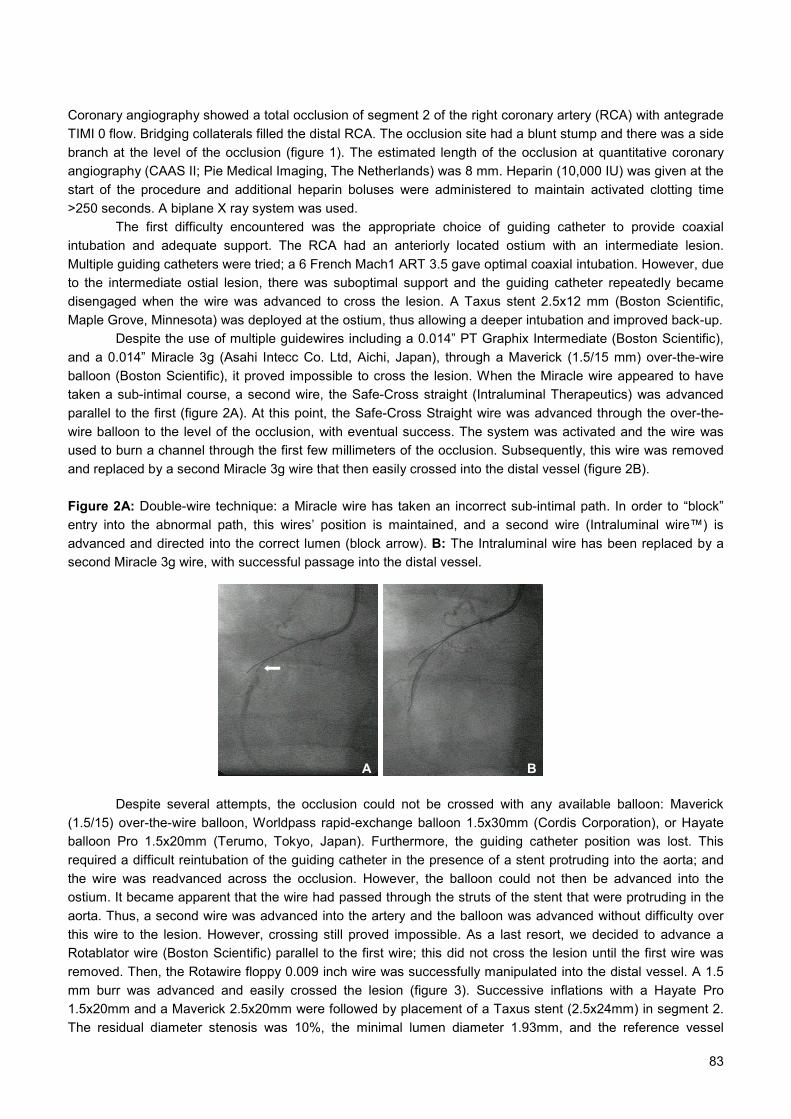
83
Coronary angiography showed a total occlusion of segment 2 of the right coronary artery (RCA) with antegradeTIMI 0 flow. Bridging collaterals filled the distal RCA. The occlusion site had a blunt stump and there was a sidebranch at the level of the occlusion (figure 1). The estimated length of the occlusion at quantitative coronaryangiography (CAAS II; Pie Medical Imaging, The Netherlands) was 8 mm. Heparin (10,000 IU) was given at thestart of the procedure and additional heparin boluses were administered to maintain activated clotting time>250 seconds. A biplane X ray system was used.
The first difficulty encountered was the appropriate choice of guiding catheter to provide coaxialintubation and adequate support. The RCA had an anteriorly located ostium with an intermediate lesion.Multiple guiding catheters were tried; a 6 French Mach1 ART 3.5 gave optimal coaxial intubation. However, dueto the intermediate ostial lesion, there was suboptimal support and the guiding catheter repeatedly becamedisengaged when the wire was advanced to cross the lesion. A Taxus stent 2.5x12 mm (Boston Scientific,Maple Grove, Minnesota) was deployed at the ostium, thus allowing a deeper intubation and improved back-up.
Despite the use of multiple guidewires including a 0.014” PT Graphix Intermediate (Boston Scientific),and a 0.014” Miracle 3g (Asahi Intecc Co. Ltd, Aichi, Japan), through a Maverick (1.5/15 mm) over-the-wireballoon (Boston Scientific), it proved impossible to cross the lesion. When the Miracle wire appeared to havetaken a sub-intimal course, a second wire, the Safe-Cross straight (Intraluminal Therapeutics) was advancedparallel to the first (figure 2A). At this point, the Safe-Cross Straight wire was advanced through the over-the-wire balloon to the level of the occlusion, with eventual success. The system was activated and the wire wasused to burn a channel through the first few millimeters of the occlusion. Subsequently, this wire was removedand replaced by a second Miracle 3g wire that then easily crossed into the distal vessel (figure 2B).
Figure 2A: Double-wire technique: a Miracle wire has taken an incorrect sub-intimal path. In order to “block”entry into the abnormal path, this wires’ position is maintained, and a second wire (Intraluminal wire™) isadvanced and directed into the correct lumen (block arrow). B: The Intraluminal wire has been replaced by asecond Miracle 3g wire, with successful passage into the distal vessel.
Despite several attempts, the occlusion could not be crossed with any available balloon: Maverick(1.5/15) over-the-wire balloon, Worldpass rapid-exchange balloon 1.5x30mm (Cordis Corporation), or Hayateballoon Pro 1.5x20mm (Terumo, Tokyo, Japan). Furthermore, the guiding catheter position was lost. Thisrequired a difficult reintubation of the guiding catheter in the presence of a stent protruding into the aorta; andthe wire was readvanced across the occlusion. However, the balloon could not then be advanced into theostium. It became apparent that the wire had passed through the struts of the stent that were protruding in theaorta. Thus, a second wire was advanced into the artery and the balloon was advanced without difficulty overthis wire to the lesion. However, crossing still proved impossible. As a last resort, we decided to advance aRotablator wire (Boston Scientific) parallel to the first wire; this did not cross the lesion until the first wire wasremoved. Then, the Rotawire floppy 0.009 inch wire was successfully manipulated into the distal vessel. A 1.5mm burr was advanced and easily crossed the lesion (figure 3). Successive inflations with a Hayate Pro1.5x20mm and a Maverick 2.5x20mm were followed by placement of a Taxus stent (2.5x24mm) in segment 2.The residual diameter stenosis was 10%, the minimal lumen diameter 1.93mm, and the reference vessel
A B
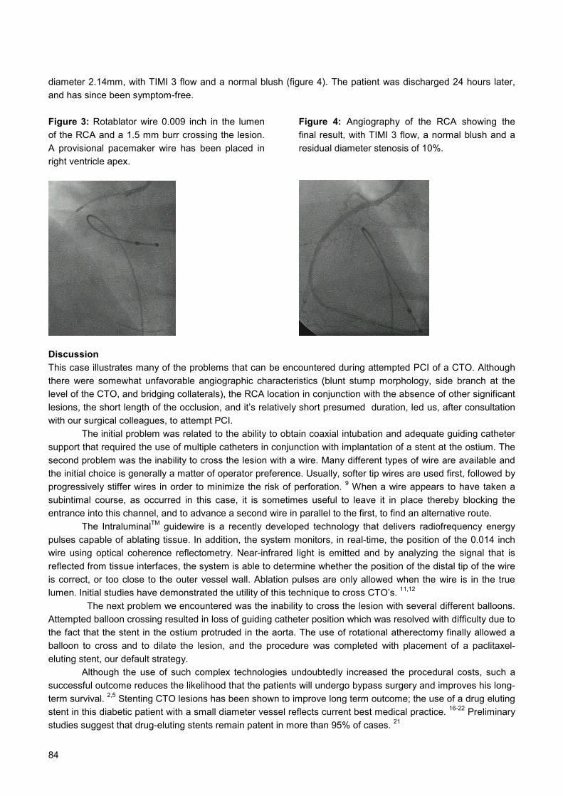
84
diameter 2.14mm, with TIMI 3 flow and a normal blush (figure 4). The patient was discharged 24 hours later,and has since been symptom-free.
Figure 3: Rotablator wire 0.009 inch in the lumenof the RCA and a 1.5 mm burr crossing the lesion.A provisional pacemaker wire has been placed inright ventricle apex.
Figure 4: Angiography of the RCA showing thefinal result, with TIMI 3 flow, a normal blush and aresidual diameter stenosis of 10%.
DiscussionThis case illustrates many of the problems that can be encountered during attempted PCI of a CTO. Althoughthere were somewhat unfavorable angiographic characteristics (blunt stump morphology, side branch at thelevel of the CTO, and bridging collaterals), the RCA location in conjunction with the absence of other significantlesions, the short length of the occlusion, and it’s relatively short presumed duration, led us, after consultationwith our surgical colleagues, to attempt PCI.
The initial problem was related to the ability to obtain coaxial intubation and adequate guiding cathetersupport that required the use of multiple catheters in conjunction with implantation of a stent at the ostium. Thesecond problem was the inability to cross the lesion with a wire. Many different types of wire are available andthe initial choice is generally a matter of operator preference. Usually, softer tip wires are used first, followed byprogressively stiffer wires in order to minimize the risk of perforation. 9 When a wire appears to have taken asubintimal course, as occurred in this case, it is sometimes useful to leave it in place thereby blocking theentrance into this channel, and to advance a second wire in parallel to the first, to find an alternative route.
The IntraluminalTM guidewire is a recently developed technology that delivers radiofrequency energypulses capable of ablating tissue. In addition, the system monitors, in real-time, the position of the 0.014 inchwire using optical coherence reflectometry. Near-infrared light is emitted and by analyzing the signal that isreflected from tissue interfaces, the system is able to determine whether the position of the distal tip of the wireis correct, or too close to the outer vessel wall. Ablation pulses are only allowed when the wire is in the truelumen. Initial studies have demonstrated the utility of this technique to cross CTO’s. 11,12
The next problem we encountered was the inability to cross the lesion with several different balloons.Attempted balloon crossing resulted in loss of guiding catheter position which was resolved with difficulty due tothe fact that the stent in the ostium protruded in the aorta. The use of rotational atherectomy finally allowed aballoon to cross and to dilate the lesion, and the procedure was completed with placement of a paclitaxel-eluting stent, our default strategy.
Although the use of such complex technologies undoubtedly increased the procedural costs, such asuccessful outcome reduces the likelihood that the patients will undergo bypass surgery and improves his long-term survival. 2,5 Stenting CTO lesions has been shown to improve long term outcome; the use of a drug elutingstent in this diabetic patient with a small diameter vessel reflects current best medical practice. 16-22 Preliminarystudies suggest that drug-eluting stents remain patent in more than 95% of cases. 21

85
References:1. Kahn JK. Angiographic suitability for catheter revascularization of total coronary occlusions in patients from a community hospital
setting. Am Heart J. 1993;126:561-4.
2. Suero JA, Marso SP, Jones PG, Laster SB, Huber KC, Giorgi LV, Johnson WL, Rutherford BD. Procedural Outcomes and Long-
Term Survival Among Patients Undergoing Percutaneous Coronary Intervention of a Chronic Total Occlusion in Native Coronary
Arteries: A 20-Year Experience. J Am Coll Cardiol 2001;38:409-14.
3. Ivanhoe RJ, Weintraub WS, Douglas JS, Jr., Lembo NJ, Furman M, Gershony G, Cohen CL, King SB 3rd. Percutaneous
transluminal coronary angioplasty of chronic total occlusions. Primary success, restenosis, and long-term clinical follow-up.
Circulation. 1992;85:106-15.
4. Olivari Z, Rubartelli P, Piscione F, Ettori F, Fontanelli A, Salemme L, Giachero C, Di Mario C, Gabrielli G, Spedicato L, Bedogni F
on behalf of the TOAST-GISE Investigators. Immediate Results and One-Year Clinical Outcome After Percutaneous Coronary
Interventions in Chronic Total Occlusions: Data From a Multicenter, Prospective, Observational Study (TOATS-GISE). J Am Coll
Cardiol 2003;41;1672-8.
5. Warren RJ, Black AJ, Valentine PA, Manolas EG, Hunt D. Coronary angioplasty for chronic total occlusion reduces the need for
subsequent coronary bypass surgery. Am Heart J 1990;120:270-274.
6. Maiello L, Colombo A, Gianossi R, Mutinelli MR, Bouzon R, Thomas J, Finci L. Coronary angioplasty of chronic occlusions:
factors of procedural success. Am Heart J 1992;124:581-584.
7. Puma JA, Sketch MH, Tcheng JE, Harrington RA, Phillips HR, Stack RS, Califf RM. Percutaneous revascularization of chronic
coronary occlusions: an overview. J Am Coll Cardiol 1995;26:1-11.Ng W,
8. Noguchi T, Miyazaki S, Morii I, Daikoku S, Goto Yoichi, Nonogi H. Percutaneous transluminal Coronary Angioplasty of Chronic
Total Occlusions. Determinants of Primary Success and Long-Term Clinical Outcome. Cathet Cardiovasc Intervent 2000;49:258-
264
9. Lefèvre T, Louvard Y, Loubeyre C, Dumas P, Piéchaud JF, Krol M, Benslimane A, Premchand RK, Morice MC. A Randomized
Study Comparing Two Guidewire Strategies for Angioplasty of Chronic total Coronary Occlusion. Am J Cardiol 2000;85:1144-7.
10. Saito S, Tanaka S, Hiroe Y, Miyashita Y, Takahashi S, Satake S, Tanaka K. Angioplasty for Chronic Total Occlusion by Using
Tapered-Tip Guidewires. Cathet Cardiovasc Diagn 2003:59:305-311
11. Chen WH, Lee PY, et al. Initial experience and safety in the treatment of chronic total coronary occlusions with a new optical
coherent reflectometry-guided radiofrequency ablation guidewire. Am J Cardiol 2003;15:732-4.
12. Chen WH, Ng W, Lee PY, Lau CP. Recanalization of chronic and long occlusive in-stent restenosis using optical coherence
reflectometry-guided radiofrequency ablation guidewire. Catheter Cardiovasc Interv. 2003;59:223-9.
13. Orlic, D, Chieffo A, Stankovic G, Sangiorgi G, Airoldi F, Corvaja N, Montorfano M, Michev I, Vitrella G, Colombo A. Prelimanary
Experience with the Front Runner Coronary Catheter, Novel Device Dedicated to Mechanical Revascularization of Chronic Total
Occlusions. Supplement to J Am Coll Cardiol 2004;43:56A.
14. Gruberg L, Mehran R, Dangas G, Hong MK, Mintz GS, Kornowski R, Lansky AJ, Kent Km, Pichard AD, Satler LF, Stone GW,
Leon MB. Effect of Plaque Debulking and Stenting on Short- and Long-term Outcomes Ater Revascularization of Chronic Total
Occlusions. J Am Coll Cardiol 2000;35:151-6.
15. Kiesz RS, Rozek MM, Mego DM, Patel V, Ebersole DG, Chilton RJ. Acute Directional Coronary Atherectomy Prior to Stenting in
Complex Coronary Lesions: ADAPTS Study. Cathet Cardiovasc Diagn 1998;45:105-112.
16. Rubartelli P, Verna E, Niccoli L, Giachero C, Zimarino M, Bernardi G, Vassanelli C, Campolo L, Martuscelli E for the GISSOC
Investigators. Coronary Stent Implantation is Superior to Balloon Angioplasty for Chronic Coronary Occlusions: six-year clinical
follow-up of the GISSOC Trial. J Am Coll Cardiol 2003;41:1488-92.
17. Lotan C, Rozenman Y, Hendler A, Turgeman Y, Ayzenberg O, Beyar R, Krakover R, Rosenfeld T, Gotsman MS. Stents in Total
Occlusion for restenosis Prevention. The multicentre randomized STOP study. Eur Heart J 2000;21:1960-66.
18. Lemos PA, Serruys PW, van Domburg RT, Saia F, Arampatzis CA, Hoye A, Degertekin M, Tanabe K, Daemen J, Liu TK,
McFadden E, Sianos G, Hofma SH, Smits PC, van der Giessen WJ, de Feyter PJ. Unrestricted utilization of sirolimus-eluting
stents compared with conventional bare stent implantation in the "real world": the Rapamycin-Eluting Stent Evaluated At
Rotterdam Cardiology Hospital (RESEARCH) registry. Circulation 2004;109:140-2.
19. Moses JW, Leon MB, Popma JJ, Fitzgeral PJ, Holmes DR, O’Shaughnessy C, Caputo RP, Kereiakes DJ, Williams DO, Teirstein
PS, Jaeger JL, Kuntz RE; SIRIUS Investigators. Sirolimus-eluting stents versus standard stents in patients with stenosis in a
native coronary artery. N Engl J Med 2003;349:1307-9.

86
20. Stone GW, Ellis SG, Cox DA, Hermiller J, O’Shaughnessy C, Mann JT, Turco M, Caputo R, Bergin P, Greenberg J, Popma JJ,
Russell ME; TAXUS-IV Investigators. A polymer-based, paclitaxel-eluting stent in patients with coronary artery disease. N Engl J
Med 2004;350:211-2.
21. Hoye A, Tanabe K, Lemos PA, Aoki J, Saia F, Arampatzis C, Degertekin M, Hofma S, Sianos G, McFadden E, van der Giessen
W, Smits P, de Feyter PJ, van Domburg R, Serruys PW. Significant Reduction in Restenosis Following the Use of Sirolimus-
Eluting Stents in the Treatment of Chronic Total Occlusions. J Am Coll Cardiol 2004;43:1954-1958.
22. Nakamura S, Selvan TS, Bae JH, Cahyadi YH, Pachirat O. Impact of Sirolimus-Eluting Stent on the Outcome of Patients With
Chronic Total Occlusions: Multicenter Registry in Asia. J Am Coll Cardiol 2004;43:35A.

87
Chapter 9
Angela HoyeKengo TanabePedro LemosJiro AokiFrancesco SaiaChourmouzios ArampatzisMuzaffer DegertekinSjoerd HofmaGeorgios SianosEugene McFaddenWillem J van der GiessenPieter C SmitsPim J de FeyterRon T van DomburgPatrick W Serruys
Journal of the AmericanCollege Of Cardiology2004;43:1954-8
Significant Reduction in RestenosisFollowing the Use of Sirolimus-Eluting
Stents in the Treatment of Chronic TotalOcclusions

88

89
Abstract
Objectives: The aim of this study was to assess sirolimus-eluting stent (SES) implantation
for the treatment of chronic total coronary occlusions (CTO).
Background: Long-term results after percutaneous coronary intervention (PCI) in the
treatment of CTOs is hindered by a significant rate of restenosis and reocclusion. In the
treatment of relatively simple nonocclusive lesions, SESs have shown dramatically reduced
restenosis rates compared with bare metal stents (BMS), but whether these results are more
widely applicable is unknown.
Methods: From April 2002, all patients at our institution were treated with SES as the device
of choice during PCI. During the first six months, 563 patients were treated solely with SES,
with treatment of a de novo CTO in 56 (9.9%). This CTO cohort was compared with a similar
group of patients (n = 28) treated in the preceding six-month period with BMS.
Results: At one year, the cumulative survival-free of major adverse cardiac events was
96.4% in the SES group versus 82.8% in the BMS group, p < 0.05. At six-month follow-up,
33 (59%) patients in the SES group underwent angiography with a binary restenosis rate
(>50% diameter stenosis) of 9.1% and in-stent late loss of 0.13 ± 0.46 mm. One patient
(3.0%) at follow-up was found to have reoccluded the target vessel.
Conclusions: The use of SESs in the treatment of chronic total coronary occlusions is
associated with a reduction in the rate of major adverse cardiac events and restenosis
compared with BMS.
90
IntroductionChronic total occlusions (CTO) are common, and found in approximately one-third of patients with significantcoronary disease who undergo angiography. 1,2 Percutaneous intervention (PCI) of CTOs accounts for 10% to15% of all angioplasties; however, after successful recanalization, there is an increased rate of subsequentrestenosis and reocclusion compared with nonocclusive stenoses. 3,4 Although several randomized trialsdemonstrated the efficacy of stent implantation over balloon-only angioplasty, even with stents there remains asignificant rate of both restenosis (32% to 55%) and reocclusion (8% to 12%). 5-9
In the treatment of relatively simple lesions, sirolimus-eluting stents (SES) markedly reduce the restenosis rate,with continued benefit documented up to two years follow-up. 10,11 Whether these results can be extrapolated tomore complex lesions such as CTOs has yet to be determined. We sought to evaluate the effectiveness of theSES in a consecutive series of patients with at least one de novo CTO compared with a similar series treatedwith bare metal stents (BMS).
MethodsPatient populationCommencing in April 2002, all PCI at our institution was done solely with SESs, irrespective of clinicalpresentation or lesion morphology; these patients comprise the Rapamycin-Eluting Stent Evaluated atRotterdam Cardiology Hospital registry (RESEARCH) registry (further details of the methodology are describedelsewhere). 12,13 Those deemed at an increased risk of restenosis (including the CTO population) wereconsidered for six-month angiographic follow-up. Sirolimus-eluting stents were available in lengths between 8mm and 33 mm, and diameters 2.25 mm to 3.0 mm. In the first six months, 563 patients were treated, including56 (9.9%) with successful revascularization of at least one CTO. These patients make up the present studycohort; all received six months dual antiplatelet therapy with clopidogrel in addition to aspirin. As predeterminedby the RESEARCH protocol, this study cohort of patients were compared with all those treated for a CTO in thepreceding six months with BMS, identified from the departments' dedicated database. Both groups were treatedby the same operators utilizing standard techniques, the only difference being the type of stent. The protocolwas approved by the local ethics committee and is in accordance with the principles of Good Clinical Practicefor Trials of Medicinal Products in the European Community and the Declaration of Helsinki. All patients signeda written informed consent
CTO definitionChronic occlusion was defined as an occlusion on angiography with no antegrade filling of the distal vesselother than via collaterals. All patients included had a native vessel occlusion estimated to be at least onemonth's duration 9 based on either a history of sudden chest pain, a previous acute myocardial infarction in thesame target vessel territory, or the time between the diagnosis made on coronary angiography and PCI.
Length of occlusionThe length of occlusion was measured by quantitative coronary angiography either utilizing antegrade filling viacollaterals, or assessment of the retrograde collateral filling. This was achieved by catheterizing both the leftand right coronary arteries, and making a simultaneous injection to delineate the distance between the site ofocclusion and the most proximal part of the vessel filled retrogradely.
Follow-upPatients were followed up prospectively and evaluated for survival-free of major adverse cardiac events(MACE) using questionnaires and telephone enquiries; MACE was predefined as: 1) death; 2) nonfatalmyocardial infarction; or 3) repeat target vessel revascularization (TVR). The diagnosis of acute myocardialinfarction required an elevation of creatine kinase to twice the upper limit of normal, together with a rise increatine kinase-MB fraction. Target vessel revascularization was defined as either surgical or percutaneousreintervention driven by significant (>50%) luminal narrowing within the treated vessel, and was undertaken inthe presence of either anginal symptoms or objective evidence of ischemia.

91
Angiographic analysisQuantitative analysis in those SES patients with follow-up angiography was undertaken in three coronarysegments: in-stent (encompassing the entire length of stented segment), and the 5-mm proximal and distaledge segments either side of the in-stent segment. The target lesion comprised the in-stent plus the proximaland distal edge segments. Binary restenosis was defined as >50% diameter stenosis within the target lesion.Late lumen loss was calculated from the difference in minimal lumen diameter between postprocedure andfollow-up.
Statistical analysisDiscrete variables are presented as percentages and compared with Fisher exact test. Continuous variablesare expressed as mean ± SD and compared with Student t test. Survival-free of adverse events was calculatedaccording to the Kaplan-Meier method. The log-rank test was used to compare MACE-free survival betweenthe two groups. All tests were two-tailed, and a p value of <0.05 was considered statistically significant.
ResultsThe baseline patient and lesion characteristics of the two groups are presented in Table 1 and Table 2. Onepatient in the BMS group underwent successful recanalization and stent implantation in two CTOs, therebymaking a total of 29 lesions in this group. Mean length of occlusion could be determined in 45 (80.4%) of theSES group and 17 (58.6%) of the BMS group. There was no significant difference between the groups withrespect to the postprocedural quantitative angiography; however, the mean diameter of stent utilized wasgreater in the BMS cohort.
Table 1: Baseline patient demographicsBare stents
n=28SESsn=56
p value
Mean age (years) 59.8 ± 11.1 60.2 ± 10.0 0.89Male sex (%) 85.7 71.4 0.2Current smoker (%) 35.7 26.8 0.5Diabetes mellitus (%) 7.1 14.3 0.5Hypertension (%) 39.3 39.3 1.0Hypercholesterolemia (%) 57.1 55.4 1.0Previous myocardial infarction (%) 46.4 55.4 0.64Previous PCI (%) 21.4 12.5 0.34Previous CABG (%) 0 0 -Glycoprotein IIb/IIIa inhibitor usage (%) 25.0 21.4 1.0Presence of multivessel disease (%) 60.7 46.3 0.25PCI in at least one additional (non-occluded) major epicardialvessel during the index procedure (%)
28.6 42.6 0.24
SES: sirolimus-eluting stents, CABG: coronary artery bypass grafting, PCI: percutaneous coronary intervention
There were no in-hospital MACE. Clinical follow-up data was obtained in 100% of both groups. There were nodeaths in either group; one non–Q-wave acute myocardial infarction occurred related to subacute stentthrombosis 11 days after SES implantation. This was successfully recanalized percutaneously; intravascularultrasound suggested underexpansion of the SES (2.5 × 33 mm), and the patient was treated with abciximaband balloon dilation of the previously implanted stent. At one year, the cumulative survival-free of MACE was96.4% in the SES group compared with 82.8% in the BMS group, p < 0.05 (Fig. 1). One patient in each grouphad a reocclusion (1.8% SES group vs. 3.6% BMS group, P = NS).

92
Table 2: Baseline procedural characteristicsBare stents
n=29SESsn=56
p value
Target vessel 0.06LAD (%) 27.6 51.8LCX (%) 27.6 25.0RCA (%) 44.8 23.2
Mean length of occlusion (mm), (range) 12.7 (2.4 - 31.8) 11.3 (4.0 - 32.1) 0.5Bifurcation stenting (%) 17.9 14.3 1.0Mean number of stents in the target vessel 1.8 2.0 1.0Mean nominal diameter of stent in the main vessel (mm) 3.03 ± 0.56 2.75 ± 0.26 <0.001Mean length of stent in the main vessel (mm) 23.31 ± 9.34 23.89 ± 9.21 0.7Mean total length of overlapping stents in the main vessel(mm), (range)
41.8 (18 - 112) 45.2 (8 – 117) 0.7
Reference diameter (mm) 2.37 ± 0.50 2.35 ± 0.46 0.9Minimal lumen diameter (mm) 2.18 ± 0.49 2.06 ± 0.48 0.3
Post-procedureQCA data
Diameter stenosis (%) 10.4 11.6 0.6LAD: left anterior descending artery, LCX: circumflex artery, RCA: right coronary artery, QCA: quantitativecoronary angiography
Figure 1: Kaplan-Meier curves for survival-free of death, acute myocardial infarction, or target vesselrevascularization.
At six months, 33 (58.9%) patients in the SES group underwent follow-up angiography (none in the BMS group)(Table 3). The binary restenosis rate was 9.1%: one occlusion, one stenosis at the ostium of a side branch afterT-stenting, and the third at the distal outflow of the SES (this is the same patient with the subacute thrombosis,and restenosis occurred at the site of balloon dilation during the second procedure). The patient with occlusionhad undergone bifurcation T-stenting after successful recanalization of a heavily calcified left anteriordescending artery. At follow-up, the artery had reoccluded, and there was new akinesis of the left ventricularanterior wall. This patient with occlusion was managed with medical therapy; the other two patients withrestenosis underwent percutaneous revascularization.
1211109876543210
100%
95%
90%
85%
80%
75%
0
Follow-up in months
Sirolimus stent: 96.4%
Bare stent: 82.1%
p<0.05 (log-rank test)

93
Table 3: Post-procedural and 6 month follow-up quantitive angiographic data for the sirolimus-eluting stent(patient number n=33)
DiscussionPrevious studies have demonstrated the importance of revascularization of CTOs, with improvement in anginalsymptoms, exercise capacity, and left ventricular function. 14-16 In addition, successful recanalization reducesthe subsequent need for bypass surgery and, importantly, long-term evaluation has shown a 10-year survivaladvantage of 73.5% after successful PCI compared with 65.1% in those with unsuccessful PCI. 4,17
To our knowledge, this is the first report regarding the efficacy of SES in CTOs, a subset of patientspreviously excluded from other protocols and, importantly, at increased risk of developing restenosis afterconventional stent implantation. 3 Of the patients who underwent follow-up angiography, both the in-stent andproximal 5-mm segments analyzed showed an encouraging late loss of 0.13 ± 0.46 mm and 0.10 ± 0.80 mm,respectively. The distal 5 mm actually showed an overall benefit, with enlargement of the vessel (late loss,−0.06 ± 0.54 mm).
In addition to the angiographic data, the clinical follow-up is very encouraging. Importantly, there wereno significant differences in baseline demographics between the SES and BMS groups, and all procedureswere carried out in the same center by the same operators. There was an episode of subacute thrombosis inthe SES group, but there appears to be an underlying mechanical cause with underexpansion of the stentdocumented on intravascular ultrasound. The restenosis rate for BMS is known to be inversely related to thepostprocedural minimal lumen diameter and the number of stents utilized. 18 In the current study, although themean diameter of stent used was significantly greater in the BMS cohort (related to a maximum available SESdiameter of 3.0 mm) with free utilization of postdilation, the postprocedural minimal lumen diameter was notsignificantly different between the two groups. The majority of events related to TVR, with, at one year, asignificantly higher rate of survival free of MACE of 96.4% in the SES group versus 82.8% in the BMS group.
Four major randomized trials have demonstrated the efficacy of stent implantation over balloon-onlyangioplasty in the treatment of CTOs, reducing the six-month restenosis rate from 68% to 74%, to 32% to 55%.5-8 Compared with this historical data, our study suggests that the SES confers a marked further advantage witha significantly lower binary restenosis rate of 9.1% (p < 0.05) (Fig. 2). In addition, we had only one patient(3.0%) with vessel reocclusion, compared with rates of between 8% to 12% in the same published trials utilizingBMS. A recent study of the clinical results of 376 patients discharged from hospital without an adverse eventafter successful intervention of a CTO showed, at one-year follow-up, a MACE rate of 12.2%; 19 our results are,therefore, quite remarkable, with a MACE-free survival rate of 96.4%.
Study limitationsThis study evaluated only a small cohort of patients, and angiographic follow-up was not obtained in all, soadditional patients with silent reocclusion cannot be excluded. However, those who did not undergo repeatangiography were all symptomatically well at follow-up. In addition, despite the discrepancy in follow-upangiography rates between the two groups, which might have biased the results towards morerevascularization in the SES group, the MACE rate remained statistically significant with a beneficial effect in
Post-procedure Proximal 5mm In-stent Distal 5mmMean diameter (mm) 2.82 ± 0.66 2.58 ± 0.55 2.10 ± 0.64Minimal lumen diameter (mm) 2.43 ± 0.51 2.04 ± 0.45 1.75 ± 0.53% diameter stenosis 14.1 12.9 21.8
6 month follow-upVessel reference diameter (mm) 3.02 ± 0.53 2.46 ± 0.81 2.12 ± 0.83Minimal lumen diameter (mm) 2.33 ± 0.90 1.91 ± 0.68 1.81 ± 0.75% diameter stenosis 20.1 21.9 18.2Late lumen loss (mm) 0.10 ± 0.80 0.13 ± 0.46 -0.06 ± 0.54
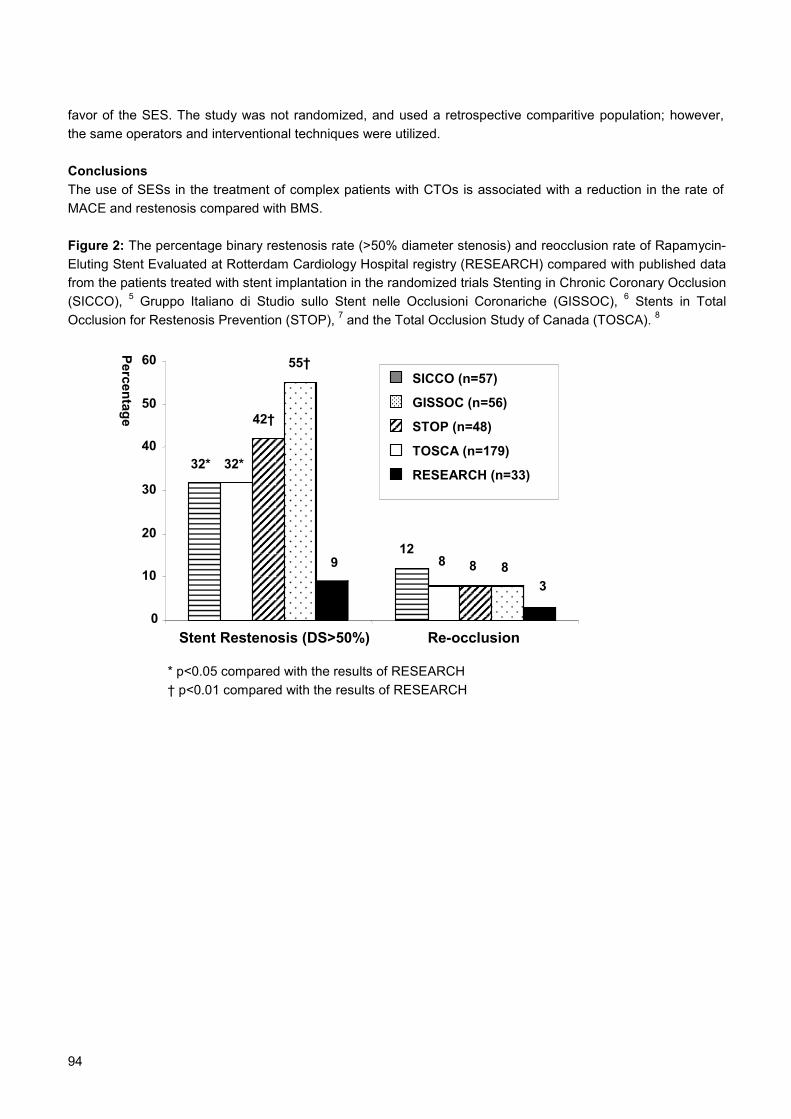
94
favor of the SES. The study was not randomized, and used a retrospective comparitive population; however,the same operators and interventional techniques were utilized.
ConclusionsThe use of SESs in the treatment of complex patients with CTOs is associated with a reduction in the rate ofMACE and restenosis compared with BMS.
Figure 2: The percentage binary restenosis rate (>50% diameter stenosis) and reocclusion rate of Rapamycin-Eluting Stent Evaluated at Rotterdam Cardiology Hospital registry (RESEARCH) compared with published datafrom the patients treated with stent implantation in the randomized trials Stenting in Chronic Coronary Occlusion(SICCO), 5 Gruppo Italiano di Studio sullo Stent nelle Occlusioni Coronariche (GISSOC), 6 Stents in TotalOcclusion for Restenosis Prevention (STOP), 7 and the Total Occlusion Study of Canada (TOSCA). 8
32* 32*
42†
55†
912
8 8 83
0
10
20
30
40
50
60
Stent Restenosis (DS>50%) Re-occlusion
Percentage* p<0.05 compared with the results of RESEARCH† p<0.01 compared with the results of RESEARCH
SICCO (n=57)
GISSOC (n=56)
STOP (n=48)
TOSCA (n=179)
RESEARCH (n=33)
95
References1. J.K. Kahn, Angiographic suitability for catheter revascularization of total coronary occlusions in patients from a community
hospital setting. Am Heart J 26 (1993), pp. 561–564.
2. P.N. Ruygrok, P.P. De Jaegere, J.J. Verploegh, R.T. Van Domburg and P.J. De Feyter, Immediate outcome following coronary
angioplasty: A contemporary single centre audit. Eur Heart J 16 Suppl L (1995), pp. 24–29.
3. G.W. Stone, B.D. Rutherford, D.R. McConahay et al., Procedural outcome of angioplasty for total coronary artery occlusion: An
analysis of 971 lesions in 905 patients. J Am Coll Cardiol 15 (1990), pp. 849–856.
4. R.J. Ivanhoe, W.S. Weintraub, J.S. Douglas, Jr. et al., Percutaneous transluminal coronary angioplasty of chronic total
occlusions: Primary success, restenosis, and long-term clinical follow-up. Circulation 85 (1992), pp. 106–115.
5. P.A. Sirnes, S. Golf, Y. Myreng et al., Stenting in Chronic Coronary Occlusion (SICCO): A randomized, controlled trial of adding
stent implantation after successful angioplasty. J Am Coll Cardiol 28 (1996), pp. 1444–1451.
6. P. Rubartelli, L. Niccoli, E. Verna et al., Stent implantation versus balloon angioplasty in chronic coronary occlusions: Results
from the Gissoc Trial (gruppo italiano di studio sullo stent nelle occlusioni coronariche). J Am Coll Cardiol 32 (1998), pp. 90–96.
7. C. Lotan, Y. Rozenman, A. Hendler et al., Stents in total occlusion for restenosis prevention: The multicentre randomized stop
study. The Israeli working group for interventional cardiology. Eur Heart J 21 (2000), pp. 1960–1966.
8. C.E. Buller, V. Dzavik, R.G. Carere et al., Primary stenting versus balloon angioplasty in occluded coronary arteries: The Total
Occlusion Study of Canada (TOSCA). Circulation 100 (1999), pp. 236–242.
9. P.W. Serruys, J.N. Hamburger, J.J. Koolen et al., Total occlusion trial with angioplasty by using laser guidewire: The total trial.
Eur Heart J 21 (2000), pp. 1797–1805.
10. M. Degertekin, P.W. Serruys, D.P. Foley et al., Persistent inhibition of neointimal hyperplasia after sirolimus-eluting stent
implantation: Long-term (up to 2 years) clinical, angiographic, and intravascular ultrasound follow-up. Circulation 106 (2002), pp.
1610–1613.
11. M.C. Morice, P.W. Serruys, J.E. Sousa et al., A randomized comparison of a sirolimus-eluting stent with a standard stent for
coronary revascularization. N Engl J Med 346 (2002), pp. 1773–1780.
12. P. Lemos, C. Lee, M. Degertekin et al., Early outcome after sirolimus-eluting stent implantation in patients with acute coronary
syndromes: Insights from the rapamycin-eluting stent evaluated at Rotterdam Cardiology Hospital (research) registry. J Am Coll
Cardiol 41 (2003), pp. 2093–2099.
13. Lemos P, Serruys PW, van Domburg RT, et al. Unrestricted utilization of sirolimus-eluting stents compared to conventional bare
stent implantation in the "real world." The Rapamycin-Eluting Stent Evaluated At Rotterdam Cardiology Hospital (RESEARCH)
registry. Circulation. In Press.
14. L. Finci, B. Meier, J. Favre, A. Righetti and W. Rutishauser, Long-term results of successful and failed angioplasty for chronic
total coronary arterial occlusion. Am J Cardiol 66 (1990), pp. 660–662.
15. J.A. Puma, M.H. Sketch, Jr., J.E. Tcheng et al., Percutaneous revascularization of chronic coronary occlusions: An overview. J
Am Coll Cardiol 26 (1995), pp. 1–11.
16. R. Rambaldi, J.N. Hamburger, M.L. Geleijnse et al., Early recovery of wall motion abnormalities after recanalization of chronic
totally occluded coronary arteries: A dobutamine echocardiographic, prospective, single-center experience. Am Heart J 136(1998), pp. 831–836.
17. J.A. Suero, S.P. Marso, P.G. Jones et al., Procedural outcomes and long-term survival among patients undergoing percutaneous
coronary intervention of a chronic total occlusion in native coronary arteries: A 20-year experience. J Am Coll Cardiol 38 (2001),
pp. 409–414.
18. A. Kastrati, A. Schomig, S. Elezi et al., Predictive factors of restenosis after coronary stent placement. J Am Coll Cardiol 30
(1997), pp. 1428–1436.
19. Z. Olivari, P. Rubartelli, F. Piscione et al., Immediate results and one-year clinical outcome after percutaneous coronary
interventions in chronic total occlusions: Data from a multicenter, prospective, obmservational study (toast-gise). J Am Coll
Cardiol 41 (2003), pp. 1672–1678.

96
97
Chapter 10
Angela HoyeAndrew TL OngJiro AokiCarlos AG van MieghemGaston A Rodriguez GranilloMarco ValgimigliGeorgios SianosEugene McFaddenWillem J van der GiessenPim J de FeyterRon T van DomburgPatrick W Serruys
Eurointervention2005;1:193-197
Drug-Eluting Stent Implantation for ChronicTotal Occlusions: Comparison between the
Sirolimus- and Paclitaxel-Eluting Stent

98
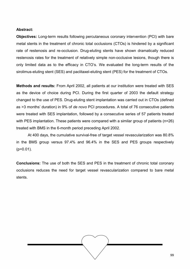
99
Abstract:
Objectives: Long-term results following percutaneous coronary intervention (PCI) with bare
metal stents in the treatment of chronic total occlusions (CTOs) is hindered by a significant
rate of restenosis and re-occlusion. Drug-eluting stents have shown dramatically reduced
restenosis rates for the treatment of relatively simple non-occlusive lesions, though there is
only limited data as to the efficacy in CTO’s. We evaluated the long-term results of the
sirolimus-eluting stent (SES) and paclitaxel-eluting stent (PES) for the treatment of CTOs.
Methods and results: From April 2002, all patients at our institution were treated with SES
as the device of choice during PCI. During the first quarter of 2003 the default strategy
changed to the use of PES. Drug-eluting stent implantation was carried out in CTOs (defined
as >3 months’ duration) in 9% of de novo PCI procedures. A total of 76 consecutive patients
were treated with SES implantation, followed by a consecutive series of 57 patients treated
with PES implantation. These patients were compared with a similar group of patients (n=26)
treated with BMS in the 6-month period preceding April 2002.
At 400 days, the cumulative survival-free of target vessel revascularization was 80.8%
in the BMS group versus 97.4% and 96.4% in the SES and PES groups respectively
(p=0.01).
Conclusions: The use of both the SES and PES in the treatment of chronic total coronary
occlusions reduces the need for target vessel revascularization compared to bare metal
stents.
100
IntroductionSuccessful percutaneous therapy of chronic total occlusions (CTOs) has been shown to improve symptoms ofangina and left ventricular function, and reduce the subsequent need for coronary artery bypass surgery. 1-5 Inaddition, in the long-term, recanalization of a CTO can reduce mortality compared with those with anunsuccessful attempt at recanalization. 6 However, the long-term outcome of percutaneous coronaryintervention (PCI) for chronic total coronary occlusions is subject to an increased risk of restenosis and re-occlusion compared with non-occlusive lesions. 1,7 The advent of drug-eluting stents is revolutionising thepractice of interventional cardiology. Several randomized trials have demonstrated a dramatic reduction inrestenosis rates compared with bare metal stents when used for the treatment of relatively simple lesions. 8-11
In addition, preliminary data has confirmed the efficacy utilizing the sirolimus-eluting stent (SES) for thetreatment of chronic total occlusions. 12 In the present report, we evaluate the use of drug-eluting stentimplantation for chronic total occlusions in a consecutive series of patients, with comparison between thesirolimus- and paclitaxel-eluting stents.
MethodsThe sirolimus-eluting stent (Cypher™, Johnson & Johnson - Cordis unit) received CE mark approval in April2002. Since that time, all patients undergoing percutaneous therapy in our institution have been treated withdrug-eluting stent implantation as the default strategy. During the first quarter of 2003, our strategy switchedfrom the sirolimus- to the paclitaxel-eluting stent (Boston Scientific) enabling a comparison of the two stenttypes. All consecutive patients with successful chronic occlusion recanalization were enrolled. Those patientstreated with drug-eluting stent implantation were compared to all those treated for a CTO in the preceding 6-months with bare metal stents (BMS), identified from the departments` dedicated database. All groups weretreated by the same operators utilizing standard techniques; the only difference being the type of stent.
During the procedure, heparin was given to maintain an activated clotting time ≥ 250 seconds. Allpatients received lifelong aspirin, and before the procedure were pre-treated with a loading dose of 300mgclopidogrel. Addition anti-platelet therapy was given with clopidogrel for 1 month in the BMS group, and for 6-months in the drug-eluting stent groups. The use of Glycoprotein IIb/IIIa inhibitor therapy was at the discretionof the operator and was only given once wire passage was confirmed as successful. The protocol wasapproved by the local ethics committee and is in accordance with the principles of Good Clinical Practice forTrials of Medicinal Products in the European Community and the Declaration of Helsinki. All patients signed awritten informed consent
Chronic total occlusion definition: Complete occlusion of a coronary artery on angiography, with noantegrade filling of the distal vessel other than via collaterals. All patients included, had a native vesselocclusion estimated to be of at least 3-months` duration, based on either a history of sudden chest pain, aprevious acute myocardial infarction in the same target vessel territory, or the time between the diagnosis madeon coronary angiography and PCI.
Length of occlusion: This was measured by quantitative coronary angiography (CAAS II; Pie MedicalImaging, The Netherlands) either utilizing antegrade filling via collaterals, or assessment of the retrogradecollateral filling achieved through making a simultaneous injection into both the left and right coronary arteriesto delineate the distance between the site of occlusion and the most proximal part of the vessel filledretrogradely. This length evaluated only the occluded vessel, and did not therefore include stenosis of thevessel pre- and post- the occlusion.
Follow-up: Patients were prospectively followed-up for clinical events, and evaluated for survival-free of majoradverse cardiac events (MACE) using questionnaires and telephone enquiries. MACE was pre-defined as: 1)death, 2) non-fatal myocardial infarction (AMI), or 3) repeat target vessel revascularization (TVR). Thediagnosis of AMI required an elevation of creatine kinase to twice the upper limit of normal, together with a risein creatine kinase-MB fraction. TVR was defined as either surgical or percutaneous reintervention driven by
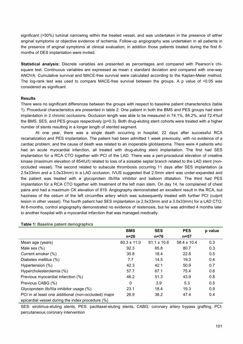
101
significant (>50%) luminal narrowing within the treated vessel, and was undertaken in the presence of eitheranginal symptoms or objective evidence of ischemia. Follow-up angiography was undertaken in all patients inthe presence of anginal symptoms at clinical evaluation; in addition those patients treated during the first 6-months of DES implantation were invited.
Statistical analysis: Discrete variables are presented as percentages and compared with Pearson’s chi-square test. Continuous variables are expressed as mean ± standard deviation and compared with one-wayANOVA. Cumulative survival and MACE-free survival were calculated according to the Kaplan-Meier method.The log-rank test was used to compare MACE-free survival between the groups. A p value of <0.05 wasconsidered as significant.
ResultsThere were no significant differences between the groups with respect to baseline patient characteristics (table1). Procedural characteristics are presented in table 2. One patient in both the BMS and PES groups had stentimplantation in 2 chronic occlusions. Occlusion length was able to be measured in 74.1%, 84.2%, and 72.4%ofthe BMS, SES, and PES groups respectively (p=0.3). Both drug-eluting stent cohorts were treated with a highernumber of stents resulting in a longer length of stented segment.
At one year, there was a single death occurring in hospital, 22 days after successful RCArecanalization and PES implantation. The patient had been admitted 1 week previously, with no evidence of acardiac problem, and the cause of death was related to an inoperable glioblastoma. There were 4 patients whohad an acute myocardial infarction, all treated with drug-eluting stent implantation. The first had SESimplantation for a RCA CTO together with PCI of the LAD. There was a peri-procedural elevation of creatinekinase (maximum elevation of 854IU/l) related to loss of a sizeable septal branch related to the LAD stent (non-occluded vessel). The second related to subacute thrombosis occurring 11 days after SES implantation (a2.5x33mm and a 3.0x33mm) in a LAD occlusion. IVUS suggested that 2.5mm stent was under-expanded andthe patient was treated with a glycoprotein IIb/IIIa inhibitor and balloon dilatation. The third had PESimplantation for a RCA CTO together with treatment of the left main stem. On day 14, he complained of chestpains and had a maximum CK elevation of 819. Angiography demonstrated an excellent result in the RCA, buthaziness of the ostium of the left circumflex artery which was subsequently treated with further PCI (culpritlesion in other vessel). The fourth patient had SES implantation (a 2.5x33mm and a 3.0x33mm) for a LAD CTO.At 6-months, control angiography demonstrated no evidence of restenosis, but he was admitted 4 months laterto another hospital with a myocardial infarction that was managed medically.
Table 1: Baseline patient demographicsBMSn=26
SESn=76
PESn=57
p value
Mean age (years) 60.3 ± 11.0 61.1 ± 10.6 58.4 ± 10.4 0.3Male sex (%) 92.3 65.8 80.7 0.3Current smoker (%) 30.8 18.4 22.8 0.5Diabetes mellitus (%) 7.7 14.5 19.3 0.4Hypertension (%) 42.3 42.1 50.9 0.7Hypercholesterolemia (%) 57.7 67.1 75.4 0.6Previous myocardial infarction (%) 46.2 51.3 43.9 0.8Previous CABG (%) 0 3.9 5.3 0.5Glycoprotein IIb/IIIa inhibitor usage (%) 23.1 18.4 19.3 0.9PCI in at least one additional (non-occluded) majorepicardial vessel during the index procedure (%)
26.9 38.2 47.4 0.4
SES: sirolimus-eluting stents, PES: paclitaxel-eluting stents, CABG: coronary artery bypass grafting, PCI:percutaneous coronary intervention

102
Post-procedure QC
A data
Diam
eter stenosis (%)
Minim
al lumen diam
eter (mm
)
Reference vessel diam
eter (mm
)
Mean total lengths of stent in the m
ain vessel (mm
)
Mean nom
inal diameter of stent in the m
ain vessel (mm
)
Mean num
ber of stents in the target vessel
Mean length of occlusion (m
m)
Bifurcation stenting (%)
RC
A (%
)
LCX (%
)
LAD
(%)
Target vessel
Num
ber of CTO
lesions treated
11.6
2.12 ± 0.51
2.34 ± 0.43
41.5 ± 23.3
3.0 ± 0.6
1.8 ± 0.8
13.0 ± 7.2
7.4
44.4
25.9
29.6
BM
Sn=27
12.9
2.04 ± 0.43
2.35 ± 0.51
48.8 ± 27.4
2.8 ± 0.3
2.2 ± 1.2
10.3 ± 5.9
13.2
34.2
19.7
46.1
SESn=76
14.1
2.26 ± 0.42
2.60 ± 0.49
58.0 ± 32.8
2.8 ± 0.4
2.6 ± 1.3
11.2 ± 6.6
13.8
50.0
27.6
22.4
PESn=58
0.6
0.02
0.008
0.04
<0.001
0.03
0.2
0.7
0.5
p value
Table 2: Baseline procedural characteristics

103
All events in the bare stent group related to the need for target vessel revascularization. At one year,the survival-free of target vessel revascularization was significantly higher in the SES and PES groupscompared with the BMS group (97.4% and 96.4% versus 80.8% respectively, p=0.01). Figure 1.
Figure 1: Kaplan-Meier estimates of the cumulative survival-free of target vessel revascularization followingstent implantation in a chronic total occlusion for patients treated with sirolimus-eluting (SES), paclitaxel-eluting(PES), or bare metal stent (BMS) implantation.
In the present report we have demonstrated the efficacy of drug-eluting stent implantation for the percutaneoustreatment of chronic total occlusions when compared to bare metal stents. In addition, we have shown that boththe sirolimus- and paclitaxel-eluting stent are associated with a low rate of target vessel revascularization at6months.
There have been several randomized trials that have demonstrated the efficacy of stent implantationover balloon-only angioplasty for the percutaneous treatment of CTOs, reducing the 6-month restenosis ratefrom 68-74% to 32-55%. 13-17 Initial randomized studies of drug-eluting stent implantation, demonstratedefficacy in reducing restenosis compared to conventional stent implantation, but excluded patients with CTOs.8-11 However, recent preliminary data from our own group have shown that the efficacy of the SES is applicablein the treatment of CTOs (defined as >1 months’ duration), with a one year cumulative survival-free of majoradverse cardiac events of 96.4%. 12 In the present study, we evaluate a larger series of consecutive patientstreated for a truly chronic total occlusion (>3 months in duration) with drug-eluting stent implantation. We haveshown that both the SES and PES significantly reduce the need for TVR, with a cumulative survival-free of TVRof 80.8% in the BMS group versus 97.4% and 96.4% in the SES and PES groups respectively (p=0.01). Figure1.
Importantly, there were no significant differences in baseline demographics between the groups, and allprocedures were carried out in the same centre by the same operators. Restenosis following BMS implantationis known to be inversely related to the post-procedural MLD and the number of stents utilized. 18 In the currentstudy, the mean nominal diameter of stent used was significantly greater in the BMS cohort, related to amaximum available SES and PES diameter of 3.0mm and 3.5mm respectively. In addition, despite utilizing agreater number of stents, both the SES and PES demonstrated efficacy over the BMS. Furthermore, thebeneficial effect of the SES occurred despite a smaller post-procedural MLD.
100
95
SES: 97.4%PES: 96.4%
BMS: 80.8%
Follow-up (months)1260
80
75
90
85
Log rank p=0.01
104
All major adverse cardiac events in the BMS group related to the need for TVR, including 1 patient whorequired coronary artery bypass surgery. Within the drug-eluting stent groups there were 5 additional non-TVRevents. One patient had a subacute thrombosis, but this might have been avoidable with evidence from IVUSdemonstrating a possible underlying mechanism of inadequate stent expansion. In addition, there is goodevidence in a further 3 of these cases that the event was unrelated to treatment of the occluded vessel. Onepatient died of non-cardiac causes, and 2 of the myocardial infarctions were thought to be related tointervention carried out in another (non-occluded) vessel. The fifth patient presented with an AMI in the territoryof the target vessel, 4 months after control angiography demonstrated patent stents. Clopidogrel medicationhad been stopped at the time of the follow-up angiogram, such that the patient was on aspirin therapy alone.The duration of dual anti-platelet therapy needed to reduce / abolish the risk of late stent thrombosis in patientstreated with DES, particularly for complex disease, is still unclear. Recently, Ong et al reported on late (>30days) stent thrombosis following DES implantation in a consecutive cohort of >2000 patients, they found a lowincidence of 0.35% (95% confidence limits 0.17% to 0.72%). 19 Importantly, there were no episodes in patientscontinuing on dual anti-platelet therapy. However, whether there is a true benefit in continuing clopidogrel inaddition to aspirin, over and above the possible disadvantages, requires further large scale evaluation.
In patients with significant coronary artery disease, although a CTO is found in at least one third, themajority are treated with either medical therapy or are referred for coronary artery bypass surgery, withpercutaneous treatment of CTOs accounting for only 10-15% PCI procedures. 20 The major limitation of PCI forCTOs is the inability to cross the lesion with a wire, however great advancements have been made in themanufacture of specialized wires, and there are additionally promising novel technologies such as theIntraluminal wire™ and Frontrunner catheter. 21-23 The current report has demonstrated the efficacy of drug-eluting stent implantation in CTOs and, together with improvements in recanalization rates, a strategy ofpercutaneous therapy of CTOs will become more widely applicable.
Study LimitationsThe study was not randomized, and angiographic follow-up data was not routinely obtained in all patients, soadditional events such as silent re-occlusion cannot be excluded. However, clinical follow-up was obtained in>99% patients (all but one patient), and assessment of symptomatic status in those that did not require re-intervention, showed that all were symptomatically well at follow-up. The study was not randomized, and used aretrospective comparative population; however the same operators and interventional techniques were utilized.
105
References1. Ivanhoe RJ, Weintraub WS, Douglas JS, Jr., Lembo NJ, Furman M, Gershony G, Cohen CL, King SB, 3rd. Percutaneous
transluminal coronary angioplasty of chronic total occlusions. Primary success, restenosis, and long-term clinical follow-up.
Circulation. 1992;85:106-15.
2. Warren RJ, Black AJ, Valentine PA, Manolas EG, Hunt D. Coronary angioplasty for chronic total occlusion reduces the need for
subsequent coronary bypass surgery. Am Heart J. 1990;120:270-4.
3. Finci L, Meier B, Favre J, Righetti A, Rutishauser W. Long-term results of successful and failed angioplasty for chronic total
coronary arterial occlusion. Am J Cardiol. 1990;66:660-2.
4. Puma JA, Sketch MH, Jr., Tcheng JE, Harrington RA, Phillips HR, Stack RS, Califf RM. Percutaneous revascularization of
chronic coronary occlusions: an overview. J Am Coll Cardiol. 1995;26:1-11.
5. Rambaldi R, Hamburger JN, Geleijnse ML, Poldermans D, Kimman GJ, Aiazian AA, Fioretti PM, Ten Cate FJ, Roelandt JR,
Serruys PW. Early recovery of wall motion abnormalities after recanalization of chronic totally occluded coronary arteries: a
dobutamine echocardiographic, prospective, single-center experience. Am Heart J. 1998;136:831-6.
6. Suero JA, Marso SP, Jones PG, Laster SB, Huber KC, Giorgi LV, Johnson WL, Rutherford BD. Procedural outcomes and long-
term survival among patients undergoing percutaneous coronary intervention of a chronic total occlusion in native coronary
arteries: a 20-year experience. J Am Coll Cardiol. 2001;38:409-14.
7. Stone GW, Rutherford BD, McConahay DR, Johnson WL, Jr., Giorgi LV, Ligon RW, Hartzler GO. Procedural outcome of
angioplasty for total coronary artery occlusion: an analysis of 971 lesions in 905 patients. J Am Coll Cardiol. 1990;15:849-56.
8. Stone GW, Ellis SG, Cox DA, Hermiller J, O'Shaughnessy C, Mann JT, Turco M, Caputo R, Bergin P, Greenberg J, Popma JJ,
Russell ME. A polymer-based, paclitaxel-eluting stent in patients with coronary artery disease. N Engl J Med. 2004;350:221-31.
9. Morice MC, Serruys PW, Sousa JE, Fajadet J, Ban Hayashi E, Perin M, Colombo A, Schuler G, Barragan P, Guagliumi G,
Molnar F, Falotico R. A randomized comparison of a sirolimus-eluting stent with a standard stent for coronary revascularization.
N Engl J Med. 2002;346:1773-80.
10. Moses JW, Leon MB, Popma JJ, Fitzgerald PJ, Holmes DR, O'Shaughnessy C, Caputo RP, Kereiakes DJ, Williams DO,
Teirstein PS, Jaeger JL, Kuntz RE. Sirolimus-eluting stents versus standard stents in patients with stenosis in a native coronary
artery. N Engl J Med. 2003;349:1315-23.
11. Grube E, Silber S, Hauptmann KE, Mueller R, Buellesfeld L, Gerckens U, Russell ME. TAXUS I: six- and twelve-month results
from a randomized, double-blind trial on a slow-release paclitaxel-eluting stent for de novo coronary lesions. Circulation.
2003;107:38-42.
12. Hoye A, Tanabe K, Lemos PA, Aoki J, Saia F, Arampatzis C, Degertekin M, Hofma SH, Sianos G, McFadden E, Van Der
Giessen WJ, Smits PC, De Feyter PJ, Van Domburg RT, Serruys PW. Significant reduction in restenosis after the use of
sirolimus-eluting stents in the treatment of chronic total occlusions. J Am Coll Cardiol. 2004;43:1954-8.
13. Sirnes PA, Golf S, Myreng Y, Molstad P, Emanuelsson H, Albertsson P, Brekke M, Mangschau A, Endresen K, Kjekshus J.
Stenting in Chronic Coronary Occlusion (SICCO): a randomized, controlled trial of adding stent implantation after successful
angioplasty. J Am Coll Cardiol. 1996;28:1444-51.
14. Rubartelli P, Niccoli L, Verna E, Giachero C, Zimarino M, Fontanelli A, Vassanelli C, Campolo L, Martuscelli E, Tommasini G.
Stent implantation versus balloon angioplasty in chronic coronary occlusions: results from the GISSOC trial. Gruppo Italiano di
Studio sullo Stent nelle Occlusioni Coronariche. J Am Coll Cardiol. 1998;32:90-6.
15. Lotan C, Rozenman Y, Hendler A, Turgeman Y, Ayzenberg O, Beyar R, Krakover R, Rosenfeld T, Gotsman MS. Stents in total
occlusion for restenosis prevention. The multicentre randomized STOP study. The Israeli Working Group for Interventional
Cardiology. Eur Heart J. 2000;21:1960-6.
16. Buller CE, Dzavik V, Carere RG, Mancini GB, Barbeau G, Lazzam C, Anderson TJ, Knudtson ML, Marquis JF, Suzuki T, Cohen
EA, Fox RS, Teo KK. Primary stenting versus balloon angioplasty in occluded coronary arteries: the Total Occlusion Study of
Canada (TOSCA). Circulation. 1999;100:236-42.
17. Hoher M, Wohrle J, Grebe OC, Kochs M, Osterhues HH, Hombach V, Buchwald AB. A randomized trial of elective stenting after
balloon recanalization of chronic total occlusions. J Am Coll Cardiol. 1999;34:722-9.
18. Kastrati A, Schomig A, Elezi S, Schuhlen H, Dirschinger J, Hadamitzky M, Wehinger A, Hausleiter J, Walter H, Neumann FJ.
Predictive factors of restenosis after coronary stent placement. J Am Coll Cardiol. 1997;30:1428-36.

106
19. Ong AT, McFadden EP, Regar E, de Jaegere PP, van Domburg RT, Serruys PW. Late angiographic stent thrombosis (LAST)
events with drug-eluting stents. J Am Coll Cardiol. 2005;45:2088-92.
20. Kahn JK. Angiographic suitability for catheter revascularization of total coronary occlusions in patients from a community hospital
setting. Am Heart J. 1993;126:561-4.
21. Orlic D CA, Stankovic G, Sangiori G, Airoldi F, Corvaja N, Montorfano M, Michev I, Vitrella G, Colombo A. Preliminary experience
with the Frontrunner coronary catheter, novel device dedicated to mechanical revascularization of chronic total occlusions. J Am
Coll Cardiol. 2004;43:56A.
22. Ng W, Chen WH, Lee PY, Lau CP. Initial experience and safety in the treatment of chronic total coronary occlusions with a new
optical coherent reflectometry-guided radiofrequency ablation guidewire. Am J Cardiol. 2003;92:732-4.
23. Hoye A, Onderwater, E. Cummins, P. Sianos, G. Serruys, PW. Improved Recanalization of Chronic Total Coronary Occlusions
Using an Optical Coherence Reflectometry-Guided Guidewire. Cathet Cardiovasc Interv. 2004;63:158-63.

107
Chapter 11
Jiro AokiAngela HoyeAV StaferovBG AlekyanPatrick W Serruys
Journal of InterventionalCardiology2005;18(1):65-9
Sirolimus-Eluting Stent Implantation forChronic Total Occlusion of the Left Main
Coronary Artery

108

109
Abstract
Chronic total occlusion of the left main coronary artery (LMCA) is rare. Recently,
percutaneous coronary intervention has been increasingly applied to unprotected LMCA
lesions. We describe a patient with chronic total occlusion of the left main coronary artery
who was successfully treated with bifurcation stenting with sirolimus eluting stents.

110
IntroductionChronic total occlusion of the left main coronary artery (LMCA) is a very unusual manifestation of coronaryatherosclerotic disease in clinical practice. 1-4 The rarity of this lesion may be accounted for by the relativelyhigh incidence of death in these patients. Coronary artery bypass graft surgery (CABG) has been the standardof care for LMCA disease, though recently, percutaneous coronary intervention (PCI) has been increasinglyapplied to unprotected LMCA lesions. 5-12 However, the development of restenosis remains a major limitation oflate outcomes after PCI, with the occurrence of restenosis particularly associated with hazardous clinicalmanifestations. Sirolimus-eluting stents (SES) have been shown to dramatically reduce the restenosis rate inselected patients with relatively simple lesions. 13-15 We report a patient with chronic total occlusion of the leftmain coronary artery who was successfully treated percutaneously with SES implantation.
Case reportA 35-year-old male presented with an acute anterior myocardial infarction that was managed medically. Hesubsequently complained of on-going chest pain (CCS class II-III angina 16) and underwent coronaryangiography 9-month later. His resting 12-ECG revealed evidence of a previous Q-wave anteroseptalmyocardial infarction and echocardiography demonstrated hypokinesis of the anteroseptal wall without leftventricle aneurysm. He was referred for coronary angiography which showed a total occlusion of the LMCA(Figure 1); the left anterior descending coronary artery (LAD) and left circumflex coronary artery (LCX) wereretrogradely filled via Rentrop grade III collaterals 17 from the RCA, which was itself not significantly stenosed(Figure 2). The patient rejected coronary artery bypass grafting, but consented to undergo attemptedrevascularization with percutaneous coronary intervention.
A 6Fr introducer sheath was inserted in the right femoral artery. In addition, a 5Fr introducer sheathwas inserted in the left femoral artery to enable simultaneous right and left coronary injections. In the absenceof antegrade flow through the occlusion, such a dual injection technique allows visualization of the distalvessels (LAD and LCX) via the collateral filling from the RCA, thereby facilitating correct positioning of the wire.A 6Fr XB 3.5 guiding catheter (Cordis) was placed in the left main coronary segment, and successfulrecanalization was achieved with a 0.0014” Shinobi wire (Cordis), which was advanced into the distal LAD. Asecond 0.0014” Shinobi wire was taken, and passage in to the LCX was attempted. Unfortunately this wascomplicated by catheter / wire-induced dissection from the LMCA to the mid-LCX (Figure 3), and the wire waswithdrawn. The LAD / distal LMCA was stabilized through stent implantation with a 3.0 x 23mm SES (Cypher,Cordis), which was then post-dilated with a 3.5mm balloon (U-pass, Cordis) giving a good result (Figure 4).After several attempts, a 0.0014” Sinobi wire was eventually successfully crossed, via the SES struts, into thetrue lumen of the LCX. After sequential predilatation using a 2.0mm balloon (Stomer, Medtronic), both thelesion and the mid-LCX dissection (Figure 5) were treated with implantation of a 2.5 x 33 mm SES deployedfrom LCX ostium. The final angiogram showed a good result with TIMI III flow in both the LAD and LCX 18
(Figure 6).The patient made an unremarkable recovery and was allowed home. There were no major adverse
cardiac events during the in-hospital period, and at 9-months clinical follow-up he remains well with norecurrence of angina.
DiscussionChronic total occlusion (CTO) of the LMCA is rare. In patients who are investigated in the catheter laboratoryits` prevalence varies from 0.04 percent to 0.4 percent. 1-4 CABG has been considered the treatment of choicein LMCA disease and in particular, is the most favored strategy for chronic total occlusion of the LMCA. Thereare a few published reports showing that the results of CABG for this group of CTO`s are beneficial comparedto medical therapy, 1,2 however, there are no studies comparing PCI with CABG for such patients. The adventof improved PCI equipment including stents and atherectomy devices, have been shown to be a safe andeffective in selected patients with unprotected elective LMCA stenosis, 5-12 and recently Trehan et al havereported a single case of successful percutaneous stenting of a CTO of an unprotected left main coronaryartery. 19

111
Figure 1: Left coronary artery angiogram,revealing total occlusion of the left main stemartery.
Figure 3: Left coronary angiogram showing thedissection of the left main stem artery to the mid-circumflex artery.
Figure 5: Left coronary angiogram showing that awire has successfully crossed through the stentstruts and into the true lumen of the distal leftcircumflex artery. The region of dissection canclearly be seen in the proximal and mid parts ofthe vessel (contrast staining).
Figure 2: Right coronary angiogramdemonstrating grade III retrograde collateralsarising from the right coronary artery to both theleft anterior descending and left circumflexarteries.
Figure 4: Left coronary angiogram followingstent deployment with a 3.0 x 23mm sirolimus-eluting stent in the left main stem / left anteriordescending artery.
Figure 6: The final angiogram followingadditional stent implantation with a 2.5 x 33mmsirolimus-eluting stent up to the ostium of thecircumflex artery, showing a good result in boththe left anterior descending and left circumflexarteries.
112
There are 2 major problems associated with PCI for chronic total occlusion of the LMCA. The firstrelates to the initial procedural difficulty of crossing the occlusion with a wire; published procedural successrates for CTO`s are generally in the range of 40-81%. 20,21 The second relates to restenosis; both bifurcationlesions and CTO`s are subject to a higher rate of restenosis compared with simpler lesions, 22-25 andimportantly, the occurrence of restenosis in the LMCA may be associated with a significant rate of mortality. Inparticular, PCI for bifurcation lesions of the distal LMCA whereby both the LAD and LCX arteries are stented, isboth technically demanding and at high risk of restenosis. 26-28 Drug eluting stents have been shown todramatically reduce the restenosis rate in elective patients with simple de novo lesions. 13-15 The developmentand more widespread application of drug eluting stents hold the promise of a significant reduction in restenosisand the need for repeat revascularization. Arampatzis et al have reported the effectiveness of SES for thetreatment of LMCA. A total of 31 consecutive patients were treated solely with SESs either electively, for acutemyocardial infarction, or due to procedural complication-related LMCA dissection. In this study, the rate of out-of-hospital clinical events was extremely low (mortality rate was 0% and target vessel revascularization ratewas 4%). 29 In addition, low subsequent binary restenosis rates following SES implantation have beendocumented both in CTO`s (9.1% at 6 months) and bifurcation lesions (22.7% at 6 months). 30,31 The techniqueof bifurcation stenting (T, culotte, kissing, or crush stenting) with drug-eluting stents is still controversial. 30
Those techniques resulting in overlapping stent struts lead to an increase in the local concentration of drug,which may induce endothelial function impairment and thus be associated with an increased rate of stentthrombosis. In the current report, we present a patient who underwent successful recanalization of a LMCACTO without the need for a cardiac support device, and underwent bifurcation stenting, with SESs. There wereno major adverse cardiac events either in-hospital, or over the subsequent 9 months follow up period. Furtherdata is needed to fully evaluate the use of this strategy in such an unusual patient population, however, SESsupported angioplasty may be a reasonable alternative to CABG in the treatment of LMCA chronic totalocclusion.
113
References1. Zimmern SH, Rogers WJ, Bream PR et al. Total occlusion of the left main coronary artery: the Coronary Artery Surgery Study
(CASS) experience. Am J Cardiol 1982;49(8):2003-2010.
2. Ward DE, Valantine H, Hui W. Occluded left main stem coronary artery. Report of five patients and review of published reports.
Br Heart J 1983;49(3):276-279.
3. Herregods MC, Piessens J, Vanhaecke J et al. Complete occlusion of the main left coronary artery. A clinical study. Acta Cardiol
1987;42(1):23-35.
4. de Feyter PJ, Serruys PW. Thrombolysis of acute total occlusion of the left main coronary artery in evolving myocardial infarction.
Am J Cardiol 1984;53(11):1727-1728.
5. Kosuga K, Tamai H, Ueda K et al. Initial and long-term results of angioplasty in unprotected left main coronary artery. Am J
Cardiol 1999;83(1):32-37.
6. Silvestri M, Barragan P, Sainsous J et al. Unprotected left main coronary artery stenting: immediate and medium-term outcomes
of 140 elective procedures. J Am Coll Cardiol 2000;35(6):1543-1550.
7. Tan WA, Tamai H, Park SJ et al. Long-term clinical outcomes after unprotected left main trunk percutaneous revascularization in
279 patients. Circulation 2001;104(14):1609-1614.
8. Park SJ, Hong MK, Lee CW et al. Elective stenting of unprotected left main coronary artery stenosis: effect of debulking before
stenting and intravascular ultrasound guidance. J Am Coll Cardiol 2001;38(4):1054-1060.
9. Black A, Cortina R, Bossi I et al. Unprotected left main coronary artery stenting: correlates of midterm survival and impact of
patient selection. J Am Coll Cardiol 2001;37(3):832-838.
10. Takagi T, Stankovic G, Finci L et al. Results and long-term predictors of adverse clinical events after elective percutaneous
interventions on unprotected left main coronary artery. Circulation 2002;106(6):698-702.
11. Park SJ, Park SW, Hong MK et al. Long-term (three-year) outcomes after stenting of unprotected left main coronary artery
stenosis in patients with normal left ventricular function. Am J Cardiol 2003;91(1):12-16.
12. Brueren BR, Ernst JM, Suttorp MJ et al. Long term follow up after elective percutaneous coronary intervention for unprotected
non-bifurcational left main stenosis: is it time to change the guidelines? Heart 2003;89(11):1336-1339.
13. Holmes DR, Jr., Leon MB, Moses JW et al. Analysis of 1-year clinical outcomes in the SIRIUS trial: a randomized trial of a
sirolimus-eluting stent versus a standard stent in patients at high risk for coronary restenosis. Circulation 2004;109(5):634-640.
14. Stone GW, Ellis SG, Cox DA et al. A polymer-based, paclitaxel-eluting stent in patients with coronary artery disease. N Engl J
Med 2004;350(3):221-231.
15. Morice MC, Serruys PW, Sousa JE et al. A randomized comparison of a sirolimus-eluting stent with a standard stent for coronary
revascularization. N Engl J Med 2002;346(23):1773-1780.
16. Campeau L. Letter: Grading of angina pectoris. Circulation 1976;54(3):522-523.
17. Cohen M, Rentrop KP. Limitation of myocardial ischemia by collateral circulation during sudden controlled coronary artery
occlusion in human subjects: a prospective study. Circulation 1986;74(3):469-476.
18. Anderson JL, Karagounis LA, Becker LC et al. TIMI perfusion grade 3 but not grade 2 results in improved outcome after
thrombolysis for myocardial infarction. Ventriculographic, enzymatic, and electrocardiographic evidence from the TEAM-3 Study.
Circulation 1993;87(6):1829-1839.
19. Trehan V, Mehta V, Mukhopadhyay S et al. Percutaneous stenting of chronic total occlusion of unprotected left main coronary
artery. Indian Heart J 2003;55(2):172-174.
20. Puma JA, Sketch MH, Jr., Tcheng JE et al. Percutaneous revascularization of chronic coronary occlusions: an overview. J Am
Coll Cardiol 1995;26(1):1-11.
21. Serruys PW, Hamburger JN, Koolen JJ et al. Total occlusion trial with angioplasty by using laser guidewire. The TOTAL trial. Eur
Heart J 2000;21(21):1797-1805.
22. Rubartelli P, Niccoli L, Verna E et al. Stent implantation versus balloon angioplasty in chronic coronary occlusions: results from
the GISSOC trial. Gruppo Italiano di Studio sullo Stent nelle Occlusioni Coronariche. J Am Coll Cardiol 1998;32(1):90-96.
23. Buller CE, Dzavik V, Carere RG et al. Primary stenting versus balloon angioplasty in occluded coronary arteries: the Total
Occlusion Study of Canada (TOSCA). Circulation 1999;100(3):236-242.
24. Al Suwaidi J, Berger PB, Rihal CS et al. Immediate and long-term outcome of intracoronary stent implantation for true bifurcation
lesions. J Am Coll Cardiol 2000;35(4):929-936.

114
25. Yamashita T, Nishida T, Adamian MG et al. Bifurcation lesions: two stents versus one stent--immediate and follow-up results. J
Am Coll Cardiol 2000;35(5):1145-1151.
26. Park SJ, Lee CW, Kim YH et al. Technical feasibility, safety, and clinical outcome of stenting of unprotected left main coronary
artery bifurcation narrowing. Am J Cardiol 2002;90(4):374-378.
27. Lee CH, Degertekin M, van Domburg RT et al. Impact of different anatomical patterns of left main coronary stenting on long-term
survival. Am J Cardiol 2003;92(6):718-720.
28. Hu FB, Tamai H, Kosuga K et al. Intravascular ultrasound-guided directional coronary atherectomy for unprotected left main
coronary stenoses with distal bifurcation involvement. Am J Cardiol 2003;92(8):936-940.
29. Arampatzis CA, Lemos PA, Tanabe K et al. Effectiveness of sirolimus-eluting stent for treatment of left main coronary artery
disease. Am J Cardiol 2003;92(3):327-329.
30. Tanabe K, Hoye A, Lemos PA et al. Restenosis rates following bifurcation stenting with sirolimus-eluting stents for de novo
narrowings. Am J Cardiol 2004;94:115-8
31. Hoye A, Tanabe K, Lemos PA, Aoki J, SaiaF, Arampatzis CA, Degertekin M, Hofma S, Sianos G, McFadden and others.
Significant reduction in restenosis following the use of sirolimus-eluting stents in the treatment of chronic total occlusions. J Am
Coll Cardiol 2004;43:1954-8.

Part 2
BIFURCATIONS
115

116

Chapter 12
Angela Hoye
Chapter in A ColouHandbook of AdultInterventional Cardiology Manson Publishing
Bifurcations
117
r
(in press)

118

Percutaneous coronary intervention (PCI) of bifurcation lesions is associated with a lower rate of proceduralsuccess,1 and an increased subsequent rate of major adverse cardiac events (MACE) and restenosiscompared with PCI of non-bifurcated lesions. The complexity of bifurcation lesions relates to the need tomaintain patency of both the main vessel and (sizeable) side branch. However, the term “bifurcation lesion”covers a range of anatomical variations, and, at present, the most effective strategy of PCI for individual lesionsis currently unknown. Indeed studies published thus far have evaluated the efficacy of different stentingstrategies when applied to treat bifurcations in general, and have not been targeted to the individual lesion.There have been several proposed classifications of bifurcation lesions, one of these, the Duke classification, isdepicted in figure 1. The recently introduced Medina classification is a simple binary system whereby thepresence of significant plaque is represented by a 1 for the proximal main vessel, distal main vessel, and sidebranch. The corresponding Medina classification for each lesion subtype is also presented in figure 1. However,even when only one of the two branches is significantly stenosed at baseline, plaque shift or the “snow-plougheffect” can pose a problem. In addition to plaque distribution, lesions also differ in respect of the degree ofangulation between the main vessel and side branch, something which is of particular importance when utilizingtechniques such as “T-stenting” and specialized bifurcation stents.
Figure 1: The Duke Classification of bifurcation lesions together with the corresponding Medina binaryclassification with respect to variations of plaque distribution.
Plaque shiftFollowing balloon dilatation or stent impproximally, longitudinally, and/or circumferesevere compromise of flow within the othercan lead to side branch occlusion, particulbranch is of relatively small diameter, osyndromes. The clinical consequences ofassociated with short-lived chest pain anangiography may demonstrate restoration may result in a rise in cardiac enzymes, andhave prognostic implications after PCI. Thefollow-up (figure 2).5,6
B
Bifurcation
Pre-branchonly
lantation, shifntially.2,3 Imp vessel. Indeearly when ther in the pres loss of the sd a (modestof patency in even a relat greater the e
Post-branchonly
t of atheromatous / thrombotortantly, plaque shift is unpredd, following dilatation of the m
ostium of the side branch is ience of soft thrombotic mateide branch depend on the ve
) rise in cardiac enzymes. Fo up to 82%.4 However even teively small cardiac enzyme relenzyme rise, the higher the mo
1
Parent vesselonly
Ostial
iiat
mar
Pre-branch andostial
Duke: A
CF
E Duke: DMedina: 1.0.0
0.1.0 1.1.01.0.
0.0.1 Medina: 1.1.1119
c material may occurctable, but can lead toin vessel, plaque shift
self diseased, the siderial in acute coronaryssel size, but may bertunately at follow-up,porary vessel closurese has been shown totality rate at long-term
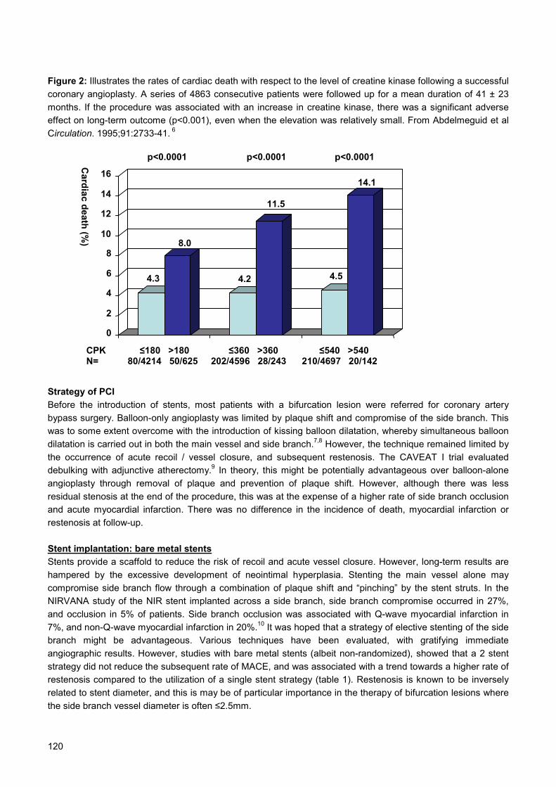
120
Figure 2: Illustrates the rates of cardiac death with respect to the level of creatine kinase following a successfulcoronary angioplasty. A series of 4863 consecutive patients were followed up for a mean duration of 41 ± 23months. If the procedure was associated with an increase in creatine kinase, there was a significant adverseeffect on long-term outcome (p<0.001), even when the elevation was relatively small. From Abdelmeguid et alCirculation. 1995;91:2733-41. 6
StrategBeforebypasswas todilatatiothe ocdebulkangiopresiduaand acresteno
Stent iStents hampecomproNIRVAand oc7%, anbranchangiogstrategrestenorelatedthe sid
4.3
8.0
4.2
11.5
4.5
14.1
0
2
4
6
8
10
12
14
16
Cardiac death (%
)
CPK ≤180 >180 ≤360 >360 ≤540 >540 N= 80/4214 50/625 202/4596 28/243 210/4697 20/142
y of PCI the introduction surgery. Balloo some extent ovn is carried out
currence of acuing with adjunctlasty through rel stenosis at theute myocardial sis at follow-up.
mplantation: baprovide a scaffored by the excmise side bran
NA study of theclusion in 5% od non-Q-wave m might be adraphic results. Hy did not reducesis compared to to stent diametee branch vessel
p<0.0001
of stents, most patienn-only angioplasty was ercome with the introdu in both the main vesselte recoil / vessel closuive atherectomy.9 In themoval of plaque and end of the procedure, tinfarction. There was n
re metal stentsld to reduce the risk of essive development ofch flow through a comb NIR stent implanted acf patients. Side branch yocardial infarction in 2
vantageous. Various towever, studies with ba the subsequent rate of the utilization of a singr, and this is may be of
diameter is often ≤2.5m
p<0.0001
ts with a bifurcationlimited by plaque shifction of kissing balloo and side branch.7,8 Hre, and subsequent ory, this might be po
prevention of plaquehis was at the expenso difference in the in
recoil and acute vess neointimal hyperplasination of plaque shifross a side branch, socclusion was assoc0%.10 It was hoped thechniques have beere metal stents (albe
MACE, and was assole stent strategy (tab
particular importancem.
p<0.0001
lesion were referred for coronary arteryt and compromise of the side branch. Thisn dilatation, whereby simultaneous balloonowever, the technique remained limited byrestenosis. The CAVEAT I trial evaluatedtentially advantageous over balloon-alone
shift. However, although there was lesse of a higher rate of side branch occlusioncidence of death, myocardial infarction or
el closure. However, long-term results areia. Stenting the main vessel alone mayt and “pinching” by the stent struts. In theide branch compromise occurred in 27%,
iated with Q-wave myocardial infarction inat a strategy of elective stenting of the siden evaluated, with gratifying immediateit non-randomized), showed that a 2 stentciated with a trend towards a higher rate ofle 1). Restenosis is known to be inversely in the therapy of bifurcation lesions where

121
Table 1StudyLength of clinical follow-up
Strategy No. In-hospital MACE(%)
Restenosis(%)
TLR (%) MACE(%)
1 stent 47 4 - 17 25Pan et al 11
18 months FU 2 stents 23 8 - 39 561 stent 39 0 48 36 38Yamashita et al 12
6 months FU 2 stents 53 13 62 38 511 stent 77 3 - 21 27Al Suwaidi et al 13
1 year FU 2 stents 54 6 - 19 481 stent 45 4 28 16 20Anzuini et al 14
1 year FU 2 stents 45 4 43 36 39Sheiban et al 15
9 months FU2 stents 54 0 63 33 33
Frontier stent 16 6 months FU
Bifurcationstent
105 3 45 13 17
1 stent 22 9 19 5 14Colombo et al 17
6-months FU 2 stents 63 10 28 10 191 stent 47 4 7 2 9Pan et al 18
11-months FU 2 stents 44 2 20 5 7Tanabe et al 19 6-months FU
2 stents 58 2 23 9 10
MACE: Major adverse cardiac events; TLR: target lesion revascularization
Stent implantation: drug-eluting stentsThe advent of drug-eluting stents, with a marked reduction in the development of restenosis, has lead to the re-evaluation of bifurcation lesion stenting strategies. Preliminary data of the sirolimus-eluting stent (SES) hasconfirmed efficacy when compared with historical data, with overall rates of restenosis of 23-26%, and need fortarget lesion revascularization in 8-9%.17,19 In an observational study of the SES in a consecutive group ofpatients, restenosis occurred particularly at the ostium of the side branch following the use of a T-stentingstrategy (described below).19 This might reflect incomplete coverage of the ostium thereby reducing the efficacyof the drug-elution, and suggested that it may be beneficial when using drug-eluting stents to adopt a strategythat ensures complete lesion coverage. There are currently several on-going randomised trials underway toevaluate this.
Preliminary data from Colombo et al of 85 patients randomized to either a single SES versus a 2-SESstrategy, found no significance difference in restenosis rates between the two groups (19% for provisional side-branch stenting versus 28% for double-stenting).17 However, these results are limited because of a notably highrate of crossover in the single-stent group to the 2-stent strategy because of a sub-optimal result (51%). Inaddition, the strategy of 2-stent implantation was not standardised. Pan et al also randomized patients with abifurcation lesion to a single versus a 2-stent strategy, and showed no difference in terms of clinical outcomes.18 At 6-months angiographic follow-up, restenosis occurred in the main vessel in 2% and 10%, and in the sidebranch in 5% and 15%, of patients treated with a single versus a 2-stent strategy respectively.
Techniques of stentingGeneral principles Angiography must adequately evaluate the bifurcation, in particular whether the side branch ostium is involvedor not. The use of 2 wires is a simple method to ensure that access to each branch is preserved throughout theintervention. In general the use of pre-dilatation is recommended, and has the advantage of assessing how thelesion behaves with evaluation of any plaque shift, and facilitation of subsequent stent implantation. Whateverthe stenting strategy chosen, kissing balloon post-dilatation is strongly recommended particularly when both
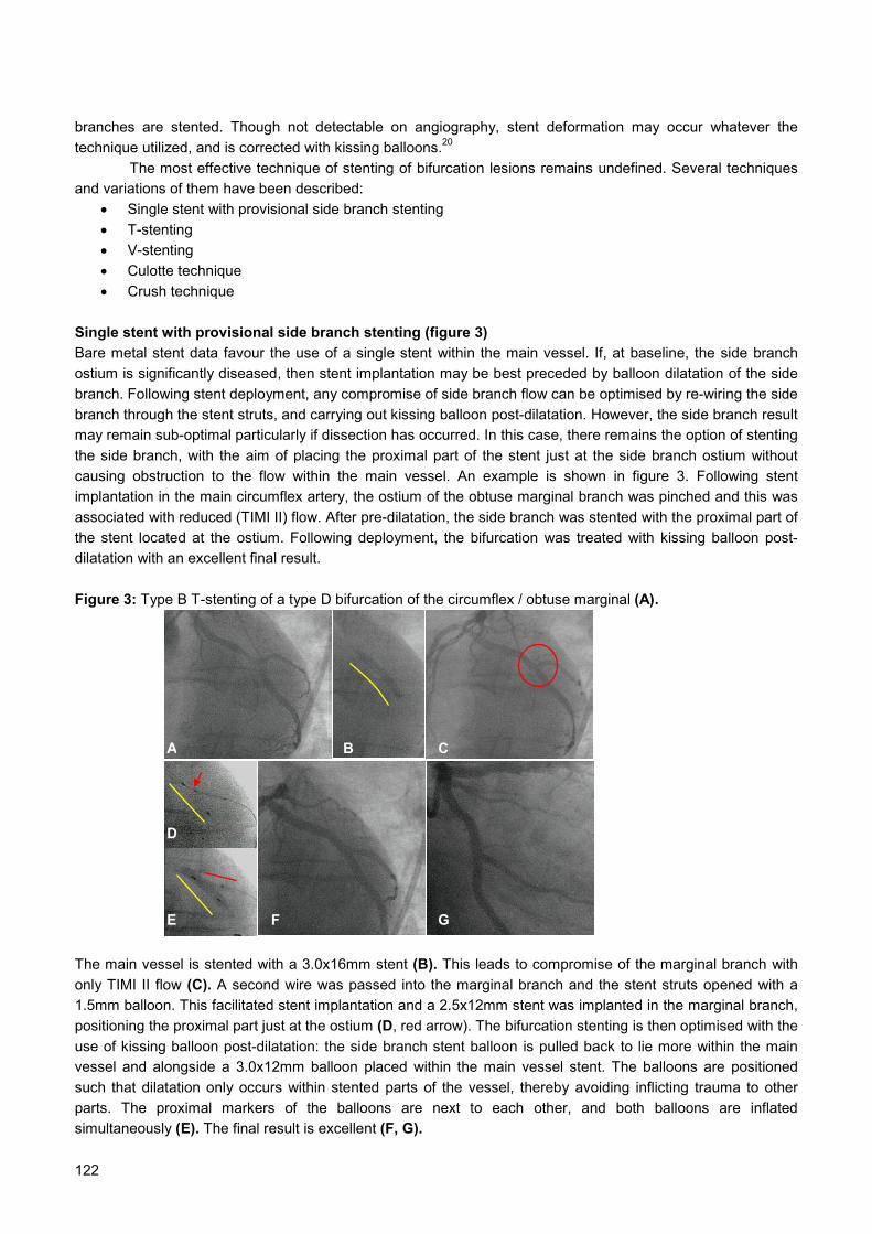
122
branches are stented. Though not detectable on angiography, stent deformation may occur whatever thetechnique utilized, and is corrected with kissing balloons.20 The most effective technique of stenting of bifurcation lesions remains undefined. Several techniquesand variations of them have been described:
• Single stent with provisional side branch stenting• T-stenting• V-stenting• Culotte technique• Crush technique
Single stent with provisional side branch stenting (figure 3)Bare metal stent data favour the use of a single stent within the main vessel. If, at baseline, the side branchostium is significantly diseased, then stent implantation may be best preceded by balloon dilatation of the sidebranch. Following stent deployment, any compromise of side branch flow can be optimised by re-wiring the sidebranch through the stent struts, and carrying out kissing balloon post-dilatation. However, the side branch resultmay remain sub-optimal particularly if dissection has occurred. In this case, there remains the option of stentingthe side branch, with the aim of placing the proximal part of the stent just at the side branch ostium withoutcausing obstruction to the flow within the main vessel. An example is shown in figure 3. Following stentimplantation in the main circumflex artery, the ostium of the obtuse marginal branch was pinched and this wasassociated with reduced (TIMI II) flow. After pre-dilatation, the side branch was stented with the proximal part ofthe stent located at the ostium. Following deployment, the bifurcation was treated with kissing balloon post-dilatation with an excellent final result.
Figure 3: Type B T-stenting of a type D bifurcation of the circumflex / obtuse marginal (A).
The main vessel is stented with a 3.0x1only TIMI II flow (C). A second wire wa1.5mm balloon. This facilitated stent imppositioning the proximal part just at the ouse of kissing balloon post-dilatation: thvessel and alongside a 3.0x12mm ballsuch that dilatation only occurs within sparts. The proximal markers of the simultaneously (E). The final result is ex
B
D
6mm stent (Bs passed into lantation and astium (D, red ae side branch oon placed wittented parts oballoons are cellent (F, G).
C
F
E GA
). This leads to compromise of the marginal branch withthe marginal branch and the stent struts opened with a 2.5x12mm stent was implanted in the marginal branch,rrow). The bifurcation stenting is then optimised with thestent balloon is pulled back to lie more within the mainhin the main vessel stent. The balloons are positionedf the vessel, thereby avoiding inflicting trauma to othernext to each other, and both balloons are inflated

T-stenting (figure 4)As shown in figure 4, this involves electively stenting first the side branch, positioning the proximal part at theostium, followed by stent implantation within the main vessel. However, the efficacy of both provisional sidebranch stenting and T-stenting are limited due to the angle between the main vessel and side branch.Complete lesion coverage can be accomplished only when there is a large angle between the vessels (a T-shape). Figure 5. However, in >75% bifurcations, this angle is significantly <70˚ and more of a “Y” shape.21
Even with precise stent positioning, this means that either the ostium of the side branch is incompletely covered(figure 5a), or the side branch stent protrudes into the main vessel (figure 5b).
Figure 4: Type A T-stenting of a type F bifurcation lesion of the LAD / diagonal (A). Both vessels were pre-dilated, and the diagonal branch was stented first with a 2.75x23mm stent placed at the ostium (B), and with agood result (C). The main vessel was then stented with a 3.0x23mm stent positioned to cover the lesionthereby covering the ostium of the diagonal (D,E). The final result in both vessels is good (F).
Figure 5: Only one quarter of bifurcations are of a“T” shape such that the angle between the 2branches is >70˚. The majority of bifurcations aremore of a “Y” shape. This means that whenstenting both the main vessel and side branch,even with precise positioning, either part of the sidebranch stent lies within the main vessel therebycompromising flow (a), or part of the ostium of theside branch is incompletely covered by stent (b).
A
B
C
D
E F
>70˚
T-shape:
Y-shape:a
b
<70˚
<70˚
123

124
V-stenting and kissing stents (figure 6)These techniques similarly involve implantation of both stents simultaneously and therefore require a guiding catheter of at least 7F. In general for these techniques both branches are of similar reference diameter. Duringkissing stent implantation, the proximal parts of each of the 2 stents are positioned at the same level and thestents lay side-by-side, similar to a “double-barrelled shotgun,” and thereby bringing forward the carina. In V-stenting, the proximal parts of the stents only just touch and form a “V” shape. If necessary, a third stent can beimplanted in the main vessel in a so-called “trouser stent” formation.
Figure 6: “V” stenting technique: there is significant plaque in the ostium of a large diagonal branch, and furtherplaque more distally within the LAD (A). To minimise problems associated with plaque shift from the diagonalinto the main vessel, both are stented simultaneously. Each vessel is wired, and both stents positioned with theproximal markers lying alongside each other (arrows, B). The stents are deployed at the same time (C), with anexcellent final result (D). This technique requires a guiding catheter of at least 7F. If there is additionally plaquewithin the proximal part of the main vessel, and in the situation whereby both branches are of similar diameter,the technique can be modified to a “kissing stents” strategy which is done in a similar manner but with theproximal parts of the stents lying parallel to ensure complete lesion coverage.
Culotte technique (figure 7)Unlike T-stenting strategies, the Culotte technconcentration of metal with a double layer of sThe technique fell out of favour, as studies witstudy of 50 patients, though the target vessel angiography, restenosis was demonstrated insignificantly higher rate of MACE at 1-yeap=0.004).13 However, preliminary results with an 8-month rate of survival-free of TLR of 95%
An example is depicted in figure 7. Tsimilar diameter, and gives excellent initial crossing of the struts with a guidewire / balloon
Crush technique (figure 8)The crush technique was first described bystraightforward method utilizing drug-eluting ostium.24 A case example is demonstrated in dilatation is important in reducing the risk oinvolves re-wiring of the side branch through ptime-consuming. To facilitate this, the stentpressure inflation with a balloon ≥nominal ste
B
ique ensures comtent struts at theh bare metal sterevascularizatio
14 (56%).22 Furtr following Cu
SES have been .23 he technique isangiographic res may, at times,
Colombo et astents, which e
figure 8. Recent f developing suotentially 3 laye within the maint diameter size
D
C Aplete coverage of the lesion, but leads to a high carina and in the proximal part of the bifurcation.nts demonstrated high rates of restenosis. In onen rate was 24%, of the 25 patients with follow-uphermore, an observational study demonstrated alotte versus T-stenting (86.3% versus 30.4%,more encouraging, with one study demonstrating
particularly useful when both branches are of aults. However, it can be time consuming, and
be difficult.
l in 2003 and was designed as a technicallynsures complete coverage of the side branchdata has demonstrated that kissing balloon post-bsequent restenosis of the side branch.25 Thisrs of struts and can therefore be both difficult andn vessel should first be post-dilated with high. Following successful wiring of the side branch,

125
the struts are opened initially with a small balloon, and then with a balloon of ≥nominal stent diameter sizetaken to high pressure. The final result is optimised with kissing balloon post-dilatation.
Figure 7: Culotte stenting of a type D bifurcation lesion of the distal left main stem (A).
Both vessels are wired and pre-dilated to facilitate subsequent stent implantation. The circumflex vessel (LCx)is, in this case, the branch associated with the greatest degree of angulation from the main vessel and istherefore stented first (B), ensuring the proximal part lies well within the main vessel. This will “trap” the wire inthe left anterior descending (LAD), and another wire is taken and passed through the LCx stent struts to liewithin the lumen of the LAD. The first LAD wire is then withdrawn. After pre-dilatation to open the struts (C), theLAD is stented such that the proximal part lies in the main vessel within the LCx stent at the same level (D).This will “trap” the LCx wire, which will need to be withdrawn and the vessel re-wired through the LAD stentstruts. A small balloon is used to open the struts of the LAD stent thereby facilitating passage of a largerballoon and enabling optimisation with kissing balloon post-dilatation (E), leading to an excellent final result(F,G).
Figure 8: Crush stenting of a type F bifurcation lesion of the LAD / diagonal (A).
Both vessels are wired, and pre-dilated to facilitated stent implantation and positioning. The stents arepositioned as shown in B. Notably, the proximal part of the side branch stent (red arrow) lies well within themain vessel thereby avoiding incomplete stent coverage of the ostium. Importantly, the entire side branch stentis covered by the stent within the main vessel (yellow arrow, B). The side branch stent is implanted (C) and theballoon withdrawn carefully to avoid displacing the stent within the main vessel. The main vessel stent is thendeployed, thereby crushing the proximal part of the side branch stent against the vessel wall (D). The finalresult is excellent (E).
A
B
C
D
E
F G
A D E
C
B
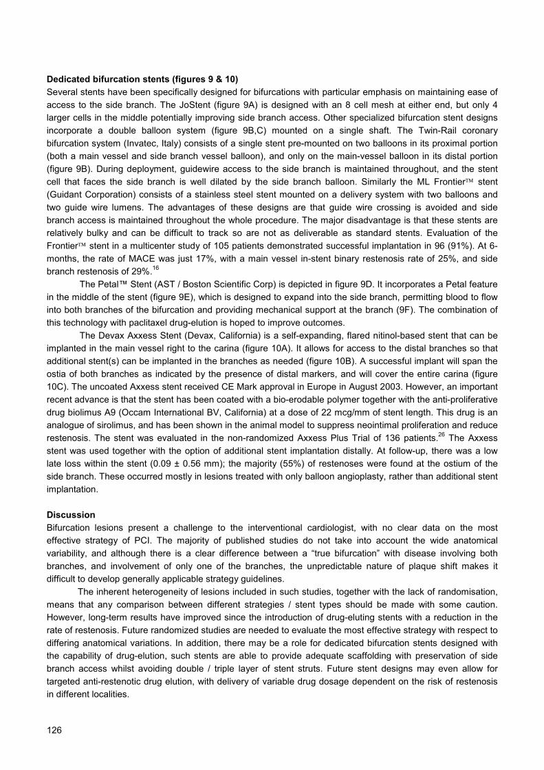
126
Dedicated bifurcation stents (figures 9 & 10)Several stents have been specifically designed for bifurcations with particular emphasis on maintaining ease ofaccess to the side branch. The JoStent (figure 9A) is designed with an 8 cell mesh at either end, but only 4larger cells in the middle potentially improving side branch access. Other specialized bifurcation stent designsincorporate a double balloon system (figure 9B,C) mounted on a single shaft. The Twin-Rail coronarybifurcation system (Invatec, Italy) consists of a single stent pre-mounted on two balloons in its proximal portion(both a main vessel and side branch vessel balloon), and only on the main-vessel balloon in its distal portion(figure 9B). During deployment, guidewire access to the side branch is maintained throughout, and the stentcell that faces the side branch is well dilated by the side branch balloon. Similarly the ML Frontier stent(Guidant Corporation) consists of a stainless steel stent mounted on a delivery system with two balloons andtwo guide wire lumens. The advantages of these designs are that guide wire crossing is avoided and sidebranch access is maintained throughout the whole procedure. The major disadvantage is that these stents arerelatively bulky and can be difficult to track so are not as deliverable as standard stents. Evaluation of theFrontier stent in a multicenter study of 105 patients demonstrated successful implantation in 96 (91%). At 6-months, the rate of MACE was just 17%, with a main vessel in-stent binary restenosis rate of 25%, and sidebranch restenosis of 29%.16
The Petal™ Stent (AST / Boston Scientific Corp) is depicted in figure 9D. It incorporates a Petal featurein the middle of the stent (figure 9E), which is designed to expand into the side branch, permitting blood to flowinto both branches of the bifurcation and providing mechanical support at the branch (9F). The combination ofthis technology with paclitaxel drug-elution is hoped to improve outcomes.
The Devax Axxess Stent (Devax, California) is a self-expanding, flared nitinol-based stent that can beimplanted in the main vessel right to the carina (figure 10A). It allows for access to the distal branches so thatadditional stent(s) can be implanted in the branches as needed (figure 10B). A successful implant will span theostia of both branches as indicated by the presence of distal markers, and will cover the entire carina (figure10C). The uncoated Axxess stent received CE Mark approval in Europe in August 2003. However, an importantrecent advance is that the stent has been coated with a bio-erodable polymer together with the anti-proliferativedrug biolimus A9 (Occam International BV, California) at a dose of 22 mcg/mm of stent length. This drug is ananalogue of sirolimus, and has been shown in the animal model to suppress neointimal proliferation and reducerestenosis. The stent was evaluated in the non-randomized Axxess Plus Trial of 136 patients.26 The Axxessstent was used together with the option of additional stent implantation distally. At follow-up, there was a lowlate loss within the stent (0.09 ± 0.56 mm); the majority (55%) of restenoses were found at the ostium of theside branch. These occurred mostly in lesions treated with only balloon angioplasty, rather than additional stentimplantation.
Discussion Bifurcation lesions present a challenge to the interventional cardiologist, with no clear data on the mosteffective strategy of PCI. The majority of published studies do not take into account the wide anatomicalvariability, and although there is a clear difference between a “true bifurcation” with disease involving bothbranches, and involvement of only one of the branches, the unpredictable nature of plaque shift makes itdifficult to develop generally applicable strategy guidelines. The inherent heterogeneity of lesions included in such studies, together with the lack of randomisation,means that any comparison between different strategies / stent types should be made with some caution.However, long-term results have improved since the introduction of drug-eluting stents with a reduction in therate of restenosis. Future randomized studies are needed to evaluate the most effective strategy with respect todiffering anatomical variations. In addition, there may be a role for dedicated bifurcation stents designed withthe capability of drug-elution, such stents are able to provide adequate scaffolding with preservation of sidebranch access whilst avoiding double / triple layer of stent struts. Future stent designs may even allow fortargeted anti-restenotic drug elution, with delivery of variable drug dosage dependent on the risk of restenosisin different localities.

127
Figure 9: Illustrations of some of the different stent types available that are specifically designed for side-branch access or bifurcation treatment. A: The middle section of the Jostent has fewer and larger cells to allowfor easier side-branch access. B: The Twin-Rail coronary bifurcation system (Invatec, Italy). C: The MLFrontier™ stent (Guidant). D,E,F: The Petal™ Stent (AST / Boston Scientific Corp) incorporates a Petal featurein the middle of the stent to scaffold the ostium of the side branch.
Figure 10: The Devax Axxess Stent (Devax, California) is a self-expanding, flared nitinol-based stent. It isimplanted in the main vessel right to the carina (figure 10B, red line); it allows for access to both the distalbranches so that additional stent(s) can be implanted in the branches as needed (yellow lines). The markersdemonstrate that the stent has covered the carina and the ostia of both branches (figure 10C).
A
B
C
D
E
F
A C
B

128
References1. Al Suwaidi J, Yeh W, Cohen HA et al. Immediate and one-year outcome in patients with coronary bifurcation lesions in the
modern era (NHLBI dynamic registry). Am J Cardiol. 2001;87:1139-44.
2. Ahmed JM, Mintz GS, Weissman NJ et al. Mechanism of lumen enlargement during intracoronary stent implantation: an
intravascular ultrasound study. Circulation. 2000;102:7-10.
3. Mintz GS, Pichard AD, Kent KM et al. Axial plaque redistribution as a mechanism of percutaneous transluminal coronary
angioplasty. Am J Cardiol. 1996;77:427-30.
4. Poerner TC, Kralev S, Voelker W et al. Natural history of small and medium-sized side branches after coronary stent
implantation. Am Heart J. 2002;143:627-35.
5. Califf RM, Abdelmeguid AE, Kuntz RE et al. Myonecrosis after revascularization procedures. J Am Coll Cardiol. 1998;31:241-51.
6. Abdelmeguid AE, Whitlow PL, Sapp SK et al. Long-term outcome of transient, uncomplicated in-laboratory coronary artery
closure. Circulation. 1995;91:2733-41.
7. George BS, Myler RK, Stertzer SH et al. Balloon angioplasty of coronary bifurcation lesions: the kissing balloon technique.
Cathet Cardiovasc Diagn. 1986;12:124-38.
8. Meier B. Kissing balloon coronary angioplasty. Am J Cardiol. 1984;54:918-20.
9. Brener SJ, Leya FS, Apperson-Hansen C et al A comparison of debulking versus dilatation of bifurcation coronary arterial
narrowings (from the CAVEAT I Trial). Am J Cardiol. 1996;78:1039-41.
10. Bhargava B, Waksman R, Lansky AJ et al. Clinical outcomes of compromised side branch (stent jail) after coronary stenting with
the NIR stent. Catheter Cardiovasc Interv. 2001;54:295-300.
11. Pan M, Suarez de Lezo J, Medina A et al. Simple and complex stent strategies for bifurcated coronary arterial stenosis involving
the side branch origin. Am J Cardiol. 1999;83:1320-5.
12. Yamashita T, Nishida T, Adamian MG et al. Bifurcation lesions: two stents versus one stent--immediate and follow-up results. J
Am Coll Cardiol. 2000;35:1145-51.
13. Al Suwaidi J, Berger PB, Rihal CS et al. Immediate and long-term outcome of intracoronary stent implantation for true bifurcation
lesions. J Am Coll Cardiol. 2000;35:929-36.
14. Anzuini A, Briguori C, Rosanio S et al. Immediate and long-term clinical and angiographic results from Wiktor stent treatment for
true bifurcation narrowings. Am J Cardiol. 2001;88:1246-50.
15. Sheiban I, Albiero R, Marsico F et al. Immediate and long-term results of "T" stenting for bifurcation coronary lesions. Am J
Cardiol. 2000;85:1141-4, A9.
16. Lefevre T, Ormiston J, Guagliumi G et al. The Frontier stent registry: safety and feasibility of a novel dedicated stent for the
treatment of bifurcation coronary artery lesions. J Am Coll Cardiol. 2005;46:592-8.
17. Colombo A, Moses JW, Morice MC et al. Randomized study to evaluate sirolimus-eluting stents implanted at coronary bifurcation
lesions. Circulation. 2004;109:1244-9.
18. Pan M, de Lezo JS, Medina A et al. Rapamycin-eluting stents for the treatment of bifurcated coronary lesions: a randomized
comparison of a simple versus complex strategy. Am Heart J. 2004;148:857-64.
19. Tanabe K, Hoye A, Lemos PA et al. Restenosis rates following bifurcation stenting with sirolimus-eluting stents for de novo
narrowings. Am J Cardiol. 2004;94:115-8.
20. Ormiston JA, Webster MW, Ruygrok PN et al. Stent deformation following simulated side-branch dilatation: a comparison of five
stent designs. Catheter Cardiovasc Interv. 1999;47:258-64.
21. Lefevre T, Louvard Y, Morice MC et al. Stenting of bifurcation lesions: classification, treatments, and results. Catheter Cardiovasc
Interv. 2000;49:274-83.
22. Chevalier B, Glatt B, Royer T, Guyon P. Placement of coronary stents in bifurcation lesions by the "culotte" technique. Am J
Cardiol. 1998;82:943-9.
23. Hoye A, van Mieghem CA, Ong AT et al. Percutaneous therapy of bifurcation lesions with drug-eluting stent implantation: the
Culotte technique revisited. Int J Cardiovasc Intervent. 2005;7:36-40.
24. Colombo A, Stankovic G, Orlic D et al. Modified T-stenting technique with crushing for bifurcation lesions: immediate results and
30-day outcome. Catheter Cardiovasc Interv. 2003;60:145-51.
25. Ge L, Airoldi F, Iakovou I et al. Clinical and angiographic outcome after implantation of drug-eluting stents in bifurcation lesions
with the crush stent technique: importance of final kissing balloon post-dilation. J Am Coll Cardiol. 2005;46:613-20.
26. Grube E, presented at DES Revolution IV, American College of Cardiology, Orlando, 2005

Chapter 13
AnW
JoCa20
New approaches to ostial and bifurcationlesions
129
gela Hoyeillem van der Giessen
urnal of Interventional rdiology04;17(6):397-403

130
131
Abstract
Percutaneous coronary intervention of bifurcation lesions is associated with lower procedural
success rates, and an increased subsequent rate of major adverse cardiac events and
restenosis. Currently, an array of stenting possibilities suggests a rational approach to treat
various bifurcation lesions with appropriate techniques. This is however seldom the case.
The main problems of treating bifurcation lesions remain plaque shift leading to (threatened)
side branch occlusion, and either too much or insufficient side branch ostial stent coverage
predisposing to impaired side branch access or restenosis respectively. This paper reviews
the available technologies and their relative merits.

132
IntroductionPercutaneous coronary intervention of bifurcation lesions is associated with lower procedural success rates, 1
and an increased subsequent rate of major adverse cardiac events (MACE) and restenosis compared with non-bifurcated lesions. Various techniques and strategies have been applied in attempt to improve outcomesincluding double wire technique, kissing balloon pre- and post-dilatation, stent implantation in the main branchor both main and side branch by T-, Y-, culotte-, trousers-, skirt-, kissing stents-, crush-, or touching stentstechnique. 2-5 This array of stenting possibilities suggests a rational approach to treat various bifurcation lesionswith appropriate techniques. This is however seldom the case. Usually new inventions are applied to mostlesions in an attempt to treat bifurcations in general, and the most effective strategy for different anatomicalvariations is currently unknown. The present paper attempts to familiarize the reader with the successivetechnical advances available to the interventional community and discuss their successes and failures.
Coronary bifurcation anatomy Lesions at a coronary bifurcation may involve either the main vessel alone, and / or the ostium of a side branch.A classical scheme to categorize coronary bifurcation lesions is the Duke classification (figure 1 below):
However, even when only one of the two branches is significantly stenosed at baseline, plaque shift or the“snow-plough effect” can pose a problem. Following balloon dilatation or stent implantation, shift ofatheromatous material may occur proximally, longitudinally, and/or circumferentially. 6 When treating the mainvessel, such shift of material can lead to side branch occlusion particularly when the ostium of the side branchis itself diseased, the side branch is of relatively small diameter, or in the presence of thrombus in acutecoronary syndromes. The clinical consequences of loss of the side branch are dependant on the vessel size,and are not usually serious with short-lived chest pain and only a modest rise in cardiac enzymes. In addition,follow-up evaluation, frequently demonstrates restoration of patency. 7 However, following PCI, a more thanthree-fold rise in cardiac enzymes above the upper limit of normal has been shown to have prognosticimplications. 8 In the NIRVANA study of the NIR stent implanted across a side branch, side branch occlusionoccurred in 4.7% patients. Of these, occlusion was associated with acute myocardial infarction (creatine-kinase-MB ≥5x normal) in 40%, including Q-wave infarction in 7%. 9
Balloon angioplastyIn the 1980’s, the majority of patients with a bifurcation lesion were referred for coronary artery bypass surgery.However, in those who were treated with percutaneous intervention, following identification of the problemsassociated with plaque shift, kissing balloon dilatation became the technique of choice. 10,11 The major limitationof balloon only angioplasty was the occurrence of acute recoil and vessel closure, and subsequent restenosis.Technical advances in the 1990’s, led to the evaluation of debulking techniques and scaffolding of the vessel(s)with stent implantation.
B
E
Postbranch
Ostial
A
D
Prebranch
Bifurcation
C
F
Parent vessel only
Prebranch and ostial
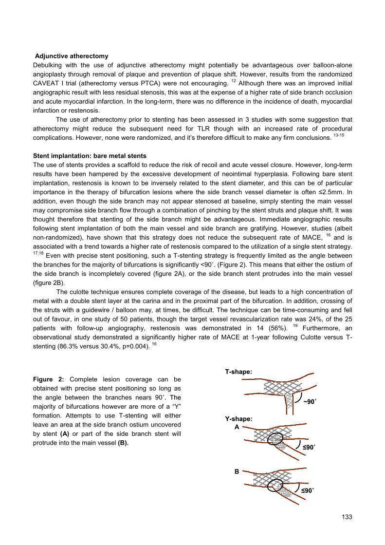
Adjunctive atherectomyDebulking with the use of adjunctive atherectomy might potentially be advantageous over balloon-aloneangioplasty through removal of plaque and prevention of plaque shift. However, results from the randomizedCAVEAT I trial (atherectomy versus PTCA) were not encouraging. 12 Although there was an improved initialangiographic result with less residual stenosis, this was at the expense of a higher rate of side branch occlusionand acute myocardial infarction. In the long-term, there was no difference in the incidence of death, myocardialinfarction or restenosis.
The use of atherectomy prior to stenting has been assessed in 3 studies with some suggestion thatatherectomy might reduce the subsequent need for TLR though with an increased rate of proceduralcomplications. However, none were randomized, and it’s therefore difficult to make any firm conclusions. 13-15
Stent implantation: bare metal stentsThe use of stents provides a scaffold to reduce the risk of recoil and acute vessel closure. However, long-termresults have been hampered by the excessive development of neointimal hyperplasia. Following bare stentimplantation, restenosis is known to be inversely related to the stent diameter, and this can be of particularimportance in the therapy of bifurcation lesions where the side branch vessel diameter is often ≤2.5mm. Inaddition, even though the side branch may not appear stenosed at baseline, simply stenting the main vesselmay compromise side branch flow through a combination of pinching by the stent struts and plaque shift. It wasthought therefore that stenting of the side branch might be advantageous. Immediate angiographic resultsfollowing stent implantation of both the main vessel and side branch are gratifying. However, studies (albeitnon-randomized), have shown that this strategy does not reduce the subsequent rate of MACE, 16 and isassociated with a trend towards a higher rate of restenosis compared to the utilization of a single stent strategy.17,18 Even with precise stent positioning, such a T-stenting strategy is frequently limited as the angle betweenthe branches for the majority of bifurcations is significantly <90˚. (Figure 2). This means that either the ostium ofthe side branch is incompletely covered (figure 2A), or the side branch stent protrudes into the main vessel(figure 2B).
The culotte technique ensures complete coverage of the disease, but leads to a high concentration ofmetal with a double stent layer at the carina and in the proximal part of the bifurcation. In addition, crossing ofthe struts with a guidewire / balloon may, at times, be difficult. The technique can be time-consuming and fellout of favour, in one study of 50 patients, though the target vessel revascularization rate was 24%, of the 25patients with follow-up angiography, restenosis was demonstrated in 14 (56%). 19 Furthermore, anobservational study demonstrated a significantly higher rate of MACE at 1-year following Culotte versus T-stenting (86.3% versus 30.4%, p=0.004). 16
Figure 2: Complete lesion coverage can beobtained with precise stent positioning so long asthe angle between the branches nears 90˚̊.. TThheemajority of bifurcations however are more of a “Y”formation. Attempts to use T-stenting will eitherleave an area at the side branch ostium uncoveredby stent (A) or part of the side branch stent willprotrude into the main vessel (B).
~~9900˚̊
TT--sshhaappee::
YY--sshhaappee::
A≤≤9900˚̊
B
133
≤≤9900˚̊

134
Dedicated stentsSeveral stents have been specifically designed for bifurcations with particular emphasis on maintaining ease ofaccess to the side branch. The JoStent is designed with an eight-cell mesh at either end, and only 4 cells in themiddle potentially improving side branch access (figure 3A). 20 The Devax Access Stent is a self-expanding,flared stent to allow for better access to bifurcation lesions. Additional stents are needed to treat the mainbranch and the side branch. Other specialized bifurcation stent designs incorporate a double balloon system(figure 3B, C). Most available data are in the form of case reports or registries.
The ML Frontier stent consists of a balloon expandable stainless steel stent mounted on a deliverysystem with two balloons and two guide wire lumens designed to maintain wire access to the side-branchvessel during stent deployment. The advantages of this design are that guide wire crossing is avoided and sidebranch access is maintained during the whole procedure. This two-balloon, two-wire system requires a 7F-guiding catheter. Evaluation of this device was carried out in 105 patients treated at 11 international sites (datapresented by T. Lefèvre at TCT 2003). The ML Frontier stent was successfully implanted in 96/105 patients,with the LAD/diagonal bifurcation as the target in 80% of cases. Acute device success by quantitative coronaryangiography was 92%. Two patients had in-hospital myocardial infarction secondary to side branch occlusion(1 Q-wave and 1 non-Q-wave MI), and one patient underwent planned in-hospital CABG after PCI failure. Noother MACE was observed during the 30-day clinical follow-up. At 180 days, the MACE rate was 17.1% (nodeath; Q-wave-MI: 1.9%; non-Q-wave-MI: 1.9%; TLR: 13.3%) and the main branch in-stent and in-segmentbinary restenosis rates were 25.3% and 29.9% respectively. These results are certainly comparable with bestresults obtained utilizing conventional bare metal stents (see table).
The DBS stent (Cordis) has been evaluated in a multicenter study of 34 patients. Procedural successwas achieved in 94% with, at 6 months, a restenosis rate of 33% and TLR rate of 19%. 21 Other bifurcationstent devices have also been developed and some data exists in the form of case reports. The Bard-XT Carinastent is pre-mounted on 2 balloons which connect to a single shaft and are simultaneously deployed with asingle indeflator (figure 4). 22 The AVE bifurcation stent is similarly delivered using the Bard delivery system.The AST SLK-View™ stent has an aperture in the middle segment of the stent to allow access to the sidebranch using a guiding tube. In an initial study of 8 patients, the stent was successfully delivered in all but one,with no adverse clinical events at 1 month. 23 The Invatec bifurcation stent has either a single or double balloonstent delivery system. The single balloon system is similar to the AST system mentioned above. The doubleballoon system is more like the ML Frontier™ stent except that the side branch balloon is a full dilatationballoon (but only 1.5 mm diameter) to be used for the kissing technique whereas the ML Frontier™ stent has ashort tapered side branch balloon to allow for side branch access and the physicians choice in treating the sidebranch or not. Especially the double balloon stent systems have a higher profile and are less flexible than the otherdesigns. That means that, in particular, in calcified coronaries their optimal deployment may be less successful.24
Table: Comparative Data of Bifurcation Stenting StudiesStudy N In-hosp
MACE6-Month
RR6-Month TLR
(total)6-Month MACE
(total)Lefevre 3 Period 1 182 5.1 - 20.6 29.2
Period 2 191 4.2 - 13.8 17.1Yamashita 18 2 stents 53 13 62 38 51
1 Stent 39 0 48 36 38Colombo 27 2 stents 63 9.5 28 9.5 19
1 Stent 22 9.1 18.7 4.5 13.6Frontier (Eur Heart J 2004;25
(Abstract Suppl): 309)
105 2.7 44.8 13.3 17.1
Unless indicated, all parameters presented as percent of patientsRR=Restenosis Rate >50% binary restenosis rate

135
Figure 3: Illustration of different stent types designed for side-branch access or bifurcation treatment. A: Themiddle section of the Jostent has fewer and larger cells to allowfor easier side-branch access. B: The earlyBARD side-branch stent carrying a side-branch wire crimped under the proximal main branch stent half. C: TheML Frontier has a main branch balloon holding the distal stent and a side-branch balloon for ostial protectionthat contains the OTW side-branch guide wire. D: The AVE bifurcated stent that was built on the former BARDplatform. This stent has been implanted in but a few patients.
Figure 4: Clinical implantation of one of the very first bifurcated stents in a 47-year-old patient in 1997. 31 A:LAD/diagonal type-D bifurcation lesion at baseline. B: Positioning of the bifurcated stent at the carina after priorpredilatation. C: Acute angiographic result. D: At 6-months follow-up the bifurcation still demonstrates nicepatency. At 6-years clinical follow-up she was free of symptoms, and working full time.
A
DC
B
A B
C D

136
Stent implantation: drug-eluting stentsBoth the sirolimus- and paclitaxel-eluting stents have demonstrated significantly reduced rates of restenosiscompared with bare metal stents, though bifurcation lesions were excluded from initial randomised studies. 25,26
Preliminary data of the sirolimus-eluting stent (SES) for bifurcation lesions has recently become available, andhas confirmed efficacy when compared with historical data with overall rates of restenosis of 23-26%, and needfor target lesion revascularization in 8-9%. 27,28 One of these studies randomized patients to either a strategy ofusing a single SES versus two stents and found that restenosis rates were similar between the two groups(19% for provisional side-branch stenting versus 28% for double-stenting). However, there was notably a highrate of crossover to the 2 stent strategy of 51%. 27 In an observational study of the sirolimus-eluting stent in aconsecutive group of patients, restenosis occurred particularly at the ostium of the side branch following theuse of T-stenting. 28 This might reflect incomplete coverage of the ostium thereby reducing the efficacy of thedrug-elution, and suggesting that it may be beneficial when using these stents to adopt a strategy that ensurescomplete coverage. Colombo et al first reported the crush technique utilizing SES in 2003, as a technicallystraightforward method that ensures complete coverage of the side branch ostium. 29 Of 20 patients, an in-hospital adverse event occurred in 3 (2 myocardial infarctions, one re-PTCA related to dissection of the mainvessel distal to the bifurcation), with no further events at 1 month. In particular there were no episodes of stentthrombosis. Further angiographic data on the subsequent rates of restenosis with this technique are pending.
General principlesAngiography must adequately evaluate the bifurcation, in particular whether the side branch ostium is involvedor not. The use of two wires is a simple method to ensure that access to each branch is preserved throughoutthe intervention. In general the use of pre-dilatation is recommended, and has the advantage of assessing howthe lesion behaves with evaluation of any plaque shift, and facilitation of subsequent stent implantation. Themajor drawback however, is the risk of dissection, or distal embolization particularly of thrombotic material inacute coronary syndromes. Whatever the stenting strategy chosen, post-dilatation with kissing balloondilatation is strongly recommended particularly when both branches are stented. Though not detectable onangiography, stent deformation may occur whatever the technique utilized, and is corrected with kissingballoons. 30
Ostial lesions represent a specific subset of bifurcations (Duke classification E). Although it may beattractive to simply stent the side branch, plaque shift into the main vessel may pose a problem, and the use oftwo wires is to be recommended to protect access to the other vessel. In addition, as with most bifurcations, theangle at the carina is not usually 90˚, so even with precise stent positioning either the disease is not entirelycovered, or some stent struts protrude into the main vessel. Discussion Bifurcation lesions still present a challenge to the interventional cardiologist, with no clear data on the mosteffective strategy during PCI. Thus far, the majority of published studies do not take into account the wideanatomical variability, and although there is a clear difference between a “true bifurcation” with diseaseinvolving both branches, and involvement of only one of the branches, the unpredictable nature of plaque shiftmakes the development of hard and fast guidelines more difficult. The inherent heterogeneity of lesions included in such studies, together with the lack of randomization,means that any comparison between different strategies / stent types should be made with some caution.However, through a reduction in restenosis, drug-eluting stents are certainly having a dramatic impact on thepractice of interventional cardiology, and efficacy certainly appears to be applicable for the treatment ofbifurcations (see table). However, restenosis at the side branch ostium can be a problem following T-stenting,and alternative strategies such as the crush or culotte technique automatically lead to a region of a double /triple layer of stent. This increased local dosage of drug could be potentially harmful with delayed re-endothelialization. In addition, both techniques involve the need to cross stent struts with a guidewire.Dedicated bifurcation stent systems can be bulky to implant and are, as yet, bare and subject to not

137
insignificant rates of restenosis. However, maintenance of side branch access is improved, and the need forseveral stent layers is avoided.
The introduction of drug-eluting stents means that new randomized studies are needed to defineguidelines for PCI strategy in the modern era. These studies need also to better evaluate the best technique ofstent implantation particularly with respect to anatomical variations. In addition, there may be a role fordedicated bifurcation stents designed with the capability of drug-elution. These may be able to provideadequate scaffolding with preservation of side branch access while avoiding double / triple layer of stent struts.In addition, future stent designs may even allow for targeted anti-restenotic drug elution, with delivery ofvariable drug dosage dependent on the risk of restenosis in different localities.

138
References1. Al Suwaidi J, Yeh W, Cohen HA et al. Immediate and one-year outcome in patients with coronary bifurcation lesions in the
modern era (NHLBI dynamic registry). Am J Cardiol. 2001;87:1139-44.
2. Lefevre T, Louvard Y, Morice MC et al. Stenting of bifurcation lesions: a rational approach. J Interv Cardiol. 2001;14:573-85.
3. Lefevre T, Louvard Y, Morice MC et al. Stenting of bifurcation lesions: classification, treatments, and results. Catheter Cardiovasc
Interv. 2000;49:274-83.
4. Louvard Y, Lefevre T, Morice MC. Percutaneous coronary intervention for bifurcation coronary disease. Heart. 2004;90:713-22.
5. Melikian N, Di Mario C. Treatment of bifurcation coronary lesions: a review of current techniques and outcome. J Interv Cardiol.
2003;16:507-13.
6. Ahmed JM, Mintz GS, Weissman NJ et al. Mechanism of lumen enlargement during intracoronary stent implantation: an
intravascular ultrasound study. Circulation. 2000;102:7-10.
7. Poerner TC, Kralev S, Voelker W et al. Natural history of small and medium-sized side branches after coronary stent
implantation. Am Heart J. 2002;143:627-35.
8. Califf RM, Abdelmeguid AE, Kuntz RE et al. Myonecrosis after revascularization procedures. J Am Coll Cardiol. 1998;31:241-51.
9. Bhargava B, Waksman R, Lansky AJ et al. Clinical outcomes of compromised side branch (stent jail) after coronary stenting with
the NIR stent. Catheter Cardiovasc Interv. 2001;54:295-300.
10. Meier B. Kissing balloon coronary angioplasty. Am J Cardiol. 1984;54:918-20.
11. George BS, Myler RK, Stertzer SH et al. Balloon angioplasty of coronary bifurcation lesions: the kissing balloon technique.
Cathet Cardiovasc Diagn. 1986;12:124-38.
12. Brener SJ, Leya FS, Apperson-Hansen C et al. A comparison of debulking versus dilatation of bifurcation coronary arterial
narrowings (from the CAVEAT I Trial). Coronary Angioplasty Versus Excisional Atherectomy Trial-I. Am J Cardiol. 1996;78:1039-
41.
13. Gambhir DS, Singh S, Sinha SC et al. Treatment of true bifurcation stenosis by elective stent implantation in parent vessel and
non-stent dilatation of side branch: immediate and follow-up results. Indian Heart J. 2000;52:289-96.
14. Chieffo A. Stankovic G, Briguori C et al. Acute and late outcome after directional atherectomy plus stenting versus stenting alone
in true bifurcation lesions. Am J Cardiol. 2002;90(suppl 6A):44H.
15. Karvouni E, Di Mario C, Nishida T et al. Directional atherectomy prior to stenting in bifurcation lesions: a matched comparison
study with stenting alone. Catheter Cardiovasc Interv. 2001;53:12-20.
16. Al Suwaidi J, Berger PB, Rihal CS et al. Immediate and long-term outcome of intracoronary stent implantation for true bifurcation
lesions. J Am Coll Cardiol. 2000;35:929-36.
17. Sheiban I, Albiero R, Marsico F et al. Immediate and long-term results of "T" stenting for bifurcation coronary lesions. Am J
Cardiol. 2000;85:1141-4, A9.
18. Yamashita T, Nishida T, Adamian MG et al. Bifurcation lesions: two stents versus one stent--immediate and follow-up results. J
Am Coll Cardiol. 2000;35:1145-51.
19. Chevalier B, Glatt B, Royer T, Guyon P. Placement of coronary stents in bifurcation lesions by the "culotte" technique. Am J
Cardiol. 1998;82:943-9.
20. Lowe HC, Kumar R, Roy PR. New balloon expandable stent for bifurcation lesions. Cathet Cardiovasc Diagn. 1997;42:235-6.
21. Dibie A, Chevalier B, Fajadet J et al. First in man study with a coronary stent specifically designed for true bifurcated lesions: the
DBS stent. Am J Cardiol. 2002;90(suppl 6A):13H.
22. Cervinka P, Foley DP, Sabate M et al. Coronary bifurcation stenting using dedicated bifurcation stents. Catheter Cardiovasc
Interv. 2000;49:105-11.
23. Toutouzas K, Stankovic G, Takagi T et al. A new dedicated stent and delivery system for the treatment of bifurcation lesions:
preliminary experience. Catheter Cardiovasc Interv. 2003;58:34-42.
24. Van der Giessen WJ, Smits PC, de Feyter PJ et al. Stenting Under Intravascular Ultrasound Guidance of Coronary Bifurcation
Lesions With a New Device Allowing Provisional Side Branch Treatment. J Am Coll Cardiol. 2004;43(Suppl A):Abstract 878-6.
25. Morice MC, Serruys PW, Sousa JE et al. A randomized comparison of a sirolimus-eluting stent with a standard stent for coronary
revascularization. N Engl J Med. 2002;346:1773-80.
26. Grube E, Silber S, Hauptmann KE et al. TAXUS I: six- and twelve-month results from a randomized, double-blind trial on a slow-
release paclitaxel-eluting stent for de novo coronary lesions. Circulation. 2003;107:38-42.

139
27. Colombo A, Moses JW, Morice MC et al. Randomized study to evaluate sirolimus-eluting stents implanted at coronary bifurcation
lesions. Circulation. 2004;109:1244-9.
28. Tanabe K, Hoye A, Lemos PA et al. Restenosis rates following bifurcation stenting with sirolimus-eluting stents for de novo
narrowings. Am J Cardiol. 2004;94:115-8.
29. Colombo A, Stankovic G, Orlic D et al. Modified T-stenting technique with crushing for bifurcation lesions: immediate results and
30-day outcome. Catheter Cardiovasc Interv. 2003;60:145-51.
30. Ormiston JA, Webster MW, Ruygrok PN et al. Stent deformation following simulated side-branch dilatation: a comparison of five
stent designs. Catheter Cardiovasc Interv. 1999;47:258-64.
31. Carlier SG, van der Giessen WJ, Foley DP et al. Stenting with a true bifurcated stent: acute and mid-term follow-up results.
Catheter Cardiovasc Interv. 1999;47:361-96.

140
141
Chapter 14
Kengo TanabeAngela HoyePedro A LemosJiro AokiChourmouzios ArampatzisFrancesco SaiaCH LeeMuzaffer DegertekinSjoerd H HofmaGeorgios SianosEugene McFaddenPieter C SmitsWillem J van der GiessenPim J de FeyterRon T van DomburgPatrick W Serruys
American JournalOf Cardiology2004;94(1):115-8
Restenosis Rates Following BifurcationStenting with Sirolimus-Eluting Stents for
De Novo Narrowings
142
Abstract
The percutaneous treatment of coronary bifurcation stenoses is hampered by an increased
rate of subsequent restenosis. The present study reports on the outcomes of a consecutive
series of 58 patients with 65 de novo bifurcation stenoses treated with sirolimus-eluting stent
implantation in both the main vessel and side branch. At 6 months, the incidence of major
adverse cardiac events was 10.3% (1 death and 5 target lesion revascularizations) with no
episodes of acute myocardial infarction or stent thrombosis.
143
Percutaneous coronary intervention of bifurcation lesions is associated with lower procedural success rates 1
and an increased subsequent rate of major adverse cardiac events (MACEs) and restenosis. Varioustechniques and strategies have been applied in an attempt to improve outcomes, including kissing balloondilatation and the use of stent implantation in both branches. 2 The use of adjunctive atherectomy was found tobe disadvantageous in the Coronary Angioplasty Versus Excisional Atherectomy Trial (CAVEAT-I) trial. 3
Although there was an improved initial angiographic result with less residual stenosis, this was at the expenseof a higher rate of side branch occlusion and acute myocardial infarction. In the long-term, results ofangioplasty in bifurcations have been hampered by problems of restenosis, particularly after stent implantationwithin the side branch. 4,5 Recently, sirolimus-eluting stents (SESs) have demonstrated dramatically reducedrestenosis rates in patients with relatively simple lesions. 6,7 We sought to investigate the safety and efficacy ofSESs in a consecutive series of unselected patients with de novo bifurcation lesions enrolled in the Rapamycin-Eluting Stent Evaluation At Rotterdam Cardiology Hospital (RESEARCH) registry. 8
Since April 2002, SES implantation (Cypher, Johnson & Johnson–Cordis, Miami, Florida) has beenused as the default strategy for all patients treated in our institution, as part of the RESEARCH registry. 8
Briefly, this single-center registry aims to evaluate the efficacy of SES implantation in the "real world" ofinterventional cardiology. All consecutive patients were enrolled, irrespective of clinical presentation and lesioncharacteristics, and the incidence of MACEs was prospectively evaluated during follow-up. At 6 months, a totalof 563 consecutive patients were treated solely with SESs. Of these, 58 patients (10.3%) with de novobifurcation lesions were treated with SES implantation in both the main and side branches; these patientscomprise the present study population. The patients' informed written consent was obtained in accordance withthe rules of the institutional ethics committee, which approved the study.
All procedures were performed with standard interventional techniques, except with the use of the SESas the device of choice. The strategy of bifurcation stenting employed and the use of kissing balloon dilatationafter procedure was at the operators' discretion. One of 4 methods of stenting was used: T-stenting, culottestenting, kissing stents, or the "crush" technique. T-stenting and culotte stenting have been previouslydescribed. 5,9 Kissing stents involved simultaneous implantation of the stents within both branches, with theproximal edges alongside each other, thereby bringing forward the point of divergence. The crush techniqueinvolves positioning both stents, with the proximal part of the side branch stent lying well within the main vessel,while ensuring that the edge of the stent in the main vessel is more proximal than the side branch stent. Theside branch stent is deployed first, and the balloon and wire are carefully withdrawn. The main vessel stent isthen deployed, thereby crushing the proximal part of the side branch stent. 10 SESs were available in diametersfrom 2.25 to 3.00 mm and lengths from 8 to 33 mm. During the procedure, intravenous heparin was given tomaintain an activated clotting time of ≥250 seconds. All patients were prescribed lifelong aspirin and clopidogrelfor 6 months. The use of glycoprotein IIb/IIIa inhibitors was at the discretion of the operator.
Clinical and angiographic follow-up was performed at 6 months. MACEs were predefined as death,myocardial infarction, or target lesion revascularization. The diagnosis of myocardial infarction required anelevation of creatine kinase levels to twice the upper limit of normal, together with an increase in the creatinekinase-MB fraction. Target lesion revascularization was defined as either surgical or percutaneousreintervention driven by significant (>50%) luminal diameter narrowing either within the stent or the 5-mmborders proximal and distal to the stent, and was undertaken in the presence of either anginal symptoms orobjective evidence of ischemia.
Coronary angiograms were obtained in multiple views after intracoronary injection of nitrates. For themain branches, 3 coronary segments were subjected to quantitative angiography: in-stent, proximal edge, anddistal edge segment. The in-stent analysis encompassed the length of all stents used during the procedure.The proximal and distal edge segment included up to 5 mm from the proximal and distal edge of the totalsegment treated with the study stents, respectively. For the side branches, 2 segments were analyzed: in-stentand distal edge 5-mm segment. Quantitative coronary angiographic (QCA) analysis was performed using theCardiovascular Angiography Analysis System II (CAAS II; Pie Medical, Maastricht, The Netherlands). Thereference vessel diameter, minimal lumen diameter, and percent diameter stenosis were measured before andafter the procedure and at follow-up. The late loss was calculated as the difference between the minimal lumen

144
diameter after the procedure and that at follow-up. Binary restenosis was defined as the presence of >50%diameter stenosis within the target lesion.
Fifty-eight patients with 65 bifurcation lesions were included in this study. Baseline patientcharacteristics are listed in Table 1. The lesion characteristics and stenting technique utilized are presented inTable 2. At 6 months, the survival-free of MACEs was 89.7%. One patient died after bifurcation stentimplantation of the left main stem for an acute myocardial infarction. This patient was admitted in cardiogenicshock, and despite the use of abciximab and intra-aortic balloon pump support, died shortly after the proceduredue to left ventricular failure. There were no episodes of acute or subacute stent thrombosis, and no patienthad a myocardial infarction. Target lesion revascularization was undertaken in 5 patients (8.6%) as outlined inthe following.
Table 1: Baseline Clinical Characteristics (n=58)Age (yrs) 63 ± 10Men 42 (72%)Hypertension 26 (45%)Hypercholesterolemia 35 (60%)Diabetes Mellitus 16 (28%)Current smoker 16 (28%)Previous myocardial infarction 22 (38%)Previous coronary angioplasty 5 (9%)Previous coronary artery bypass surgery 3 (5%)
1 15 (26%)2 28 (48%)
No. of coronary arteries significantly narrowed
3 15 (26%)Presentation with an acute coronary syndrome 18 (31%)
Values are presented as the numbers (relative percentages) or mean value ± SD.
Table 2: Lesion and Procedural Characteristics (number of lesions = 65)
Left anterior descending / diagonal 39 60%)
Left circumflex / obtuse marginal 16 (25%)
Right coronary / posterior descending 4 (6%)
Coronary artery treated withbifurcation stenting
Left main stem – left anterior descending / circumflex 6 (9%)
T-stenting 41 (63%)
Culotte stenting 5 (8%)
Kissing stenting 2 (3%)
Stenting technique
Crush stenting 17 (26%)
Kissing balloon dilatation after stenting 20 (31%)
Glycoprotein IIb/IIIa inhibitor use 20 (31%)
Values are presented as the numbers (relative percentages).
Of 65 lesions, 6-month angiographic follow-up was performed in 44 lesions. The binary restenosis rate was22.7% (10 of 44 lesions). QCA data are presented in Table 3. Angiographic restenosis occurred in 4 lesionswithin the main branch (1 in the proximal segment; 3 in the in-stent segment), yielding a restenosis rate of

145
9.1%. Angiographic restenosis occurred in 6 of the side branches, all within the in-stent segment. Of these 6restenoses, 5 occurred at the ostium of side branch after the use of T-stenting (Figure 1). All 4 patients with arestenosis within the main vessel and 1 patient with a restenosis at the ostium of a side branch underwentpercutaneous target lesion revascularization with new drug-eluting stent implantation. Directional coronaryatherectomy was additionally used in 1 patient. The remaining 5 patients, all with ostial side branch restenoses,were asymptomatic and treated with medical therapy alone.
Table 3: Quantitative Coronary AngiographyProximal Segment In-stent Segment Distal Segment
Main branch (n=44)Reference diameter (mm) N/A 2.64 N/A
Preprocedure N/A 0.64 N/APost-procedure 2.39 2.19 1.86
Minimal lumendiameter (mm)
6-mo follow-up 2.26 2.07 1.85Diameter stenosis at 6 mo (%) 28.3 22.9 25.4Late lumen loss (mm) 0.12 0.12 0.01Restenosis rate (%) 2.3 6.8 0
Side branch (n=44)Reference diameter (mm) 1.99 N/A
Preprocedure 0.61 N/APost-procedure 1.80 1.57
Minimal lumendiameter (mm)
6-mo follow-up 1.49 1.47Diameter stenosis at 6 mo (%) 31.0 21.9Late lumen loss (mm) 0.31 0.09Restenosis rate (%) 13.6 0
Values are presented as mean values or relative percentages.
Figure 1: A 3.0 × 33 mm SES was implanted in the circumflex artery, and a 2.25 × 8 mm SES was implanted inthe side branch (obtuse marginal) with T-stenting technique (A). At 6-month angiographic follow-up, restenosisoccurred at the ostium of the side branch (arrowhead) (B).
A B
SES2.25 x 8SES
3.0 x 33

146
The major findings of this study of bifurcation stenting include the following. (1) SES implantation in both themain and side branches is feasible and associated with a low procedural complication rate and no episodes ofstent thrombosis. (2) The target lesion revascularization rate of 8.6% is seemingly diminished compared withhistorical controls. (3) Angiographic restenosis rates of the main and side branches are 9.1% and 13.6%,respectively, with an overall restenosis rate of 22.7%. (4) Five of the 6 restenoses occurring in the side branchwere located at the ostium after using the T-stenting technique.
Drug-eluting stent deployment in both vessels to treat bifurcation lesions may raise theoreticalconcerns that it could result in a propensity to stent thrombosis. When we treat bifurcation lesions with SESsusing the culotte, kissing, or crush stenting techniques, there are some overlapping stent struts, where thehigher concentration of sirolimus may induce endothelial function impairment and thus be associated with anincreased rate of stent thrombosis. Although these stenting techniques were applied in 37% of the lesionstreated, no stent thrombosis was reported during follow-up, implying that sirolimus has a wide safety margin.
Several strategies have been advocated to treat bifurcation lesions with percutaneous coronaryintervention, such as deployment of stents in both vessels, stenting in 1 branch with balloon angioplasty in theother, and mechanical debulking. The published reports regarding the subsequent need for target lesionrevascularization utilizing bare stents range from 17% to 53%; 5,11,12 thus, the rate of 8.6% in our study is veryfavorable. In addition, the rate observed in the present study may underestimate the true beneficial treatmenteffect of SES as explained in the following.
Five of the 6 restenoses in the side branch occurred at the ostium after T-stenting. When we apply T-stenting, stent positioning must be extremely accurate to ensure complete coverage of the side branch ostium.This is particularly difficult and/or impossible to achieve when the angle between the 2 branches is much <90°.Restenosis at this site may therefore be mainly a reflection of incomplete coverage. The restenosis rate in theside branch following T-stenting was 16.7% (5 of 30 lesions), whereas that following the other stent techniqueswas 7.1% (1 of 14 lesions). The present study is limited because the choice of strategy was nonrandomized,and there is no comparison with alternative strategies, such as the use of stent implantation in the main vesselalone, with balloon-only angioplasty of the side branch. In addition, the sample size was relatively small, andany difference between the different techniques was not statistically significant. However, our results suggestthat it seems wise to ensure the complete coverage of the ostium with SESs using stenting techniques otherthan T-stenting. The crush technique is technically easier and quicker to do than a culotte, but further data withlonger follow-up from a larger population are needed to fully determine the efficacy of these techniques.

147
References1. Al Suwaidi J, Yeh W, Cohen HA, Detre KM, Williams DO, Holmes DR, Jr. Immediate and one-year outcome in patients with
coronary bifurcation lesions in the modern era (NHLBI dynamic registry). Am J Cardiol. 2001;87:1139-44.
2. Lefevre T, Louvard Y, Morice MC, Loubeyre C, Piechaud JF, Dumas P. Stenting of bifurcation lesions: a rational approach. J
Interv Cardiol. 2001;14:573-85.
3. Brener SJ, Leya FS, Apperson-Hansen C, Cowley MJ, Califf RM, Topol EJ. A comparison of debulking versus dilatation of
bifurcation coronary arterial narrowings (from the CAVEAT I Trial). Coronary Angioplasty Versus Excisional Atherectomy Trial-I.
Am J Cardiol. 1996;78:1039-41.
4. Anzuini A, Briguori C, Rosanio S, Tocchi M, Pagnotta P, Bonnier H, Gimelli G, Airoldi F, Margonato A, Legrand V, Colombo A.
Immediate and long-term clinical and angiographic results from Wiktor stent treatment for true bifurcation narrowings. Am J
Cardiol. 2001;88:1246-50.
5. Pan M, Suarez de Lezo J, Medina A, Romero M, Hernandez E, Segura J, Castroviejo JR, Pavlovic D, Melian F, Ramirez A,
Castillo JC. Simple and complex stent strategies for bifurcated coronary arterial stenosis involving the side branch origin. Am J
Cardiol. 1999;83:1320-5.
6. Degertekin M, Serruys PW, Foley DP, Tanabe K, Regar E, Vos J, Smits PC, van der Giessen WJ, van den Brand M, de Feyter P,
Popma JJ. Persistent inhibition of neointimal hyperplasia after sirolimus-eluting stent implantation: long-term (up to 2 years)
clinical, angiographic, and intravascular ultrasound follow-up. Circulation. 2002;106:1610-3.
7. Morice MC, Serruys PW, Sousa JE, Fajadet J, Ban Hayashi E, Perin M, Colombo A, Schuler G, Barragan P, Guagliumi G,
Molnar F, Falotico R. A randomized comparison of a sirolimus-eluting stent with a standard stent for coronary revascularization.
N Engl J Med. 2002;346:1773-80.
8. Lemos PA, Lee CH, Degertekin M, Saia F, Tanabe K, Arampatzis CA, Hoye A, van Duuren M, Sianos G, Smits PC, de Feyter P,
van der Giessen WJ, van Domburg RT, Serruys PW. Early outcome after sirolimus-eluting stent implantation in patients with
acute coronary syndromes: insights from the Rapamycin-Eluting Stent Evaluated At Rotterdam Cardiology Hospital
(RESEARCH) registry. J Am Coll Cardiol. 2003;41:2093-9.
9. Chevalier B, Glatt B, Royer T, Guyon P. Placement of coronary stents in bifurcation lesions by the "culotte" technique. Am J
Cardiol. 1998;82:943-9.
10. Colombo A, Stankovic G, Orlic D, Corvaja N, Liistro F, Airoldi F, Chieffo A, Spanos V, Montorfano M, Di Mario C. Modified T-
stenting technique with crushing for bifurcation lesions: immediate results and 30-day outcome. Catheter Cardiovasc Interv.
2003;60:145-51.
11. Al Suwaidi J, Berger PB, Rihal CS, Garratt KN, Bell MR, Ting HH, Bresnahan JF, Grill DE, Holmes DR, Jr. Immediate and long-
term outcome of intracoronary stent implantation for true bifurcation lesions. J Am Coll Cardiol. 2000;35:929-36.
12. Yamashita T, Nishida T, Adamian MG, Briguori C, Vaghetti M, Corvaja N, Albiero R, Finci L, Di Mario C, Tobis JM, Colombo A.
Bifurcation lesions: two stents versus one stent--immediate and follow-up results. J Am Coll Cardiol. 2000;35:1145-51.

148

149
Chapter 15
Angela HoyeCarlos AG van MieghemAndrew TL OngJiro AokiGaston A Rodriguez GranilloMarco ValgimigliGeorgios SianosEugene McFaddenWillem J van der GiessenPim J de FeyterRon T van DomburgPatrick W Serruys
Eurointervention:2005;1:24-30
Treatment of De Novo Bifurcation Lesions:Comparison of Sirolimus- and Paclitaxel-
Eluting Stents

150
151
Abstract
Objective: Both the sirolimus-(SES) and paclitaxel-eluting (PES) stents have been shown to
reduce restenosis rates when used in relatively simple lesions. This study aimed to evaluate
the results of a consecutive series of patients treated with drug-eluting stent implantation for
de novo bifurcation lesions, and compared outcomes with respect to stenting strategy and
stent type.
Patients: From April 2002 to September 2003, all patients at our institution were treated with
drug-eluting stent implantation. A consecutive series of 144 patients were treated for 167 de
novo bifurcation lesions with SES, followed by 104 patients treated with PES for 113 lesions.
Results: Clinical follow-up at 9-months was obtained in 99% patients with survival-free of
major adverse cardiac events (MACE) of 86.7% for SES versus 78.6% for PES, p=0.09.
Independent predictors of MACE were diabetes mellitus, previous CABG, multivessel
disease, and treatment for acute myocardial infarction. Survival-free of target lesion
revascularization (TLR) was 90.9% for SES versus 81.6% for PES, p=0.03, with stent type
being the only independent predictor. Technique of stenting was not a predictor of either
MACE or TLR.
Conclusions: MACE rates for both the SES and PES are low compared with historical data
of bare metal stents. The most effective techniques for bifurcation stenting remain undefined.
Our data suggests a higher need for TLR for the PES compared with the SES, however
further randomized studies are needed to fully evaluate both stenting strategy, and any
difference between the stents.

152
IntroductionThe outcome of percutaneous therapy (PCI) of bifurcation lesions with bare metal stents is hindered by anincreased rate of procedural complications, 1 and a high rate of restenosis particularly when both the mainvessel and side branch are stented. 2-6 The advent of drug-eluting stents is revolutionising the practice ofinterventional cardiology by demonstrating a reduction in the subsequent rate of restenosis. There is evidenceof efficacy in randomized trials for both the sirolimus- (SES) and paclitaxel-eluting (PES) stents for thetreatment of relatively simple lesions. 7,8 In addition, the sirolimus-eluting stent for the treatment of bifurcationlesions has demonstrated a low rate of adverse cardiac events compared with historical data utilizing baremetal stents. 9,10 However, the most effective technique of stenting for bifurcation lesions with drug-elutingstents is currently unknown. In the present report we evaluate the rate of major adverse cardiac eventsfollowing PCI for bifurcation lesions treated with either SESs or PESs in a consecutive series of patients. Inaddition, outcomes were assessed with respect to the baseline bifurcation anatomy and type of stentingstrategy employed.
MethodsBifurcation classification: All lesions were classified on baseline angiography according to the Dukeclassification (figure 1).
Figure 1: the Duke classification of bifurcation lesions
Procedure: The sirolimus-eluting stent (Cypher™, Johnson & Johnson - Cordis unit) received CE markapproval in April 2002. Since that time, all patients undergoing percutaneous therapy in our institution havebeen treated with drug-eluting stent implantation as the default strategy. During the first quarter of 2003, ourstrategy switched from the sirolimus- to the paclitaxel-eluting stent (Boston Scientific) enabling a comparison ofthe two stent types. All consecutive patients were enrolled irrespective of clinical presentation and lesioncharacteristics, and the incidence of major adverse cardiac events (MACE) was prospectively evaluated duringthe follow-up.
All procedures were performed with standard interventional techniques. The strategy of bifurcationstenting employed, and the use of kissing balloon dilatation post-procedure were at the operators’ discretion.One of 6 methods of stenting was used: stenting of the main vessel with balloon-only angioplasty of the sidebranch; type A T-stenting (stenting first of the side branch, followed by stenting of the main vessel); type B T-stenting (stenting of the main vessel followed by stenting of the side branch because of a sub-optimal result); 2
the `crush` technique; 11 culotte stenting; 12 or kissing stents (simultaneous implantation in the main vessel andside branch with the proximal edges of the stents side by side). SESs were available in diameters from 2.25mmto 3.00mm and lengths from 8mm to 33mm. PESs were available in diameters from 2.25mm to 3.5mm and
B
E
Postbranch
Ostial
A
D
Prebranch
Bifurcation
C
F
Parent vessel only
Prebranch and ostial
153
lengths from 8mm to 32mm. During the procedure, intravenous heparin was given to maintain an activatedclotting time ≥250 seconds. Patients were preloaded with 300mg clopidogrel, and received life-long aspirintogether with 75mg clopidogrel per day for 6-months. The use of glycoprotein IIb/IIIa inhibitors was at thediscretion of the operator. The protocol was approved by the Institutional ethics committee and is in accordancewith the principles of Good Clinical Practice for Trials of Medicinal Products in the European Community andthe Declaration of Helsinki. All patients signed a written informed consent
Follow-up: Clinical follow-up was obtained by investigators who were blinded to the PCI procedural details,using telephone calls and questionnaires. Follow-up evaluated the rate of major adverse cardiac events(MACE) which were pre-defined as death, acute myocardial infarction (AMI), or target vessel revascularization(TVR). The diagnosis of AMI required an elevation of creatine kinase levels to twice the upper limit of normal,together with a rise in creatine kinase-MB fraction. Target lesion revascularization was defined as eithersurgical or percutaneous reintervention driven by significant (>50%) luminal diameter narrowing either withinthe stent or the 5mm borders proximal and distal to the stent, and was undertaken in the presence of eitheranginal symptoms or objective evidence of ischemia. Target vessel revascularization was defined asrevascularization within the target vessel including encompassing the target lesion. The definition of stentthrombosis was the presence of intra-stent thrombosis, with or without stent occlusion, documented onangiography, and was categorized as acute if occurring within 24 hours or subacute if within 30 days after stentimplantation.
Statistical analysis: Discrete variables are presented as percentages and compared with Fisher exact test.Continuous variables are expressed as mean ± standard deviation and compared with Student’s t test.Cumulative survival and MACE-free survival were calculated according to the Kaplan-Meier method. The log-rank test was used to compare MACE-free survival between the two groups. The variables shown in Tables 1and 2 were tested in Cox regression modeling to identify the independent predictors of adverse events. Oddsratio with corresponding 95% confidence intervals are reported. All tests were two-tailed, and a p value of <0.05was considered as significant.
ResultsThe baseline patient and procedural characteristics for the SES and PES cohorts are presented in tables 1 and2 respectively. There were no significant differences between the 2 groups with respect to baseline patientcharacteristics, though there was a trend towards an increased usage of glycoprotein IIb/IIIa inhibitors in thePES group (38.5% versus 27.8% in the SES group, p=0.07). There was no significant difference in the numberof stents used, however, the mean nominal diameter of stent used in the main vessel was greater with the PES(2.93 ± 0.34mm versus 2.85 ± 0.23 for the SES, p=0.007). For those patients treated with stent implantation inthe side branch, though there was no significant difference in the number of stents used, the total length ofstented segment in the side branch was longer for the PES-treated patients (18.8 ± 10.5mm versus 14.1 ±7.6mm, p=0.0001). The choice of stenting strategy during the 2 treatment periods is presented in figure 2. Thetotal number of lesions treated with each stenting technique was single stent utilization in 55 (19.6%), type A T-stenting in 47 (16.8%), type B T-stenting in 46 (16.4%), crush stenting in 88 (31.4%), culotte stenting in 24(8.6%), and kissing stents in 20 (7.1%). There was no difference with respect to the use of kissing balloon post-dilatation between the SES and PES cohorts.
Clinical follow-up was obtained in 99.2% patients. Stent thrombosis occurred in 2 patients treated withSES (1.4%) and 3 patients treated with PES (2.9%), p=0.4. The demographics of these patients are in table 3.All episodes of stent thrombosis were subacute (within 30 days following stent implantation), and were treatedpercutaneously with kissing balloon dilatation and administration of a glycoprotein IIb/IIIa inhibitor. The onlyindependent predictor for stent thrombosis was therapy for AMI (HR 9.4; 95% CI 1.57 to 56.21, p=0.01).

154
Table 1: Baseline patient demographicsSES
n=144PES
n=104p value
Mean age (years) 62.4 ± 10.5 60.3 ± 11.8 0.1Male sex (%) 74.3 73.1 1Current smoker (%) 27.1 27.9 1Diabetes mellitus (%) 18.8 17.3 1Hypertension (%) 43.1 46.2 0.7Hypercholesterolemia (%) 56.9 62.5 0.3Previous myocardial infarction (%) 35.4 38.5 0.2Previous CABG (%) 4.9 3.8 0.9Clinical presentation 0.4
Stable angina (%) 65.3 67.3Unstable angina (%) 21.5 17.3Acute ST-elevation myocardial infarction (%) 13.2 16.3
Glycoprotein IIb/IIIa inhibitor usage (%) 27.8 38.5 0.07PCI in at least one additional major epicardial vessel duringthe index procedure (%)
40.3 39.4 1
Table 2: Baseline procedural characteristics
Total number of bifurcation lesions treatedSES167
PES113
p value
Target vessel 0.3LAD / diagonal (%) 61.1 56.6LCX / obtuse marginal (%) 19.2 17.7RCA bifurcation (%) 9.6 8.0LMS (%) 10.2 17.7
Bifurcation classification 0.4A (%) 4.8 3.5B (%) 7.2 5.3C (%) 8.4 6.2D (%) 17.5 20.4E (%) 8.4 3.5F (%) 44.0 50.4Total occlusion (TIMI 0 flow) (%) 9.6 10.6
Pre-dilatation of main vessel (%) 59.3 54.0 0.4Pre-dilatation of the side branch (%) 42.5 31.9 0.07Pre-dilatation with kissing balloons (%) 15.0 13.3 0.9Mean number of stents in the main vessel 1.56 ± 0.84 1.48 ± 0.67 0.4Mean nominal diameter of stent in the main vessel (mm) 2.85 ± 0.23 2.93 ± 0.34 0.007Mean total lengths of stent in the main vessel (mm) 30.4 ± 17.7 30.3 ± 17.8 1.0Mean number of stents in side branch 1.11 ± 0.36 1.13 ± 0.39 0.8Mean nominal diameter of stent in the side branch (mm) 2.53 ± 0.29 2.60 ± 0.35 0.06Mean total lengths of stent in the side branch (mm) 14.1 ± 7.6 18.8 ± 10.5 0.0001Nominal diameter of balloon in side branch for POBA 2.28 ± 0.44 2.19 ± 0.49 0.5Post-dilatation with kissing balloons (%) 47.3 45.1 0.9SES: sirolimus-eluting stents, PES: paclitaxel-eluting stents, LAD: left anterior descending artery, LCX:circumflex artery, RCA: right coronary artery, LMS: left main stem, POBA: plain old balloon angioplasty, CABG:coronary artery bypass grafting, PCI: percutaneous coronary intervention

155
Figure 2: the type of stenting strategy employed for the sirolimus-eluting (SES) and paclitaxel-eluting stent(PES)
Figure 3: Kaplan-Meier curves for survival-free of major adverse cardiac events (MACE) for the sirolimus-eluting (SES) and paclitaxel-eluting stent (PES)
0
5
10
15
20
25
30
35
40
45
50
%SES
PES
Single stent Type A T-stenting
Type B T-stenting
Crush Culotte Kissingstents
9630
100
90
80
70
60
SES: 86.7%
PES: 78.6%
Cum
ulative survival-free of MA
CE (%
)
Follow-up (months)
Log rank p=0.09

156
5 4 3 2 1 Pt.no
51yrF 46yrF 66yrM 57yrM 74yrF Age
/ sex
PES
PES
PES
SES
SES
Stenttype
N N N Y N
Diabetes
mellitus
Y N Y N Y
Multivesseldisease
N N N N N
PreviousC
AB
G
LCx
LAD
LCx
LAD
LAD
Targetvessel
AMI
AMI
Unstableangina
AMI
Stableangina
Indexpresentation
Y N N Y N
Index use ofG
P IIb/IIIainhibitor
Type A T-stenting
Type B T-stenting
Crush
Type A T-stenting
Crush
Stentingstrategy
Y N N N Y
Kissing
balloondilatation
4 6 7 18 1
Time to
thrombosis,
days
Table 3: Dem
ographic data for the patients with subacute stent throm
bosis

157
The cumulative incidence of MACE at 9-months for the SES and PES groups are presented in table 4,and the survival-free of MACE at 9-months is illustrated in figure 3. The independent predictors for MACE andTLR are shown in table 5. The only factor found to be predictive for TLR was stent type. Neither the baselinebifurcation anatomy, nor the type of stenting strategy utilized, were predictive of events. At 9-months, survival-free of TLR was 90.9% for SES versus 81.6% for PES, p=0.03 (figure 4). TLR was for subacute thrombosis in 5patients (see above), was for restenosis of the main vessel in 4 lesions treated with SES (2.4%) and 6 lesionstreated with PES (5.3%), for restenosis of the side branch in 3 lesions treated with SES (1.8%) and 3 treatedwith PES (2.7%), and for restenosis of both branches in 2 lesions treated with SES (1.2%) and 2 treated withPES (1.8%).
Table 4: Cumulative incidence of major adverse cardiac events at 9-months for the sirolimus- and paclitaxel-eluting stents
SESn=144
PESn=104
p value (log rank)
Death (%) 2.8 4.8 0.4Death or AMI (%) 6.2 11.4 0.2Death, AMI, or TLR (%) 11.3 20.5 0.04Death, AMI, or TVR (%) 13.3 21.4 0.09SES: sirolimus-eluting stents, PES: paclitaxel-eluting stents, AMI: acute myocardial infarction, TLR: targetlesion revascularization, TVR: target vessel revascularization
Table 5: Independent predictors of major adverse cardiac events and target lesion revascularization at 9-months
Oddsratio
95% confidenceintervals
p value
MACEPrevious CABG 3.24 1.13 to 9.34 0.03Multivessel disease 1.63 1.14 to 2.34 0.008Diabetes mellitus 2.29 1.17 to 4.48 0.02Presentation with acute myocardial infarction 2.85 1.31 to 6.24 0.009
TLRTherapy with sirolimus-eluting stent 0.35 0.14 to 0.84 0.02
Figure 4: Kaplan-Meier curves for survival-free of target lesion revascularization (TLR) for the sirolimus-eluting(SES) and paclitaxel-eluting stent (PES)
9630
100
90
80
70
60
SES: 90.9%
PES: 81.6%
Cum
ulative survival-free of TLR (%
)
Follow-up (months)
Log rank p=0.03
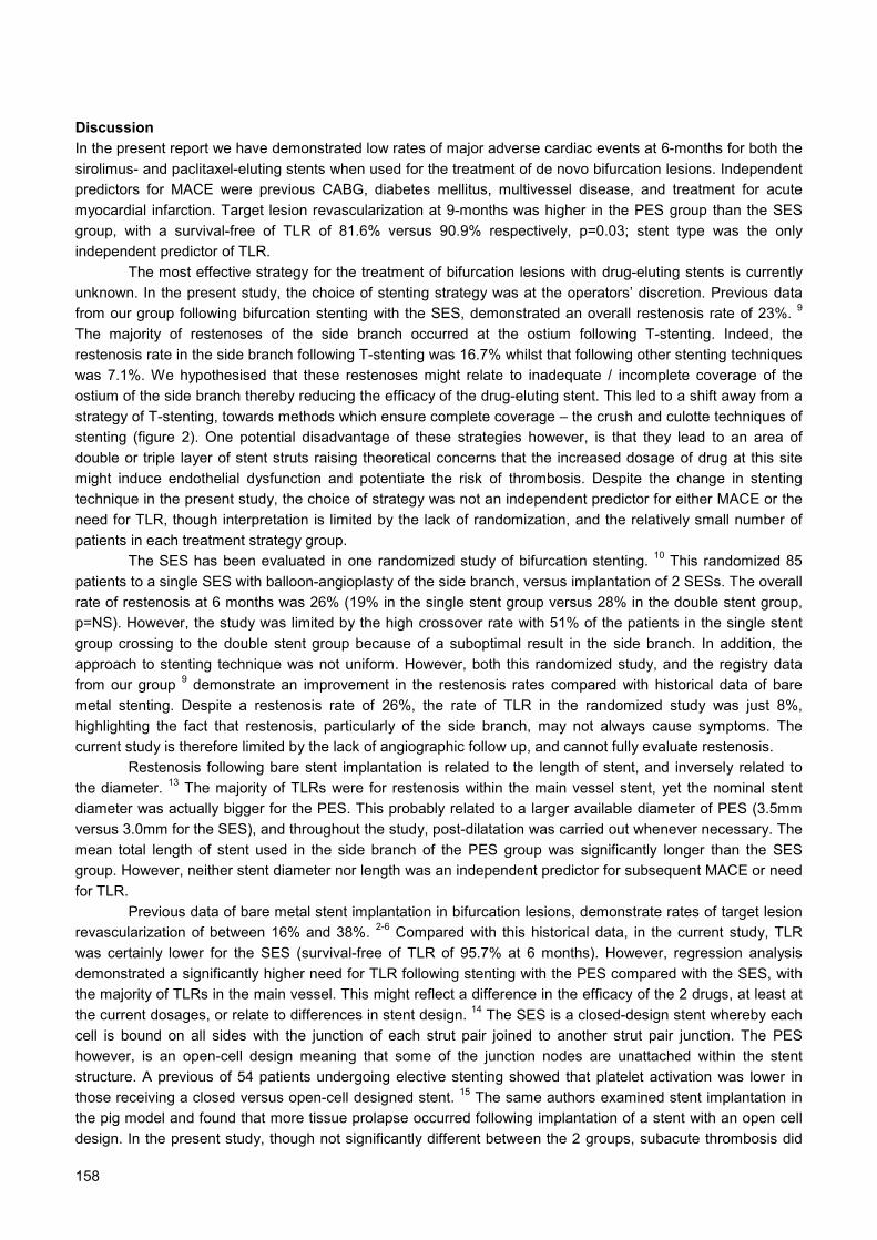
158
DiscussionIn the present report we have demonstrated low rates of major adverse cardiac events at 6-months for both thesirolimus- and paclitaxel-eluting stents when used for the treatment of de novo bifurcation lesions. Independentpredictors for MACE were previous CABG, diabetes mellitus, multivessel disease, and treatment for acutemyocardial infarction. Target lesion revascularization at 9-months was higher in the PES group than the SESgroup, with a survival-free of TLR of 81.6% versus 90.9% respectively, p=0.03; stent type was the onlyindependent predictor of TLR.
The most effective strategy for the treatment of bifurcation lesions with drug-eluting stents is currentlyunknown. In the present study, the choice of stenting strategy was at the operators’ discretion. Previous datafrom our group following bifurcation stenting with the SES, demonstrated an overall restenosis rate of 23%. 9
The majority of restenoses of the side branch occurred at the ostium following T-stenting. Indeed, therestenosis rate in the side branch following T-stenting was 16.7% whilst that following other stenting techniqueswas 7.1%. We hypothesised that these restenoses might relate to inadequate / incomplete coverage of theostium of the side branch thereby reducing the efficacy of the drug-eluting stent. This led to a shift away from astrategy of T-stenting, towards methods which ensure complete coverage – the crush and culotte techniques ofstenting (figure 2). One potential disadvantage of these strategies however, is that they lead to an area ofdouble or triple layer of stent struts raising theoretical concerns that the increased dosage of drug at this sitemight induce endothelial dysfunction and potentiate the risk of thrombosis. Despite the change in stentingtechnique in the present study, the choice of strategy was not an independent predictor for either MACE or theneed for TLR, though interpretation is limited by the lack of randomization, and the relatively small number ofpatients in each treatment strategy group.
The SES has been evaluated in one randomized study of bifurcation stenting. 10 This randomized 85patients to a single SES with balloon-angioplasty of the side branch, versus implantation of 2 SESs. The overallrate of restenosis at 6 months was 26% (19% in the single stent group versus 28% in the double stent group,p=NS). However, the study was limited by the high crossover rate with 51% of the patients in the single stentgroup crossing to the double stent group because of a suboptimal result in the side branch. In addition, theapproach to stenting technique was not uniform. However, both this randomized study, and the registry datafrom our group 9 demonstrate an improvement in the restenosis rates compared with historical data of baremetal stenting. Despite a restenosis rate of 26%, the rate of TLR in the randomized study was just 8%,highlighting the fact that restenosis, particularly of the side branch, may not always cause symptoms. Thecurrent study is therefore limited by the lack of angiographic follow up, and cannot fully evaluate restenosis.
Restenosis following bare stent implantation is related to the length of stent, and inversely related tothe diameter. 13 The majority of TLRs were for restenosis within the main vessel stent, yet the nominal stentdiameter was actually bigger for the PES. This probably related to a larger available diameter of PES (3.5mmversus 3.0mm for the SES), and throughout the study, post-dilatation was carried out whenever necessary. Themean total length of stent used in the side branch of the PES group was significantly longer than the SESgroup. However, neither stent diameter nor length was an independent predictor for subsequent MACE or needfor TLR.
Previous data of bare metal stent implantation in bifurcation lesions, demonstrate rates of target lesionrevascularization of between 16% and 38%. 2-6 Compared with this historical data, in the current study, TLRwas certainly lower for the SES (survival-free of TLR of 95.7% at 6 months). However, regression analysisdemonstrated a significantly higher need for TLR following stenting with the PES compared with the SES, withthe majority of TLRs in the main vessel. This might reflect a difference in the efficacy of the 2 drugs, at least atthe current dosages, or relate to differences in stent design. 14 The SES is a closed-design stent whereby eachcell is bound on all sides with the junction of each strut pair joined to another strut pair junction. The PEShowever, is an open-cell design meaning that some of the junction nodes are unattached within the stentstructure. A previous of 54 patients undergoing elective stenting showed that platelet activation was lower inthose receiving a closed versus open-cell designed stent. 15 The same authors examined stent implantation inthe pig model and found that more tissue prolapse occurred following implantation of a stent with an open celldesign. In the present study, though not significantly different between the 2 groups, subacute thrombosis did
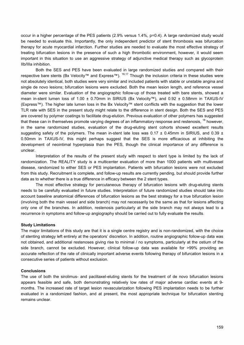
159
occur in a higher percentage of the PES patients (2.9% versus 1.4%, p=0.4). A large randomized study wouldbe needed to evaluate this. Importantly, the only independent predictor of stent thrombosis was bifurcationtherapy for acute myocardial infarction. Further studies are needed to evaluate the most effective strategy oftreating bifurcation lesions in the presence of such a high thrombotic environment, however, it would seemimportant in this situation to use an aggressive strategy of adjunctive medical therapy such as glycoproteinIIb/IIIa inhibition.
Both the SES and PES have been evaluated in large randomized studies and compared with theirrespective bare stents (Bx Velocity™ and Express™). 16,17 Though the inclusion criteria in these studies werenot absolutely identical, both studies were very similar and included patients with stable or unstable angina andsingle de novo lesions; bifurcation lesions were excluded. Both the mean lesion length, and reference vesseldiameter were similar. Evaluation of the angiographic follow-up of those treated with bare stents, showed amean in-stent lumen loss of 1.00 ± 0.70mm in SIRIUS (Bx Velocity™), and 0.92 ± 0.58mm in TAXUS-IV(Express™). The higher late lumen loss in the Bx Velocity™ stent conflicts with the suggestion that the lowerTLR rate with SES in the present study might relate to the difference in stent design. Both the SES and PESare covered by polymer coatings to facilitate drug-elution. Previous evaluation of other polymers has suggestedthat these can in themselves promote varying degrees of an inflammatory response and restenosis, 18 however,in the same randomized studies, evaluation of the drug-eluting stent cohorts showed excellent resultssuggesting safety of the polymers. The mean in-stent late loss was 0.17 ± 0.45mm in SIRIUS, and 0.39 ±0.50mm in TAXUS-IV, this might perhaps suggest that the SES is more efficacious at inhibiting thedevelopment of neointimal hyperplasia than the PES, though the clinical importance of any difference isunclear.
Interpretation of the results of the present study with respect to stent type is limited by the lack ofrandomization. The REALITY study is a multicenter evaluation of more than 1000 patients with multivesseldisease, randomized to either SES or PES implantation. Patients with bifurcation lesions were not excludedfrom this study. Recruitment is complete, and follow-up results are currently pending, but should provide furtherdata as to whether there is a true difference in efficacy between the 2 stent types.
The most effective strategy for percutaneous therapy of bifurcation lesions with drug-eluting stentsneeds to be carefully evaluated in future studies. Interpretation of future randomized studies should take intoaccount baseline anatomical differences of bifurcation lesions as the best strategy for a true bifurcation lesion(involving both the main vessel and side branch) may not necessarily be the same as that for lesions affectingonly one of the branches. In addition, restenosis particularly at the side branch may not always lead to arecurrence in symptoms and follow-up angiography should be carried out to fully evaluate the results.
Study LimitationsThe major limitations of this study are that it is a single centre registry and is non-randomized, with the choiceof stenting strategy left entirely at the operators’ discretion. In addition, routine angiographic follow-up data wasnot obtained, and additional restenoses giving rise to minimal / no symptoms, particularly at the ostium of theside branch, cannot be excluded. However, clinical follow-up data was available for >99% providing anaccurate reflection of the rate of clinically important adverse events following therapy of bifurcation lesions in aconsecutive series of patients without exclusion.
ConclusionsThe use of both the sirolimus- and paclitaxel-eluting stents for the treatment of de novo bifurcation lesionsappears feasible and safe, both demonstrating relatively low rates of major adverse cardiac events at 9-months. The increased rate of target lesion revascularization following PES implantation needs to be furtherevaluated in a randomized fashion, and at present, the most appropriate technique for bifurcation stentingremains unclear.

160
References1. Al Suwaidi J, Yeh W, Cohen HA, et al. Immediate and one-year outcome in patients with coronary bifurcation lesions in the
modern era (NHLBI dynamic registry). Am J Cardiol. 2001;87:1139-44.
2. Lefevre T, Louvard Y, Morice MC, et al. Stenting of bifurcation lesions: classification, treatments, and results. Catheter
Cardiovasc Interv. 2000;49:274-83.
3. Anzuini A, Briguori C, Rosanio S, et a. Immediate and long-term clinical and angiographic results from Wiktor stent treatment for
true bifurcation narrowings. Am J Cardiol. 2001;88:1246-50.
4. Pan M, Suarez de Lezo J, Medina A, et al. Simple and complex stent strategies for bifurcated coronary arterial stenosis involving
the side branch origin. Am J Cardiol. 1999;83:1320-5.
5. Yamashita T, Nishida T, Adamian MG, et al. Bifurcation lesions: two stents versus one stent--immediate and follow-up results. J
Am Coll Cardiol. 2000;35:1145-51.
6. Al Suwaidi J, Berger PB, Rihal CS, et al. Immediate and long-term outcome of intracoronary stent implantation for true bifurcation
lesions. J Am Coll Cardiol. 2000;35:929-36.
7. Morice MC, Serruys PW, Sousa JE, et al. A randomized comparison of a sirolimus-eluting stent with a standard stent for
coronary revascularization. N Engl J Med. 2002;346:1773-80.
8. Grube E, Silber S, Hauptmann KE, et al. TAXUS I: six- and twelve-month results from a randomized, double-blind trial on a slow-
release paclitaxel-eluting stent for de novo coronary lesions. Circulation. 2003;107:38-42.
9. Tanabe K, Hoye A, Lemos PA, et al. Restenosis rates following bifurcation stenting with sirolimus-eluting stents for de novo
narrowings. Am J Cardiol. 2004;94:115-8.
10. Colombo A, Moses JW, Morice MC, et al. Randomized study to evaluate sirolimus-eluting stents implanted at coronary
bifurcation lesions. Circulation. 2004;109:1244-9.
11. Colombo A, Stankovic G, Orlic D, et al. Modified T-stenting technique with crushing for bifurcation lesions: immediate results and
30-day outcome. Catheter Cardiovasc Interv. 2003;60:145-51.
12. Chevalier B, Glatt B, Royer T, et al. Placement of coronary stents in bifurcation lesions by the "culotte" technique. Am J Cardiol.
1998;82:943-9.
13. de Feyter PJ, Kay P, Disco C, et al. Reference chart derived from post-stent-implantation intravascular ultrasound predictors of 6-
month expected restenosis on quantitative coronary angiography. Circulation. 1999;100:1777-83.
14. Morton AC, Crossman D, Gunn J. The influence of physical stent parameters upon restenosis. Pathol Biol (Paris). 2004;52:196-
205.
15. Gurbel PA, Cummings CC, Bell CR, et al. Onset and extent of platelet inhibition by clopidogrel loading in patients undergoing
elective coronary stenting: the Plavix Reduction Of New Thrombus Occurrence (PRONTO) trial. Am Heart J. 2003;145:239-47.
16. Moses JW, Leon MB, Popma JJ, et al. Sirolimus-eluting stents versus standard stents in patients with stenosis in a native
coronary artery. N Engl J Med. 2003;349:1315-23.
17. Stone GW, Ellis SG, Cox DA, et al. A polymer-based, paclitaxel-eluting stent in patients with coronary artery disease. N Engl J
Med. 2004;350:221-31.
18. van der Giessen WJ, Lincoff AM, Schwartz RS, et al. Marked inflammatory sequelae to implantation of biodegradable and
nonbiodegradable polymers in porcine coronary arteries. Circulation. 1996;94:1690-7.

161
Chapter 16
Angela HoyeIoannis IakovouLei GeCarlos AG van MieghemAndrew TL OngJohn CosgraveGiuseppe M SangiorgiFlavio AiroldiMatteo MontorfanoIassen MichevAlaide ChieffoMauro CarlinoNicola CorvajaJiro AokiGaston A Rodriguez GranilloMarco ValgimigliGeorgios SianosWillem J van der GiessenPim J de FeyterRon T van DomburgPatrick W SerruysAntonio Colombo
Journal of the AmericanCollege of CardiologyIn press
Long-term Outcomes Following Stenting ofBifurcation Lesions Utilizing the “Crush”
Technique: Predictors of an AdverseOutcome

162

163
AbstractObjectives: To evaluate predictors of an adverse outcome following “crush” bifurcation
stenting.
Background: The “crush” technique is a recently introduced strategy, with limited data
regarding long-term outcomes.
Methods: We identified 231 consecutive patients who underwent “crush” stenting for 241 de
novo bifurcation lesions. Stents used were sirolimus-eluting in 137(56.8%), and paclitaxel-eluting in 104(43.2%). Clinical follow-up was obtained in 99.6%.
Results: The in-hospital major adverse cardiac event (MACE) rate was 5.2%. At 9-months,10 (4.3%) patients had an event consistent with post-procedural stent thrombosis. The
survival-free of target lesion revascularisation (TLR) rate was 90.3%; the independent
predictor of TLR was left main stem (LMS) therapy (OR 4.97; 95%CI 2.00 to 12.37,p=0.001). Survival-free of MACE was 83.5%, independent predictors of MACE were LMS
therapy (OR 3.79; 95%CI 1.76 to 8.14, p=0.001), and treatment of patients with multivessel
disease (OR 4.21; 95%CI: 0.95 to 18.56, p=0.058).Angiographic follow-up (in 77% lesions) demonstrated a mean late loss of the main
vessel and side branch of 0.30±0.64mm and 0.41±0.67mm respectively, with binary
restenosis rates of 9.1% and 25.3%. Kissing balloon post-dilatation significantly reduced theside branch late lumen loss (0.24±0.50mm versus 0.58±0.77mm, p<0.001).
Conclusions: With drug-eluting stents, the crush technique is associated with favourableoutcomes for most lesions; however, efficacy appears significantly reduced in LMS
bifurcations, and further research is needed before the technique can be routinely
recommended in this group. The incidence of possible stent thrombosis is of concern andrequires further investigation. Kissing balloon post-dilatation is mandatory to reduce the rate
of side branch restenosis.
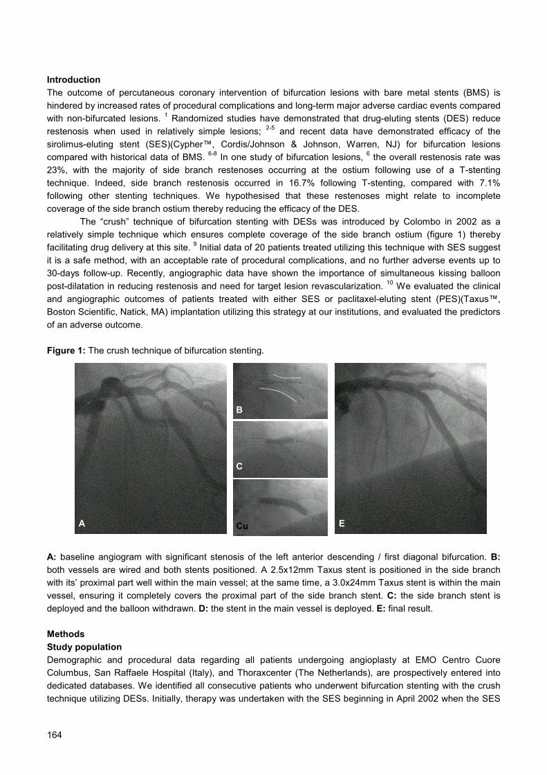
164
IntroductionThe outcome of percutaneous coronary intervention of bifurcation lesions with bare metal stents (BMS) ishindered by increased rates of procedural complications and long-term major adverse cardiac events comparedwith non-bifurcated lesions. 1 Randomized studies have demonstrated that drug-eluting stents (DES) reducerestenosis when used in relatively simple lesions; 2-5 and recent data have demonstrated efficacy of thesirolimus-eluting stent (SES)(Cypher™, Cordis/Johnson & Johnson, Warren, NJ) for bifurcation lesionscompared with historical data of BMS. 6-8 In one study of bifurcation lesions, 6 the overall restenosis rate was23%, with the majority of side branch restenoses occurring at the ostium following use of a T-stentingtechnique. Indeed, side branch restenosis occurred in 16.7% following T-stenting, compared with 7.1%following other stenting techniques. We hypothesised that these restenoses might relate to incompletecoverage of the side branch ostium thereby reducing the efficacy of the DES.
The “crush” technique of bifurcation stenting with DESs was introduced by Colombo in 2002 as arelatively simple technique which ensures complete coverage of the side branch ostium (figure 1) therebyfacilitating drug delivery at this site. 9 Initial data of 20 patients treated utilizing this technique with SES suggestit is a safe method, with an acceptable rate of procedural complications, and no further adverse events up to30-days follow-up. Recently, angiographic data have shown the importance of simultaneous kissing balloonpost-dilatation in reducing restenosis and need for target lesion revascularization. 10 We evaluated the clinicaland angiographic outcomes of patients treated with either SES or paclitaxel-eluting stent (PES)(Taxus™,Boston Scientific, Natick, MA) implantation utilizing this strategy at our institutions, and evaluated the predictorsof an adverse outcome.
Figure 1: The crush technique of bifurcation stenting.
A: baseline angiogram with significant stenosis of the left anterior descending / first diagonal bifurcation. B:both vessels are wired and both stents positioned. A 2.5x12mm Taxus stent is positioned in the side branchwith its’ proximal part well within the main vessel; at the same time, a 3.0x24mm Taxus stent is within the mainvessel, ensuring it completely covers the proximal part of the side branch stent. C: the side branch stent isdeployed and the balloon withdrawn. D: the stent in the main vessel is deployed. E: final result.
MethodsStudy populationDemographic and procedural data regarding all patients undergoing angioplasty at EMO Centro CuoreColumbus, San Raffaele Hospital (Italy), and Thoraxcenter (The Netherlands), are prospectively entered intodedicated databases. We identified all consecutive patients who underwent bifurcation stenting with the crushtechnique utilizing DESs. Initially, therapy was undertaken with the SES beginning in April 2002 when the SES
A E
B
C
Cum

165
received CE mark approval. In the first quarter of 2003, patients could also be treated with the PES. Patientswith either stable or unstable angina were included if they were treated for a de novo bifurcation lesion. Thosewith acute ST-elevation myocardial infarction were excluded. SESs were available in diameters from 2.25mm to3.00mm and lengths from 8mm to 33mm; PESs were available in diameters from 2.25mm to 3.5mm andlengths from 8mm to 32mm.
Procedures and intervention medicationsThe crush technique is depicted in figure 1, and has been previously described. 9 In short, the procedurerequires a guide catheter of ≥7F. Both the main vessel and side branch are wired and prepared for stentimplantation with pre-dilatation as necessary. The stents are both positioned such that the proximal part of theside branch lies well within the main vessel, but is completely covered by the stent within the main vessel(figure 1B). The side branch stent is deployed and the balloon carefully removed ensuring that the stent in themain vessel remains fixed. The wire within the side branch is commonly also removed, though providing thewire is not hydrophilic, it may be kept in position. The stent in the main vessel is deployed thereby crushing theproximal part of the side branch stent (and trapping the side branch wire if still in situ). If present, the wire in theside branch can then be withdrawn, and post-dilatation of the main vessel stent with high pressure ballooninflation facilitates use of the side branch wire to re-cross into the side branch to allow kissing balloon post-dilatation. Kissing balloon post-dilatation was undertaken at the operator’s discretion. The protocol wasapproved by the Institutional ethics committees and is in accordance with the principles of Good ClinicalPractice for Trials of Medicinal Products in the European Community and the Declaration of Helsinki. Allpatients signed a written informed consent.
During the procedure, intravenous heparin was given to maintain an activated clotting time ≥250seconds. Patients were preloaded with 300mg clopidogrel, and received life-long aspirin together with 75mgclopidogrel per day for at least 6-months. The use of glycoprotein IIb/IIIa inhibitors was at the operator’sdiscretion.
Clinical Definitions and Follow-upClinical follow-up was obtained using either telephone calls or office visit, and evaluated the rate of majoradverse cardiac events (MACE), pre-defined as death, acute myocardial infarction (AMI), or target vesselrevascularization (TVR). The diagnosis of AMI both peri-procedural and at follow-up, required an elevation ofcreatine kinase levels to twice the upper limit of normal, together with a rise in creatine kinase-MB fraction.When in addition to enzyme elevation there were new pathological Q waves on the electrocardiogram, theevent was defined as Q-wave MI. Target lesion revascularization (TLR) was defined as either surgical orpercutaneous reintervention driven by significant (>50%) luminal diameter narrowing either within the stent orthe 5mm borders proximal and distal to the stent, and was undertaken in the presence of either anginalsymptoms or objective evidence of ischemia. TVR was defined as revascularization within the target vesselincluding encompassing the target lesion.
Stent thrombosis was defined as an acute coronary syndrome with angiographic documentation ofeither vessel occlusion or thrombus within or adjacent to a previously successfully stented vessel, or, in theabsence of angiographic confirmation, either acute AMI in the distribution of the treated vessel, or death notclearly attributable to other causes. 11,12 Stent thrombosis was categorized according to the timing of the eventinto: intra-procedural (angiographic confirmed intra-luminal filling defect within the stent that occurred during theindex procedure), [13] acute (occurred within the first 24 hours after the procedure), subacute (from 24 hours to30 days) and late (>30 days after the index procedure).
Angiographic evaluation:Procedural angiographic success was defined as a post-procedural final residual stenosis less than 50% withThrombolysis In Myocardial infarction (TIMI) flow 3 in both the main vessel and side branch. Between 6 and 12months after the index procedure, all living patients were invited back for angiographic follow-up. Coronary

166
angiograms were obtained in multiple views after intracoronary injection of nitrates. Quantitative coronaryangiographic (QCA) analysis was performed using one of two validated edge detection systems (CMS, version5.2, MEDIS, the Netherlands, and the Cardiovascular Angiography Analysis System II (CAAS II), Pie Medical,The Netherlands. The reference vessel diameter, minimal lumen diameter (MLD) and percent diameterstenosis were measured at pre-, post-procedure and follow-up. Reference vessel diameter for the side branchwas taken as the diameter of the normal vessel distal to the bifurcation. The late lumen loss was calculated asthe difference between the post procedure and follow-up MLD. 14 Binary restenosis was defined as thepresence of >50% diameter stenosis within the target lesion.
Statistical analysis:Discrete variables are presented as percentages and compared with Fisher exact test. Continuous variablesare expressed as mean ± standard deviation and compared with Student’s t test. Cumulative survival-free ofadverse events were calculated according to the Kaplan-Meier method. Logistic regression models wereestablished to investigate independent predictors of TLR and MACE. The following clinical variables wereentered into the analysis model: age, gender, diabetes, stent type, unstable angina, premature antiplatelettherapy discontinuation, LMS (left main stem) bifurcation, glycoprotein IIb/IIIa inhibitor use, kissing balloon post-dilatation, nominal stent diameter, and stent length. Odds ratio with corresponding 95% confidence intervals arereported. All tests were two-tailed, and a p value of <0.05 was considered significant.
ResultsThe crush technique was utilized in 231 patients (241 lesions), with SES in 137 (56.8%), and PES in 104(43.2%). The baseline patient and procedural characteristics are presented in tables 1 and 2. The use ofglycoprotein IIb/IIIa inhibitor therapy was significantly higher in the SES group than the PES group (40.8%versus 19.8%, p=0.001). Attempted kissing balloon post-dilatation was undertaken in 128 lesions, and wassuccessful in 122 (95%) cases; it was carried out more frequently in the PES group (61.5% versus 42.3% inSES, p=0.004).
Table 1: Baseline patient demographics
Alln=231
SESn=130
PESn=101
p value*
Mean age (years) 62.8 ± 11.2 62.9 ± 11.2 62.6 ± 11.1 0.76Male sex (%) 193 (83.5) 113 (86.9) 80 (79.2) 0.15Current smoker (%) 44 (19.0) 24 (18.5) 20 (19.8) 0.87Diabetes mellitus (%) 46 (19.9) 25 (19.2) 21 (20.8) 0.74Hypertension (%) 125 (54.1) 76 (58.5) 49 (48.5) 0.14Hypercholesterolemia (%) 162 (70.1) 91 (70.0) 71 (70.3) 1.0Family history (%) 103 (44.6) 53 (40.8) 50 (49.5) 0.23Previous myocardial infarction (%) 95 (41.1) 48 (36.9) 47 (46.5) 0.22Previous CABG (%) 33 (14.3) 18 (13.8) 15 (14.9) 0.85Multivessel disease (%) 174 (75.3) 98 (75.4) 76 (75.2) 1.0Clinical presentation 1.0
Stable angina (%) 172 (74.5) 96 (73.8) 76 (75.2)Unstable angina (%) 59 (25.5) 34 (26.2) 25 (24.8)
Glycoprotein IIb/IIIa inhibitor usage (%) 73 (31.6) 53 (40.8) 20 (19.8) 0.001*p value for the SES group versus PES groupCABG: coronary artery bypass graft surgery; PES: paclitaxel-eluting stent; SES: sirolimus-eluting stent
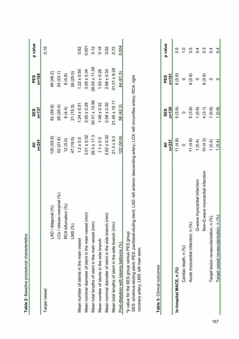
167
BBBBBBBBBBBBBBBBBBBBBBBBBBBBp
valu
e
0.15
0.82
0.00
1
0.12
0.18
0.02
0.72
0.00
4
p va
lue
0.5
1.0
0.5
0.4
0.3
0.4
0.4
PES
n=10
4
48 (4
6.2)
24 (2
3.1)
6 (5
.8)
26 (2
5.0)
1.22
± 0
.50
3.09
± 0
.34
28.0
5 ±
11.5
8
1.03
± 0
.26
2.68
± 0
.34
21.0
1 ±
8.28
64 (6
1.5)
PES
n=10
16
(5.9
)
0
6 (5
.9)
0
6 (5
.9)
0 0
SES
n=13
7
82 (5
9.9)
28 (2
0.4)
6 (4
.4)
21 (1
5.3)
1.24
± 0
.51
2.95
± 0
.29
30.3
1 ±
10.9
6
1.08
± 0
.32
2.58
± 0
.30
21.4
5 ±
10.1
1
58 (4
2.3)
SES
n=13
05
(3.8
)
0
5 (3
.8)
1 (0
.8)
4 (3
.1)
1 (0
.8)
1 (0
.8)
All
n=24
1
130
(53.
9)
52 (2
1.6)
12 (5
.0)
47 (1
9.5)
1.2
± 0.
5
3.01
± 0
.32
29.3
± 1
1.3
1.1
± 0.
3
2.62
± 0
.32
21.3
± 9
.3
122
(50.
6)
All
n=23
111
(4.8
)
0
11 (4
.8)
1 (0
.4)
10 (4
.3)
1 (0
.4)
1 (0
.4)
Q-w
ave
myo
card
ial i
nfar
ctio
n
Non
-Q w
ave
myo
card
ial i
nfar
ctio
n
LAD
/ di
agon
al (%
)
LCx
/ obt
use
mar
gina
l (%
)
RC
A bi
furc
atio
n (%
)
LMS
(%)
Car
diac
dea
th, n
(%)
Acut
e m
yoca
rdia
l inf
arct
ion,
n (%
)
Targ
et le
sion
reva
scul
ariz
atio
n, n
(%)
Targ
et v
esse
l rev
ascu
lariz
atio
n, n
(%)
Tabl
e 2:
Bas
elin
e pr
oced
ural
cha
ract
eris
tics
Targ
et v
esse
l
Mea
n nu
mbe
r of s
tent
s in
the
mai
n ve
ssel
Mea
n no
min
al d
iam
eter
of s
tent
in th
e m
ain
vess
el (m
m)
Mea
n to
tal l
engt
hs o
f ste
nt in
the
mai
n ve
ssel
(mm
)
Mea
n nu
mbe
r of s
tent
s in
the
side
bra
nch
Mea
n no
min
al d
iam
eter
of s
tent
in th
e si
de b
ranc
h (m
m)
Mea
n to
tal l
engt
hs o
f ste
nt in
the
side
bra
nch
(mm
)
Post
-dila
tatio
n w
ith k
issi
ng b
allo
ons
(%)
*p
valu
e fo
r the
SE
S gr
oup
vers
us P
ES
grou
p
SES
: siro
limus
-elu
ting
sten
t; P
ES:
pac
litax
el-e
lutin
g st
ent;
LAD
: lef
t ant
erio
r des
cend
ing
arte
ry; L
CX:
left
circ
umfle
x ar
tery
; RC
A: ri
ght
co
rona
ry a
rtery
; LM
S: le
ft m
an s
tem
Tabl
e 3:
Clin
ical
out
com
es
In-h
ospi
tal M
AC
E, n
(%)
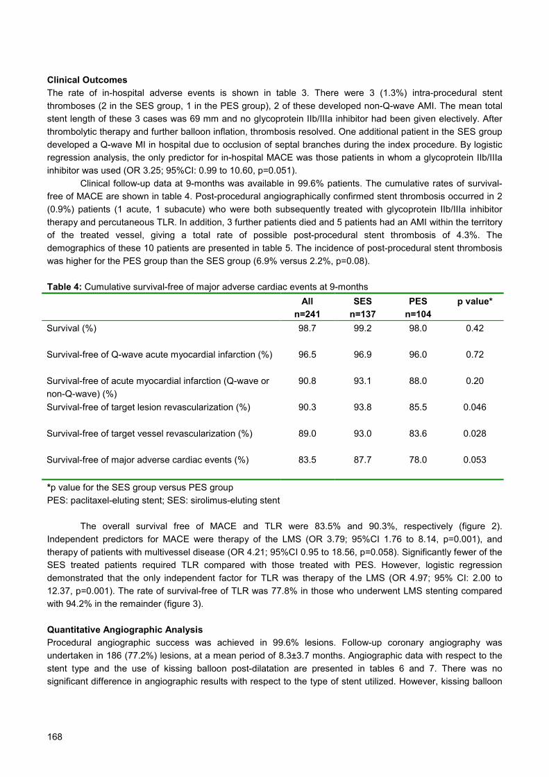
168
Clinical OutcomesThe rate of in-hospital adverse events is shown in table 3. There were 3 (1.3%) intra-procedural stentthromboses (2 in the SES group, 1 in the PES group), 2 of these developed non-Q-wave AMI. The mean totalstent length of these 3 cases was 69 mm and no glycoprotein IIb/IIIa inhibitor had been given electively. Afterthrombolytic therapy and further balloon inflation, thrombosis resolved. One additional patient in the SES groupdeveloped a Q-wave MI in hospital due to occlusion of septal branches during the index procedure. By logisticregression analysis, the only predictor for in-hospital MACE was those patients in whom a glycoprotein IIb/IIIainhibitor was used (OR 3.25; 95%CI: 0.99 to 10.60, p=0.051).B
Clinical follow-up data at 9-months was available in 99.6% patients. The cumulative rates of survival-free of MACE are shown in table 4. Post-procedural angiographically confirmed stent thrombosis occurred in 2(0.9%) patients (1 acute, 1 subacute) who were both subsequently treated with glycoprotein IIb/IIIa inhibitortherapy and percutaneous TLR. In addition, 3 further patients died and 5 patients had an AMI within the territoryof the treated vessel, giving a total rate of possible post-procedural stent thrombosis of 4.3%. Thedemographics of these 10 patients are presented in table 5. The incidence of post-procedural stent thrombosiswas higher for the PES group than the SES group (6.9% versus 2.2%, p=0.08).
Table 4: Cumulative survival-free of major adverse cardiac events at 9-monthsAll
n=241SES
n=137PES
n=104p value*
Survival (%) 98.7 99.2 98.0 0.42
Survival-free of Q-wave acute myocardial infarction (%) 96.5 96.9 96.0 0.72
Survival-free of acute myocardial infarction (Q-wave ornon-Q-wave) (%)
90.8 93.1 88.0 0.20
Survival-free of target lesion revascularization (%) 90.3 93.8 85.5 0.046
Survival-free of target vessel revascularization (%) 89.0 93.0 83.6 0.028
Survival-free of major adverse cardiac events (%) 83.5 87.7 78.0 0.053
*p value for the SES group versus PES groupPES: paclitaxel-eluting stent; SES: sirolimus-eluting stent
The overall survival free of MACE and TLR were 83.5% and 90.3%, respectively (figure 2).Independent predictors for MACE were therapy of the LMS (OR 3.79; 95%CI 1.76 to 8.14, p=0.001), andtherapy of patients with multivessel disease (OR 4.21; 95%CI 0.95 to 18.56, p=0.058). Significantly fewer of theSES treated patients required TLR compared with those treated with PES. However, logistic regressiondemonstrated that the only independent factor for TLR was therapy of the LMS (OR 4.97; 95% CI: 2.00 to12.37, p=0.001). The rate of survival-free of TLR was 77.8% in those who underwent LMS stenting comparedwith 94.2% in the remainder (figure 3).
Quantitative Angiographic AnalysisProcedural angiographic success was achieved in 99.6% lesions. Follow-up coronary angiography wasundertaken in 186 (77.2%) lesions, at a mean period of 8.3±3.7 months. Angiographic data with respect to thestent type and the use of kissing balloon post-dilatation are presented in tables 6 and 7. There was nosignificant difference in angiographic results with respect to the type of stent utilized. However, kissing balloon

169
post-dilatation significantly reduced the side branch late lumen loss and binary restenosis. Among the 47restenotic lesions at the side branch, 34 (72.3%) were focal (<10mm) and located at the ostium.
DiscussionThe main findings of this report are: (1) treatment of most bifurcation lesions with DES by the “crush” techniqueis associated with low rates of TLR and MACE at 9-months. However, therapy of the left main stem was anindependent predictor of both TLR and MACE; (2) at 9-months, the incidence of possible post-procedural stentthrombosis was 4.3%; (3) the rate of side branch restenosis was significantly lower in lesions treated withkissing balloon post-dilatation compared to those without.
Figure 2: Cumulative survival-free of target lesion revascularization (TLR), and major adverse cardiac events(MACE) following bifurcation stenting utilizing the crush technique.
Figure 3: Cumulative survival-free of target lesion revascularization (TLR) for patients treated with the crushtechnique of bifurcation stenting for a left main stem (LMS) lesion compared with those treated for lesionsoutside the left main stem (non-LMS).
90
100
90
80
70
60
50
63Follow-up (months)
Cum
ulative survival-free of adverse events (%)
TLR: 90.3%
MACE: 83.5%
9630
100
90
80
70
Cum
ulative survival-free of TLR (%
)
Follow-up (months)
Non-LMS lesions: 94.2%
LMS lesions: 77.8%
Log rank p<0.001

170
10 9 8 7 6 5 4 3 2 1 Ptno.
80yrM 65yrF
71yrM 61yrM 82yrM 41yrM 73yrM 67yrM 66yrF
74yrF Age/sex
SES
PES
PES
PES
SES
PES
PES
PES
PES
SES
Stenttype
Y Y N N Y N Y N N N DM
Y N Y Y Y Y Y Y Y Y
Multivesseldisease
N N N Y N N Y Y N N
PreviousC
AB
G
LAD
LAD
LMS
LMS
LMS
LAD
LMS
LMS
LCx
LAD
Targetvesse
l
SA
SA
SA
SA
UA
UA
SA
SA
UA
SA
Indexpresentation
Y Y N Y Y N N N N N
Index useof G
PIIb/IIIa
N N Y Y Y N N Y N Y
Kissing
balloon post-dilatation
26
166
117
63 55
211
204
145 7 1
Time to definite
or probablethrom
bosis,
NS
toppedclopidogrelbecause of
abdominal surgery
Y Y Y NS
toppedclopidogrelbecause ofpancreatitis
NS
toppedclopidogrel at 6-
months
NS
toppedclopidogrel at 6-
months
Y Y Y
Dual anti-platelettherapy at the
time of the event
AM
I: managed
medically; alive
AM
I: managed
medically; alive
AM
I: managed
medically; alive
AM
I and death
Death
AM
I: managed
medically; alive
Sudden death
AM
I: managed
medically; alive
AM
I: underwent TLR
with P
CI; alive
AM
I: underwent TLR
with P
CI; alive
Presentation andtherapy of
thrombosis
Table 5: Patients with a definite or probable post-procedural stent throm
bosis

171
Stent coverage of the ostium of the side branch and clinical outcomesBifurcation lesions are subject to increased rates of restenosis and need for TLR compared with non-bifurcatedlesions. Historical data of BMSs suggest a TLR rate of 16-38%, with a tendency to increased restenosisfollowing stenting of both the main vessel and side branch compared with single vessel stenting. 15-19 In thesame studies, the rate of MACE at 6-months ranged between 17-51%. In randomized studies of relativelysimple lesions, DESs reduce restenosis compared with BMSs, though bifurcation lesions were excluded. 2-5
Preliminary data for the SES has recently suggested efficacy in bifurcation lesions. 6-8 However, the mosteffective stenting strategy is currently unknown. Previous data of the SES suggested a higher restenosis ratefollowing T-stenting, with the hypothesis that this might relate to incomplete coverage of the side branch ostium.6,7 In most bifurcations, the angle at the carina is significantly smaller than 90˚ meaning that even with precisepositioning, the stents are unable to make a “T” and completely cover the bifurcation. 18 The crush technique isa relatively simple strategy that ensures complete lesion coverage, even for bifurcation lesions that haveextensive disease within the side branch. Preliminary data have suggested acceptable short-term resultssuggesting it may therefore be an effective strategy for bifurcation lesions. 9
In the present study, we have demonstrated encouraging long-term results, with a high rate of survival-free of TLR of 90.3%. Although TLR rates were higher in those treated with PES compared to SES, there weremore patients in this group treated for LMS bifurcation (25.0% versus 15.3%). By logistic regression analysis,therapy of the LMS was the only independent predictor of TLR. Indeed, at 9-months, the survival-free of TLRwas 77.8% in those who underwent LMS stenting, compared with 94.2% in the remainder. The rate of in-hospital MACE was 4.8%, the majority comprising non-Q wave AMI. The MACE rate was higher in those whoreceived a glycoprotein IIb/IIIa inhibitor. However, this is likely to reflect the operators’ decision to use such anagent only in the situation of a difficult or complicated procedure. There is evidence to suggest improvedefficacy of glycoprotein IIb/IIIa inhibitors with early administration. Further work is needed to evaluate whetherroutine pre-procedural administration of such agents to all patients undergoing crush stenting might reduce theoccurrence of in-hospital events.
At 9-months, the overall rate of survival-free of MACE was 83.5%. Independent predictors were thetreatment of multivessel disease, and treatment of the LMS. Recent data has been published specificallyevaluating the results of LMS stenting with DES implantation. 20-23 In all these studies, results suggested lowerrates of restenosis compared with historical data of BMS. The incidence of TLR ranged from 2.0-14.1%. Thisappears to be much lower than the LMS cohort in our study where the rate of survival-free of TLR was just77.8% (compared with 94.2% for non-LMS lesions). However, in the aforementioned studies, restenosisfollowing LMS bifurcation stenting was higher compared with lesions localised to the ostium or body of theLMS. Chieffo et al 22 evaluated 85 patients including 69 (81.2%) with disease of the distal LMS. The majority ofthese patients were treated with stent implantation to both branches, most (59%) with crush stenting. All 12patients who required TLR were initially treated with a 2 stent strategy. Park et al 21 demonstrated excellentresults following LMS angioplasty, with a binary restenosis rate of 7.0%. In this study, 70.6% patients weretreated for the LMS bifurcation, and all restenoses occurred in these patients.
Kissing balloon post-dilatationAlthough the overall rate of TLR in the present study was relatively low, kissing balloon post-dilatation had asignificant impact on the angiographic results, leading to a significantly larger post-procedural MLD within boththe main vessel and side branch. This larger MLD was maintained in both vessels at follow-up, but wasparticularly evident within the side branch. As previously demonstrated by Ge et al, 10 in the present paper,kissing balloon post-dilatation in the present study significantly reduced both the side branch late lumen loss(0.24 ± 0.50mm versus 0.58 ± 0.77mm, p<0.001), and binary restenosis rate (9.6% versus 41.3%,p<0.000001).
The majority (72.3%) of these side branch restenoses were focal and occurred at the ostium. Benchstudy results have demonstrated the crush technique to effectively cover the bifurcation lesion, however, theabsence of kissing balloon post-dilatation leads to under-expansion and mal-apposition of the side branch stentstruts. 24 Kissing balloon post-dilatation opens the struts thereby facilitating access to the side branch, andcorrects stent deformation to provide optimal scaffolding and delivery of drug. The crush technique is
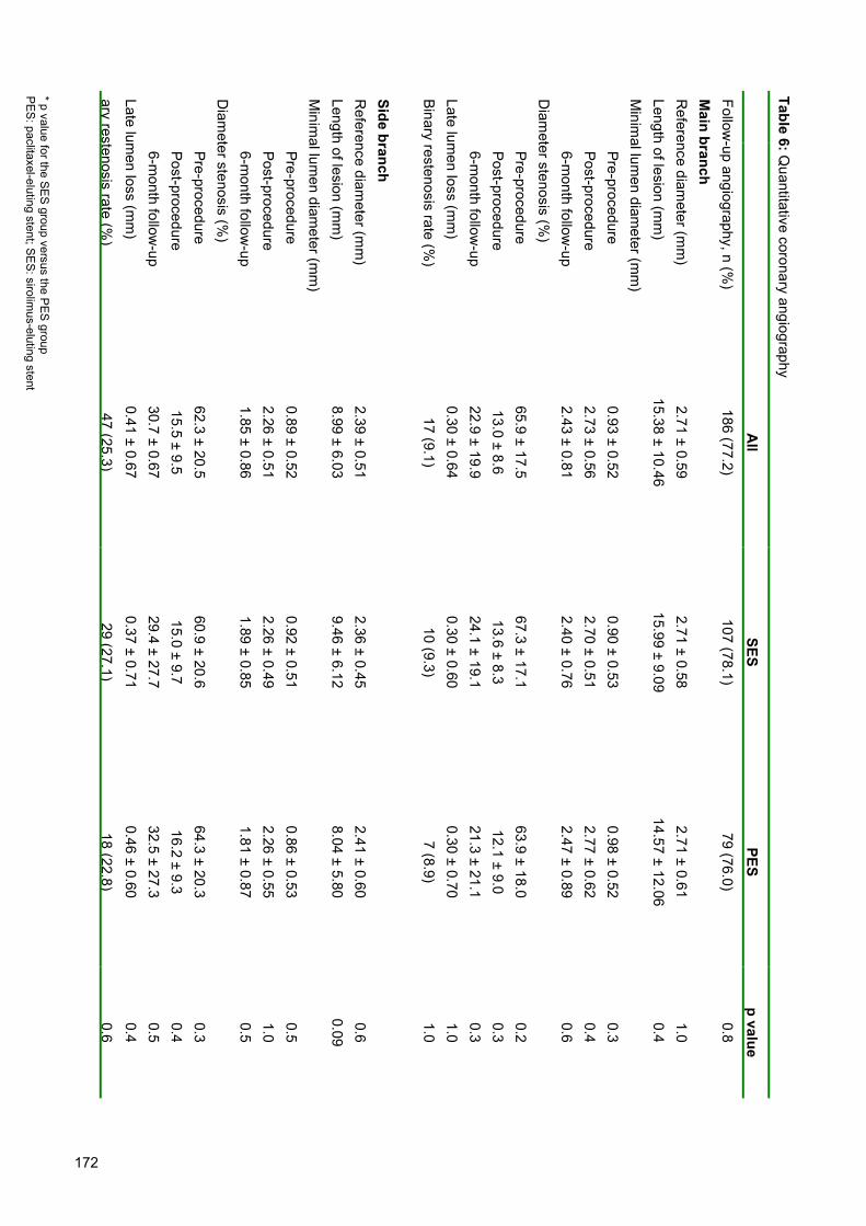
172
ary restenosis rate (%)
Late lumen loss (m
m)
6-month follow
-upPost-procedurePre-procedure
Diam
eter stenosis (%)
6-month follow
-upPost-procedurePre-procedure
Minim
al lumen diam
eter (mm
)Length of lesion (m
m)
Reference diam
eter (mm
)Side branch
Binar y restenosis rate (%)
Late lumen loss (m
m)
6-month follow
-upPost- procedurePre- procedure
Diam
eter stenosis (%)
6-month follow
-upPost-procedurePre- procedure
Minim
al lumen diam
eter (mm
)Length of lesion (m
m)
Reference diam
eter (mm
)M
ain branchFollow
-up angiography, n (%)
47 (25.3)0.41 ± 0.6730.7 ± 0.6715.5 ± 9.562.3 ± 20.5
1.85 ± 0.862.26 ± 0.510.89 ± 0.52
8.99 ± 6.032.39 ± 0.51
17 (9.1)0.30 ± 0.6422.9 ± 19.913.0 ± 8.665.9 ± 17.5
2.43 ± 0.812.73 ± 0.560.93 ± 0.52
15.38 ± 10.462.71 ± 0.59
186 (77.2)
All
29 (27.1)0.37 ± 0.7129.4 ± 27.715.0 ± 9.760.9 ± 20.6
1.89 ± 0.852.26 ± 0.490.92 ± 0.51
9.46 ± 6.122.36 ± 0.45
10 (9.3)0.30 ± 0.6024.1 ± 19.113.6 ± 8.367.3 ± 17.1
2.40 ± 0.762.70 ± 0.510.90 ± 0.53
15.99 ± 9.092.71 ± 0.58
107 (78.1)
SES
18 (22.8)0.46 ± 0.6032.5 ± 27.316.2 ± 9.364.3 ± 20.3
1.81 ± 0.872.26 ± 0.550.86 ± 0.53
8.04 ± 5.802.41 ± 0.60
7 (8.9)0.30 ± 0.7021.3 ± 21.112.1 ± 9.063.9 ± 18.0
2.47 ± 0.892.77 ± 0.620.98 ± 0.52
14.57 ± 12.062.71 ± 0.61
79 (76.0)
PES
0.60.40.50.40.3
0.51.00.5
0.090.6
1.01.00.30.30.2
0.60.40.3
0.41.0
0.8
p value
Table 6: Quantitative coronary angiography
* p value for the SES group versus the P
ES group
PES
: paclitaxel-eluting stent; SES: sirolim
us-eluting stent

173
p va
lue
1.0
0.1
0.5
0.3
<0.0
001
<0.0
01
0.7
0.2
0.04 0.3
0.2
0.1
1.0
0.8
<0.0
0001
<0.0
0000
01
0.8
<0.0
001
<0.0
0000
1<0
.001
<0.0
0000
1
No
kiss
ing
ballo
on p
ost-d
ilata
tion
92 (7
7.3)
2.64
± 0
.57
15.9
7 ±
10.5
5
0.89
± 0
.52
2.55
± 0
.53
2.21
± 0
.75
66.4
± 1
8.0
13.8
± 8
.526
.1 ±
19.
30.
35 ±
0.6
411
(12.
0)
2.32
± 0
.49
8.97
± 6
.03
0.88
± 0
.52
2.10
± 0
.44
1.52
± 0
.86
61.9
± 2
0.3
18.3
± 9
.541
.0 ±
31.
50.
58 ±
0.7
738
(41.
3)
Kis
sing
bal
loon
pos
t-dila
tatio
n
94 (7
7.0)
2.78
± 0
.61
14.8
4 ±
10.4
0
0.97
± 0
.53
2.89
± 0
.54
2.64
± 0
.81
65.5
± 1
7.1
12.2
± 8
.719
.9 ±
20.
20.
26 ±
0.6
56
(6.4
)
2.45
± 0
.53
9.01
± 6
.06
0.90
± 0
.53
2.43
± 0
.53
2.18
± 0
.71
62.7
± 2
0.7
12.8
± 8
.720
.5 ±
17.
90.
24 ±
0.5
09
(9.6
)
Pre-
proc
edur
ePo
st-p
roce
dure
6-m
onth
follo
w-u
p
Pre-
proc
edur
ePo
st-p
roce
dure
6-m
onth
follo
w-u
p
Pre-
proc
edur
ePo
st-p
roce
dure
6-m
onth
follo
w-u
p
Pre-
proc
edur
ePo
st-p
roce
dure
6-m
onth
follo
w-u
p
Follo
w-u
p an
giog
raph
y, n
(%)
Mai
n br
anch
Ref
eren
ce d
iam
eter
(mm
)Le
ngth
of l
esio
n (m
m)
Min
imal
lum
en d
iam
eter
(mm
)
Dia
met
er s
teno
sis
(%)
Late
lum
en lo
ss (m
m)
Bina
ry re
sten
osis
rate
(%)
Side
bra
nch
Ref
eren
ce d
iam
eter
(mm
)Le
ngth
of l
esio
n (m
m)
Min
imal
lum
en d
iam
eter
(mm
)
Dia
met
er s
teno
sis
(%)
Late
lum
en lo
ss (m
m)
Bina
ry re
sten
osis
rate
(%)
Tabl
e 7:
Qua
ntita
tive
coro
nary
ang
iogr
aphy
with
resp
ect t
o th
e us
e of
kis
sing
bal
loon
pos
t-dila
tatio
n

174
technically relatively quick and simple; kissing balloon post-dilatation increases the procedural time and cost,however, our results suggest that it is a necessity.
Following stent implantation, it can be difficult and time-consuming to re-cross the side branch with awire and/or balloon. We recommend routine post-dilatation of the main vessel stent with a balloon (≥nominalstent diameter) taken to high pressure. Following this, successful access of the side branch and subsequentpost-dilatation, can be achieved in >95% procedures. Stent under-expansion remains one of the major reasonsfor restenosis, 25 even in the DES era. 26-28 To enable full stent strut expansion at the side branch ostium, weinitially perform high pressure (>12atm) balloon inflation in the side branch with a balloon ≥nominal stentdiameter. 29 Once both the main vessel and side branch stents have been individually post-dilated at highpressure, kissing balloon post-dilatation is then undertaken. Notably, the aforementioned bench study 24
emphasised that optimal kissing dilatation requires the size of the balloon in the main vessel ≥nominal stentdiameter.
Bifurcation stenting is known to increase the risk of restenosis with BMS. Accordingly, compared withthe results of randomized studies of non-bifurcation lesions, our results suggest that this also applies to DESs.Compared with the SIRIUS study, 4 the SES patients in our study demonstrated a higher rate of TLR (5.4%versus 4.1%) Similarly, compared with the results of TAXUS IV, 5 the PES patients in our study demonstrated ahigher rate of TLR (11.9% versus 3.0%). In addition, compared with these published studies, both groups ofpatients in our study had a higher main vessel late lumen loss (0.30±0.60mm versus 0.24±0.47mm for the SES,and 0.30±0.70mm versus 0.23±0.44mm for the PES).
Stent thrombosisThe 1.3% incidence of intra-procedural stent thrombosis in the present report is slightly higher than theincidence previously reported in a large series of patients treated with SES (0.7%). 12 The 4.3% incidence ofpost-procedural stent thrombosis is of concern, and is higher than the findings of the trials that evaluated DESimplantation in relatively simple lesions (0.4% for SES and 0.6% for PES). 4,5 This may reflect the complexity ofthe technique with an increased risk of thrombosis perhaps reflecting the triple layer of stent struts, polymer anddrug at the site of the carina. Notably, in the present study, kissing balloon post-dilatation did not appear toreduce the risk of stent thrombosis.
The incidence of post-procedural stent thrombosis tended to be higher in the cohort treated with thePES compared with the SES (6.9% versus 2.2%). This is in accordance with the recently presented results ofthe REALITY study. 30 This multicenter study randomized 1353 patients (1911 lesions), to therapy with eitherSES or PES. There was a higher rate of stent thrombosis in the PES-treated group (1.8% versus 0.4%,p=0.02). In the present study, such a high incidence of thrombosis emphasises the importance of anaggressive strategy of anti-platelet therapy, with administration of dual anti-platelet therapy for a prolonged(though as yet undefined) period of time. Indeed, 4 of the 10 patients had stopped clopidogrel prior to thepresumed thrombotic event. A recent study has shown that premature discontinuation of dual antiplatelettherapy is associated with an approximately 30 fold greater risk of stent thrombosis after SES implantation. 11
For patients treated with the crush technique, premature discontinuation of anti-platelet therapy has beenshown to be a predictor of stent thrombosis, 10 and, in conjunction with our results, suggests that the techniqueshould not be recommended in patients who cannot receive or tolerate dual anti-platelet therapy. Further workis needed to evaluate the potential benefit of routine pre-procedural administration of glycoprotein IIb/IIIainhibitor therapy to all patients treated utilizing this technique.
Study limitationsThe main limitation of the present report is its non-randomized design; therefore caution must be taken inevaluating any differences between the stent types. Furthermore, the study does not make comparison withalternative stenting strategies. The decision to use both glycoprotein IIb/IIIa inhibitor therapy, and the use ofkissing balloon post-dilatation was at the operators’ discretion and was therefore also not randomized.

175
ConclusionsThe crush technique of bifurcation stenting with DESs is associated with favourable clinical outcomes whencompared with historical data of BMS. However, the incidence of possible post-procedural stent thrombosis isof concern, and is higher than that following therapy of more simple lesions, suggesting that an aggressivestrategy of anti-platelet therapy maybe of importance. Importantly, the efficacy of the technique appears to bereduced in LMS bifurcation lesions, and further research is needed before the technique can be routinelyrecommended in this group of patients. When utilizing this technique, kissing balloon post-dilatation ismandatory to reduce the rate of restenosis of the side branch. Randomized studies are warranted to directlycompare the technique with other bifurcation stenting strategies.

176
References1. Al Suwaidi J, Yeh W, Cohen HA, Detre KM, Williams DO, Holmes DR, Jr. Immediate and one-year outcome in patients with
coronary bifurcation lesions in the modern era (NHLBI dynamic registry). Am J Cardiol. 2001;87:1139-44.
2. Grube E, Silber S, Hauptmann KE, et al. TAXUS I: six- and twelve-month results from a randomized, double-blind trial on a slow-
release paclitaxel-eluting stent for de novo coronary lesions. Circulation. 2003;107:38-42.
3. Morice MC, Serruys PW, Sousa JE, et al. A randomized comparison of a sirolimus-eluting stent with a standard stent for
coronary revascularization. N Engl J Med. 2002;346:1773-80.
4. Moses JW, Leon MB, Popma JJ, et al. Sirolimus-eluting stents versus standard stents in patients with stenosis in a native
coronary artery. N Engl J Med. 2003;349:1315-23.
5. Stone GW, Ellis SG, Cox DA, et al. A polymer-based, paclitaxel-eluting stent in patients with coronary artery disease. N Engl J
Med. 2004;350:221-31.
6. Tanabe K, Hoye A, Lemos PA, et al. Restenosis rates following bifurcation stenting with sirolimus-eluting stents for de novo
narrowings. Am J Cardiol. 2004;94:115-8.
7. Colombo A, Moses JW, Morice MC, et al. Randomized study to evaluate sirolimus-eluting stents implanted at coronary
bifurcation lesions. Circulation. 2004;109:1244-9.
8. Pan M, de Lezo JS, Medina A, et al. Rapamycin-eluting stents for the treatment of bifurcated coronary lesions: a randomized
comparison of a simple versus complex strategy. Am Heart J. 2004;148:857-64.
9. Colombo A, Stankovic G, Orlic D, et al. Modified T-stenting technique with crushing for bifurcation lesions: immediate results and
30-day outcome. Catheter Cardiovasc Interv. 2003;60:145-51.
10. Ge L, Airoldi F, Iakovou I, et al. Clinical and angiographic outcome after implantation of drug-eluting stents in bifurcation lesions
with the crush stent technique: importance of final kissing balloon post-dilation. J Am Coll Cardiol. 2005;46:613-20.
11. Cutlip DE, Baim DS, Ho KK, et al. Stent thrombosis in the modern era: a pooled analysis of multicenter coronary stent clinical
trials. Circulation. 2001;103:1967-71.
12. Jeremias A, Sylvia B, Bridges J, et al. Stent thrombosis after successful sirolimus-eluting stent implantation. Circulation.
2004;109:1930-2.
13. Chieffo A, Bonizzoni E, Orlic D, et al. Intraprocedural stent thrombosis during implantation of sirolimus-eluting stents. Circulation.
2004;109:2732-6.
14. Lansky AJ, Dangas G, Mehran R, et al. Quantitative angiographic methods for appropriate end-point analysis, edge-effect
evaluation, and prediction of recurrent restenosis after coronary brachytherapy with gamma irradiation. J Am Coll Cardiol.
2002;39:274-80.
15. Pan M, Suarez de Lezo J, Medina A, et al. Simple and complex stent strategies for bifurcated coronary arterial stenosis involving
the side branch origin. Am J Cardiol. 1999;83:1320-5.
16. Yamashita T, Nishida T, Adamian MG, et al. Bifurcation lesions: two stents versus one stent--immediate and follow-up results. J
Am Coll Cardiol. 2000;35:1145-51.
17. Al Suwaidi J, Berger PB, Rihal CS, et al. Immediate and long-term outcome of intracoronary stent implantation for true bifurcation
lesions. J Am Coll Cardiol. 2000;35:929-36.
18. Lefevre T, Louvard Y, Morice MC, et al. Stenting of bifurcation lesions: classification, treatments, and results. Catheter
Cardiovasc Interv. 2000;49:274-83.
19. Anzuini A, Briguori C, Rosanio S, et al. Immediate and long-term clinical and angiographic results from Wiktor stent treatment for
true bifurcation narrowings. Am J Cardiol. 2001;88:1246-50.
20. Arampatzis CA, Lemos PA, Hoye A, et al. Elective sirolimus-eluting stent implantation for left main coronary artery disease: six-
month angiographic follow-up and 1-year clinical outcome. Catheter Cardiovasc Interv. 2004;62:292-6; discussion 297.
21. Park SJ, Kim YH, Lee BK, et al. Sirolimus-eluting stent implantation for unprotected left main coronary artery stenosis:
comparison with bare metal stent implantation. J Am Coll Cardiol. 2005;45:351-6.
22. Chieffo A, Stankovic G, Bonizzoni E, et al. Early and mid-term results of drug-eluting stent implantation in unprotected left main.
Circulation. 2005;111:791-5.
23. Valgimigli M, van Mieghem CA, Ong AT, et al. Short- and long-term clinical outcome after drug-eluting stent implantation for the
percutaneous treatment of left main coronary artery disease: insights from the Rapamycin-Eluting and Taxus Stent Evaluated At
Rotterdam Cardiology Hospital registries (RESEARCH and T-SEARCH). Circulation. 2005;111:1383-9.

177
24. Ormiston JA, Currie E, Webster MW, et al. Drug-eluting stents for coronary bifurcations: insights into the crush technique.
Catheter Cardiovasc Interv. 2004;63:332-6.
25. Castagna MT, Mintz GS, Leiboff BO, et al. The contribution of "mechanical" problems to in-stent restenosis: An intravascular
ultrasonographic analysis of 1090 consecutive in-stent restenosis lesions. Am Heart J. 2001;142:970-4.
26. Fujii K, Mintz GS, Kobayashi Y, et al. Contribution of stent underexpansion to recurrence after sirolimus-eluting stent implantation
for in-stent restenosis. Circulation. 2004;109:1085-8.
27. Takebayashi H, Kobayashi Y, Mintz GS, et al. Intravascular ultrasound assessment of lesions with target vessel failure after
sirolimus-eluting stent implantation. Am J Cardiol. 2005;95:498-502.
28. Takebayashi H, Kobayashi Y, Dangas G, et al. Restenosis due to underexpansion of sirolimus-eluting stent in a bifurcation
lesion. Catheter Cardiovasc Interv. 2003;60:496-9.
29. Colombo A. Bifurcational lesions and the "crush" technique: understanding why it works and why it doesn't-a kiss is not just a
kiss. Catheter Cardiovasc Interv. 2004;63:337-8.
30. Morice MC. A prospective, randomized, multi-center comparison study of the Cypher Sirolimus-eluting and Taxus paclitaxel-
eluting stent systems. Results presented at the American College of Cardiology Annual Scientific Meeting. 2005.

178

179
Chapter 17
Angela HoyeCarlos AG van MieghemAndrew TL OngJiro AokiGaston A Rodriguez GranilloMarco ValgimigliKeiichi TsuchidaGeorgios SianosEugene P McFaddenWillem J van der GiessenPim J de FeyterRon T van DomburgPatrick W Serruys
International JournalOf CardiovascularInterventions2005;7(1):36-40
Percutaneous Therapy of BifurcationLesions with Drug-Eluting Stent
Implantation: the Culotte TechniqueRevisited

180

181
AbstractIntroduction: The most effective strategy for bifurcation stenting is currently undefined. The
Culotte technique was developed as a method that ensures complete bifurcation lesioncoverage. However, it went out of favour due to a high rate of restenosis when utilizing bare
metal stents. Drug-eluting stents reduce the rate of restenosis and need for repeat lesion
revascularization compared with bare metal stents; we re-evaluated this technique with drug-eluting stent implantation.
Methods: Between April 2002 and October 2003, 207 patients were treated for at least onebifurcation lesion with drug-eluting stent implantation to both the main vessel and side
branch. Of these, 23 were treated with the Culotte technique (11.1%) for 24 lesions.
Sirolimus-eluting stents were used in 8.3%, and paclitaxel-eluting stents in the remaining92.7%.
Results: Clinical follow-up was obtained in 100%. One patient had a myocardial infarction at14 days (maximum rise in creatine kinase 872IU/L) related to thrombosis occurring in
another lesion, and underwent repeat revascularization. There were no episodes of stent
thrombosis in the Culotte lesions. At 8 months follow-up, there were no deaths and no furthermyocardial infarction. One patient required target lesion revascularization (TLR), and a
second underwent target vessel revascularization. The cumulative rates of survival-free of
TLR and major adverse cardiac events were 94.7% and 84.6% respectively.Angiographic follow-up was obtained in 16 patients (69.6%) at a mean period of 8.3 ±
4.3months. The late lumen loss for the main vessel and side branch were 0.48 ± 0.56mm
and 0.53 ± 0.33mm respectively, with binary restenosis rates of 18.8% and 12.5%.
Conclusions: In this small study of bifurcation stenting utilizing the Culotte technique with
drug-eluting stent implantation, there was a low rate of major adverse events and need fortarget lesion revascularization at 8 months, when compared with historical data of bifurcation
stenting with bare metal stents. Further re-evaluation of this technique utilizing drug-eluting
stents, is warranted in the setting of larger randomized studies.
182
IntroductionThe outcome of PCI of bifurcation lesions with is hindered by an increased rate of procedural complications, 1
and a high rate of restenosis compared with non-bifurcated lesions. The most effective strategy of bifurcationstenting is currently undefined, though the majority of historical data assessing bare metal stents evaluated a T-stenting strategy. 2-6 The angle between the main vessel and side branch in most bifurcation lesions issignificantly smaller than 90˚ making it impossible to make an exact “T” shape even with precise stentpositioning. 4 The Culotte technique ensures complete lesion coverage, however it fell out of favour as studieswith bare metal stents demonstrated a high rate of restenosis at follow-up.
Randomized studies have demonstrated that percutaneous coronary intervention (PCI) with drug-eluting stent implantation reduces restenosis when used in relatively simple lesions; 7-10 and recent data havedemonstrated efficacy of the sirolimus-eluting stent (SES) for bifurcation lesions compared with historical dataof bare metal stents. 11,12 Data from our group demonstrated that the majority of restenoses of the side branchoccurred at the ostium following T-stenting. 11 Indeed, the restenosis rate in the side branch following T-stentingwas 16.7% whilst that following other stenting techniques was 7.1%. We hypothesised that these restenosesmight relate to inadequate / incomplete coverage of the ostium of the side branch thereby reducing the efficacyof the drug-eluting stent. In the present study we evaluate the outcome of the Culotte technique utilizing drug-eluting stents.
MethodsThe sirolimus-eluting stent (Cypher™, Johnson & Johnson - Cordis unit) received CE mark approval in April2002. Since that time, all patients in our institution undergoing PCI have been treated with drug-eluting stentimplantation as the default strategy. During the first quarter of 2003, our strategy switched from the sirolimus-(SES) to the paclitaxel-eluting stent (PES) (Boston Scientific). All consecutive patients were enrolledirrespective of clinical presentation and lesion characteristics, and the incidence of major adverse cardiacevents (MACE) was evaluated prospectively during the follow-up. SESs were available in diameters from2.25mm to 3.00mm and lengths from 8mm to 33mm. PESs were available in diameters from 2.25mm to 3.5mmand lengths from 8mm to 32mm. Between April 2002 and October 2003, 207 patients with bifurcation lesionswere treated with stenting of both the main vessel and side branch. The choice of stenting strategy was at theoperators’ discretion, and 23 patients (11.1%) were treated utilizing the Culotte technique.
The protocol was approved by the Institutional ethics committee and is in accordance with theprinciples of Good Clinical Practice for Trials of Medicinal Products in the European Community and theDeclaration of Helsinki. All patients signed a written informed consent.
Outline of the Culotte technique: The Culotte technique has been well described previously, 13 and anexample is depicted in figure 1. Both vessels are wired and pre-dilatation is generally recommended to facilitatesubsequent stent passage. The vessel with the most acute angulation (usually the side branch) is stented first.This will “trap” the first wire placed within the main vessel behind the stent struts. The main vessel is thereforere-wired through the struts of the deployed stent, and the trapped wire is withdrawn. The struts are opened withballoon dilatation to enable passage of the main vessel stent through the struts such that it lies within the mainvessel, but with its’ proximal part lying within the proximal part of the side branch stent. The main vessel stent isdeployed, thereby “trapping” the wire in the side branch. The side branch is therefore re-wired, and the“trapped” wire withdrawn. The side branch ostium is dilated to open up the struts of the main vessel, and thefinal result is optimised with the use of kissing balloon post-dilatation.
Intervention medications: Before the procedure, patients were preloaded with 300mg clopidogrel, andreceived life-long aspirin together with 75mg clopidogrel per day for 6-months. During the procedure,intravenous heparin was given to maintain an activated clotting time ≥250 seconds; the use of a glycoproteinIIb/IIIa inhibitor was at the discretion of the operator.

183
Figure 1: The Culotte technique of stenting a bifurcation lesion of the left main stem (LMS)(A). Both the leftanterior descending (LAD) and circumflex (LCX) arteries are wired and pre-dilated to facilitate stentimplantation. As the vessel with the most acute angle from the LMS, the LCX is stented first (B). The LAD is re-wired through the LCX stent struts, and the initial “trapped” LAD wire is withdrawn. Following balloon pre-dilatation, the LAD stent is positioned through the LCX stent, into the LMS and deployed (C,D). The LCX is re-wired through the LAD stent struts, and the “trapped” LCX wire withdrawn. Kissing balloon post-dilatation canthen be carried out (E), with an excellent final result (F).
Clinical follow-up: Survival status was assessed by written inquires to the Municipal Civil Registries. Clinicalfollow-up was obtained using telephone calls and questionnaires to all living patients, with the referringphysician and general practitioner directly approached whenever necessary. Clinical follow-up evaluated therate of major adverse cardiac events (MACE), pre-defined as death, acute myocardial infarction (AMI), or targetvessel revascularization (TVR). The diagnosis of AMI required an elevation of creatine kinase levels to twicethe upper limit of normal, together with a rise in creatine kinase-MB fraction. Target lesion revascularization(TLR) was defined as either surgical or percutaneous reintervention driven by significant (>50%) luminaldiameter narrowing either within the stent or the 5mm borders proximal and distal to the stent, and wasundertaken in the presence of either anginal symptoms or objective evidence of ischemia. TVR was defined asrevascularization within the target vessel including encompassing the target lesion. The definition of stentthrombosis was the presence of intra-stent thrombosis, with or without stent occlusion, documented onangiography.
Angiographic follow-up: Between 6 and 12 months after the index procedure, all patients were invited backfor angiographic follow-up. Coronary angiograms were obtained in multiple views after administration ofintracoronary nitrate. Quantitative coronary angiographic (QCA) analysis was performed using theCardiovascular Angiography Analysis System II (CAAS II) (Pie Medical, Maastricht, The Netherlands). Thereference vessel diameter, minimal lumen diameter and percent diameter stenosis were measured at pre-,post-procedure and follow-up. The late lumen loss was calculated as the difference in post procedure minimallumen diameter and that at follow-up. Binary restenosis was defined as the presence of >50% diameterstenosis within the target lesion.
A
B
C F
E
D

184
Statistical analysis:Discrete variables are presented as percentages and continuous variables are expressed as mean ± standarddeviation. Cumulative survival-free of adverse events were calculated according to the Kaplan-Meier method.
ResultsThe Culotte technique of stenting was utilized in 24 lesions in 23 patients. The baseline patient and proceduralcharacteristics are presented in tables 1 and 2. Clinical follow-up data was obtained in 100% patients. Onepatient had a myocardial infarction at 14 days (maximum rise in creatine kinase 872IU/L) related to thrombosisoccurring in another lesion, and underwent repeat percutaneous revascularization. There were no episodes ofstent thrombosis in the Culotte lesions. At 8 months follow-up, there were no deaths and no further myocardialinfarction. One patient required target lesion revascularization for restenosis within both the main vessel andside branch stents. A second patient underwent target vessel revascularization. The cumulative rates ofsurvival-free of TLR and MACE were 94.7% and 84.6% respectively.
Table 1: Baseline patient demographicsPatients treated with Culotte stenting
n=23Mean age (years) 63.0 ± 11.8Male sex (%) 58.3Current smoker (%) 13.0Diabetes mellitus (%) 21.7Hypertension (%) 56.5Hypercholesterolemia (%) 73.9Previous myocardial infarction (%) 43.5Previous CABG (%) 4.3Multivessel disease (%) 60.9Clinical presentation
Stable angina (%) 73.9Unstable angina (%) 21.7Acute ST-elevation myocardial infarction (%) 4.3
Glycoprotein IIb/IIIa inhibitor usage (%) 30.4PCI in at least one additional lesion during the indexprocedure (%)
43.5
CABG: coronary artery bypass grafting, PCI: percutaneous coronary intervention
Angiographic follow-up was obtained in 16 patients (69.6%) at a mean period of 8.3 ± 4.3months, and resultsare presented in table 3.
DiscussionIn the present study we have demonstrated efficacy of the Culotte technique of bifurcation stenting when usingdrug-eluting stent implantation, with, at 8-months, a high rate of survival-free of major adverse cardiac events(84.6%) and target lesion revascularization (94.7%), as compared with historical data of alternative stentingstrategies that utilized bare metal stents. These results suggest that the utilization of this technique needs to bere-evaluated in current practice with drug-eluting stents.
Bifurcation lesions are subject to an increased rate of restenosis and need for repeat lesionrevascularization as compared with non-bifurcated lesions. 1 The Culotte technique ensures complete lesioncoverage, however, studies with bare metal stents demonstrated relatively high rates of TLR of 24-33%, 5,13
with an angiographic restenosis rate in one of these studies of 56%. 13 In addition, the rate of MACE at 1-yearin the smaller study was extremely high at 86.3%. 5 Indeed, several studies of bare metal stents have
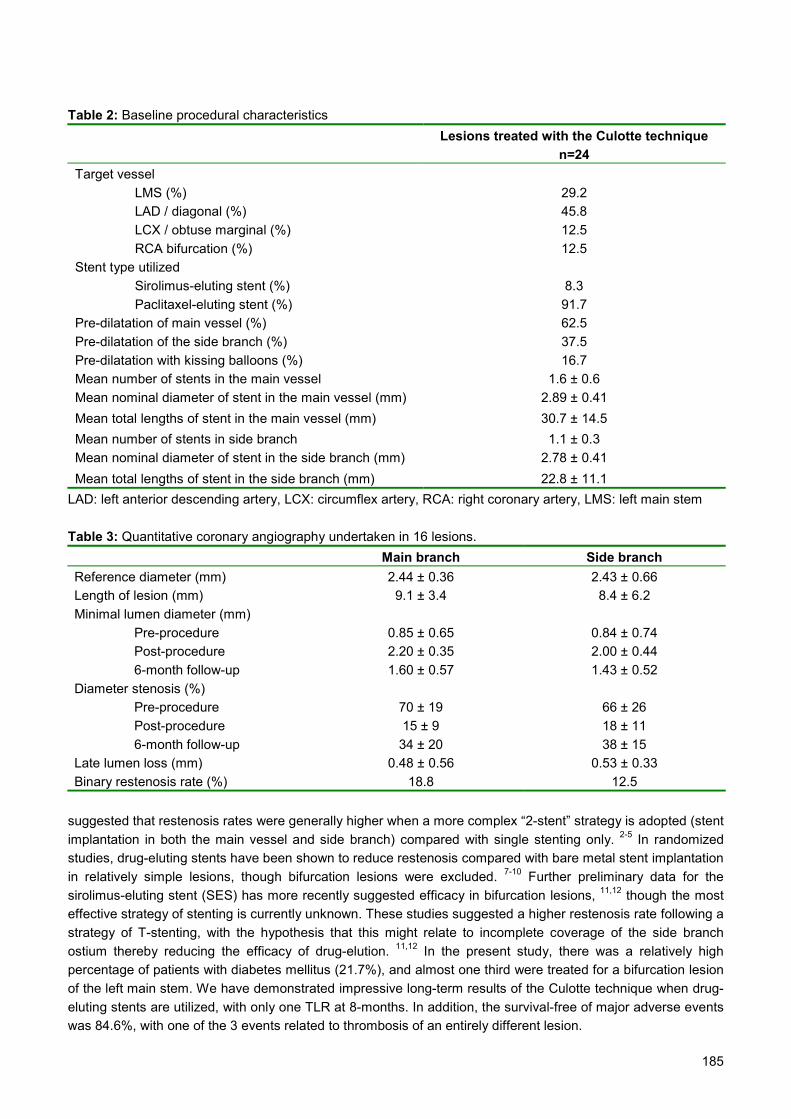
185
Table 2: Baseline procedural characteristicsLesions treated with the Culotte technique
n=24Target vessel
LMS (%) 29.2LAD / diagonal (%) 45.8LCX / obtuse marginal (%) 12.5RCA bifurcation (%) 12.5
Stent type utilizedSirolimus-eluting stent (%) 8.3Paclitaxel-eluting stent (%) 91.7
Pre-dilatation of main vessel (%) 62.5Pre-dilatation of the side branch (%) 37.5Pre-dilatation with kissing balloons (%) 16.7Mean number of stents in the main vessel 1.6 ± 0.6Mean nominal diameter of stent in the main vessel (mm) 2.89 ± 0.41Mean total lengths of stent in the main vessel (mm) 30.7 ± 14.5Mean number of stents in side branch 1.1 ± 0.3Mean nominal diameter of stent in the side branch (mm) 2.78 ± 0.41Mean total lengths of stent in the side branch (mm) 22.8 ± 11.1
LAD: left anterior descending artery, LCX: circumflex artery, RCA: right coronary artery, LMS: left main stem
Table 3: Quantitative coronary angiography undertaken in 16 lesions.Main branch Side branch
Reference diameter (mm) 2.44 ± 0.36 2.43 ± 0.66Length of lesion (mm) 9.1 ± 3.4 8.4 ± 6.2Minimal lumen diameter (mm)
Pre-procedure 0.85 ± 0.65 0.84 ± 0.74Post-procedure 2.20 ± 0.35 2.00 ± 0.446-month follow-up 1.60 ± 0.57 1.43 ± 0.52
Diameter stenosis (%)Pre-procedure 70 ± 19 66 ± 26Post-procedure 15 ± 9 18 ± 116-month follow-up 34 ± 20 38 ± 15
Late lumen loss (mm) 0.48 ± 0.56 0.53 ± 0.33Binary restenosis rate (%) 18.8 12.5
suggested that restenosis rates were generally higher when a more complex “2-stent” strategy is adopted (stentimplantation in both the main vessel and side branch) compared with single stenting only. 2-5 In randomizedstudies, drug-eluting stents have been shown to reduce restenosis compared with bare metal stent implantationin relatively simple lesions, though bifurcation lesions were excluded. 7-10 Further preliminary data for thesirolimus-eluting stent (SES) has more recently suggested efficacy in bifurcation lesions, 11,12 though the mosteffective strategy of stenting is currently unknown. These studies suggested a higher restenosis rate following astrategy of T-stenting, with the hypothesis that this might relate to incomplete coverage of the side branchostium thereby reducing the efficacy of drug-elution. 11,12 In the present study, there was a relatively highpercentage of patients with diabetes mellitus (21.7%), and almost one third were treated for a bifurcation lesionof the left main stem. We have demonstrated impressive long-term results of the Culotte technique when drug-eluting stents are utilized, with only one TLR at 8-months. In addition, the survival-free of major adverse eventswas 84.6%, with one of the 3 events related to thrombosis of an entirely different lesion.

186
The SES has been evaluated in one randomised study of bifurcation comparing a single stent with a 2-stent strategy. 12 The results suggested that, even with drug-eluting stents, it may be preferable to only stentthe main vessel, and carry out balloon-only angioplasty in the side branch. On follow-up angiography, therewas a trend towards a lower rate of restenosis in those treated with 1 stent compared with a 2-stent strategy(18.7% versus 28.0%, p=0.5). However, there was a very high rate of crossover from the 1 stent group to the 2stent group of 51%, related to a sub-optimal result in the side branch. Furthermore, the strategy used in the 2stent group was at the operators’ discretion and therefore heterogeneous. This study emphasises some of thedifficulties faced when treating bifurcation lesions. At baseline, a true bifurcation will have significant narrowingof both the main vessel and side branch. However, whatever the baseline anatomy, implantation of a stentwithin the main vessel can lead to plaque shift into the side branch therefore compromising side branch flow.Importantly, occurrence of such shift is unpredictable, 14 and the operator is then faced with the question as tohow to deal with the side branch most effectively. The Culotte technique ensures complete lesion coverageand, with drug-eluting stents, may offer a useful strategy for treating bifurcation lesions. In particular, it can bedone using a 6F sheath (other techniques such as the crush technique 15 or kissing stents require a guide of atleast 7F), and, by covering the entire lesion, will minimise the importance of any plaque shift. In addition, it canprovide a “backup” strategy for stenting of the side branch if the result following single stent implantation in themain vessel is sub-optimal. The major drawback to the technique is that it tends to be quite time-consuming,predominantly related to the need to re-wiring the vessels. Though it has not occurred in our experience, failureto re-wire the vessels could lead to procedural failure. In addition, there will be a double layer of stent struts inthe proximal vessel. This raises some concerns, as the increased dosage of drug at this site may predispose tostent thrombosis. The technique needs to be fully evaluated in large studies, and randomization is needed tocompare the relative efficacy of different stenting strategies particularly related to baseline anatomy.
Study limitationsThe present study comprises a consecutive series of patients treated with the Culotte technique. However, thestudy was small and non-randomized, with no direct comparison of drug-eluting stent implantation utilizingalternative stenting strategies.
ConclusionsIn this small study of bifurcation stenting utilizing the Culotte technique with drug-eluting stent implantation,there was a low rate of major adverse events and need for target lesion revascularization at 8-months whencompared with historical data of bifurcation stenting with bare metal stents. Further re-evaluation of thistechnique utilizing drug-eluting stents, is warranted in the setting of larger randomized studies.

187
References1. Al Suwaidi J, Yeh W, Cohen HA, Detre KM, Williams DO, Holmes DR, Jr. Immediate and one-year outcome in patients with
coronary bifurcation lesions in the modern era (NHLBI dynamic registry). Am J Cardiol. 2001;87:1139-44.
2. Anzuini A, Briguori C, Rosanio S, Tocchi M, Pagnotta P, Bonnier H, Gimelli G, Airoldi F, Margonato A, Legrand V, Colombo A.
Immediate and long-term clinical and angiographic results from Wiktor stent treatment for true bifurcation narrowings. Am J
Cardiol. 2001;88:1246-50.
3. Pan M, Suarez de Lezo J, Medina A, Romero M, Hernandez E, Segura J, Castroviejo JR, Pavlovic D, Melian F, Ramirez A,
Castillo JC. Simple and complex stent strategies for bifurcated coronary arterial stenosis involving the side branch origin. Am J
Cardiol. 1999;83:1320-5.
4. Lefevre T, Louvard Y, Morice MC, Dumas P, Loubeyre C, Benslimane A, Premchand RK, Guillard N, Piechaud JF. Stenting of
bifurcation lesions: classification, treatments, and results. Catheter Cardiovasc Interv. 2000;49:274-83.
5. Al Suwaidi J, Berger PB, Rihal CS, Garratt KN, Bell MR, Ting HH, Bresnahan JF, Grill DE, Holmes DR, Jr. Immediate and long-
term outcome of intracoronary stent implantation for true bifurcation lesions. J Am Coll Cardiol. 2000;35:929-36.
6. Yamashita T, Nishida T, Adamian MG, Briguori C, Vaghetti M, Corvaja N, Albiero R, Finci L, Di Mario C, Tobis JM, Colombo A.
Bifurcation lesions: two stents versus one stent--immediate and follow-up results. J Am Coll Cardiol. 2000;35:1145-51.
7. Grube E, Silber S, Hauptmann KE, Mueller R, Buellesfeld L, Gerckens U, Russell ME. TAXUS I: six- and twelve-month results
from a randomized, double-blind trial on a slow-release paclitaxel-eluting stent for de novo coronary lesions. Circulation.
2003;107:38-42.
8. Morice MC, Serruys PW, Sousa JE, Fajadet J, Ban Hayashi E, Perin M, Colombo A, Schuler G, Barragan P, Guagliumi G,
Molnar F, Falotico R. A randomized comparison of a sirolimus-eluting stent with a standard stent for coronary revascularization.
N Engl J Med. 2002;346:1773-80.
9. Moses JW, Leon MB, Popma JJ, Fitzgerald PJ, Holmes DR, O'Shaughnessy C, Caputo RP, Kereiakes DJ, Williams DO,
Teirstein PS, Jaeger JL, Kuntz RE. Sirolimus-eluting stents versus standard stents in patients with stenosis in a native coronary
artery. N Engl J Med. 2003;349:1315-23.
10. Stone GW, Ellis SG, Cox DA, Hermiller J, O'Shaughnessy C, Mann JT, Turco M, Caputo R, Bergin P, Greenberg J, Popma JJ,
Russell ME. A polymer-based, paclitaxel-eluting stent in patients with coronary artery disease. N Engl J Med. 2004;350:221-31.
11. Tanabe K, Hoye A, Lemos PA, Aoki J, Arampatzis CA, Saia F, Lee CH, Degertekin M, Hofma SH, Sianos G, McFadden E, Smits
PC, van der Giessen WJ, de Feyter P, van Domburg RT, Serruys PW. Restenosis rates following bifurcation stenting with
sirolimus-eluting stents for de novo narrowings. Am J Cardiol. 2004;94:115-8.
12. Colombo A, Moses JW, Morice MC, Ludwig J, Holmes DR, Jr., Spanos V, Louvard Y, Desmedt B, Di Mario C, Leon MB.
Randomized study to evaluate sirolimus-eluting stents implanted at coronary bifurcation lesions. Circulation. 2004;109:1244-9.
13. Chevalier B, Glatt B, Royer T, Guyon P. Placement of coronary stents in bifurcation lesions by the "culotte" technique. Am J
Cardiol. 1998;82:943-9.
14. Ahmed JM, Mintz GS, Weissman NJ, Lansky AJ, Pichard AD, Satler LF, Kent KM. Mechanism of lumen enlargement during
intracoronary stent implantation: an intravascular ultrasound study. Circulation. 2000;102:7-10.
15. Colombo A, Stankovic G, Orlic D, Corvaja N, Liistro F, Airoldi F, Chieffo A, Spanos V, Montorfano M, Di Mario C. Modified T-
stenting technique with crushing for bifurcation lesions: immediate results and 30-day outcome. Catheter Cardiovasc Interv.
2003;60:145-51.

188
189
Part 3
THE UNRESTRICTED USE OF DRUG-ELUTING STENTS: PREDICTORS OF AN
ADVERSE OUTCOME

190

191
Chapter 18
Andrew OngAngela HoyeJiro AokiCarlos AG van MieghemGaston Rodríguez GranilloKaren SonnenscheinEvelyn RegarEugene McFaddenGeorgios SianosWillem J van der GiessenPieter de JaegerePim J de FeyterRon T van DomburgPatrick W Serruys
Journal of the AmericanCollege of Cardiology2005; 45(6):947-53
Thirty-day incidence and six-monthclinical outcome of thrombotic stent
occlusion after bare-metal, sirolimus, orpaclitaxel stent implantation

192

193
AbstractObjectives: We sought to determine the real-world incidence of angiographically confirmed
and possible stent thrombosis (ST) in an unrestricted population during the first 30 days afterbare-metal stent (BMS), sirolimus-eluting stent (SES), and paclitaxel-eluting stent (PES)
implantation.
Background: Current data on ST in drug-eluting stents (DES) have come from randomized
trials with strict entry criteria, which limits their generalizability to daily practice.
Methods: The study population comprised three sequential cohorts of 506 consecutive
patients with BMS, 1,017 consecutive patients with SES, and 989 consecutive patients
treated with PES.
Results: In the first 30 days after stent implantation, 6 BMS (1.2%, 95% confidence interval
[CI] 0.5% to 2.6%; p = 0.9), 10 SES (1.0%, 95% CI 0.5% to 1.8%), and 10 PES (1.0%, 95%CI 0.6% to 1.9%) patients developed angiographically proven ST. Multiple potential risk
factors were identified in most patients with ST. Bifurcation stenting in the setting of acute
myocardial infarction was an independent risk factor for angiographic ST in the entirepopulation (odds ratio [OR] 12.9, 95% CI 4.7 to 35.8, p < 0.001). In patients with DES who
had angiographic ST, 30-day mortality was 15%, whereas another 60% suffered a nonfatal
myocardial infarction; no further deaths occurred during six months of follow-up. Includingpossible cases, 7 BMS (1.4%, 95% CI 0.7% to 2.8%), 15 SES (1.5%, 95% CI 0.9% to 2.4%),
and 16 PES (1.6%, 95% CI 1.0% to 2.6%) patients had ST.
Conclusions: The unrestricted use of SES or PES is associated with ST rates in the range
expected for BMS. Stent thrombosis was associated with a high morbidity and mortality.
Bifurcation stenting, when performed in patients with acute myocardial infarction, wasassociated with an increased risk of ST.
194
IntroductionDrug-eluting stents (DES) reduce clinical events related to restenosis. Concerns have been raised regardingthe incidence of stent thrombosis (ST) with the unrestricted use of these stents. Data from the bare-metal stent(BMS) era report a high morbidity and mortality with ST. 1,2 Evidence for ST in DES has come from randomizedcontrolled trials with strict entry criteria for the treatment of single lesions, limiting conclusions that areapplicable to the real-world setting. 3-6 Other information has come from electronic registries with inherentbiases that preclude generalization of the findings. A single-center registry recently reported its results withsirolimus-eluting stents (SES). 7 The aim of this present study is to describe the incidence of ST (bothangiographically proven and including possible cases) in three consecutive populations while analyzing theunrestricted use of a control BMS group, SES, and paclitaxel-eluting stents (PES).
MethodsStudy design and patient populationSince April 2002, SES (Cypher; Cordis Corp., Miami Lakes, Florida, a Johnson & Johnson Company) havebeen the stents of choice for all percutaneous coronary interventions irrespective of their clinical presentation orclinical outcome. 8 In the first quarter of 2003, PES (Taxus; Boston Scientific Corp., Natick, Massachusetts)replaced SES as the default stent. This present study comprises three sequential cohorts: a control group ofthe last 506 consecutive patients treated with BMS before April 2002; 1,017 consecutive patients with SEStreated between April 2002 and February 2003; and 989 consecutive patients with PES treated betweenFebruary 2003 and December 2003.
Procedure and antiplatelet managementAll interventions were performed according to current standard guidelines, and the final interventional strategyincluding periprocedural glycoprotein IIb/IIIa and intravascular ultrasound use, was left to the discretion of theoperator. Patients were pretreated with aspirin and a loading dose of 300 mg of clopidogrel. After theirprocedure, all patients were prescribed a lifelong aspirin regimen. Clopidogrel was prescribed for at least onemonth in the BMS group, for at least three months in the SES group, 8 and for at least six months in the PESgroup.
Follow-upAs part of the national health system, our institution as a tertiary referral center is the only interventional facilitywithin our catchment area. The survival status of our patients at one and six months after discharge wasobtained from the Municipal Civil Registries. Details of all repeat interventions (surgical and percutaneous)were collected prospectively during follow-up. Referring physicians and institutions were contacted whenevernecessary for additional information. This protocol was approved by the Hospital Ethics Committee, and written,informed consent was obtained from every patient.
DefinitionsStent thrombosis was considered to have occurred when confirmed angiographically: either Thrombolysis InMyocardial Infarction (TIMI) flow grade 0 or 1 or the presence of flow-limiting thrombus (TIMI flow grade 1 or 2)occurring in an acute (within 24 h of stent implantation) or subacute (between 1 and 30 days) time period afterstent implantation. 9 In addition, a clinical definition of “possible stent thrombosis” was used for patients whowithin the first 30 days experienced sudden death, who suffered a fatal out-of-hospital cardiac arrest, or whosuffered a myocardial infarction (MI) that was not clearly attributable to another coronary lesion and who did notundergo repeat angiography. All deaths and MIs were reviewed independently by two interventionalcardiologists (A.O., E.Mc.F) for “possible stent thrombosis.”
Statistical analysisCategorical variables were compared using the Fisher exact test and continuous variables with the Student ttest or one-way analysis of variance where appropriate. Univariate and forward stepwise (entry criteria of 0.05
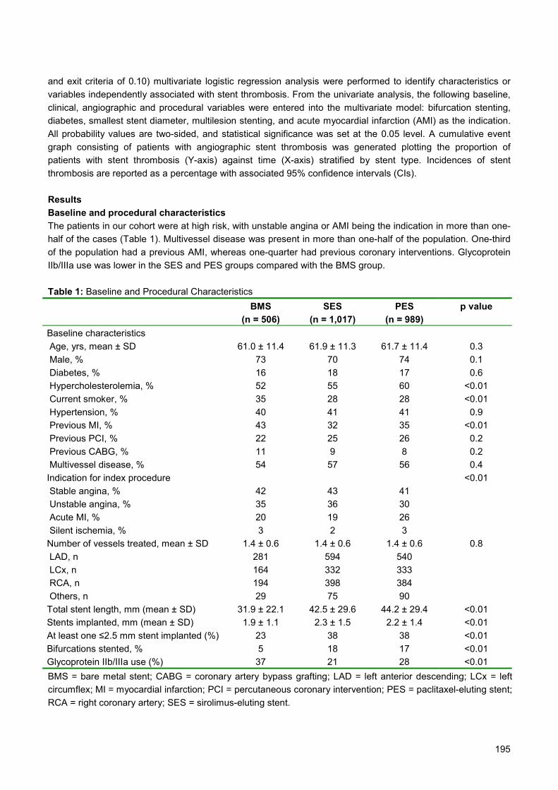
195
and exit criteria of 0.10) multivariate logistic regression analysis were performed to identify characteristics orvariables independently associated with stent thrombosis. From the univariate analysis, the following baseline,clinical, angiographic and procedural variables were entered into the multivariate model: bifurcation stenting,diabetes, smallest stent diameter, multilesion stenting, and acute myocardial infarction (AMI) as the indication.All probability values are two-sided, and statistical significance was set at the 0.05 level. A cumulative eventgraph consisting of patients with angiographic stent thrombosis was generated plotting the proportion ofpatients with stent thrombosis (Y-axis) against time (X-axis) stratified by stent type. Incidences of stentthrombosis are reported as a percentage with associated 95% confidence intervals (CIs).
ResultsBaseline and procedural characteristicsThe patients in our cohort were at high risk, with unstable angina or AMI being the indication in more than one-half of the cases (Table 1). Multivessel disease was present in more than one-half of the population. One-thirdof the population had a previous AMI, whereas one-quarter had previous coronary interventions. GlycoproteinIIb/IIIa use was lower in the SES and PES groups compared with the BMS group.
Table 1: Baseline and Procedural CharacteristicsBMS
(n = 506)SES
(n = 1,017)PES
(n = 989)p value
Baseline characteristics Age, yrs, mean ± SD 61.0 ± 11.4 61.9 ± 11.3 61.7 ± 11.4 0.3 Male, % 73 70 74 0.1 Diabetes, % 16 18 17 0.6 Hypercholesterolemia, % 52 55 60 <0.01 Current smoker, % 35 28 28 <0.01 Hypertension, % 40 41 41 0.9 Previous MI, % 43 32 35 <0.01 Previous PCI, % 22 25 26 0.2 Previous CABG, % 11 9 8 0.2 Multivessel disease, % 54 57 56 0.4Indication for index procedure <0.01 Stable angina, % 42 43 41 Unstable angina, % 35 36 30 Acute MI, % 20 19 26 Silent ischemia, % 3 2 3Number of vessels treated, mean ± SD 1.4 ± 0.6 1.4 ± 0.6 1.4 ± 0.6 0.8 LAD, n 281 594 540 LCx, n 164 332 333 RCA, n 194 398 384 Others, n 29 75 90Total stent length, mm (mean ± SD) 31.9 ± 22.1 42.5 ± 29.6 44.2 ± 29.4 <0.01Stents implanted, mm (mean ± SD) 1.9 ± 1.1 2.3 ± 1.5 2.2 ± 1.4 <0.01At least one ≤2.5 mm stent implanted (%) 23 38 38 <0.01Bifurcations stented, % 5 18 17 <0.01Glycoprotein IIb/IIIa use (%) 37 21 28 <0.01BMS = bare metal stent; CABG = coronary artery bypass grafting; LAD = left anterior descending; LCx = leftcircumflex; MI = myocardial infarction; PCI = percutaneous coronary intervention; PES = paclitaxel-eluting stent;RCA = right coronary artery; SES = sirolimus-eluting stent.
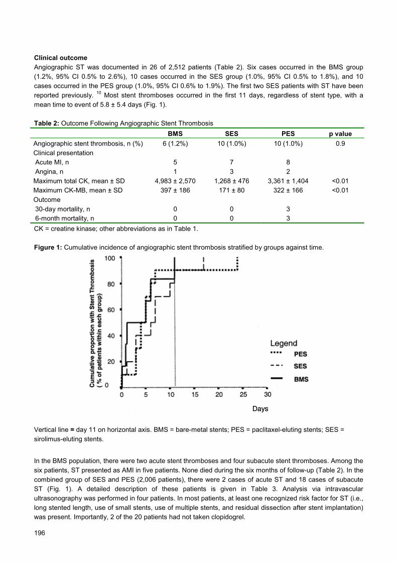
196
Clinical outcomeAngiographic ST was documented in 26 of 2,512 patients (Table 2). Six cases occurred in the BMS group(1.2%, 95% CI 0.5% to 2.6%), 10 cases occurred in the SES group (1.0%, 95% CI 0.5% to 1.8%), and 10cases occurred in the PES group (1.0%, 95% CI 0.6% to 1.9%). The first two SES patients with ST have beenreported previously. 10 Most stent thromboses occurred in the first 11 days, regardless of stent type, with amean time to event of 5.8 ± 5.4 days (Fig. 1).
Table 2: Outcome Following Angiographic Stent ThrombosisBMS SES PES p value
Angiographic stent thrombosis, n (%) 6 (1.2%) 10 (1.0%) 10 (1.0%) 0.9Clinical presentation Acute MI, n 5 7 8 Angina, n 1 3 2Maximum total CK, mean ± SD 4,983 ± 2,570 1,268 ± 476 3,361 ± 1,404 <0.01Maximum CK-MB, mean ± SD 397 ± 186 171 ± 80 322 ± 166 <0.01Outcome 30-day mortality, n 0 0 3 6-month mortality, n 0 0 3CK = creatine kinase; other abbreviations as in Table 1.
Figure 1: Cumulative incidence of angiographic stent thrombosis stratified by groups against time.
Vertical line = day 11 on horizontal axis. BMS = bare-metal stents; PES = paclitaxel-eluting stents; SES =sirolimus-eluting stents.
In the BMS population, there were two acute stent thromboses and four subacute stent thromboses. Among thesix patients, ST presented as AMI in five patients. None died during the six months of follow-up (Table 2). In thecombined group of SES and PES (2,006 patients), there were 2 cases of acute ST and 18 cases of subacuteST (Fig. 1). A detailed description of these patients is given in Table 3. Analysis via intravascularultrasonography was performed in four patients. In most patients, at least one recognized risk factor for ST (i.e.,long stented length, use of small stents, use of multiple stents, and residual dissection after stent implantation)was present. Importantly, 2 of the 20 patients had not taken clopidogrel.

197
Mortality and morbidityOverall, 20 of 26 patients (77%) re-presented with an AMI, whereas the other 6 re-presented with anginapectoris (Table 2). Of these 26 patients, 3 (Patients #12, #18, and #20 from Table 3—all in the DES population)died at days 11, 5, and 3, respectively. Two patients died during reintervention from intractable ventricularfibrillation, whereas the third underwent emergency surgery after a suboptimal reintervention and could not beweaned from bypass. The incidence of death at 30 days was 12%, whereas another 65% suffered a nonfatalMI. Among the survivors of ST, there were no further deaths in the six months after reintervention.
Possible STThirty-day survival data was complete for 98% of patients (Table 4). There were 12 patients who were judgedwith “possible stent thrombosis,” of which 9 died and 3 had nonfatal MIs. Of the nine deaths, four were out-of-hospital sudden deaths, three occurred in hospital with ventricular tachycardia as the initiating preterminalrhythm, and two had ST-segment elevation and died before they could undergo reangiography. Among thosewith MIs, one patient developed a postprocedural enzyme leak, and another developed ventricular fibrillationrequiring multiple cardioversions the day after the procedure. Repeat coronary angiography six months laterdemonstrated occluded stents in both of these patients; whereas a third underwent coronary angiography 14days after stent implantation because of an increase in cardiac enzyme levels, which demonstrated an in-stentfilling defect which was treated with abciximab, and subsequently underwent repeat percutaneous coronaryintervention two weeks later. Including the suspected cases, the combined incidence of angiographic andpossible ST was 1.4% (95% CI 0.7% to 2.8%) in the BMS control group, 1.5% (95% CI 0.9% to 2.4%) in theSES group, and 1.6% (95% CI 1.0% to 2.6%) in the PES group. In the combined total of 38 documented andpossible ST, there were 12 deaths (32%) and 20 nonfatal MIs (53%) in the first 30 days.
Table 4: Incidence of Stent Thrombosis Classified by DefinitionStentType
Number ofPatients
Angiographically ProvenStent Thrombosis
n (% [95% CI])
Possible StentThrombosis
n (% [95% CI])
All Stent Thrombosis
n (% [95% CI])BMS 506 6 (1.2% [0.5%–2.6%]) 1 (0.2% [0.0%–1.1%]) 7 (1.4% [0.7%–2.8%])SES 1,017 10 (1.0% [0.5%–1.8%]) 5 (0.5% [0.2%–1.1%]) 15 (1.5% [0.9%–2.4%])PES 989 10 (1.0% [0.6%–1.9%]) 6 (0.6% [0.3%–1.3%]) 16 (1.6% [1.0%–2.6%])CI = confidence interval; other abbreviations as in Table 1.
Multivariate analysisBy univariate analysis, bifurcation stenting was the only significant factor (p = 0.01). Multivariate analysis wasperformed with the following covariates based on their significance on univariate analysis as well as theirpotential clinical impact: diabetes (p = 0.07), smallest stent diameter (p = 0.13), multilesion stenting (p = 0.17),AMI as the indication (p = 0.3), and bifurcation stenting. By multivariate analysis, bifurcation stenting was theonly independent predictor of ST (odds ratio [OR] 3.0, 95% CI 1.3 to 6.8, p < 0.01). When the interaction ofbifurcation stenting by AMI was entered as a covariate, it was highly significant (OR 12.9, 95% CI 4.7 to 35.8, p< 0.001), and bifurcation stenting as a covariate was no longer significant.
DiscussionThe main findings in this study can be summarized as follows: 1) the incidence of angiographic ST in anunselected, complex DES population was low ( 1.0%), within the same range as the corresponding BMSpopulation and concordant with previously published results from the BMS era; 2) the inclusion of possible STincreases the overall incidence of ST to 1.5%; 3) angiographically proven ST was associated with a highmortality and morbidity; 4) patients who developed ST often had multiple high-risk features, regardless of stenttype; and 5) the association of bifurcation stenting for AMI was a highly significant independent risk factor forST.
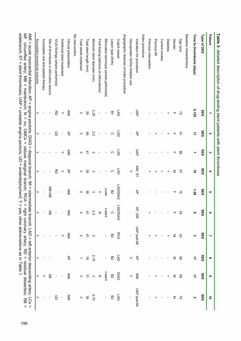
198
Successful procedural outcom
e
Incomplete oral anti-platelet therapy
Site of throm
bosis in bifurcation lesions
IVU
S findings (w
here performed)
Additional stent im
planted
Clinical presentation
Re intervention
Total stents implanted
Total stent length (mm
)
Minim
um stent diam
eter (mm
)
Final kissing balloons (in bifurcations)
Bifurcation technique (w
here performed)
Lesion type (AH
A)
Culprit vessel
Angiographic features of index procedure
Glycoprotein IIb/IIIa inhibitor use
Indication for procedure
Index procedure
Previous intervention
Previous M
I
Current sm
oker
Diabetes
Gender
Age (yrs)
Baseline characteristics
Time to throm
bosis (days)
Type of DES
Patient
Y - - RD Y AM
I
2 26
2.25 - - B1
LAD -
UA
P - - - + F 72
0.125
SES 1
Y - - UD - AP 2 66
2.5 - - C LAD - AP - + - + F 61 11
SES 2
Y - - - Y AM
I
3 41 3 - - C LAD -
UA
P - - - - F 86 7
SES 3
Y - - RD Y AP 2 26 3 - - C LAD Y
AM
I, ST
+ + + + M 57 10
SES 4
Y -
MB
+SB
- -
AM
I
2 36 3 Y
crush
B2
LAD
/DIA
G
- AP - - - - F 75
1.08
SES 5
Y - SB - -
AM
I
2 41
2.5 N
t-stent
C
LAD
/DIA
G
-
AP, IS
R
+ - - - F 55 6
SES 6
Y - - - Y AM
I
2 41 3 - B2
RC
A -
UA
P post-M
I
+ + - + M 53 3
SES 7
Y - - AP 1 18
2.75 - B2
LAD Y AP - - - - M 58 17
SES 8
Y Y SB - -
AM
I
2 31 3 N
t-stent
B2
DIA
G
Y AM
I
- + - + M 58 17
SES 9
Y - - UD -
AM
I
2 36
2.75 - B2
LAD -
UA
P post-M
I
- + - - M 74 3
SES
10
Table 3: detailed description of drug-eluting stent patients with stent throm
bosis
AMI = acute m
yocardial infarction; AP = angina pectoris; D
IAG = diagonal branch; IM
= intermediate branch; LA
D = left anterior descending artery; LC
x =left circum
flex artery; MB = m
ainbranch; N = no; O
MC
x = obtuse marginal branch; R
CA = right coronary artery; R
D = residual dissection; S
B =sidebranch; ST = stent throm
bosis; UA
P = unstable angina pectoris; UD
= underdeployment; Y
= yes; other abbreviations as in Table 1.

199
Mea
n ±
SD, %
6.3
± 5.
7
59.7
± 1
1.9
13M
7F
30%
30%
45%
25%
30%
40%
2.7
± 0.
3
41.2
± 2
7.8
2.2
± 1.
5
v
AM
I = 7
5%
Yes
= 3
5%
10%
Dea
th =
15%
20 PES 3 31 M - + - + A
P -
LAD
B2 - N 3 84 4 A
MI
- - - -
Die
d
19 PES 5 65 M - - + - A
P Y
LAD
/IM/L
Cx
CC
ulot
tecr
ush
Y 2.25
140 8 A
MI
- -
MB
+SB
-
Die
d
18 PES 5 54 M - + - -
AM
I
Y
LAD
/DIA
G
C
Cru
sh N 2.75 36 2 AM
I
- - SB - Y
17 PES
24 60 M - - + - AP -
LCX C - -
2.25 32 2
UA
P
Y -
MB
+SB
- Y
16 PES 4 52 F - + - -
AM
I
-
LCX
/OM
CX
C
t-ste
nt
N 2.25 44 2
UA
P - - SB - Y
15 PES 3 61 M + + + - A
P -
LCx
B2 - - 2.5
20 1 AM
I
Y - - - Y
14 PES 6 47 F - + - -
AM
I
Y
LAD
/DIA
G
C
Cru
sh N 2.5
36 2 AM
I
- - SB - Y
13 PES 7 67 M - - + + A
P -
OM
CX
C
Cru
sh N 2.5
32 2 AM
I
Y - SB - Y
12 PES 4 50 M - - - -
AM
I
-
LAD
B2 - - 3.5
24 1 AM
I
- - - -
Die
d
11 PES
0.04 59 M - - - -
AM
I
-
RC
A
B2 - - 3 28 1 A
MI
- - - - Y
Patie
nt
Type
of D
ES
Tim
e to
thro
mbo
sis
(day
s)
Bas
elin
e ch
arac
teris
tics
A
ge (y
rs)
G
ende
r
D
iabe
tes
C
urre
nt s
mok
er
P
revi
ous
MI
P
revi
ous
inte
rven
tion
Inde
x pr
oced
ure
I
ndic
atio
n fo
r pro
cedu
re
G
lyco
prot
ein
IIb/II
Ia in
hibi
tor u
se
Ang
iogr
aphi
c fe
atur
es o
f ind
ex p
roce
dure
C
ulpr
it ve
ssel
L
esio
n ty
pe (A
HA
)
B
ifurc
atio
n te
chni
que
(whe
re p
erfo
rmed
)
F
inal
kis
sing
bal
loon
(in
bifu
rcat
ions
)
M
inim
um s
tent
dia
met
er (m
m)
T
otal
ste
nt le
ngth
(mm
)
T
otal
ste
nts
impl
ante
d
Re
inte
rven
tion
C
linic
al p
rese
ntat
ion
A
dditi
onal
ste
nt im
plan
ted
I
VU
S fi
ndin
gs (w
here
per
form
ed)
S
ite o
f thr
ombo
sis
in b
ifurc
atio
n le
sion
s
I
ncom
plet
e or
al a
nti-p
late
let t
hera
py
S
ucce
ssfu
l pro
cedu
ral o
utco
me
AMI =
acu
te m
yoca
rdia
l inf
arct
ion;
AP
= an
gina
pec
toris
; DIA
G =
dia
gona
l bra
nch;
IM =
inte
rmed
iate
bra
nch;
LA
D =
left
ante
rior d
esce
ndin
g ar
tery
; LC
x =
left
circ
umfle
x ar
tery
; M
B =
mai
nbra
nch;
N =
no;
OM
Cx
= ob
tuse
mar
gina
l br
anch
; R
CA
= rig
ht c
oron
ary
arte
ry;
RD
= r
esid
ual
diss
ectio
n; S
B =
side
bran
ch; S
T =
sten
t thr
ombo
sis;
UA
P =
unst
able
ang
ina
pect
oris
; UD
= u
nder
depl
oym
ent;
Y =
yes
; oth
er a
bbre
viat
ions
as
in T
able
1.

200
The availability of DES as the default stent at our institution has allowed us to analyze this newtechnology in an unrestricted population, 8 a population that would have comprised any BMS population in thepre-DES era. Therefore, this availability allows us to analyze incidences in an “all-comers” population becausepatients were enrolled irrespective of clinical presentation or outcome. In this population sample, angiographicST rates in the first 30 days for both DES, i.e., SES and PES, occurred within the range as that reported in theBMS era. 1,2,11,12
The angiographic definition used is the most accurate for diagnosis but may underestimate the trueincidence of ST because some patients who have a presumed ST may die before receiving medical attention.Conversely, the use of major adverse cardiac events (i.e., death and MI in addition to the angiographic findings)to define ST overestimates the true incidence because not all patients who die suddenly or suffer a MI do sobecause of ST. 13 This consideration is important in our heterogeneous unrestricted population with multivesseldisease, previous MI, and previous revascularization. Furthermore, not all patients who die will undergoautopsy studies to determine the cause of death. To attenuate this overestimation and to provide an accuratefigure, we have adjudicated all deaths and noncatheterized, nonfatal MIs within the first 30 days in the threegroups and included them with the angiographically proven patients to provide an overall incidence for eachgroup.
The incidences of ST for both groups of DES are within the range reported in the larger randomizedclinical trials of DES 3-6 despite longer total stent length, multivessel treatment, and a heterogeneous population(Table 5). This incidence complements information already available from the randomized trials regarding thesafety of these new devices.
Table 5: Clinical Trials on Drug-Eluting StentsTrial Name Number of Patients in Drug-
Eluting ArmTotal Stent Length mm
(Mean ± SD)Incidence of Stent Thrombosis
in the First 30 Days (%)SIRIUS (3) 533 23.0 ± 8.6 0.2*E-SIRIUS (6) 157 21.5 ± 6.7 1.1*C-SIRIUS (5) 50 23.8 ± 8.4 2.0*TAXUS-IV (4) 662 21.9 ± 8.1 0.3†SES group 1,017 42.5 ± 29.6 ‡1.0–1.5§PES group 989 44.2 ± 29.4 ‡1.0–1.6§Abbreviations as in Table 1.* Definition of stent thrombosis was not stated.† Stent thrombosis defined as angiographically proven, or cardiac death or myocardial infarction in the first 30days.‡ Stent thrombosis defined as angiographically proven.§ Stent thrombosis defined as angiographically proven, or adjudicated death or myocardial infarction in the first30 days.
Angiographic ST was associated with a high mortality and morbidity in our study. Within the DES population, 15patients (75%) experienced a MI as their diagnosis at the second presentation, and 3 (15%) died during thereintervention procedure. The inclusion of possible ST patients increased the mortality to 32%. Given the smallnumber of events, the fact that no deaths occurred in the BMS group was most likely due to chance. Theseresults are in concordance with the results of a large BMS registry. 2
Previous studies have demonstrated that residual dissection, 1,11 long stents, 1 small final lumendiameter, 1 and use of multiple stents 2 are risk factors for the development of ST. In our series, multiple riskfactors were identified in most patients who developed ST. Patients with ST had more multiple lesions treated,smaller minimum stent diameters, and longer stent lengths compared with those without ST; however, thesefactors were not significant on univariate analysis. What did emerge and which has not been previouslyreported is that patients undergoing bifurcation stenting had a higher incidence of ST compared with those

201
without bifurcation stenting. A recent study on bifurcations reported a 3.5% incidence of ST, which is higherthan the overall incidence in this population. 14
Although stent implantation for AMI was not significant on univariate analysis, the interaction of AMIand bifurcation stenting when entered as a covariate for ST on multivariate analysis emerged as a highlysignificant independent predictor, and bifurcation stenting as a covariate was no longer significant. This resultconfirms a clinical suspicion in our department regarding the increased risk of ST in patients treated withbifurcation stenting in the setting of AMI.
Mechanical reasons that predispose to ST can be modified by interventional technique. Optimizingstent placement including, if necessary, intravascular ultrasound-guided postdilation, kissing balloonpostdilation with bifurcation stenting, and careful inspection for residual dissection after stent implantation, mayfurther reduce the incidence of ST.
Pharmacologic reasons for ST, i.e., inadequate antiplatelet therapy, are patient-specific factors. Recentresearch literature has focused on “resistance” to either aspirin 15 or to clopidogrel. 16 Currently, mostlaboratories do not routinely test for antiplatelet resistance. In our series, two patients who had not taken theirprescribed clopidogrel after the procedure developed ST.
This report covers ST occurring in the first 30 days after stent implantation only, during which allpatients received dual antiplatelet therapy. The duration of clopidogrel therapy differed among the three groups;in part, it reflects uncertainty with regards to re-endothelialization after DES implantation. Late ST has beenreported to occur with BMS 17 and with DES, 18 including a reported fatality 19 after clopidogrel discontinuation.At this stage, the incidence of late ST in the DES era is unknown, and further studies are required to clarify thispotential late complication.
Comment on sample size and statistical comparisonsBecause ST occurs at a low incidence ( 1.0 to 1.5%), a small sample size may underestimate or overestimatethe true incidence. In a previously published report from our institution, we reported an angiographic incidenceof 0.4% in 508 patients. 8 In the present study we extended the population to incorporate the entire period ofDES used to date at our institution (n = 2,006) to allow a more accurate analysis of the true incidence of ST inthe DES population. Despite having 2,512 patients, the low and small/negligible absolute difference inincidence precludes formal statistical comparisons of ST rates among the three groups because it lackssufficient statistical power. To achieve adequate power would require sample sizes in the order of >100,000patients. To date, this study is the largest series of patients reported on in the DES era.
Study limitationsThese single-center registry data complement available randomized data, as they reflect the results ofunrestricted DES use.
ConclusionsDespite having a more complex cohort with high-risk inclusion criteria, longer stent lengths, and more complexprocedural features, the incidence of ST with DES are in the same range as the BMS population observed inour present study. They also are in agreement with previously reported data by others from the BMS era andwith those results reported on in the earlier randomized DES trials. Furthermore, the two groups of DES, i.e.,SES and PES, share an incidence of 1.0% to 1.5%. Stent thrombosis is associated with a high morbidity andmortality.As extensively documented in previous reports with BMS, mechanical reasons were observed to be frequentassociations for ST with DES. In this study, bifurcation stenting in the setting of AMI was a highly significantindependent predictor for angiographic ST.

202
References1. D.E. Cutlip, D.S. Baim and K.K. Ho et al., Stent thrombosis in the modern era: a pooled analysis of multicenter coronary stent
clinical trials, Circulation 103 (2001), pp. 1967–1971.
2. J.L. Orford, R. Lennon and S. Melby et al., Frequency and correlates of coronary stent thrombosis in the modern era: analysis of
a single center registry, J Am Coll Cardiol 40 (2002), pp. 1567–1572.
3. J.W. Moses, M.B. Leon and J.J. Popma et al., Sirolimus-eluting stents versus standard stents in patients with stenosis in a native
coronary artery, N Engl J Med 349 (2003), pp. 1315–1323.
4. G.W. Stone, S.G. Ellis and D.A. Cox et al., A polymer-based, paclitaxel-eluting stent in patients with coronary artery disease, N
Engl J Med 350 (2004), pp. 221–231.
5. E. Schampaert, E.A. Cohen and M. Schluter et al., The Canadian study of the sirolimus-eluting stent in the treatment of patients
with long de novo lesions in small native coronary arteries (C-SIRIUS), J Am Coll Cardiol 43 (2004), pp. 1110–1115.
6. J. Schofer, M. Schluter and A.H. Gershlick et al., Sirolimus-eluting stents for treatment of patients with long atherosclerotic
lesions in small coronary arteries: double-blind, randomised controlled trial (E-SIRIUS), Lancet 362 (2003), pp. 1093–1099.
7. A. Jeremias, B. Sylvia and J. Bridges et al., Stent thrombosis after successful sirolimus-eluting stent implantation, Circulation 109
(2004), pp. 1930–1932.
8. P.A. Lemos, P.W. Serruys and R.T. van Domburg et al., Unrestricted utilization of sirolimus-eluting stents compared with
conventional bare stent implantation in the “real world”: the Rapamycin-Eluting Stent Evaluated At Rotterdam Cardiology Hospital
(RESEARCH) registry, Circulation 109 (2004), pp. 190–195.
9. P.A. Lemos, C.H. Lee and M. Degertekin et al., Early outcome after sirolimus-eluting stent implantation in patients with acute
coronary syndromes: insights from the Rapamycin-Eluting Stent Evaluated At Rotterdam Cardiology Hospital (RESEARCH)
registry, J Am Coll Cardiol 41 (2003), pp. 2093–2099.
10. E. Regar, P.A. Lemos and F. Saia et al., Incidence of thrombotic stent occlusion during the first three months after sirolimus-
eluting stent implantation in 500 consecutive patients, Am J Cardiol 93 (2004), pp. 1271–1275.
11. E. Cheneau, L. Leborgne and G.S. Mintz et al., Predictors of subacute stent thrombosis: results of a systematic intravascular
ultrasound study, Circulation 108 (2003), pp. 43–47.
12. T.R. Tolleson, L.K. Newby and R.A. Harrington et al., Frequency of stent thrombosis after acute coronary syndromes (from the
SYMPHONY and 2nd SYMPHONY trials), Am J Cardiol 92 (2003), pp. 330–333.
13. Y. Honda and P.J. Fitzgerald, Stent thrombosis: an issue revisited in a changing world, Circulation 108 (2003), pp. 2–5.
14. A. Colombo, J.W. Moses and M.C. Morice et al., Randomized study to evaluate sirolimus-eluting stents implanted at coronary
bifurcation lesions, Circulation 109 (2004), pp. 1244–1249.
15. P.A. Gum, K. Kottke-Marchant, P.A. Welsh, J. White and E.J. Topol, A prospective, blinded determination of the natural history of
aspirin resistance among stable patients with cardiovascular disease, J Am Coll Cardiol 41 (2003), pp. 961–965.
16. W.C. Lau, P.A. Gurbel and P.B. Watkins et al., Contribution of hepatic cytochrome P450 3A4 metabolic activity to the
phenomenon of clopidogrel resistance, Circulation 109 (2004), pp. 166–171.
17. F. Wang, G.A. Stouffer, S. Waxman and B.F. Uretsky, Late coronary stent thrombosis: early vs, late stent thrombosis in the stent
era. Catheter Cardiovasc Interv 55 (2002), pp. 142–147.
18. A. Colombo, J. Drzewiecki and A. Banning et al., Randomized study to assess the effectiveness of slow- and moderate-release
polymer-based paclitaxel-eluting stents for coronary artery lesions, Circulation 108 (2003), pp. 788–794.
19. R. Virmani, G. Guagliumi and A. Farb et al., Localized hypersensitivity and late coronary thrombosis secondary to a sirolimus-
eluting stent: should we be cautious?, Circulation 109 (2004), pp. 701–705.

203
Chapter 19
Pedro LemosAngela HoyeDirk GoedhartChourmouzios ArampatzisFrancesco SaiaWillem J van der GiessenEugene McFaddenGeorgios SianosPieter C SmitsSjoerd H HofmaPim J de FeyterRon T van DomburgPatrick W Serruys
Circulation2004;109(11):1366-70
Clinical, angiographic, and proceduralpredictors of angiographic restenosis after
sirolimus-eluting stent implantation incomplex patients: an evaluation from the
Rapamycin-Eluting Stent Evaluated AtRotterdam Cardiology Hospital
(RESEARCH) study

204

205
Abstract
Background: The factors associated with the occurrence of restenosis after sirolimus-eluting
stent (SES) implantation in complex cases are currently unknown.
Methods and Results: A cohort of consecutive complex patients treated with SES
implantation was selected according to the following criteria: (1) treatment of acute
myocardial infarction, (2) treatment of in-stent restenosis, (3) 2.25-mm diameter SES, (4) left
main coronary stenting, (5) chronic total occlusion, (6) stented segment >36 mm, and (7)
bifurcation stenting. The present study population was composed of 238 patients (441
lesions) for whom 6-month angiographic follow-up data were obtained (70% of eligible
patients). Significant clinical, angiographic, and procedural predictors of post-SES restenosis
were evaluated. Binary in-segment restenosis was diagnosed in 7.9% of lesions (6.3% in-
stent, 0.9% at the proximal edge, 0.7% at the distal edge). The following characteristics were
identified as independent multivariate predictors: treatment of in-stent restenosis (OR 4.16,
95% CI 1.63 to 11.01; P<0.01), ostial location (OR 4.84, 95% CI 1.81 to 12.07; P<0.01),
diabetes (OR 2.63, 95% CI 1.14 to 6.31; P=0.02), total stented length (per 10-mm increase;
OR 1.42, 95% CI 1.21 to 1.68; P<0.01), reference diameter (per 1.0-mm increase; OR 0.46,
95% CI 0.24 to 0.87; P=0.03), and left anterior descending artery (OR 0.30, 95% CI 0.10 to
0.69; P<0.01).
Conclusions: Angiographic restenosis after SES implantation in complex patients is an
infrequent event, occurring mainly in association with lesion-based characteristics and
diabetes mellitus.

206
IntroductionIn-stent restenosis is the major limitation hampering the medium-term efficacy of coronary stenting. Severalreports have evaluated the impact of baseline and procedural characteristics on the risk of subsequentrestenosis after bare metal stent implantation, with a number of high-risk parameters, such as diabetes, lesionlength, and vessel size, being consistently identified in most studies.1–7 Unfortunately, these characteristics arecommonly found in the daily practice, where treatment of complex patients frequently appears as a challengingtherapeutic dilemma.
Sirolimus-eluting stents (SESs) have been proven to strikingly decrease neointimal growth, leading to amarked reduction in restenosis rates.8–10 In the RAndomized study with the sirolimus-eluting Bx VElocityballoon-expandable stent in the treatment of patients with de novo native coronary artery Lesions (RAVEL),8 nocases of binary angiographic restenosis were seen after SES implantation. Moreover, restenosis wassignificantly reduced from 36.3% with conventional stents to 8.9% with SESs in the randomized SIRolImUS-eluting Bx velocity balloon expandable stent trial (SIRIUS)9 and from 42.3% to 5.9% in the E-SIRIUS trial,10 withdiabetes, small vessel size, and long lesions being identified as predictors of post-SES restenosis in the SIRIUStrial.9 Nevertheless, these randomized studies have been largely restricted to selected patients treated withsingle-lesion elective stenting. The factors related to angiographic restenosis after SES implantation in highlycomplex subsets are currently unknown.
SES implantation was recently shown to effectively improve the 1-year clinical outcomes in "real world"practice in patients enrolled in the Rapamycin-Eluting Stent Evaluated At Rotterdam Cardiology Hospital(RESEARCH) study.11 In the RESEARCH study, a parallel angiographic substudy was conducted to evaluatethe late angiographic findings of complex patients treated with SESs. The present report aimed to evaluate thevalue of clinical, angiographic, and procedural factors in predicting the risk of binary restenosis in highlycomplex patients treated with SES implantation in the RESEARCH study.
MethodsStudy Design and Patient PopulationThe design of the RESEARCH study has been reported previously.11 In brief, SES implantation (Cypher;Johnson & Johnson-Cordis unit, Cordis Europa NV) was introduced as the default strategy for all patientsundergoing percutaneous coronary interventions in our institution after April 2002. All procedures wereperformed according to standard techniques, and the final interventional strategy was left to the discretion of theoperator, with the aim of achieving a final residual stenosis <50% by online quantitative coronary angiography inthe presence of TIMI (Thrombolysis In Myocardial Infarction) 3 grade flow. The use of periproceduralglycoprotein IIb/IIIa inhibitors and antithrombotic medications was left entirely to the discretion of the attendingteam.
Patients receiving SESs were considered candidates for angiographic reevaluation if they had at least 1of the following characteristics: (1) treatment of acute myocardial infarction, (2) treatment of in-stent restenosis,(3) use of a very small SES (2.25-mm nominal diameter), (4) treatment of left main coronary, (5) treatment ofchronic total occlusion (>3 months), (6) total adjacent stented segment longer than 36 mm, and (7) bifurcationstenting (SES implanted in both the main vessel and the side branch). Patients with the aforementionedcharacteristics who had not undergone repeat intervention in the first month and had not presented any formalmedical contraindication for angiographic restudy were considered eligible for angiographic follow-up at 6 to 8months. Coronary angiograms performed prematurely because of clinical indications were used as the follow-up angiography if performed after 4 months or if restenosis was detected. In other cases, a second angiogramwas obtained between 6 and 8 months. Importantly, although all patients were approached for angiographicfollow-up, patient refusal was not considered as an exclusion criterion to be enrolled in the RESEARCH study.Angiographic restudy was not requested for nonresidents of the Netherlands.
During the first 6 months of enrollment, a total of 362 consecutive patients had at least 1 of the high-riskcriteria listed above (57% of all patients treated with SESs in the period). Of these, 2 patients moved to anothercountry, 10 had died within the first 6 months of follow-up, 6 had repeat intervention before 30 days (surgical orpercutaneous), and 3 were considered to have a medical contraindication to the angiographic follow-up (1

207
patient with previous stroke and disabling dementia, 1 with severe allergic contrast reaction at the indexprocedure, and 1 with end-stage hepatic failure due to autoimmune hepatitis). Of the remaining 341 patients,angiographic reevaluation at 204±34 days was obtained for 238 patients (70% of eligible patients), whocompose the present study population.
Quantitative Coronary AngiographyQuantitative coronary angiographic analysis was performed as described previously with a validated computer-based edge-detection system (CASS II, Pie Medical).12 Interpolated reference diameter, minimal luminaldiameter, and diameter stenosis were obtained at baseline, after stenting, and at follow-up. In-stent restenosiswas defined by diameter stenosis >50% and was classified as in-stent if inside the stent or in-segment if locatedwithin the stented segment plus the 5-mm segments distal or proximal to the stent margins.9 Restenosis at anostial location (within 3 mm of the vessel origin) was classified as in-stent unless clearly located outside thelimits of the SES.13
Statistical AnalysisContinuous variables are presented as mean±SD and were compared with Student’s unpaired t test.Categorical variables are presented as counts and percentages and compared with the Fisher exact test.Demographic, clinical, procedural, and angiographic variables were tested in univariate and multivariate logisticanalyses for their value in predicting binary restenosis. All variables shown in Tables 1 and 2 were consideredin multivariate logistic regression analyses regardless of their univariate findings. The final model was builtiteratively and evaluated for lack of fit with the Hosmer-Lemeshow test. Global predictive accuracy wasassessed by means of the C-index (area under the receiver operating characteristic curve). Finally, an internalvalidation was performed with a bootstrap technique.14 The model was repeatedly applied to 1000 replicatedbootstrap samples, and the C-index for each individual sample was calculated. The C-index obtained from eachbootstrap sample was then subtracted from the initial C-index value of the original population. The average ofthe differences was considered as a measure of optimism in the model fit. Finally, a corrected C-index wascalcuTable 1: Clinical Characteristics of 238 Patients Treated With SES ImplantationCharacteristic No.Male gender 73Age, y 60 ± 12Height, cm 172 ± 9Weight, kg 82 ± 14Hypercholesterolemia 58Hypertension 56Diabetes mellitus 22Insulin-dependent diabetes 6Non-insulin-dependent diabetes 16Previous myocardial infarction 32Previous bypass surgery 11Previous percutaneous intervention 28
1-vessel disease 402-vessel disease 35
Vessel disease
3-vessel disease 25Stable angina 54Unstable angina 21
Clinical presentation
Acute myocardial infarction 26Periprocedural IIb/IIIa inhibitor 27Values are percentages or mean±SD*Total cholesterol >200 mg/dL or receiving lipid-lowering treatment
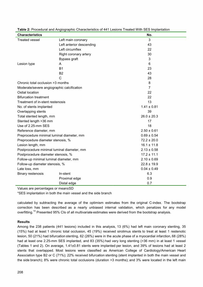
208
Table 2: Procedural and Angiographic Characteristics of 441 Lesions Treated With SES ImplantationCharacteristics No.
Left main coronary 3Left anterior descending 43Left circumflex 22Right coronary artery 30
Treated vessel
Bypass graft 3A 6B1 23B2 43
Lesion type
C 28Chronic total occlusion >3 months 8Moderate/severe angiographic calcification 7Ostial location 22Bifurcation treatment 22Treatment of in-stent restenosis 13No. of stents implanted 1.41 ± 0.81Overlapping stents 39Total stented length, mm 26.0 ± 20.3Stented length >36 mm 17Use of 2.25-mm SES 18Reference diameter, mm 2.50 ± 0.61Preprocedure minimal luminal diameter, mm 0.69 ± 0.54Preprocedure diameter stenosis, % 72.2 ± 20.0Lesion length, mm 16.1 ± 11.8Postprocedure minimal luminal diameter, mm 2.13 ± 0.58Postprocedure diameter stenosis, % 17.2 ± 11.1Follow-up minimal luminal diameter, mm 2.10 ± 0.69Follow-up diameter stenosis, % 22.8 ± 19.9Late loss, mm 0.04 ± 0.49
In-stent 6.3Proximal edge 0.9
Binary restenosis
Distal edge 0.7Values are percentages or mean±SD*SES implantation in both the main vessel and the side branch
calculated by subtracting the average of the optimism estimates from the original C-index. The bootstrapcorrection has been described as a nearly unbiased internal validation, which penalizes for any modeloverfitting.14 Presented 95% CIs of all multivariate estimates were derived from the bootstrap analysis.
ResultsAmong the 238 patients (441 lesions) included in this analysis, 13 (6%) had left main coronary stenting, 35(15%) had at least 1 chronic total occlusion, 45 (19%) received sirolimus stents to treat at least 1 restenoticlesion, 50 (21%) had bifurcation stenting, 62 (26%) were in the acute phase of a myocardial infarction, 68 (28%)had at least one 2.25-mm SES implanted, and 83 (35%) had very long stenting (>36 mm) in at least 1 vessel(Tables 1 and 2). On average, 1.41±0.81 stents were implanted per lesion, and 39% of lesions had at least 2stents that overlapped. Most lesions were classified as American College of Cardiology/American HeartAssociation type B2 or C (71%); 22% received bifurcation stenting (stent implanted in both the main vessel andthe side branch); 8% were chronic total occlusions (duration >3 months); and 3% were located in the left main

209
coronary. Mean vessel size was 2.50±0.61 mm (range 1.00 to 4.59 mm), and the average stented length was26.0±20.3 mm (range 8 to 117 mm).
At the follow-up angiogram, 7.9% of lesions had binary in-segment restenosis. Of these, 6.3% werelocated inside the stent (in-stent), 0.9% were located in the proximal edge, and the remaining 0.7% occurred atthe distal edge. Because of the limited number of lesions with edge restenosis (7 observations), additionalanalyses were performed for all lesions grouped as in-segment restenosis.
Table 3: Clinical, Procedural, and Angiographic Univariate Predictors of In-Segment Restenosis After SESRestenosis
OR 95% CI pBypass graft 4.61 1.39 – 15.33 0.01Treatment of in-stent restenosis 3.66 1.68 – 7.96 <0.01Previous bypass surgery 3.24 1.42 – 7.41 <0.01Bifurcation stenting (side branch position) 2.77 1.15 – 6.33 0.02Ostial location 2.66 1.30 – 5.46 <0.01Diabetes mellitus 2.54 1.24 – 5.21 0.01No. of stents implanted 1.62 1.19 – 2.22 <0.01Postprocedure diameter stenosis (per 10% increase) 1.55 1.14 – 2.10 <0.01Total stented length (per 10-mm increase) 1.30 1.14 – 1.48 <0.01Preprocedure minimal luminal diameter 0.46 0.22 – 0.95 0.04Postprocedure minimal luminal diameter 0.39 0.20 – 0.76 <0.01Left anterior descending artery 0.37 0.16 – 0.82 0.02Acute myocardial infarction 0 <0.01
Table 4: Clinical, Procedural, and Angiographic Multivariate Predictors of In-Segment Restenosis After SESRestenosis*
OR 95% CI pTreatment of in-stent restenosis 4.16 1.63 – 11.01 <0.01Ostial location 4.84 1.81 – 12.07 <0.01Diabetes mellitus 2.63 1.14 – 6.31 0.02Total stented length (per 10-mm increase) 1.42 1.21 – 1.68 <0.01Reference diameter (per 1.0-mm increase) 0.46 0.24 – 0.87 0.03Left anterior descending artery 0.30 0.10 – 0.69 <0.01*Intercept coefficient, -2.34.
Table 5: Actual Rates of Post-SES In-Segment Restenosis According to the Presence of High-RiskCharacteristics*
In-Segment Restenosis Rate, %Treatment of in-stent restenosis 19.6Ostial location 14.7Diabetes mellitus 14.3Stented length >26 mm 13.9Reference diameter <2.17 mm 10.3Non-LAD location 10.8LAD indicates left anterior descending artery. *Presence of multivariate independent predictors.Higher tercile for stented length.Lower tercile for reference diameter.

210
The Figure shows the univariate relationship between demographic, angiographic, and proceduralcharacteristics and the incidence of post-SES restenosis, and significant univariate parameters are shown inTable 3. In the multivariate analysis, the following variables were identified as independent predictors ofrestenosis: treatment of in-stent restenosis, ostial location, presence of diabetes mellitus, total stented length,reference diameter, and left anterior descending artery location (Table 4). The final multivariate model fit thedata well (Hosmer-Lemeshow test P=0.94; 2=2.93; df=8) and had a good predictive accuracy (C-index 0.83),which was virtually unchanged after the bootstrap correction (corrected C-index 0.82). Actual restenosis ratesfor patients with "high-risk" characteristics (derived from the multivariate model) are shown in Table 5.
DiscussionThe present study reported on the predictors of angiographic restenosis after SES implantation in complexpatients. Overall, our series included patients with smaller vessels and longer lesions than in other trialsconducted to date.8–10 Moreover, a considerable proportion of patients had previous in-stent restenosis,bifurcation stenting, chronic total occlusions, thrombus-containing lesions, and calcified vessels, conditions thatwere formally excluded from previous trials. Nevertheless, binary restenosis after SES implantation in such acomplex patient population was detected in only a minority of cases (7.9% of lesions). The expected restenosisrate for de novo lesions included in the present report would range from 40.1% to 43.0% if treated with baremetal stents, as calculated from prediction equations derived from previous meta-analysis with conventionalstents.3,6
In the SIRIUS trial, small vessel size, long lesion length, and diabetes were shown to significantlyincrease the incidence of restenosis after SES.9 These characteristics were confirmed as predictors of post-SES restenosis in the present study, which additionally extended the list of independent parameters to includeostial location and treatment of in-stent restenosis (as negative factors) and left anterior descending arterylocation (as a protective factor). Interestingly, most characteristics identified as predictors of post-SESrestenosis have long been recognized as major predictors of restenosis after balloon angioplasty orconventional bare stent implantation.1–7,15–17 It seems intuitive to assume that the increased incidence ofrestenosis after SES implantation in patients with these risk factors may reflect an extreme backgroundtendency to tissue reaction and neointimal growth, which was not sufficiently inhibited by the antiproliferativeaction of the drug.
Restenosis after SES has been shown to be associated with incomplete lesion coverage in somecases, as detected by intravascular ultrasound.13 In the present study, lesions involving ostial sites had a higherrisk of restenosis, which may be related, at least in part, to technical difficulties in stent positioning and vesselscaffolding at the ostium. We may speculate that the presence of "traditional" risk factors for restenosis maypotentially act as a predisposing factor that will lead to restenosis in case a subtle device-related or procedure-related local failure is eventually superimposed. Unfortunately, small gaps between stents and minor ruptures inthe metallic stent mesh or in the polymer integrity are not detectable by conventional coronary angiography13
and could not be evaluated in the present report.The treatment of in-stent restenosis with SES was associated with a more than 4-fold increase in the
risk of restenosis after adjustment for other independent variables. Although SES implantation has beenassociated with low rates of repeat restenosis after treatment of noncomplex in-stent restenosis,18,19 the efficacyof this device for more complicated cases remains to be established.20,21 Redilation of restenotic lesions (ie,exposure to "double injury") has been shown to trigger a peculiar local vascular response, distinct from thatobserved after the first dilation.22 Modifications in the reparative mechanisms, especially after endovascularbrachytherapy,20 may decrease the responsiveness of restenotic lesions to the antiproliferative drug.
Curiously, lesions located in the left anterior descending artery had a decreased restenosis rate in thepresent series. Whether this factor represents a true protective characteristic has to be further investigated infuture studies. Although post-SES restenosis was not detected in any patient admitted with acute myocardialinfarction, this characteristic was not included in our final multivariate model, which suggests that perhaps acutemyocardial infarction at admission per se was not an important factor affecting restenosis in the present studyjjjjk
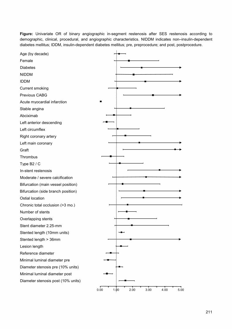
211
Figure: Univariate OR of binary angiographic in-segment restenosis after SES restenosis according todemographic, clinical, procedural, and angiographic characteristics. NIDDM indicates non–insulin-dependentdiabetes mellitus; IDDM, insulin-dependent diabetes mellitus; pre, preprocedure; and post, postprocedure.
Diabetes
NIDDM
IDDM
Previous CABG
Stable angina
0.00 1.00 2.00 3.00 4.00 5.00
Thrombus
Type B2 / C
Chronic total occlusion (>3 mo.)
Ostial location
Moderate / severe calcification
In-stent restenosis
Bifurcation (main vessel position)
Bifurcation (side branch position)
Stented length (10mm units)
Number of stents
Stent diameter 2.25-mm
Age (by decade)
Female
Left anterior descending
Left circumflex
Right coronary artery
Left main coronary
Graft
Acute myocardial infarction
Minimal luminal diameter post
Minimal luminal diameter pre
Diameter stenosis pre (10% units)
Diameter stenosis post (10% units)
Lesion length
Current smoking
Abciximab
Reference diameter
Overlapping stents
Stented length > 36mm

212
population. Post-SES restenosis in the present study was almost entirely restricted to the segment inside thestent ( 80% of restenoses). This finding represents a major difference from previous trials with SES, in whichrestenosis more frequently occurred at the stent edges.9,10 In the RESEARCH study, all operators were stronglyadvised to actively cover the entire injured vessel area and to avoid both residual dissection at stent bordersand gaps between stents. In addition, the stent placement strategy aimed to cover the treated segment "fromhealthy tissue to healthy tissue," to avoid having the free borders of the stents terminate in grossly diseasedsegments. However, it remains speculative whether these procedural strategies might have had any impact inreducing the incidence of restenosis at the stent edges.
Study LimitationsThe present report may suffer from its relatively limited study population, which was restricted to complexpatients who fulfilled predefined criteria to be included in this angiographic substudy. Therefore, our resultscannot be directly extrapolated to the entire cohort of consecutive patients treated in the RESEARCH study,and further analyses are needed to fully assess the angiographic outcomes of subsets not included in thepresent study. Ten patients with early death could not be restudied at 6 months, and a higher rate ofangiographic follow-up ( 70% in this study) would be desirable for a comprehensive evaluation. However, thepresent study was designed to enroll all unselected patients treated in our institution, and patient refusal forangiographic follow-up did not preclude enrollment in the RESEARCH study. Obviously, this real-life scenariodiffers substantially from that of randomized trials and limits the compliance to angiographic restudy.
ConclusionsAngiographic restenosis after SES implantation in complex patients is an infrequent event (7.9% of lesions),occurring mainly in association with local, lesion-based characteristics and diabetes mellitus.
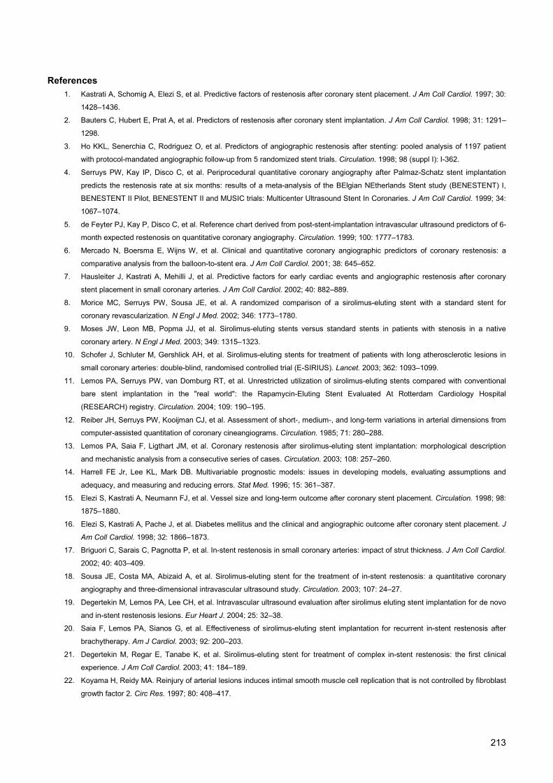
213
References1. Kastrati A, Schomig A, Elezi S, et al. Predictive factors of restenosis after coronary stent placement. J Am Coll Cardiol. 1997; 30:
1428–1436.
2. Bauters C, Hubert E, Prat A, et al. Predictors of restenosis after coronary stent implantation. J Am Coll Cardiol. 1998; 31: 1291–
1298.
3. Ho KKL, Senerchia C, Rodriguez O, et al. Predictors of angiographic restenosis after stenting: pooled analysis of 1197 patient
with protocol-mandated angiographic follow-up from 5 randomized stent trials. Circulation. 1998; 98 (suppl I): I-362.
4. Serruys PW, Kay IP, Disco C, et al. Periprocedural quantitative coronary angiography after Palmaz-Schatz stent implantation
predicts the restenosis rate at six months: results of a meta-analysis of the BElgian NEtherlands Stent study (BENESTENT) I,
BENESTENT II Pilot, BENESTENT II and MUSIC trials: Multicenter Ultrasound Stent In Coronaries. J Am Coll Cardiol. 1999; 34:
1067–1074.
5. de Feyter PJ, Kay P, Disco C, et al. Reference chart derived from post-stent-implantation intravascular ultrasound predictors of 6-
month expected restenosis on quantitative coronary angiography. Circulation. 1999; 100: 1777–1783.
6. Mercado N, Boersma E, Wijns W, et al. Clinical and quantitative coronary angiographic predictors of coronary restenosis: a
comparative analysis from the balloon-to-stent era. J Am Coll Cardiol. 2001; 38: 645–652.
7. Hausleiter J, Kastrati A, Mehilli J, et al. Predictive factors for early cardiac events and angiographic restenosis after coronary
stent placement in small coronary arteries. J Am Coll Cardiol. 2002; 40: 882–889.
8. Morice MC, Serruys PW, Sousa JE, et al. A randomized comparison of a sirolimus-eluting stent with a standard stent for
coronary revascularization. N Engl J Med. 2002; 346: 1773–1780.
9. Moses JW, Leon MB, Popma JJ, et al. Sirolimus-eluting stents versus standard stents in patients with stenosis in a native
coronary artery. N Engl J Med. 2003; 349: 1315–1323.
10. Schofer J, Schluter M, Gershlick AH, et al. Sirolimus-eluting stents for treatment of patients with long atherosclerotic lesions in
small coronary arteries: double-blind, randomised controlled trial (E-SIRIUS). Lancet. 2003; 362: 1093–1099.
11. Lemos PA, Serruys PW, van Domburg RT, et al. Unrestricted utilization of sirolimus-eluting stents compared with conventional
bare stent implantation in the "real world": the Rapamycin-Eluting Stent Evaluated At Rotterdam Cardiology Hospital
(RESEARCH) registry. Circulation. 2004; 109: 190–195.
12. Reiber JH, Serruys PW, Kooijman CJ, et al. Assessment of short-, medium-, and long-term variations in arterial dimensions from
computer-assisted quantitation of coronary cineangiograms. Circulation. 1985; 71: 280–288.
13. Lemos PA, Saia F, Ligthart JM, et al. Coronary restenosis after sirolimus-eluting stent implantation: morphological description
and mechanistic analysis from a consecutive series of cases. Circulation. 2003; 108: 257–260.
14. Harrell FE Jr, Lee KL, Mark DB. Multivariable prognostic models: issues in developing models, evaluating assumptions and
adequacy, and measuring and reducing errors. Stat Med. 1996; 15: 361–387.
15. Elezi S, Kastrati A, Neumann FJ, et al. Vessel size and long-term outcome after coronary stent placement. Circulation. 1998; 98:
1875–1880.
16. Elezi S, Kastrati A, Pache J, et al. Diabetes mellitus and the clinical and angiographic outcome after coronary stent placement. J
Am Coll Cardiol. 1998; 32: 1866–1873.
17. Briguori C, Sarais C, Pagnotta P, et al. In-stent restenosis in small coronary arteries: impact of strut thickness. J Am Coll Cardiol.
2002; 40: 403–409.
18. Sousa JE, Costa MA, Abizaid A, et al. Sirolimus-eluting stent for the treatment of in-stent restenosis: a quantitative coronary
angiography and three-dimensional intravascular ultrasound study. Circulation. 2003; 107: 24–27.
19. Degertekin M, Lemos PA, Lee CH, et al. Intravascular ultrasound evaluation after sirolimus eluting stent implantation for de novo
and in-stent restenosis lesions. Eur Heart J. 2004; 25: 32–38.
20. Saia F, Lemos PA, Sianos G, et al. Effectiveness of sirolimus-eluting stent implantation for recurrent in-stent restenosis after
brachytherapy. Am J Cardiol. 2003; 92: 200–203.
21. Degertekin M, Regar E, Tanabe K, et al. Sirolimus-eluting stent for treatment of complex in-stent restenosis: the first clinical
experience. J Am Coll Cardiol. 2003; 41: 184–189.
22. Koyama H, Reidy MA. Reinjury of arterial lesions induces intimal smooth muscle cell replication that is not controlled by fibroblast
growth factor 2. Circ Res. 1997; 80: 408–417.

214

215
Chapter 20
Andrew TL OngPatrick W SerruysJiro AokiAngela HoyeCarlos AG van MieghemGaston A Rodriguez-GranilloMarco ValgimigliKaren SonnenscheinEvelyn RegarMartin van der EntPeter PT de JaegereEugene McFaddenGeorgios SianosWillem J van der GiessenPim J de FeyterRon T van Domburg
Journal of the AmericanCollege of Cardiology2005; 45(7):1135-41
The unrestricted use of paclitaxel- versussirolimus-eluting stents for coronary arterydisease in an unselected population: one-
year results of the Taxus-Stent Evaluated AtRotterdam Cardiology Hospital (T-SEARCH)
registry

216
217
AbstractObjectives: We investigated the efficacy of paclitaxel-eluting stents (PES) compared to
sirolimus-eluting stents (SES) when used without restriction in unselected patients.
Background: Both SES and PES have been separately shown to be efficacious when
compared to bare stents. In unselected patients, no direct comparison between the twodevices has been performed.
Methods: Paclitaxel-eluting stents have been used as the stent of choice for allpercutaneous coronary interventions in the prospective Taxus-Stent Evaluated At Rotterdam
Cardiology Hospital (T-SEARCH) registry. A total of 576 consecutive patients with de novo
coronary artery disease exclusively treated with PES were compared with 508 patientstreated with SES from the Rapamycin-Eluting Stent Evaluated At Rotterdam Cardiology
Hospital (RESEARCH) registry.
Results: The PES patients were more frequently male, more frequently treated for acute
myocardial infarction, had longer total stent lengths, and more frequently received
glycoprotein IIb/IIIa inhibitors. At one year, the raw cumulative incidence of major adversecardiac events was 13.9% in the PES group and 10.5% in the SES group (unadjusted
hazard ratio [HR] 1.33, 95% confidence interval [CI] 0.95 to 1.88, p = 0.1). Correction for
differences in the two groups resulted in an adjusted HR of 1.16 (95% CI 0.81 to 1.64, p =0.4, using significant univariate variables) and an adjusted HR of 1.20 (95% CI 0.85 to 1.70,
p = 0.3, using independent predictors). The one-year cumulative incidence of clinically driven
target vessel revascularization was 5.4% versus 3.7%, respectively (HR 1.38, 95% CI 0.79to 2.43, p = 0.3).
Conclusions: The universal use of PES in an unrestricted setting is safe and is associatedwith a similar adjusted outcome compared to SES. The inferior trend in crude outcome seen
in PES was due to its higher-risk population. A larger, randomized study enrolling an
unselected population may assist in determining the relative superiority of either device.

218
IntroductionSirolimus-eluting stents (SES, Cypher, Cordis, Johnson and Johnson, Miami Lakes, Florida) 1 and paclitaxel-eluting stents (PES, TAXUS, Boston Scientific Corp., Natick, Massachusetts) 2 have both been independentlyshown to reduce the need for repeat intervention when compared to bare-metal stents (BMS) in separaterandomized clinical trials. The Food and Drug Administration approvals for these devices were granted in 2003and 2004, respectively, and it is estimated that drug-eluting stents (DES) currently comprise 70% of the stentmarket in the U.S. The randomized controlled trials on which approval was granted enrolled highly selectedpatients with single lesions that could be covered with one DES and were compared against BMS which is notrepresentative of daily clinical practice.
Our group has previously published the results of the Rapamycin-Eluting Stent Evaluated At RotterdamCardiology Hospital (RESEARCH) registry, which demonstrated that routine implantation of SES resulted in areduction in major adverse cardiac events (MACE), principally driven by a reduction in target vesselrevascularization (TVR) when compared with a historical BMS control group. 3 The PES were commercializedsubsequent to SES, based on the results of randomized controlled trials. 4,5 The beneficial effect of PES inpatients treated in daily practice remains to be defined. The aim of this study was to report the one-yearoutcomes of unrestricted/universal use of PES in patients with de novo coronary artery lesions and to compareits efficacy against our historical SES cohort. 3
MethodsStudy design and patient populationThe Taxus-Stent Evaluated At Rotterdam Cardiology Hospital (T-SEARCH) registry is a prospective single-center registry with the main purpose of evaluating the safety and efficacy of PES implantation for consecutiveunselected patients treated in daily practice. Its conceptual design and methodology are similar to that of theRESEARCH registry 6 and follows the dynamic registry design described by Rothman and Greenland. 7
Since February 16, 2003, when PES was granted Conformité Européenne approval, it replaced SES asthe default stent for every percutaneous coronary intervention. Up until September 30, 2003, a total of 576patients with de novo lesions were treated exclusively with PES and are included in the present report (PESgroup). This comprised 83.7% of all patients with de novo disease who received coronary stents. In this period,only 12 patients received BMS exclusively (11 were due to requirement for stents >3.5mm, 1 patient hadelevated liver enzymes that precluded long-term clopidogrel therapy). Patients treated with PES and BMS inthe same procedure (20 patients), those treated with PES and SES (20 patients), those treated with SES only(15 patients), and patients enrolled in other drug-eluting trials (44 patients) were not included in the presentreport. The PES are available in diameters of 2.25 mm, 2.5 mm, 3.0 mm, and 3.5 mm and in lengths of 8 to 32mm in 4-mm increments for each available diameter.
This PES group was compared with a control group that comprised the active arm of the RESEARCHregistry, that is the 508 patients with de novo disease treated solely with SES (SES group). Thus, the reportconsists of 1,084 patients treated with DES, differentiated by the type of drug coating on the stent, eithersirolimus or paclitaxel.
Procedures and postintervention medicationsInterventions were performed according to current standard procedures, with the final interventional strategy(including direct stenting, postdilation, periprocedural glycoprotein IIb/IIIa inhibitor, and use of intravascularultrasound) left entirely up to the operator's discretion. 6 Angiographic success was defined as residual stenosis≤30% by visual analysis in the presence of Thrombolysis In Myocardial Infarction (TIMI) flow grade 3. Patientswere advised to maintain lifelong aspirin (at least 80 mg/day) and were pretreated with 300 mg clopidogrel.Postprocedural clopidogrel treatment differed between the two groups. Patients treated with PES wereprescribed at least six months of clopidogrel (75 mg/day), based on existing data from randomized, controlledtrials. 5 For patients treated with SES, clopidogrel was prescribed for at least three months, unless one of thefollowing was present (in which case clopidogrel was maintained for at least six months): multiple SESimplantation (≥3 stents), total stent length ≥36 mm, chronic total occlusion, and bifurcations.

219
End point definitions and clinical follow-upThe primary outcome was the occurrence of MACE, defined as a composite of: 1) all cause death, 2) nonfatalmyocardial infarction (MI), or 3) TVR. Myocardial infarction was diagnosed by a rise in the creatine kinase-MBfraction (CK-MB) of more than three times the upper limit of normal according to American HeartAssociation/American College of Cardiology guidelines. 8 In patients who underwent coronary artery bypasssurgery during the follow-up period, a periprocedural MI was diagnosed by a rise in the CK-MB level of fivetimes the upper limit of normal. 9 For patients who presented with an acute MI, a diagnosis of re-MI in the acutephase required a fall and rise of CK-MB of 50% above the previous level. 10 Target lesion revascularization wasdefined as a repeat intervention (surgical or percutaneous) to treat a luminal stenosis within the stent or in the5-mm distal or proximal segments adjacent to the stent. Target vessel revascularization was defined as a re-intervention driven by any lesion located in the same epicardial vessel. Thrombotic stent occlusion was definedas angiographically documented complete occlusion (TIMI flow grade 0 or 1) or flow-limiting thrombus (TIMIflow grade 1 or 2) in a previously successfully treated artery. A committee of three cardiologists (A.O., J.A., andE.M.F.) reviewed all MACE.
All patients underwent clinical follow-up. Information about the in-hospital outcomes was obtained fromour institutional electronic clinical database and by review of the hospital records for those discharged toreferring hospitals (patients were referred from a total of 14 local hospitals). Postdischarge survival status wasobtained from the Municipal Civil Registries at 1, 6, and 12 months. All repeat interventions (surgical andpercutaneous) and re-hospitalizations were prospectively collected during the follow-up. Questionnairesregarding adverse events, anginal status, and medication use were sent to all living patients at 6 and 12months. Referring physicians and institutions were contacted for additional information if required.
In both groups, follow-up coronary angiography was clinically driven by symptoms or signs suggestiveof myocardial ischemia or mandated by the operator at the end of the index procedure predominantly forcomplex procedures. In the PES group, three specific subgroups were restudied: left main stem stenting, crush-bifurcation procedures, and patients who were concomitantly in a vulnerable plaque study involving non-treatedvessels (in total, 27% [n = 154] of PES patients underwent re-study during follow-up, including 14% [n = 81]that were clinically driven). In the SES group, the following “complex patient” subgroups were re-studied:bifurcation lesions, left main stem stenting, chronic total occlusions, very small vessels, long stent length (36mm), and acute MI (in total, 40% [n = 204] of SES patients were re-studied, including 8% [n = 40] that wereclinically driven). Because of the well-known effect of angiographic re-evaluation in increasing the incidence ofrepeat revascularization, 11 all re-interventions were retrospectively adjudicated and classified as eitherclinically driven or non-clinically driven. Clinically driven repeat revascularization was defined as anyintervention motivated by a significant luminal stenosis (≥50% diameter stenosis) in the presence of anginalsymptoms and/or proven myocardial ischemia in the target vessel territory by noninvasive testing.
Statistical analysisContinuous variables are presented as mean ± standard deviation, and were compared using the Studentunpaired t test. Categorical variables are presented as counts and percentages and compared by means of theFisher exact test. All statistical tests were two-tailed. Patients lost to follow-up were considered at risk until thedate of last contact, at which point they were censored. The cumulative incidence of adverse events wasestimated according to the Kaplan-Meier method, and Cox proportional hazards models were used to assessdifferences between the two strategies. Separate Cox regression analyses were performed to identifyindependent predictors of adverse events, using clinical, angiographic, and procedural variables contained inTable 1 and Table 2. The Cox proportional hazards regression models were used to control for differencesbetween groups, and the final results are presented as adjusted hazard ratios (HRs).
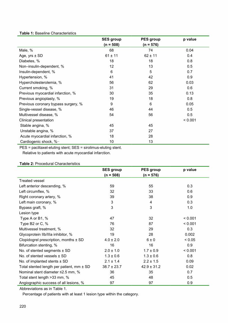
220
Table 1: Baseline CharacteristicsSES group(n = 508)
PES group(n = 576)
p value
Male, % 68 74 0.04Age, yrs ± SD 61 ± 11 62 ± 11 0.4Diabetes, % 18 18 0.8Non–insulin-dependent, % 12 13 0.5Insulin-dependent, % 6 5 0.7Hypertension, % 41 42 0.9Hypercholesterolemia, % 56 62 0.03Current smoking, % 31 29 0.6Previous myocardial infarction, % 30 35 0.13Previous angioplasty, % 19 18 0.8Previous coronary bypass surgery, % 9 6 0.05Single-vessel disease, % 46 44 0.5Multivessel disease, % 54 56 0.5Clinical presentation < 0.001 Stable angina, % 45 45 Unstable angina, % 37 27 Acute myocardial infarction, % 18 28 Cardiogenic shock, %� 10 13PES = paclitaxel-eluting stent; SES = sirolimus-eluting stent.� Relative to patients with acute myocardial infarction.
Table 2: Procedural CharacteristicsSES group(n = 508)
PES group(n = 576)
p value
Treated vesselLeft anterior descending, % 59 55 0.3Left circumflex, % 32 33 0.6Right coronary artery, % 39 38 0.9Left main coronary, % 3 4 0.3Bypass graft, % 3 3 1.0Lesion type�
Type A or B1, % 47 32 < 0.001 Type B2 or C, % 76 87 < 0.001Multivessel treatment, % 32 29 0.3Glycoprotein IIb/IIIa inhibitor, % 19 28 0.002Clopidogrel prescription, months ± SD 4.0 ± 2.0 6 ± 0 < 0.05Bifurcation stenting, % 16 16 0.9No. of stented segments ± SD 2.0 ± 1.0 1.7 ± 0.9 < 0.001No. of stented vessels ± SD 1.3 ± 0.6 1.3 ± 0.6 0.8No. of implanted stents ± SD 2.1 ± 1.4 2.2 ± 1.5 0.09Total stented length per patient, mm ± SD 38.7 ± 23.7 42.9 ± 31.2 0.02Nominal stent diameter ≤2.5 mm, % 36 35 0.7Total stent length >33 mm, % 45 48 0.5Angiographic success of all lesions, % 97 97 0.9Abbreviations as in Table 1.� Percentage of patients with at least 1 lesion type within the category.

221
ResultsBaseline and procedural characteristicsThe PES patients were more often male, had more MI as their presenting symptom, more cardiogenic shock,more complex lesions treated, longer total stent lengths, and more frequently received glycoprotein IIb/IIIainhibitors (Table 1 and Table 2). Fewer PES patients had a history of previous bypass surgery, and fewersegments per patient were stented, although the number of vessels treated per patient was identical. Otherbaseline and procedural characteristics were similar.
Clinical outcomeFirst 30 daysNo significant differences were noted between groups with respect to the incidences of death, death or MI,TVR, or MACE in the first month (Table 3). Mortality in the first 30 days was 2.1% in the PES group and 1.6% inthe SES group (p = 0.7). In both groups, most deaths occurred in patients with cardiogenic shock.Angiographically proven stent thrombosis occurred in six patients in the PES group, four of whom were treatedfor AMI, the other two presented with unstable angina. Two patients with AMI also underwent bifurcationstenting, as did one with unstable angina. In total, three patients with bifurcation stenting experienced stentthrombosis. In the SES group, two patients were diagnosed with stent thrombosis. One patient died as a resultof stent thrombosis in the PES group.
Table 3: Major Adverse Cardiac Events in the First Month Following Stent Implantation0 to 1 Month SES group
(n = 508)PES group(n = 576)
p value�
Death, n (%) 8 (1.6) 12 (2.1) 0.7Nonfatal myocardial infarction, n (%) 12 (2.4) 17 (3.0) 0.6Target lesion revascularization, n (%) 6 (1.2) 7 (1.2) 1.0Target vessel revascularization, n (%)† 6 (1.2) 13 (2.3) 0.2Any event, n (%) 23 (4.5) 34 (5.9) 0.3Stent thrombosis, n (%)‡ 2 (0.4) 6 (1.0) 0.3Abbreviations as in Table 1.� By Fisher exact test.† Includes target lesion revascularization.‡ Angiographically documented stent thrombosis requiring repeat intervention.
One yearThe MACE components are presented in Figure 1 and Figure 2. At one year, 5.3% of patients in the PES groupand 3.4% in the SES group had died (HR 1.69, 95% confidence interval [CI] 0.93 to 3.00, p = 0.08). In total,8.8% of patients in the PES group versus 7.0% in the SES group had either died or suffered a nonfatal re-MI(HR 1.28, 95% CI 0.84 to 1.95, p = 0.3). The incidence of TVR was similar in the SES and PES groups: 7.3%versus 5.1% (HR 1.31, 95% CI 0.81 to 2.13, p = 0.3). Clinically driven TVR was reduced by a similar magnitudein both groups, specifically 3.7% versus 5.4%, respectively (HR 1.38, 95% CI 0.79 to 2.43, p = 0.3). Post-hocanalysis of clinically driven TVR demonstrates that confidence limits crossed unity, with point estimates close tounity in the subgroups analyzed (Fig. 3). Regarding the primary end point of MACE (the composite of death, MI,or TVR), Kaplan-Meier estimates were 13.9% in the PES group versus 10.5% in the SES group (unadjustedHR 1.33, 95% CI 0.95 to 1.88, p = 0.10).

222
Figure 1: Unadjusted Kaplan-Meier event curves at one year.
CI = confidence interval; PES = paclitaxel-eluting stent; SES = sirolimus-eluting stent.
A: Cumulative risk of death.
B: Cumulative risk of death or myocardial infarction.
Time (Months)129630
Cum
ulat
ive
Eve
nts
(%)
20
15
10
5
0SES
PES
Patients At Risk (n)
PES 576 550 546 538 500
SES 508 493 489 488 442
Events (%, 95% CI)
PES 3.5 (2.0-5.1) 5.3 (3.5-7.1)
SES 2.8 (1.3-4.2) 3.4 (1.8-5.0)
Hazard Ratio 1.69 (95% CI 0.93 - 3.00), p=0.08
Time (Months)129630
Cum
ulat
ive
Eve
nts
(%)
20
15
10
5
0SES
PES
Patients At Risk (n)
PES 576 550 546 538 500
SES 508 493 489 488 442
Events (%, 95% CI)
PES 3.5 (2.0-5.1) 5.3 (3.5-7.1)
SES 2.8 (1.3-4.2) 3.4 (1.8-5.0)
Hazard Ratio 1.69 (95% CI 0.93 - 3.00), p=0.08
129630
Cum
ulat
ive
Eve
nts
(%)
20
15
10
5
0
Time (Months)
SES
PES
Patients At Risk (n)
PES 576 534 530 520 484
SES 508 480 475 471 429
Events (%, 95% CI)
PES 6.5 (4.4-8.6) 8.8 (6.5-11.1)
SES 5.6 (3.6-7.6) 7.0 (4.7-9.2)
Hazard Ratio 1.28 (95% CI 0.84 - 1.95), p=0.3
129630
Cum
ulat
ive
Eve
nts
(%)
20
15
10
5
0
Time (Months)
SES
PES
Patients At Risk (n)
PES 576 534 530 520 484
SES 508 480 475 471 429
Events (%, 95% CI)
PES 6.5 (4.4-8.6) 8.8 (6.5-11.1)
SES 5.6 (3.6-7.6) 7.0 (4.7-9.2)
Hazard Ratio 1.28 (95% CI 0.84 - 1.95), p=0.3

223
C: Cumulative risk of death, myocardial infarction, or target vessel revascularization.
Figure 2: Unadjusted one-year cumulative risk of clinically driven target vessel revascularization.
Abbreviations as in Figure 1.
Time (Months)129630
Cum
ulat
ive
Even
ts (%
)20
15
10
5
0
SES
PES
Hazard Ratio 1.33 (95% CI 0.95 - 1.88), p=0.10
Patients At Risk (n)
PES 576 525 511 492 456
SES 508 475 468 453 409
Events (%, 95% CI)
PES 9.9 (7.4-12.3) 13.9 (11.1-16.8)
SES 7.2 (4.9-9.4) 10.5 (7.9-13.2)
Time (Months)129630
Cum
ulat
ive
Even
ts (%
)20
15
10
5
0
SES
PES
Hazard Ratio 1.33 (95% CI 0.95 - 1.88), p=0.10
Patients At Risk (n)
PES 576 525 511 492 456
SES 508 475 468 453 409
Events (%, 95% CI)
PES 9.9 (7.4-12.3) 13.9 (11.1-16.8)
SES 7.2 (4.9-9.4) 10.5 (7.9-13.2)
Patients At Risk (n)
PES 576 541 524 513 495
SES 508 486 481 470 430
Events (%)
PES 4.1 (2.5-5.8) 5.4 (3.5-7.3)
SES 1.6 (0.5-2.7) 3.7 (2.0-5.3)
Time (months)129630
Cum
ulat
ive
Eve
nts
(%)
20
15
10
5
0
SES
PES
Hazard Ratio 1.38 (95% CI 0.79 - 2.43), p=0.26
Patients At Risk (n)
PES 576 541 524 513 495
SES 508 486 481 470 430
Events (%)
PES 4.1 (2.5-5.8) 5.4 (3.5-7.3)
SES 1.6 (0.5-2.7) 3.7 (2.0-5.3)
Time (months)129630
Cum
ulat
ive
Eve
nts
(%)
20
15
10
5
0
SES
PES
Hazard Ratio 1.38 (95% CI 0.79 - 2.43), p=0.26
Time (months)129630
Cum
ulat
ive
Eve
nts
(%)
20
15
10
5
0
Time (months)129630
Cum
ulat
ive
Eve
nts
(%)
20
15
10
5
0
SES
PES
Hazard Ratio 1.38 (95% CI 0.79 - 2.43), p=0.26

224
Figure 3: Hazard ratios (HR) of stent type at one-year follow-up for clinically driven target vesselrevascularization in subgroups of patients according to baseline and procedural characteristics.MI = myocardial infarction; other abbreviations as in Figure 1.
There were two cases of late (>6 months to 1 year) stent thrombosis documented angiographically in the PESgroup. In one, it occurred eight months after the index procedure while the patient was on antiplateletmonotherapy with aspirin. The second occurred 11 months after the index procedure after the patient hadtemporarily suspended antiplatelet therapy (aspirin) for noncardiac surgery.
Predictors of adverse eventsTo assess the independent predictors of MACE at one year, two separate multivariate analyses wereperformed. First, a model was built using all baseline and procedural characteristics shown in Table 1 andTable 2. Forward stepwise regression was performed with entry and stay criteria of 0.05 and 0.10, respectively.The following variables were significant: cardiogenic shock, female gender, multivessel disease, diabetesmellitus, left main stenting, bifurcation stenting, and lesion type B2/C (Table 4). A second model built using thesame variables with the end point of TVR at one year revealed bifurcation stenting was the only significantindependent predictor of TVR.
Adjustment for differences between groupsThe Cox regression models were used to adjust the two groups by correcting for multiple potential confoundersin the baseline and procedural characteristics. First, a model was built forcing stent type and all independentpredictors from Table 4 (see Table 5). All previously significant variables remained significant except for lesiontype B2/C. The adjusted HR for use of PES became even less significant, decreasing from 1.33 (95% CI 0.95to 1.88, p = 0.10) to 1.20 (95% CI 0.85 to 1.70, p = 0.3), after controlling for the increased complexity in thePES group.
0 51PES Better SES Better
p value95% CIHR
0.7(0.64-1.99)1.130.2(0.74-4.57)1.840.06(0.99-4.66)2.150.5(0.32-1.84)0.760.3(0.63-4.64)1.720.2(0.79-4.06)1.790.7(0.53-2.51)1.150.8(0.31-4.97)1.240.4(0.22-1.80)0.620.4(0.7-2.69)1.380.5(0.49-3.87)1.380.4(0.41-9.61)2.00.03(1.11-10.96)3.490.6(0.38-1.71)0.800.1(0.88-5.40)2.280.8(0.52-2.22)1.080.1(0.89-3.82)1.840.7(0.35-2.13)0.870.3(0.79-2.43)1.38
Subgroup
Lesion type B2/CLesion type A/B1< 2 stents implanted> 2 stents implantedBifurcationMultivessel treatment1 vessel treatmentUse of IIb/IIIa inhibitorDiabetes mellitusMultivessel disease1 vessel diseaseAcute MIUnstable anginaStable anginaFemaleMaleAge < 65Age > 65All patients

225
Table 4: Multivariate Predictors of Major Adverse Cardiac Events at One Year (Cox Proportional HazardsModel)
HR 95% CI p valueMajor adverse cardiac events�
Cardiogenic shock (stable angina as reference variable) 4.54 2.44–8.48 < 0.001 Female gender 1.72 1.22–2.43 0.002 Multivessel disease 1.74 1.19–2.55 0.005 Diabetes mellitus 1.65 1.12–2.42 0.01 Left main stenting 1.96 1.10–3.48 0.02 Bifurcation stenting 1.59 1.06–2.38 0.03 Lesion type B2 or C 1.85 1.01–3.40 0.047Target vessel revascularization Bifurcation stenting 2.77 1.68–4.57 < 0.001
CI = confidence interval; HR = hazard ratio.� Major adverse cardiac events: death, myocardial infarction, or target vessel revascularization.
Table 5: Hazard Ratios by Stent Type of Major Adverse Cardiac Events After Adjustment�
HR 95% CI p valueMACE† Unadjusted 1.33 0.95–1.88 0.10 Adjusted for significant predictors of MACE 1.20 0.85–1.70 0.3 Adjusted for significant univariate variables‡ 1.16 0.81–1.64 0.4TVR Unadjusted 1.31 0.81–2.13 0.26 Adjusted for significant predictors of TVR 1.33 0.82–2.15 0.25
Abbreviations as in Table 4.� Stent type coded as: 0 = sirolimus-eluting stent, 1 = paclitaxel eluting stent.† Major adverse cardiac events: death, myocardial infarction, or target vessel revascularization (TVR).‡ Significant univariate variables for major adverse cardiac event (MACE) were the significant predictors plustotal stent length and number of stents implanted.
A second model was then built forcing stent type and significant univariate variables (independent predictorsplus total stent length and number of stents), and the adjusted outcome of MACE at one year was similarbetween SES and PES (adjusted HR 1.16, 95% CI 0.81 to 1.64, p = 0.4). Finally, stent type was also not asignificant predictor of TVR when adjusted for bifurcation stenting (adjusted HR 1.33, 95% CI 0.82 to 2.15, p =0.25).
DiscussionThe major finding of this report is that the unrestricted use of PES in de novo lesions is associated with anonsignificant difference in outcome compared to SES, both unadjusted and when controlled for significantbaseline and procedural characteristics. The trend toward an inferior crude outcome with PES was due to themore complex characteristics of the group.
The two sequential registries were separated by a four-month interval. Several differences in baselinecharacteristics were noted. More MIs including patients in cardiogenic shock were treated in the T-SEARCHregistry because of the implementation of a local pre-hospital protocol that triaged more patients to primarypercutaneous coronary intervention. More complex lesions were treated in the T-SEARCH registry, with a shiftfrom type A/B1 to B2/C lesions, with more stents being implanted in the T-SEARCH registry. This in partreflects the increased complexity of cases being performed with time and as operators and referring physiciansbecoming more aware and familiar with DES.

226
The primary end point of this trial was overall MACE, and the results for this comparison are presentedboth unadjusted and following adjustment for significant predictive variables (Table 5). With thecommercialization of PES, our institution switched completely from SES to PES, precluding randomization.Therefore, it was intuitive to present the data as such and imperative to statistically correct by using significantpredictive variables to account for the increased complexity seen in the PES group. To preserve theprospective, consecutive, and unselected nature of both registries, and the requirement to control for multiplesignificant variables, the Cox regression model was used. Our results demonstrate that, following adjustment,the HR was closer to unity compared to the crude result, further confirming the increased complexity in the PESgroup.
The multivariate analysis (Table 4) for independent predictors of MACE is unique as it is an analysis of1,084 DES patients treated in an unrestricted setting. In a total cohort of DES patients, cardiogenic shock,female gender, multivessel disease, diabetes mellitus, left main stenting, bifurcation stenting, and treatment ofa complex lesion significantly predicted an adverse outcome. From this list, patients who possess thesecharacteristics should undergo more regular clinical surveillance.
The major advantage of DES has been to reduce the need for repeat revascularization. 1-3 In our study,the incidence TVR at one year with PES was not significantly different from the results obtained with SES.Furthermore, when the adjusted end point of clinically driven TVR was used (Fig. 2), similar outcomes werereproduced, thus confirming that both drug-eluting systems serve to reduce clinical restenosis in an unselectedpopulation.
A nonsignificantly higher incidence of angiographic stent thrombosis in the first 30 days was noted inthe PES cohort (1.0% in SES vs. 0.4% in PES, p = 0.3). However, it is important to emphasize that, owing tothe infrequent occurrence of this event, large numbers of patients are required to assess this complicationproperly. We have shown that in a larger population, the incidence rates in both DES were in the same range:1.0% (95% CI 0.6% to 1.9%) in PES and 1.0% (95% CI 0.5% to 1.8%) in SES. 12
At the time the T-SEARCH registry was conducted, TAXUS II 5 and the Randomized Comparison of aSirolimus-Eluting Stent with a Standard Stent for Coronary Revascularization (RAVEL) 13 were the twopublished trials available with one-year MACE results from the eluting stent arms of 10.9% (slow-release arm)and 5.8%, respectively. Based on those results, the group sample sizes of our study would have beenadequately powered to show a difference.
Subsequent to that, the results of larger trials of both devices—TAXUS IV and Sirolimus-Eluting Stentin Coronary Lesions (SIRIUS)—were published and demonstrated a smaller difference (8.4% vs. 7.1%,respectively). The population of this registry is an all-inclusive unrestricted one, a sample that is representativeof the population seen in a tertiary catheterization laboratory. Therefore, this population is directly comparableto daily practice and the results do not require extrapolation as for randomized trials. The results of this registrycomplement published randomized trials.
ConclusionsThe universal use of PES in an unrestricted setting is safe, and associated with a non-significant adjusteddifference in outcome at one year compared to SES, with a trend toward worse outcomes in the PES cohort, inpart owing to its higher-risk profile. Both DES reduce the need for repeat intervention in the real world setting ofcomplex patient and procedural characteristics.

227
References1. J.W. Moses, M.B. Leon and J.J. Popma et al., Sirolimus-eluting stents versus standard stents in patients with stenosis in a native
coronary artery, N Engl J Med 349 (2003), pp. 1315–1323.
2. G.W. Stone, S.G. Ellis and D.A. Cox et al., A polymer-based, paclitaxel-eluting stent in patients with coronary artery disease, N Engl J
Med 350 (2004), pp. 221–231.
3. P.A. Lemos, P.W. Serruys and R.T. van Domburg et al., Unrestricted utilization of sirolimus-eluting stents compared with conventional
bare stent implantation in the “real world”: the Rapamycin-Eluting Stent Evaluated At Rotterdam Cardiology Hospital
(RESEARCH) registry, Circulation 109 (2004), pp. 190–195.
4. E. Grube, S. Silber and K.E. Hauptmann et al., TAXUS I: six- and twelve-month results from a randomized, double-blind trial on a
slow-release paclitaxel-eluting stent for de novo coronary lesions, Circulation 107 (2003), pp. 38–42.
5. A. Colombo, J. Drzewiecki and A. Banning et al., Randomized study to assess the effectiveness of slow- and moderate-release
polymer-based paclitaxel-eluting stents for coronary artery lesions, Circulation 108 (2003), pp. 788–794.
6. P.A. Lemos, C.H. Lee and M. Degertekin et al., Early outcome after sirolimus-eluting stent implantation in patients with acute coronary
syndromes: insights from the Rapamycin-Eluting Stent Evaluated At Rotterdam Cardiology Hospital (RESEARCH) registry, J Am
Coll Cardiol 41 (2003), pp. 2093–2099.
7. K.J. Rothman and S. Greenland, Cohort studies. In: K.J. Rothman and S. Greenland, Editors, Modern Epidemiology (2nd edition),
Lippincott Williams & Wilkins Publishers, Philadelphia, PA (1998), pp. 79–80.
8. S.C. Smith Jr, J.T. Dove and A.K. Jacobs et al., ACC/AHA guidelines of percutaneous coronary interventions (revision of the 1993
PTCA guidelines) A report of the American College of Cardiology/American Heart Association Task Force on Practice Guidelines
(Committee to Revise the 1993 Guidelines for Percutaneous Transluminal Coronary Angioplasty), J Am Coll Cardiol 37 (2001), p.
2239i –lxvi.
9. P.W. Serruys, F. Unger and J.E. Sousa et al., Comparison of coronary-artery bypass surgery and stenting for the treatment of
multivessel disease, N Engl J Med 344 (2001), pp. 1117–1124.
10. G.W. Stone, C.L. Grines and D.A. Cox et al., Comparison of angioplasty with stenting, with or without abciximab, in acute myocardial
infarction, N Engl J Med 346 (2002), pp. 957–966.
11. P.N. Ruygrok, R. Melkert and M.A. Morel et al., Does angiography six months after coronary intervention influence management and
outcome? Benestent II Investigators, J Am Coll Cardiol 34 (1999), pp. 1507–1511.
12. A.T. Ong, A. Hoye and J. Aoki et al., Thirty-day incidence and six-month clinical outcome of thrombotic stent occlusion following bare
metal, sirolimus or paclitaxel stent implantation, J Am Coll Cardiol 45 (2005), pp. 944–950.
13. M.C. Morice, P.W. Serruys and J.E. Sousa et al., A randomized comparison of a sirolimus-eluting stent with a standard stent for
coronary revascularization, N Engl J Med 346 (2002), pp. 1773–1780.

228

229
Part 4
SUMMARY AND CONCLUSIONS

230
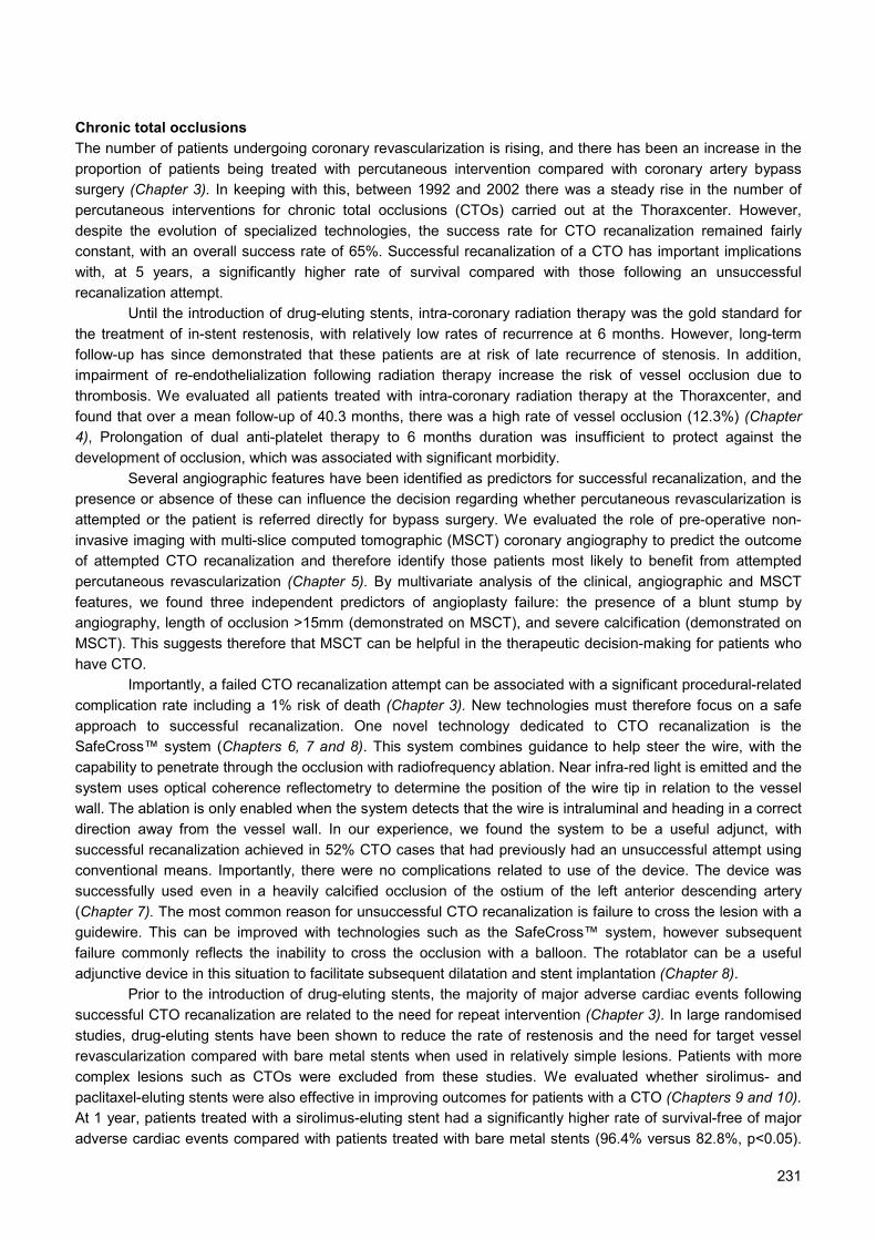
231
Chronic total occlusionsThe number of patients undergoing coronary revascularization is rising, and there has been an increase in theproportion of patients being treated with percutaneous intervention compared with coronary artery bypasssurgery (Chapter 3). In keeping with this, between 1992 and 2002 there was a steady rise in the number ofpercutaneous interventions for chronic total occlusions (CTOs) carried out at the Thoraxcenter. However,despite the evolution of specialized technologies, the success rate for CTO recanalization remained fairlyconstant, with an overall success rate of 65%. Successful recanalization of a CTO has important implicationswith, at 5 years, a significantly higher rate of survival compared with those following an unsuccessfulrecanalization attempt.
Until the introduction of drug-eluting stents, intra-coronary radiation therapy was the gold standard forthe treatment of in-stent restenosis, with relatively low rates of recurrence at 6 months. However, long-termfollow-up has since demonstrated that these patients are at risk of late recurrence of stenosis. In addition,impairment of re-endothelialization following radiation therapy increase the risk of vessel occlusion due tothrombosis. We evaluated all patients treated with intra-coronary radiation therapy at the Thoraxcenter, andfound that over a mean follow-up of 40.3 months, there was a high rate of vessel occlusion (12.3%) (Chapter4), Prolongation of dual anti-platelet therapy to 6 months duration was insufficient to protect against thedevelopment of occlusion, which was associated with significant morbidity.
Several angiographic features have been identified as predictors for successful recanalization, and thepresence or absence of these can influence the decision regarding whether percutaneous revascularization isattempted or the patient is referred directly for bypass surgery. We evaluated the role of pre-operative non-invasive imaging with multi-slice computed tomographic (MSCT) coronary angiography to predict the outcomeof attempted CTO recanalization and therefore identify those patients most likely to benefit from attemptedpercutaneous revascularization (Chapter 5). By multivariate analysis of the clinical, angiographic and MSCTfeatures, we found three independent predictors of angioplasty failure: the presence of a blunt stump byangiography, length of occlusion >15mm (demonstrated on MSCT), and severe calcification (demonstrated onMSCT). This suggests therefore that MSCT can be helpful in the therapeutic decision-making for patients whohave CTO.
Importantly, a failed CTO recanalization attempt can be associated with a significant procedural-relatedcomplication rate including a 1% risk of death (Chapter 3). New technologies must therefore focus on a safeapproach to successful recanalization. One novel technology dedicated to CTO recanalization is theSafeCross™ system (Chapters 6, 7 and 8). This system combines guidance to help steer the wire, with thecapability to penetrate through the occlusion with radiofrequency ablation. Near infra-red light is emitted and thesystem uses optical coherence reflectometry to determine the position of the wire tip in relation to the vesselwall. The ablation is only enabled when the system detects that the wire is intraluminal and heading in a correctdirection away from the vessel wall. In our experience, we found the system to be a useful adjunct, withsuccessful recanalization achieved in 52% CTO cases that had previously had an unsuccessful attempt usingconventional means. Importantly, there were no complications related to use of the device. The device wassuccessfully used even in a heavily calcified occlusion of the ostium of the left anterior descending artery(Chapter 7). The most common reason for unsuccessful CTO recanalization is failure to cross the lesion with aguidewire. This can be improved with technologies such as the SafeCross™ system, however subsequentfailure commonly reflects the inability to cross the occlusion with a balloon. The rotablator can be a usefuladjunctive device in this situation to facilitate subsequent dilatation and stent implantation (Chapter 8).
Prior to the introduction of drug-eluting stents, the majority of major adverse cardiac events followingsuccessful CTO recanalization are related to the need for repeat intervention (Chapter 3). In large randomisedstudies, drug-eluting stents have been shown to reduce the rate of restenosis and the need for target vesselrevascularization compared with bare metal stents when used in relatively simple lesions. Patients with morecomplex lesions such as CTOs were excluded from these studies. We evaluated whether sirolimus- andpaclitaxel-eluting stents were also effective in improving outcomes for patients with a CTO (Chapters 9 and 10).At 1 year, patients treated with a sirolimus-eluting stent had a significantly higher rate of survival-free of majoradverse cardiac events compared with patients treated with bare metal stents (96.4% versus 82.8%, p<0.05).

232
Furthermore, follow-up angiography at 6-months showed that the sirolimus-eluting stent effectively suppressedneointimal proliferation, and was associated with a low late lumen loss of just 0.13 ± 0.46mm. We subsequentlyalso evaluated the clinical outcomes of patients treated with paclitaxel-eluting stents (Chapter 10). Followingtherapy of a CTO of >3 months duration, the cumulative survival-free of target vessel revascularization was97.4% for patients treated with sirolimus-eluting stents, 96.4% for paclitaxel-eluting stents, and significantlylower at 80.8% for bare metal stents (p=0.01). These data demonstrate efficacy of both the sirolimus- andpaclitaxel-eluting stents in reducing restenosis and the need for repeat reintervention compared with bare metalstents. This improvement in outcomes means an expansion in the complexity of those lesions deemed suitablefor percutaneous intervention. Chronic total occlusion of the left main stem is a very rare lesion, but wedemonstrate that in contemporary practice, it can be successfully treated with drug-eluting stent implantation(Chapter 11).
BifurcationsA variety of techniques have been proposed to treat bifurcation lesions (Chapters 12 and 13). However, whenbare metal stents are utilised, techniques involving stenting of the side branch are associated with a higher rateof restenosis compared with strategies whereby only the main vessel is stented. We evaluated the efficacy ofdrug-eluting stents in consecutive patients treated for at least one bifurcation lesion (Chapter 14). In thesepatients, the side branch was stented in addition to the main vessel as it seemed intuitive when using thesestents to cover the entire lesion. At 6 months, the incidence of major adverse cardiac events was 10.3%, whichis lower than that documented in published data of bare metal stents. The incidence of target lesionrevascularization was 8.6%. Follow-up angiography demonstrated restenosis rates of the main vessel and sidebranch of 9.1% and 13.6% respectively, with an overall restenosis rate of 22.7%. The majority of restenoses ofthe side branch occurred at the ostium following T-stenting. Indeed, the restenosis rate in the side branchfollowing T-stenting was 16.7% whilst that following other stenting techniques was 7.1%. The major problemwith T-stenting relates to the degree of angulation between the main vessel and side branch. If this angleapproximates 90˚, precise stent positioning in both vessels can completely cover the lesion. However, themajority of bifurcation lesions have an angle of <70˚. We hypothesised that the side branch restenoses seen inour study might relate to inadequate or incomplete coverage of the ostium thereby reducing the efficacy of thedrug-eluting stent. This led to a shift away from a strategy of T-stenting, towards methods which ensurecomplete coverage – the crush and Culotte techniques of stenting (Chapter 15). However, despite the changein stenting technique, the choice of strategy was not an independent predictor for either major adverse cardiacevents or the need for target lesion revascularization. It is unlikely that any single strategy will be optimal for allbifurcations due to the diversity of these lesions with differences of vessel size, angulation, and plaquedistribution.
Following stenting of bifurcation lesions, we found a difference in outcomes with respect to stent type infavour of fewer repeat target lesion revascularization in the sirolimus-eluting stent group as compared withthose treated with paclitaxel-eluting stents (Chapter 15). However, this result must be taken with caution in viewof the lack of randomisation of the data. Indeed, in chapter 20, we show that for the total populations treatedwith drug-eluting stent implantation, stent type (sirolimus-eluting versus paclitaxel-eluting) was not anindependent predictor of either major adverse cardiac events or the need for target vessel revascularization.
The crush technique of bifurcation stenting is a new strategy that ensures complete lesion coverage,and was introduced specifically to be used with drug-eluting stents. We evaluated the outcomes of 231consecutive patients treated using this technique for 241 de novo bifurcation lesions (Chapter 16). The rate ofin-hospital major adverse cardiac events was 5.2%. At 9-months, the incidence of possible post-proceduralstent thrombosis was of concern, with 10 (4.3%) patients having had a possible event. The rate of survival-freeof target lesion revascularization was 90.3%, and survival-free of major adverse cardiac events was 83.5%.Therapy of the left main stem was an independent predictor for adverse events, and further research is neededbefore this technique can be routinely recommended in this group. Angiographic follow-up demonstrated amean late loss of 0.30±0.64mm and 0.41±0.67mm for the main vessel and side branch respectively, with binaryrestenosis rates of 9.1% and 25.3%. Kissing balloon post-dilatation significantly reduced the side branch late
233
lumen loss (0.24±0.50mm versus 0.58±0.77mm, p<0.001) and binary restenosis rates (9.6% versus 41.3%,p<0.0001), and should therefore be carried out routinely in all cases to reduce the rate of restenosis.
The Culotte technique of bifurcation stenting is also a strategy that ensures complete lesion coverage.However, the technique fell out of favour because of high restenosis rates following bare metal stentimplantation. We evaluated the outcomes of a series of patients treated with this technique using drug-elutingstents and showed promising clinical and angiographic outcomes (Chapter 17). These results warrant the re-evaluation of the Culotte technique utilizing drug-eluting stents, in the setting of large randomized studies.
Chronic total occlusions, bifurcations, and the RESEARCH and T-SEARCH RegistriesFrom April 2002, drug-eluting stents were used as the default strategy for all patients undergoing percutaneouscoronary intervention. Initially, patients were treated with the sirolimus-eluting stent, then during the first quarterof 2003, our strategy switched to using paclitaxel-eluting stents in all patients. These patients comprise theRESEARCH and T-SEARCH Registries respectively. All consecutive patients were enrolled irrespective ofclinical presentation and lesion characteristics, and the incidence of major adverse cardiac events wasprospectively evaluated during follow-up.
By multivariate analysis, the treatment of chronic total occlusions was not found to be an independentpredictor of stent thrombosis, restenosis, or adverse cardiac events including target vessel revascularization.However, our data show that in the era of drug-eluting stents, percutaneous coronary intervention of bifurcationlesions is associated with an increased risk of adverse events. Drug-eluting stents impair the process of re-endothelialization compared with bare metal stents, and there are concerns that this might lead to an increasedrisk of stent thrombosis. We evaluated >2,500 consecutive patients, and show that in unselected patientpopulations treated with either sirolimus-eluting or paclitaxel-eluting stents, the incidence of stent thrombosis at30 days is within the range seen following bare stent implantation (Chapter 18). The only independent predictorfor stent thrombosis was bifurcation stenting in the setting of acute myocardial infarction. This emphasises theimportance of optimal pharmacotherapy in this group, and suggests that it may be preferable to use a singlestent strategy for bifurcation lesions treated in this setting.
During the first 6 months of the RESEARCH Registry, patients treated for complex disease were invitedback for follow-up coronary angiography. These were patients treated for acute myocardial infarction, in-stentrestenosis, left main stem stenting, chronic total occlusion, bifurcation stenting, those who received a 2.25mmstent, and those treated with a stented segment length >36mm. Bifurcation stenting (side branch position) wasa univariate predictor of restenosis (OR 2.77; 95% CI: 1.15-6.33, p=0.02), though this was no longer a predictoron multivariate analysis (Chapter 19). We subsequently evaluated the clinical outcomes of all patients treated with drug-eluting stents for denovo lesions (Chapter 20). Use of the paclitaxel-eluting stent was associated with a non-significant difference inoutcome compared with the sirolimus-eluting stent. However, when the entire population was evaluated forpredictors of adverse outcomes, bifurcation stenting was an independent predictor of major adverse cardiacevents (HR 1.59; 95%CI: 1.06-2.38, p=0.03), and was the only independent predictor of target vesselrevascularization (HR 2.77; 95%CI: 1.68-4.57, p<0.001).
ConclusionsIn the therapy of chronic total occlusions and bifurcation lesions, sirolimus- and paclitaxel-eluting stents areeffective in reducing restenosis compared with bare metal stents. Successful chronic total occlusion therapyinfers a significant improvement in long-term survival, and therefore justifies an aggressive strategy ofrecanalization. However, failed recanalization may be associated acutely with an adverse event, and newtechnologies must focus on a safe approach to successful recanalization.
The most effective strategy of stenting of bifurcation lesions is currently undefined, and furtherrandomised studies are warranted taking into account factors such as vessel size, angulation between the mainvessel and side branch, and plaque distribution at baseline. In contemporary coronary interventional practiceutilizing drug-eluting stents, bifurcation lesions continue to pose a problem in terms of being associated with anincreased risk of major adverse cardiac events and target vessel revascularization.

234

235
Samenvatting en Conclusies

236

237
Chronische totale occlusiesHet aantal patienten dat een coronaire revascularisatie ondergaat neemt gestaag toe en deze tendens isgrotendeels toe te schrijven aan de toename van het aantal percutane coronaire interventies ten nadele vancoronaire bypass operaties (hoofdstuk 3). In analogie hiermee was er in het Thoraxcentrum tussen 1992 en2002 eveneens een duidelijke toename in het aantal percutane interventies voor chronische totale occlusies(CTO’s). Hoewel er in deze periode een belangrijke technologische vooruitgang is geweest, bleef de kans opsuccesvolle recanalisatie relatief constant, namelijk om en bij de 65%. Een succesvolle recanalisatie van eenCTO heeft nochtans belangrijke prognostische implicaties: deze patienten hebben een betere 5-jaarsoverlevingin vergelijking met de groep waarbij recanalisatie niet lukt.
Tot de introductie van drug-eluting-stents was intracoronaire brachytherapie de aangewezen therapievoor de behandeling van in-stent-restenose aangezien met deze therapie de kans op recidief restenose relatieflaag was 6 maanden behandeling. Lange-termijn resultaten hebben evenwel aangetoond dat patientenbehandeld met brachytherapie een hoger risico vertonen op het ontwikkelen van laattijdige restenose.Bovendien verhoogt brachytherapie de kans op coronaire thrombose doordat het interfereert met het normalere-endothelialisatieproces. In hoofdstuk 4 rapporteren we de resultaten van alle patienten die in hetThoraxcentrum behandeld werden met coronaire brachytherapie: na een gemiddelde follow-up van 40.3maanden was er een hoge mate van bloedvatocclusie (12.3%). Combinatietherapie met 2 klassen vanbloedplaatjesaggregantia gedurende een periode van 6 maanden heeft bovendien geen beschermend effecttegen het ontstaan van bloedvatocclusie, hetgeen gepaard gaat met een belangrijke morbiditeit.
Er zijn verschillende angiografische karakteristieken gekend die het succes van een recanalisatievoorspellen. Het aan- of afwezig zijn van deze kenmerken beinvloedt mee de beslissing om een patient voorpercutane coronaire revascularisatie dan wel voor coronaire bypass chirurgie te verwijzen. In hoofdstuk 5beschrijven we de toegevoegde waarde van niet-invasieve beeldvorming met multi-slice computer tomografie(MSCT) van de coronaire bloedvaten als voorbereidend onderzoek voor het uitvoeren van de revascularisatie.We hebben de rol van MSCT onderzocht voor het voorspellen van de kans op succes van een percutanecoronaire interventie van een CTO en met andere woorden nagegaan welke patienten een redelijk hoogslaagpercentage hebben. Multivariaat analyse van klinische, angiografische en MSCT kenmerken leverde 3onafhankelijke variabelen op die geassocieerd zijn met een lage slaagkans van de percutane interventie: deaanwezigheid van een abrupt stopbeeld op het conventionele angiogram, een geoccludeerd bloedvat segmentvan >15mm (MSCT variabele) evenals een sterk verkalkt segment van de coronair arterie (MSCT variabele).Deze bevindingen suggereren dat MSCT mogelijk een rol heeft bij de beslissing om een patient met een CTOal dan niet via percutane weg te behandelen.
In hoofdstuk 3 beschrijven we dat een niet succesvolle percutane recanalisatiepoging voor een CTOkan gerelateerd zijn aan majeure complicaties waaronder een mortaliteitsrisico van 1%. Nieuwe technologischeverbeteringen zijn bijgevolg noodzakelijk om de recanalisatiepoging succesvoller en tegelijkertijd veiliger temaken. Een van deze nieuwe technieken voor de behandeling van CTO’s is het SafeCross™ system(hoofdstuk 6 tot en met 8). Dit systeem combineert 2 eigenschappen: (1) het biedt ondersteuning bij hetmaneuvreren van de draad, en (2) door middel van radiofrekwentie ablatie is het mogelijk hard materiaal tedoorbreken. Het systeem maakt gebruik van optical coherence tomografie om de positie van de draadtip inrelatie tot de bloedvatwand te bepalen. Ablatie is enkel mogelijk wanneer de tip van de draad centraal in hetbloedvat wordt gedetecteerd en in de juiste richting, weg van de bloedvatwand, opschuift. Onze ervaring metdit systeem is gunstig aangezien het toeliet 52% van de patiënten, die reeds een eerste niet-succesvollepercutane revascularisatiepoging achter de rug hadden, deze keer met succes te behandelen. Belangrijk tevermelden is dat er bij gebruik van dit systeem geen complicaties optraden. Zelfs bij een ernstig verkalkteocclusie vanaf de oorsprong van de LAD (left anterior descending arterie) was het SafeCross™ systeemsuccesvol (hoofdstuk 7). De voornaamste reden voor het niet slagen van een recanalisatiepoging van een CTOis de onmogelijkheid om met een draad de occlusie te passeren. Het SafeCross™ systeem biedt wat dit betrefteen hogere slaagkans. Casussen die dan nog niet lukken zijn gewoonlijk gerelateerd aan de ballon die deocclusie niet kan passeren. Rotablatie kan in deze situatie zinvol zijn aangezien het de ballondilatatie envervolgens de stentplaatsing vergemakkelijkt (hoofdstuk 8).
238
In de periode voor de introductie van drug-eluting stents, was het merendeel van de majeure cardialeevents na succesvolle recanalisatie van een CTO te wijten aan de noodzaak tot herinterventie (hoofdstuk 3). Ingrote gerandomiseerde studies werd aangetoond dat het gebruik van drug-eluting stents bij de behandelingvan relatief eenvoudige lesies de incidentie van restenose en de noodzaak tot herinterventie van hetbehandelde bloedvat (ook wel ‘target vessel’ revascularisatie genoemd) in belangrijke mate hebbengereduceerd in vergelijking met niet-gecoate stents (ook wel ‘bare metal stents’ genoemd). Patienten metcomplexe lesies zoals CTO’s werden evenwel niet geincludeerd in deze studies. In hoofdstuk 9 en 10beschrijven we of drug-eluting stents, met name sirolimus-eluting en paclitaxel-eluting stent, ook de outcomeverbeteren van patienten met CTO’s. Patienten die behandeld werden met een sirolimus-eluting stent hadden 1jaar na de interventie een significant hogere overleving vrij van majeure cardiale events in vergelijking met decontrolegroep waarin ‘bare metal stents’ werden gebruikt (96.4% versus 82.8%, p<0.05). Bovendien bleek uitde controle angiografie 6 maanden na de interventie dat sirolimus-eluting stents effectief de ontwikkeling vanneointimahyperplasie onderdrukken hetgeen resulteerde in een ‘late lumen loss’ van slechts 0.13 ± 0.46mm. Inhoofdstuk 10 beschrijven we de klinische outcome van patienten die met een paclitaxel-eluting stent werdenbehandeld. Na behandeling van een CTO van minstens 3 maanden oud, was het percentage patienten waarbijgeen nieuwe herinterventie van het behandelde bloedvat meer noodzakelijk was significant hoger in de drug-eluting stent groep (97.4% voor sirolimus, 96.4% voor paclitaxel) in vergelijking met de ‘bare metal stent’ groep(80.8%) (P-waarde= 0.01). Deze bevindingen bewijzen de efficientie van zowel de sirolimus- als paclitaxel-eluting stent wat betreft afname van restenose en de nood tot herinterventie in vergelijking met b’are metalstents’. Deze duidelijke verbetering in outcome, ook na behandeling van meer complexe lesies, laat eenverruiming toe van de indicaties voor percutane coronaire interventies. Een chronische totale occlusie van delinker hoofdstam is een uitzonderlijk gegeven: in hoofdstuk 11 illustreren we dat dit type lesie tegenwoordigsuccesvol kan behandeld worden mits gebruik gemaakt wordt van een drug-eluting stent.
BifurcatiesDe aanpak van coronaire bifurcatie lesies is niet uniform en verklaart het grote aantal percutanebehandelingsmethoden (hoofdstuk 12 en 13). Wanneer ‘bare metal stents’ worden gebruikt voor debehandeling van het zijvat, blijkt de incidentie van restenose hoger te zijn in vergelijking met een meereenvoudige behandelingsstrategie waarbij enkel het hoofdvat gestent wordt. In hoofdstuk 14 rapporteren we deefficientie van drug-eluting stents in een consecutieve groep van patienten waarbij percutane interventie vanminstens 1 bifurcatielesie noodzakelijk was. In deze groep patienten werd afgezien van het hoofdvatsystematisch ook het zijvat gestent aangezien het intuitief gezien beter lijkt om gans de lesie te overdekken meteen drug-eluting stent. Na 6 maanden was de incidentie van majeure cardiale events 10.3%, een duidelijkeafname in vergelijking met gepubliceerde data met ‘bare metal stents’. De incidentie van ‘target lesie’revascularisatie was 8.6%. Bij controle angiografie werd restenose gedocumenteerd bij 22.7% der patienten:9.1% vertoonde restenose in het hoofdvat, 13.6% in het zijvat. Restenose van het zijvat deed zich voornamelijkvoor ter hoogte van het ostium, met name bij patienten behandeld volgens de T-stent techniek: in deze groeppatienten kwam restenose voor bij 16.7% der patienten, in tegenstelling tot andere behandelingsstrategieenwaar restenose voorkwam bij 7.1% der patienten. Het belangrijkste probleem van de T-stent techniek isgerelateerd aan de hoek tussen het hoofd- en zijvat. Een hoek van 90 graden laat een accurate positioneringvan de stent toe waarbij de lesie volledig overdekt wordt. In het merendeel der bifurcatielesies bedraagt dehoek evenwel minder dan 70 graden. Onze hypothese was dat het optreden van restenose van de zijtak in debeschreven groep patienten mogelijks gerelateerd is aan inadekwate of onvolledige overdekking van hetostium waardoor de effectiviteit van drug-eluting stents afneemt. Deze bevinding heeft ertoe geleid dat T-stenting minder gebruikt wordt ten voordele van behandelingsmethoden die per definitie het ostium van hetzijvat mee overdekken, met name de Crush en Culotte techniek (hoofdstuk 15). Deze verandering inbehandelingsmethode was evenwel geen onafhankelijke predictor voor majeure cardiale events of ‘target lesie’revascularisatie. Het is onwaarschijnlijk dat 1 bepaalde behandelingsstrategie optimaal is voor ieder typebifurcatie lesie als gevolg van de grote verscheidenheid aan bifurcatiepathologie: belangrijke elementen hierin

239
zijn de verschillen in grootte van het bloedvat, de grootte van de hoek tussen hoofd- en zijvat, en de distributievan de plaque.
Na stenting van bifurcatie lesies vonden wij een verschil in outcome in functie van het type drug-elutingstent dat werd gebruikt: ‘target lesie’ revascularisatie was minder frekwent bij patiënten behandeld met eensirolimus-eluting stent in vergelijking met patiënten behandeld met een paclitaxel-eluting stent (hoofdstuk 15).Dit verschil moet evenwel kritisch bekeken worden aangezien het geen gerandomiseerde studie betrof. Inhoofdstuk 20 tonen we namelijk aan dat in de totale populatie die met een drug-eluting stent werd behandeld,het type drug-eluting stent (sirolimus- versus paclitaxel-eluting stent) geen onafhankelijk predictor was voor watbetreft majeure cardiale events of de nood voor ‘target vessel’ revascularisatie.
Bifurcatiestenting volgens de Crush techniek is een nieuwe behandelingsmethode die specifiekgeïntroduceerd werd voor gebruik met drug-eluting stents en maakt het mogelijk de lesie in hoofd- en zijvatvolledig te overdekken. Hoofdstuk 16 beschrijft de outcome van 231 consecutieve patiënten, met in totaal 241de novo bifurcatie lesies, die volgens de Crush methode werden behandeld. In-hospitaal majeure cardialeevents deden zich voor bij 5.2% der patienten. De resultaten na een gemiddelde follow-up van 9 maandenwaren de volgende: 1/ 10 patienten (4.3% der studiepopulatie) ontwikkelden mogelijks een subacute oflaattijdige stent thrombose, een zorgwekkende bevinding; 2/ de overleving vrij van ‘target lesion’revascularisatie was 90.3%, en de overleving vrij van majeure cardiale events was 83.5%; 3/ Crush stentingvan de hoofdstam was een onafhankelijke predictor voor het optreden van cardiale events: deze bevindingvereist verder onderzoek vooraleer Crush stenting kan worden aanbevolen als een routinetechniek voor debehandeling van een significante hoofdstamlesie; 4/ follow-up door middel van angiografie toonde aan dat degemiddelde ‘late loss’ voor het hoofd- en zijvat respectievelijk 0.3±0.64mm en 0.41±0.67mm was en deincidentie van restenose respectievelijk 9.1% en 25.3%; 5/ ‘Kissing balloon’ postdilatatie reduceerde opsignificante wijze ‘late lumen loss’ (0.24±0.50mm versus 0.58±0.77mm, p<0.001) evenals de incidentie vanrestenose (9.6% versus 41.3%, p<0.0001) van het zijvat, en zou om die reden systematisch moeten wordengebruikt ter reductie van de incidentie van restenose.
Bifurcatiestenting volgens de Culotte techniek is een ander type strategie die eveneens zorgt voor eenvolledige overdekking van de lesie. Wegens een hoge incidentie van restenose bij patiënten behandeld met‘bare metal stents’ werd deze techniek evenwel minder populair. Hoofdstuk 17 beschrijft een serie patiëntenbehandeld met drug-eluting stents volgens de Culotte techniek waarbij de klinische en angiografische outcomeveelbelovend was. Deze resultaten creëeren de noodzaak de Culotte techniek in de setting van drug-elutingstents te herevalueren in grote gerandomiseerde studies.
Chronische totale occlusies, bifurcaties, en de RESEARCH en T-SEARCH registratiesSedert april 2002 worden in het Thoraxcentrum te Rotterdam drug-eluting stents gebruikt als standaardbehandeling bij alle patiënten die een percutane coronaire interventie ondergaan. Tot het eerste kwartaal van2003 werden alle patiënten behandeld met een sirolimus-eluting stent en vervolgens werd overgeschakeld oppaclitaxel-eluting stents. Deze patiënten maken deel uit van respectievelijk de RESEARCH en T-SEARCHregistratie. In deze registraties werden systematisch alle patiënten geincludeerd ongeacht de klinischepresentatie of lesie karakteristieken. In beide registraties wordt de incidentie van majeure cardiale eventsprospectief geëvalueerd.
Uit multivariaat analyse blijkt dat het gebruik van drug-eluting stents voor chronisch totale occlusiesgeen onafhankelijke predictor is voor stent thrombose, restenose of het optreden van cardiale events, inclusiefde nood aan ‘target vessel’ revascularisatie. De registratiegegevens tonen evenwel aan de percutanebehandeling van bifurcatielesies geassocieerd is met een hoger risico op cardiale events. Drug-eluting stentsverstoren het normale re-endothelialisatieproces in vergelijking met ‘bare metal stents’, en hebben aanleidinggegeven tot de bezorgdheid dat dit kan leiden tot een hoger risico op stent thrombose. In een serie van meerdan 2500 consecutieve patienten hebben we aangetoond dat in een niet-geselecteerde patientenpopulatie diebehandeld werden met een sirolimus- of paclitaxel-eluting stent, de incidentie van stent thrombose gedurendede eerste 30 dagen na de behandeling vergelijkbaar is met de incidentie bij gebruik van ‘bare metal stents’(hoofdstuk 18). De enige onafhankelijke predictor voor het optreden van stent thrombose was bifurcatiestenting

240
bij patiënten behandeld voor een acuut myocardinfarct. Deze bevinding benadrukt het belang van optimalemedicamenteuse therapie in deze patientengroep en suggereert dat bij de behandeling van bifurcatielesies inde setting van een acuut myocardinfarct een eenvoudige strategie waarbij enkel het hoofdvat gestent wordt teverkiezen is.
Gedurende de eerste 6 maanden van de RESEARCH registratie werden alle patiënten die behandeldwerden voor complexe lesies uitgenodigd voor een controle angiografie. Het betrof patiënten behandeldwegens een acuut myocardinfarct, in-stent restenose, stenting van de hoofdstam, chronische totale occlusies,bifurcatiestenting, vernauwingen van kleine bloedvaten waarbij een 2.25mm stent gebruikt werd, of patiëntenwaarbij het gestente bloedvat segment meer dan 36mm betrof. Bifurcatiestenting was een predictor voor hetoptreden van restenose op niveau van het zijvat bij univariaat analyse (OR 2.77; 95% CI: 1.15-6.33, p=0.02),doch niet bij multivariaat analyse (hoofdstuk 19).
Hoofdstuk 20 beschrijft de klinische outcome van alle patiënten die behandeld werden met een drug-eluting stent voor de novo lesies. Het gebruik van een paclitaxel-eluting stent was geassocieerd met een niet-significant verschil in outcome in vergelijking met de sirolimus-eluting stent. Bifurcatiestenting was evenwel eenonafhankelijke predictor van majeure cardiale events (HR 1.59; 95% CI: 1.06-2.38, p=0.03), en de enigeonafhankelijke predictor voor wat betreft ‘ target vessel’ revascularisatie (HR 2.77; 95% CI: 1.68-4.57,p<0.001).
ConclusiesVoor de behandeling van chronische totale occlusies en bifurcatielesies zijn de sirolimus- en paclitaxel-elutingstent effectief in vergelijking met ‘bare metal stents’ voor wat betreft reductie van restenose. Succesvollebehandeling van een chronische totale occlusie gaat gepaard met een significante verbetering van deoverleving op lange termijn en rechtvaardigt bijgevolg een agressieve recanalisatiestrategie. Een niet-succesvolle recanalisatiepoging kan in de acute fase evenwel geassocieerd zijn met een majeur event enmaakt het noodzakelijk nieuwe technologiëen te ontwikkelen die op een veilige manier de kans op succesverhogen.
De meest effectieve strategie bij de behandeling van bifurcatielesies is tot op heden niet gekend.Gerandomiseerde studies die rekening houden met de grootte van het bloedvat, de hoek tussen hoofd- enzijvat, en de distributie van de plaque zijn noodzakelijk om hierin duidelijkheid te verschaffen. Ook in de huidigepraktijk van interventiecardiologie waarbij gebruik gemaakt wordt van drug-eluting stents, is hetbehandelingsresultaat van bifurcatie lesies niet optimaal aangezien dit type lesies geassocieerd blijft met eenverhoogd risico op majeure cardiale events en ‘target vessel’ revascularisatie.

241
Acknowledgements

242

243
It seemed like a goal that was perhaps just that step too ambitious. But you don’t get anywhere if you don’t try.When I decided on a career in cardiology, it became apparent that there were going to be several hurdles.Firstly, it is a very popular medical speciality and entrance into training is highly competitive. Secondly, somecolleagues suggested that it was “not a good career choice” for a female. Still, I stuck to my guns and mustthank the support I received from several cardiologists in Sheffield, particularly David Oakley and Julian Gunn. Istarted training in Hull in 1998 on a career path destined for life as a general cardiologist in a district generalhospital. However, I discovered I really enjoyed operating and my trainers suggested that I should considerformal training in coronary intervention. I leapt at the opportunity and must thank all the guidance I received atthe time from the interventional cardiologists in Hull – Mike Norell, Farqad Alamgir, John Caplin, and GerryKaye.
I was determined to have a career in Interventional Cardiology in a tertiary referral hospital setting andsoon realised that to achieve this, I needed to do something to “stand out from the rest.” Thoraxcenter,Rotterdam. Professor Patrick Serruys. “I’ll never get accepted there”. I cautiously wrote the letter and waspleased to receive a fairly optimistic response inviting me for an interview. As many Fellows before me haveexperienced, that first encounter was extremely nerve wracking – but needn’t have been. For such a wellrenowned and intelligent man I discovered that Professor Serruys was actually nice, indeed quite charming!
I was invited to start work in September 2002, but there was one small problem. I wanted to further mytraining in intervention and work in the cath lab – “you must be fluent in Dutch”. No problem I thought? I wasterrible at languages at school and knew that the language center is one part of my brain that does not functionvery well. Maybe it’s because I’m English - we’re not a country known for our language skills but after all,doesn’t everyone in the world speak English?
On my first day in September, I will admit to being nervous – I have always been quite shy amongstpeople that I don’t know very well. However, everyone was really welcoming, and I even discovered a couple offellow “ex-pats” working in the lab. Each member of the cath lab team quickly earnt my respect. Not onlybecause they all spoke impeccable English – but mainly because of their expertise and dedication. I hope Idon’t forget anyone. I thank the nurses for their patience – Marjo, Jeanine, Tieneke, Marielle, Marianne, Elza,Caroline, Kim, Denis, Helen, Nuhren, Marieke, Marianne, Stijn, Nico, Dick, and Samantha and Fiona my fellowEnglish workers. I learnt a great deal from each of the technicians – Gio, Emile, Jurgen, Anne-marie, John,Ben, Maaike, Elco, and Max. When I arrived, I didn’t even know how to move the intensifier, let alone move thetable in the right direction! I must thank Paul and Arno – you bridged the language gap on many occasions andworked very hard completing mountains of paperwork, “persuading” the ethics committee, and patientlyobtaining informed consent. Paul, I hope my British successors will be able to keep you supplied with yourcustard powder and burger relish! There are many more members of the team who keep the whole systemrunning and deserve a great deal of recognition – Anja, Elles, Edith, Laetitia, Mieke, Marijke, Sjaan. I must alsothank Ron van Domburg for his statistical expertise, and Jan Tuin for the hours spent making images andediting movies (compensated by a good taste in music). To develop a center of excellence takes greatteamwork and dedicated members of staff. I feel privileged to have been accepted as a part of this, albeit for arelatively brief 2-year period.
I will always remember that first day when I started clinical duties working in the lab with ProfessorSerruys. I couldn’t put it off – I needed to speak Dutch with enough clarity to be understood and provide thepatients with proper informed consent. I had been given some cassette tapes to learn Dutch “easily”. Nochance. I was destined for a baptism of fire on my first day. I did my best but got some very strange looks fromthe patients. Thank goodness for the help from the nursing staff of the wards, and the Dutch Fellows especiallyBen Gho, Eric Duckers, Michelle Michels, Kadir Caliskan, and Robert Jan van Geuns.

244
Each of the seniors shared with me their many and varied “tips and tricks” and I learnt from them all.Professor Patrick Serruys, an astonishing individual, intellectual, creative, technically brilliant, and extremelyhard-working. If I only learnt one word in Dutch it was “uitstekend” – Professor you are truly uitstekend. Thehours we spent attempting chronic occlusions, usually successfully of course – in the words of Freddie Mercury“We are the Champions”. Professor Pim de Feyter, you are a true gentleman, and great to work with. Wim vander Giessen, thankyou for all your support, and congratulations on your “Professorship”, your wickedly drysense of humour was typically “English” at times. George Sianos, you are an extremely competent operatorand became a good teacher equipped with your large armory of strategies to overcome any obstacle. I know –“push the guide”. Eugene McFadden, not quite British (and proud of it), you learnt a great deal of Dutch –certainly better than mine, I’m sure this was to ensure you were up to date with all the gossip. I will alwaysappreciate your patience and great ability to stay cool and calm under pressure, we all respect your skill andcompetence. I hope that you are enjoying you new life in Ireland (even though it’s a non-smoking country), andremember how to be a general cardiologist. How are your ECHO skills? Thanks also to Pieter Smits, EvelynRegar, Martin van der Ent, and Sjoerd Hofma, I wish you all luck in your future careers.
One of the most valuable experiences of being at the Thoraxcenter is being part of the team of Fellowsfrom around the world. I made friends with people that I’m sure I would never have met otherwise - a trulycultural experience! We had tremendous team spirit and comradeship that was matched only by the work thatwas produced. The list of fellows and nationalities is varied – Pedro (Brazil), Francesco and Marco (Italy), Akis(Greece), Muzaffer (Turkey), Andrew (Australia), Kengo, Jiro and Keiichi (Japan), Gaston (Argentina), Carlosand Nico (Belgium), Vijay (India), Ronald (Singapore), Hector (Mexico). Pedro was the key to the entireoperation, one of the most thoughtful and intellectual people I will ever meet. Pedro, I owe you a great deal.You tolerated the European climate remarkably well, but I’m sure you are enjoying life back in warmer climates(remember that the British are obsessed with the weather and it was a stable part of lunchtime conversation).As a group we worked hard but enjoyed the occasional night out – pizza on a Wednesday, Francesco, what isrucola? The occasional drink of course – though don’t even think of trying to keep up with the big Greek guy.Akis, surprisingly dextrous, mildly eccentric, obsessed with cinema, I have never seen a bottle of Jack Danielsdisappear so quickly! You could be forgiven for thinking that these three comprise the three musketeers, in factthey were more like the “three tenors”. I remember one night, working very late to submit abstracts, when I was“serenaded” by Pedro, Francesco, and Akis. Perhaps the fluorescent lighting had made them go a little crazy. Isuppose there was a weak resemblance to what might be termed music but take it from me – singing is not oneof your talents. I never spent much time in the Z building – Muzaffer and Kengo seemed to be always working.Muzaffer you make a great cup of Turkish coffee. Kengo, Jiro, and Keiichi – the Japanese Fellows, you are alltruly inspiring with your dedication, patience and generosity of spirit. I look forward to visiting you in Japan inthe near future. Gaston, you made us all feel very old! You have achieved alot already in your career, good luckwith the rest of your training. Hector, it was a shame that we didn’t really get the opportunity to work together -good luck with continuing on the CTO road.
A special mention for my paranymphs – Carlos and Nico the two Belgian guys, and my honouraryparanymph Andrew. Andrew, you made my life in Rotterdam much more pleasant - even offering to bring mechicken soup when I was ill in bed! You put me to shame – in 2 years, you visited more places in Europe than Ihave travelled to in my entire life! You and May were so welcoming and great hosts, it is just a shame you liveso far away. It is a pity that you can’t attend the defense, but it was a good excuse being back in Australia.Crazy Andrew did agree to travel on a plane for 3 days just to attend the 1 hour defense and then leaveimmediately afterwards to get back to work in Oz! The voice of reason (mine) stepped in to advise against therisk of DVT! Nico – thanks for stepping in at the last minute! That CT paper will always be memorable not least

245
because of the time it took to finally complete it – thank goodness Pedro was able to help with the stats! Carlos,you are a great guy and I hope that we stay in touch. You were absolutely invaluable when making contact withall those bifurcation patients, a very time consuming and rather unrewarding job, I was really grateful. Also thesamenvatting en conclusies – I’m afraid my Dutch wasn’t quite good enough to even think about attemptingsuch a translation, and it must have taken ages. To all the fellows, it was a privilege to work with each andevery one of you. You and your families will always be welcome to visit me in the UK – there is a lot more to theUK than just London, the North of England is a great place.
I would like to thank all the thesis committee members for their presence at this thesis, it is indeed anhonour for me – Professor de Feyter, Professor van der Giessen, Professor van der Steen, ProfessorPattynama, Professor Piek, Professor di Mario, Professor Sivanathan, and of course Professor Serruys.
Many people think that I made some sort of sacrifice to come to the Thoraxcenter, and it is true thatthere are short-term financial disadvantages. But the people who make the real sacrifices are the familymembers. I know that the 2-years I spent away were much more difficult for my husband Phil than for me. Icame home as often as I could and can fondly remember spending precious weekends together - I think they’rewhat kept us both sane. Phil, I will always be grateful to you for your support and for taking good care of Fluffy!

246

247
Curriculum Vitae

248

249
Angela Hoye was born on 30th September 1970 in Nottingham, England. Following schooling in Guildford, she
went to the University of Sheffield in 1988, and graduated with the degree of Batchelor of Medicine and Surgery
in 1993. She undertook training in general internal medicine in Sheffield, and successfully gained full
Membership of the Royal College of Physicians (UK) in 1996. Following junior positions in cardiology, she
started formal cardiology training on the East Yorkshire rotation in January 1998. In September 2002, she
started a clinical and research fellowship in the catheterisation laboratory of the Thoraxcenter, Erasmus
Medical Centre in Rotterdam, under the supervision of Prof Patrick Serruys. In 2004, she returned to England
and completed her cardiology training in January 2005. She is currently working as an Interventional
Cardiologist in the University Hospitals of Hull, East Yorkshire.
Angela Hoye is a member of the British Cardiac Society, the British Cardiovascular Interventional
Society, and the Royal College of Physicians (London).

250

251
List of Publications

252

253
MANUSCRIPTS
1. Percutaneous recanalization of chronically occluded coronary arteries: a consensus document: part IIStone GW, Reifart NJ, Moussa I, Hoye A, Cox DA, Colombo A, Baim DS, Teirstein PS, Strauss BH, Selmon M, Mintz GS, Katoh
O, Mitsudo K, Suzuki T, Tamai H, Grube E, Cannon LA, Kandzari DE, Reisman M, Schwartz RS, Bailey S, Dangas G, Mehran R,
Abizaid A, Moses JW, Leon MB, Serruys PW
Circulation. 2005 Oct 18;112(16):2530-7
2. Percutaneous recanalization of chronically occluded coronary arteries: a consensus document: part IStone GW, Kandzari DE, Mehran R, Colombo A, Schwartz RS, Bailey S, Moussa I, Teirstein PS, Dangas G, Baim DS, Selmon
M, Strauss BH, Tamai H, Suzuki T, Mitsudo K, Katoh O, Cox DA, Hoye A, Mintz GS, Grube E, Cannon LA, Reifart NJ, Reisman
M, Abizaid A, Moses JW, Leon MB, Serruys PW
Circulation. 2005 Oct 11;112(15):2364-72
3. Treatment of De Novo Bifurcation Lesions: Comparison of Sirolimus- and Paclitaxel-Eluting StentsHoye A, van Mieghem CA, Ong ATL, Aoki J, Rodriguez Granillo G, Valgimigli M, Tsuchida K, Sianos G, McFadden E, van der
Giessen WJ, de Feyter P, Serruys PW
Eurointervention 1:24-30
4. Low Rates of Target Vessel Revascularization Following Drug-Eluting Stent Implantation for Chronic Total Occlusions:
Comparison between the Sirolimus- and Paclitaxel-Eluting StentHoye A, Ong ATL, Lemos PA, Aoki J, van Mieghem CA, Valgimigli M, Rodriguez Granillo G, Sianos G, McFadden E, van der
Giessen WJ, de Feyter WJ, van Domburg RT, Serruys PW
Eurointervention 1:193-197
5. Multimodality Plaque Ablation following Failure of Conventional Wires and Balloons to Cross and to Allow Successful
Paclitaxel-Eluting Stent Implantation of a Chronic Total OcclusionRuiz-Cantador J, Hoye A, McFadden EP
J Invasive Cardiol. 2005 Oct;17(10):E7-E10
6. Percutaneous coronary intervention for chronic total occlusions: the Thoraxcenter experience 1992-2002
Hoye A, van Domburg RT, Sonnenschein K, Serruys PW
Eur Heart J. 2005 Sep 23; [Epub ahead of print]
7. Percutaneous recanalization of chronically occluded coronary arteries: Procedural techniques, devices, and results
Stone GW, Colombo A, Teirstein PS, Moses JW, Leon MB, Reifart NJ, Mintz GS, Hoye A, Cox DA, Baim DS, Strauss BH,
Selmon M, Moussa I, Suzuki T, Tamai H, Katoh O, Mitsudo K, Grube E, Cannon LA, Kandzari DE, Reisman M, Schwartz RS,
Bailey S, Dangas G, Mehran R, Abizaid A, Serruys PW
Catheter Cardiovasc Interv. 2005 Oct;66(2):217-236
8. Paclitaxel eluting stents for the treatment of angiographically non-significant atherosclerotic lesions
Rodriguez-Granillo G, Valgimigli M, Ong AT, Aoki J, van Mieghem CA, Hoye A, Tsuchida K, McFadden E, de Feyter P, Serruys
PW
Int J Cardiovasc Intervent. 2005;7(2):68-71
9. Percutaneous therapy of bifurcation lesions with drug-eluting stent implantation: the Culotte technique revisited.
Hoye A, van Mieghem CA, Ong AT, Aoki J, Rodriguez Granillo GA, Valgimigli M, Tsuchida K, Sianos G, McFadden EP, van der
Giessen WJ, de Feyter PJ, van Domburg RT, Serruys PW
Int J Cardiovasc Intervent. 2005;7(1):36-40.

254
10. Long term outcome after intracoronary beta radiation therapy
Sianos G, Hoye A, Saia F, van der Giessen W, Lemos P, de Feyter PJ, Levendag PC, van Domburg R, Serruys PW
Heart. 2005 Jul;91(7):942-7
11. Successful use of a new guidewire with radiofrequency ablation capability for the treatment of chronic total occlusion at
the ostium of the left anterior descending artery.Hoye A, Lemos PA, Serruys PW
J Invasive Cardiol. 2005 May;17(5):277-9.
12. Five year clinical effect of coronary stenting and coronary artery bypass grafting in renal insufficient patients with
multivessel coronary artery disease: insights from ARTS trial
Aoki J, Ong AT, Hoye A, van Herwerden LA, Sousa JE, Jatene A, Bonnier JJ, Schonberger JP, Buller N, Bonser R, Lindeboom
W, Unger F, Serruys PW
Eur Heart J. 2005 Aug;26(15):1488-93
13. The unrestricted use of paclitaxel- versus sirolimus-eluting stents for coronary artery disease in an unselected
population: one-year results of the Taxus-Stent Evaluated at Rotterdam Cardiology Hospital (T-SEARCH) registry
Ong AT, Serruys PW, Aoki J, Hoye A, van Mieghem CA, Rodriguez-Granillo GA, Valgimigli M, Sonnenschein K, Regar E, van
der Ent M, de Jaegere PP, McFadden EP, Sianos G, van der Giessen WJ, de Feyter PJ, van Domburg RT
J Am Coll Cardiol. 2005 Apr 5;45(7):1135-41
14. Sirolimus-eluting stent implantation for chronic total occlusion of the left main coronary artery
Aoki J, Hoye A, Staferov AV, Alekyan BG, Serruys PW
J Interv Cardiol. 2005 Feb;18(1):65-9
15. Thirty-day incidence and six-month clinical outcome of thrombotic stent occlusion after bare-metal, sirolimus, or
paclitaxel stent implantationOng AT, Hoye A, Aoki J, van Mieghem CA, Rodriguez Granillo GA, Sonnenschein K, Regar E, McFadden EP, Sianos G, van der
Giessen WJ, de Jaegere PP, de Feyter P, van Domburg RT, Serruys PW
J Am Coll Cardiol. 2005 Mar 15;45(6):947-53.
16. Incomplete Stent Apposition After Implantation of Paclitaxel-Eluting Stents or Bare Metal Stents. Insights From the
Randomized TAXUS II TrialTanabe K, Serruys PW, Degertekin M, Grube E, Guagliumi G, Urbaszek W, Bonnier J, Lablanche JM, Siminiak T, Nordrehaug J,
Figulla H, Drzewiecki J, Banning A, Hauptmann K, Dudek D, Bruining N, Hamers R, Hoye A, Ligthart JM, Disco C, Koglin J,
Russell ME, Colombo A
Circulation 2005 Feb 14th; epub
17. Value of Pre-Procedure Multislice CT Coronary Angiography to Predict Percutaneous Recanalization of Chronic TotalOcclusions
Mollet N, Hoye A, Lemos P, Sianos G, Cadimitri F, Serruys PW, de Feyter P
Am J Cardiol, 2005 Jan 15;95(2):240-3
18. Sirolimus-eluting stents for the Treatment of Atherosclerotic Ostial Lesions
Vijaykumar M, Rodriguez Granillo GA, Lemos PA, Aoki J, Hoye A, Ong AT, McFadden EP, Sianos G, Hofma SH, Smits PC, van
der Giessen WJ, de Feyter P, van Domburg RT, Serruys PW
J Invasive Cardiol 2005 Jan;17(1):10-2

255
19. Impact of Baseline Renal Function on Mortality After Percutaneous Coronary Intervention with Sirolimus-Eluting Stents
or Bare Metal Stents
Lemos PA, Arampatzis CA, Hoye A, Daemen J, Ong ATL, Saia F, van der Giessen WJ, McFadden E, Sianos G, Smits PC, de
Feyter P, Hofma SH, van Domburg RT, Serruys PW
Am J Cardiol, 2005 Jan 15;95(2):167-72
20. Treatment of Coronary Artery Disease in Dialysis Patients with Sirolimus-Eluting Stent: 1-year Clinical Follow-up of a
Consecutive Series of Cases
Daemon J, Lemos P, Aoki J, Arampatzis C, Hoye A, McFadden E, Serruys P
J Invasive Cardiol 2004 Dec;16(12):658-7
21. Predictors of Survival after Contemporary Percutaneous Revascularization for Acute Myocardial Infarction in the RealWorld
Lee CH, van Domburg RT, Hoye A, Lemos PA, Tanabe K, Smits PC, van der Giessen WJ, de Feyter P, Serruys PW
J Invasive Cardiol 2004 Nov;16(11):627-31
22. New approaches to ostial and bifurcation lesions
Hoye A, van der Giessen WJ
J Interv Cardiol. 2004 Dec;17(6):397-403.
23. Effectiveness of Sirolimus-Eluting Stent Implantation for the Treatment of Coronary Artery Disease in OctogenariansVijayakumar M, Lemos PA, Hoye A, Ong ATL, Aoki J, Rodriguez Granillo G, McFadden EP, Sianos G, Hofma SH, Smits PC, van
der Giessen WJ, de Feyter P, van Domburg RT, Cummins PA, Serruys PW
Am J Cardiol 2004 Oct 1;94(7):909-13
24. Improved recanalization of chronic total coronary occlusions using an optical coherence reflectometry-guided
guidewireHoye A, Onderwater E, Cummins P, Sianos G, Serruys PW
Catheter Cardiovasc Interv. 2004 Oct;63(2):158-63.
25. A European multi-center trial investigating the anti-restenotic effect of intravascular sonotherapy after stenting of de
novo
Serruys P, Hoye A, Grollier G, Colombo A, Symons J, Mudra H.
Int J Cardiovasc Intervent. 2004;6(2):53-60.
26. Routine sirolimus eluting stent implantation for unselected in-stent restenosis: insights from the rapamycin elutingstent evaluated at Rotterdam Cardiology Hospital (RESEARCH) registry
Saia F, Lemos PA, Arampatzis CA, Hoye A, Degertekin M, Tanabe K, Sianos G, Smits PC, van der Giessen WJ, de Feyter PJ,
van Domburg RT, Serruys PW.
Heart. 2004 Oct;90(10):1183-8.
27. Elective sirolimus-eluting stent implantation for multivessel disease involving significant LAD stenosis: one-yearclinical outcomes of 99 consecutive patients - the Rotterdam experience
Arampatzis CA, Hoye A, Lemos PA, Saia F, Tanabe K, Degertekin M, Sianos G, Smits PC, van der Giessen WJ, McFadden E,
van Domburg R, de Feyter P, Serruys PW.
Catheter Cardiovasc Interv. 2004 Sep;63(1):57-60.
lesions (EUROSPAH: EUROpean Sonotherapy Prevention of Arterial Hyperplasia).

256
28. Elective Sirolimus-Eluting Stent Implantation for Left Main Coronary Disease: 6-month Angiographic Follow-up and 1-
year Clinical Outcome
Arampatzis CA, Lemos PA, Tanabe K, Hoye A, Degertekin M, Saia F, Lee CH, Ruiter A, McFadden E, Sianos G, Smits PC, van
der Giessen W, de Feijter P, van Domburg R, Serruys PW
Catheter Cardiovasc Interv 2004 July;62(3):292-6
29. Clinical Outcomes for Sirolimus-Eluting Stent Implantation and Vascular Brachytherapy for the Treatment of In-stent
Restenosis
Saia F, Lemos PA, Hoye A, Sianos G, Arampatzis CA, de Feyter PJ, van der Giessen WJ, Smits PC, van Domburg RT, Serruys
PW
Catheter Cardiovasc Interv 2004 July;62(3):283-8
30. Restenosis Rates Following Bifurcation Stenting with Sirolimus-Eluting Stents for De Novo Narrowings
Tanabe K, Hoye A, Lemos PA, Aoki J, Arampatzis CA, Saia F, Lee CH, Degertekin M, Sianos G, Smits PC, van der Giessen WJ,
de Feyter P, van Domburg RT, Serruys PW
Am J Cardiol, 2004 Jul 1;94(1):115-8
31. Effectiveness of sirolimus-Eluting stent implantation for coronary narrowings <50% in diameterHoye A, Lemos PA, Arampatzis CA, Saia F, Tanabe K, Degertekin M, Daemen J, Smits PC, McFadden E, Hofma SJ, Sianos G,
de Feyter P, van der Giessen WJ, van Domburg RT, Serruys PW
Am J Cardiol. 2004 Jul 1;94(1):112-4.
32. Recurrent Angina after Revascularization: an Emerging Problem for the Clinician
Lemos PA, Hoye A, Serruys PW
Coron Artery Dis 2004 Jun;15(4):S11-5
33. Significant reduction in restenosis after the use of sirolimus-eluting stents in the treatment of chronic total occlusionsHoye A, Tanabe K, Lemos PA, Aoki J, Saia F, Arampatzis CA, Degertekin M, Hofma SJ, Sianos G, McFadden E, van der
Giessen WJ, Smits PC, de Feyter PJ, van Domburg RT, Serruys PW
J Am Coll Cardiol. 2004 Jun 2;43(11):1954-8.
34. Effectiveness of the sirolimus-eluting stent in the treatment of saphenous vein graft disease
Hoye A, Lemos PA, Arampatzis CA, Saia F, Tanabe K, Degertekin M, Hofma SH, McFadden E, Sianos G, Smits PC, van der
Giessen WJ, de Feyter P, van Domburg RT, Serruys PW
J Invasive Cardiol. 2004 May;16(5):230-3.
35. Post-Sirolimus-Eluting Stent Restenosis Treated with Repeat Percutaneous Intervention. Late Angiographic and Clinic
Outcomes
Lemos PA, Van Mieghem CA, Arampatzis CA, Hoye A, Ong AT, McFadden E, Sianos G, van der Giessen WJ, de Feyter PJ, van
Domburg RT, Serruys PW
Circulation, 2004 Jun 1;109(21):2500-2
36. Incidence of Thrombotic Stent Occlusion During the First 3 Months After Sirolimus-eluting Stent Implantation in 500
Consecutive Patients Treated in the Real World
Regar E, Lemos PA, Degertekin M, Saia F, Tanabe K, Lee CH, Arampatzis CA, Hoye A, Sianos G, deFeyter P, can der Giessen
WJ, Smits PC, van Domburg R, Serruys PW
Am J Cardiol 2004 May, 15;93(10):1271-5
257
37. Effectiveness of the sirolimus-eluting stent in the treatment of patients with a prior history of coronary artery bypass
graft surgery
Hoye A, Lemos PA, Arampatzis CA, Saia F, Tanabe K, Degertekin M, Hofma SH, McFadden E, Sianos G, Smits PC, van der
Giessen WJ, de Feyter P, van Domburg RT, Serruys PW
Coron Artery Dis. 2004 May;15(3):171-5.
38. Clinical and Angiographic Outcomes after Overdilation of Undersized Sirolimus-Eluting Stents with Largely Oversized
Balloons: an Observational Study
Saia F, Lemos PA, Arampatzis CA, Hoye A, McFadden E, Sianos G, Smits PC, van der Giessen WJ, de Feyter PJ, van
Domburg RT, Serruys PW
Catheter Cardiovasc Interv 2004 April;61(4): 455-60
39. Very Long Sirolimus-Eluting Stent Implantation for de novo Coronary Lesions
Degertekin M, Arampatzis CA, Lemos PA, Saia F, Hoye A, Daemon J, Tanabe K, Lee CH, Hofma SJ, Sianos G, McFadden E,
van der Giessen W, Smits PC, deFeyter PJ, van Domburg RT, Serruys PW
Am J Cardiol 2004 April 1;93(7):826-9
40. Treatment of Very Small Vessels with 2.25-mm Diameter Sirolimus-Eluting Stents (from the RESEARCH Registry)Lemos PA, Arampatzis CA, Saia F, Hoye A, Degertekin M, Tanabe K, Lee CH, Cummins P, Smits PC, McFadden E, Sianos G,
de Feyter P, van der Giessen WJ, van Domburg RT, Serruys PW
Am J Cardiology 2004 Mar 1;93(5):633-6
41. Short- and Long-Term Clinical Benefit of Sirolimus- Eluting Stents Compared to Conventional Bare Stents for Patients
With Acute Myocardial InfarctionLemos PA, Saia F, Hofma SJ, Daemen J, Ong ATL, Arampatzis CA, Hoye A, McFadden E, Sianos G, Smits PC, van der
Giessen WJ, de Feyter P, van Domburg RT, Serruys PW
J Am Coll Cardiol 2004 Feb 18;43(4):704-8
42. Long-term Outcome of Percutaneous Coronary Intervention after Failed Brachytherapy
Saia F, Sianos G, Hoye A, Lemos PA, van der Giessen WJ, de Feyter P, van Domburg RT, Serruys PW
J Invasive Cardiol 2004 Feb;16(2):60-4
43. Clinical, Angiographic, and Procedural Predictors of Angiographic Restenosis After Sirolimus-Eluting StentImplantation in Complex Patients: an Evaluation from the Rapamycin-Eluting Stent Evaluated at Rotterdam Cardiology
Hospital (RESEARCH) Study
Lemos PA, Hoye A, Goedhart D, Arampatzis CA, Saia F, van der Giessen WJ, McFadden E, Sianos G, Smits PC, Hofma SH, de
Feyter PJ, van Domburg RT, Serruys PW
Circulation 2004 Mar 23;109(11):1366-70
44. Local Drug Delivery Using Coated Stents: New Developments and Future Perspectives
Tanabe K, Regar E, Lee CH, Hoye A, van der Giessen WJ, Serruys PW
Curr Pharm Des 2004;10(4):357-67
45. Unrestricted Utilization of Sirolimus-Eluting Stents Compared With Conventional Bare Stent Implantation in the "Real
World." The Rapamycin-Eluting Stent Evaluated At Rotterdam Cardiology Hospital (RESEARCH) RegistryLemos PA, Serruys PW, Van Domburg RT, Saia F, Arampatzis CA, Hoye A, Degertekin M, Tanabe K, Daemen J, Liu TK,
McFadden E, Sianos G, Hofma SH, Smits PC, Van Der Giessen WJ, De Feyter PJ
Circulation. 2004 Jan 20;109(2):190-5

258
46. Stent fracture and restenosis in the drug-eluting stent era
Sianos G, Hofma S, Ligthart JM, Saia F, Hoye A, Lemos PA, Serruys PW
Catheter Cardiovasc Interv. 2004 Jan;61(1):111-116
47. Sirolimus-eluting stent implantation in ST-elevation acute myocardial infarction: a clinical and angiographic study
Saia F, Lemos PA, Lee CH, Arampatzis CA, Hoye A, Degertekin M, Tanabe K, Sianos G, Smits PC, McFadden E, Hofma SH,
van der Giessen WJ, de Feyter PJ, van Domburg RT, Serruys PW
Circulation. 2003 Oct 21;108(16):1927-9
48. Effectiveness of sirolimus-eluting stent for treatment of left main coronary artery disease.
Arampatzis CA, Lemos PA, Tanabe K, Hoye A, Degertekin M, Saia F, Lee CH, Ruiter A, McFadden E, Sianos G, Smits PC, van
der Giessen WJ, de Feijter P, van Domburg R, Serruys PW
Am J Cardiol. 2003 Aug 1;92(3):327-9
.
49. Coronary restenosis after sirolimus-eluting stent implantation: morphological description and mechanistic analysisfrom a consecutive series of cases.
Lemos PA, Saia F, Ligthart JM, Arampatzis CA, Sianos G, Tanabe K, Hoye A, Degertekin M, Daemen J, McFadden E, Hofma S,
Smits PC, de Feyter P, van der Giessen WJ, van Domburg RT, Serruys PW
Circulation. 2003 Jul 22;108(3):257-60
50. Effectiveness of sirolimus-eluting stent implantation for recurrent in-stent restenosis after brachytherapy.Saia F, Lemos PA, Sianos G, Degertekin M, Lee CH, Arampatzis CA, Hoye A, Tanabe K, Regar E, van der Giessen WJ, Smits
PC, de Feyter P, Ligthart J, van Domburg RT, Serruys PW
Am J Cardiol. 2003 Jul 15;92(2):200-3
51. Iatrogenic hyperkalaemia
Hoye A, Clark A
Lancet. 2003 Jun 21;361(9375):2124
52. Early outcome after sirolimus-eluting stent implantation in patients with acute coronary syndromes: insights from theRapamycin-Eluting Stent Evaluated At Rotterdam Cardiology Hospital (RESEARCH) registry
Lemos PA, Lee CH, Degertekin M, Saia F, Tanabe K, Arampatzis CA, Hoye A, van Duuren M, Sianos G, Smits PC, de Feyter P,
van der Giessen WJ, van Domburg RT, Serruys PW
J Am Coll Cardiol. 2003 Jun 4;41(11):2093-9
53. Long-term Outcomes Following Stenting of Bifurcation Lesions Utilizing the “Crush” Technique:Predictors of an Adverse Outcome
Hoye A, Iakovou I, Ge L, van Mieghem CA, Ong ATL, Cosgrave J, Sangiorgi GM, Airoldi F, Montorfano M, Michev I, Chieffo A,
Carlino M, Corvaja N, Aoki J, Rodriguez Granillo G, Valgimigli M, Sianos G, van der Giessen WJ, de Feyter P, van Domburg RT,
Colombo A, Serruys PW
J Am Coll Cardiol, in press
54. Drug-eluting stents in the ACS population
Hoye A, Lemos PA, Ong ATL, Serruys PW
Acute Coronary Syndromes, in press
55. Drug-eluting stents in the ACS population
Hoye A, Lemos PA, Ong ATL, Serruys PW
Acute Coronary Syndromes, in press
259
56. Effect of stent-based delivery of paclitaxel on restenosis
Tanabe K, Hoye A, Degertekin M, Serruys PW
In press
Incidence Predictors and Predictors, Incidence and Prognosis of Coronary Occlusion following Intracoronary Beta-radiation
TherapyHoye A, Sianos G, Saia F, Lemos P, van der Giessen W, de Feyter PJ, Coen VLMA, van Domburg RT, Levendag PC, Serruys PW
Submitted for publication
BOOK CHAPTERS
1. Glossary of trialsAngela Hoye, Mark Walters
in Essential Interventional Cardiology, published by Elsevier
2. Stent design
Angela Hoye
in A Year in Interventional Cardiology 2003, published by Clinical Publishing
3. Chronic Total Occlusion
Bernard Chevalier, Philippe Guyon, Angela Hoye, Bernard Glatt, Thierry Royerin EURO-PCR book 2004
4. Diagnosing the vulnerable plaque in the cardiac catheterization laboratoryJohannes Schaar, Evelyn Regar, Francesco Saia, Chourmouzios A Arampatzis, Angela Hoye, Frits Mastik, Rob Krams, Cornelis
J Slager, Frank J Gijsen, Jolanda J Wentzel, Pim J de Feyter, Anton FW van der Steen, Patrick W Serruys
in Handbook of the Vulnerable Plaque, published by Taylor & Francis 2004
ABSTRACTS
TCT 2005 (oral presentation)
1. Bifurcation Stenting with the Crush Technique: 9-month Clinical and Angiographic OutcomesHoye A, Iakovou I, Ge L, van Mieghem CAG, Ong ATL, Cosgrave J, Sangiorgi GM, Airoldi F, Montorfano M, Michev I, Chieffo A,
Carlino M, Corvaja N, Aoki J, Rodriguez Granillo G, Valgimigli M, Sianos G,van der Giessen WJ, de Feyter PJ, Serruys PW,
Colombo A
American College of Cardiology - 54rd Annual Scientific Session, 2005
2. Percutaneous Coronary Intervention of De Novo Bifurcation Lesions: Therapy With Sirolimus-Eluting Stents isAssociated With Fewer Major Adverse Cardiac Events and Target Lesion Revascularization Compared With Paclitaxel-
Eluting Stent Implantation
Hoye A, van Mieghem C, Ong ATL, Aoki J, Rodriguez Granillo GA, Valgimigli M, Tsuchida K, Sianos G, McFadden E, van der
Giessen WJ, de Feyter PJ, van Domburg RT, Serruys PW
American Heart Association 2004Circulation 2004: 110 (17)
3. Multislice CT Coronary Angiography Prior to Percutaneous Treatment of Chronic Total Occlusions for the Prediction of
Procedural FailureMollet NR, Cademartiri F, Hoye A, Lemos P, Baks T, Krestin GP, de Feyter P
260
4. Clinical and Angiographic Outcomes Following “Crush” Bifurcation Stenting with Drug-Eluting Stents (oral
presentation)
Hoye A, van Mieghem C, Ong ATL, Aoki J, Valgimigli M, Rodriguez Granillo G, Sianos G, McFadden E, van der Giessen WJ, de
Feyter P, van Domburg RT, Serruys PW
5. The Efficacy of Sirolimus-Eluting Stents versus Bare Metal Stents for Diabetic Patients Undergoing PercutaneousCoronary Intervention
Aoki J, Lemos PA, Hoye A, Ong AT, Rodriguez Granillo G, van Mieghem C, Valgimigli M, van Domburg RT, de Feyter P,
Serruys PW
Australian Cardiac Society Meeting 2004
6. Comparative incidence of Angiographically Proven Early Stent Thrombosis in Unselected Sirolimus- and Paclitaxel-Eluting Stent Populations
Ong ATL, Hoye A, Aoki J, van Mieghem CA, Rodriguez Granillo G, Lemos PA, van der Giessen WJ, de Feyter P, McFadden E,
Serruys PW
7. Safety and Feasibility of Paclitaxel-Eluting Stents in the Treatment of ST-Elevation Myocardial Infarction
Ong ATL, Hoye A, Aoki J, van Mieghem CA, Rodriguez Granillo G, Smits PC, van der Giessen WJ, de Feyter P, McFadden E,
Serruys PW
British cardiac society (oral presentations): Heart 2004 90: Suppl 18. Improved rate of chronic total occlusion recanalisation utilising the Safe-Cross™ system: a wire with the capacity of
radiofrequency ablation that is guided by optical coherence reflectometry
A Hoye, P Cummins, E Onderwater, G Sianos, PW Serruys
9. Incidence of angiographically proven subacute stent thrombosis in 1,750 patients treated with drug-eluting stents
A Hoye, ATL Ong, P Lemos, C Arampatzis, F Saia, J Aoki, C van Mieghem, G Rodriguez, W van der Giessen, G Sianos, S
Hofma, E McFadden P de Feyter, PW Serruys
American College of Cardiology - 53rd Annual Scientific Session, 2004 JACC 2004: 43 (5 [Suppl A])
10. Low Repeat Revascularization Rates Following Drug-Eluting Stent Implantation in De Novo Bifurcation Lesions 36A
A Hoye, PA Lemos, K Tanabe, C Arampatzis, F Saia, J Aoki, M Degertekin, ATL Ong, C van Mieghem, SH Hofma, G Sianos,
PC Smits, W van der Giessen, E McFadden, P de Feyter, PW Serruys
11. Restenosis After Sirolimus-Eluting Stent Implantation: Long-Term Evaluation Following Repeat PercutaneousIntervention 71A
PA Lemos, Arampatzis CA, Hoye A, Daemon J, Saia F, Ong ATL, Sianos G, Aoki J, Smits PC, van der Giessen WJ, deFeyter P,
McFadden E, Hofma SH, van Domburg RT, Serruys PW
12. Similar Clinical Outcomes for Sirolimus-Eluting Stent Implantation and Coronary Brachytherapy for the Treatment of In-
Stent Restenosis 90ASaia F, Lemos PA, Hoye A, Sianos G, Arampatzis CA, van der Giessen W,J, Smits PC, van Domburg RT, de Feyter P, Serruys
PW
13. Comparative Incidence of Angiographically Proven Early Stent Thrombosis in Unselected Sirolimus- and Paclitaxel-
Eluting Stent Populations 98A
Ong AT, Lemos PA, Hoye A, Arampatzis CA, Saia F, Aoki J, van Mieghem C, Hofma SH, Smits PC, van der Giessen WJ,
Sianos G, McFadden E, de Feyter P, Serruys PW
261
14. Safety and Feasibility of Paclitaxel-Eluting Stents in the Treatment of ST-Elevation Acute Myocardial Infarction 251A
Ong ATL, Lemos PA, Hoye A, Aramptazis CA, Saia F, Aoki J, van Mieghem C, Hofma SH, Smits PC, van der Giessen W,
Sianos G, McFadden E, deFeyter P, Serruys PW
American Heart Association Scientific Sessions 2003
15. Unrestricted Utilization of Sirolimus-Eluting Stent Implantation in the “Real World” Reduces Events Compared withPrevious Strategies Using Conventional Bare Stents – A Study of 1200 Consecutive Patients from the RESEARCH
registry.
Lemos PA, Saia F, Arampatzis CA, Hoye A, Tanabe K, Degertekin M, Sianos G, Smits PC, van der Giessen WJ, de Feyter P,
McFadden E, Hofma S, van Domburg RT, Serruys PW
Circulation 2003; 108 (17 [Suppl]): IV-532
16. Comparison between sirolimus-eluting stents and conventional interventional strategies for patients with acute
myocardial infarction – Results from the RESEARCH registry
Saia F, Lemos PA, Lee CH, Arampatzis CA, Hoye A, Tanabe K, Degertekin M, Smits PC, Sianos G, van der Giessen WJ,
McFadden E, Hofma S, de Feyter P, van Domburg RT, Serruys PW
Circulation 2003; 108 (17 [Suppl]): IV-409
European Society of Cardiology meeting, Vienna, 2003
17. Long term outcome after intracoronary beta radiation therapy.
A Hoye, G Sianos, R T van Domburg, P W Serruys
18. Sirolimus-Eluting Stent Implantation in Very Small Coronary Vessels Treated in RESEARCH Registry
Lemos PA, Arampatzis CA, Hoye A, Tanabe K, van der Giessen WJ, deFeyter P, van Domburg /rt, /Serruys PW
19. Safety of Sirolimus-Eluting Stent Implantation in Patients with Acute Coronary Syndromes – Insights from the
RESEARCH RegistryLemos PA, Lee CH, Degertekin M, Hoye A, Sianos G, van Domburg RT, de Feyter P, Serruys PW
20. Sirolimus-Eluting Stents for the Treatment of Bifurcation LesionsTanabe K, Arampatzis CA, Hoye A, Lemos PA, Sianos G, van der Giessen WJ, van Domburg RT, Serruys PW
21. Sirolimus-Eluting Stent Implantation in Acute Myocardial Infarction: Results of the RESEARCH RegistrySaia F, Lemos PA, Arampatzis CA, Hoye A, Smits PC, van der Giessen WJ, van Domburg RT, Serruys PW
British cardiac society (oral presentations): Heart 2003 89: Suppl 122. Long term outcome following intracoronary radiation therapy A36
A Hoye, G Sianos, R T van Domburg, P W Serruys
23. Sirolimus-eluting stent implantation for restenosis following brachytherapy A36
A Hoye, F Saia, M Degertekin, PA Lemos, C Arampatzis, K Tanabe, C Lee, G Sianos, PC Smits, W van der Giessen, P de
Feyter, PW Serruys
24. Very low incidence of acute and subacute thrombosis following sirolimus-eluting stent implantation in a large series of
consecutive patients – insights from the Rapamycin Eluting Stent Evaluated At Rotterdam Cardiology Hospital(RESEARCH) Registry A54
A Hoye, PA Lemos, E Regar, F Saia, M Degertekin, C Arampatzis, K Tanabe, C Lee, R van Domburg, G Sianos, PC Smits, W
van der Giessen, P de Feyter, PW Serruys
Related Documents