UMEÅ UNIVERSITY ODONTOLOGICAL DISSERTATIONS New Series No. 95 ISSN 0345-7532 ISBN 91-7264-195-9 Kinins – Important Regulators in Inflammation Induced Bone Resorption Anna Bernhold Brechter Department of Oral Cell Biology, Umeå University, Umeå 2006

Welcome message from author
This document is posted to help you gain knowledge. Please leave a comment to let me know what you think about it! Share it to your friends and learn new things together.
Transcript
UMEÅ UNIVERSITY ODONTOLOGICAL DISSERTATIONS
New Series No. 95 ISSN 0345-7532
ISBN 91-7264-195-9
Kinins – Important Regulators in
Inflammation Induced Bone Resorption
Anna Bernhold Brechter
Department of Oral Cell Biology, Umeå University, Umeå
2006

Cover Picture: The picture on the front page illustrates the human osteosarcoma cell line, MG-63, in culture. The cells were fixed with methanol before the photograph was taken. MG-63 cells were used in a lot of different experiments in the present thesis.
Copyright © Anna Bernhold Brechter
ISBN 91-7264-195-9 Printed in Sweden by Print & Media
Umeå 2006

To Magnus, Erik and Lisa – My wonderful family
Tardi ingenii east rivulos consectari, fontes rerum non videre – Liten i anden är den som bara följer bäckarna och inte ser till tingens källor. Cicero 106-43 f.Kr.

ABSTRACT
Kinins – Important Regulators in Inflammation Induced Bone Resorption
Anna B.Brechter, Department of Oral Cell Biology, Umeå University, SE-901 87 Umeå, Sweden
Inflammatory processes in, or in close vicinity of, the skeleton often lead to loss of bone tissue. Different cytokines have been shown to be involved as stimulators of inflammatory induced osteoclastic bone resorption. During inflammatory processes also the kallikrein-kinin system is activated, leading to production of kinins that can cause pain, vasodilation and increased permeability of vessels. Kinins can also induce bone resorption in vitro. All cytokines and kinins that stimulate bone resorption stimulate in parallell prostaglandin synthesis, and prostaglandins, per se, have also been shown to induce bone resorption.
The aim of this project was to increase the knowledge about the mechanisms involved in the interactions between different inflammatory mediators (i.e. kinins, cytokines and prostaglandins) suggested to be involved in the pathogenesis of inflammatory bone resorbing diseases.
Human osteoblasts (MG-63) are equipped with both kinin B1 and B2 receptors linked to prostaglandin release and the stimulation of prostaglandin release are likely mediated via separate molecular mechanisms (Paper I). Activation of B1 or B2 receptors causes synergistic stimulation of PGE2 synthesis induced by either interleukin-1β (IL-1β) or tumour necrosis factor-α (TNF-α) (Paper II). The molecular mechanism involves increased expression of cyclooxygenase-2 (COX-2) and results in synergistic potentiation of receptor activator of NF-κB ligand (RANKL) protein expression. The synergistic interaction is dependent on the activation of NF-κB and the mitogen-activated protein kinases (MAPK) p38 and JNK (Paper II). The synergistic increase in RANKL expression might be an explanation why kinins potentiate IL-1β induced bone resorption, a mechanism likely to be important in inflammation induced bone resorption in diseases such as periodontal disease and rheumatoid arthritis.
The synergism between kinins and IL-1β or TNF-α might also be dependent on regulation of kinin receptors, since both IL-1β and TNF-α markedly upregulated B1 and B2 receptors, both at the mRNA level and protein level (Paper III). This upregulation is not further potentiated by the kinins, and different kinin receptor agonists do not regulate the receptors for IL-1β or TNF-α, in MG-63 cells. No other cytokines known to stimulate bone resorption regulates the expressions of B1 and B2 receptors. The IL-1β- or TNF-α-induced enhancements of B1 and B2 receptor expressions involve activation of NF-κB and MAPK. The enhancement of kinin receptors may also be an important mechanism in the synergistic interactions between the two pro-inflammatory cytokines and kinins (paper III). IL-4 and IL-13 are two cytokines that have been shown to inhibit bone resorption. We have shown that COX-2 and both B1 and B2 receptors are down-regulated by IL-4 and IL-13, via a ‘signal transducer and activator of transcription-6’ (STAT6) dependent pathway, which might be an important regulatory mechanism in inflammation induced bone resorption (paper IV).
In conclusion, the mechanisms behind the synergistic potentiation of prostaglandin formation
and increased bone resorption caused by co-stimulation with kinins and IL-1β or TNF-α seem to involve both potentiation of COX-2 and subsequently increased levels of RANKL, as well as upregulation of B1 and B2 kinin receptors. Interestingly, IL-4 and IL-13 decreased the expressions of COX-2 and both B1 and B2 receptors. These events might be important in the regulation of inflammation induced bone resorption in diseases such as periodontitis and rheumatoid arthritis. Key words: Bone resorption, Osteoblasts, Kinins, B1 and B2 receptors, IL-1β, TNF-α, Prostaglandin, COX-2, RANKL, Transcription factors, IL-4, IL-13

TABLE OF CONTENTS
Preface………………………………………………………………………….. 6 Abbrevations…………………………………………………………………… 7 Introduction……………………………………………………………………. 9
Bone structure and composition…………………………………………… 9
Bone cells…………………………………………...……………………... 10
Bone metabolism………………………………………………………….. 12
Regulators of bone metabolism…………………………………………… 16
The kallikrein-kinin system……………………………………………….. 27
Bradykinin receptors………………………………………………………. 30 Kinins and bone metabolism………………………………………………. 33
Biosynthesis of prostaglandins…………………………………………….. 34
Prostaglandins and bone metabolism………………………………………. 39
Inflammation induced bone remodelling………………………………….. 42
Intracellular signalling…………………………………………………….. 47
Aims…………………………………………………………………………….. 53 Methods………………………………………………………………………… 54 Results and Discussion………………………………………………………… 60 Concluding Remarks………………………………………………………….. 68 Acknowledgements……………………………………………………………. 69 References……………………………………………………………………… 71 Papers I-IV …………………………………………………………………….. 107
5

PREFACE
This thesis is based on the following papers, which will be referred to by their Roman numerals: I Brechter A.B. and Lerner U.H.
Characterization of bradykinin receptors in a human osteoblastic cell line. Regulatory Peptides 2002;103:39-51
II Brechter A.B. and Lerner U.H.
Bradykinin potentiates cytokine induced prostaglandin biosynthesis in osteoblasts by enhanced expression of COX-2 resulting in increased RANKL. Arthritis and Rheumatism, in press
III Brechter A.B., Persson E., Lundgren I. and Lerner U.H. Kinin B1 and B2 receptor expression in osteoblasts and fibroblasts is enhanced by interleukin-1β and tumour necrosis factor-α – Effects dependent on activation of NF-κB and MAP kinases. Submitted
IV Brechter A.B. and Lerner U.H.
IL-4 and IL-13 inhibit cytokine-induced enhancements of COX-2 and kinin receptor expression – Effects important for their inhibition of the synergistic stimulation of PGE2 formation caused by co-treatment with cytokines and kinins. Manuscript
Reprints were made with kind permission from the publishers.
6

ABBREVATIONS α-MEM α-modification of Minimum Essential Medium AP-1 activating protein-1 BCA bicinchoninic acid BK bradykinin B1 bradykinin receptor type 1 B2 bradykinin receptor type 2 BSA bovine serum albumin BSP bone sialoprotein cAMP cyclic 3’, 5’ adenosine monophosphate Cbfa1 core binding factor 1 cDNA complementary deoxyribonucleic acid COX cyclooxygenase CRE cAMP responsive element CREB cAMP responsive element binding protein DABK des-Arg9-bradykinin DALBK des-Arg10-Lys-bradykinin DAP12 DNAX-activating protein 12 ELISA enzyme-linked immunosorbent assay EMSA electrophoretic mobility shift assay ERK extracellular signal-regulated protein kinase FcRγ Fc receptor common γ subunit FCS foetal calf serum GAPDH glyceraldehyde-3-phosphate dehydrogenase GCF gingival crevicular fluid G protein guanine nucleotide-binding protein GPCR G-protein coupled receptor Hoe 140 D-Arg-[Hyp3, Thi5, D-Tic7,Oic8] Hyp 4-Hydroxyproline IFN interferon IGF insulin-like growth factor IκB inhibitor of NF-κB IKK IκB kinase IL interleukin IL-1R interleukin-1 receptor JAK Janus kinase JNK c-Jun N-terminal kinase LIF leukemia inhibitory factor LPS lipopolysaccharide M-CSF macrophage colony-stimulating factor MAPK mitogen-activated protein kinase MMP matrix metalloproteinase mPGES membrane associated prostaglandin E synthase NF-κB nuclear factor κB NSAID nonsteroidal anti-inflammatory drugs Oic L-(3aS,7aS)-Octahydroindol-2-yl-carbonyl OPG osteoprotegerin OSM oncostatin M
7

PBS phosphate-buffered saline PCR polymerase chain reaction PDTC pyrrolidine dithiocarbamate PG prostaglandin PGE2 prostaglandin E2 PGES prostaglandin E synthase PGI2 prostacyclin 6-keto-PGF1α 6-keto-prostaglandin F1α
PLA2 phospholipase A2PTH parathyroid hormone RANK receptor activator of nuclear factor κB RANKL receptor activator of nuclear factor κB ligand RIA radioimmunoasssay RPL13A 60S ribosomal protein L13A Sar sarcosine (N-methylglycine) SDS sodium dodecyl sulphate SDS-PAGE SDS polyacrylamide gel electrophoresis STAT signal transducer and activator of transcription TBS tris buffered saline Thi β-(2-Thienyl)alanine Tic 1,2,3,4-Tetrahydroisoquinoline-3-carboxylic acid TTBS TBS with Tween-20 solution TGF-β transforming growth factor-β TNF tumour necrosis factor TNF-R TNF receptor TRAF TNF receptor-associated factor TRAP tartrate-resistant acid phosphatase Vit D3 1α,25-(OH)2 vitamin D3
8

INTRODUCTION
Bone structure and composition
Bone is a very specialized form of connective tissue, in which the extracellular matrix is mineralized. It is a complex tissue, and the composition contributes to the skeletons ridigity and strength, and also giving the structure some level of elasticity. About 70% of the bone tissue is mineralized, and this inorganic part is mainly composed by calcium and phosphate in the form of hydroxyapatite crystals [Ca10(PO4)6(OH)2] dispersed in the matrix. 5% is water and the remaining 25% is the organic matrix in which the main component (90-95%) is type I collagen. The collagen fibrils/fibers create networks that give the bone (and also skin, tendons and ligaments) a higher degree of tissue strength and elasticity (Rossert and de Crombrugghe, 2002). The remaining 5-10% of the organic matrix is composed of a number of noncollagenous proteins, as well as bone cells. The variety of noncollagenous proteins that are present in the bone tissue may influence the organization of the matrix, the mineralization of bone and the behaviour of bone cells. These proteins include proteoglycans (e.g. decorin, biglycan, osteoglycin, osteoadherin), glycoproteins (e.g. alkaline phosphatase, osteonectin, vitronectin, osteopontin, bone sialoprotein) and glutamic acid (GLA)-containing proteins (e.g. matrix Gla protein, osteocalcin) (Robey, 2002). There have also been a number of growth factors identified in bone matrix including transforming growth factor-β (TGF- β) (Bonewald, 2002), insulin-like growth factor I and II (IGF-I, IGF-II) (Conover, 2000) and bone morphogenic proteins (BMP:s) (Rosen and Wozney, 2002). The inorganic hydroxyapatite crystals coat the fibrils in the collagen network, and thereby improving the rigidity of the tissue (Weiner and Traub, 1992). The deposition of crystals in the organic matrix of bone is under cellular control and it serves as an ion reservoir (Buckwalter et al., 1996a). In addition to its supportive and protective actions, where the skeleton protects vital organs and bone marrow from physical damage, the bone tissue serves as an ion reservoir, participating in the calcium homeostasis in the body. The skeleton also has a mechanical function, where it provides sites for muscles to attach.
The skeleton consists of two morphologically different types of bone tissue, the cortical (compact) bone and the trabecular (cancellous, spongy) bone. In cortical bone, the collagen fibrils are densely packed forming concentric lamellae, while the trabecular bone has a more loosly-organized matrix. The external dense layer of most bones is composed of cortical bone, which forms approximately 80% of the mature skeleton. The outside of the cortical bone is covered by the periosteum, which separates the bone from the surrounding tissues. The periosteum is composed of two layers, an outer denser layer of collagen containing fibroblastic cells, and networks of nerves and vessles, and an inner layer with a higher density of cells including bone cells, fibroblasts and nerve cells (Allen et al., 2004). The inside of the cortical bones and the trabecular bone surfaces are covered by the endosteum which separates the bone surface from the bone marrow. Cortical and trabecular bone consist of the same type of cells and the same matrix, but they have structural and functional differences. The more loose structure of trabecular bone, results in a higher surface area per bone unit than cortical bone, which contributes to an enhanced rate of remodeling, since the metabolic activities are dependent on the surface (Buckwalter et al., 1996a). Thus, the cortical bone provides the mechanical and protective functions while trabecular bone provides the metabolic functions.
The embryonic development of the skeleton includes two different bone forming processes, endochondral or intramembranous ossification. Most of the bones, including long
9

bones, develop through endochondral ossification. During this process, the embryonic mesenchyme is condensed and transformed by chondrocytes into a cartilage template with the shape of the forming bone. The chondrocytes undergo a strictly regulated life cycle of proliferation, maturation and apoptosis. Cells in the middle of the shaft differentiate into hypertrophic chondrocytes and secrete a matrix. This matrix contains molecules that make the tissue more susceptible to angiogenesis. As capillaries form, osteoblastic cells are carried from the blood stream and invade the cartilage tissue. The chondrocytes closest to the osteoblast zone mineralize their matrix before they undergo apoptosis. Differentiating osteoblasts replace the matrix with a collagen I-rich bone matrix (osteoid) that eventually mineralizes. Osteoclast-mediated resorption of the ossified matrix in the center of the shaft (trabecular bone) gives rise to the bone marrow cavity where bone marrow stromal cells reside (Kronenberg, 2003; Provot and Schipani, 2005). In contrast, the flat bones (i.e. the cranial vault, facial bones and parts of the mandible and clavicle), are formed using intramembraneous ossification. In this process, the embryonic mesenchyme is condensed at skeletogenic sites, and the mesechymal cells are directly transformed into bone-forming osteoblasts in the connective tissue. The osteoblasts produce an extracellular matrix that results in formation of bone islands, which increase in size and eventually develop into flat bones without the formation of a cartilagenous template (Karaplis, 2002).
Bone cells
The bone is a very dynamic tissue and the cells responsible for the remodelling of the bone, to meet the different demands of physiological changes, are the osteoblasts that produce new bone, the osteocytes entrapped in the bone and the osteoclasts, which are the cells responsible for bone resorption. The fourth type is the bone lining cells, which are the inactive osteoblasts covering the surface of the bone. These different cell types can be distinguished by there specific morphology, location and function (Fig. 1). Osteoblasts
The osteoblasts are the cells responsible for the formation of bone tissue. This process requires two steps. First, the osteoblasts form a non-mineralized extracellular matrix (osteoid) consisting mainly of collagen fibers type I. The matrix then gets impregnated with hydroxyapatite crystals, to form the mineralized bone tissue. Importantly, the mineralization is never complete, since there always is a non-mineralized zone of osteoid between the mineralized bone tissue and the osteoblasts on the surface. Osteoblasts originate from multipotent mesenchymal stem cells and they are closely related to odontoblasts, cementoblasts and fibroblasts. However, one fundamental difference between osteoblasts and fibroblasts is that osteoblasts release matrix in a polarized direction towards the bone surface, while the fibroblasts release the matrix all around them, in a pericellular way. The mesenchymal stem cells can also differentiate into chondrocytes, myoblasts, adipocytes and tendon cells. In which direction the mesenchymal stem cells develop is due to which cell type-specific factors that are activated. The cell type-specific factors that have been identified in osteoblasts are the transcription factor runt-related transcription factor 2 / core binding factor 1 (Runx2/cbfa1), and the secreted protein osteocalcin (Ducy et al., 2000; Harada and Rodan, 2003). Other factors that seem to be essential for bone formation are the transcription factors osterix (Osx) and β-catenin. β-catenin is coupled to the Lrp5/Wnt/Frizzled-system (Komori, 2006), and recently, Nishio et al., found that Osx seems to regulate the transcription of Runx2 (Nishio et al., 2006). Also the enzyme Tissue-Nonspecific Alkaline Phosphatase
10

(TNAP) seems to be important for bone formation, especially the mineralization of the bone tissue. The active osteoblasts are easily distinguished from inactive osteoblasts (lining cells) due to there cuboidal structure and the obvious endoplasmatic reticulum (ER). The bone lining cells on the other hand are flat and show only a little amount of ER. In addition to the osteoblasts ability to form bone, they are also able to control the formation and activation of the bone-resorbing osteoclasts.
Osteocytes
The osteocyte is the most abundant cell type in mature bone tissue and approximately 90% of the bone cells are osteocytes. They are more numerous in trabecular bone than in cortical bone (Noble and Reeve, 2000). During bone formation, some of the osteoblasts are incorporated into the osteoid and eventually these cells will be trapped in lacunae and transformed into more dendritic-shaped cells. Long processes extend from the osteocytes into canaliculi in the mineralized bone. These processes communicate with similar processes from other osteocytes and with processes from the osteoblasts on the surface, and thereby enhancing nutrition (Knothe Tate et al., 2004). There functions are not fully understood, but they seem to have the ability to form bone matrix (Buckwalter et al., 1996a) and it has been suggested that these cells act as mechanosensors responding to mechanical loading and thereby regulating the metabolism of bone tissue together with osteoblasts and osteoclasts (Knothe Tate, 2003).
Osteoclasts
Osteoclasts are multinucleated, non-dividing, motile giant cells with the specific capacity to resorb mineralized bone tissue. This is a unique feature for the osteoclasts. The multinucleated, terminally differentiated osteoclasts are formed through the fusion of mononuclear precursor cells, originated from the monocyte/macrophage hematopoietic stem cell linage. The osteoclast progenitors proliferate and differentiate into mononuclear preosteoclasts. They are then recruited from bone marrow or other hematopoietic sites, via the circulation. The preosteoclasts are guided to sites on the bone surface where resorption of bone tissue is going to take place, and then fusioned to multinucleated osteoclasts. These multinucleated osteoclasts are only found at or near the bone surface (Lerner 2000; Boyle et al., 2003). Both late preosteoclasts and mature multinucleated osteoclasts express receptors for calcitonin (calcium regulating hormone) and show positive staining for tartrate resistant acid phosphatase enzyme (TRAP) (Granholm et al., submitted). Active osteoclasts show characteristic nuclei polarity, meaning that their nuclei are typically located in the cytoplasm, away from the bone surface.
11

Bone cells
Bone metabolism
The activities of the cells in the skeleton vary substantially over the life span of an organism, and this reflects the temporally variations in bone metabolism. Throughout life, there are interactions between different cell types that are essential for bone metabolism. These interactions are strictly regulated by biochemical and mechanical factors. The balance between osteoclastic bone resorption and osteoblastic bone formation must be tightly controlled to maintain the normal bone homeostasis, both during de novo bone formation during post-natal growth, and to meet the demands that are placed upon the mature skeleton. The first two decades in life when the skeleton develops, and bone formation necessarily must precede and exceed bone resorption, is called modelling. After that, in the adult skeleton, there need to be a balance between bone formation and resorption, and this is called coupling. This is very important in the remodelling processes which dominate the adult skeleton’s capacity to respond and adapt to different up-coming situations (Marks and Odgren, 2002). It has been estimated that about 10% of the adult skeleton is remodelled per year, and this is of importance not only to maintain the skeletal structure and strength but also to regulate calcium homeostasis (Parfitt, 1994).
Hematopoietic stem cell
Osteoclast
Preosteoclasts
Osteoblasts
Osteocytes
Bone lining cells
Mesenchymal stem cell
Preosteoblasts
Figure 1. Schematic illustration of the cell types in the bone.
Osteoid
Mineralized bone
12

The remodelling cycle Bone tissue is continuosly replaced to respond to the changing needs of the body. This
process, called bone remodelling, occurs in restricted areas and includes recruitment and activation of both osteoblasts and osteoclasts, and involves bone formation, as well as bone resorption. The events and signalling behind the determination of location and initiation of the remodelling process are still unknown. The physiological bone remodelling takes place in so called ‘bone multi-cellular units’ (BMUs), which is initiated by recruitment, formation and activation of osteoclasts. Old bone is resorbed and subsequently new bone is synthesized by activated osteoblasts. The new bone fills up the Howships resorption lacunae. Such BMUs are present both at the surfaces of cortical and trabecular bone and in the Haversian canals of cortical bone, but are more frequent in the trabecular bone. Finally, the BMUs dissolve and leave inactive cells lying on the newly formed bone. The length of the resorption phase is very short (2-4 weeks) compared to the bone formation phase (4-6 months), and the lifetime of an osteoclast is much shorter than that of an osteoblast (Manolagas, 2000). 10-15% of the bone surfaces undergo remodelling, at any time, while the remaining surfaces are covered with inactivated bone lining cells (Ott, 2002).
Initiation
The events and signalling responsible for the determination of location and initiation of a BMU remains still unknown. What is known though, is that the remodelling process begins with activation of the inactive osteoblasts (lining cells), a process that is regulated by various factors including both systemic circulating hormones (e.g. parathyroid hormone (PTH) and 1α,25(OH)2 vitamin D3 (Vit D3), growth factors, cytokines, as well as signals from osteocytes, in response to mechanical loading. The activated osteoblasts change their apperance to more rounded cells and start secreting proteolytic enzymes which degrade the osteoid (the non-mineralized matrix) that covers all minerlized bone surfaces, to enable for the osteoclasts to reach the mineralized surface of the bone (Vaes, 1988).
When bone resorption is about to occure, osteoclast progenitor cells are attracted
to the resorptive site, from the circulation, through a ‘homing process’, which is still not understood. However, the following events in which osteoclasts differentiate and become activated have been, to some extent, elucidated. The activated osteoblasts stimulate mononucleated osteoclast progenitors in the periosteum and endosteum to differentiate to preosteoclasts and subsequently fuse to generate multinuclear osteoclasts. Activated stromal cells/osteoblasts play a key role in differentiation, fusion and activation of multinucleated osteoclasts, and it was first reported by Takahashi et al., that the presence of bone marrow stromal cells (multipotent cells that can differentiate into cells with an osteoblastic phenotype) was essential for osteoclastogenesis in cultures of hematopoietic cells (Takahashi et al., 1988). Soon thereafter it was discovered that cell-to-cell contact with stromal cells/osteoblasts is crucial for osteoclast differentiation (Udugawa et al., 1989). Evidently, there have been two proteins produced by stromal cells/osteoblasts that have been proven to be both necessary and sufficient for osteoclastogenesis, namely the cytokine macrophage colony-stimulating factor (M-CSF) and the tumour necrosis factor (TNF)-related protein receptor activator of nuclear factor κB ligand (RANKL) (Fig. 2). M-CSF is secreted from osteoblasts (and their precursors), and binds to its receptor c-Fms on osteoclast progenitor cells, and this leads to proliferation and survival of these cells (Tanaka et al., 1993; Felix et al., 1994). Cell-to cell contact with stromal cells/osteoblasts is essential
13

for further differentiation and activation of the osteoclast precursors. Remarkable progress in research has occurred during the last ten years on the molecular mechanism of osteoclast differentiation and activation, especially by the findings of RANKL. The cell-to-cell contact is mediated by binding of RANKL (which is expressed on the surface of osteoblasts) to its receptor, receptor activator of nuclear factor κB (RANK), situated on osteoclast precursors and multinuclear osteoclasts. The RANK-RANKL interaction has been shown to promote the differentiation of mononuclear osteoclast progenitor cells, fusion of preosteoclasts and activation of multinuclear osteoclasts, resulting in a mature terminally differentiated osteoclast that can resorb bone (Lacey et al., 1998; Hsu et al., 1999) (Fig. 2). These important steps can be inhibited by osteoprotegerin (OPG), a secreted glucoprotein produced by osteoblasts/stromal cells, and which, like RANK, is a member of the TNF receptor superfamily. OPG binds to RANKL, and thereby function as a decoy-receptor in the interaction between RANK and RANKL, resulting in inhibition of the development of osteoclasts and bone resorption (Simonet et al., 1997; Yasuda et al., 1998b). The importance of the RANK/RANKL/OPG system has been nicely demonstrated in mice with target deletions of these factors. So, rank -/- and rankl -/- mice have no osteoclasts and develop severe osteopetrosis, and in contrast opg -/- mice have enhanced numbers of osteoclasts leading to osteoporosis (Suda et al., 1999; Lerner, 2004).
Figure 2. Schematic illustration of osteoclast formation and activation. This process include 1) proliferation of progenitor cells induced by M-CSF, 2) RANKL-induced differentiation to preosteoclasts, 3) fusion of the preosteoclasts and finally 4) activation of the multinucleated latent osteoclast to a mature bone-resorbing osteoclast.
Osteoclast differentiation and activation
Activated Osteoclast
C-Fms M-CSF
1 2, 3
Hematopoietic progenitor cells Preosteoclasts Multinucleated
inactive osteoclast
4RANK OPGRANKL
14

Resorption During the process of bone resorption the osteoclasts undergo major cellular
alterations. After osteoid degradation, the activated osteoclasts establish contact with the mineralized bone surface through interactions between the αvβ3-integrin in the osteoclastic cell membrane and several bone matrix proteins, including osteopontin and bone sialoprotein (Reinholt et al., 1990). The attachement results in the formation of a tight ring-like zone of adhesion, called the “sealing zone”, in the periphery of the cell membrane facing the bone surface (Baron, 1989). The sealing zone isolates the resorption site, known as “Howship’s lacuna”, beneath the osteoclast, from the surrounding space. Cellular attachement to the bone also induces intracellular signalling that promotes the transport of acidifying vesicles, containing vacuolar type H+-adenosine triphosphatase (H+ATPase) proton pumps, towards the apical membrane. The following fusion of the proton pump-containing vesicles with the apical membrane, results in formation of a characteristic structure with numerous folds, called the “ruffled border”.
The first event in bone resorption is to dissolve the hydroxyapatite crystals and
after that there is the degradation of the organic matrix proteins (Lerner et al., 1997). Mineral dissolution starts with the acidification caused by the proton pumps present in the ruffled border. These pumps deliver protons (H+) generated intracellularly, by the osteoclast specific enzyme carbonic anhydrase II, to the Howship’s lacunae. In addition to the transport of H+ through the proton pumps, chloride ions (Cl-) are released into the Howship’s lacuna through chloride channels also situated in the ruffled border. This is a way to maintain the electroneutrality in the cell. Production of hydrochloride acid (HCl) in the lacuna results in a decrease of the pH to about 3-4, and this contributes to the dissolution of the inorganic components in the bone tissue. Osteoclasts also synthesize several proteolytic enzymes, including cysteine proteinases such as Cathepsin K and matrix metalloproteinases (MMPs), which are transported toward the apical side of the cells and released into Howship’s lacuna and play important roles in the degradation of proteins in the bone matrix (Teitelbaum, 2000a,b; Lerner, 2000). The degradation products seem to be ingested by the osteoclast through endocytosis and secreted on the basolateral surface of the cell, to prevent accumulation of degradation products in the lacuna (Rouselle and Heymann, 2002; Väänänen and Zhao, 2002) Formation of new bone
Following the resorption phase, a not completely elucidated process called coupling, guides the osteoblasts to the resorption site and activates the cells to produce new bone. It seems like that non-collagenous proteins, including growth factors (such as IGF-I, IGF-II and TGF-β), which have been deposited by osteoblasts during bone formation, are released from the degraded matrix during bone resorption, and these factors might function as autocrine coupling-factors (Rodan, 1991). When the osteoclasts have detached from the bone surface, these proteins are released from the resorption site, and activate the osteoblasts near the resorption pit, causing these cells to invade the lacuna and start to form new bone. The avtivated osteoblasts start to synthesize type I collagen, which together with the non-collagenous proteins, form the osteoid. The osteoid is then subsequently mineralized by the osteoblasts (Buckwalter et al., 1996b). The length of the bone formation phase is much longer (4-6 months), than the resorption phase that only takes about 2-4 weeks (Manolagas, 2000).
15
Mineralization The hydroxyapatite crystals found in mineralized bone are smaller in size (100-400
Å) and less perfect in atomic structure than natural hydroxyapatite, resulting in a more reactive and soluble mineral (Posner et al., 1969). Osteoblasts produce matrix vesicles (MVs) that arise from the basal plasma membrane areas near newly formed osteoid (Morris et al., 1992). It is generally accepted that MVs are the initial site of calcification in cartilage, bone and dentin, and these vesicles contain many different enzymes, including alkaline phosphatase (ALP), and calcium and phosphorous sources. MMPs have also been identified in association with MVs and are probably involved in MV membrane degradation which permits crystals to grow out into the extracellular matrix (D’Angelo et al., 2001). In the initial phase of bone mineralization high intra-vesicular concentrations of calcium and inorganic phosphate ions induce hydroxyapatite precipitation. Then, after accumulation and growth, the crystals become exposed to the extracellular surroundings by protruding through the membrane of the MV. The extracellular exposure of the crystals enables further growth and proliferation, and ultimately saturating the matrix with hydroxyapatite in the final stages of complete bone formation (Andersson, 1995). It is known that osteoblasts express the tissue-nonspecific alkaline phosphatase (TNAP or ALP), which is a ubiquitous plasma membrane bound enzyme and is synthesized by many cell types (Henthorn et al., 1999). An increase in the expression of ALP is associated with osteoblastic differentiation and positively correlates with ossification. Some evidence for ALP’s role in mineralization is the findings that osteoblasts from TNAP knock-out (tnap-/-) mice failed to produce mineralized bone nodules in vitro (Wennberg et al, 2000).
Regulators of bone metabolism
The development and homeostasis of the skeleton depends on dynamic balancing of the activities of osteoblastic bone formation and osteoclastic bone resorption (Karsenty and Wagner, 2002). In pathological conditions in the skeleton, there are often an imbalance between bone formation and bone resorption. To prevent this imbalance, the metabolism of the bone tissue is tightly controlled by a complex network of different factors, including both systemic factors and local factors, generated in the bone. The systemic factors include Vit D3, PTH, calcitonin, glucocorticoids, sex steroids and thyroid hormones. Also various local factors are important for the regulation of bone metabolism, including osteotropic cytokines, prostaglandins, kinins, neuropeptides and growth factors. I will here present an introduction to some of the osteotropic factors that have been investigated in this project. Though I haven’t studied M-CSF in this thesis, I think that a short presentation of this factor still is in place. M-CSF
Macrophage colony-stimulating factor (M-CSF) and granulocyte-macrophage colony-stimulating factor (GM-CSF) belong to the colony-stimulating factor (CSF) family. M-CSF was first isolated from fetal mouse yolk sac (Johnsson and Metcalf, 1978), and was found to trigger the stimulation of macrophage colony formation of mouse bone marrow cells (Johnsson and Burgess, 1978). M-CSF is a growth factor for monocytes/macrophages and GM-CSF for granulocytes and monocytes/macrophages. Both can be synthezised by osteoblasts and bone marrow stromal cells, but also by other cell types, including monocytes, granulocytes, endothelial cells and fibroblasts. M-CSF promotes the survival, proliferation and differentiation of monocytes and macrophages, and plays an important role in the survival
16

and proliferation of osteoclast progenitor cells (Tanaka et al., 1993; Felix et al., 1994). There are several forms of M-CSF, due to differential splicing and posttranslational modifications, including a secreted soluble glycoprotein, a membrane-bound glycoprotein and a secreted glycoprotein, which can be attached to an extracellular matrix (Stanley et al., 1997). The importance of M-CSF in osteoclastogenesis was demonstrated by studies on the osteopetrotic op/op mouse, which are defective in the production of functional M-CSF. This defect is due to a single base pair insertion in the coding region of the M-CSF gene that generates a stop codon (TGA) 21 base pairs downstream, causing lack of full-length M-CSF protein, although the levels of M-CSF mRNA were normal (Yoshida et al., 1990). The lack of functional M-CSF leads to systemic osteopetrosis, due to a severe deficiency in mature osteoclasts (Wiktor-Jedrzejczak et al., 1990). However, a progressive expression of the related cytokine GM-CSF seems to compensate for this effect over time (Myint et al., 1999). In contrast, there are other evidence indicating that the secreted forms of M-CSF is crucial for osteoclastogenesis (Dai et al., 2004).
M-CSF binds to its receptor c-Fms, to mediate its biological activities. This receptor is a 165 kDa glycoprotein with a single transmembraneous domain that connects the extracellular ligand-binding domain with the intracellular domain (Fixe and Praloran, 1998). When ligand binding occurs, c-Fms is homodimerized and the tyrosine kinase domain induces autophosphorylation of the receptor. The phosphorylation promotes interaction of the receptor with different adapter proteins (Src, Grb2, p85) that induce intracellular signalling, including the phosphoinositide 3-kinase (PI3K) and mitogen-activated protein (MAP) kinase cascades (Bourette and Rohrschneider, 2000). In osteoclast progenitor cells the intracellular signalling that results from the binding of M-CSF to c-Fms on the cell surface, leads to induction of cell proliferation and survival. Similar to lacking functional M-CSF, mice deficient in c-Fms develop severe osteopetrosis, due to the absence of c-Fms on osteoclast progenitor cells (Dai et al., 2002). RANKL/RANK/OPG
It has been known for some time that cell-to-cell contact is essential for osteoclast formation and activation, but it was not until the observations of the crucial role of RANKL (expressed on osteoblasts/stromal cells) binding to its cognate receptor RANK (expressed on osteoclast precursor cells and mature osteoclasts) that the molecular mechanism involved in this cell-to-cell contact was more clarified. The binding of RANK to RANKL is inhibited by OPG, which serves as a decoy receptor in the system. The RANKL-RANK signalling is crucial both for the differentiation and fusion of osteoclast progenitor cells to multinucleated preosteoclasts, as well as for the activation of mature multinucleated osteoclasts to start to resorb bone (Fuller et al., 1998; Horowitz et al., 2001). There were several research groups that simultanously discovered these important proteins, and many different names were given. Therefore, to avoid confusion, the American Society for Bone and Mineral Research, has suggested RANKL, RANK and OPG as standard nomenclature for these proteins (ASBMR Committee on Nomenclature, 2000).
RANKL The receptor activator of nuclear factor κB ligand (RANKL) is a member of the
TNF ligand superfamily of cytokines. It was discovered by several research groups. Yasoda and co-workers cloned a protein, expressed by osteoblasts/stromal cells, which enhanced osteoclast differentiation, so they called it osteoclast differentiating factor (ODF) (Yasoda et al., 1998b). The protein was shown to be identical to the earlier found cytokine TNF-related activation-induced cytokine (TRANCE) that bound to TNF-
17

receptors on T-cells (Wong et al., 1997) and RANKL, a protein that stimulated T-cell growth (Anderson et al., 1997). The same molecule was also cloned by another group, at this time, and they named the protein osteoprotegerin ligand (OPGL) (Lacey et al., 1998).
The RANKL gene is present on the human chromosome 13q14 and on the mouse
chromosome 14. The human and mouse RANKL contains 317 and 316 amino acids, respectively. Like several other TNF-like proteins, RANKL is biologically active both in a membrane-bound form and soluble cleaved form. The membrane-bound form is a type II membrane-embedded protein, with a large extracellular, receptor-binding domain, a membrane-anchoring domain, and a small connecting stem. The different forms of RANKL are produced by proteolytic shedding of the RANKL ectodomain by the metalloprotease-disintegrin TNF-α convertase (TACE) or other related metalloproteases (Lum et al., 1999). The ectodomain of murine RANKL has been crystallized, and shown to be homotrimeric. This trimeric protein contains four surface loops that promote the specificty in the interactions with its receptor RANK (Lam et al., 2001; Ito et al., 2002). Although both the soluble and the membrane-bound forms are functionally active, it has been indicated that the membrane-bound form is more efficient in inducing osteoclastogenesis in vitro (Nakashima et al., 2000).
RANKL is abundantly expressed in bone and lymphoid tissues (spleen, thymus,
lymph nodes, intestinal lymphoid patches), but can also be seen in other extraskeletal tissues (Kartsogiannis et al., 1999). The expression of RANKL in osteoblasts/stromal cells is regulated by several cytokines and hormones, which stimulate osteoclast formation and bone resorption (Lerner, 2004). The important role of RANKL in osteoclast development has been shown in mice with a targeted deletion of the RANKL gene. These rankl -/- mice have an osteopetrotic phenotype due to the absence of osteoclasts. The lack of RANKL also results in growth retardation, impaired tooth eruption, disturbed T and B lymphocyte differentiation and absence of lymph nodes (Kong et al., 1999). On the other hand, mice overexpressing soluble RANKL developed an osteoporotic phenotype, with increased numbers of osteoclasts and decreased bone mineral density (Mizuno et al., 2002).
RANK Receptor activator of nuclear factor κB (RANK) was initially discovered in
dendritic cells, and shown to be a regulator of the interactions between dendritic cells and T-cells (Anderson et al., 1997). It was later demonstrated that RANK, expressed on osteoclasts, was the receptor for RANKL (Hsu et al., 1999). RANK belongs to the TNF-R superfamily, a family of proteins containing four cysteine-rich domains, in the amino-terminal extracellular region, that are involved in ligand binding (Locksley et al., 2001). The RANK of human and mouse contains 616 and 625 amino acids respectively. In humans, RANK is situated on chromosome 18q22.1. The transmembrane part of the receptor connects the extracellular region with a long intracellular cytoplasmatic tail. The intracellular tails of the receptor interacts with a number of signalling pathways to mediate the biological responses. The adapter proteins, TNF-receptor-associated factors (TRAFs) that bind to the cytoplasmic tail of all receptors in the TNF-R family, mediate the signalling pathways downstream RANK (Arch et al., 1998). The main adapter protein involved in RANK signalling is TRAF6, which binds to the membrane-proximal domain of RANK (Galibert et al., 1998; Darnay et al., 1999). The activation of TRAF6
18

results in stimulation of a network of intracellular signalling pathways including c-Src, nuclear factor κB (NF-κB) and several mitogen-activated protein (MAP) kinases, such as p38, extracellular signal-regulated protein kinases (ERKs) and c-Jun N-terminal kinase (JNK). This activation subsequently results in nuclear translocation of a number of transcription factors, including NF-κB, activator protein-1 (AP-1) and nuclear factor of activated T-cells 2 (NFAT2) (Lerner, 2004). The transcription factors regulate the transcription on genes responsible for osteoclastogenesis.
Similar to RANKL-deficient mice, targeted deletion of RANK gives rise to severe
osteopetrosis, due to the absence of multinucleated osteoclasts (Dougall et al., 1999; Li et al., 2000a). However, the osteopetrotic phenotype of rank-/- mice could be prevented by bone marrow transplantation, in contrast to the rankl-/- mice, demonstrating the defect in the osteoclastic linage in rank-/- mice (Li et al., 2000a). The rank-/- mice also exhibit a deficiency in B-cells, lack of peripherial lymph nodes, hypocalcemia, hypophosphatemia and defective tooth eruption. The latter being a typical finding in mice with decreased osteoclastogenesis. In addition, TRAF6-deficient mice also develop an osteopetrotic phenotype, and exhibit defective tooth eruption, impaired B-cell-differentiation and lack of lymph nodes, similar to the RANK and RANKL knockout mice (Lomaga et al., 1999; Naito et al., 1999). These facts show that there can be no doubt about the importance of RANK in osteoclastogengesis and bone resorption.
OPG
Osteoprotegerin (OPG) was initially found as a secreted protein, from human skin fibroblasts, which was shown to inhibit osteoclast formation in vitro, and the protein was called osteoclast inhibitory factor (OCIF) (Tsuda et al., 1997). At the same time, another group reported about a protein cloned from fetal rat intestinal cDNA that reminded of OCIF in its actions, and they named the protein osteoprotegerin (Simonet et al., 1997). OCIF was later cloned and shown to be identical to OPG (Yasoda et al., 1998a). Another protein that inhibited osteoclastogenesis was identified in a sequence tag database and denoted TNF receptor-like molecule 1 (TR-1), because of its similarity to the TNF receptor superfamily members (Tan et al., 1997). TR-1 is also identical with OPG (Kwon et al., 1998). In addition, also follicular dendritic cell-derived receptor-1 (FDCR-1) was discovered, as a TNF-related receptor in lymphoid cells, and shown to be identical to OPG (Yun et al., 1998).
In humans, the OPG gene is situated on chromosome 8q23-24. The synthesized
product of the OPG gene, in humans, rats and mice, is a 401 amino acid pro-peptide, and after cleavage of a signal peptide (21 amino acids), the protein becomes a biologically active protein of 380 amino acids. The OPG of human, mouse and rat share 85-94% sequence homology. The binding to its ligand requires involvement of several domains, including four cysteine-rich domains in the amino-terminal of the OPG-protein. OPG lacks both a transmembrane domain and a cytoplasmic tail and therefore, unlike the other members of the TNF receptor (TNF-R) superfamily, OPG exists only as a soluble receptor. Secreted OPG has affinity to both membrane-bound and soluble RANKL, and acts as a ‘decoy-receptor’, to prevent activation of RANK. OPG inhibits osteoclast formation in bone marrow cultures, organ-cultured fetal long bones, as well as in neonatal mouse calvariae stimulated by severel different cytokines and hormones (Kwon et al., 1998; Palmqvist et al., 2002).
19

OPG is expressed in a variety of different cells, including osteoblasts, stromal cells, endothelial cells, aortic smooth-muscle cells, fibroblasts, dendritic cells and lymphoid cell lines. To show the importance of OPG, in the development of osteoclasts and bone resorption, both OPG-deficient mice and transgenic mice overexpressing OPG have been studied. Targeted deletion of the OPG gene results in mice that exhibit a substantial loss of bone density in both the cortical and trabecular bone (Bucay et al., 1998; Mizuno et al., 1998), showing that the opg-/- mice develop early-onset osteoporosis (Yasoda et al., 1998c). The decresed density of the bone is due to both increased numbers of osteoclasts and increased activity of the mature osteoclasts. In contrast, transgenic mice overexpressing OPG, have a normal appearance, but the skeleton is osteopetrotic with increased bone mineral density and fewer trabecular osteoclasts (Simonet et al., 1997). Compared to RANK and RANKL deficient mice, the OPG knockout mice have normal shapes and sizes of the bones, no defects in tooth eruption, lymphocyte development or lymph node formation. It is clear that osteoclastogenesis is very much regulated by the RANKL/RANK/OPG system, and that the ratios between these molecules will determine how many osteoclasts that are formed and activated and subsequently also determine the bone mineral density.
DAP12 and FcRγ
Recently, it has been demonstrated that activation of two adapter proteins, DNAX-activating protein 12 (DAP12) and Fc receptor common γ subunit (FcRγ), also is critical for the differentiation of osteoclasts (Takayanagi, 2005a,b). FcRγ preferentially associates with two different immunoreceptors, called paired immunoglobulin-like receptor A (PIR-A) and osteoclast-associated receptor (OSCAR) whereas DAP12 associates with a number of different ligand-recognizing immunoreceptors called DAP12-associated receptors (DARs), including ‘triggering receptor expressed by myeloid cells 2’ (TREM2), TREM3, ‘natural
activation of the immunoreceptor tyrosine-based activation motifs (ITAM) that are situated in the cytoplasmic tails of both DAP12 and FcRγ. The importance of DAP12 and FcRγ in osteoclastogenesis has been demonstated in mice deficient in these genes. DAP12 knockout mice only exhibit a mild osteopetrosis, whereas the double knockout (dap12-/-, fcrγ-/-) mice exhibit severe osteopetrosis due to a defect in the differentiation of osteoclasts (Koga et al., 2004; Mocsai et al., 2004). These data indicate that DAP12 and FcRγ are compensating for each other. Cytokines
Cytokines are small secreted proteins that mediate and regulate immunity, inflammation, and hematopoiesis. They are produced de novo in response to different immune stimuli and are used for intercellular communication. Cytokines are produced by a variety of cell types, but the predominant producers are T helper cells (Th) and macrophages. They may act on the cells that secrete them (autocrine action), on cells in the vincinity (paracrine action), or in some situations on more distant cells (endocrine action).The cytokines consist mainly of smaller water-soluble proteins and glycoproteins with a mass of 8-30 kDa. The cytokines bind to specific cell-surface receptors. Subsequent pathways of intracellular signalling then alter the cell functions, including upregulation and/or downregulation of several genes and their transcription factors, resulting in the formation of other cytokines, increasing numbers of surface receptors, or suppression of their own effects. Different cell types can secrete the
20
killer cells group 2D’ (NKG2D), ‘myeloid DAP12-associated lectin-1’ (MDL-1) and ‘signal-’regulatory protein β1 (SIRPβ1). The activation of DAP12 and FcRγ subsequently leads to

same cytokine and one cytokine may act on several different cell types (pleiotropism). They are redundant in their activity, meaning that similar functions can be stimulated by different cytokines. Their main functions are initiation and maintenance of immune and inflammatory responses, hematopoiesis, wound healing and regulation of cell growth and differentiation, as well as stimulation of other cytokines. They have a short half-life, leading to low plasma concentrations, to ensure limitation of their activities. There are different types of cytokines including lymphokines (cytokines made by lymphocytes), interleukins (cytokines made by one leukocyte and acting on other leukocytes or other cells) and chemokines (cytokines with chemotactic activities), based on their presumed function, cell of secretion or target of action. In this project we used a variety of osteotropic cytokines to stimulate/inhibit different cell responses. I here present a short introduction of the main cytokines used in this project.
Interleukin-1 (IL-1) IL-1 is primarily an inflammatory cytokine, and was one of the first cytokines ever
described. It was a factor that induced fever, controlled lymphocytes, enhanced the number of bone marrow cells and caused degeneration in the joints. In 1984 it was confirmed that there were two distinct genes for IL-1, called IL-1α and IL-1β (Dinarello, 1994a). Both forms of IL-1 seem to have similar activities and potencies (Dinarello, 1991). The IL-1 family consists of IL-1α, IL-1β and the IL-1 receptor antagonist (IL-1Ra) (Eisenberg et al., 1991).
IL-1α and IL-1β
Both IL-1α and IL-1β are produced by a variety of different cell types, including macrophages, monocytes, dendritic cells, osteoblasts, gingival and periodontal ligament fibroblasts, epithelial and endothelial cells. Thus, it appears that both hematopoietic and mesenchymal/stromal/osteoblastic cells can produce IL-1 and the production increases when both cell types are co-cultured together (Haynes et al., 1999). Observations in osteoblast-like cells from human adult bone have shown that these cells also can produce IL-1 in vitro (Keeting et al., 1991). Each gene is situated on chromosome 2q14, and codes for the IL-1α and IL-1β proteins, respectively (Webb et al., 1986). IL-1α and IL-1β are produced as precusor peptides, and for example, mature IL-1β, is released from pro-IL-1β following cleavage by an enzyme called caspase-1 or the interleukin-1 converting enzyme (ICE) (Cerretti et al., 1994). ICE is a member of the cysteine protease family. ICE does not cleave the IL-1α precursor. The mature sizes of both IL-1α and IL-1β are 17 kDa. They have different amino acid sequences, with only 22% homology. IL-1 plays an important role in the inflammatory response of the body against infection, and is biologically active in the low picomolar and femtomolar range. These cytokines enhance the expression of adhesion molecules on endothelial cells to enable transmigration of leukocytes, to sites of infection and also re-set the thermoregulatory center of hypothalamus, leading to an increased body temperature (fever), which helps the body's immune system to fight infection. Il-1 is a potent inducer of hypotension and shock. Humans are particularly sensitive to the pyrogenic and hypotensive properties of IL-1 (Smith et al., 1992). IL-1 is also an important factor in the regulation of hematopoiesis, and has been shown to be a potent stimulator of bone resorption in vitro (Lorenzo et al., 1987) and in vivo (Sabatini et al., 1988) as well.
21

IL-1Ra The third member of the IL-1 gene family is the IL-1Ra. IL-1Ra was initially
called the IL-1 inhibitor and was discovered separately in 1984 by two independent laboratories (Lomedico et al., 1984; Auron et al., 1984). IL-1Ra, is a secreted protein that binds to the same receptor on the cell surface as IL-1α and IL-1β, and thus prevents the signal transduction in that cell. It is used in the treatment of the autoimmmune disease rheumatoid arthritis, in which IL-1 plays a key role. IL-1Ra inhibits the ability of IL-1 to stimulate bone resorption and PGE2 formation in bone organ cultures (Seckinger et al., 1990). IL-1β is more closely related to IL-1Ra than to IL- 1α. The primary amino acid sequence that is identical between mature human IL-1α and mature IL-1β is 22% while it is 26% when comparing IL-1β to IL-1Ra and only 18% when comparing IL-1α to IL-1Ra (Dinarello 1994a).
IL-1 receptors (IL-1R)
Two distinct forms of receptors for IL-1 have been cloned in mammalian cells, IL-1R1 and IL-1R2 (Sims et al., 1988; McMahan et al., 1991; Chizzonite et al., 1989; Dinarello, 1993a,b). The type 1 receptor is an 80 kDa glycoprotein found on a variety of cells, but predominantly on smooth muscle cells, endothelial cells, hepatocytes, fibroblasts, keratinocytes and T-lymphocytes. It is a member of the Toll receptor family (Means et al., 2000). The IL-1R type 2 is a 68 kDa glycoprotein prominently found on monocytes, B-lymphocytes and neutrophils. The extracellular domains of IL-1R1 and IL-1R2 share only 28% amino acid homology. The IL-1R1 is the primary signal transducing receptor (Sims et al., 1993), and the post-receptor signalling involves breakdown of sphingomyelin and ceramide production (Kolesnick and Golde, 1994), activation of NF-κB (Jimi et al., 1996), mitogen-activated protein kinases (MAPK), as well as activating protein-1 (AP-1) (Suzuki et al., 2001; Rannou et al., 2006). IL-1R2 also binds IL-1 but does not transduce signals. It appears to function as a decoy receptor by preventing the binding of IL-1 to IL-1R1, and therefore inhibiting its activity (Colotta et al., 1993). Additionally, IL-1R2 can be released as a soluble protein inhibiting the interactions between IL-1 and IL-1R1 (Dinarello, 1993a,b). IL-1R2 may also synergize with IL-1Ra to inhibit IL-1’s activation of IL-1R1 (Burger et al., 1995). However, only 2% of the IL-1 receptors need to be occupied for the development of a biological response.
The members of the TRAF family are cytoplasmic adapter proteins that are
recruited by receptors of the TNF-R family, as well as the IL-1 receptor family. In mice and humans, there are six members of the TRAF family (i.e. TRAF1 to TRAF6), and these proteins have a conserved sequence of amino acids near the carboxy-terminal end, called the TRAF domain. This domain is essential for the binding of these signal-transducing adaptor proteins to the receptors. Additionally, there are at least two other functional domains, the RING finger domain and the zinc finger domain, located on the amino-terminal end of the TRAF protein, and these seem to be required for the activation of the downstream signalling (Cha et al., 2003). TRAF6 is distinct from the other TRAFs, since it is the only one involved in Toll/IL-1 receptor signalling. TRAF2 and TRAF6 have been found to mediate the transcription of downstream target genes through the activation of two different intracellular signalling pathways, namely JNK and NF-κB (Cha et al., 2003). There are two groups that have generated TRAF6-deficient mice, and these are found to be osteopetrotic and exhibit defective tooth eruption, B-cell differentiation, lymph node organogenesis and IL-1 signalling (Lomaga et al., 1999; Naito et al., 1999). Transfection of traf6-/- spleen cells with TRAF6 from
22

wild type mice restores the osteoclastogenic response to RANKL/M-CSF (Kobayashi et al., 2001).
Effects of IL-1 on bone
Horton et al., demonstrated the first proof of that immune-competent cells could influence bone cells, when peripheral blood leukocytes, stimulated with either phytohemagglutinin or dental plaque, released factors into the culture supernatants that could stimulate bone resorption (Horton et al., 1972). This activity was called osteoclast-activating factor (OAF) and this factor was then found to be the cytokine IL-1β (Dewhirst et al., 1985). Thus, IL-1 was the first protein mediator of immune cell functions which was proven to regulate bone resorption (Gowen et al., 1983) and bone formation (Canalis, 1986), and IL-1 is one of the most potent stimulators of bone resorption known today (Lorenzo et al., 1987). IL-1 has also been shown to increase prostaglandin biosynthesis in bone (Lorenzo et al., 1987), an important effect that might be responsible for some of the resorptive activity caused by IL-1, since prostaglandins themselves can stimulate bone resorption (Klein and Raisz, 1970). IL-1 has been shown to increase RANKL formation in stromal/osteoblastic cells (Hofbauer et al., 1999), and stimulate OPG formation in a human osteosarcoma cell line (Vidal et al., 1998), as well as directly stimulate the resorptive effect of mature osteoclasts, through binding to IL-1R1 (Jimi et al., 1999), by a mechanism involving activation of NF-κB (Miyazaki et al., 2000). IL-1 has also been shown to promote osteoclast survival through prevention of apoptosis (Jimi et al., 1996). Additionally, IL-1 seems to be involved in the differentiation of osteoclasts from hematopoietic progenitor cells (Akatsu et al., 1991), and has been shown to be a potent stimulator of bone resorption in vivo (Sabatini et al., 1988). Regarding bone formation IL-1 appear to be mainly inhibitory (Canalis, 1986), but it does stimulate DNA synthesis in bone organ cultures, as well as primary cultures of human bone cells (Canalis, 1986; Gowen et al., 1985). The influence of IL-1 on bone resorption will be further described in the section about inflammation induced bone remodelling.
Tumour necrosis factor-α (TNF-α) The TNF family consists of two related polypeptides, TNF-α and TNF-β, which
are two separate gene products, similar to IL-1 (Beutler and Cerami, 1989). TNF-α is also a pleiotropic inflammatory cytokine involved in systemic inflammation. It was first isolated in 1975 in an attempt to identify tumour necrosis factors responsible for necrosis of the sarcoma Meth A (Carswell et al., 1975). It seems like the cytokine possesses both growth stimulatory and growth inhibitory properties. The cytokine is produced by a variety of cell types, including monocytes/macrophages, neutrophils, osteoblasts, gingival and periodontal ligament fibroblasts, epithelial and endothelial cells. It consists of 185 amino acids, cleaved from a 212 amino acid-long propeptide on the surface of the cells. The gene is situated on chromosome 6p21.3 in humans. Its release is stimulated by several other mediators, such as IL-1 and bacterial endotoxin (Taniguchi and Yamamoto, 2005).
TNF-α acts as a key mediator in the local inflammatory immune response. It is an
acute phase protein which initiates a cascade of cytokines and increases vascular permeability, thereby recruiting macrophages and neutrophils to a site of infection. High levels of TNF-α correlate with increased risk of mortality (Rink & Kirchner, 1996). Lipopolysaccharide from from bacteria cell walls is an especially potent stimulus
23

for TNF-α biosynthesis (Tracey et al., 1988). TNF-α exhibits chronic effects, as well as resulting in acute pathologies. A locally increasing concentration of TNF-α will cause the cardinal signs of inflammation (i.e. heat, swelling, redness and pain). TNF-α promotes the inflammatory response, which in turn causes many of the clinical problems associated with autoimmune disorders such as rheumatoid arthritis, Crohn's disease, psoriasis and asthma.
TNF-α is a trimeric protein and it is produced as a noncleaved 27 kDa precursor peptide (Perez, et al., 1990). Stimulated macrophages produce TNF-α, which can either bind directly to TNFR-I (p55) or TNFR-II (p75) receptors through cell-to-cell contact, or undergo cleavage and bind in its soluble form. TNF-α shares only 36% amino acid sequence homology with TNF-β, but the tertiary structures of the two proteins are very similar so both bind to the same TNF receptors. These receptors are expressed on nearly all somatic cells.
TNF receptors (TNFR) TNF exerts its effect by binding to two cell surface receptors called TNFRI (p55)
and TNFRII (p75) (Brouckaert et al., 1993). Both receptors are present on several cell types including bone marrow hematopoietic cells (Rusten and Jacobsen, 1995; Sato et al., 1997), and both receptors transmit biological responses, although TNFRI seems to mediate most of the biological properties of TNF-α (i.e. apoptosis and activation of NF-κB) (Wiegmann et al., 1992; Hsu et al., 1995). There are occasions when both receptors appear to interact with each other (Tartaglia et al., 1993), and for full biologic effect to occur, activation of both receptors is sometimes necessary (Vandenabeele et al., 1995). However, some activities can be induced by selective activation of either receptor (Sheehan et al., 1995). Mice deficient in the TNFRI and TNFRII breed normally and appear rather healthy, but they lack normal immune response and mechanisms regarding apoptosis (Rothe et al., 1993; Erickson et al., 1994). The TNF receptors can undergo proteolytic cleavage and release the extracellular fragment (soluble TNF-receptors). Soluble TNF-receptors are able to bind to TNF and inhibit its actions (Björnberg et al.,1994). The members of the TNF-R superfamily can mediate a variety of cellular responses, including cell proliferation, differentiation and apoptosis. Mainly, these functions are mediated by a family of intracellular TNFR-binding proteins, called the TNFR-associated factors (TRAFs). TRAF2 has been shown to be the common signal transducer of TNFRI and TNFRII (Wajant et al., 2003; Liu, 2005). TRAF2 has also been found to mediate the transcription of downstream target genes through the activation JNK and NF-κB (Cha et al., 2003).
Effects of TNF-α on bone
The biologic activities of TNF-α and TNF-β are similar, and they have potent stimulatory effects on bone resorption (Bertolini et al., 1986, Lorenzo et al., 1987) and inhibitory effects on collagen formation in bone (Bertolini et al., 1986, Canalis et al., 1987). The effects of TNF on bone resorption seem to be mediated by its effects on osteoclasts, since the number of osteoclasts was enhanced after TNF treatment of bones (Johnson et al., 1989), and since bone resorption stimulated by TNF was inhibited by calcitonin (Stashenko et al., 1987). Similar to IL-1, TNF-induced formation of osteoclast-like cells in bone marrow culture (Pfeilschifter et al., 1989) is mediated by an enhancement of RANKL expression (Hofbauer et al., 1999). Additionally, TNF also increases the expresion of OPG in osteoblastic cell models (Hofbauer et al., 1998).
24

Since the RANK/RANKL signalling system was discovered, the differentiation of
osteoclasts has been regarded to be exclusively induced by RANKL. At the moment, there are discussions about whether or not TNF can stimulate formation of osteoclasts from osteoclast precursors in the absence of RANKL. In 2000, two research-groups independently found TNF-α to stimulate differentiation of osteoclasts without RANKL-RANK interaction (Kobayashi et al., 2000; Azuma et al., 2000). In contrast, other authors have found that TNF stimulate the differentiation of osteoclasts precursors into osteoclasts solely in the presence of RANKL (Lam et al., 2000). Similar to IL-1, TNF stimulates bone resorption, inhibits bone formation and seem to mimic the in vivo response of bone to haematological malignancies (Bertolini et al., 1986).
The stimulatory effects of TNF on RANKL-induced osteoclastogenesis are
mediated by activation of TNFRI (p55) (Zhang et al., 2001). Basal osteoclast formation by RANKL was decreased in TNFRI knockout mice. Further stimulation with TNF-α, of the cells derived from TNFRI-deficient mice, failed to increase osteoclastogenesis, and in these mice there was also reduced DNA-binding of the transcription factors AP-1 and NF-κB, compared to wt mice (Zhang et al., 2001). When it comes to the osteoblasts, in vitro studies have shown that TNF directly inhibits the differentiation of osteoblast precursors into mature osteoblasts (Gilbert et al., 2000). The apoptosis of osteoblasts can also be induced by TNF (Jilka et al., 1998). Human osteoblast-like cells are able to produce TNF-α (Gowen et al., 1990), and this production is stimulated by IL-1, LPS and GM-CSF. The effects of TNF-α on osteoblastic cells seem to be mediated by stimulation of NF-κB (Ali et al., 1999; Yao et al., 2000). There are a lot of data indicating that TNF-α plays an important role in the event of bone resorption in metabolic diseases such as rheumatoid arthtritis and periodontitis. This aspect will be further described in the section about inflammation induced bone resorption.
Interleukin 4 and Interleukin 13 IL-4 (19 kDa) and IL-13 (10 kDa) are multifunctional immunoregulatory
cytokines secreted mainly by activated T helper type 2 (TH2) cells, mast cells and basophils. These cytokines can cause similar responses, and many of these are associated with the regulation of immune responses, such as allergy, asthma and inhibition of autoimmunity (Nelms et al., 1999; Hershey, 2003). Some of the functions of IL-4 are the growth and activation of B-cells and the inhibition of macrophage function (Hart et al., 1989). IL-4 inhibits the ability, of cells from a macrophage linage, to produce a number of inflammatory cytokines (e.g. IL-1, TNF-α and IL-6), and has therefore been considered as an anti-inflammatory cytokine (Hart et al., 1989, 1991; Suzuki et al., 1993; Sugiyama et al.,1996; Lacey et al., 1995). Inhibitory effects on bone resorption have been seen by both IL-4 and IL-13 (Horowitz and Lorenzo, 2002). IL-4 also seems to inhibit osteoclast formation and bone resorption both in vitro and ex vivo (Miossec et al., 1994; Riancho et al.,1993; Palmqvist et al., 2006). Both IL-4 and IL-13 can inhibit bone resorption, partially by decreasing prostaglandin biosynthesis (Kawaguchi et al., 1996; Onoe et al., 1996; Palmqvist et al., 2006), but both cytokines can also inhibit bone resorption by mechanisms independent on COX-2- inhibition (Palmqvist et al., 2006).
The inhibition of bone resorption by IL-4, in vitro, seems to involve decreased
osteoclastogenesis, and target the RANK/RANKL pathway (Watanabe et al., 1990,
25

Abu-Amer, 2001). Several mechanisms have been proposed for the inhibitory actions of IL-4 on osteoclastogenesis including inhibition of NF-κB (Abu-Amer, 2001), MAPK signalling (Wei et al., 2002), and induction of peroxisome proliferator-activated receptor-γ1 (PPAR-γ1) (Bendixen et al., 2001). IL-4 has also been reported to downregulate RANK expression in osteoclast progenitor cells (Moreno et al., 2003, Palmqvist et al., 2006). The process seems to be dependent on activation of the IL-4-responsive STAT (signal transducers and activators of transcription), namely the transcription factor STAT6 (Abu-Amer, 2001; Palmqvist et al., 2006). Obsevations indicate that IL-4 and IL-13 exhibit their effects not only on osteoclasts, but also on osteoblasts since both cytokines downregulate RANKL and upregulate OPG in osteoblasts isolated from mouse cavarial bones (Palmqvist et al., 2006).
The gene coding for IL-13 is located 12 kb upstream of the gene encoding IL-4, on
the chromosome 5q31. Although IL-4 and IL-13 only have 25% homology in their amino acid sequences, they seem to share many properties including a receptor subunit (the α subunit of the IL-4 receptor (IL-4Rα)). There are at least 2 different types of IL-4 receptors, called type 1 and 2. IL4R type 1 is formed by heterodimerization of IL-4Rα chain and the common γ chain (γc), a receptor component that also is found in many other receptors (i.e. IL-2, IL-7, IL-15 and IL-21). The IL-4 receptor type 2, however, is composed of IL-4Rα and the IL-13 receptor α1 (IL-13Rα1) protein (Callard et al., 1996; Murata et al., 1998; Zurawski et al.,1995; Kelly-Welch et al., 2003; Hershey, 2003). IL-13 can also bind to its other receptor, the IL-13 receptor subtype α2 (IL-13Rα2), but it seems that this binding is not resulting in any biological response, which indicates that IL-13Rα2 may function as a form of decoy receptor.
Binding of either IL-4 to IL-4Rα in IL-4R type 1 or 2, or IL-13 to IL-13Rα1 in IL-13 receptors results in an activation of Janus tyrosine kinase 1 (JAK1). This activation leads to phosphorylation of tyrosine residues of IL-4Rα, which subsequently binds to the transcriptionfactor STAT6. STAT6 then homodimerizes and translocates into the nucleus (Hebenstreit et al., 2006). Activation of IL-4 and IL-13 receptors also results in an activation of other members in the JAK family and several signalling molecules including insulin receptor substrate 1 and 2 (IRS 1 and 2). Two pathways have been proposed to be involved in signalling down-stream IRS-1 and 2: the phosphatidylinositol 3 (PI3) kinase and the Ras/mitogen-activating protein kinase pathways (Jiang et al., 2000). The IRS cascade has shown to be involved in cellular proliferation whereas an activation of STAT6 seems to be important for the regulation of gene expression, immunoglobulin E (IgE)-production from B-cells and the development of T-helper Type 2-cells (Nelms et al., 1999). As mentioned earlier, IL-4 and IL-13 share many structural characteristics, but they also have some important differences. One difference is their ability to act across species. IL-4 is absolutely species specific (i.e. human IL-4 acts only on human cells) (Park et al., 1987), whereas IL-13 is not species specific, although it appears to be species selective (i.e. human IL-13 has greater activity on human cells than on mouse cells) (de Vries, 1996).
26

The kallikrein-kinin system
The kallikrein-kinin system was discovered nearly 60 years ago when Rocha e Silva et al., (1949) showed that ”bradykinin” was released by snake venom, and the first attempts to purify bradykinin (BK) was made by Prado et al., 1950. Not until 1960, BK was identified as a nonapeptide, Arg-Pro-Pro-Gly-Phe-Ser-Pro-Phe-Arg (Boissonnas et al., 1960). At the same time another group also solved the primary structure of BK (Elliott et al., 1960, 1961). A year later also the structure of Lys-BK (Kallidin) was identified (Werle et al. 1961). Both BK and Lys-BK are rapidly inactivated by different enzymes called Kininase I and II. Kininase I is similar to Carboxypeptidase N and M, and Kininase II was later shown to be identical with the angiotensin I-converting enzyme (Erdös, 1979a,b). This study led to the opportunity to develop synthetic inhibitors against Kininase II called ACE inhibitors. Kinins are short peptides released from precursors (kininogens) through clevage by proteolytic enzymes (kallikreins) present in several body tissues and fluids. Kinins are the molecules responsible for the biologial effects caused by the kallikrein-kinin system (Fig 3).
Kallikreins
The plasma kallikrein-kinin system is triggered following activation of the Hageman Factor (coagulation Factor XII), by endotoxin (Kalter et al., 1983) and microbial proteases (Molla et al., 1989) yielding Factor XIIa, or autoactivation initiated by injury to the endothelium. The Hageman Factor also activates the coagulation cascade. Plasma kallikrein is synthesized by hepatocytes in the liver, and released as an inactive proenzyme (prekallikrein). Factor XIIa cleaves plasma prekallikrein, to its active form (kallikrein) which subsequently acts on high molecular weight (HMW) kiningen, to release BK, a nonapeptide with arginine at both ends. Plasma kallikrein and Factor XIIa can be rapidly inactivated by the C1-inhibitor (complement system), α2-macroglobulin (α2-M) and antithrombin III (AT-III) (Schreiber, 1976; Shapira et al., 1981; de Agostini et al., 1984; Davis, 2004).
Tissue kallikrein is a member of a large multigene (KLK) family of enzymes, and is expressed in a variety of tissues, though at different levels. Tissue kallikrein acts primarly to generate Lys-BK (kallidin) from both HMW and low molecular weight (LMW) kininogens, but since LMW kininogen is the most abundant substrate, tissue kallikrein mainly uses LMW kininogen for its purpose. Tissue kallikrein is not as sensitive to inhibition as plasma kallikrein. Kininogens
Kininogens are defined as circulating proteins that contain the BK sequence. HMW (88-120 kDa) and LMW (50-68 kDa) kininogens are products from a single gene (Takagaki et al., 1985), of 11 exons, and the different forms are due to alternative splicing of the transcript. The kininogens are synthetizised by hepatocytes in the liver, as glycoproteins with an amino-terminal heavy chain and a carboxyterminal light chain. The kininogens have multiple protein domains, with different activities associated with each domain. The heavy chain consists of domain 1-3. Domain 4 is BK, and domain 5-6 build up the light chain. Domains 2 and 3 contain an amino acid sequence found in cysteine protease inhibitors (Salvesen et al., 1986), suggesting the possibility that kininogens may act as both pro- and anti-inflammatory proteins.
27

Kallikrein-kinin system Endotoxin, proteases
Bradykinin Lys-BK HMWK LMWK HMWK
Plasma prekallikrein
Tissue prekallikrein
Plasma kallikrein
Tissue kallikrein
Factor XII FXIIa Tissue injury, proteases?
des-Arg9-BK DALBK Inactive peptides
CPN/CPM CPN/CPM
Aminopeptidases
Figure 3. Schematic presentation of the two primary pathways of the kallikrein-kinin system, leading to formation of kinins.
ACE/NEP
Kinins
Kinins are short bioactive peptides related to the nonapeptide bradykinin. The term kinin originates from the Greek word kineo (= to move). Originally the word kinin was used for substances having effects on smooth musculature. The kinins are effector molecules of the kallikrein-kinin system, and are not released from cells. They are instead potent peptides cleaved from circulating kininogens (HMW or LMW), an action catalysed by either plasma- or tissue kallikrein. The HMW kininogen is cleaved by either plasma- or tissue kallikrein yielding BK or Lys-BK respectively, whereas LMW kininogen only can be cleaved by tissue kallikrein yielding Lys-BK (Margolius, 1989). The amino acid sequences for some of the natural kinins and kinin analogues with antagonistic properties are shown in Table 1. Kinins are very rapidly degraded in plasma, due to the enzymatic actions by proteases, mainly Kininase I and II. Kininase I cleaves the carboxy-terminal arginine from the kinins and des-Arg9-BK (DABK) or des-Arg10-Lys-BK (DALBK) are produced. These peptides are also bioactive in several cell types, and they are therefore also included in the group of natural kinins (Table 1).
The physiological and pathophysiological functions of BK include vasodilation and
constriction (Regoli and Barabé, 1980; Hall, 1992), increasing of microvascular permeability and promote plasma extravasation (Campos and Calixto, 1995), releasing of histamine from mast cells (Bueb et al., 1990, 1993) and stimulation of pain and hyperalgesia (Dray and Perkins, 1993). The kallikrein-kinin system plays an important role in the inflammatory response.
28
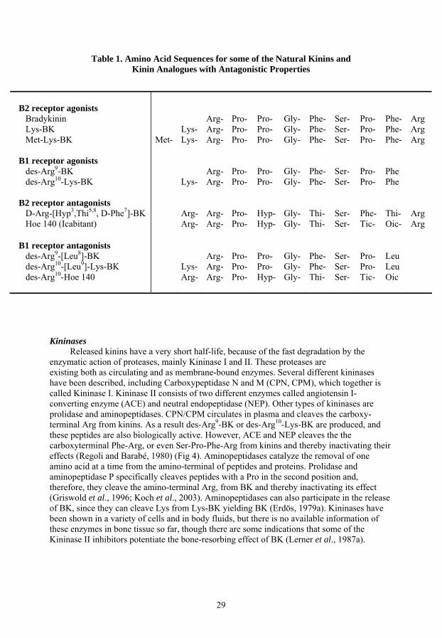
Table 1. Amino Acid Sequences for some of the Natural Kinins and Kinin Analogues with Antagonistic Properties
B2 receptor agonists
Bradykinin Arg- Pro- Pro- Gly- Phe- Ser- Pro- Phe- Arg Lys-BK Lys- Arg- Pro- Pro- Gly- Phe- Ser- Pro- Phe- Arg Met-Lys-BK Met- Lys- Arg- Pro- Pro- Gly- Phe- Ser- Pro- Phe- Arg
B1 receptor agonists
des-Arg9-BK Arg- Pro- Pro- Gly- Phe- Ser- Pro- Phe des-Arg10-Lys-BK Lys- Arg- Pro- Pro- Gly- Phe- Ser- Pro- Phe
B2 receptor antagonists
D-Arg-[Hyp3,Thi5,8, D-Phe7]-BK Arg- Arg- Pro- Hyp- Gly- Thi- Ser- Phe- Thi- Arg Hoe 140 (Icabitant) Arg- Arg- Pro- Hyp- Gly- Thi- Ser- Tic- Oic- Arg
B1 receptor antagonists
des-Arg9-[Leu8]-BK Arg- Pro- Pro- Gly- Phe- Ser- Pro- Leu des-Arg10-[Leu9]-Lys-BK Lys- Arg- Pro- Pro- Gly- Phe- Ser- Pro- Leu des-Arg10-Hoe 140 Arg- Arg- Pro- Hyp- Gly- Thi- Ser- Tic- Oic
Kininases
Released kinins have a very short half-life, because of the fast degradation by the enzymatic action of proteases, mainly Kininase I and II. These proteases are existing both as circulating and as membrane-bound enzymes. Several different kininases have been described, including Carboxypeptidase N and M (CPN, CPM), which together is called Kininase I. Kininase II consists of two different enzymes called angiotensin I-converting enzyme (ACE) and neutral endopeptidase (NEP). Other types of kininases are prolidase and aminopeptidases. CPN/CPM circulates in plasma and cleaves the carboxy-terminal Arg from kinins. As a result des-Arg9-BK or des-Arg10-Lys-BK are produced, and these peptides are also biologically active. However, ACE and NEP cleaves the the carboxyterminal Phe-Arg, or even Ser-Pro-Phe-Arg from kinins and thereby inactivating their effects (Regoli and Barabé, 1980) (Fig 4). Aminopeptidases catalyze the removal of one amino acid at a time from the amino-terminal of peptides and proteins. Prolidase and aminopeptidase P specifically cleaves peptides with a Pro in the second position and, therefore, they cleave the amino-terminal Arg, from BK and thereby inactivating its effect (Griswold et al., 1996; Koch et al., 2003). Aminopeptidases can also participate in the release of BK, since they can cleave Lys from Lys-BK yielding BK (Erdös, 1979a). Kininases have been shown in a variety of cells and in body fluids, but there is no available information of these enzymes in bone tissue so far, though there are some indications that some of the Kininase II inhibitors potentiate the bone-resorbing effect of BK (Lerner et al., 1987a).
29

Figure 4. Different kininases cleave the bradykinin molecule at different sites.
Arg1-Pro
Prolidase
Aminopeptidase P
2-Pro3-Gly4-Phe5 6-Pro7 8-Arg9
Kininase I CPN/CPM
-Ser -Phe
Kininase II NEP
Kininase II ACE
Bradykinin receptors
In the late 1970’s, Regoli and co-workers started the molecular characterization of kinin receptors by showing the existence of two types of kinin receptors, B1 and B2. These differ in their pharmacological profiles and in their expression patterns (Regoli et al., 1977, 1978; Drouin et al., 1979). 1985, there was a breakthrough when the first BK analogues with antagonistic properties for the B2 receptors were developed (Vavrek and Stewart, 1985). Another important discovery, during this time, was the identification of the B2 receptor as a G protein-coupled receptor (GPCR), which signals through the phospholipase C pathway leading to inositol 3-phosphate (IP3) formation and intracellular increase of Ca2+, as well as through the phospholipase A2 pathway resulting in the release of arachidonic acid (Burch and Axelrod, 1987). The culmination of the search for the kinin receptors came when Jarnagin and co-workers cloned the rat B2 receptor cDNA in 1991 (McEachern et al,. 1991) and soon thereafter the human B1 receptor was cloned (Menke et al,. 1994). More recently the targeted ablation of the genes for the B2 (Borkowski et al,. 1995) and B1 receptor (Pesquero et al,. 2000) in mice has started to reveale the pathophysiological functions of the kinin receptors.
There are two pharmacologically distinct subtypes of kinin receptors, called B1 and B2, which are linked to the effects of kinins (Prado et al., 2003; Leeb-Lundberg et al., 2005). They belong to the rhodopsin family of GPCR. Both receptors seem to signal through Gαq to stimulate phospholipase Cβ yielding phosphoinositide (PI) hydrolysis and an increase in intracellular free Ca2+, as well as through Gαi to downregulate adenylate cyclase and stimulate the MAPK cascades. Also stimulation of phospholipase A2 seems to occur via G protein-coupled mechanisms (Burch and Axelrod, 1987; Leeb-Lundberg et al., 2005). The distribution of BK receptors in different cells and tissues and the relative expressions of B1 and B2 receptors have been extensively studied, preferentially by using pharmacological methods, such as rank order potencies for different agonists, sensitivity to receptor antagonists, and radioligand-binding studies. Selective antagonists for the B1 receptor are for example des-Arg9[Leu8]-BK, des-Arg10[Leu9]-LysBK or des-Arg10-Hoe140 (Wirth et al., 1992), and for the B2 receptor has Hoe140 (Icatibant; D-Arg-[Hyp3, Thi5, D-Tic7,Oic8] been shown to be a very selective antagonist (Regoli et al., 1998; Simpson et al., 2000). Some non-peptide BK receptor antagonists have also been developed including WIN64338 (Salvino et al,. 1993) and FR173657 (Aramori et al, 1997; Abe et al,. 1998) for the B2 receptors, and SSR240612 (Gougat et al., 2004) for the B1 receptors.
30
The natural kinins with high affinity to the B2 receptor are BK and Lys-BK. Those are
generated in response to injury, from kininogen precursors, cleaved by either plasma or tissue kallikreins, in all mammalian species. The B2 receptor require the hole nonapeptide to be activated, and is constitutively expressed in a variety of different cell types including endothelial cells, synovial cells, sensory fibers, dermal and gingival fibroblasts, smooth muscle cells and epithelial cells. The expression of the B2 receptors can be enhanced by cytokines, cyclic AMP, estrogen and glucocorticoids (Schmidlin et al., 1998; Haddad et al., 2000; Pesquero et al., 1996., Madeddu et al., 1997; Scherrer et al., 1999) and the promoter contain binding elements for AP-1, cyclic AMP responsive element (CRE), NF-κB, estrogen receptor (ER) and glucocorticoid receptor (GR), as well as a silencer element (Baptista et al., 2002).
The most important structural determinant for high affinity to B1 receptors is the removal of the carboxy-terminal arginine from BK or Lys-BK, by carboxypeptidase N and M, yielding des-Arg9-BK or des-Arg10-Lys-BK (Regoli and Barabe, 1980; Marceau, 1995). The B2 receptors are constitutively expressed in many cell types, whereas the B1 receptors are believed to be induced during inflammation, by lipopolysaccharides, pro-inflammatory cytokines (Marceau et al., 1998), as well as by kinins themselves (Schanstra et al., 1998; Phagoo et al., 1999), although this auto-regulation model is not universally applicable or may be species-specific (Sabourin et al., 2001; Marceau et al., 2002). The B1 promoter has numerous putative binding sequences for several transcription factors including NF-κB and AP-1 (Bachvarov et al., 1996), and c-Jun has been shown to be an important mediator of B1 receptor regulation (Yang et al., 2001). It has also been shown that pharmacological inhibitors of different MAPKs and of NF-κB can reduce increased expression of B1 receptors (Larrivée et al., 1998; Sabourin et al., 2002; Ganju et al., 2001; Medeiros et al., 2004).
The signalling patterns of the B1 and B2 receptors seem to differ with respect to their
duration. B2 receptor-mediated signalling is transient, whereas B1 receptor-mediated signalling is sustained (Mathis et al., 1996; Faussner et al., 1998). At the cellular level, BK stimulation of the B2 receptor leads to rapid desensitization of the receptor response determined by both PI hydrolysis and the increase in intracellular Ca2+, in a variety of native systems including vascular endothelial cells (Smith et al., 1995), vascular smooth muscle cells (Mathis et al., 1996), as well as cell systems transfected with the B2 receptor (Blaukat et al., 1996; Fathy et al., 1999). The B2 receptor desensitization involves phosphorylation of specific serines and threonines in the receptor carboxy-terminal end. The B1 receptor differs from the B2 receptor in this aspect since it is only desensitizised to a very limited degree (Bascands et al., 1993; Smith et al., 1995; Mathis et al., 1996), it lacks serines and threonines in the carboxy-terminal tail. Moreover, human B1 receptors are not phosphorylated to a significant degree in either the presence or absence of agonist (Blaukat et al., 1999). The difference in the amount of desensitization of B2 and B1 receptors might contribute to the distinct pattern of receptor signalling (Bascands et al., 1993; Mathis et al., 1996).
The relative importance of signalling through B1 and B2 receptors in vivo is not known, but it has been proposed that B1 receptors may be important in inflammatory conditions, not only because of their induction, but also due to the fact that des-Arg metabolites of BK and Lys-BK, acting preferentially on B1 receptors, are produced in inflammatory processes by carboxypeptidases.
31

Cloning of the kinin receptors The B2 receptor was first cloned from rat uterus (McEachern et al., 1991). Subsequently
the B2 receptor has been molecularly cloned in several mammalian species including human (Hess et al., 1992). The structure of the B2 receptor protein is typical of that of a GPCR, containing seven transmembrane-spanning domains, with the amino-terminal end being extracellular and the carboxy-terminal end being intracellular, and with three extracellular loops and three intracellular loops. Three consensus sites for amino-terminal linked glycosylation are found in extracellular domains and the carboxy-terminal tail contains serines and threonines that are putative sites for acylation (Fredriksson et al., 2003). The three-exon structure of the gene (BDKRB2) has been determined, and the gene is located on the human chromosome 14q32 (Powell et al., 1993; Kammerer et al., 1995). The coding sequence of the human B2 receptor was first believed to be intronless in the exon 3, but an initiation codon has also been found in exon 2 (extends the amino-terminal with 27 residues).
The cloning of the human B1 receptor was achieved later (Menke et al., 1994). Thereafter, the B1 receptor has also been cloned in several mammalian species. The B1 receptor also possesses three consensus sites for amino-terminal linked glycosylation in extracellular domains and putative sites for phosphorylation and acylation. The three-exon structure of the human B1 receptor gene (BDKRB1) has also been determined with the protein sequence being encoded only by exon 3 (Bachvarov et al., 1996; Yang and Polgar, 1996). The gene corresponding with the B1 receptor is also located on the chromosome 14q32 in close vincinity to the B2 receptor gene, with the B2 receptor gene being proximal to the B1 receptor gene, separated by only 12 kb (Cayla et al., 2002). The close proximity of the two genes suggests that they evolved from a common ancestor by a gene duplication event. Both B1 and B2 receptors, from several species, have been found to be highly conserved (70-80% homology). In humans, the predicted sequences for the B1 and B2 receptor proteins are 353 and 364/391 amino acids respectively. The mRNA coding for the B2 receptor is large (∼4 kb) compared to that of the B1 receptor (∼1.4 kb), and the large 3’-untranslated region of the B2 receptor accounts for most of the difference.
Targeted disruption (“knockout”) of both kinin receptor genes, by homologous recombination has been reported. The B2 receptor knockout mice fail to respond to BK in assays such as smooth muscle contraction and afferent nerve stimulation (Borkowski et al., 1995). These animals seems apparently healthy, are fertile, but they may not develop and age in an entirely normal manner. Interestingly, B2 receptors might be important in senescence-associated bone loss since mice double mutant for Ins2Akita/+ and Bdkrb2-/- genes exhibit a complex phenotype including kyphosis and osteoporosis (Kakoki et al., 2006). Targeted deletion of the B2 receptor gene has also resulted in mice with severe hypertension, with end-organ damage, when they are challenged to excess dietary sodium chloride (Alfie et al., 1996).
More recently the B1 receptor knockout mice have been produced (Pesquero et al., 2000). These mice develop normally with normotension, but fail to respond to des-Arg9-BK in assays measuring contractility of the mouse-isolated stomach. However, lipopolysaccharide (LPS)-induced hypotension is blunted and the number of polymorphonuclear (PMN) leukocytes is decreased in inflamed tissues. Additionally, B1 receptor knockout mice are analgesic in behavioral experiments of nociception (Pesquero et al., 2000).
The use of mice deficient in each receptor gene, and a number of different specific B1 and B2 receptor antagonists, have suggested that both B1 and B2 receptors are potential
32

therapeutic targets in several pathophysiological events related to inflammation (e.g. pain, sepsis, allergic asthma, rhinitis, oedema), as well as diabetes and cancer (Leeb-Lundberg et al., 2005).
Kinins and Bone Metabolism
Observations of the effects of kinins on bone metabolism have been made in vitro, mainly in bone organ cultures, and most of these investigations have been done using a mouse calvarial bone culture model, in which bones, prelabelled with 45Ca or [3H]proline in vivo are used (Lerner et al., 1987a; Ljunggren et al., 1991a). Treatment of mouse calvarial bones with BK for 72-96 hours resulted in increased bone resorption, as assessed by the release of 45Ca or by the mobilization of stable calcium and inorganic phosphate (Gustafson and Lerner, 1984; Lerner et al., 1987a). BK also increases the bone matrix degradation, as assessed by the release of 3H from [3H]proline-labelled bones (Lerner et al., 1987a). The minimal concentration for action of BK in mouse calvarial bones is 3nM, and half-maximal stimulation (EC50) is achieved at 100 nM (Lerner et al., 1987a). The action of BK on bone resorption is delayed, with no effect seen until after 24 hours (Lerner et al., 1987a), and this might indicate that BK stimulates bone resorption due to enhanced osteoclast recruitment. .
BK and Lys-BK have been shown to stimulate bone resorption in mouse calvariae,
indicating that there are B2 receptors present (Gustafson et al., 1986; Lerner et al., 1987a; Ljunggren and Lerner, 1988). This observation is further supported by the fact that the B1 receptor antagonist des-Arg9-[Leu8]-BK does not inhibit the bone resorptive effect caused by BK (Lerner et al., 1987a), an observation that opens the possibility that the effect of BK is not due to the conversion of BK by carboxypetidases to the B1 receptor agonist des-Arg9-BK. The addition of des-Arg9-BK to mouse calvarial bones results in increased release of 45Ca (Lerner et al., 1987a; Ljunggren and Lerner, 1990), an effect that is inhibited by the B1 receptor antagonist des-Arg9-[Leu8]-BK (Ljunggren and Lerner, 1990), suggesting that bone cells also are equipped with B1 receptors.
Inflammatory induced bone loss may not only be due to increased bone resorption, but
also decreased bone formation. Very little is known about the possible effects of BK regarding osteoblast cell proliferation, biosynthesis of bone matrix proteins, and the activity of alkaline phosphatase. In the human osteosarcoma cell line MG-63, BK does not stimulate cell proliferation or the biosynthesis of type I collagen or osteocalcin (Rosenqvist et al., 1996). In agreement with the findings in the MG-63 cells, BK has no effect on the proliferation of osteoblast-like cells isolated from human bone (Frost et al., 1999). However, there are observations showing that HMW kininogens might be involved in the growth-promoting activity of milk (Yamamura et al., 2000), and these observations suggest that kininogens may not only be important for BK formation, but also for bone growth. Prostaglandins – Mediators of BK induced Bone Resorption
It is well known that bone resorption induced by BK is inhibited by indomethacin, a potent inhibitor of prostaglandin formation (Gustafson and Lerner, 1984). It has also been shown that a lot of inhibitors of the cyclooxygenase pathway, in the arachidonic acid cascade, including indomethacin, flurbiprofen, naproxen and meclofenamic acid, all abolish BK-induced mineral mobilization and bone matrix degradation (Lerner et al., 1987a). These nonsteroidal anti-inflammatory drugs (NSAID) also clearly inhibit the bone resorptive effects
33

of Lys-BK and Met-Lys-BK (Gustafson et al., 1986; Ljunggren and Lerner, 1988). Thus, these observations indicate that the bone resorptive effect caused by BK is totally dependent on its capacity to activate prostaglandin biosynthesis. Moreover, most stimulators of bone resorption in vitro also stimulate prostaglandin biosynthesis in bone tissue and bone cells, although the level of the prostaglandin response varies between different stimulators. However, there are stimulators of bone resorption, including PTH, vit D3 and transforming growth factor-β (TGF-β) that are totally independent of prostaglandin formation, meaning that they neither stimulate prostaglandin production nor are inhibited by NSAIDs (Ljunggren and Lerner, 1989; Lerner, 1996). In addition, IL-1β, another stimulator of bone resorption, can stimulate bone resorption both dependent and independent of prostaglandin formation (Lerner et al., 1991), and then there are the kinins, which are unable to stimulate bone resorption in the absence of prostanoid production.
In primary cultures of mouse calvarial osteoblasts, BK causes a rapid burst of PGE2 and 6-keto-PGF1α (the stable breakdown product of PGI2) that is maximal after only 5-10 minutes (Lerner et al., 1989). A similar effect has been found in the mouse calvarial osteoblastic cell line MC3T3-E1 (Lerner et al., 1989) and in isolated human osteoblast-like cells (Ljunggren et al., 1990; Rahman et al., 1992). By removal of the carboxyterminal arginine from BK, by CPN/CPM, des-Arg9-BK is formed, and this peptide has also been found to stimulate bone resorption and prostaglandin formation, but the effect is very much delayed compared to the effect caused by BK (Lerner et al., 1987a; Ljunggren and Lerner, 1990).
Results achieved with mouse calvarial bones demonstrate the presence of both B1 and B2 receptors linked to bone resorption, by a process depending on the stimulation of prostaglandin formation. Differences in the kinetics for the prostanoid response indicate different molecular mechanisms of action in B1 and B2 receptor stimulation of bone resorption.
Biosynthesis of prostaglandins Eicosanoids
Eicosanoids are a group of polyunsaturated hydrophobic molecules, with both autocrine and paracrine functions. They are not found preformed in the tissues but are generated de novo from phospholipids in response to a wide range of different stimuli. They are involved in the control of many physiological processes and are one of the most important mediators in an inflammatory reaction. The term ”eicosanoid” is originally from eicosa, meaning that there are 20 carbon atoms, and enoic means that it contains ’double bonds’. The main source of the eicosanoids is arachidonic acid (5,8,11,14-eicosatetraenoic acid), a 20-carbon unsaturated fatty acid, containing four double bonds. Arachidonic acid (AA) is esterfied, mainly in the sn-2 position of membrane phospholipids (Irvine, 1982). The amount of free AA under normal physiological conditions is very low. However, different stimuli can trigger the release of AA by phospholipases. The eicosanoids are usually referring to prostaglandins, thromboxanes, leukotrienes and also lipoxins. The term prostanoid is used to encompass both prostaglandins and thromboxanes. Prostaglandins
Prostaglandins are potent biologically active lipid mediators derived from arachidonic acid in the plasma membrane. They were first extracted from semen, prostate and seminal
34

vesicles and proven to lower blood pressure and cause smooth muscle contraction (Goldblatt, 1933; von Euler, 1935). In 1936, von Euler named the substance ”prostaglandin”, deriving from prostate gland, since it was first believed to be part of the prostatic secretions (von Euler, 1936). Prostaglandin was first purified during the early 1960’s, and two research groups indepentently identified AA, a 20-carbon polyunsaturated fatty acid (C20:4,ω6) as the precursor to prostaglandins in 1964 (van Dorp et al., 1964; Bergström et al., 1964). Prostaglandins can be produced by a variety of different cell types, and act as local mediators. Different stimulatory agents such as cytokines, hormones, neurotransmitters, antigens, endotoxin or mechanical stress, trigger the release of AA by phospholipase A2 enzymes. In activated cells, phospholipase A2-derived AA is oxidized by different cyclooxygenases (COXs) to form PGG2 and then subsequently PGH2. PGH2 is a common substrate for several downstream enzymes involved in the biosynthesis of different prostanoids, i.e. PGE2, PGD2, PGF2α, TXA2 or PGI2 (Fig 5). One of these downstream enzymes is the membrane-associated prostaglandin E synthase-1 (mPGES-1), which is an enzyme specifically catalyzing the conversion of PGH2 to PGE2 (Jakobsson et al., 1999; Murakami et al., 2000), and is expressed especially in cells activated by inflammation. After the prostanoids are biosynthesized, they are released from the cells and bind to specific receptors, to exert their biological effects. Prostaglandins are potent but have a short half-life before they will be inactivated and excreted. Therefore, they only exert a paracrine or autocrine function.
Membrane Phospholipids
Arachidonic acid
PLA2
Cyclooxygenases Lipoxygenases
Leukotrienes
PGG2
TXA2PGE2
PGD2, PGF2α, PGI2
PGEsynthases
Arachidonic acid cascade
PGH2
Specific Synthases
Hydroperoxyeicosatetraenoic acids (HPETEs)
Figure 5. Schematic diagram over two of the major families of eicosanoids derived from arachidinic acid: the prostanoids and the leukotriens.
35

Phospholipase A2 enzymes release arachidonic acid
Phospholipase A2 enzymes (PLA2) catalyze the hydrolysis of membrane phospholipids, releasing free fatty acids, and arachicidonic acid is the most common fatty acid in human cells. A number of stimulatory mediators trigger the release of AA by PLA2 enzymes. The release of free AA is tightly regulated step in the biosynthesis of eicosanoids. The family of PLA2 enzymes are subdivided into several classes: secretory PLA2 (sPLA2), cytosolic Ca2+-dependent PLA2 (cPLA2), cytosolic Ca2+-independent PLA2 (iPLA2) and platelet-activating factor (PAF) acetyl hydrolase (Kudo and Murakami, 2002). At least two classes of PLA2 enzymes have important functions in prostanoid production – the group II sPLA2 enzymes and cPLA2 (Murakami et al., 1997).
sPLA2 enzymes, with a molecular mass of 14–19 kDa, nonselectively release fatty acids
from the phospholipids, and need a mM concentration of Ca2+ to be activated. sPLA2 enzymes can be induced by proinflammatory stimuli (Oka and Arita, 1991). Two sPLA2 enzymes, type IIA and V, have been proven to be involved in prostaglandin production (Bingham and Austen, 1999; Sawada et al., 1999). Secretory PLA2-IIA is the most common isozyme in human.
cPLA2 is consitutively expressed in the cytoplasm of a large variety of cells. There are at
least four different subgroups of cPLA2, namely cPLA2α, cPLA2β, cPLA2γ and cPLA2δ, and cPLA2α has been shown to be highly selective for phospholipids containing AA in the sn-2 position (Leslie et al., 1988, Clark et al., 1991; Hirabayashi et al., 2004). cPLA2α is activated at sub-μM concentrations of Ca2+, and this leeds to translocation of cPLA2α from the cytosol to the nuclear membrane, which is essential for the release of AA (Reynolds et al., 1993). Cells and tissues from cPLA2α−deficient mice fail to produce prostaglandins, leukotrienes or PAF (Uozumi et al., 1997; Bonventre et al., 1997).
Cyclooxygenases
Cyclooxygenase (COX), also known as prostaglandin endoperoxide H synthase or prostaglandin G/H synthase (PGHS), is membrane-bound, and catalyses the formation of cyclic endoperoxides, PGG2, and subsequently PGH2 by oxidation of arachidonic acid. There are two isoforms of cyclooxygenase, called COX-1 and COX-2, encoded by separate genes. COX-1 is expressed at relatively constant levels in most tissues and is therefore considered to be constitutively expressed, whereas COX-2 is generally expresssed at very low levels, but can be induced to much higher levels by different inflammatory mediators (Herschman, 1994). PGH2 is then converted into different bioactive prostanoids like PGE2, PGD2, PGF2α, TXA2 or PGI2 by specific downstream enzymes in a cell-specific manner. Both COX-1 and COX-2 are situated similarly on the inner and outer membranes of the nuclear envelope and on the lumenal surface of the endoplasmic reticulum (Morita el al., 1995; Smith et al., 1996; Spencer et al., 1998), and share 60% identity in the amino acid sequence (Simmons et al., 2004).
COX-1
The genes for COX-1 and COX-2 from mouse or human origin were isolated by different laboratories (Fletcher et al., 1992; Kraemer et al., 1992; Appleby et al., 1994). These results show that the COX-1 gene contains 11 exons and is transcribed as 2.8 kb mRNA, which then is translated to form a protein with a molecular mass of ∼68 kDa (Kraemer et al., 1992). The gene is located on the human chromosome 9q32-q33.3 and
36

is approximately 25 kb in size. In the promoter region of this gene, there are a number of transcriptional regulatory elements (SP1, AP-1 and NF-IL-6), but there is not so much known about the promoter elements that control the transcription (Kraemer et al., 1992; Smith and Dewitt, 1996). The expression of the COX-1 gene is often constitutive and ubiquitous, and COX-1 is preferentially utilized in the immediate PG-biosynthesis, which takes place within minutes after stimulation with Ca2+-mobilizers. COX-2
COX-2 was discovered independently by two research groups in 1991 (Xie et al., 1991; Kujubu et al., 1991). The COX-2 gene is approximately 8 kb in size, contains 10 exons and is situated on the human chromosome 1q25.2-25.3. The mRNA transcript is about 4.1-4.5 kb and this encodes the COX-protein with a molecular weight of about 72 kDa. The promoter region of the COX-2 gene has been more extensively characterized. The cAMP response element (CRE) seems to be important in COX-2 transcription (Xie et al., 1994), and activation of different MAP kinase pathways, results in the activation of transcription factors binding to the CRE element (Herschman et al., 1997; Mestre et al., 2001). Other researchers have found that CREB/NF-IL-6 elements or NF-κB sites, either together or alone, are responsible for maximal induction of transcription by cytokines and growth factors (Crofford et al., 1997; Herschman et al., 1997). Not only the activation of transcription seems to be important, but also the stability of the mRNA. It has been shown that pro-inflammatory agents have increased the stability of COX-2 mRNA and proteins (Ristimäki et al., 1996; Sheng et al., 2000; Cok and Morrison, 2001). NF-κB-dependent transcription of COX-2 has been shown to be inhibited by high concentrations of NSAIDs, that block IκB kinase activation (Yan and Polk, 1999). The induction of COX-2 is also downregulated by anti-inflammatory glucocorticoids. This downregulation of COX-2 induction seems to be dependent on AP-1 and NF-κB transcription (Auphan et al., 1995; Scheinman et al., 1995), as well as destabilization and degradation of COX-2 mRNA and protein (Dixon et al., 2000; Newton et al., 1998; Cok and Morrison, 2001). Finally, the regulation of transport of the COX-2 mRNA, from the nucleus to the rough endoplasmic reticulum (ER), may also have functional significance (Jang et al., 2003). The inducible COX-2 is absolutely essential for the delayed prostaglandin biosynthesis, which lasts for several hours following various stimuli.
COX-3
The exon/intron organization is very similar between the COX-1 and COX-2 genes, however, the COX-1 gene contains an extra intron (intron-1), that participates in the alternative splicing, yielding COX-3 and PCOX-1 (partial COX-1) proteins (Chandrasekharan et al., 2002). COX-3 consists of the COX-1 mRNA that retains intron-1. Intron-1 is a small intron, and in dogs it contains 90 nucleotides. The COX-3 variant generates a protein containing the encoded intron-1 sequence (also called COX-1b). The protein does not have as good effect on prostaglandin biosynthesis as COX-1, but analgesic/antipyretic drugs such as acetaminophen (Paracetamol) preferentially inhibit its activity. In human, COX-3 mRNA is transcribed as a 5.2 kb transcript and is abundantly expressed in cerebral cortex and heart.
37

Prostaglandin E synthases
Prostaglandin E synthases are the enzymes involved in the conversion of cyclooxygenase derived PGH2 into PGE2. Until now, three proteins have been identified in this conversion, namely membrane-associated PGES-1 (mPGES-1), mPGES-2 and cytosolic PGES (cPGES) (Jakobsson et al., 1999; Murakami et al., 2000; Tanikawa et al., 2002; Tanioka et al., 2000).
Membrane-associated PGE synthase-1
Membrane-associated PGE synthase-1 is a gluthathion (GSH)-requiring perinuclear protein belonging to the MAPEG (for Membrane-Associated Proteins involved in Eicosanoid and GSH metabolism) family (Jakobsson et al., 1999; Murakami et al., 2000; Mancini et al., 2001). The mPGES-1 gene is located on chromosome 9q34.3 (Forsberg et al., 2000), and is about 15 kb in size, devided into three exons. The promoter of the human mPGES-1 gene contains binding sites for C/EBPα and β, AP-1, progesterone receptors, GRE, CRE and serum response elements (SRE) (Naraba et al., 2002). mPGES-1 is markedly induced by pro-inflammatory agents, downregulated by anti-inflammatory glucocorticoids, and is functionally coupled with COX-2 in marked preference to COX-1 (Murakami et al., 2000; Kamei et al., 2004). Induction of mPGES-1 expression has also been observed in several systems in which COX-2 derived PGE2 has been implicated to play an important role, such as inflammation, fever, pain, female reproduction, tissue repair and cancer (Kudo and Murakami, 2005). Co-localization of COX-2 and mPGES-1 in the same perinuclear membrane may contribute to their efficient functional coupling. Inducible expression of mPGES-1 is partially regulated by the MAP kinase pathways (Han et al., 2002). Very recently Degousee et al., demonstrated the role of JNK in mPGES-1 expression, in rat neonatal cardiomyocytes. There was no evidence about increased transcriptional activity, but the induction of mPGES-1 mRNA was found to be dependent on mRNA stability (Degousee et al., 2006). Microsomal PGE synthase-2
The second membrane-bound form of PGES is mPGES-2 (Tanikawa et al., 2002). This enzyme is initially produced as a Golgi membrane-associated protein followed by proteolytic removal of the amino-terminal hydrodophic domain, leading to the formation of a mature cytosolic enzyme (Tanikawa et al., 2002; Murakami et al., 2003). The mPGES-2 gene is localized on human chromosome 9q33-34 in the vicinity to the genes for mPGES-1 and COX-1 (Tanikawa et al., 2002). Co-transfection of mPGES-2 with either COX-1 or COX-2 (in a human embryonic kidney cell line, HEK293), demonstrated that mPGES-2 is functionally coupled with both COX isoforms (Murakami et al., 2003). The expression of mPGES-2 seems to be constitutive in a variety of cells and tissues, and is not increased during inflammation or tissue damage. However, a marked elevation of mPGES-2 expression has been observed in human colorectal cancer (Murakami et al., 2003). Cytosolic PGE synthase
Cytosolic PGE synthase (cPGES) is a glutathione-requiring enzyme that is constitutively expressed in a wide variety of cells. cPGES is a protein of 23 kDa, and identical to p23, a binding protein for heat shock protein 90 (Hsp90) (Tanioka et al,. 2000). Cotransfection and antisense experiments indicated that cPGES is functionally linked to COX-1, and not COX-2, to promote immediate PGE2 biosynthesis, elicited by
38
Ca2+-evoked stimuli (Tanioka et al., 2000). In another study by Tanioka et al, it was shown that Hsp90 significantly affects the activity of PGE2-production of cPGES (Tanioka et al., 2003). Although cPGES expression is constitutive and not affected by pro-inflammatory stimuli in most cases, some exceptions have been reported. For example, Moore et al., have shown that administration of IL-1 into the mouse cortex, led to an elevation of PGE2, which was accompanied by increased expression of cPGES, as well as that of COX-2 and mPGES-1, with differences in the kinetics (Moore et al., 2004).
Prostaglandin receptors There are currently nine known receptors of prostaglandins on various cell types. The
prostaglandin receptors are seven-transmembrane G-protein-coupled receptors that are designated by the letter ”P” and a prefix of ”D, E, F, I or T” to suggest preference for prostaglandin D, E, F, I or thromboxane, respectively. Up to date there are four different subtypes of EP receptors identified, called EP1-EP4. PGE2 contracts gastrointestinal smooth muscle through stimulation of EP1 receptors. The EP2 receptors generally mediate arterial dilatation, but are also involved in the salt-sensitive hypertension (Kennedy et al., 1999). The pyretic action of PGE2 is mediated by the EP3 receptor, since mutant mice lacking this receptor, do not develop fever after administration of either PGE2, IL-1 or LPS (Uschikubi et al., 1998). The EP4, as well as EP2 receptors might be important for angiogenesis induced by PGE2 (Harada et al., 1994; Ben-Av et al., 1995). Stimulation of the TP receptor on platelets leads to their aggregation, and TP-deficient mice have very prolonged bleeding times, demonstrating the importance of TXA2 in hemostasis (Thomas et al., 1998). Normal production of PGI2 is crucial for vessel tone control and inhibition of thrombosis. This is because the PGI2/TXA2 balance is critical. PGI2 is mainly secreted by endothelial cells. It binds to the IP receptors on vascular smooth muscle cells and reduces vascular contraction (FitzGerald et al., 1983; Murata et al., 1997). Prostaglandin receptors in bone cells are presented below.
Prostaglandins and Bone Metabolism
Prostaglandins are involved in a wide variety of actions, including muscular
constriction, inflammation, pain, fever, vascular homeostasis and thrombosis; they are “cytoprotective” in the gastrointestinal tract and have effects on bone tissue. They are potent but have a short half-life before being inactivated. Therefore, they exert mainly a paracrine (locally active) or autocrine function. As earlier mentioned, prostaglandins are metabolites from arachidonic acid, and PGE2 is a very potent stimulator of bone resorption in organ cultures and osteoclastogenesis in bone marrow and spleen cell cultures (Pilbeam et al., 2002). Prostaglandin receptors
There are a lot of complexity around the prostaglandins effects on bone tissues, and this can be partially explained by the fact that there are several transmembrane G-protein-coupled receptors that prostaglandins can bind to, and that may produce altered effects trough different signalling pathways. There are four different types of receptors for PGE2. The EP1 receptor acts mainly by enhancing the calcium influx and phosphatidyl inositol turnover and has been suggested to be involved in mineralized nodule formation (Fujieda et al., 1999), as well as the stimulation of c-fos and c-jun expression (Suda et al., 2000). Additionally, EP1 has been
39

shown to be involved in the induction of IL-6 and COX-2 (Kozawa et al., 1998; Suda et al., 1998). EP2 and EP4 receptors activate adenylyl cyclase, and both receptors seem to be involved in the regulation of osteoblast function, and they have also been reported to be important for the effects of PGE2 on osteoclastogenesis and bone resorption (Li et al., 2000b; Suzawa et al., 2000). At least two different mechanisms are involved in PGE2-induced bone resorption. The first is due to activation of prostaglandin receptors in osteoblasts/stromal cells, resulting in an increased RANKL expression (Yasuda et al., 1998b). The other mechanism involves potentiation of RANK signaling, caused by activation of prostaglandin receptors on osteoclast progenitor cells (Ono et al., 2005). The EP3 receptor acts mainly by inhibiting cyclic AMP formation and has not been shown to play a specific role in bone. There is also evidence for the existence of receptors for PGI2 on bone, and one study demonstrated IP-receptors in mouse osteoblastic MC3T3-E1 cells induced by TNF-α (Wang et al., 1999). Regulation of prostaglandin production in osteoblasts
Prostanoids produced by bone cells include PGE2, 6-keto-PGF1α (the stable breakdown product of PGI2), PGF2α and some PGD2 and thromboxane (Raisz et al., 1979; Feyen et al., 1984). There are a lot of factors that stimulate prostaglandin production in bone, including inflammatory mediators such as IL-1 and TNF-α (Sato et al., 1986; Tashjian et al., 1987); regulators of cell growth and differentiation, such as transforming growth factor (TGF)-α and β (Tashjian et al., 1985, Hurley et al., 1989; Sumitani et al., 1989), systemic calcium-regulating hormones, such as PTH and Vit D3 (Ljunggren and Lerner, 1989; Pilbeam et al., 1989; Klein-Nulend et al., 1991a,b), as well as, mechanical loading of bone (Rawlinson et al., 1991, Cheng et al., 1997). Also bradykinin (Ljunggren et al., 1991b) and thrombin (Ljunggren et al., 1991c) can stimulate prostaglandin production in osteoblasts.
The most commonly used osteoblastic cell models constitutively express COX-1 and can be induced to express COX-2. Induction of COX-2 in osteoblastic cells seems to be required for most stimulated prostaglandin responses. Several different agonists are capable of inducing COX-2 in osteoblastic cells, including IL-1, TNF-α, IL-6, TGF-α, TGF-β, PTH, VitD3 and mechanical loading (Harrison et al., 1994; Kawaguchi et al., 1994, 1996; Pilbeam et al., 1997b; Min et al., 1998; Okada et al., 2000a; Klein-Nulend et al., 1997). It has been shown that prostaglandins induce COX-2 expression and therefore can amplify prostaglandin responses from other agonists (Kawaguchi et al., 1994; Pilbeam et al., 1995). This mechanism of amplifying may be important in sustaining the production of prostaglandins in cell, as well as organ cultures. Inhibition of prostaglandin production
Glucocorticosteroids have been reported to be potent inhibitors of prostaglandin formation in many studies. They can inhibit phospholipase A2 production, as well as inhibit the inducible COX-2 mRNA and protein expression, in bone and other tissues (Kawaguchi et al., 1994). The cytokines IL-4 and IL-13 have also been shown to inhibit COX-2 expression and prostaglandin formation in bone cell and organ cultures (Kawaguchi et al., 1996; Onoe et al., 1996). Nonsteroidal antiinflammatory drugs (NSAIDs) reduce prostaglandin production by competing directly with arachidinic acid for binding to the COX catalytic site (Pilbeam et al., 1997a). Therefore, NSAIDs will not inhibit leukotriene production. Most available NSAIDs inhibit both COX-1 and COX-2 activity, but the development of the selective COX-2 inhihitors (coxibs; e.g. celecoxib (Celebra) etc.) have made it possible to avoid some of the negative gastrointestinal effects while still effectively reducing inflammation (Silverstein et al., 2000). COX-2 is the enzyme responsible for the prostaglandin enhancement of stimulated osteoclastogegensis. In bone marrow cultures from COX-2-deficient mice, osteoclast
40
formation stimulated by PTH or Vit D3 was reduced by 60-70% compared to the wild type mice cultures (Okada et al., 2000a). The PGE2 formation was also markedly decreased, and the inhibited osteoclast formation was completely reversed by addition of PGE2 to the cultures. Prostaglandins produced by COX-2 seems to be essential for in vitro osteoclastogenesis in response to several agents. Prostaglandins in bone resorption
The effects of prostaglandins on bone resorption is predominantly caused by PGE2 (Raisz and Martin, 1983), but PGI2 and PGF2α can also induce bone resorption, though to a lesser extent. The ability of PGF2α to stimulate bone resorption is partially dependent on PGE2-induction (Raisz et al., 1990). PGE2, but not PGF2α, can stimulate osteoclast formation in bone marrow cultures (Collins and Chambers, 1991; Kaji et al., 1996). There are many different agonists reported to stimulate prostaglandin-dependent osteoclastogenesis in bone marrow cultures, including IL-1, TNF-α, PTH, Vit D3, IL-11, IL-6 and IL-17. The formation of mature active bone-resorbing osteoclasts requires an interaction with cells from the osteoblastic linage (Suda et al., 1999). The molecules mediating this interaction is RANKL (Anderson et al., 1997; Wong et al., 1997; Yasoda et al., 1998b), binding to its cognate receptor RANK on the osteoclast precursor. OPG works as a decoy receptor for RANKL as mentioned earlier. Osteoblastic cells produce both RANKL and OPG, as well as M-CSF, also essential for osteoclastogenesis, whereas osteoclastic cells express RANK. The bone resorption caused by PGE2 seems to be dependent on the induction of RANKL (Tsukii et al., 1998). Additionally, prostaglandins are not only able to act on osteoblastic cells to enhance the expression of RANKL, but they can also act directly on osteoclastic progenitor cells to induce formation of preosteoclasts. PGE2 has been reported to increase the combined effects caused by RANKL and M-CSF, to induce osteoclastogenesis in spleen cultures (Wani et al., 1999). In contrast, when PGE2 was added to isolated osteoclasts in vitro, it transiently inhibited bone resorption (Fuller and Chambers, 1989). Such transient inhibition of bone resorption, by prostaglandins, has also been observed in mouse calvarial bone cultures (Lerner et al., 1987b). There is not much known about the role of prostaglandins in bone resorption in vivo, but one important role might be the ability to generate new osteoclasts from osteoclastic precursors, since this becomes the factor limiting the rate of resorption. The prolonged bone resorption in vivo, caused by IL-1, was found to depend on prostaglandins (Boyce et al., 1989). Thus, prostaglandins increase the formation and differentiation of osteoclast precursors, as well as act on the osteoblasts to increase their endogenous prostaglandin production. This leads to an enhanced osteoclast formation. When fully differentiated mature osteoclasts are formed, prostaglandins are inhibitory. These effects seem to be cAMP-mediated (Lerner et al., 1987b). Prostaglandins in bone formation
Studies in vitro have shown that prostaglandins seem to have both stimulatory and inhibitory effects on bone formation, although in vivo studies have more prominently shown a potent anabolic effect (Jee and Ma, 1997). PGE2 has been shown to stimulate both cell replication and differentiation, in rat calvarial organ cultures (Woodiel et al., 1996). In addition, marrow stromal cells or primary calvarial cells from COX-2-deficient mice seem to have delayed osteoblastic differentiation compared to wt mice (Okada et al., 2000b). At high concentrations though, prostaglandins seem to inhibit collagen formation in cell and organ cultures (Fall et al., 1994). Prostaglandins can be produced by osteoblasts, osteocytes and adjacent hematopoietic cells. They seem to increase both the replication and the differentiation of the osteoblastic precursors, but inhibit collagen synthesis in mature osteoblasts as well.
41

Inflammation induced bone remodelling
In general, inflammation is the first response of the immune system to infection or other
exogenous or endogenous stimuli and may be referred to as the acute/innate response. Inflammation is characterised by: redness (rubor), heat (calor), swelling (tumor), pain (dolor) and dysfunction of the organs involved (functio laesa). Inflammation consists of two main events including a cellular and an exudative event. The cellular component involves the movement of the leukocytes from blood vessels into the inflamed tissue. The leukocytes extravasate from the capillaries into tissue, and act as phagocytes. The exudative events lead to accumulation of fluid and leukocytes in extravascular tissues. Various leukocytes are involved in the initiation and maintenance of the inflammatory process.
Acute inflammation is predominantly mediated by neutrophils (polymorphonuclear
leukocytes), and is modulated by molecules that induce vasodilatation (bradykinin and prostaglandins) increase vascular permeability (bradykinin, histamine and leukotriene) and possess chemotactic properties (complement products) to recruit additional inflammatory cells. Neutrophils only live for a couple of days, so if the inflammation persists for a longer time, they are gradually replaced by longer lived monocytes. The subsequent immune response is initiated when antigen-presenting cells become involved and present the foreign antigens or microorganisms to immunocompetent cells. This leads to the expansion of antibody-secreting plasma cells and the development of a chronic lesion. Chronic inflammation is mainly mediated by mononuclear cells such as monocytes and macrophages. These cells can be further stimulated to maintain inflammation through the action of an adaptive response involving lymphocytes (T-cells, B-cells), and antibodies. The adaptive response is more precise, reacting on specific antigens, but also takes a longer time.
During physiological conditions, the skeleton is continuously remodelled, by bone-forming osteoblasts and bone-resorbing osteoclasts. In pathological processes, the activities of the bone cells are un-coupled, leading to either increased bone resorption or increased bone formation. In some diseases, the amount of resorbed bone exceeds that of formed, leading to bone loss. This is often seen in patients with inflammatory processes in the vicinity of the bone tissue (e.g. periodontal disease, rheumatoid arthritis, osteomyelitis etc.). In some situations, however, more bone is produced than resorbed, leading to sclerosis of the bone. This can be seen in patients with apical periodontitis, but not normally in patients with marginal periodontitis. However, there are som evidence showing that increased isotope uptake have been observed also in patients with marginal periodontitis (Jeffcoat et al., 1991), indicating that increased bone formation also is a part of this process. The isotope used is incorporated into the skeleton during osteoblastic bone formation, and a high uptake indicating high rates of bone formation. This suggests that, accompanying the inflammation induced bone resorption, there is also increased bone formation. There are some evidence indicating that prostaglandins, as well as PTH (Hodsman et al., 2005), can stimulate not only bone resorption, but also bone formation (Hartke and Lundy, 2001; Pilbeam et al., 2002; Raisz and Woodiel, 2003; Vrotsos et al., 2003).
In several inflammatory diseases such as rheumatoid arthritis, periodontitis and osteomyelitis, increased osteoclast differentiation and activity can be stimulated by several bone resorbing pro-inflammatory cytokines, leading to resorption of bone tissue in areas adjacent to the inflammatory site. The group of bone-resorbing cytokines currently includes IL-1, IL-6, IL-11, IL-17, TNF-α, leukemia inhibitory factor (LIF) and oncostatin M (OSM)
42

(Martin et al., 1998; Horowitz and Lorenzo, 2002; Nakamura and Jimi, 2006) (Fig. 6). In an inflammatory process, there are also a number of factors leaking out from the blood vessels, due to the increased permeability, that are important for the regulation of bone metabolism. These factors are produced in the liver, and as long as they circulate in the bloodstream they are inactive, but as soon as they leak out in the inflammatory site, they are activated locally. Important such factors are the molecules generated from the kallikrein-kinin system and the coagulation cascade (i.e. kinins and thrombin). The pro-inflammatory peptide BK, as well as other kinin analogues has been reported to stimulate bone resorption (Lerner et al., 1987a; Ljunggren and Lerner, 1990; Worthy et al., 1990; Lerner, 1997; Couture et al., 2001; Moreau et al., 2005). Importantly, kinins, acting on both B1 and B2 receptors, not only stimulate bone resorption per se, but also synergistically potentiate the bone resorptive effect of IL-1 and TNF-α (Lerner, 1991; Lerner and Modéer, 1991; Lerner and Lundberg, 2002), by a prostaglandin dependent mechanism (Lerner et al., 1989). The effects of cytokines stimulating osteoclast formation and bone resorption seem to be prevented by other cytokines that inhibit the process of bone resorption (Fig. 6). Such cytokines are IL-4, IL-10, IL-12, IL-13, IL-18, interferon-β (IFN-β) and IFN-γ, and all are able to inhibit osteoclast formation and bone resorption (Horowitz and Lorenzo, 2002; Palmqvist et al., 2006). Thus, a balance between stimulatory and inhibitory mediators of inflammation, together with the regulation of their receptors, as well as signal-transducing mechanisms, will determine the amount and the activity of the osteoclasts formed, and thus the degree of bone resorption. The important role of osteoclasts in bone loss has been shown in several inflammatory induced diseases, and here follows an introduction to two of those diseases, namely rheumatoid arthritis and periodontal disease.
43

Inflammation
Inhibitory cytokines (IL-4, IL-10, IL-12, IL-13, IL-18, IFN-β, IFN-γ)
Kinins Thrombin
Stimulatory cytokines (IL-1, IL-6, IL-11, IL-17, TNF-α, LIF, OSM)
Osteoblasts/stromal cells
Preosteoclasts
RANKL/OPG
Figure 6. Different cytokines and inflammatory mediators have been shown to regulate osteoclast formation and bone resorption. The stimulatory mediators exert their effects by stimulating the RANKL/OPG-ratio in periosteal osteoblasts. The inhibitory cytokines are able to cause their effects either indirectly, by affecting the osteoblasts, or sometimes, directly, by affecting the osteoclast precursors.
Rheumatoid Arthritis
Rheumatoid arthritis (RA) is a chronic, inflammatory, autoimmune disorder characterized by joint inflammation, synovial lining hyperplasia, pain, cartilage destruction and bone resorption. There are symptoms that distinguish rheumatoid arthritis from other forms of arthritis: 1) Inflammation and soft-tissue swelling of several joints at the same time. 2) Arthritis of hand joints. 3) The joints are usually affected asymmetrically in the beginning, and then in a symmetrical fashion as the disease progresses. 3) The pain of rheumatoid arthritis is usually worse in the morning (morning stiffness), but generally improves with use of the affected joints. 4) As the pathology progresses the inflammatory activity leads to destruction and erosion of the joint surface, which worsens the range of movement and leads to deformity.
During initiation of the disease, the synovial membrane becomes hyperplastic because
of the accumulation of fibroblasts and cells of hematopoietic origin. This modification of the
44

synovial membrane is followed by the destruction of adjacent structures, such as articular cartilage and bone, due to the invasive properties of synovial tissue. This pathologic events leading to arthritis is governed by several proinflammatory cytokines that allow communication between inflammatory cells, leading to concerted actions such as the tissue invasion. IL-1, TNF-α and IL-6 are highly expressed in inflamed synovium and in synovial fluid from patients with RA, and have been implicated in the pathogenetic mechanism leading to bone resorption in this disease (Choy and Panayi, 2001; Udagawa et al., 2002; Dinarello, 2002; Firestein, 2003; Gravallese, 2003; Kay and Calabrese, 2004). These proinflammatory cytokines are of particular clinical importance, because their blockade improves the features of RA (Dinarello, 1994b; Feldmann and Maini, 1999; Dayer, 2002; Nishimoto et al., 2004; Cohen et al., 2004; St Clair, 2002; Zwerina et al., 2006a).
Kinins have significant stimulatory effects on TNF-α and IL-1 release from
macrophages, and this release may be mediated by the activation of B1 receptors (Tiffany and Burch, 1989). TNFs and IL-1 can stimulate bone resorption, cartilage damage, hypertrophic synovitis, inflammatory cell migration, angiogenesis and pannus formation. TNF receptors have been localized in synovial tissue and cartilage-pannus junction in patients with RA, and this may play a major role in the genesis of the disease (Deleuran et al., 1992). In synovial fluid from patients with RA, increased kinin levels, increased kallikrein activity, as well as kininogens, has been demonstrated (Kellermeyer and Graham, 1968; Selwyn et al., 1989; Worthy et al, 1990; Sharma and Buchanan, 1994). These findings, together with the observations that kinins can stimulate bone resorption in neonatal mouse calvariae, indicate that also kinins could be responsible for osteoclastic activation in inflammatory conditions (Lerner et al., 1987a; Lerner and Lundberg, 2002). The kinin induced bone resorption is associated with an induced production of PGE2 (Ljunggren and Lerner, 1990; Ljunggren et al., 1991b). Non-cytokine inflammatory mediators such as PGE2, PGI2 and leukotriens are released by kinin stimulation (Sharma & Mohsin, 1990; Sharma, 1991a,b). High levels of extracellular phospholipase A2 have been detected from human synovial fluid (Hara et al., 1989), and this might be released from macrophages, granulocytes and lymphocytes. PLA2 is activated by kinins to release the arachidonic acid metabolites, prostaglandins and leukotriens, in the process of RA (Sharma, 1991a,b). Kinins are capable of stimulating prostaglandin release in different cell types, including osteoblasts, fibroblasts and endothelial cells (Conklin et al., 1988; Lerner et al., 1989; Lerner and Modéer, 1991; Ljunggren and Lerner 1990; Ljunggren et al., 1990, 1991b).
The proinflammatory and destructive properties of TNF and IL-1 are mediated through
activation of several intracellular signal transduction pathways. Among these molecules p38 MAPK is proven to be one of the most important signals for TNF-mediated inflammatory responses (Geng et al., 1996; Kotlyarov et al., 1999; Schett et al., 2000; Revesz et al., 2004). Interestingly, inhibition of p38 MAPK leads to inhibition of the arthritis caused by overexpression of TNF, and, in adddition, inflammatory bone loss depends on activation of p38 MAPK, since osteoclastogenesis in the inflamed synovium depends on intact activation of the p38 MAPK signalling pathway (Zwerina et al., 2006b).
45
Periodontal disease Periodontal diseases are mainly initiated by bacterial infections inducing an
inflammatory response, that in some situations are leading to destruction of the periodontal ligament, as well as the underlying alveolar bone tissue (Offenbacher, 1996). It is one of the most common infectious diseases among adults, and it can be influenced by a variety of modifying factors, including the microorganisms present in the subgingival lesion, systemic disorders that might have negative effects on the host response, environmental factors (e.g. plaque and smoking) or one's inherited or genetic susceptibilty to periodontitis.
Results from human and animal research have clearly shown that PGE2 levels within periodontal tissues and within the gingival crevicular fluid (GCF) correlate with the clinical expression of periodontal disease (Offenbacher et al., 1993). This indicates that NSAIDs might be able to suppress inflammation and alveolar bone resorption in periodontitis, and there are several studies showing that different NSAIDs can inhibit the periodontal ligament destruction, as well as the rate of alveolar bone loss, both in vitro and in vivo (Nyman et al., 1979; Williams et al., 1984, 1988a,b; Offenbacher et al., 1987, 1992; Salvi et al., 1997). Also the effect of more selective COX-2 inhibitors, on alveolar bone loss, have been studied in rats (Bezerra et al., 2000; Holzhausen et al., 2002), showing a significant reduction in bone loss compared to controls (Salvi and Lang, 2005).
In addition, increased kinin levels have been found in inflammatory exudates collected from dogs with periodontal disease (Montgomery et al., 1986). These observations, together with findings that kinins can stimulate bone resorption in neonatal mouse calvariae, as well as synergistically potentiate the bone resorptive effect of IL-1 (Lerner, 1991), have raised the possibility that also kinins should be regarded as candidates responsible for osteoclastic activation in inflammatory conditions such as periodontal disease and rheumatoid arthritis (Lerner et al., 1987a; Lerner and Lundberg, 2002). The synergistic stimulation of IL-1 induced bone resorption caused by kinins is associated with a synergistic potentiation by kinins on IL-1 induced production of prostaglandin E2 (PGE2) and PGI2 as well (Lerner, 1991; Lerner and Lundberg, 2002). IL-1, TNF-α and IL-6 have been found to be generously expressed in inflamed gingiva, and increased levels have been found in the crevicular fluid from patients with periodontitis (Okada and Murakami, 1998; Mogi et al., 1999; Boch et al., 2001; Graves and Cochran, 2003). Graves and Cochran have also shown that a lot of the damage that occurs during periodontal tissue destruction can be caused by IL-1 and TNF activity. This destruction might be a hyperreaction of the host response to the periodontal pathogens, caused by an extensive production of IL-1 and TNF (Graves and Cochran, 2003; Nakamura and Jimi, 2006). Thus, the same types of cytokines seem to be responsible for the stimulation of bone resorption in periodontitis, as well as in rheumatoid arthritis (Lerner, 2006). There are also observations indicating that upregulation of RANKL mRNA, in both epithelium and inflammatory cells, might be associated with the activation of osteoclastic bone resorption in periodontitis (Liu et al., 2003). In addition, it has recently been shown that transvascular dissemination of Porphyromonas gingivalis from a sequestered site is dependent on activation of the kallikrein-kinin pathway, indicating that activation of the kinin system is involved in the connection between human periodontitis and systemic diseases (Hu et al., 2006).
46

Intracellular signalling
Signal transduction is a process in which a cell converts one type of signal/stimulus into another. These processes often involve a sequence of reactions inside the cell, which are performed by enzymes and linked through second messengers. These are very fast processes (milliseconds-seconds). Signal transduction usually involves the binding of extracellular factors to receptors embedded in the outer cell membrane and trigger events inside the cell. This often happens via a change in the shape of the receptor that occurs when the signal molecule "docks" or binds. Receptors respond only to the specific ligand for which they have affinity, and molecules that are different in shape have no effect or maybe act as inhibitors. Often, the intracellular events triggered by the external stimulus are considered distinct from the event of "transduction" itself, because this term refers only to the step that converts the extracellular stimulus to an intracellular signal. There are a variety of intracellular signalling molecules including heterotrimeric G protein, cyclic AMP (cAMP), Ca2+, phosphatidylinositol-triphosphate (PIP3), diacylglycerol (DAG), inositol-triphosphate (IP3), and several protein kinases and phosphatases. Some of these are also referred to as second messengers.
A transcription factor is a protein that acts as a regulator of gene expression, specifically regulating the activation or inhibition of transcription in the nucleus. They mainly exert their effects through binding to specific response elements, either through direct binding to DNA or through binding other DNA-bound proteins, in the promoter sequence of target genes and thereby regulate transcription. Here follows an introduction to the transcription factor pathways studied in this thesis. Activating protein-1 (AP-1)
One of the first transcription factors identified was AP-1. After phorbol ester stimulation AP-1 was found to interact with specific DNA sequences in the gene promoters (Angel et al., 1987; Lee et al., 1987). Much of the current knowledge about the characteristics of transcription factors comes from the discovery and study of this transcription factor. AP-1 controls both basal and inducible transcription of several genes containing the AP-1 sites (Angel and Karin, 1991). AP-1 proteins are generally expressed and utilize their function in almost all cell types. It is a dimeric protein predominantly consisiting of members of the Jun (i.e. c-Jun, JunB and JunD) and Fos (i.e. c-Fos, FosB, Fra-1 and Fra-2) families of nuclear phosphoproteins. Jun proteins exist as homo- and heterodimers, whereas the Fos proteins, which cannot homodimerize, form stable heterodimers with Jun proteins and thereby enhance
their DNA-binding activity (Halazonetis et al., 1988). Jun-Jun and Jun-Fos dimers bind to the AP-1 site. Each of these proteins is differentially expressed and regulated, meaning that every cell type has a complex mixture of AP-1 dimers with different functions (Wagner, 2001).
The activity and abundance of AP-1 is mainly regulated by kinases and extracellular stimuli including cytokines, growth factors, stress signals and infections, as well as oncogenic
factors. Phosphorylation of AP-1 is achieved by JNK, as well as other kinases and this event increases the transcriptional activity and the stability of AP-1, because of the prevention of ubiquitination and subsequent proteasomal degradation. JNK is a member of the mitogen-activated protein kinase (MAPK) family (Davis, 2000). Activated by the MAPK cascade, JNK translocates to the nucleus, where Jun is phosphorylated and thereby enhancing the transactivation potential (Karin et al., 1997; Wagner, 2001). The extracellular stimuli lead to activation of a MAP kinase via a signaling cascade, and mainly regulate AP-1 through control of de novo formation of both Jun and Fos proteins (Hess et al., 2004) (Fig.7).
47

There are studies showing an important role for AP-1 components, primarily members
of the Fos family, in osteoblasts (Grigoriadis et al., 1995; Wang et al., 1991; Eferl and Wagner, 2003; Eferl et al., 2004; Sunters et al., 2004). There are some evidence that suggest that members of the AP-1 family are involved in the regulation of osteoblast differentiation, including the observations that functional AP-1-binding sites have been found in the promoter region of several genes expressed in osteoblasts, such as alkaline phosphatase, α1(I) collagen and osteocalcin (McCabe et al., 1996). Various members of the AP-1 family have also been shown to be expressed in osteoblast cultures and can be detected at sites of active bone formation in vivo (Dony and Gruss, 1987). Members of the Fos family are also involved in osteoclastogenesis, since c-Fos-deficient mice lack osteoclasts and therefore develop severe osteopetrosis that is characterized by increased bone mass due to reduced bone resorption
(Johnson et al., 1992; Wang et al., 1992). Fos expression in osteoclasts is induced by binding of RANKL to its receptor RANK (Wagner, 2002), resulting in the recruitment of TRAF-family proteins, which activate MAPK pathways that include JNK. There is also evidence that activation of JNK alters osteoclastogenesis through both Jun-dependent and Jun-independent mechanisms (David et al., 2002), showing that Jun proteins contribute partially to this process. JunB has also been shown to be important for efficient osteoclast differentiation, since mice missing JunB have decreased osteoclast formation (Kenner et al., 2004).
Nuclear factor κB (NF-κB)
In 1986, Sen and Baltimore detected a nuclear DNA binding protein that bound to a sequence prestent in the intronic enhancer element of the immunoglobulin κ light chain (Igκ) gene, in B-cells. This protein was called nuclear factor κB, NF-κB (Sen and Baltimore, 1986). Despite its initial discovery in B-cells, NF-κB is now known to be present in many other cell types, although in an inactive cytoplasmic form. Upon cellular stimulation, NF-κB can be induced to translocate into the nucleus where it binds to, as well as regulates several enhancers and promoters. NF-κB is a family of dimeric transcription factors that share a highly conserved amino-terminal domain called “Rel homology domain” (RHD), responsible for dimerisation, DNA binding, nuclear import and interactions with the family of inhibitory κB proteins (IκBs). In mammalian cells, NF-κB exists as homo- and heterodimers of the family members p50, p52, p65 (also called RelA), RelB and c-Rel (Ghosh et al., 1998).
In contrast to several other transcription factors, NF-κB is always present in the cytoplasm waiting for a signal, which leads to its activation. This makes a rapid activation possible, since de novo synthesis is not required. In unstimulated cells, the NF-κB transcription factors are kept inactive by members of another protein family, namely the inhibitor of NF-κB (IκB) family, consisting of IκBα, IκBβ, IκBε, IκBγ and Bcl-3. A common feature of the IκB family members is that they accommodate protein interaction motifs called ankyrin repeats, mediating the interaction with the RHD of NF-κB. The initial step in the NF-κB pathway is the activation of IκB kinase (IKK) that subsequently phosphorylates IκB on two amino-terminal serine residues (Karin and Delhase, 2000). IKK is composed of two catalytic subunits (IKKα and IKKβ), and a regulatory subunit (IKKγ; also called NEMO). IKKα and IKKβ are both capable of phosphorylating IκB in vitro, and IKKγ is thought to link the IKK complex to more upstream signalling molecules that regulate its activity. The phosphorylation of IκB enables the binding of ubiquitin, leading to rapid proteasomal degradation (Wei et al., 2001). Dissociation of IκB from NF-κB reveals the nuclear localisation sequence that promotes nuclear import of NF-κB. In the nucleus, NF-κB regulates the expression of genes containing the specific κB binding site (Miyamoto and
48

Verma, 1995) (Fig. 7). Since one of the NF-κB target genes is IκBα, the NF-κB activation results in new formation of its own inhibitor, and IκBα therefore promotes an effective negative feedback regulation of the activity of NF-κB. IκBα also appears to be involved in the nuclear export of NF-κB, since there are studies demonstrating the shuttling of IκBα between the cytoplasm and the nucleus, thereby mediating the export of the inactivated NF-κB dimer from the nucleus (Huang et al., 2000).
Activation of NF-κB has been suggested in the pathogenesis of inflammatory diseases, and patients with rheumatoid arthritis, asthma, periodontitis, atherosclerosis and inflammatory bowel disease have enhanced levels of NF-κB in the affected tissues. Proteins encoded by certain NF-κB target genes are probably of importance for these inflammatory responses (Yamamoto and Gaynor, 2001). The NF-κB pathway is very important for osteoclastogenesis, demonstrated by the observations that mice lacking both NF-κB subunits p50 and p52 are osteopetrotic, with bone marrow cavities filled with unremodelled osteocartilaginous matrix (Franzoso et al., 1997; Iotsova et al., 1997). The p50/p52 knockout mice lack both mature osteoclasts and TRAP-positive mononuclear progenitor cells. The osteopetrotic phenotype of p50/p52-/- mice can be prevented by bone marrow transplantation, but not by co-culture of wild-type osteoblasts with spleen cells from p50/p52-deficient mice, indicating that it is the lack of p50/p52 expression in osteoclast progenitor cells that is the cause of the defective osteoclastogenesis (Lerner, 2004).
Nucleus
Cytoplasm
Cell membrane
MKK IKKα
IκB NF-κB
IκB NF-κB
P IκB
NF-κB
P
p38 P
ERK P JNK
P
AP-1 (Fos/Jun) P
P Fos NF-κB Jun
Extracellular stimuli
Figure 7. Schematic illustration of the signalling pathways for NF-κB and AP-1.
49

Signal Transducers and Activator of Transcription (STAT) The STAT family of proteins regulates many events of cell differentiation, growth and
survival (Darnell et al., 1994; Horvath, 2000). There are seven mammalian STAT family members identified today (i.e. STAT1, STAT2, STAT3, STAT4, STAT5a, STAT5b and STAT6). The transcription factors of this family are activated by the Janus Kinase (JAK).This kinase was initially called JAK after "just another kinase", but later, it was said to be named after Janus, the Greek gatekeeper of heaven, described to have two faces. The kinase is named after him because of its two phosphate-transferring domains.
Cytokines are secreted proteins regulating several biological functions by binding to cell
surface receptors, leading to an activation of signal transduction pathways. A number of cytokines activates the Janus kinase-signal transducer and activator of transcription (JAK-STAT) pathway in this event. Extracellular binding of cytokines leads to activation of the intracellular JAK that phosphorylates the specific tyrosine residue in the STAT protein. This event promotes the dimerization of STAT monomers via their Src homology 2 (SH2) domains, and the phosphorylated dimer is then translocated rapidly into the nucleus. Once inside the nucleus the active STAT dimer binds to the promoter region of genes containing a gamma activated site (GAS) motif and activates transcription of this protein. The STAT protein can be de-phosphorylated by nuclear phosphatases leading to inactivation of STAT and subsequently the transcription factor becomes transported out of the nucleus (Levy and Darnell, 2002; Silva, 2004). To ensure an appropriate and controlled cellular response, intracellular signalling is regulated by inhibitors. There are three families of proteins (i.e. the SH2-containing phosphatases (SHP), the protein inhibitors of activated STATs (PIAS), and the suppressors of cytokine signaling (SOCS)) that inhibit specific aspects of cytokine signal transduction, and these inhibitors control different steps in the intracellular signalling cascade (Sasaki et al., 1999; Wormald et al., 2004).
STAT6 STAT6 is a member of the STAT protein family. In 1988, Boothby et al., reported,
for the first time, the presence of a DNA binding factor in B-cells that could be increased by IL-4 (Boothby et al., 1988). Classically, STAT6 is tightly connected to IL-4 and IL-13 signalling. There are three different receptors known to bind to and transduce signals of IL-4 and/or IL-13. Signalling is mediated predominatly through IL-4Rα and results in activation of the JAK/STAT and IRS-1/IRS-2 pathways. The activation of JAK1, leads to phosphorylation of tyrosine residues of IL-4Rα, which subsequently binds to the transcription factor STAT6. Due to activation/phosphorylation, STAT6 homodimerizes and translocates into the nucleus, where it binds to sequences found within promoters of the responsive genes (i.e. IL-4 and IL-13 regulated genes) (Kelly-Welch et al., 2003; Hebenstreit et al., 2006). STAT6 seems to be important for the regulation of gene expression, immunoglobulin E (IgE)-production from B-cells and the development of T-helper Type 2-cells (Nelms et al., 1999). STAT6 binding to DNA alone is usually not enough to stimulate the activation of a specific position of a particular gene. Initiation of transcription often requires the interaction of STAT6 with the basal transcription machinery and is depending on other transcriptional co-regulatory proteins. Two important factors proposed are CBP, the binding protein for cAMP response element binding protein (CREB) and p300. Both are generally expressed nuclear proteins that are highly homologous and share several functional properties. Due to this redundancy they are often addressed together as CBP/p300. CBP/p300 serves as a bridging factor to the basal transcription machinery (McDonald and Reich, 1999; Gingras et al., 1999).
50

Mitogen-activated protein kinase (MAPK) Among the most widespread mechanisms of cellular regulation are the MAP kinase
signal transduction pathways. MAP kinases are serine/threonine-specific protein kinases that respond to extracellular stimuli (i.e. hormones, mitogens, inflammatory cytokines, vasoactive peptides and envirionmental stress) and regulate several cellular activities, such as gene expression, differentiation, mitosis, cell survival and apoptosis (Pearson et al., 2001).
The extracellular stimuli lead to activation of a MAP kinase via a signaling cascade (the
MAPK cascade), consisting of MAP kinase kinase kinase (MKKK or MAP3K), MAP kinase kinase (MKK or MAP2K) and MAP kinase. When a MAP3K is activated by extracellular stimuli, it phosphorylates a MAP2K on its serine/threonine residues, and subsequently this MAP2K activates a MAP kinase through phosphorylation on its serine and tyrosine residues (Seger and Krebs, 1995). This MAP kinase signaling cascade has been evolutionarily well-conserved. To date, there are four different subfamilies of MAPKs that have been characterized in mammals, i.e. extracellular signal-regulated protein kinases (ERKs), JNKs, p38 MAP kinases and ERK5.
Extracellular signal-regulated protein kinases (ERKs)
ERKs are also known as the classical MAP kinases. They are extensively expressed and involved in the regulation of mitosis and meiosis in differentiated cells. Many different extracellular stimuli, including cytokines, growth factors, virus infection and ligands for G protein-coupled receptors, activate the ERK signalling pathway, and this regulates cell differentiation and cell proliferation. Two similar (85% amino acid sequence identity) protein kinases were originally called ERK1 and ERK2 (Boulton and Cobb, 1991). Transgenic gene knockout mice that lack ERK2 have major defects in early development (Yao et al., 2003). In contrast, mice deficient in ERK1 are viable and it is believed that ERK2 can fulfill most of the ERK1 functions in a variety of cell types (Pages et al., 1999). Phosphorylation of ERKs leads to the activation of their kinase activity. The molecular events linking cell surface receptors to activation of ERKs are complex. It has been found that Ras GTP-binding proteins are involved in the activation of ERKs (Leevers and Marshall, 1992). Another protein kinase, Raf-1, has been shown to phosphorylate a "MAPK kinase", and therefore qualifying as a "MAPK kinase kinase" (Kyriakis et al., 1992). This MAPK kinase was called MAPK/ERK kinase (MEK) (Crews and Erikson, 1992). Phospho-ERK can translocate into the nucleus and activate the transcrition factor Elk-1, leading to c-Fos transcription (Gille et al., 1992; Marais et al., 1993).
C-Jun N-terminal kinases (JNKs)
The JNKs are also known as stress-activated protein kinases (SAPKs). They are mitogen-activated protein kinases mainly activated in response to stress stimuli, e.g. inflammatory cytokines, ultraviolet irradiation and heat shock, and are involved in cell differentiation and apoptosis. The JNKs consists of three different isoforms. JNK1 and JNK2 are generally spread and JNK3 is mainly found in neuronal tissue. Activation of JNK1 occurs by phosphorylation at Thr183/Tyr185 by MKK4. Activated JNK1 can translocate into the nucleus where it regulates transcription through its effects on AP-1 and other transcription factors. JNK1 is involved in inflammatory conditions and cytokine production.
The involvement of the MAPK pathway in osteoclast formation is indicated by the fact that JNK is not activated in traf6-/- mice (Lomaga et al., 1999), as well as the
51

obsevation that RANK overexpression leads to increased activation of JNK and NF-κB (Hsu et al., 1999). RANKL seems to preferentially activate JNK1, and lack of JNK1 leads to decreased (50%) osteoclast formation (David et al., 2002).
p38 mitogen-activated protein kinase (p38 MAPK)
p38 MAPK is a well known major signalling molecule of inflammation, and is activated by a range of cellular stress stimuli, including osmotic shock, LPS, pro-inflammatory cytokines, UV light and growth factors, and it is involved in cell differentiation and apoptosis. Importantly, p38 MAPK is also involved in the proinflammatory cytokine production by activating transcription factors that bind to the promoter regions of several proinflammatory cytokines, including TNF and IL-1 (Kumar et al., 2001). There are four different isoforms of p38 MAPK, and the α and β isoforms are mainly involved in the signalling of cytokines. MKK3 activate p38α by phosphorylation at Thr180/Tyr182. Activated p38α has been shown to phosporylate different transcription factors, including AP-1 and Max.
RANKL stimulates phosphorylation of JNK, ERK and p38 MAPK in an osteclast
progenitor cell line (RAW 264.7 cells) that can differentiate to mature osteoclasts in the presence of RANKL (Matsumoto et al., 2000). Several pharmacological inhibitors have been used to show that osteoclast formation is associated with p38 MAPK, and there is strong evidence for the important role of this kinase, since the dominant negative form of p38 MAPK inhibited RANKL-induced osteoclastogenesis. RANKL induced stimulation of p38 MAPK seems to be involved in the differentiation of osteoclasts, but not for their function (Li et al., 2002). There are also observations indicating that p38 MAPK is a major signalling pathway involved in IL-1β and TNF-α induced RANKL expression in bone marrow stromal cells (Rossa et al., 2006).
ERK5
ERK5 is a MAP kinase that has been discovered rather recently and is activated both by growth factors and by stress stimuli and it is involved in cell proliferation and cell differentiation. ERK5 is suspected to mediate the effects of numerous oncogenes. Recently, the targeted deletions of the erk5 and the mek5 genes in mice have provided genetic evidence that the ERK5 cascade is a non-redundant signalling pathway essential for normal cardiovascular development (Wang and Tournier, 2006). ERK5 plays also a key role in neural differentiation. ERK5 nuclear translocation is controlled by its own nuclear localizing and nuclear export activities (Nishimoto and Nishida, 2006).
52

AIMS
The overall aim with this project was to increase the knowledge about the mechanisms involved in the interactions between inflammatory mediators involved in the pathogenesis of inflammatory bone resorbing diseases.
Paper I
The aim of the first study was to characterize the kinin receptors in a human osteoblastic osteosarcoma cell line (MG-63), using different kinins with specific agonistic and antagonistic properties for different kinin receptor subtypes.
Paper II
The aims of the second paper were to study the interactions between kinins and osteotropic cytokines on i) prostaglandin production, ii) expression of enzymes involved in the arachidonic acid cascade, iii) expression of RANKL, RANK and OPG and iv) finally to evaluate the role of NF-κB and MAPK, using the human osteoblastic cell line MG-63 and neonatal mouse calvarial bones.
Paper III
The aim of paper III was to study if the synergistic interactions between kinins and IL-1β or TNF-α respectively, could involve regulation of receptor expression and we therefore studied the effect of cytokines on kinin receptors and, vice versa, the effect of kinins on cytokine receptor expression. Another aim was to investigate the signal transduction pathways involved in these interactions. We used MG-63 cells, primary human gingival fibroblasts ae well as mouse calvarial bones in these studies.
Paper IV
The aim of this paper was to investigate how the inhibitory cytokines IL-4 and IL-13 may affect inflammation induced bone resorption by regulating the expressions of kinin receptors, as well as the enzymes involved in prostaglandin biosynthesis, in osteoblasts, human gingival fibroblasts and neonatal mouse calvarial bone.
53
METHODS
Animals (Paper II-IV)
CsA mice from our own inbred colony were used in several experiments. Mice homozygous for the STAT6 tm1Gru mutation in a Balb/c background (C.129S2-Stat6tm1Gru/J; stock no. 002828) and their corresponding wild type mice Balb/cJ (stock no. 000651) were purchased from JAX®MICE, The Jackson Laboratory and were bred in our animal facility unit. The stat6-/- mice were confirmed not to express STAT6 by reverse transcriptase polymerase chain reaction (RT-PCR) analyses. Animal care and experiments were approved and made in accordance with accepted standards of humane animal care and use, as considered appropriate by the Animal Care and Use Committee of Umeå University, Umeå, Sweden.
Bone cell culture (Paper I-IV)
MG-63 cells (obtained at passage 87 from American Type Culture Collection) are a
human osteoblastic osteosarcoma cell line which expresses several osteoblastic phenotypes including biosynthesis of type I collagen and osteocalcin (Clover and Gowen, 1994). For experiments, cells were seeded at an initial density of 4-5 x 10⁴ cells/cm2 in either 2 cm2
multiwell culture plates for prostaglandin analysis, or 9.5 cm2 or 20 cm2 culture dishes for gene expression analysis or 60 cm2 for preparation of total cell lysates or nuclear extracts. They were cultured in α-MEM, supplemented with 10% FCS, L-glutamine and antibiotics (bensylpenicillin, gentamycin sulphate, streptomycin), at 37 °C in humidified air containing 5% CO2. The cells were cultured for 1-2 days until 80-90% confluent monolayers were obtained. Then, the cells were washed 2 x in PBS and 1 x in serum free α-MEM, and subsequently incubated in α-MEM/1% FCS, with or without test substances for different periods of time.
Culture of calvarial bones (Paper II-IV)
Calvarial bones from 5-7 days old mice (CsA, stat6-/- and Balb/cJ) were dissected and devided into two halves along the sagittal suture. The bones were preincubated for 18-24 hours in α-MEM containing 0.1% BSA and 1 μM indomethacin, to prevent initial prostaglandin induced bone resorption, caused by handling trauma. Following preincubation, the bones were extensively washed and subsequently incubated for 24 h, in 24-wells culture plates, containing 1.0 ml indomethacin-free α-MEM, supplemented with 0.1% BSA, 1 μg/ml Fe(NO3)3, 0.1 mg/ml ascorbic acid, L-glutamine and antibiotics, with or without test-substances, at 37 °C in humidified air containing 5% CO2 (Lerner, 1987). The bones were homogenized and RNA was extracted from individual bones (5-6/group) and used for gene expression analyses. CsA mice were from our own inbred colony.
Isolation of human gingival fibroblasts (Paper III-IV)
Human gingival fibroblasts were isolated from explants of human papillar gingiva obtained by surgery from clinically healthy gingiva as previously described (Lerner and Hänström, 1987). The explants were dissected into small pieces (0.5 cm2) and placed in
54

culture flasks containing α-MEM supplemented with 10% foetal calf serum (FCS), L-glutamine and antibiotics (bensylpenicillin, gentamycin sulphate, streptomycin), followed by incubation at 37 °C in humidified air containing 5% CO2. Cells from passages 4-7 were used in the experiments. The cells growing out from the explants were subcultured in α-MEM/10% FCS and then seeded at a density of 80% in 6-wells plates (9.5 cm2), and then cultured with or without different test substances. After 24 h of incubation, RNA was extracted for subsequent analysis of gene expression. The study was approved by the Human Studies Ethical Committee of Umeå University and informed consent was obtained by all patients.
Measurements of prostaglandin release (Paper I-IV)
Prostaglandin biosynthesis in MG-63 cells and calvarial bones (paper II) was assessed by analyzing the amounts of PGE2 and 6-keto-PGF1α (the stable breakdown product of PGI2; paper I) in the media at the end of the cultures. MG-63 cells were cultured to 70-90% confluent monolayers in 2 cm2 multiwell dishes. In short-term experiments (< 30 min), the cells were extensively washed and subsequently incubated in serum-free MEM/HEPES medium with or without different test substances. These incubations were performed in air at 37 °C in a water bath. In long-term experiments (1-48 h), the cells were incubated in α-MEM/1% FCS in the absence or presence of test substances, and the incubations were performed at 37 °C in a humidified incubator gassed with 5% CO2 in air. At the end of the culture period, the media were withdrawn, acidified, frozen and stored at -20°C. PGE2 and 6-keto-PGF1α were determined using commercially available radioimmuno-assay kits, by following the instructions of the manufacturer.
Radioligand binding (Paper I and III)
MG-63 cells were cultured to 80% subconfluent monolayers in 2 cm2 multiwell plates. The cells were washed once in PBS/0.1% BSA and once in MEM/HEPES/0.1% BSA. In paper I, the cells were then incubated in 250 μl MEM/HEPES/0.1% BSA containing 10 μmol/L phosphoramidone, 4 nM [3H]-BK (~ 120 000 DPM/well) or 7 nM [3H]-DALBK (~ 240 000 DPM/well), with or without different kinin receptor agonists or antagonists at different concentrations. Cells were incubated at 4°C for 5-300 min. In displacement experiments, the cells were incubated for 150 min. In paper III, the cells were preincubated in MEM/HEPES/0.1% BSA with different cytokines (for different periods of time or different concentrations), kinins or in plain control medium. Subsequently, the cells were incubated in 250 μl MEM/HEPES/0.1% BSA containing 10 μmol/L phosphoramidone, 4 nM [3H]-BK (~ 120 000 DPM/well) or 7 nM [3H]-DALBK (~ 240 000 DPM/well), with or without different kinin receptor agonists. Cells were incubated for 150 min, at 4°C. At the end of the incubation, medium was aspirated and cells were washed 5 times with PBS/0.1% BSA. Finally, 500 μl of Ca2+- and Mg2+- free phosphate buffer (137 mM NaCl, 2.7 mM KCl, 3 mM NaH2PO4 x H2O ; pH=7.2) containing EDTA (187 mg/l) and trypsin (100 mg/l) was added for 10 min and then the radioactivity in the suspension was analyzed using a liquid scintillation counter.
55

Protein analysis by ELISA (Paper II)
The protein synthesis of RANKL was assessed by enzyme-linked immunosorbent assay (ELISA), measuring either protein release from cells to conditioned media or cellular protein content in calvarial bone lysates, using a commercially available ELISA kit (Swanson et al., 2006). Calvarial bones were dissected from 5-7 days old mice (CsA) and divided into two halves along the sagittal suture. Following preincubation, a total of 6-8 calvarial bone halves per group were individually incubated in 24 well plates in the absence (control) or presence of test substances for 24 h. The amounts of released RANKL protein were measured in conditioned media collected following incubation. For measurement of RANKL protein content in the calvarial bones, the bones were treated with 0.2% Triton-X 100 for 24 hours in room temperature. The bone lysates and the conditioned media were analyzed by ELISA according to the manufacturer’s protocol. The sensitivitie of the immunoassay is 5 pg/ml. Vit D3 was used as a positive control and resulted in an increased protein level of RANKL in both culture media and bone lysates, as expected.
RNA isolation and first-strand cDNA synthesis (Paper I-IV)
After incubation with or without test substances, the MG-63 cells or gingival fibroblasts were washed 2 x in PBS and total RNA was isolated using Trizol LS Reagent or RNAqueousTM-4PCR kit, by following the manufacturer’s protocol. The calvarial bones were homogenized (Ultra-Turrax®, Jenke & Kunkel KG, Staufen, Germany) before RNA extraction using RNAqueousTM-4PCR kit, by following the manufacturer’s recommendations. Extracted RNA was quantified spectrophotometrically and its integrity was analyzed by agarose gel electrophoresis. The RNA isolated was DNase-treated with a commercially available DNA-freeTM-kit. Only RNA preparations showing intact species were used for subsequent analysis. 0.25-1.0 μg of total RNA was reverse transcribed into single-stranded cDNA with a 1st strand cDNA Synthesis Kit, using random p(dN)6 or oligo-p(dT)15 primers. After incubation at 25°C for 10 min and at 42°C for 60 min, the AMW reverse transcriptase was was finally denatured at 99°C for 5 min, followed by cooling to 4°C for 5 min. The cDNA was kept at -20°C until used for polymerase chain reaction (PCR).
Semi-quantitative reverse transcription polymerase chain reaction (Paper I-IV)
Total RNA (1 μg) from MG-63 cells was reverse transcribed into single stranded cDNA with a 1st Strand cDNA synthesis kit using random p(dN)6 primers or oligo-p(dT)15 primers. The cDNA was amplified in polymerase chain reactions (PCR) utilizing a PCR Core Kit and PC-960 G Gradient Thermal Cycler (Corbett Research, Australia). The PCR reaction conditions were performed in a reaction volume of 100 μl containing 2-4 μl template, 0.4 μM of each primer, 2.5 U HotStar Taq DNA polymerase, 1 x PCR buffer, 0.2 μM dNTPs and 1.5 mM MgCl2. The reaction conditions included a hot start at 95°C for 15 min, denaturing at 94°C for 40 s, annealing at individual temperatures for 40 s, followed by extension at 72°C for 40 s. After the last cycle, the samples were incubated at 72°C for 6 min, followed by cooling to 4°C. The annealing temperature was optimized for the individual primer pairs. For normalization, glycerylaldehyde-3-phosphate dehydrogenase (GAPDH) was used as an internal standard. For all genes, no amplification was detected in samples where the RT-reaction had been excluded. The PCR products were fractionated by 1.5% agarose gel electrophoresis and visualized using ethidium bromide staining. The identity of the PCR
56

products was confirmed using QIAquick purification kit and a Thermo SequenaseTM II DYEnamic ET® terminator cycle sequencing premix kit with sequences analysed on an ABI 377 XL DNA Sequencer.
Quantitative real-time polymerase chain reaction (Paper II-IV)
0.25-1.0 μg of total RNA, following DNase treatment, was reverse transcribed into single-stranded cDNA with a 1st Strand cDNA Synthesis Kit using random p(dN)6 primers. The obtained cDNA were diluted 5-20 times, with nuclease-free water and analysed using a TaqMan Universal PCR Master Mix kit. PCR reactions were carried out in 384-well plates with 5, 10 or 20 μl volume per well and each sample analysed in duplicate or triplicate. The concentrations of primers and probes were individually optimized for each gene. Quantitative real-time PCR (q-RT-PCR) analyses were performed using the TaqMan kinetics with fluorescence labelled probes (reporter fluorescent dye VIC or FAM at the 5’ end and quencher fluorescent dye TAMRA at the 3’ end). The amplifications were performed on an ABI PRISM 7900 HT Sequence Detection System and software (Applied Biosystems, Foster City, CA, USA). The reaction conditions included an initial step for 2 min at 50°C and 10 min at 95°C , followed by 40 cycles of denaturation at 95°C for 15 s and annealing/extension at 60°C for 1 min. No amplification was detected in samples where the RT-reaction had been excluded. To control for variability in amplification due to differences in starting mRNA concentrations, 60S ribosomal protein L13A (RPL13A; human) and β-actin (mouse) were used as internal standards. The relative expressions of target genes were calculated as differences in threshold cycle (Ct) value for target genes compared to internal standards, using the standard curve method. The Ct-values for RPL13A and β-actin did not vary with time or treatment. Most of the oligonucleotide primers and TaqMan probes were designed using Primer ExpressTM 2.0 (Applied Biosystems), based on the sequences from the Genbank database (http://www.ncbi.nlm.nih.gov). Pre-made TaqMan® Gene Expression Assays for mouse B1- and B2 receptors were ordered from Applied Biosystems.
Preparation of total cell lysates (Paper II and III)
MG-63 cells were cultured to 80-90% confluent monolayers in 60 cm2 dishes, washed 2 x in PBS and 1 x in serum-free α-MEM, and then incubated in α-MEM (without serum), with or without test substances for different periods of time. After the incubation, the cells were washed twice in PBS before adding the lysis buffer (1% igepal CA-630, 0.1% SDS, 2 mM EDTA, 50 mM NaF, 0.1 mg/ml PMSF, 10 μg/ml leupeptin, 10 μg/ml pepstatin A, in PBS). The dishes were kept on ice for 15 min followed by scraping and collection of cell lysates. Before using the cell lysates in Western blot, they were concentrated using microcon centrifugal filter devices according to manufacturer’s recommendations. Protein concentrations of the cell lysates were measured using the BCA technique with bovine albumin as standard.
Western blot (Paper II and III)
Concentrated cell lysates were mixed with sample buffer (200 mM Tris-HCl, pH 6.7, 20% glycerol, 10% β-mercaptoethanol, 5% SDS, 0.01% pyronin Y) and boiled for 3 min. Protein samples were then loaded on Tris-HCl polyacrylamide gels and electrophoresis was
57

performed according to the Laemmli method (SDS-PAGE). Electrophoresed proteins were then blotted onto a PVDF-membrane, which was blocked (1% milk, 1% BSA in Tris buffered saline (TBS); 5% milk, 0.1% Tween-20 in TBS(TTBS) or 3% gelatine in TBS) overnight or 2 hours at room temperature. For detection, the membrane was incubated with primary antibody (diluted in 1% milk, 1% BSA, 0.05% TTBS; 2% milk, 0.05% TTBS or 1% gelatine, 0.05% TTBS) for 60 min at room temperature or overnight at 4°C respectively, depending on the blocking time. After incubation the membrane was washed three times, for 10 min, in TBS with 0.05% TTBS, followed by incubation with HRP-conjugated secondary antibody (diluted in 1% milk, 1% BSA, 0.05%TTBS; 2% milk, 0.05%TTBS or in 1% gelatine, 0.05% TTBS) for 60 min at room temperature. Finally, the membrane was washed extensively with TTBS and TBS followed by development using a chemiluminence detection kit according to the manufacturer’s protocol.
Preparation of Nuclear Extracts (Paper III)
MG-63 cells were plated at a density of 3-4 x 10⁴ cells/cm2 in culture dishes (60 cm2) containing α-MEM with 10% FBS, L-glutamine and antibiotics. After 1-2 days, when 80-90% confluent monolayers were obtained, the cells were incubated in the without (control) or with test substances for 5-30 min. After the incubation, the cells were washed with ice cold PBS and scraped. Cell suspensions were centrifuged briefly and pelleted cells homogenized in lysis buffer A (10 mM HEPES, pH 7.9, 0.1 mM EDTA, 10 mM KCl, 625 μg/ml spermidine, 625 μg/ml spermine, 0.5 mM DTT, 0.5 mM PMSF, 1 μg/ml leupeptin, 1 μg/ml pepstatin A). After 15 min on ice, Igepal CA-630 was added to a final concentration of 0.5%, and the nuclei were collected by centrifugation at 12000 x g for 2 min. Pelleted nuclei were lysed by incubation for 30 min on ice in lysis buffer B (20 mM HEPES, pH 7.9, 0.2 mM EDTA, 0.42 M NaCl, 25% glycerol, 625 μg/ml spermidine, 625 μg/ml spermine, 0.5 mM DTT, 0.5 mM PMSF, 1 μg/ml leupeptin, 1 μg/ml pepstatin A). Supernatants were collected by centrifugation at 16000 x g for 10 min. The protein concentration of the samples was determined by the Bradford method and aliquots were stored at -80°C until use in electrophoretic mobility shift assays (EMSAs).
Electrophoretic Mobility Shift Assay (EMSA; Paper III)
Oligonucleotide probes including a κB site (CACTTTTGCGGCAATCCCCACAAT) and an AP-1 site (TCCCGAAAGACTCACTTTTG) of the human B1 promoter (Ni et al., 1998) were end-labelled with [γ-32P]ATP using T4 kinase according to manufacturer’s instructions. Mutated forms of the NF-κB (CACTTTTGCCTCAATCCCCACAAT) and AP-1 (TCCCGAAAGAGCTCCTTTTG) oligonucleotides were used in competition studies. Annealing of complementary strands of both labelled and unlabelled oligonucleotides was performed before used in EMSA. Reaction mixtures containing 8 μg of nuclear extract, 0.5-1.0 ng of probe (50 000 cpm), 6 μg poly(dI-dC)•poly(dI-dC), 20 nM DTT, and reaction buffer (50 mM Tris-HCl, pH 7.5, 0.25 M NaCl, 5 mM EDTA, 25% glycerol) were incubated at room temperature for 30 min. In antibody supershifts and competition studies, 2 mg/ml of antibody or 50- or 100-fold excess of unlabelled probe was preincubated with reaction mixture without probe for 30 min before addition of 32P-labelled probe. After incubation for 30 min at room temperature, the samples were loaded onto a non-denaturing polyacrylamide gel and subsequently electrophoresed, followed by drying of the gel and autoradiography, a technique using X- ray film to visualize molecules that have been radioactively labeled.
58

Phosphorylation of proteins analyzed by Cellular Activation of Signalling ELISA (CASE) kits (Paper III)
The effects of IL-1β on the protein phosphorylations of p38 and JNK MAP kinases were
analyzed using Cellular Activation of Signalling ELISA, CASE™, kits by following the manufacturer´s protocol. In short, MG-63 cells were seeded (2 x 10⁴ cells/well) in α-MEM/10% FCS in 96-well plates, attached overnight and then serumstarved for 24 h. Subsequently, cells were incubated with or without IL-1β (100 pg/ml) in HEPES-buffered MEM for 5 or 15 min at room temperature. Cells were then fixed and separated wells were incubated with primary antibodies recognizing either JNK phosphorylated at threonine 183 and tyrosine 185, p38 phosphorylated at threonine 180 and tyrosine 182, total JNK or total p38, followed by HRP-conjugated secondary antibodies and then colorometric absorbance (450 nm and 595 nm) was quantified using a plate reader. The antibody readings were normalized to the relative cell number by deviding the OD450 readings for each well by its OD595 reading. To determine the relative extent of target protein phosphorylation, the phospho-protein specific antibody OD450:OD595 ratio was normalized to the pan-protein specific antibody OD450:OD595 ratio, for the same experimental conditions.
59

RESULTS AND DISCUSSION
Characterization of bradykinin B1 and B2 receptors in osteoblasts (Paper I)
It has been shown that osteoclastic bone resorption can be stimulated by bradykinin (BK), a B2 receptor agonist, in mouse calvariae (Lerner et al., 1987a), and this effect is clearly reduced by inhibitors of prostaglandin formation. In this paper, we report that the human osteoblastic cell line, MG-63, responds to BK, as well as other BK analogues, with a burst of PGE2 and 6-keto-PGF1α-release within minutes. This effect requires the whole nonapeptide (Arg-Pro-Pro-Gly-Phe-Ser-Pro-Phe-Arg), since neither des-Arg1-BK nor des-Arg9-BK or des-Arg10-Lys-BK (DALBK) can cause this burst of prostaglandin release. These observations demonstrate that the cleaving of either the amino- or the carboxyterminal arginine from BK leads to inactivation of its capacity to cause a burst of prostaglandin release.
We tested a wide variety of natural kinins, their breakdown products and different kinin
analogues as stimulators of rapid (10 min) prostaglandin release, and found the following rank order of response: Lys-BK > BK ≥ Met-Lys-BK >Ile-Ser-BK>[Tyr8]-BK ≥ [Hyp3]-BK >>>>> des-Arg9-BK = DALBK = des-Arg1-BK = [Thi5,8, D-Phe7]-BK = Sar-[D-Phe8]-des-Arg9-BK = Tyr-Gly-Lys-Aca-Lys-des-Arg9-BK, indicating that B2 receptors are responible for this effect (Marceau et al., 1998; Regoli et al., 1998). This is further supported by the observations that Hoe 140 (a B2 receptor antagonist), dose-dependently inhibited the stimulatory effect of BK, whereas des-Arg9-[Leu8]-BK, des-Arg10-[Leu9]-Lys-BK or des-Arg10-Hoe 140 (all B1 receptor antagonists) were without effects. Additionally, radioligand binding showed specific binding sites for [3H]-BK, which were inhibited by B2 receptor agonists/antagonists but not by B1 receptor agonists/antagonists. These observations, together with the findings that MG-63 cells express mRNA for the human B2 receptor indicates that these human osteoblastic cells are equipped with functional B2 receptors.
There is also evidence showing that not only B2 receptor agonists, but also B1 receptor
agonists (i.e. des-Arg9-BK and DALBK) are able to induce a bone resorptive response in mouse calvarial bones, cultured for 72 hours. This effect is dependent on prostaglandin biosynthesis since the effect is abolished by COX-inhibitors. Here, the enhancement of PGE2 release is much more delayed (Ljunggren and Lerner, 1990). Similar to these findings, we demonstrate, in this paper, that des-Arg9-BK and DALBK cause a delayed PGE2 and PGF1α response, in the MG-63 cells, with stimulatory effects seen at and after 4 hours. This late prostaglandin response was also observed with other B1 receptor agonists, and the rank order of response, in long term experiments (24 h), for the B1 receptor agonists was: Tyr-Gly-Lys-Aca-Lys-des-Arg9-BK >> DALBK > Sar-[D-Phe8]-des-Arg9-BK > des-Arg9-BK. BK also caused a PGE2 response in the 24-hours incubations, but this effect was unaffected by the three different B1 receptor antagonists, but markedly reduced by Hoe 140, suggesting that this enhancement still was caused by the rapid initial B2 receptor mediated burst of PGE2. In contrast, the delayed response to the B1 receptor agonist DALBK was inhibited by all three B1 receptor antagonists used. These obseravtions indicate that the delayed prostaglandin response is mediated by B1 receptors.
Using radioligand binding and RT-PCR, we could further demonstrate the presence of
B1 receptors on MG-63 cells. When we used the B1 receptor agonist DALBK as a ligand, we could observe specific binding sites on these cells that were inhibited by several B1 receptor agonists/antagonists. Using RT-PCR, the B1 receptor mRNA was also shown to be
60

constitutively expressed in the MG-63 cells. The common view that a B1 receptor agonist-induced response is due to “culture-induced” B1 receptor expression in different in vitro systems, is not the situation in the MG-63 cells, since both specific binding and mRNA expression for the B1 receptors were seen in cells without any preculture. It has been observed that B1 receptors not only can be upregulated by cytokines and endotoxin (Campos et al., 1999; Phagoo et al., 2000; McLean et al., 1999) but also by B1 receptor agonists themselves (Phagoo et al., 1999), although this seems not to be the case in MG-63 cells since the B1 receptor agonist DALBK does neither increase the mRNA for the B1 receptor, nor the binding of [3H]-DALBK. In contrast, pretreatment with IL-1β or TNF-α markedly enhanced the binding of [3H]-DALBK (paper III).
Thus, human osteoblasts seem to constitutively express B1 receptors linked to
stimulation of prostaglandin release, by a mechanism that are temporarily different from the mechanism involved in B2 receptor-induced prostaglandin release. The difference in time-course of action between B1 and B2 receptors has also been observed in a rat paw model of hyperalgesia (Poole et al., 1999), in the endotoxemic heart (McLean et al., 1999), in rat renal mesenteric arteries (Bagaté et al., 1999), in relaxation of rabbit mesenteric arteries (Churchill and Ward, 1986) and dog renal arteries (Rhaleb et al., 1989). The delayed response to B1 receptor agonists in several in vivo and in vitro models may not be due to upregulation of B1 receptor expression but instead to a delayed release of prostaglandins, in contrast to the rapid release caused by B2 receptor agonists. This view is in line with our more recent observations in MG-63 cells, showing that B1 receptors agonists, also in cells that have been pretreated with IL-1β, to upregulate B1 receptors, cause a delayed PGE2 release (paper III).
In conclusion, our data indicates that human osteoblastic cells are equipped with
functional B1 and B2 receptors coupled to prostaglandin release and that the stimulation of prostaglandin release is probably mediated by different molecular mechanisms. Due to our previous findings that both B1 and B2 receptors can stimulate bone resorption in neonatal mouse calvariae and here demonstrate that both receptors are expressed on human osteoblasts, there are a possibility that inflammation-induced bone resorption in diseases such as periodontitis and rheumatoid arthritits may be induced not only by BK and Lys-BK but also by their breakdown pruducts des-Arg9-BK and DALBK.
Interactions between kinins and pro-inflammatory cytokines (Paper II)
Our observations that different kinins, both B1 and B2 receptor agonists, synergistically stimulated PGE2 biosynthesis induced by either IL-1β or TNF-α, demonstrate that signalling through both B1 and B2 receptors interact with the receptor signalling of IL-1 and TNF, to cause synergistic stimulation. This synergistic interaction, as assessed by PGE2 release, was delayed, indicating that this mechanism involves induction of gene expression. Therefore we studied the expression of two important enzymes involved in the conversion of arachidonic acid to PGE2, namely COX-2 and m-PGES-1. Co-stimulation of IL-1β or TNF-α with either BK or DALBK resulted in a potentiation of COX-2 expression, by the kinins, both in human osteoblastic MG-63 cells, as well as in mouse calvarial bones. This response could be seen at both mRNA and protein levels. The increased expression of COX-2, caused by co-treatment with IL-1β and kinins, was unaffected by indomethacin in MG-63 cells, but clearly inhibited by indomethacin in mouse calvarial bones, indicating that the interactions between B1, B2, IL-1β and TNF-α receptors lead to either PGE2-independent or dependent induction of COX-
61

2 expression, in different cells. The kinins also potentiated the IL-1β and TNF-α induced formation of mPGES-1 mRNA, in MG-63 cells. These effects were also independent on PGE2 formation. No effects were seen on cPGES or mPGES-2 mRNA expression, by the kinins. The enhanced mPGES-1 mRNA levels caused by IL-1β resulted in increased protein levels of mPGES-1, but we could not see any further potentiation, by B1 or B2 receptor agonists, at the protein level.
These data indicate that enhanced expression of COX-2 is involved in the mechanism by
which kinins synergistically potentiate IL-1β and TNF-α induced PGE2 biosynthesis. This observation is further supported by the fact that potentiation of COX-2 expression preceded the synergistic effect on PGE2 formation. In contrast, the kinin-induced enhancement of mPGES-1 does not seem to be involved in the interactions between kinins and IL-1β.
In an attempt to study intracellular signalling mechanisms we evaluated the importance
of the transcription factor NF-κB and three MAP kinases related to activation of the transcription factor AP-1. The NF-κB inhibitor, as well as the specific inhibitors of p38, JNK and ERK MAPK substantially inhibited the PGE2 release caused by co-treatment with either BK or DALBK and IL-1β, indicating that both NF-κB and MAPKs are involved. NF-κB has been regarded as a key transcription factor regulating COX-2 gene expression in a variety of cell systems (Newton et al., 1997; Jobin et al., 1998; Yan et al., 2002; Nakao et al., 2000; Chen et al., 2004; Lee et al., 2004). In the present study, however, the effect of the NF-κB inhibitor PDTC was associated with only a marginal decrease of COX-2 mRNA and a partial decrease of mPGES-1 mRNA, which indicates that other mechanisms also are involved in the large inhibition of PGE2 release.
In contrast, p38 and JNK MAPK inhibitors caused a substantial decrease in both COX-2
and mPGES-1 mRNA expressions, whereas the ERK inhibitor was without effect. Similarly, Nie et al., found that NF-κB did not play a key role in IL-1β-induced COX-2 transcription in human ASM cells, but may be required for optimized effect (Nie et al., 2003). In addition, it has been reported that IL-1β induces the COX-2 expression and PGE2 formation through JNK and p38 MAPK, in renal mesangial cells (Guan et al., 1998). Our date indicates that p38 and JNK MAPK are important signalling pathways in the interactions between kinins and IL-1β on PGE2 release and COX-2 expression.
In agreement with observations in other cell types (Bradbury et al., 2003; Kondo and
Togari, 2004), stimulation of B2 receptors caused an increase in COX-2 mRNA in the human osteoblastic cell line MG-63 and mouse calvarial bones. We also observed that a B1 receptor agonist significantly increased COX-2 mRNA in MG-63 cells, as well as in mouse calvarial bones. This indicates that the delayed PGE2 response after stimulation with B1 receptor agonists might be due to the induction of COX-2.
We next wanted to evaluate if the interactions between the kinins and IL-1β might
involve effects on the expression of molecules involved in osteoclast differentiation and activity. Since the RANK-RANKL-OPG system is an important event in inflammation induced bone resorption, including in inflammatory diseases such as periodontitis (Crotti et al., 2003; Lerner, 2006) and rhematoid arthitis (Gravallese et al., 2000; Shigeyama et al., 2000; Romas et al., 2002; Gravallese, 2002), we concentrated on these molecules. IL-1β caused a significant increase in the expression of RANKL mRNA and protein in mouse calvarial bones, but had no effect on the mRNA expression of OPG. In contrast, other
62

observations show that IL-1 increases the OPG expression, in MG-63 cells, primary human osteoblastic cells and human bone marrow stromal cells (Vidal et al., 1998; Hofbauer et al., 1999; Brändström et al., 2001; Pantouli et al., 2005). BK itself did not affect the expression of RANKL mRNA or protein in the mouse calvariae, but co-treatment with IL-1β and BK caused a synergistic potentiation of IL-1β-induced expression of RANKL mRNA, as well as protein. Co-treatment did not influence the expressions of OPG or RANK mRNA. These data suggest that enhanced RANKL/OPG ratio expression in osteoblasts is a possible explanation for the synergistic potentiation of bone resorption caused by kinins.
In summary, our observations show that kinins, acting via both B1 and B2 receptors,
interact with the signalling of receptors for IL-1β and TNF-α, causing a synergistic potentiation of cytokine-induced PGE2 synthesis, by a mechanism depending on increased expression of COX-2. Interestingly, these interactions also enhanced the expression of RANKL, one of the most crucial activators of osteoclast differentiation and activity in inflammatory diseases such as periodontitis and rheumatoid arthritis.
Up-regulation of kinin receptors by pro-inflammatory cytokines (Paper III)
Previous findings show that co-treatment of cells with B1 or B2 receptor agonists and either IL-1β or TNF-α, leads to a remarkably large stimulation of prostaglandin formation (paper II; Lerner, 1991; Lerner and Modéer, 1991). This stimulation can partially be explained by potentiation at the level of COX-2 mRNA and protein (paper II). In this paper, we report that the interactions also can be explained by more up-stream effects, at the level of receptor expression. The osteosarcoma cell line MG-63, express B1 and B2 receptor mRNA and specific binding sites for ligands that specifically recognize B1 and B2 receptors (Paper I, III). B1 receptor expression does not seem to be regulated by stimulation of either B1 or B2 receptors, in MG-63 cells. However, B2 receptor expression was down-regulated by activation of B2 receptors, but unaffected by B1 receptor stimulation. This homologous down-regulation of B2 receptors was also observed in intact bone, but not in gingival fibroblasts. Similar to the data obtained in MG-63 cells, B1 receptor mRNA was unaffected by stimulation of B1 and B2 receptors in gingival fibroblasts, as well as in intact mouse calvarial bones. Although, there are observations in the human lung fibroblastic cell line, IMR-90, showing that B1 receptor expression is upregulated by B1, as well as by B2 receptor activation (Phagoo et al., 1999, 2001; Schanstra et al., 1998), this up-regulation do not appear to be a general phenomenon, since we could not see any regulation of the B1 receptor expression by stimulation of either B1 or B2 receptors, in the three different human and mouse cells used. Moreover, Sabourin et al., found no regulation of B1 receptor mRNA or binding, by a B1 receptor agonist, in rabbit aortic smooth muscle cells (Sabourin et al., 2001).
The MG-63 cells were also equipped with IL-1R1, but not IL-1R2, as well as both
TNFRI and TNFRII. IL-1β and TNF-α up-regulated the expressions for IL-1R1 and TNFRII, but not the expression for TNFRI. None of these receptors were regulated, at the mRNA level, by kinins, neither in the absence or presence of IL-1β or TNF-α. However, the activation of IL-1 or TNF-receptors led to increased expression of both B1 and B2 receptor expression, as assessed by semi-quantitative RT-PCR, q-RT-PCR, as well as by radioligand binding assays, with a more profound potentiation of the B1 receptors. Additionally, no effects on kinin receptor mRNA, or radioligand binding, were observed when the MG-63 cells were stimulated with other pro-inflammatory cytokines known to stimulate bone resorption,
63

showing a specificity in this event for IL-1 and TNF-α. The increase in B1 and B2 receptor expression by IL-1β or TNF-α was also observed in primary human gingival fibroblasts and in mouse calvarial bones. Increased B1 receptor expression due to IL-1β or TNF-α, assessed by either mRNA analysis or binding assays, have previously been demonstrated in a variety of cell types (Ni et al., 1998; Schanstra et al., 1998; Zhou et al., 1998, 2000; Tsukagoshi et al., 1999; Phagoo et al., 1999, 2000, 2001; Haddad et al., 2000; Newton et al., 2002; Sabourin et al., 2002). IL-1β or TNF-α-induced mRNA expression and/or binding of B2 receptors have previously been observed to a much lesser extent (Schmidlin et al., 1998; Phagoo et al., 2000; Haddad et al., 2000; Newton et al., 2002; Imai et al., 2005).
Thus, our observations demonstrate that B1 and B2 receptor expressions, at the level of
both mRNA and binding, are increased by IL-1β and TNF-α, in connective tissue cells including the human osteoblastic cell line MG-63, primary human gingival fibroblasts and in intact mouse calvarial bones. The IL-1β- and TNF-α-induced kinin receptor expression might be one important mechanism involved in the synergistic potentiation of prostaglandin synthesis caused by co-treatment with kinins and either IL-1β or TNF-α. In addition, these interactions and down-stream effects on the expression of RANKL mRNA and protein (paper II), might be important in the pathogenic mechanisms involved in inflammation induced bone resorption in disorders like rheumatoid arthritis and periodontal disease.
Next, we wanted to study the molecular mechanism in the regulation of kinin receptors
by the cytokines IL-1β and TNF-α. Since the B1 receptor promoter contains binding sites for NF-κB and AP-1 (Ni et al., 1998), we made oligonucleotide probes based on the sequences in the NF-κB and AP-1 sites in the B1 promoter. In EMSA analyses we could show that IL-1β, as well as TNF-α enhanced the specific DNA binding of both NF-κB and AP-1, in the MG-63 cells. When we made supershift analyses, we found that the bound NF-κB dimer contained the p52 and p65 subunits, in IL-1β stimulated cells, and p50 and p65 subunits, in TNF-α stimulated cells, suggesting that the activation of B1 receptor expression caused by TNF-α is mediated by the canonical NF-κB pathway, whereas the IL-1β stimulation of B1 receptor expression, is caused by a non-canonical pathway. Regarding AP-1, the bound AP-1 dimer was partially composed by c-Jun, in both IL-1β and TNF-α-stimulated MG-63 cells, a finding in line with the observation that c-Jun is important in the activation of B1 receptor expression (Yang et al., 2001). The activation of AP-1, by IL-1β and TNF-α, seems to involve different subunits in their complex, since c-Fos was supershifted in TNF-α- but not in IL-1β-stimulated cells.
The activation of the NF-κB pathway starts with the phosphorylation of the NF-κB-
inhibitor IκBα, by IκB kinases, and then IκBα dissociates from the NF-κB/IκBα complex, followed by the ubiquitination of IκBα, and subsequently its proteasomal degradation. Released NF-κB dimers are then translocated into the nucleus and there it binds to its responsible elements in different promoters. The activation of NF-κB, by IL-1β and TNF-α, observed in EMSA analyses was also confirmed in Western blot analyses, where we found a rapid reduction of IκBα protein expression in both IL-1β and TNF-α-stimulated MG-63 cells. In previous studies, the role of NF-κB in the regulation of B1 receptor expression have been evaluated (Campos et al., 1999; Phagoo et al., 2001; Sardi et al., 2002; Sabourin et al., 2002; Medeiros et al., 2004), but its role in IL-1β-induced regulation of B2 receptors, as well as in TNF-α-induced activation of B1 and B2 receptor expression is not known. We, therefore, investigated the importance of the increased NF-κB activation found in IL-1β and TNF-α-
64

induced B1 and B2 receptor expression using a pharmacological inhibitor for NF-κB, namely PDTC. Interestingly, PDTC inhibited the IL-1β-induced enhanced expression of B1 receptor mRNA (49%), as well as the expression of B2 receptor mRNA (46%), in the MG-63 cells, indicating that regulation of B1 and B2 receptor expression is at least partially dependent on the activation of NF-κB. However, TNF-α-induced enhancement of B1 mRNA expression was unaffected by PDTC, whereas the TNF-α-stimulated increase in B2 mRNA expression was inhibited by 30%. These data suggest that NF-κB is not the only transcription factor involved in the regulation of B1 and B2 receptors. The data also indicates that there are different mechanisms involved in different cell types and apperantly by different stimuli.
The activation of AP-1, by IL-1β and TNF-α, observed in EMSA analyses led us to
investigate the possible role of MAPK in the stimulation of B1 and B2 receptor expression by IL-1β and TNF-α. It has previously only been shown that pharmacological inhibitors for p38 and JNK, but not ERK, can reduce the spontaneous enhancement of B1 mRNA in rat portal vein explants (Medeiros et al., 2004), and that the IL-1β-induced hyperalgesia response to B1 receptor agonists in vivo can be reduced by a p38-inhibitor (Ganju et al., 2001). When we used SB203580 as an inhibitor for p38 MAPK, we found that activation of p38 is important for the stimulatory effects of IL-1β, TNF-α on both B1 and B2 receptor mRNA expression. Similarly, when we used SP600125 as an inhibitor for JNK, we found that activation of JNK also appear to be important for the effects of both IL-1β and TNF-α on the expression of B1 receptor mRNA, as well as TNF-α-induced enhancement of B2 mRNA expression. However, JNK does not seem to be involved in the IL-1β-induced expression of B2 receptor mRNA, in MG-63 cells, and the ERK inhibitor, PD98059, did not affect any of the stimulatory effects of IL-1β or TNF-α, on either B1 or B2 receptor mRNA expression. Thus, our results suggest that IL-1β and TNF-α share some pathways in their regulations of B1 and B2 receptor transcription, but the data also demonstrate that separate pathways seem to be involved, further indicating that B1 and B2 receptor expression is regulated by different pathways in different situations.
We have previously demonstrated that stimulation of B2 receptors in osteoblasts,
gingival fibroblasts, periodontal ligament cells and intact bone, causes a rapid burst of prostaglandin release, whereas stimulation of B1 receptors leads to a more delayed prostaglandin response (Lerner, 1997; Lerner and Lundberg, 2002). It is speculated that this might depend on delayed induction of B1 receptor expression during culture or by homologous induction. However, we demonstrate in this paper that stimulation of B1 receptors does not lead to enhanced expression of B1 receptors in the MG-63 cells. We also found that the delayed concentration-dependent PGE2 release in response to DALBK, sensitive to inhibition by a B1 receptor antagonist, also could be obtained in MG-63 cells pre-stimulated with IL-1β, to increase the number of B1 receptors. These results strongly indicate that the delayed PGE2 response, caused by B1 receptor agonists, is not due to delayed B1 receptor expression. The different time-courses seen for PGE2 formation after stimulation with B1 or B2 receptors agonists, indicate that the down-stream signalling mechanisms linked to prostaglandin release are different for the two kinin receptor subtypes.
In conclusion, IL-1β and TNF-α stimulate the expression of both B1 and B2 receptors in
connective tissue cells by pathways that involve the activation of NF-κB and MAPK. The enhancement of the kinin receptors, caused by either IL-1β or TNF-α, may be an important mechanism in the synergistic interactions between the the two pro-inflammatory cytokines
65

and kinins on prostaglandin biosynthesis and bone resorption in inflammatory diseases such as rheumatoid arthritis and periodontal disease.
The effects of IL-4 and IL-13 on cytokine-induced enhancements of COX-2 and kinin receptors (Paper IV)
IL-4 and IL-13 are multifunctional immune cytokines shown to inhibit bone resorption,
both dependent and independent of prostaglandins (Rianhco et al., 1993; Miossec et al., 1994; Kawaguchi et al., 1996; Onoe et al., 1996; Palmqvist et al., 2006). In this paper, we examined if IL-4 or IL-13 could inhibit the synergistic potentiation of PGE2 release caused by co-treatment with BK and pro-inflammatory cytokines, in the osteoblastic cell line, MG-63. Both IL-4 and IL-13 markedly decreased the IL-1β, as well as the TNF-α induced PGE2 release and the synegistic stimulation of PGE2 caused by co-stimulation with BK and IL-1β. The inhibition of IL-1β-induced PGE2 formation seems to be dependent on the reduction of the expression of COX-2, since the enhanced expression of COX-2 mRNA induced by IL-1β was inhibited by both IL-4 and IL-13, in MG-63 cells, as well as in human gingival fibroblasts. In contrast, the TNF-α induced stimulation of COX-2 was not inhibited by IL-4 or IL-13, in either MG-63 cells or human gingival fibroblasts, indicating that different mechanisms are involved in mediating the effects caused by IL-1β and TNF-α. However, this do not seem to be a general phenomenon since other studies are showing that IL-4 and IL-13 can reduce the TNF-α induced COX-2 level in neonatal mouse parietal bones (Kawaguchi et al., 1996) and human osteoarthritic synovial fibroblasts (Alaaeddine et al., 1999).
We then evaluated if the more terminal enzyme in the arachidonic cascade, mPGES-1,
could be involved in the inhibition of PGE2 release. Our data show that IL-4 or IL-13 could neither inhibit the IL-1β nor the TNF-α induced expression of mPGES-1 mRNA, in MG-63 cells, similar to findings in human non-small cell lung cancer cells (Cui et al., 2006). Thus, our studies suggest that IL-4 and IL-13 have the capacity to inhibit PGE2 formation, and that the inhibition of IL-1β stimulated PGE2 synthesis is mainly dependent on COX-2.
To confirm our data and also investigate the mechanisms involved in the inhibitory
effects of IL-4 and IL-13, we used calvarial bones from STAT6-deficient mice. The transcription factor STAT6 has been shown to be essential for gene expression by receptors for IL-4 and IL-13 (Kelly-Welch et al., 2003; Hebenstreit et al., 2006). Similarly, we found that the inhibitory effect, caused by IL-4 or IL-13, on IL-1β induced COX-2 mRNA expression, in calvarial bones from wt mice could not be seen in calvarial bones from stat6-/- mice. These data indicate that the transcription factor STAT6 is crucial in this event.
We have previously shown that interactions between the pro-inflammatory cytokines,
i.e. IL-1β or TNF-α, and kinins also are dependent on cytokine induced potentiation of B1, as well as B2 receptors (paper III), so we next sought to evaluate the effects of IL-4 and IL-13 on the expression of B1 and B2 receptors. We here demonstrate that IL-4 and IL-13 can inhibit both the basal and the IL-1β or TNF-α induced expressions of B1, as well as B2 receptors, in MG-63 cells and human gingival fibroblasts. Other observations show that IL-4 can inhibit the effects of kinin receptors in human umbelicial veins (Sardi et al., 2002), and in a hyperalgesic model in rats (Cunha et al., 1999). We also used calvarial bones from stat6-/- mice to confirm our data and to further analyse the mechanisms involved in the inhibitory effects of IL-4 and IL-13, and the inhibition seen in wt mice could again not be observed in
66

the calvarial bones from stat6-/- mice, indicating the importance for STAT6 in the inhibitory regulation of B1 and B2 receptor expression.
In summary, these studies demonstrate that COX-2, as well as B1 and B2 receptor
expressions are down-regulated by IL-4 and IL-13, and that the regulation involves activation of the transcription factor STAT6. The inhibitory effects caused by IL-4 and IL-13 might be of importance in the reduction of inflammation induced bone resorption, in diseases such as rheumatoid arthritis and periodontal disease.
67

CONCLUDING REMARKS
In several inflammatory diseases including rheumatoid arthritis, periodontitis and osteomyelitis, bone resorption occur in areas adjacent to the inflammatory process, mainly due to recruitment and activation of osteoclasts, by locally produced cytokines and other inflammatory mediators. Some of these inflammatory mediators seem to interact with each other to regulate the response of their actions. This thesis aimed to investigate the mechanisms behind the interactions between different inflammatory mediators involved in the pathogenesis of inflammatory bone resorbing disorders. In conclusion, the findings presented in thesis are:
• Human osteoblastic cells are equipped with functional B1 and B2 receptors coupled to prostaglandin release, and the stimulated prostaglandin release is mediated by different molecular mechanisms.
• Kinins, acting via both B1 and B2 receptors, interact with the signalling of receptors
for IL-1β and TNF-α, causing a synergistic potentiation of cytokine-induced PGE2 synthesis, by a mechanism mainly depending on increased expression of COX-2. Both NF-κB and MAPKs seem to be involved in the intracellular signalling
• Interestingly, these interactions also enhanced the expression of RANKL, a crucial
activator of osteoclastogenesis and bone resorption, in inflammatory diseases such as periodontitis and rheumatoid arthritis.
• IL-1β and TNF-α increase the expression of both B1 and B2 receptors in connective
tissue cells by pathways including the activation of JNK and p38 MAPK, as well as the transcription factors NF-κB and AP-1.
• The anti-inflammatory cytokines IL-4 and IL-13 down-regulate the expressions of
COX-2, as well as B1 and B2 receptor in human osteoblastic cells, human gingival fibroblasts and mouse cavarial bones. These effects are dependent on the activation of the transcription factor STAT6.
68

ACKNOWLEDGEMENTS
I would like to express my gratitude and warmest thanks to my family, friends and colleagues, who have supported and inspired me during these years. You made this journey possible!! I especially want to thank the following persons: Prof. Ulf Lerner, my supervisor. Thank you for introducing me to the world of science and sharing with me your fabulous expertise on bone biology. Dr. Maria Ransjö, my examinator. Thank you for good advice in both research and orthodontic matters. Dr. Pernilla Lundberg, my dear friend and collegue. Thank you for all your support and good advice. All interesting discussions about both research and odontology have been invaluable. You have really inspired me!! Dr. Py Palmqvist, my dear friend, collegue and the best of room-mates. Thank you for all great discussions about everything in life, your tremendous support, and always being there when I needed you. You have been invaluable to me during these years. Dr. Emma Persson, my co-author and friend. Thank you for sharing your great knowledge about molecular biological techniques and intracellular signalling. It’s also good to have someone around that understand all the great things about horses! Susanne Granholm, my former room-mate and friend. Thank you for your support and all the nice chats, as well as our interesting discussions about research. Cecilia Koskinen I hope you will enjoy your postgraduate studies here at OCB. It is really nice to have you around. Inger Lundgren, my co-author and friend. Thank you for your personal support and skilful technical assistance during all these years. Your help has been very much apprechiated! Ingrid Boström Thank you very much for helping me out with the Western blots, when time was short and bands were missing…. Birgit Andertun Thank you for your help and support. Chrissie Roth Thank you, for being such a nice and helpful person and for your great support during these years. Anita Lie Thank you for your help with developing better methods and your great problem-solving skills when there are things not working at the lab. Britt-Inger Gladsky Thank you for taking care of the animals so well. Rosie Forsgren and Marie Bergström Without your help no experiments would have been possible. Thank you. Birgitta Wiklund Thank you for all your help and support during the first half of my postgraduate studies. I still miss you at the department. Susanne Lindgren It is very nice to have you at the department, helping out with the administrative business. Stephen Matemba, Annika Sahlin-Platt and Peyman Kelk Thank you for all your support. Dr. Anders Johansson Thank you for your help with photographing my cells, introducing me to Western blot and all other nice discussions we have had through the years. Assar and Stina Bäckman Thank you for introducing me to real-time q-RT-PCR and learning me how design my own primers and probes. Thank you also for your postitive attitude!! It was really nice to have you at the department and I still miss you a lot. Arne Olovsson Thank you, for helping me with the cover of this thesis. Dr. Herschel Conaway Thank you for your inspiring way and all the nice chats. Prof. Östen Ljunggren Thank you for increasing the knowledge about kinins, their receptors, and their effects on bone. I’m probably the most enthusiastic reader of your thesis…
69

People at the department of Orthodontics Thank you for always being so nice and encourageing. I’m really glad to have the opportunity to work with you all. Ronny Fors, my collegue, friend and former room-mate. Thank you for all the help and support, and off coarse all great discussions about orthodontic, as well as research matters. Eva Holmgren, Alexandra Ioannidis Olsson, Håkan Olsson, Peter Berglund and Anna Eriksson Lorenzo, my collegues and former ST-pals. Thank you for taking such good care of me when I was a new resident at the department of Orthodontics. I have learned a lot from you all. Umeå Studentkör/Vuxna Röster Singing in these great choirs have given me so much positive energy. Thank you! Malin Björkman-Hietala Your tremendous support and positive attitude have really ment a lot. Thank you for being such a great friend. You are my true singing and laughing-partner! Kristina Löfdahl, my dear friend. Thank you for always being there, helping out, as well as all the good dinners and chats about everything in life. You are really apprechiated! Sofia Lundgren, my good friend and collegue. Thank you for all good advice and support, as well as all great dinnerparties that you have arranged during these years. Ewa Lampa, my dear friend and collegue. Thank you for all the nice chats about almost everything. Your great support during all these years has been invaluable. My mother Berit and father Mats Thank you for bringing me up believing that nothing is impossible. Your encouragement and great support have shaped me as a person. Peter and Andreas, my dear brothers. Thank you for being there! Johanna, my little sister. Thank you for brighten up the day! Catharina and Carl Thank you for being the best mother/father in law a girl could have. I am really thankful for all your support during these years, and Calle I think you are one of the few that will really read the whole thesis…. Louise, Susanne and Viktoria You are like my own sisters, and that means a lot to me. Agneta Thank you for being such a warm and kind person. Your concern about other people is tremendous. Family and friends Thank you for all your inspiration and support! Magnus, my wonderful husband. I cannot ever thank you enough for your never ending love and support. You are the best and I love you!! Lisa and Erik, our marvellous children. You have made me realize what is important in life. I’m so proud of you!
70

REFERENCES
Abe Y, Kayakiri H, Satoh S, Inoue T, Sawada Y, Inamura N, Asano M, Aramori I, Hatori C, Sawai H, Oku T and Tanaka H (1998) A novel class of orally active non-peptide bradykinin B2 receptor antagonists. 4. Discovery of novel frameworks mimicking the active conformation. J Med Chem 41:4587-4598 Abu-Amer Y (2001) IL-4 abrogates osteoclastogenesis through STAT6-dependent inhibition of NF-kappaB. J Clin Invest 107:1375-1385 Akatsu T, Takahashi N, Udagawa N, Imamura K, Yamaguchi A, Sato K, Nagata N and Suda T (1991) Role of prostaglandins in interleukin-1-induced bone resorption in mice in vitro. J Bone Miner Res 6:183-189 Alaaeddine N, Di Battista JA, Pelletier JP, Kiansa K, Cloutier JM and Martel-Pelletier J (1999) Inhibition of tumor necrosis factor alpha-induced prostaglandin E2 production by the antiinflammatory cytokines interleukin-4, interleukin-10, and interleukin-13 in osteoarthritic synovial fibroblasts: distinct targeting in the signaling pathways. Arthritis Rheum 42:710-718 Alfie ME, Yang XP, Hess F and Carretero OA (1996) Salt-sensitive hypertension in bradykinin B2 receptor knockout mice. Biochem Biophys Res Commun 224:625-630 Ali NN, Gilston V and Winyard PG (1999) Activation of NF-kappaB in human osteoblasts by stimulators of bone resorption. FEBS Lett 460:315-320 Allen MR, Hock JM and Burr DB (2004) Periosteum: biology, regulation, and response to osteoporosis therapies. Bone 35:1003-1012 American Society for Bone and Mineral Research President’s Committee on Nomeclature (2000) Proposed standard nomenclature for new tumor necrosis factor family members involved in the regulation of bone resorption. J Bone Miner Res 15:2293-2296 Anderson DM, Maraskovsky E, Billingsley WL, Dougall WC, Tometsko ME, Roux ER, Teepe MC, DuBrose RF, Cosman D and Galibert L (1997) A homologue of the TNF receptor and its ligand enhance T-cell growth and dendritic-cell function. Nature 390:175-179 Anderson HC (1995) Molecular biology of matrix vesicles. Clin Orthop Relat Res 314:266-280 Angel P, Imagawa M, Chiu R, Stein B, Imbra RJ, Rahmsdorf HJ, Jonat C, Herrlich P and Karin M (1987) Phorbol ester-inducible genes contain a common cis element recognized by a TPA-modulated trans-acting factor. Cell 49:729-739 Angel P and Karin M (1991) The role of Jun, Fos and the AP-1 complex in cell-proliferation and transformation. Biochim Biophys Acta 1072:129-157 Appleby SB, Ristimaki A, Neilson K, Narko K and Hla T (1994) Structure of the human cyclo-oxygenase-2 gene. Biochem J 302:723-727 Aramori I, Zenkoh J, Morikawa N, O'Donnell N, Asano M, Nakamura K, Iwami M, Kojo H and Notsu Y (1997) Novel subtype-selective nonpeptide bradykinin receptor antagonists FR167344 and FR173657. Mol Pharmacol 51:171-176 Arch RH, Gedrich RW and Thompson CB (1998) Tumor necrosis factor receptor-associated factors (TRAFs) – a family of adapter proteins that regulates life and death. Genes Dev 1:2821-2830
71

Auphan N, DiDonato JA, Rosette C, Helmberg A and Karin M (1995) Immunosuppression by glucocorticoids: inhibition of NF-kappa B activity through induction of I kappa B synthesis. Science 270:286-290 Auron PE, Webb AC, Rosenwasser LJ, Mucci SF, Rich A, Wolff SM and Dinarello CA (1984) Nucleotide sequence of human monocyte interleukin 1 precursor cDNA. Proc Natl Acad Sci USA 81:7907-7911 Azuma Y, Kaji K, Katogi R, Takeshita S and Kudo A (2000) Tumor necrosis factor-alpha induces differentiation of and bone resorption by osteoclasts. J Biol Chem 275:4858-4864 Bachvarov DR, Hess JF, Menke JG, Larrivée JF and Marceau F (1996) Structure and genomic organization of the human B1 receptor gene for kinins (BDKRB1). Genomics 33:374-381 Bagaté K, Develioglu L, Imbs JL, Michel B, Helwig JJ and Barthelmebs M (1999) Vascular kinin B(1) and B(2) receptor-mediated effects in the rat isolated perfused kidney - differential regulations. Br J Pharmacol 128:1643-1650 Baptista HA, Avellar MCW, Araujo RC, Pesquero JL, Schanstra JP, Bascands JL, Esteve JP, Paiva ACM, Bader M and Pesquero JB (2002) Transcriptional regulation of the rat bradykinin B2 receptor gene: identification of a silencer element. Mol Pharmacol 62:1344-1355 Baron R (1989) Molecular mechanisms of bone resorption by the osteoclast. Anat Rec 224:317-324 Bascands JL, Pecher C, Rouaud S, Emond C, Tack JL, Bastie MJ, Burch R, Regoli D and Girolami JP (1993) Evidence for existence of two distinct bradykinin receptors on rat mesangial cells. Am J Physiol 264:F548-F556 Ben-Av P, Crofford LJ, Wilder RL and Hla T (1995) Induction of vascular endothelial growth factor expression in synovial fibroblasts by prostaglandin E and interleukin-1: a potential mechanism for inflammatory angiogenesis. FEBS Lett 372:83-87 Bendixen AC, Shevde NK, Dienger KM, Willson TM, Funk CD and Pike JW (2001) IL-4 inhibits osteoclast formation through a direct action on osteoclast precursors via peroxisome proliferator-activated receptor gamma 1. Proc Natl Acad Sci USA 98:2443-2448 Bergström S, Danielsson H and Samuelsson B (1964) The enzymic formation of prostaglandin E2 from arachidonic acid. Prostaglandins and related factors 32. Biochim Biophys Acta 90:207-210 Bertolini DR, Nedwin GE, Bringman TS, Smith DD and Mundy GR (1986) Stimulation of bone resorption and inhibition of bone formation in vitro by human tumour necrosis factors. Nature 319:516-518 Beutler B and Cerami A (1989) The biology of cachectin/TNF--a primary mediator of the host response. Annu Rev Immunol 7:625-655 Bezerra MM, de Lima V, Alencar VB, Vieira IB, Brito GA, Ribeiro RA and Rocha FA (2000) Selective cyclooxygenase-2 inhibition prevents alveolar bone loss in experimental periodontitis in rats. J Periodontol 71:1009-1014 Bingham CO and Austen KF (1999) Phospholipase A2 enzymes in eicosanoid generation. Proc Assoc Am Physicians 111:516-524
72

Björnberg F, Lantz M, Olsson I and Gullberg U (1994) Mechanisms involved in the processing of the p55 and the p75 Tumor Necrosis Factor (TNF) receptors to soluble receptor forms. Lymphokine Cytokine Res 13:203-211 Blaukat A, Alla SA, Lohse MJ and Muller-Esterl W (1996) Ligand-induced phosphorylation/dephosphorylation of the endogenous bradykinin B2 receptor from human fibroblasts. J Biol Chem 271:32366-32374 Blaukat A, Herzer K, Schroeder C, Bachmann M, Nash N and Muller-Esterl W (1999) Overexpression and functional characterization of kinin receptors reveal subtype-specific phosphorylation. Biochemistry 38:1300-1309 Boch JA, Wara-aswapati N and Auron PE (2001) Interleukin 1 signal transduction--current concepts and relevance to periodontitis. J Dent Res 80:400-407 Boissonnas RA, Guttmann S, Jaquenoud PA, Konzett H and Stuermer E (1960) Synthesis and biological activity of peptides related to bradykinin. Experientia 16:326 Bonewald LF (2002) Transforming growth factor-ß. In: Principles of Bone Biology, 2nd edition. Bilezikian JP, Raisz LG, Rodan GA editors. San Diego: Academic Press, 903-918 Bonventre JV, Huang Z, Taheri MR, O'Leary E, Li E, Moskowitz MA and Sapirstein A (1997) Reduced fertility and postischaemic brain injury in mice deficient in cytosolic phospholipase A2. Nature 390:622-625 Boothby M, Gravallese E, Liou HC and Glimcher LH (1988) A DNA binding protein regulated by IL-4 and by differentiation in B cells. Science 242:1559-1562 Borkowski JA, Ransom RW, Seabrook GR, Trumbauer M, Chen H, Hill RG, Strader CD and Hess JF (1995) Targeted disruption of a B2 bradykinin receptor gene in mice eliminates bradykinin action in smooth muscle and neurons. J Biol Chem 270:13706-13710
Boulton TG and Cobb MH (1991) Identification of multiple extracellular signal-regulated kinases (ERKs) with antipeptide antibodies. Cell Regulation 2:357-571
Bourette RP and Rohrschneider LR (2000) Early events in M-CSF receptor signalling. Growth Factors 17:155-166 Boyce BF, Aufdemorte TB, Garrett IR, Yates AJ and Mundy GR (1989) Effects of interleukin-1 on bone turnover in normal mice. Endocrinology 125:1142-1150 Boyle WJ, Simonet WS and Lacey DL (2003) Osteoclast differentiation and activation. Nature 423:337-342 Bradbury DA, Newton R, Zhu YM, El-Haroun H, Corbett L and Knox A (2003) Cyclooxygenase-2 induction by bradykinin in human pulmonary artery smooth muscle cells is mediated by the cyclic AMP response element through a novel autocrine loop involving endogenous prostaglandin E2, E-prostanoid 2 (EP2), and EP4 receptors. J Biol Chem 278:49954-49964 Brouckaert P, Libert C, Everaerdt B, Takahashi N, Cauwels A and Fiers W (1993) Tumor necrosis factor, its receptors and the connection with interleukin 1 and interleukin 6. Immunobiology 187:317-329 Brändström H, Björkman T and Ljunggren Ö (2001) Regulation of osteoprotegerin secretion from primary cultures of human bone marrow stromal cells. Biochem Biophys Res Commun 280:831-835
73

Bucay N, Sarosi I, Dunstan CR, Morony S, Tarpley J, Capparelli C, Scully S, Tan HL, Xu W, Lacey DL, Boyle WJ and Simonet WS (1998) Osteoprotegerin-deficient mice develop early onset of osteoporosis and arterial calcification. Genes Dev 12:1260-1268 Buckwalter JA, Glimcher MJ, Cooper RR and Recker R (1996a) Bone biology. Part I: Structure, blood supply, cells, matrix, and mineralization. Instr Course Lect 45:371-386 Buckwalter JA, Glimcher MJ, Cooper RR and Recker R (1996b) Bone biology. Part II: Formation, form, modeling, remodeling, and regulation of cell function. Instr Course Lect 45:387-399 Bueb JL, Mousli M, Landry Y and Bronner C (1990) A pertussis toxin-sensitive G protein is required to induce histamine release from rat peritoneal mast cells by bradykinin. Agents Actions 30:98-101 Bueb JL, Mousli M, Landry Y and Regoli D (1993) Structure-activity studies of bradykinin analogues on rat mast cell histamine release. Peptides 14:685-689 Burch RM and Axelrod J (1987) Dissociation of bradykinin-induced prostaglandin formation from phosphatidylinositol turnover in Swiss 3T3 fibroblasts: evidence for G protein regulation of phospholipase A2. Proc Natl Acad Sci USA 84:6374-6378 Burger D, Chicheportiche R, Giri JG and Dayer JM (1995) The inhibitory activity of human interleukin-1 receptor antagonist is enhanced by type II interleukin-1 soluble receptor and hindered by type I interleukin-1 soluble receptor. J Clin Invest 96:38-41 Callard RE, Matthews DJ and Hibbert L (1996) IL-4 and IL-13 receptors: are they one and the same? Immunol Today 17:108-110 Campos MM and Calixto JB (1995) Involvement of B1 and B2 receptors in bradykinin-induced rat paw oedema. Br J Pharmacol 114:1005-1013 Campos MM, Souza GEP and Calixto JB (1999) In vivo B1 kinin-receptor upregulation. Evidence for involvement of protein kinases and nuclear factor kappaB pathways. Br J Pharmacol 127:1851-1859 Canalis E (1986) Interleukin-1 has independent effects on deoxyribonucleic acid and collagen synthesis in cultures of rat calvariae. Endocrinology 118:74-81 Canalis E (1987) Effects of tumor necrosis factor on bone formation in vitro. Endocrinology 121:1596-1604
Carswell EA, Old LJ, Kassel RL, Green S, Fiore N and Williamson B (1975) An endotoxin-induced serum factor that causes necrosis of tumors. Proc Natl Acad Sci USA 72:3666-3670
Cayla C, Merino VF, Cabrini DA, Silva JA Jr, Pesquero JB and Bader M (2002) Structure of the mammalian kinin receptor gene locus. Int Immunopharmacol 2:1721-1727
Cerretti DP, Hollingsworth LT, Kozlosky CJ, Valentine MB, Shapiro DN, Morris SW and Nelson N (1994) Molecular characterization of the gene for human interleukin-1 beta converting enzyme (IL1BC). Genomics 20:468-473
Cha GH, Cho KS, Lee JH, Kim M, Kim E, Park J, Lee SB and Chung J (2003) Discrete functions of TRAF1 and TRAF2 in Drosophila melanogaster mediated by c-Jun N-terminal kinase and NF-kappaB-dependent signaling pathways. Mol Cell Biol 23:7982-91
74

Chandrasekharan NV, Dai H, Roos KL, Evanson NK, Tomsik J, Elton TS and Simmons DL (2002) COX-3, a cyclooxygenase-1 variant inhibited by acetaminophen and other analgesic/antipyretic drugs: cloning, structure, and expression. Proc Natl Acad Sci USA 99:13926-13931
Chen BC, Chung CC, Lei HC, Chang MS, Hsu MJ, Huang CL, Chen MC, Sheu JR, Chen TF, Chen TL, Inoue H and Lin CH (2004) Bradykinin B2 receptor Mediates NF-κB Activation and Cyclooxygenase-2 Expression via the Ras/Raf-1/ERK Pathway in Human Airway Epithelial Cells. J Immunol 173:5219-5228 Cheng MZ, Zaman G, Rawlinson SC, Pitsillides AA, Suswillo RF and Lanyon LE (1997) Enhancement by sex hormones of the osteoregulatory effects of mechanical loading and prostaglandins in explants of rat ulnae. J Bone Miner Res 12:1424-1430 Chizzonite R, Truitt T, Kilian PL, Stern AS, Nunes P, Parker KP, Kaffka KL, Chua AO, Lugg DK and Gubler U (1989) Two high-affinity interleukin 1 receptors represent separate gene products. Proc Natl Acad Sci USA 86:8029-8033 Choy EH and Panayi GS (2001) Cytokine pathways and joint inflammation in rheumatoid arthritis. N Engl J Med 344:907-916 Churchill L and Ward PE (1986) Relaxation of isolated mesenteric arteries by des-Arg9-bradykinin stimulation of B1 receptors. Eur J Pharmacol 130:11-18 Clark JD, Lin LL, Kriz RW, Ramesha CS, Sultzman LA, Lin AY, Milona N and Knopf JL (1991) A novel arachidonic acid-selective cytosolic PLA2 contains a Ca(2+)-dependent translocation domain with homology to PKC and GAP. Cell 65:1043-1051 Clover J and Gowen M (1994) Are MG-63 and HOS TE85 human osteosarcoma cell lines representative models of the osteoblastic phenotype? Bone 15:585-591 Cohen SB, Moreland LW, Cush JJ, Greenwald MW, Block S, Shergy WJ, Hanrahan PS, Kraishi MM, Patel A, Sun G and Bear MB (2004) A multicentre, double blind, randomised, placebo controlled trial of anakinra (Kineret), a recombinant interleukin 1 receptor antagonist, in patients with rheumatoid arthritis treated with background methotrexate. Ann Rheum Dis 63:1062-1068 Cok SJ and Morrison AR (2001) The 3'-untranslated region of murine cyclooxygenase-2 contains multiple regulatory elements that alter message stability and translational efficiency. J Biol Chem 276:23179-23185 Collins DA and Chambers TJ (1991) Effect of prostaglandins E1, E2, and F2 alpha on osteoclast formation in mouse bone marrow cultures. J Bone Miner Res 6:157-164 Colotta F, Re F, Muzio M, Bertini R, Polentarutti N, Sironi M, Giri JG, Dower SK, Sims JE and Mantovani A (1993) Interleukin-1 type II receptor: a decoy target for IL-1 that is regulated by IL-4. Science 261:472-475 Conklin BR, Burch RM, Steranka LR and Axelrod J (1988) Distinct bradykinin receptors mediate stimulation of prostaglandin synthesis by endothelial cells and fibroblasts. J Pharmacol Exp Ther 244:646-649 Conover CA (2000) In vitro studies of insulin-like growth factor I and bone. Growth Horm IGF Res 10:S107-S110 Couture R, Harrisson M, Vianna RM and Cloutier F (2001) Kinin receptors in pain and inflammation. European J Pharmacol 429:161-176
75

Crews CM and Erikson RL (1992) Purification of a murine protein-tyrosine/threonine kinase that phosphorylates and activates the Erk-1 gene product: relationship to the fission yeast byr1 gene product. Proc Natl Acad Sci USA 89:8205-8209
Crofford LJ, Tan B, McCarthy CJ and Hla T (1997) Involvement of nuclear factor kappa B in the regulation of cyclooxygenase-2 expression by interleukin-1 in rheumatoid synoviocytes. Arthritis Rheum 40:226-236
Crotti T, Smith MD, Hirsch R, Soukoulis S, Weedon H, Capone M, Ahern MJ and Haynes D (2003) Receptor activator of NF kappaB ligand (RANKL) and osteoprotegerin (OPG) protein expression in periodontitis. J Periodontal Res 38:380-387 Cui X, Yang S-C, Sharma S, Heuze-Vourc’h N and Dubinett SM (2006) IL-4 regulates COX-2 and PGE2 production in human non-small cell lung cancer. Biochem Biophys Res Communis 343:995-1001 Cunha FQ, Poole S, Lorenzetti BB, Veiga FH and Ferreira SH (1999) Cytokine-mediated inflammatory hyperalgesia limited by interleukin-4. Br J Pharmacol 126:45-50 Dai XM, Ryan GR, Hapel AJ, Dominguez MG, Russell RG, Kapp S, Sylvestre V and Stanley ER (2002) Targeted disruption of the mouse colony-stimulating factor-1 receptor gene results in osteopetrosis, mononuclear phagocyte deficiency, increased primitive progenitor cell frequencies, and reproductive defects. Blood 99:111-120 Dai XM, Zong XH, Sylvestre V and Stanley ER (2004) Incomplete restoration of colony- stimulating factor 1 (CSF-1) function in CSF-1-deficient Csf1op/Csf1op mice by transgenic expression of cell surface CSF-1. Blood 103:1114-1123 D'Angelo M, Billings PC, Pacifici M, Leboy PS and Kirsch T (2001) Authentic matrix vesicles contain active metalloproteases (MMP), a role for matrix vesicle-associated MMP-13 in activation of transforming growth factor-beta. J Biol Chem 276:11347-11353 Darnay BG, Ni J, Moore PA and Aggarwal BB (1999) Activation of NF-κB by RANK requires tumor necrosis factor receptor-associated factor (TRAF) 6 and NF-κB-inducing kinase. Identification of a novel TRAF6 interaction motif. J Biol Chem 274:7724-7731 Darnell JE Jr, Kerr IM and Stark GR (1994) Jak-STAT pathways and transcriptional activation in response to IFNs and other extracellular signaling proteins. Science 264:1415-1421 David JP, Sabapathy K, Hoffmann O, Idarraga MH and Wagner EF (2002) JNK1 modulates osteoclastogenesis through both c-Jun phosphorylation-dependent and -independent mechanisms. J Cell Sci 115:4317-4325 Davis RJ (2000) Signal transduction by the JNK group of MAP kinases. Cell 103:239-252 Davis AE (2004) Biological effects of C1 inhibitor. Drug News Perspect 17:439-446 Dayer J-M (2002) The saga of the discovery of IL-1 and TNF and their specific inhibitors in the pathogenesis and treatment of rheumatoid arthritis. Joint Bone Spine 69:123-132
de Agostini A, Lijnen HR, Pixley RA, Colman RW and Schapira M (1984) Inactivation of factor XII active fragment in normal plasma. Predominant role of C-1-inhibitor. J Clin Invest 73:1542-1549
76

Degousee N, Angoulvant D, Fazel S, Stefanski E, Saha S, Iliescu K, Lindsay TF, Fish JE, Marsden PA, Li RK, Audoly LP, Jakobsson PJ and Rubin BB (2006) c-Jun N-terminal kinase-mediated stabilization of microsomal prostaglandin E2 synthase-1 mRNA regulates delayed microsomal prostaglandin E2 synthase-1 expression and prostaglandin E2 biosynthesis by cardiomyocytes. J Biol Chem 281:16443-16452
Deleuran BW, Chu CQ, Field M, Brennan FM, Mitchell T, Feldmann M and Maini RN (1992) Localization of tumor necrosis factor receptors in the synovial tissue and cartilage-pannus junction in patients with rheumatoid arthritis. Implications for local actions of tumor necrosis factor alpha. Arthritis Rheum 35:1170-1178
de Vries JE (1996) Molecular and biological characteristics of interleukin-13. Chem Immunol 63:204-218
Dewhirst FE, Stashenko PP, Mole JE and Tsurumachi T (1985) Purification and partial sequence of human osteoclast-activating factor: identity with interleukin 1 beta. J Immunol 135:2562-2568
Dinarello CA (1991) Inflammatory cytokines: interleukin-1 and tumor necrosis factor as effector molecules in autoimmune diseases. Curr Opin Immunol 3:941-948
Dinarello CA (1993a) Modalities for reducing interleukin 1 activity in disease. Trends Pharmacol Sci 14:155-159
Dinarello CA (1993b) Blocking interleukin-1 in disease. Blood Purif 11:118-127
Dinarello CA (1994a) The interleukin-1 family: 10 years of discovery. FASEB J 8:1314-1325
Dinarello CA (1994b) Blocking interleukin-1 receptors. Int J Clin Lab Res 24:61-79
Dinarello CA (2002) The IL-1 family and inflammatory diseases. Clin Exp Rheumatol 20:S1-S13 Dixon DA, Kaplan CD, McIntyre TM, Zimmerman GA and Prescott SM (2000) Post-transcriptional control of cyclooxygenase-2 gene expression. The role of the 3'-untranslated region. J Biol Chem 275:11750-11757 Dony C and Gruss P (1987) Proto-oncogene c-fos expression in growth regions of fetal bone and mesodermal web tissue. Nature 328:711-714 Dougall WC, Glaccum M, Charnier K, Rohrbach K, Brasel K, De Smedt T, Daro E, Smith J, Tometsko ME, Maliszewski CR, Armstrong A, Shen V, Bain S, Cosman D, Anderson D, Morrissey PJ, Peschon JJ and Schuh J (1999) RANK is essential for osteoclast and lymph node development. Genes Dev 13:2412-2424 Dray A and Perkins M (1993) Bradykinin and inflammatory pain. Trends Neurosci 16:99-104 Drouin JN, St-Pierre SA and Regoli D (1979) Receptors for bradykinin and kallidin. Can J Physiol Pharmacol 57:375-379 Ducy P, Schinke T and Karsenty G (2000) The osteoblast: a sophisticated fibroblast under central surveillance. Science 289:1501-1504
Eferl R and Wagner EF (2003) AP-1: a double-edged sword in tumorigenesis. Nat Rev Cancer 3:859-868
77

Eferl R, Hoebertz A, Schilling AF, Rath M, Karreth F, Kenner L, Amling M and Wagner EF (2004). The Fos-related antigen Frau-1 is an activator of bone matrix formation. EMBO J 23:2789-2799
Eisenberg SP, Brewer MT, Verderber E, Heimdal P, Brandhuber BJ and Thompson RC (1991) Interleukin 1 receptor antagonist is a member of the interleukin 1 gene family: evolution of a cytokine control mechanism. Proc Natl Acad Sci USA 88:5232-5236
Elliott DF, Horton EW and Lewis GP (1960) Actions of pure bradykinin. J Physiol 153:473-480
Elliott DF, Horton EW and Lewis GP (1961) The isolation of bradykinin, a plasma kinin from ox blood. Biochem J 78:60-65
Erdös EG (1979a) Enzyme inhibitors of the kallikrein and renin systems: introduction. Fed Proc 38:2751-2752
Erdös EG (1979b) Inhibitors of kininases. Fed Proc 38:2774-2777
Erickson SL, de Sauvage FJ, Kikly K, Carver-Moore K, Pitts-Meek S, Gillett N, Sheehan KC, Schreiber RD, Goeddel DV and Moore MW (1994) Decreased sensitivity to tumour-necrosis factor but normal T-cell development in TNF receptor-2-deficient mice. Nature 372:560-563
Fall PM, Breault DT and Raisz LG (1994) Inhibition of collagen synthesis by prostaglandins in the immortalized rat osteoblastic cell line Py1a: structure-activity relations and signal transduction mechanisms. J Bone Miner Res 9:1935-1943
Fathy DB, Leeb T, Mathis SA and Leeb-Lundberg LM (1999) Spontaneous human B2 bradykinin receptor activity determines the action of partial agonists as agonists or inverse agonists. Effect of basal desensitization. J Biol Chem 274:29603-29606
Faussner A, Proud D, Towns M and Bathon JM (1998) Influence of the cytosolic carboxyl termini of human B1 and B2 kinin receptors on receptor sequestration, ligand internalization, and signal transduction. J Biol Chem 273:2617-2623
Feldmann M and Maini RN (1999) The role of cytokines in the pathogenesis of rheumatoid arthritis. Rheumatology 38:3-7 Felix R, Hofstetter W, Wetterwald A, Cecchihi MG and Fleisch H (1994) Role of colony- stimulating factor-1 in bone metabolism. J Cell Biochem 55:340-349 Feyen JH, van der Wilt G, Moonen P, Di Bon A and Nijweide PJ (1984) Stimulation of arachidonic acid metabolism in primary cultures of osteoblast-like cells by hormones and drugs. Prostaglandins 28:769-781 Firestein GS (2003) Evoling concepts of rheumatoid arthritis. Nature 423:356-361
FitzGerald GA, Pedersen AK and Patrono C (1983) Analysis of prostacyclin and thromboxane biosynthesis in cardiovascular disease. Circulation 67:1174-1177 Fixe P and Praloran V (1998) M-CSF: Haematopoietic growth factor or inflammatory cytokine? Cytokine 10:32-37 Fletcher BS, Kujubu DA, Perrin DM and Herschman HR (1992) Structure of the mitogen-inducible TIS10 gene and demonstration that the TIS10-encoded protein is a functional prostaglandin G/H synthase. J Biol Chem 267:4338-4344
78

Forsberg L, Leeb L, Thoren S, Morgenstern R and Jakobsson P (2000) Human glutathione dependent prostaglandin E synthase: gene structure and regulation. FEBS Lett 471:78-82 Franzoso G, Carlson L, Xing L, Poljak L, Shores EW, Brown KD, Leonardi A, Tran T, Boyce BF and Siebenlist U (1997) Requirement for NF-kappaB in osteoclast and B-cell development. Genes Dev 11:3482-3496 Fredriksson R, Lagerstrom MC, Lundin LG and Schioth HB (2003) The G-protein-coupled receptors in the human genome form five main families. Phylogenetic analysis, paralogon groups, and fingerprints. Mol Pharmacol 63:1256-1272 Frost A, Jonsson KB, Ridefelt P, Nilsson O, Ljunghall S and Ljunggren Ö (1999) Thrombin, but not bradykinin, stimulates proliferation in isolated human osteoblasts, via a mechanism not dependent on endogenous prostaglandin formation. Acta Orthop Scand 70:497-503 Fujieda M, Kiriu M, Mizuochi S, Hagiya K, Kaneki H and Ide H (1999) Formation of mineralized bone nodules by rat calvarial osteoblasts decreases with donor age due to a reduction in signaling through EP(1) subtype of prostaglandin E(2) receptor. J Cell Biochem 75:215-225 Fuller K and Chambers TJ (1989) Effect of arachidonic acid metabolites on bone resorption by isolated rat osteoclasts. J Bone Miner Res 4:209-215 Fuller K, Wong B, Fox S, Choi Y and Chambers TJ (1998) TRANCE is necessary and sufficient for osteoblast-mediated activation of bone resorption in osteoclasts. J Exp Med 188:997-1001 Galibert L, Tometsko ME, Anderson DM, Cosman D and Dougall WC (1998) The involvement of multiple tumor necrosis factor receptor (TNFR)-associated factors in the signaling mechanisms of receptor activator of NF-κB, a member of the TNFR superfamily. J Biol Chem 273:34120-34127 Ganju P, Davis A, Patel S, Núnez X and Fox A (2001) p38 stress-activated protein kinase inhibitor reverses bradykinin B1 receptor-mediated component of inflammatory hyperalgesia. Europ J Pharmacol 421:191-199 Geng Y, Valbracht J and Lotz M (1996) Selective activation of the mitogen-activated protein kinase subgroups c-Jun NH2 terminal kinase and p38 by IL-1 and TNF in human articular chondrocytes. J Clin Invest 98:2425-2430 Ghosh S, May MJ and Kopp EB (1998) NF-κB and Rel proteins: evolutionary conserved mediators of immune response. Ann Rev Immunol 16:225-260 Gilbert L, He X, Farmer P, Boden S, Kozlowski M, Rubin J and Nanes MS (2000) Inhibition of osteoblast differentiation by tumor necrosis factor-alpha. Endocrinology 141:3956-3964 Gille H, Sharrocks AD and Shaw PE (1992) Phosphorylation of transcription factor p62TCF by MAP kinase stimulates ternary complex formation at c-fos promoter. Nature 358:414-417 Gingras S, Simard J, Groner B and Pfitzner E (1999) p300/CBP is required for transcriptional induction by interleukin-4 and interacts with Stat6. Nucleic Acids Res 27:2722-2729
Goldblatt MW (1933) A depressor substance in seminal fluid. J Soc Chem Ind 52:1056-1057
Gougat J, Ferrari B, Sarran L, Planchenault C, Poncelet M, Maruani J, Alonso R, Cudennec A, Croci T, Guagnini F, Urban-Szabo K, Martinolle JP, Soubrie P, Finance O and Le Fur G (2004) SSR240612
79

[(2R)-2-[((3R)-3-(1,3-benzodioxol-5-yl)-3-[[(6-methoxy-2-naphthyl)sulfonyl]amino]propanoyl)amino]-3-(4-[[2R,6S)-2,6-dimethylpiperidinyl]methyl]phenyl)-N-isopropyl-N-methylpropanamide hydrochloride], a new nonpeptide antagonist of the bradykinin B1 receptor: biochemical and pharmacological characterization. J Pharmacol Exp Ther 309:661-669 Gowen M, Wood DD, Ihrie EJ, McGuire MK and Russell RG (1983) An interleukin 1 like factor stimulates bone resorption in vitro. Nature 306:378-380 Gowen M, Wood DD and Russell RG (1985) Stimulation of the proliferation of human bone cells in vitro by human monocyte products with interleukin-1 activity. J Clin Invest 75:1223-1229 Gowen M, Chapman K, Littlewood A, Hughes D, Evans D and Russell G (1990) Production of tumor necrosis factor by human osteoblasts is modulated by other cytokines, but not by osteotropic hormones. Endocrinology 126:1250-1255 Gravallese EM, Manning C, Tsay A, Naito A, Pan C, Amento E and Goldring SR (2000) Synovial tissue in rheumatoid arthritis is a source of osteoclast differentiation factor. Arthritis Rheum 43:250-258 Gravallese EM (2002) Bone destruction in arthritis. Ann Rheum Dis 61:84-86
Gravallese EM (2003) Osteopontin: a bridge between bone and the immune system. J Clin Invest 112:147-149 (Erratum in: J Clin Invest 112:627) Graves DT and Cochran D (2003) The contribution of interleukin-1 and tumor necrosis factor to periodontal tissue destruction. J Periodontol 74:391-401 Grigoriadis A E, Wang ZQ and Wagner EF (1995). Fos and bone cell development: lessons from a nuclear oncogene. Trends Genet 11:436-441 Griswold JA, Beall CV, Baker CR Jr, Little DT, Little GH and Behal FJ (1996) Bradykinin metabolism in the liver and lung of the rat. J Surg Res 66:12-20 Guan Z, Buckman SY, Pentland AP, Templeton DJ and Morrison AR (1998) Induction of cyclooxygenase-2 by the activated MEKK1 --> SEK1/MKK4 --> p38 mitogen-activated protein kinase pathway. J Biol Chem 273:12901-12908 Gustafson GT and Lerner U (1984) Bradykinin stimulates bone resorption and lysosomal-enzyme release in cultured mouse calvaria. Biochem J 219:329-332 Gustafson GT, Ljunggren Ö, Boonekamp P and Lerner U (1986) Stimulation of bone resorption in cultured mouse calvaria by Lys-bradykinin (kallidin), a potential mediator of bone resorption linking anaphylaxis processes to rarefying osteitis. Bone Miner 1:267-277 Haddad E, Fox AJ, Rousell J, Burgess G, McIntyre P, Barnes PH and Chung F (2000) Post-transcriptional regulation of bradykinin B1 and B2 receptor gene expression in human lung fibroblasts by tumour necrosis factor-α: modulation by dexamethasone. Mol Pharmacol 57:1123-1131 Halazonetis TD, Georgopoulos K, Greenberg ME and Leder P (1988) c-Jun dimerizes with itself and with c-Fos, forming complexes of different DNA binding affinities. Cell 55:917-924 Hall JM (1992) Bradykinin receptors: pharmacological properties and biological roles. Pharmacol Ther 56:131-190
80

Han R, Tsui S and Smith TJ (2002) Up-regulation of prostaglandin E2 synthesis by interleukin-1beta in human orbital fibroblasts involves coordinate induction of prostaglandin-endoperoxide H synthase-2 and glutathione-dependent prostaglandin E2 synthase expression. J Biol Chem 277:16355-16364 Hara S, Kudo I, Chang HW, Matsuta K, Miyamoto T and Inoue K (1989) Purification and characterization of extracellular phospholipase A2 from human synovial fluid in rheumatoid arthritis. J Biochem (Tokyo) 105:395-399 Harada S, Nagy JA, Sullivan KA, Thomas KA, Endo N, Rodan GA and Rodan SB (1994) Induction of vascular endothelial growth factor expression by prostaglandin E2 and E1 in osteoblasts. J Clin Investig 93: 2490-2496 Harada S and Rodan GA (2003) Control of osteoblast function and regulation of bone mass. Nature 423:349-355 Harrison JR, Lorenzo JA, Kawaguchi H, Raisz LG and Pilbeam C (1994) Stimulation of prostaglandin E2 production by interleukin-1 alpha and transforming growth factor alpha in osteoblastic MC3T3-E1 cells. J Bone Miner Res 9:817-823 Hart PH, Vitti GF, Burgess DR, Whitty GA, Piccoli DS and Hamilton JA (1989) Potential antiinflammatory effects of interleukin 4: suppression of human monocyte tumor necrosis factor alpha, interleukin 1, and prostaglandin E2. Proc Natl Acad Sci 86:3803-3807 Hart PH, Cooper RL and Finlay-Jones JJ (1991) IL-4 suppresses IL-1 beta, TNF-alfa and PGE2 production by human peritoneal macrophages. Immunology 72:344-349 Hartke JR and Lundy MW (2001) Bone anabolic therapy with selective prostaglandin analogs. J Musculoskelet Neuronal Interact 2:25-31 Haynes DR, Atkins GJ, Loric M, Crotti TN, Geary SM and Findlay DM (1999) Bidirectional signaling between stromal and hemopoietic cells regulates interleukin-1 expression during human osteoclast formation. Bone 25:269-278 Hebenstreit D, Wirnsberger G, Horejs-Hoeck J and Duschl A (2006) Signaling mechanisms, interaction partners, and target genes of STAT6. Cytokine Growth Factor Rev 17:173-188 Henthorn PS, Millán JL and Leboy P (1999) Acid and alkaline phosphatases. In: Seibel MJ et al. editors. Dynamics of Bone and Cartilage Metabolism. San Diego, Academic Press, 127-136 Herschman HR (1994) Regulation of prostaglandin synthase-1 and prostaglandin synthase-2. Cancer Metastasis Rev 13:241-256 Herschman HR, Reddy ST and Xie W (1997) Function and regulation of prostaglandin synthase-2. Adv Exp Med Biol 407: 61-66 Hershey GK (2003) IL-13 receptors and signaling pathways: an evolving web. J Allergy Clin Immunol 111:677-690 Hess JF, Borkowski JA, Young GS, Strader CD and Ransom RW (1992) Cloning and pharmacological characterization of a human bradykinin (BK-2) receptor. Biochem Biophys Res Commun 184:260-268 Hess J, Angel P and Schorpp-Kistner M (2004) AP-1 subunits: quarrel and harmony among siblings. J Cell Sci 117:5965-5973
81

Hirabayashi T, Murayama T and Shimizu T (2004) Regulatory mechanism and physiological role of cytosolic phospholipase A2. Biol Pharm Bull 27:1168-1173 Hodsman AB, Bauer DC, Dempster DW, Dian L, Hanley DA, Harris ST, Kendler DL, McClung MR, Miller PD, Olszynski WP, Orwoll E and Yuen CK (2005) Parathyroid hormone and teriparatide for the treatment of osteoporosis: a review of the evidence and suggested guidelines for its use. Endocr Rev 26:688-703 Hofbauer LC, Dunstan CR, Spelsberg TC, Riggs BL and Khosla S (1998) Osteoprotegerin production by human osteoblast lineage cells is stimulated by vitamin D, bone morphogenetic protein-2, and cytokines. Biochem Biophys Res Commun 250:776-781 Hofbauer LC, Lacey DL, Dunstan CR, Spelsberg TC, Riggs BL and Khosla S (1999) Interleukin-1β and Tumor Necrosis Factor-α, But Not Interleukin-6, stimulate Osteoprotegerin Ligand Gene Expression in Human Osteoblastic Cells. Bone 25:255-259 Holzhausen M, Rossa Junior C, Marcantonio Junior E, Nassar PO, Spolidorio DM and Spolidorio LC (2002) Effect of selective cyclooxygenase-2 inhibition on the development of ligature-induced periodontitis in rats. J Periodontol 73:1030-1036 Horowitz MC, Xi Y, Wilson K and Kacena MA (2001) Control of osteoclastogenesis and bone resorption by members of the TNF family of receptors and ligands. Cytokine Growth Factor Rev 12:9-18 Horowitz MC and Lorenzo JA (2002) Local regulators of bone – IL-1, TNF, lymphotoxin, interferon-γ, IL-8, IL-10, IL-4, the LIF/IL-6 family, and additional cytokines. In: Principles of Bone Biology, 2nd edition. Bilezikian JP, Raisz LG, Rodan GA editors. San Diego: Academic Press, 961-978 Horton JE, Raisz LG, Simmons HA, Oppenheim JJ and Mergenhagen SE (1972) Bone resorbing activity in supernatant fluid from cultured human peripheral blood leukocytes. Science 177:793-795 Horvath CM (2000) STAT proteins and transcriptional responses to extracellular signals. Trends Biochem Sci 25:496–502 Hsu H, Xiong J and Goeddel DV (1995) The TNF receptor 1-associated protein TRADD signals cell death and NF-kappa B activation. Cell 81:495-504 Hsu H, Lacey DL, Dunstan CR, Solovyev I, Colombero A, Timms E, Tan HL, Elliott G, Kelley MJ, Sarosi I, Wang L, Xia XZ, Elliott R, Chiu L, Black T, Scully S, Capparelli C, Morony S, Shimamoto G, Brass MB and Boyle WJ (1999) Tumor necrosis factor receptor family member RANK mediates osteoclast differentiation and activation induced by osteoprotegerin ligand. Proc Natl Acad Sci USA 96:3540-3545 Hu SW, Huang CH, Huang HC, Lai YY and Lin YY (2006) Transvascular dissemination of Porphyromonas gingivalis from a sequestered site is dependent upon activation of the kallikrein/kinin pathway. J Periodontal Res 41:200-207 Huang TT, Kudo N, Yoshida M and Miyamoto S (2000) A nuclear export signal in the N- terminal regulatory domain of IkappaBalpha controls cytoplasmic localization of inactive NF- kappaB/IkappaBalpha complexes. Proc Natl Acad Sci USA 97:1014-1019 Hurley MM, Fall P, Harrison JR, Petersen DN, Kream BE and Raisz LG (1989) Effects of transforming growth factor alpha and interleukin-1 on DNA synthesis, collagen synthesis, procollagen
82

mRNA levels, and prostaglandin E2 production in cultured fetal rat calvaria. J Bone Miner Res 4:731-736 Imai C, Okamure A, Peng JF, Kitamura Y and Printz MP (2005) Interleukin-1β enhanced action of kinins on extracellular matrix of spontaneous hypertensive rat cardiac fibroblasts. Clin Exp Hyperten 1:59-69 Iotsova V, Caamano J, Loy J, Yang Y, Lewin A and Bravo R (1997) Osteopetrosis in mice lacking NF-kappaB1 and NF-kappaB2. Nat Med 3:1285-1289 Irvine RF (1982) How is the level of free arachidonic acid controlled in mammalian cells? Biochem J 204:3-16 Ito S, Wakabayashi K, Ubukata O, Hayashi S, Okada F and Hata T (2002) Crystal structure of the extracellular domain of mouse RANK ligand at 2.2-Å resolution. J Biol Chem 277:6631-6636 Jakobsson P-J, Thorén S, Morgenstern R and Samuelsson B (1999) Identification of human prostaglandin E synthase: A microsomal, glutathione-dependent, inducible enzyme, constituting a potential novel drug target. Proc Natl Acad Sci USA 96:7220-7225 Jang BC, Munoz-Najar U, Paik JH, Claffey K, Yoshida M and Hla T (2003) Leptomycin B, an inhibitor of the nuclear export receptor CRM1, inhibits COX-2 expression. J Biol Chem 278:2773-2776 Jee WS and Ma YF (1997) The in vivo anabolic actions of prostaglandins in bone. Bone 21:297-304 Jeffcoat MK, Page R, Reddy M, Wannawisute A, Waite P, Palcanis K, Cogen R, Williams RC and Basch C (1991) Use of digital radiography to demonstrate the potential of naproxen as an adjunct in the treatment of rapidly progressive periodontitis. J Periodontal Res 26:415-421 Jiang H, Harris MB and Rothman P (2000) IL-4/IL-13 signaling beyond JAK/STAT. J Allergy Clin Immunol 105:1063-1070 Jilka RL, Weinstein RS, Bellido T, Parfitt AM and Manolagas SC (1998) Osteoblast programmed cell death (apoptosis): modulation by growth factors and cytokines. J Bone Miner Res 13:793-802 Jimi E, Ikebe T, Takahashi N, Hirata M, Suda T and Koga T (1996) Interleukin-1 alpha activates an NF-kappaB-like factor in osteoclast-like cells. J Biol Chem 271:4605-4608 Jimi E, Nakamura I, Duong LT, Ikebe T, Takahashi N, Rodan GA and Suda T (1999) Interleukin 1 induces multinucleation and bone-resorbing activity of osteoclasts in the absence of osteoblasts/stromal cells. Exp Cell Res 247:84-93 Jobin C, Morteau O, Han DS and Balfour SR (1998) Specific NF-kappaB blockade selectively inhibits tumour necrosis factor-alpha-induced COX-2 but not constitutive COX-1 gene expression in HT-29 cells. Immunology 95:537-543 Johnson GR and Burgess AW (1978) Molecular and biological properties of a macrophage colony-stimulating factor from mouse yolk sacs. J Cell Biol 77, 35-47 Johnson GR and Metcalf D (1978) Sources and nature of granulocyte-macrophage colony stimulating factor in fetal mice. Exp Hematol 6:327-335
83

Johnson RA, Boyce BF, Mundy GR and Roodman GD (1989) Tumors producing human tumor necrosis factor induced hypercalcemia and osteoclastic bone resorption in nude mice. Endocrinology 124:1424-1427
Johnson RS, Spiegelmann BM and Papaioannou V (1992). Pleiottropic effects of a null mutation in the c-fos proto-oncogene. Cell 71:577-586
Kaji H, Sugimoto T, Kanatani M, Fukase M, Kumegawa M and Chihara K (1996) Prostaglandin E2 stimulates osteoclast-like cell formation and bone-resorbing activity via osteoblasts: role of cAMP-dependent protein kinase. J Bone Miner Res 11:62-71
Kakoki M, Kizer CM, Yi X, Takahashi N, Kim H-S, Bagnell CR, Edgell C-JS, Maeda N, Jennette C and Smithies O (2006) Senescence-associated phemotypes in Akita diabetic mice are enhanced by absence of bradykinin B2 receptors. J Clin Invest 116:1302-1309 Kalter ES, van Dijk WC, Timmerman A, Verhoef J and Bouma BN (1983) Activation of purified human plasma prekallikrein triggered by cell wall fractions of Escherichia coli and Staphylococcus aureus. J Infect Dis 148:682-691 Kamei D, Yamakawa K, Takegoshi Y, Mikami-Nakanishi M, Nakatani Y, Oh-Ishi S, Yasui H, Azuma Y, Hirasawa N, Ohuchi K, Kawaguchi H, Ishikawa Y, Ishii T, Uematsu S, Akira S, Murakami M and Kudo I (2004) Reduced pain hypersensitivity and inflammation in mice lacking microsomal prostaglandin e synthase-1. J Biol Chem 279:33684-33695 Kammerer S, Braun A, Arnold N and Roscher AA (1995) The human bradykinin B2 receptor gene: full length cDNA, genomic organization and identification of the regulatory region. Biochem Biophys Res Commun 211:226-233 Karaplis AC (2002) Embryonic development of bone and the molecular regulation of intramembranous and endochondral bone formation. In: Principles of Bone Biology, 2nd edition. Bilezikian JP, Raisz LG, Rodan GA editors. San Diego: Academic Press, 33-58 Karin M, Liu Z and Zandi E (1997) AP-1 function and regulation. Curr Opin Cell Biol 9:240-246 Karin M and Delhase M (2000) The I kappa B kinase (IKK) and NF-kappa B: key elements of proinflammatory signalling. Semin Immunol 12:85-98 Karsenty G and Wagner EF (2002) Reaching a genetic and molecular understanding of skeletal development. Dev Cell 2:389-406 Kartsogiannis V, Zhou H, Horwood NJ, Thomas RJ, Hards DK, Quinn JMW, Niforas P, Ng KW, Martin TJ and Gillespie MT (1999) Localization of RANKL (receptor activator of NF- κB) mRNA and protein in skeletal and extraskeletal tissues. Bone 25:525-534 Kawaguchi H, Raisz LG, Voznesensky OS, Alander CB, Hakeda Y and Pilbeam CC (1994) Regulation of the two prostaglandin G/H synthases by parathyroid hormone, interleukin-1, cortisol, and prostaglandin E2 in cultured neonatal mouse calvariae. Endocrinology 135:1157-1164 Kawaguchi H, Nemoto K, Raisz LG, Harrison JR, Voznesensky OS, Alander CB and Pilbeam CC (1996) Interleukin-4 inhibits prostaglandin G/H synthase-2 and cytosolic phospholipase A2 induction in neonatal mouse parietal bone cultures. J Bone Miner Res 11:358-366 Kay J and Calabrese L (2004) The role of interleukin-1 in the pathogenesis of rheumatoid arthritis. Rheumatology (Oxford) 43:iii2-iii9
84

Keeting PE, Rifas L, Harris SA, Colvard DS, Spelsberg TC, Peck WA and Riggs BL (1991) Evidence for interleukin-1 beta production by cultured normal human osteoblast-like cells. J Bone Miner Res 6:827-833 Kellermeyer RW and Graham RC (1968) Kinins--possible physiologic and pathologic roles in man. N Engl J Med 279:859-866 Kelly-Welch AE, Hanson EM, Boothby MR and Keegan AD (2003) Interleukin-4 and interleukin-13 signaling connections maps. Science 300:1527-1528 Kennedy CR, Zhang Y, Brandon S, Guan Y, Coffee K, Funk CD, Magnuson MA, Oates JA, Breyer MD and Breyer RM (1999) Salt-sensitive hypertension and reduced fertility in mice lacking the prostaglandin EP2 receptor. Nat Med 5:217-220
Kenner L, Hoebertz A, Beil T, Keon N, Karreth F, Eferl R, Scheuch H, Szremska A, Amling M, Schorpp-Kistner M, Angel P and Wagner EF (2004). Mice lacking JunB are osteopenic due to cell-autonomous osteoblast and osteoclast defects. J Cell Biol 164:613-623
Klein DC and Raisz LG (1970) Prostaglandins: stimulation of bone resorption in tissue culture. Endocrinology 86:1436-1440
Klein-Nulend J, Pilbeam CC, Harrison JR, Fall PM and Raisz LG (1991a) Mechanism of regulation of prostaglandin production by parathyroid hormone, interleukin-1, and cortisol in cultured mouse parietal bones. Endocrinology 128:2503-2510
Klein-Nulend J, Pilbeam CC and Raisz LG (1991b) Effect of 1,25-dihydroxyvitamin D3 on prostaglandin E2 production in cultured mouse parietal bones. J Bone Miner Res 6:1339-1344
Klein-Nulend J, Burger EH, Semeins CM, Raisz LG and Pilbeam CC (1997) Pulsating fluid flow stimulates prostaglandin release and inducible prostaglandin G/H synthase mRNA expression in primary mouse bone cells. J Bone Miner Res 12:45-51
Knothe Tate ML (2003) “Whither flows the fluid in bone?” An osteocyte’s perspective. J Biomech 36:1409-1424 Knothe Tate ML, Adamson JR, Tami AE and Bauer TW (2004) The osteocyte. Int J Biochem Cell Biol 36:1-8 Kobayashi K, Takahashi N, Jimi E, Udagawa N, Takami M, Kotake S, Nakagawa N, Kinosaki M, Yamaguchi K, Shima N, Yasuda H, Morinaga T, Higashio K, Martin TJ and Suda T (2000) Tumor necrosis factor alpha stimulates osteoclast differentiation by a mechanism independent of the ODF/RANKL-RANK interaction. J Exp Med 191:275-286 Kobayashi N, Kadono Y, Naito A, Matsumoto K, Yamamoto T, Tanaka S and Inoue J (2001) Segregation of TRAF6-mediated signaling pathways clarifies its role in osteoclastogenesis. EMBO J 20:1271-1280 Koch M, Wendorf M, Dendorfer A, Wolfrum S, Schulze K, Spillmann F, Schultheiss HP and Tschope C (2003) Cardiac kinin level in experimental diabetes mellitus: role of kininases. Am J Physiol Heart Circ Physiol 285:H418-H423
85

Koga T, Inui M, Inoue K, Kim S, Suematsu A, Kobayashi E, Iwata T, Ohnishi H, Matozaki T, Kodama T, Taniguchi T, Takayanagi H and Takai T (2004) Costimulatory signals mediated by the ITAM motif cooperate with RANKL for bone homeostasis. Nature 428:758-763 Kolesnick R and Golde DW (1994) The sphingomyelin pathway in tumor necrosis factor and interleukin-1 signaling. Cell 77:325-328 Komori T (2006) Regulation of osteoblast differentiation by transcription factors. J Cell Biochem 99:1233-1239 Kondo A and Togari A (2004) Activation of osteoblastic functions by a mediator of pain, bradykinin. Biochem Pharmacol 68:1423-1431 Kong YY, Yoshida H, Sarosi I, Tan HL, Timms E, Capparelli C, Morony S, Oliveira-dos- Santos AJ, Van G, Itie A, Khoo W, Wakeham A, Dunstan CR, Lacey DL, Mak TW, Boyle WJ and Penninger JM (1999) OPGL is a key regulator of osteoclastogenesis, lymphocyte development and lymph-node organogenesis. Nature 397:315-323 Kotlyarov A, Neininger A, Schubert C, Eckert R, Birchmeier C, Volk HD and Gaestel M (1999) MAPKAP kinase 2 is essential for LPS-induced TNF-alpha biosynthesis. Nat Cell Biol 1:94-97 Kozawa O, Suzuki A, Tokuda H, Kaida T and Uematsu T (1998) Interleukin-6 synthesis induced by prostaglandin E2: cross-talk regulation by protein kinase C. Bone 22:355-360 (Erratum in: Bone 23:177) Kraemer SA, Meade EA and DeWitt DL (1992) Prostaglandin endoperoxide synthase gene structure: identification of the transcriptional start site and 5'-flanking regulatory sequences. Arch Biochem Biophys 293:391-400 Kronenberg HM (2003) Developmental regulation of the growth plate. Nature 423:332-336 Kudo I and Murakami M (2002) Phospholipase A2 enzymes. Prostaglandins Other Lipid Mediat 68-69:3-58 Kudo I and Murakami M (2005) Prostaglandin e synthase, a terminal enzyme for prostaglandin E2 biosynthesis. J Biochem Mol Biol 38:633-638 Kujubu DA, Fletcher BS, Varnum BC, Lim RW and Herschman HR (1991) TIS10, a phorbol ester tumor promoter-inducible mRNA from Swiss 3T3 cells, encodes a novel prostaglandin synthase/cyclooxygenase homologue. J Biol Chem 266:12866-12872 Kumar S, Votta BJ, Rieman DJ, Badger AM, Gowen M and Lee JC (2001) IL-1- and TNF-induced bone resorption is mediated by p38 mitogen activated protein kinase. J Cell Physiol 187:294-303 Kwon BS, Wang S, Udagawa N, Haridas V, Lee ZH, Kim KK, Oh KO, Greene J, Li Y, Su J, Gentz R, Aggarwal BB and Ni J (1998) TR1, a new member of the tumor necrosis factor receptor superfamily, induces fibroblast proliferation and inhibits osteoclastogenesis and bone resorption. FASEB J 12:845-854
Kyriakis JM, App H, Zhang XF, Banerjee P, Brautigan DL, Rapp UR and Avruch J (1992) Raf-1 activates MAP kinase-kinase. Nature 358:417-421
Lacey DL, Erdmann JM, Teitelbaum SL, Tan HL, Ohara J and Shioi A (1995) Interleukin 4, interferon-gamma, and prostaglandin E impact the osteoclastic cell-forming potential of murine bone marrow macrophages. Endocrinology 136:2367-2376
86
Lacey DL, Timms E, Tan HL, Kelley MJ, Dunstan CR, Burgess T, Elliott R, Colombero A, Elliott G, Scully S, Hsu H, Sullivan J, Hawkins N, Davy E, Capparelli C, Eli A, Qian YX, Kaufman S, Sarosi I, Shalhoub V, Senaldi G, Guo J, Delaney J and Boyle WJ (1998) Osteoprotegerin ligand is a cytokine that regulates osteoclast differentiation and activation. Cell 93:165-176 Lam J, Takeshita S, Barker JE, Kanagawa O, Ross FP and Teitelbaum SL (2000) TNF-alpha induces osteoclastogenesis by direct stimulation of macrophages exposed to permissive levels of RANK ligand. J Clin Invest 106:1481-1488 Larrivée JF, Bachvarov DR, Houle F, Landry J, Huot J and Marceau F (1998) Role of mitogen-activated protein kinases in the expression of the kinin B1 receptors induced by tissue injury. J Immunol 160:1419-1426 Lee KM, Kang BS, Lee HL, Son SJ, Hwang SH, Kim DS, Park JS and Cho HJ (2004) Spinal NF-kB activation induces COX-2 upregulation and contributes to inflammatory pain hypersensitivity. Eur J Neurosci 19:3375-3381 Lee W, Mitchell P and Tjian R (1987) Purified transcription factor AP-1 interacts with TPA- inducible enhancer elements. Cell 49:741-752 Leeb-Lundberg LM, Marceau F, Muller-Esterl W, Pettibone DJ and Zuraw BL (2005) International union of pharmacology. XLV. Classification of the kinin receptor family: from molecular mechanisms to pathophysiological consequences. Pharmacol Rev 57:27-77
Leevers SJ and Marshall CJ (1992) Activation of extracellular signal-regulated kinase, ERK2, by p21ras oncoprotein. EMBO J 11:569-574
Lerner UH (1987) Modifications of the mouse calvarial technique improve the responsiveness to stimulators of bone resorption. J Bone Miner Res 2:375-383 Lerner UH (1991) Bradykinin synergistically potentiates interleukin-1 induced bone resorption and prostanoid biosynthesis in neonatal mouse calvarial bones. Biochem Biophys Res Commun 175:775-783 Lerner UH (1996) Transforming growth factor-beta stimulates bone resorption in neonatal mouse calvariae by a prostaglandin-unrelated but cell proliferation-dependent pathway. J Bone Miner Res 11:1628-1639 Lerner UH (1997) The role of the kallikrein-kinin system in inflammation-induced bone metabolism. In: Farmer SG, editor. The Kinin System - The Handbook of Immunopharmacology. Academic press, 219-234 Lerner UH (2000) Osteoclast formation and resorption. Matrix Biol 19:107-120 Lerner UH (2004) New molecules in the tumor necrosis factor ligand and receptor superfamilies with importance for physiological and pathological bone resorption. Crit Rev Oral Biol Med 15:64-81 Lerner UH (2006) Inflammation-induced bone remodelling in periodontal disease and the influence of post-menopausal osteoporosis. J Dent Res 85:596-607 Lerner UH and Hänström L (1987) Human gingival fibroblasts secrete non-dialyzable, prostanoid-independent products which stimulate bone resorption in vitro. J Peroidontal Res 22:284-289
87

Lerner UH, Jones IL and Gustafson GT (1987a) Bradykinin, a new potential mediator of inflammation-induced bone resorption. Studies of the effects on mouse calvarial bones and articular cartilage in vitro. Arthritis Rheum 30:530-540 Lerner UH, Ransjö M and Ljunggren Ö (1987b) Prostaglandin E2 causes a transient inhibition of mineral mobilization, matrix degradation, and lysosomal enzyme release from mouse calvarial bones in vitro. Calcif Tissue Int 40:323-331 Lerner UH, Ransjö M and Ljunggren Ö (1989) Bradykinin stimulates production of prostaglandin E2 and prostacyclin in murine osteoblasts. Bone Miner 5:139-154 Lerner UH and Modéer T (1991) Bradykinin B1 and B2 receptor agonists synergistically potentiate interleukin-1-induced prostaglandin biosynthesis in human gingival fibroblasts. Inflammation 15:427-436 Lerner UH, Ljunggren Ö, Dewhirst FE and Boraschi D (1991) Comparison of human interleukin-1 beta and its 163-171 peptide in bone resorption and the immune response. Cytokine 3:141-148 Lerner UH, Johansson L, Ranjsö M, Rosenquist JB, Reinholt FP and Grubb A (1997) Cystatin C, and inhibitor of bone resorption produced by osteoblasts. Acta Physiol Scand 161:81-92 (Erratum in: Acta Physiol Scand 1998;162:175) Lerner UH and Lundberg P (2002) Kinins and neuro-osteogenic factors. In: Principles of Bone Biology, 2nd edition. Bilezikian JP, Raisz LG, Rodan GA, editors. San Diego: Academic Press, 773-799 Leslie CC, Voelker DR, Channon JY, Wall MM and Zelarney PT (1988) Properties and purification of an arachidonoyl-hydrolyzing phospholipase A2 from a macrophage cell line, RAW 264.7. Biochim Biophys Acta 963:476-492 Levy DE and Darnell JE (2002) STATS: Transcriptional control and biological impact. Nature Rev Mol Cell Biol 3:651-662 Li J, Sarosi I, Yan XQ, Morony S, Capparelli C, Tan HL, McCabe S, Elliott R, Scully S, Van G, Kaufman S, Juan SC, Sun Y, Tarpley J, Martin L, Christensen K, McCabe J, Kostenuik P, Hsu H, Fletcher F, Dunstan CR, Lacey DL and Boyle WJ (2000a) RANK is the intrinsic hematopoiteic cell surface receptor that controls osteoclastogenesis and regulation of bone mass and calcium metabolism. Proc Natl Acad Sci USA 97:1566-1571 Li X, Okada Y, Pilbeam CC, Lorenzo JA, Kennedy CR, Breyer RM and Raisz LG (2000b) Knockout of the murine prostaglandin EP2 receptor impairs osteoclastogenesis in vitro. Endocrinology 141:2054-2061 Li X, Udagawa N, Itoh K, Suda K, Murase Y, Nishihara T, Suda T and Takahashi N (2002) p38 MAPK-mediated signals are required for inducing osteoclast differentiation but not for osteoclast function. Endocrinology 143:3105-3113 Liu D, Xu JK, Figliomeni L, Huang L, Pavlos NJ, Rogers M, Tan A, Price P and Zheng MH (2003) Expression of RANKL and OPG mRNA in periodontal disease: possible involvement in bone destruction. Int J Mol Med 11:17-21 Liu ZG (2005) Molecular mechanism of TNF signaling and beyond. Cell Res 15:24-27
88
Ljunggren Ö and Lerner UH (1988) Stimulation of bone resorption in cultured mouse calvaria by met-lys-bradykinin. J Periodontal Res 23:75-77 Ljunggren Ö and Lerner UH (1989) Parathyroid hormone stimulates prostanoid formation in mouse calvarial bones. Acta Endocrinol 120:357-361 Ljunggren Ö and Lerner UH (1990) Evidence for BK1 bradykinin-receptor-mediated prostaglandin formation in osteoblasts and subsequent enhancement of bone resorption. Br J Pharmacol 101:382-386 Ljunggren Ö, Rosenquist J, Ransjö M and Lerner UH (1990) Bradykinin stimulates prostaglandin E2 formation in isolated human osteoblast-like cells. Biosci Rep 10:121-126 Ljunggren Ö, Ransjö M and Lerner UH (1991a) In vitro studies on bone resorption in neonatal mouse calvariae using a modified dissection technique giving four samples of bone from each calvaria. J Bone Miner Res 6:543-550 Ljunggren Ö, Vavrek R, Stewart JM and Lerner UH (1991b) Bradykinin-induced burst of prostaglandin formation in osteoblasts is mediated via B2 bradykinin receptors. J Bone Miner Res 6:807-815 Ljunggren Ö, Johansson H, Ljunghall S and Lerner UH (1991c) Thrombin increases cytoplasmic Ca2+ and stimulates formation of prostaglandin E2 in the osteoblastic cell line MC3T3-El. Bone Miner 12:81-90 Locksley RM, Killeen N and Lenardo MJ (2001) The TNF and TNF receptor superfamilies: integrating mammalian biology. Cell 104:487-501 Lomaga MA, Yeh, WC, Sarosi I, Duncan GS, Furlonger C, Ho A, Morony S, Capparelli C, Van G, Kaufman S, van der Heiden A, Itie A, Wakeham A, Khoo W, Sasaki T, Cao Z, Penninger JM, Paige CJ, Lacey DL, Dunstan CR, Boyle WJ, Goeddel DV and Mak TW (1999) TRAF6 deficiency results in osteopetrosis and defective interleukin-1, CD40, and LPS signaling. Genes Dev 13:1015-1024 Lomedico PT, Gubler R, Hellman CP, Dukovich M, Giri JG, Pan YE, Collier K, Semionow R, Chua AO and Mizel SB (1984) Cloning and expression of murine interleukin-1 cDNA in Escherichia coli. Nature 312:458-462 Lorenzo JA, Sousa SL, Alander C, Raisz LG and Dinarello CA (1987) Comparison of the bone-resorbing activity in the supernatants from phytohemagglutinin-stimulated human peripheral blood mononuclear cells with that of cytokines through the use of an antiserum to interleukin 1. Endocrinology 121:1164-1170 Lum L, Wong BR, Josien R, Becherer JD, Erdjument-Bromage H, Schlöndorff J, Tempst P, Choi Y and Blobel CP (1999) Evidence for a role of a tumor necrosis factor-a (TNF-a)- converting enzyme-like protease in shedding of TRANCE, a TNF family member involved in osteclastogenesis and dendritic cell suvival. J Biol Chem 274:13613-13618 Madeddu P, Emanueli C, Varoni MV, Demontis MP, Anania V, Gorioso N and Chao J (1997) Regulation of bradykinin B2-receptor expression by oestrogen. Br J Pharmacol 121:1763-1769 Mancini JA, Blood K, Guay J, Gordon R, Claveau D, Chan CC and Riendeau D (2001) Cloning, expression, and up-regulation of inducible rat prostaglandin e synthase during lipopolysaccharide-induced pyresis and adjuvant-induced arthritis. J Biol Chem 276:4469-4475
89

Manolagas SC (2000) Birth and death of bone cells: basic regulatory mechanisms and implications for the pathogenesis and treatment of osteoporosis. Endocr Rev 21:115-137 Marais R, Wynne J and Treisman R (1993) The SRF accessory protein Elk-1 contains a growth factor-regulated transcriptional activation domain. Cell 73:381-393 Marceau F (1995) Kinin B1 receptors: a review. Immunopharmacology 30:1-26 Marceau F, Hess JF and Bachvarov DR (1998) The B1 receptors for kinins. Pharmacol Rev 50:357-386 Marceau F, Sabourin T, Houle S, Fortin JP, Petitclerc E, Molinaro G and Adam A (2002) Kinin receptors: functional aspects. Int Immunopharmacol 2:1729-1739 Margolius HS (1989) Tissue kallikreins and kinins: roles in human disease and target for new drug development. Adv Exp Med Biol 247A:1-6 Marks SC and Odgren PR (2002) Structure and development of the skeleton. In: Principles of Bone Biology, 2nd edition. Bilezikian JP, Raisz LG, Rodan GA, editors. San Diego: Academic Press, 3-15 Martin TJ, Romas E and Gillespie MT (1998) Interleukins in the control of osteoclast differentiation. Crit Rev Eukaryot Gene Expr 8:107-123 Mathis SA, Criscimagna NL and Leeb-Lundberg LM (1996) B1 and B2 kinin receptors mediate distinct patterns of intracellular Ca2+ signaling in single cultured vascular smooth muscle cells. Mol Pharmacol 50:128-139 Matsumoto M, Sudo T, Saito T, Osada H and Tsujimoto M (2000) Involvement of p38 mitogen-activated protein kinase signaling pathway in osteoclastogenesis mediated by receptor activator of NF-kappa B ligand (RANKL). J Biol Chem 275:31155-31161 McCabe LR, Banerjee C, Kundu R, Harrison RJ, Dobner PR, Stein JL, Lian JB and Stein GS (1996) Developmental expression and activities of specific fos and jun proteins are functionally related to osteoblast maturation: role of Fra-2 and Jun D during differentiation. Endocrinology 137:4398-4408 McDonald C and Reich NC (1999) Cooperation of the transcriptional coactivators CBP and p300 with Stat6. J Interferon Cytokine Res 19:711-722 McEachern AE, Shelton ER, Bhakta S, Obernolte R, Bach C, Zuppan P, Fujisaki J, Aldrich RW and Jarnagin K (1991) Expression cloning of a rat B2 bradykinin receptor. Proc Natl Acad Sci USA 88:7724-7728 McLean PG, Perretti M and Ahluwalia A (1999) Inducible expression of the kinin B1 receptor in the endotoxemic heart: mechanisms of des-Arg9bradykinin-induced coronary vasodilation. Br J Pharmacol 128:275-82 McMahan CJ, Slack JL, Mosley B, Cosman D, Lupton SD, Brunton LL, Grubin CE, Wignall JM, Jenkins NA, Brannan CI, Copeland NG, Huebner K, Croce CM, Cannizzarro A, Benjamin D, Dower SK, Spriggs MK and Sims JE (1991) A novel IL-1 receptor, cloned from B cells by mammalian expression, is expressed in many cell types. EMBO J 10:2821-2832 Means TK, Golenbock DT and Fenton MJ (2000) Structure and function of Toll-like receptor proteins. Life Sci 68:241-258
90

Medeiros R, Cabrini DA, Ferreira J, Fernandes ES, Mori MAS, Pesquero JB, Bader M, Avellar MCW, Campos MM and Calixto JB (2004) Bradykinin B1 receptor expression induced by tissue damage in the rat portal vein. A critical role for mitogen-activated protein kinase and nuclear factor-κB signalling pathways. Circ Res 94:1375-1382 Menke JG, Borkowski JA, Bierilo KK, MacNeil T, Derrick AW, Schneck KA, Ransom RW, Strader CD, Linemeyer DL and Hess JF (1994) Expression cloning of a human B1 bradykinin receptor. J Biol Chem 269:21583-21586 Mestre JR, Rivadeneira DE, Mackrell PJ, Duff M, Stapleton PP, Mack-Strong V, Maddali S, Smyth GP, Tanabe T and Daly JM (2001) Overlapping CRE and E-box promoter elements can independently regulate COX-2 gene transcription in macrophages. FEBS Lett 496:147-151 Min YK, Rao Y, Okada Y, Raisz LG and Pilbeam CC (1998) Regulation of prostaglandin G/H synthase-2 expression by interleukin-1 in human osteoblast-like cells. J Bone Miner Res 13:1066-1075 Miossec P, Chomarat P, Dechanet J, Moreau JF, Roux JP, Delmas P and Banchereau J (1994) Interleukin-4 inhibits bone resorption through an effect on osteoclasts and proinflammatory cytokines in an ex vivo model of bone resorption in rheumatoid arthritis. Arthritis Rheum 37:1715-1722 Miyamoto S and Verma IM (1995) Rel/NF-kappa B/I kappa B story. Adv Cancer Res 66:255-292 Miyazaki T, Katagiri H, Kanegae Y, Takayanagi H, Sawada Y, Yamamoto A, Pando MP, Asano T, Verma IM, Oda H, Nakamura K and Tanaka S (2002) Reciprocal role of ERK and NF-kappaB pathways in survival and activation of osteoclasts. J Cell Biol 148:333-342 Mizuno A, Amizuka N, Irie K, Murakami A, Fujise N, Kanno T, Sato Y, Nakagawa N, Yasuda H, Mochizuki S, Gomibuchi T, Yano K, Shima N, Washida N, Tsuda E, Morinaga T, Higashio K and Ozawa H (1998) Severe osteoporosis in mice lacking osteoclastogenesis inhibitory factor/osteoprotegerin. Biochem Biophys Res Commun 247:610-615 Mizuno A, Kanno T, Hoshi M, Shibata O, Yano K, Fujise N, Kinosaki M, Yamaguchi K, Tsuda E, Murakami A, Yasuda H and Higashio K (2002) Transgenic mice overexpressing soluble osteoclast differentiation factor (sODF) exhibit severe osteoporosis. J Bone Miner Metab 20:337-344 Mocsai A, Humphrey MB, Van Ziffle JA, Hu Y, Burghardt A, Spusta SC, Majumdar S, Lanier LL, Lowell CA and Nakamura MC (2004) The immunomodulatory adapter proteins DAP12 and Fc receptor γ-chain (FcRγ) regulate development of functional osteoclasts through the Syk tyrosine kinase. Proc Natl Acad Sci USA 101:6158-6163 Mogi M, Otogoto J, Ota N, Inagaki H, Minami M and Kojima K (1999) Interleukin 1 beta, interleukin 6, beta 2-microglobulin, and transforming growth factor-alpha in gingival crevicular fluid from human periodontal disease. Arch Oral Biol 44:535-539 Molla A, Yamamoto T, Akaike T, Miyoshi S and Maeda H (1989) Activation of hageman factor and prekallikrein and generation of kinin by various microbial proteinases. J Biol Chem 264:10589-10594 Montgomery EH and White RR (1986) Kinin generation in the gingival inflammatory response to topically applied bacterial lipopolysaccharides. J Dent Res 65:113-117 Moore AH, Olschowka JA and O'Banion MK (2004) Intraparenchymal administration of interleukin-1beta induces cyclooxygenase-2-mediated expression of membrane- and cytosolic-associated prostaglandin E synthases in mouse brain. J Neuroimmunol 148:32-40
91

Moreau ME, Garbacki N, Molinaro G, Brown NJ, Marceau F and Adam A (2005) The kallikrein-kinin system: current and future pharmacological targets. J Pharmacol Sci 99:6-38 Moreno JL, Kaczmarek M, Keegan AD and Tondravi M (2003) IL-4 suppresses osteoclast development and mature osteoclast function by a STAT6-dependent mechanism: irreversible inhibition of the differentiation program activated by RANKL. Blood 102:1078-1086 Morita I, Schindler M, Regier MK, Otto JC, Hori T, DeWitt DL and Smith WL (1995) Different intracellular locations for prostaglandin endoperoxide H synthase-1 and -2. J Biol Chem 270:10902-10908 Morris DC, Moylan PE and Anderson HC (1992) Immunochemical and immunocytochemical identification of matrix vesicle proteins. Bone Miner 17:209-213 Murakami M, Nakatani Y, Atsumi G, Inoue K and Kudo I (1997) Regulatory functions of phospholipase A2. Crit Rev Immunol 17:225-283 Murakami M, Naraba H, Tanioka T, Semmyo N, Nakatani Y, Kojima F, Ikeda T, Fueki M, Ueno A, Oh-ishi S and Kudo I (2000) Regulation of Prostaglandin E2 Biosynthesis by Inducible Membrane-associated Prostaglandin E2 Synthase That Acts in Consert with Cyclooxygenase-2. J Biol Chem 275:32783-32792 Murakami M, Nakashima K, Kamei D, Masuda S, Ishikawa Y, Ishii T, Ohmiya Y, Watanabe K and Kudo I (2003) Cellular prostaglandin E2 production by membrane-bound prostaglandin E synthase-2 via both cyclooxygenases-1 and -2. J Biol Chem 278:37937-37947 Murata T, Ushikubi F, Matsuoka T, Hirata M, Yamasaki A, Sugimoto Y, Ichikawa A, Aze Y, Tanaka T, Yoshida N, Ueno A, Oh-ishi S and Narumiya S (1997) Altered pain perception and inflammatory response in mice lacking prostacyclin receptor. Nature 388:678-682 Murata T, Obiri NI and Puri RK (1998) Structure of and signal transduction through interleukin-4 and interleukin-13 receptors. Int J Mol Med 1:551-557 Myint YY, Miyakawa K, Naito M, Shultz LD, Oike Y, Yamamura K and Takahashi K (1999) Granulocyte/macrophage colony-stimulating factor and interleukin-3 correct osteopetrosis in mice with osteopetrosis mutation. Am J Pathol 154:553-566 Naito A, Azuma S, Tanaka S, Miyazaki T, Takaki S, Takatsu K, Nakao K, Nakamura K, Katsuki M, Yamamoto T and Inoue J (1999) Severe osteopetrosis, defective interleukin-1 signalling and lymph node organogenesis in TRAF6-deficient mice. Genes Cells 4:353-362 Nakamura I and Jimi E (2006) Regulation of osteoclast differentiation and function by interleukin-1. Vitam Horm 74:357-370 Nakao S, Ogata Y, Shimizu-Sasaki E, Yamazaki M, Furuyama S and Sugiya H (2000) Activation of NFkappaB is necessary for IL-1beta-induced cyclooxygenase-2 (COX-2) expression in human gingival fibroblasts. Mol Cell Biochem 209:113-118 Nakashima T, Kobayashi Y, Yamasaki S, Kawakami A, Eguchi K, Sasaki H and Sakai H (2000) Protein expression and functional difference of membrane-bound and soluble receptor activator of NF-κB ligand: modulation of the expression by osteotropic fcators and cytokines. Biochem Biophys Res Commun 275:768-775
92

Naraba H, Yokoyama C, Tago N, Murakami M, Kudo I, Fueki M, Oh-Ishi S and Tanabe T (2002) Transcriptional regulation of the membrane-associated prostaglandin E2 synthase gene. Essential role of the transcription factor Egr-1. J Biol Chem 277:28601-28608 Nelms K, Keegan AD, Zamorano J, Ryan JJ and Paul WE (1999) The IL-4 receptor: signaling mechanisms and biologic functions. Annu Rev Immunol 17:701-738 Newton RLM, Kuitert M, Bergmann M, Adcock IM and Barnes PJ (1997) Evidence for involvement of NF-κB in the transcriptional control of COX-2 gene expression by IL-1β Biochem Biophys Res Commun 237:28-32 Newton R, Seybold J, Kuitert LM, Bergmann M and Barnes PJ (1998) Repression of cyclooxygenase-2 and prostaglandin E2 release by dexamethasone occurs by transcriptional and post-transcriptional mechanisms involving loss of polyadenylated mRNA. J Biol Chem 273:32312-32321 Newton R, Eddleston J, Haddad EB, Hawisa S, Mak J, Lim S, Fox AJ, Donnelly LE and Chung KF (2002) Regulation of kinin receptors in airway epithelial cells by inflammatory cytokines and dexamethasone. Europ J Pharmacol 441:193-202 Ni A, Chao L and Chao J (1998) Transcription factor nuclear factor κB regulates the inducible expression of the human B1 receptor gene in inflammation. J Biol Chem 273:2784-2791 Nie M, Pang L, Inoue H and Knox A (2003). Transcriptional Regulation of Cyclooxygenase 2 by Bradykinin and Interleukin-1β in Human Airway Smooth Muscle Cells: Involvement of Different Promotor Elements, Transcription Factors, and Histone H4 Acetylation. Mol Cell Biol 23:9233-9244 Nishimoto N, Yoshizaki K, Miyasaka N, Yamamoto K, Kawai S, Takeuchi T, Hashimoto J, Azuma J and Kishimoto T (2004) Treatment of rheumatoid arthritis with humanized anti-interleukin-6 receptor antibody: a multicenter, double-blind, placebo-controlled trial. Arthritis Rheum 50:1761-1769 Nishimoto S and Nishida E (2006) MAPK signalling: ERK5 versus ERK1/2. EMBO Rep 7:782-786 Nishio Y, Dong Y, Paris M, O'Keefe RJ, Schwarz EM and Drissi H (2006) Runx2-mediated regulation of the zinc finger Osterix/Sp7 gene. Gene 372:62-70 Noble BS and Reeve J (2000). Osteocyte function, osteocyte death and bone fracture resistance. Mol Cell Endocrinol 159:7-13 Nyman S, Schroeder HE and Lindhe J (1979) Suppression of inflammation and bone resorption by indomethacin during experimental periodontitis in dogs. J Periodontol 50:450-461 Offenbacher S (1996) Periodontal diseases: pathogenesis. Ann Periodontol 1:821-878 Offenbacher S, Braswell LD, Loos AS, Johnson HG, Hall CM, McClure H, Orkin JL, Strobert EA, Green MD and Odle BM (1987) Effects of flurbiprofen on the progression of periodontitis in Macaca mulatta. J Periodontal Res 22:473-481 Offenbacher S, Williams RC, Jeffcoat MK, Howell TH, Odle BM, Smith MA, Hall CM, Johnson HG and Goldhaber P (1992) Effects of NSAIDs on beagle crevicular cyclooxygenase metabolites and periodontal bone loss. J Periodontal Res 27:207-213 Offenbacher S, Heasman PA and Collins JG (1993) Modulation of host PGE2 secretion as a determinant of periodontal disease expression. J Periodontol 64:432-444
93

Oka S and Arita H (1991) Inflammatory factors stimulate expression of group II phospholipase A2 in rat cultured astrocytes. Two distinct pathways of the gene expression. J Biol Chem 266:9956-9960 Okada H and Murakami S (1998). Cytokine expression in periodontal health and disease. Crit Rev Oral Biol Med 9:248-266 Okada Y, Lorenzo JA, Freeman AM, Tomita M, Morham SG, Raisz LG and Pilbeam CC (2000a) Prostaglandin G/H synthase-2 is required for maximal formation of osteoclast-like cells in culture. J Clin Invest 105:823-832 Okada Y, Tomita M, Gronowicz G, Kawaguchi H, Sohn J, Tanaka Y, Morimoto I, Nakamura T, Raisz LG and Pilbeam CC (2000b) Effects of cyclooxygenase-2 gene disruption on osteoblastic function. J Bone Miner Res 15(S1):S217 (abstract) Ono K, Kaneko H, Choudhary S, Pilbeam CC, Lorenzo AJ, Akatsu T, Kugai N and Raisz LG (2005) Biphasic effect of prostaglandin E2 on osteoclast formation in spleen cell cultures: role of the EP2 receptor. J Bone Miner Res 20:23-29 Onoe Y, Miyaura C, Kaminakayashiki T, Nagai Y, Noguchi K, Chen QR, Seo H, Ohta H, Nozawa S, Kudo S and SudaT (1996) IL-13 and IL-4 inhibit bone resorption by suppressing cyclooxygenase-2-dependent prostaglandin synthesis in osteoblasts. J Immunol 156:758-764 Ott SM (2002) Histomorphometric analysis of bone remodeling. In: Principles of Bone Biology, 2nd edition. Bilezikian JP, Raisz LG, Rodan GA, editors. San Diego: Academic Press, 303-319 Pages G, Guerin S, Grall D, Bonino F, Smith A, Anjuere F, Auberger P and Pouyssegur J (1999) Defective thymocyte maturation in p44 MAP kinase (Erk 1) knockout mice. Science 286:1374-1377 Palmqvist P, Persson E, Conaway HH and Lerner UH (2002) IL-6, leukemia inhibitory factor, and oncostatin M stimulate bone resorption and regulate the expression of receptor activator of NF-kappa B ligand, osteoprotegerin, and receptor activator of NF-kappa B in mouse calvariae. J Immunol 169:3353-3362 Palmqvist P, Lundberg P, Persson E, Johansson A, Lundgren I, Lie A, Conaway HH and Lerner UH (2006) Inhibition of hormone and cytokine-stimulated osteoclastogenesis and bone resorption by interleukin-4 and interleukin-13 is associated with increased osteoprotegerin and decreased RANKL and RANK in a STAT6-dependent pathway. J Biol Chem 281:2414-2429 Pantouli E, Boehm MM and Koka S (2005) Inflammatory cytokines activate p38 MAPK to induce osteoprotegerin synthesis in MG-63 cells. Biochem Biophys Res Commun 329:224-229 Parfitt AM (1994) Osteonal and hemi-osteonal remodeling: the spatial and temporal framework for signal traffic in adult human bone. J Cell Biochem 55:273-286 Park LS, Friend D, Sassenfeld HM and Urdal DL (1987) Characterization of the human B cell stimulatory factor 1 receptor. J Exp Med 166:476-488
Pearson G, Robinson F, Beers Gibson T, Xu BE, Karandikar M, Berman K and Cobb MH (2001) Mitogen-activated protein (MAP) kinase pathways: regulation and physiological functions. Endocr Rev 22:153-183
Perez C, Albert I, DeFay K, Zachariades N, Gooding L and Michael K (1990) A nonsecretable cell surface mutant of tumor necrosis factor (TNF) kills by cell-to-cell contact. Cell 63:251-258
94

Pesquero JB, Lindsey CJ, Paiva ACM, Ganten D and Bader M (1996) Transcriptional regulatory elements in the rat bradykinin B2 receptor gene. Immunopharmacology 33:33-41 Pesquero JB, Araujo RC, Heppenstall PA, Stucky CL, Silva JA Jr, Walther T, Oliveira SM, Pesquero JL, Paiva AC, Calixto JB, Lewin GR and Bader M (2000) Hypoalgesia and altered inflammatory responses in mice lacking kinin B1 receptors. Proc Natl Acad Sci USA 97:8140-8145 Pfeilschifter J, Chenu C, Bird A, Mundy GR and Roodman GD (1989) Interleukin-1 and tumor necrosis factor stimulate the formation of human osteoclastlike cells in vitro. J Bone Miner Res 4:113-118 Phagoo SB, Poole S and Leeb-Lundberg LMF (1999) Autoregulation of bradykinin receptors: agonists in the presence of interleukin-1beta shift the repertoire of receptor subtypes from B2 to B1 in human lung fibroblasts. Mol Pharmacol 56:325-333 Phagoo SB, Yaqoob M, Herrera-Martinez E, McIntyre P, Jones C and Burgess GM (2000) Regulation of bradykinin receptor gene expression in human lung fibroblasts. Eur J Pharmacol 397:237-246 Phagoo SB, Reddi K, Anderson KD, Leeb-Lundberg LMF and Warburton D (2001). Bradykinin B1 Receptor Up-Regulation by Interleukin-1β and B1 Agonist Occurs through Independent and Synergistic Intracellular Signaling Mechanisms in Human Lung Fibroblasts. J Pharmacol Expr Ther 298:77-85 Pilbeam CC, Klein-Nulend J and Raisz LG (1989) Inhibition by 17 beta-estradiol of PTH stimulated resorption and prostaglandin production in cultured neonatal mouse calvariae. Biochem Biophys Res Commun 163:1319-1324 Pilbeam CC, Raisz LG, Voznesensky O, Alander CB, Delman BN and Kawaguchi H (1995) Autoregulation of inducible prostaglandin G/H synthase in osteoblastic cells by prostaglandins. J Bone Miner Res 10:406-414 Pilbeam CC, Fall PM, Alander CB and Raisz LG (1997a) Differential effects of nonsteroidal anti-inflammatory drugs on constitutive and inducible prostaglandin G/H synthase in cultured bone cells. J Bone Miner Res 12:1198-1203 Pilbeam CC, Kawaguchi H, Voznesensky OS, Alander CB and Raisz LG (1997b) Regulation of inducible prostaglandin G/H synthase by interleukin-1, transforming growth factors-alpha and -beta, and prostaglandins in bone cells. Adv Exp Med Biol 400B:617-623 Pilbeam C, Harrison JR and Raisz LG (2002). Prostaglandins and bone metabolism. In: Principles of Bone Biology, 2nd edition. Bilezikian JP, Raisz LG, Rodan GA, editors. San Diego: Academic Press, 979-994 Poole S, Lorenzetti BB, Cunha JM, Cunha FQ and Ferreira SH (1999) Bradykinin B1 and B2 receptors, tumour necrosis factor alpha and inflammatory hyperalgesia. Br J Pharmacol 126:649-656 (Erratum in: Br J Pharmacol 127:314) Posner AS (1969) Crystal chemistry of bone mineral. Physiol Rev 49:760-792 Powell SJ, Slynn G, Thomas C, Hopkins B, Briggs I and Graham A (1993) Human bradykinin B2 receptor: nucleotide sequence analysis and assignment to chromosome 14. Genomics 15:435-438 Prado ES, Beraldo WT and Rocha e Silva M (1950) Bradykinin, assay of purification. Arch Biochem 27:410-417
95

Prado GN, Taylor L, Zhou X, Ricupero D, Mierke DF and Polgar P (2003). Mechanisms regulating the expression, self-maintenance, and signalling-function of the bradykinin B2 and B1 receptors. J Cell Physiol 193:275-286 Provot S and Schipani E (2005). Molecular mechanisms of endochondral bone development. Biochem Biophys Res Commun 328:658-665 Rahman S, Bunning RA, Dobson PR, Evans DB, Chapman K, Jones TH, Brown BL and Russell RG (1992) Bradykinin stimulates the production of prostaglandin E2 and interleukin-6 in human osteoblast-like cells. Biochim Biophys Acta 1135:97-102 Raisz LG, Vanderhoek JY, Simmons HA, Kream BE and Nicolaou KC (1979) Prostaglandin synthesis by fetal rat bone in vitro: evidence for a role of prostacyclin. Prostaglandins 17:905-914 Raisz LG and Martin TJ (1983) Prostaglandins in bone and mineral metabolism. In: Peck WA, editor. Bone and Mineral Research, Annual 2. Amsterdam: Elsevier Science, 286-310 Raisz LG, Alander CB, Fall PM and Simmons HA (1990) Effects of prostaglandin F2 alpha on bone formation and resorption in cultured neonatal mouse calvariae: role of prostaglandin E2 production. Endocrinology 126:1076-1079 Raisz LG and Woodiel FN (2003) Effects of selective prostaglandin EP2 and EP4 receptor agonists on bone resorption and formation in fetal rat organ cultures. Prostaglandins Other Lipid Mediat 71:287-292 Rannou F, Francois M, Corvol MT and Berenbaum F (2006) Cartilage breakdown in rheumatoid arthritis. Joint Bone Spine 73:29-36 Rawlinson SC, el-Haj AJ, Minter SL, Tavares IA, Bennett A and Lanyon LE (1991) Loading-related increases in prostaglandin production in cores of adult canine cancellous bone in vitro: a role for prostacyclin in adaptive bone remodeling? J Bone Miner Res 6:1345-1351 Regoli D, Barabe J and Park WK (1977) Receptors for bradykinin in rabbit aortae. Can J Physiol Pharmacol 55:855-867 Regoli D, Marceau F and Barabe J (1978) De novo formation of vascular receptors for bradykinin. Can J Physiol Pharmacol 56:674-677 Regoli D and Barabé J (1980). Pharmacology of bradykinin and related kinins. Pharmacological Reviews 32:1-46 Regoli D, Nsa Allogho S, Rizzi A and Gobeil FJ (1998). Bradykinin receptors and their antagonists. Eur J Pharmacol 348:1-10 Reinholt FP, Hultenby K, Oldberg A and Heinegard D (1990) Osteopontin--a possible anchor of osteoclasts to bone. Proc Natl Acad Sci USA 87:4473-4475 Revesz L, Blum E, Di Padova FE, Buhl T, Feifel R, Gram H, Hiestand P, Manning U and Rucklin G (2004) Novel p38 inhibitors with potent oral efficacy in several models of rheumatoid arthritis. Bioorg Med Chem Lett 14:3595-3599 Reynolds LJ, Hughes LL, Louis AI, Kramer RM and Dennis EA (1993) Metal ion and salt effects on the phospholipase A2, lysophospholipase, and transacylase activities of human cytosolic phospholipase A2. Biochim Biophys Acta 1167:272-280
96

Rhaleb N, Dion S, Barabé J, Rouissi N, Jukic D, Drapeau G and Regoli D (1989) Receptors for kinins in isolated arterial vessels of dogs. Eur J Pharmacol 162:419-427 Riancho JA, Zarrabeita MT, Olmos JM, Amado JA and Gonzales-Macias J (1993) Effects of interleukin-4 on human osteoblast-like cells. Bone Miner 21:53-61 Rink L and Kirchner H (1996) Recent Progress in the Tumor Necrosis Factor-Alpha Field. International Archives of Allergy and Immunology 111:199-209 Ristimäki A, Narko K and Hla T (1996) Down-regulation of cytokine-induced cyclo-oxygenase-2 transcript isoforms by dexamethasone: evidence for post-transcriptional regulation. Biochem J 318:325-331 Robey PG (2002) Bone matrix proteoglycans and glycoproteins. In: Principles of Bone Biology, 2nd edition. Bilezikian JP, Raisz LG, Rodan GA, editors. San Diego: Academic Press, 225-237 Rocha e Silva M, Beraldo WT and Rosenfeld G (1949) Bradykinin, a hypotensive and a smooth muscle stimulating factor released from plasma by snake venoms and by trypsin. Am J Physiol 156:261-273 Rodan GA (1991) Mechanical loading, estrogen deficiency, and the coupling of bone formation to bone resorption. J Bone Miner Res 6:527-530 Romas E, Gillespie MT and Martin TJ (2002) Involvement of receptor activator of NFkappaB ligand and tumor necrosis factor-alpha in bone destruction in rheumatoid arthritis. Bone 30:340-346 Rosen V and Wozney JM (2002) Bone morphogenetic proteins. In: Principles of Bone Biology, 2nd edition. Bilezikian JP, Raisz LG, Rodan GA, editors. San Diego: Academic Press, 919-928 Rosenquist JB, Ohlin A and Lerner UH (1996) Cytokine-induced inhibition of bone matrix proteins is not mediated by prostaglandins. Inflamm Res 45:457-463 Rossa C, Ehmann K, Liu M, Patil C and Kirkwood KL (2006) MKK3/6-p38 MAPK signaling is required for IL-1beta and TNF-alpha-induced RANKL expression in bone marrow stromal cells. J Interferon Cytokine Res 26:719-729 Rossert J, and de Crombrugghe B (2002). Type I collagen: structure, synthesis, and regulation. In: Principles of Bone Biology, 2nd edition. Bilezikian JP, Raisz LG, Rodan GA editors. San Diego: Academic Press, 189-210 Rothe J, Lesslauer W, Lotscher H, Lang Y, Koebel P, Kontgen F, Althage A, Zinkernagel R, Steinmetz M and Bluethmann H (1993) Mice lacking the tumour necrosis factor receptor 1 are resistant to TNF-mediated toxicity but highly susceptible to infection by Listeria monocytogenes. Nature 364:798-802 Rousselle AV and Heymann D (2002) Osteoclastic acidification pathways during bone resorption. Bone 30:533-540 Rusten LS and Jacobsen SE (1995) Tumor necrosis factor (TNF)-alpha directly inhibits human erythropoiesis in vitro: role of p55 and p75 TNF receptors. Blood 85:989-996 Sabatini M, Boyce B, Aufdemorte T, Bonewald L and Mundy GR (1988) Infusions of recombinant human interleukins 1 alpha and 1 beta cause hypercalcemia in normal mice. Proc Natl Acad Sci USA 85:5235-5239
97

Sabourin T, Guay K, Houle S, Bouthillier J, Bachvarov DR, Adam A and Marceau F (2001) Absence of ligand-induced regulation of kinin receptor expression in the rabbit. Br J Pharmacol 133:1154-1162 Sabourin T, Morissette G, Bouthillier J, Levesque L and Marceau F (2002) Expression of kinin B1 receptor in fresh or cultures rabbit aortic smooth muscle: role of NF-κB. Am J Physiol Heart Circ Physiol 283:H227-H237 Salvesen G, Parkes C, Abrahamson M, Grubb A and Barrett AJ (1986) Human low-Mr kininogen contains three copies of a cystatin sequence that are divergent in structure and in inhibitory activity for cysteine proteinases. Biochem J 234:429-434 Salvi GE, Williams RC and Offenbacher S (1997) Nonsteroidal anti-inflammatory drugs as adjuncts in the management of periodontal diseases and peri-implantitis. Curr Opin Periodontol 4:51-58
Salvi GE and Lang NP (2005) The effects of non-steroidal anti-inflammatory drugs (selective and non-selective) on the treatment of periodontal diseases. Curr Pharm Des 11:1757-1769 Salvino JM, Seoane PR, Douty BD, Awad MM, Dolle RE, Houck WT, Faunce DM and Sawutz DG (1993) Design of potent non-peptide competitive antagonists of the human bradykinin B2 receptor. J Med Chem 36:2583-2584 Sardi SP, Rey-Ares V, Pujol-Lereis VA, Serrano SA and Rothlin RP (2002) Further pharmacological evidence of nuclear factor-κB pathway involvement in bradykinin B B1 receptor-sensitized responses in human umbilical vein. J Pharmacol Exp Ther 301:975-980 Sasaki A, Yasukawa H, Suzuki A, Kamizono S, Syoda T, Kinjyo I, Sasaki M, Johnston JA and Yoshimura A (1999). Cytokine-inducible SH2 protein-3 (CIS3/SOCS3) inhibits Janus tyrosine kinase by binding through the N-terminal kinase inhibitory region as well as SH2 domain. Genes Cells 4:339-351 Sato K, Fujii Y, Asano S, Ohtsuki T, Kawakami M, Kasono K, Tsushima T and Shizume K (1986) Recombinant human interleukin 1 alpha and beta stimulate mouse osteoblast-like cells (MC3T3-E1) to produce macrophage-colony stimulating activity and prostaglandin E2. Biochem Biophys Res Commun 141:285-291 Sato T, Selleri C, Anderson S, Young NS and Maciejewski JP (1997) Expression and modulation of cellular receptors for interferon-gamma, tumour necrosis factor, and Fas on human bone marrow CD34+ cells. Br J Haematol 97:356-365 Sawada H, Murakami M, Enomoto A, Shimbara S and Kudo I (1999) Regulation of type V phospholipase A2 expression and function by proinflammatory stimuli. Eur J Biochem 263:826-835 Schanstra JP, Bataillé E, Castano MEM, Barascud Y, Hirtz C, Pesquero JB, Pecher C, Gauthier F, Girolami JP and Bascands JL (1998) The B1-agonist [des-Arg10]-kallidin activates transcription factor NF-κB and induces homologous upregulation of the bradykinin B1-receptor in cultured human lung fibroblasts. J Clin Invest 101:2080-2091 Scheinman RI, Gualberto A, Jewell CM, Cidlowski JA and Baldwin AS (1995) Characterization of mechanisms involved in transrepression of NF-kappa B by activated glucocorticoid receptors. Mol Cell Biol 15:943-953 Scherrer D, Schmidlin F, Haddad EB, Kassel O, LandryY and Gies JP (1999) Glucocorticoids increase bradykinin B2 receptor gene transcription in cultured guinea-pig tracheal smooth muscle cells. Naunyn-Schmiedeberg´s Arch Pharmacol 359:153-159
98

Schett G, Tohidast-Akrad M, Smolen JS, Schmid BJ, Steiner CW, Bitzan P, Zenz P, Redlich K, Xu Q and Steiner G (2000) Activation, differential localization, and regulation of the stress-activated protein kinases, extracellular signal-regulated kinase, c-JUN N-terminal kinase, and p38 mitogen-activated protein kinase, in synovial tissue and cells in rheumatoid arthritis. Arthritis Rheum 43:2501-2512 Schmidlin F, Scherrer D, Daeffler L, Bertrand C, Landry Y and Gies JP (1998) Interleukin-1β induces bradykinin B2 receptor gene expression through a prostanoid cyclic AMP-dependent pathway in human bronchial smooth muscle cells. Mol Pharmacol 53:1009-1015 Schreiber AD (1976) Plasma inhibitors of the Hageman factor dependent pathways. Semin Thromb Hemost 3:32-51 Seckinger P, Klein-Nulend J, Alander C, Thompson RC, Dayer JM and Raisz LG (1990) Natural and recombinant human IL-1 receptor antagonists block the effects of IL-1 on bone resorption and prostaglandin production. J Immunol 145:4181-4184 Seger R and Krebs EG (1995) The MAPK signaling cascade. FASEB J 9:726-735 Selwyn BM, Figueroa CD, Fink E, Swan A, Dieppe PA and Bhoola KD (1989) A tissue kallikrein in the synovial fluid of patients with rheumatoid arthritis. Ann Rheum Dis 48:128-133 Sen R and Baltimore D (1986) Multiple nuclear factors interact with the immunoglobulin enhancer sequences. Cell 46:705-707 Shapira M, Scott CF and Colman RW (1981) Protection of human plasma kallikrein from inactivation by C1 inhibitor and other protease inhibitors. The role of high molecular weight kininogen. Biochemistry 20:2738-2743 Sharma JN (1991a) The role of the kallikrein-kinin system in joint inflammatory disease. Pharmacol Res 23:105-112 Sharma JN (1991b) The role of kinin system in joint inflammatory disease. Eur J Rheumatol Inflamm 11:30-37 Sharma JN and Mohsin SS (1990) The role of chemical mediators in the pathogenesis of inflammation with emphasis on the kinin system. Exp Pathol 38:73-96 Sharma JN and Buchanan WW (1994) Pathogenic responses of bradykinin system in chronic inflammatory rheumatoid disease. Exp Toxicol Pathol 46:421-433 Sheehan KC, Pinckard JK, Arthur CD, Dehner LP, Goeddel DV and Schreiber RD (1995) Monoclonal antibodies specific for murine p55 and p75 tumor necrosis factor receptors: identification of a novel in vivo role for p75. J Exp Med 181:607-617 Sheng H, Shao J, Dixon DA, Williams CS, Prescott SM, DuBois RN and Beauchamp RD (2000) Transforming growth factor-beta1 enhances Ha-ras-induced expression of cyclooxygenase-2 in intestinal epithelial cells via stabilization of mRNA. J Biol Chem 275:6628-6635 Shigeyama Y, Pap T, Kunzler P, Simmen BR, Gay RE and Gay S (2000) Expression of osteoclast differentiation factor in rheumatoid arthritis. Arthritis Rheum 43:2523-2530
Silva CM (2004) Role of STATs as downstream signal transducers in Src family kinase-mediated tumorigenesis. Oncogene 23:8017-8023
99

Silverstein FE, Faich G, Goldstein JL, Simon LS, Pincus T, Whelton A, Makuch R, Eisen G, Agrawal NM, Stenson WF, Burr AM, Zhao WW, Kent JD, Lefkowith JB, Verburg KM and Geis GS. (2000) Gastrointestinal toxicity with celecoxib vs nonsteroidal anti-inflammatory drugs for osteoarthritis and rheumatoid arthritis. The CLASS study: a randomized controlled trial. JAMA 284:1247-55
Simmons DL, Botting RM and Hla T (2004) Cyclooxygenase isozymes: the biology of prostaglandin synthesis and inhibition. Pharmacol Rev 56:387-437 Simonet WS, Lacey DL, Dunstan CR, Kelley M, Chang M-S, Lüthy R, Nguyen HQ, Wooden S, Bennett L, Boone T, Shimamoto G, DeRose M, Elliott R, Colombero A, Tan H-L, Trail G, Sullivan J, Davy E, Bucay N, Renshaw-Gegg L, Hughes TM, Hill D, Pattison W, Campbell P, Sander S, Van G, Tarpley J, Derby P, Lee R and Boyle WJ (1997) Osteoprotegerin: A novel secreted protein involved in the regulation of bone density. Cell 89:309-319 Simpson PB, Woollacott AJ, Hill RG and Seabrook GR (2000) Functional characterization of bradykinin analogues on recombinant human bradykinin B1 and B2 receptors. Eur J Pharmacol 392:1-9 Sims JE, March CJ, Cosman D, Widmer MB, MacDonald HR, McMahan CJ, Grubin CE, Wignall JM, Jackson JL and Call SM (1988) cDNA expression cloning of the IL-1 receptor, a member of the immunoglobulin superfamily. Science 241:585-589 Sims JE, Gayle MA, Slack JL, Alderson MR, Bird TA, Giri JG, Colotta F, Re F, Mantovani A, Shanebeck K, Grabstein KH and Dower SK (1993) Interleukin 1 signaling occurs exclusively via the type I receptor. Proc Natl Acad Sci USA 90:6155-6159 Smith JW, Urba WJ, Curti BD, Elwood LJ, Steis RG, Janik JE, Sharfman WH, Miller LL, Fenton RG and Conlon KC (1992) The toxic and hematologic effects of interleukin-1 alpha administered in a phase I trial to patients with advanced malignancies. J Clin Oncol 10:1141-1152 Smith JA, Webb C, Holford J and Burgess GM (1995) Signal transduction pathways for B1 and B2 bradykinin receptors in bovine pulmonary artery endothelial cells. Mol Pharmacol 47:525-534 Smith WL, Garavito RM and DeWitt DL (1996) Prostaglandin endoperoxide H synthases (cyclooxygenases)-1 and -2. J Biol Chem 271:33157-33160 Smith WL and Dewitt DL (1996) Prostaglandin endoperoxide H synthases-1 and -2. Adv Immunol 62:167-215 Spencer AG, Woods JW, Arakawa T, Singer II and Smith WL (1998) Subcellular localization of prostaglandin endoperoxide H synthases-1 and -2 by immunoelectron microscopy. J Biol Chem 273:9886-9893 Stanley ER, Berg KL, Einstein DB, Lee PS, Pixley FJ, Wang Y and Yeung YG (1997) Biology and action of colony-stimulating factor-1. Mol Reprod Dev 46:4-10 Stashenko P, Dewhirst FE, Peros WJ, Kent RL and Ago JM (1987) Synergistic interactions between interleukin 1, tumor necrosis factor, and lymphotoxin in bone resorption. J Immunol 138:1464-1468 St Clair EW (2002) Infliximab treatment for rheumatic disease: clinical and radiological efficacy. Ann Rheum Dis 61:ii67-ii69 Suda M, Tanaka K, Yasoda A, Natsui K, Sakuma Y, Tanaka I, Ushikubi F, Narumiya S and Nakao K (1998) Prostaglandin E2 (PGE2) autoamplifies its production through EP1 subtype of PGE receptor in mouse osteoblastic MC3T3-E1 cells. Calcif Tissue Int 62:327-331
100

Suda M, Tanaka K, Sakuma Y, Yasoda A, Ozasa A, Fukata J, Tanaka I, Narumiya S and Nakao K (2000) Prostaglandin E(2) (PGE(2)) induces the c-fos and c-jun expressions via the EP(1) subtype of PGE receptor in mouse osteoblastic MC3T3-E1 cells. Calcif Tissue Int 66:217-223 Suda T, Takahashi N, Udagawa N, Jimi E, Gillespie MT and Martin TJ (1999) Modulation of osteoclast differentiation and function by the new members of the tumor necrosis factor receptor and ligand families. Endocr Rev 20:345-357 Sugiyama E, Taki H, Kuroda A, Mino T, Yamashita N and Kobayashi M (1996) Interleukin-4 inhibits prostaglandin E2 production by freshly prepared adherent rheumatoid synovial cells via inhibition of biosynthesis and gene expression of cyclo-oxygenase II but not of cyclo-oxygenase I. Ann Rheum Dis 55:375-382 Sumitani K, Kawata T, Yoshimoto T, Yamamoto S and Kumegawa M (1989) Fatty acid cyclooxygenase activity stimulated by transforming growth factor-beta in mouse osteoblastic cells (MC3T3-E1). Arch Biochem Biophys 270:588-595 Sunters A, Thomas D P, Yeudall WA and Grigoriadis AE (2004) Accelerated cell cycle progression in osteoblasts overexpressing the c-fos proto-oncogene: induction of cyclin A and enhanced CDK2 activity. J Biol Chem 279:9882-9891 Suzawa T, Miyaura C, Inada M, Maruyama T, Sugimoto Y, Ushikubi F, Ichikawa A, Narumiya S and Suda T (2000) The role of prostaglandin E receptor subtypes (EP1, EP2, EP3, and EP4) in bone resorption: an analysis using specific agonists for the respective EPs. Endocrinology 141:1554-1559 Suzuki H, Sugiyama E, Tunru IS, Yamashita N, Matsuno H, Hamazaki T and Kobayashi M (1993) Suppressive effect of interleukin-4 (IL-4) on IL-6 production by adherent rheumatoid synovial cells. Clin Immunol Immunopathol 66:67-72 Suzuki K, Hino M, Kutsuna H, Hato F, Sakamoto C, Takahashi T, Tatsumi N and Kitagawa S (2001) Selective activation of p38 mitogen-activated protein kinase cascade in human neutrophils stimulated by IL-1beta. J Immunol 167:5940-5947 Swanson C, Lorentzon M, Conaway HH and Lerner UH (2006) Glucocorticoid regulation of osteoclast differentiation and expression of receptor activator of nuclear factor-kappaB (NF-kappaB) ligand, osteoprotegerin, and receptor activator of NF-kappaB in mouse calvarial bones. Endocrinology 147:3613-3622 Takagaki Y, Kitamura N and Nakanishi S (1985) Cloning and sequence analysis of cDNAs for human high molecular weight and low molecular weight prekininogens. Primary structures of two human prekininogens. J Biol Chem 260:8601-8609 Takahashi N, Akatsu T, Udagawa N, Sasaki T, Yamaguchi A, Moseley JM, Martin TJ and Suda T (1988) Osteoblastic cells are involved in osteoclast formation. Edocrinology 123, 2600-2602 Takayanagi H (2005a) Mechanistic insight into osteoclast differentiation in osteoimmunology. J Mol Med 83:170-179 Takayanagi H (2005b) Inflammatory bone destruction and osteoimmunology. J Periodont Res 40:287-293 Tan KB, Harrop J, Reddy M, Young P, Terrett J, Emery J, Moore G and Truneh A (1997) Characterization of a novel TNF-like ligand and recently described TNF ligand and TNF receptor superfamily genes and their constitutive and inducible expression in hematopoietic
101

and non-hematopoietic cells. Gene 204:35-46 Tanaka S, Takahashi N, Udagawa N, Tamura T, Akatsu T, Stanley ER, Kurokawa T and Suda T (1993) Macrophage colony-stimulating factor is indispensable for both proliferation and differentiation of osteoclast progenitors. J Clin Invest 91:257-263 Taniguchi T and Yamamoto K (2005) Anti-inflammatory effects of intravenous anesthetics on endotoxemia. Mini Rev Med Chem 5:241-245 Tanikawa N, Ohmiya Y, Ohkubo H, Hashimoto K, Kangawa K, Kojima M, Ito S and Watanabe K (2002) Identification and characterization of a novel type of membrane-associated prostaglandin E synthase. Biochem Biophys Res Commun 291:884-889 Tanioka T, Nakatani Y, Semmyo N, Murakami M and Kudo I (2000) Molecular Identification of Cytocolic Prostaglandin E2 Synthase That Is Functionally Coupled with Cyclooxygenase-1 in Immediate Prostaglandin E2 Biosynthesis. J Biol Chem 275:32775-32783 Tanioka T, Nakatani Y, Kobayashi T, Tsujimoto M, Oh-ishi S, Murakami M and Kudo I (2003) Regulation of cytosolic prostaglandin E2 synthase by 90-kDa heat shock protein. Biochem Biophys Res Commun 303:1018-1023 Tartaglia LA, Pennica D and Goeddel DV (1993) Ligand passing: the 75-kDa tumor necrosis factor (TNF) receptor recruits TNF for signaling by the 55-kDa TNF receptor. J Biol Chem 268:18542-18548 Tashjian AH, Voelkel EF, Lazzaro M, Singer FR, Roberts AB, Derynck R, Winkler ME and Levine L (1985) Alpha and beta human transforming growth factors stimulate prostaglandin production and bone resorption in cultured mouse calvaria. Proc Natl Acad Sci USA 82:4535-4538 Tashjian AH, Voelkel EF, Lazzaro M, Goad D, Bosma T and Levine L (1987) Tumor necrosis factor-alpha (cachectin) stimulates bone resorption in mouse calvaria via a prostaglandin-mediated mechanism. Endocrinology 120:2029-2036 Teitelbaum SL (2000a) Bone resorption by osteoclasts. Science 289:1504-1508 Teitelbaum SL (2000b) Osteoclasts, integrins, and osteoporosis. J Bone Miner Metab 18:344-349 Thomas DW, Mannon RB, Mannon PJ, Latour A, Oliver JA, Hoffman M, Smithies O, Koller BH and Coffman TM (1998) Coagulation defects and altered hemodynamic responses in mice lacking receptors for thromboxane A2. J Clin Invest 102:1994-2001 Tiffany CW and Burch RM (1989) Bradykinin stimulates tumor necrosis factor and interleukin-1 release from macrophages. FEBS Lett 247:189-192
Tracey KJ, Lowry SF and Cerami A (1988) Cachectin/TNF mediates the pathophysiological effects of bacterial endotoxin/lipopolysaccharide (LPS). Prog Clin Biol Res 272:77-88
Tsuda E, Goto M, Mochizuki S, Yano K, Kobayashi F, Morinaga T and Higashio K (1997) Isolation of a novel cytokine from human fibroblasts that specifically inhibits osteoclastogenesis. Biochem Biophys Res Commun 234:137-142 Tsukagoshi H, Shimizu Y, Horie T, Fukabori Y, Shimizu Y, Iwamae S, Hisada T, Ishizuka T, Iizuka K, Domashi K and Mori M (1999) Regulation by interleukin-1β of gene expression of bradykinin B1 receptor in MH-S murine alveolar macrophage cell line. Biochem Biophys Res Commun 259:476-482
102

Tsukii K, Shima N, Mochizuki S, Yamaguchi K, Kinosaki M, Yano K, Shibata O, Udagawa N, Yasuda H, Suda T and Higashio K (1998) Osteoclast differentiation factor mediates an essential signal for bone resorption induced by 1 alpha,25-dihydroxyvitamin D3, prostaglandin E2, or parathyroid hormone in the microenvironment of bone. Biochem Biophys Res Commun 246:337-341 Udagawa N, Takahashi N, Akatsu T, Sasaki T, Yamaguchi A, Kodama H, Martin TJ and Suda T (1989) The bone marrow-derived stromal cell lines MC3T3-G2/PA6 and ST2 support osteoclast-like cell differentiation in co-cultures with mouse spleen cells. Edocrinology 125: 1805-1813 Udagawa N, Kotake S, Kamatani N, Takahashi N and Suda T (2002) The molecular mechanism of osteoclastogenesis in rheumatoid arthritis. Arthritis Res 4:281-289 Uozumi N, Kume K, Nagase T, Nakatani N, Ishii S, Tashiro F, Komagata Y, Maki K, Ikuta K, Ouchi Y, Miyazaki J and Shimizu T (1997) Role of cytosolic phospholipase A2 in allergic response and parturition. Nature 390:618-622 Ushikubi F, Segi E, Sugimoto Y, Murata T, Matsuoka T, Kobayashi T, Hizaki H, Tuboi K, Katsuyama M, Ichikawa A, Tanaka T, Yoshida N and Narumiya S (1998) Impaired febrile response in mice lacking the prostaglandin E receptor subtype EP3. Nature 395:281-284 Vaes G (1988) Cellular biology and biochemical mechanisms of bone resorption. Clin Orthop Rel Res 231:239-271 Vandenabeele P, Goossens V, Beyaert R, Declercq W, Grooten J, Vanhaesebroeck B, Van de Craen M, Vercammen D, Depuydt B and Denecker G (1994) Functional requirement of the two TNF receptors for induction of apoptosis in PC60 cells and the role of mitochondria in TNF-induced cytotoxicity. Circ Shock 44:196-200 Van Dorp, Beerthuis RK, Nugteren DH and Vonkeman (1964) The biossynthesis of prostaglandins. Biochim Biophys Acta 90:204-207 Vavrek RJ and Stewart JM (1985) Competitive antagonists of bradykinin. Peptides 6:161-164 Vidal ON, Sjögren K, Eriksson BI, Ljunggren Ö and Ohlsson C (1998) Osteoprotegerin mRNA is increased by interleukin-1 alpha in the human osteosarcoma cell line MG-63 and in human osteoblast-like cells. Biochem Biophys Res Commun 248:696-700 von Euler US (1935) The specific blood pressure lowering substance in human prostate and seminal secretions. Klin Wochenschr 14:1182-1183 von Euler US (1936) On the specific vaso-dilating and plain muscle stimulating substances from accessory genital glands in man and certain animals (prostaglandin and vesiglandin). J Physiol 88:213-234 Vrotsos Y, Miller SC and Marks SC (2003) Prostaglandin E--a powerful anabolic agent for generalized or site-specific bone formation. Crit Rev Eukaryot Gene Expr 13:255-263 Väänänen K and Zhao H (2002) Osteoclast function – biology and mechanisms. In: Principles of Bone Biology, 2nd edition. Bilezikian JP, Raisz LG, Rodan GA editors. San Diego: Academic Press, 127-139
Wagner EF (2001) AP-1 reviews. Oncogene 20:2333-2497
Wagner EF (2002) Functions of AP1 (Fos/Jun) in bone development. Ann Rheum Dis 61:40-42
103

Wajant H, Pfizenmaier K and Scheurich P (2003) Tumor necrosis factor signaling. Cell Death Differ 10:45-65 Wang J, Yamamoto K, Sugimoto Y, Ichikawa A and Yamamoto S (1999) Induction of prostaglandin I(2) receptor by tumor necrosis factor alpha in osteoblastic MC3T3-E1 cells. Biochim Biophys Acta 1441:69-76 Wang X and Tournier C (2006) Regulation of cellular functions by the ERK5 signalling pathway. Cell Signal 18:753-60
Wang ZQ, Grigoriadis AE, Mohle-Steinlein U and Wagner EF (1991). A novel target cell for c-fos-induced oncogenesis: development of chondrogenic tumours in embryonic stem cell chimeras. EMBO J 10:2437-2450
Wang ZQ, Ovitt C, Grigoriadis AE, Mohle-Steinlein U, Ruther U and Wagner EF (1992). Bone and hematopoietic defects in mice lacking c-fos. Nature 360:741-745
Wani MR, Fuller K, Kim NS, Choi Y and Chambers T (1999) Prostaglandin E2 cooperates with TRANCE in osteoclast induction from hemopoietic precursors: synergistic activation of differentiation, cell spreading, and fusion. Endocrinology 140:1927-1935
Watanabe K, Tanaka Y, Morimoto I, Yahata K, Zeki K, Fujihira T, Yamashita U and Eto S (1990) Interleukin-4 as a potent inhibitor of bone resorption. Biochem Biophys Res Commun 172:1035-1041
Webb AC, Collins KL, Auron PE, Eddy RL, Nakai H, Byers MG, Haley LL, Henry WM and Shows TB (1986) Interleukin-1 gene (IL1) assigned to long arm of human chromosome 2. Lymphokine Res 5:77-85
Wei S, Teitelbaum SL, Wang MW and Ross FP (2001) Receptor activator of nuclear factor-kappa b ligand activates nuclear factor-kappa B in osteoclast precursors. Endocrinology 142:1290-1295
Wei S, Wang MW, Teitelbaum SL and Ross FP (2002) Interleukin-4 reversibly inhibits osteoclastogenesis via inhibition of NF-kappa B and mitogen-activated protein kinase signaling. J Biol Chem 277:6622-6630
Weiner S and Traub W (1992) Bone structure: from Ångstroms to microns. FASEB J 6:879- 885 Wennberg C, Hessle L, Lundberg P, Mauro S, Narisawa S, Lerner UH and Millan JL (2000) Functional characterization of osteoblasts and osteoclasts from alkaline phosphatase knockout mice. J Bone Miner Res 15:1879-1888 Werle E, Trautschold I and Leysath G (1961) Isolation and structure of kallidin. Hoppe Seylers Z Physiol Chem 326:174-176 Wiegmann K, Schutze S, Kampen E, Himmler A, Machleidt T and Kronke M (1992) Human 55-kDa receptor for tumor necrosis factor coupled to signal transduction cascades. J Biol Chem 267:17997-18001 Wiktor-Jedrzejczak W, Bartocci A, Ferrante Jr AW, Ahmed-Ansari A, Sell KW, Pollard JW and Stanley ER (1990) Total absence of colony-stimulating factor-1 in the macrophage- deficient osteopetrotic (op/op) mouse. Proc Natl Acad Sci USA 87:4828-4832
104

Williams RC, Jeffcoat MK, Wechter WJ, Johnson HG, Kaplan ML and Goldhaber P (1984) Non-steroidal anti-inflammatory drug treatment of periodontitis in beagles. J Periodontal Res 19:633-637 Williams RC, Offenbacher S, Jeffcoat MK, Howell TH, Johnson HG, Hall CM, Wechter WJ and Goldhaber P (1988a) Indomethacin or flurbiprofen treatment of periodontitis in beagles: effect on crevicular fluid arachidonic acid metabolites compared with effect on alveolar bone loss. J Periodontal Res 23:134-138 Williams RC, Jeffcoat MK, Howell TH, Reddy MS, Johnson HG, Hall CM and Goldhaber P (1988b) Ibuprofen: an inhibitor of alveolar bone resorption in beagles. J Periodontal Res 23:225-229 Wirth KJ, Wiemer G and Sholkens BA (1992) Des-Arg10[Hoe 140] is a potent B1 bradykinin antagonist. Agents Actions Suppl 38:406-413 Wong BR, Rho J, Arron J, Robinson E, Orlinick J, Chao M, Kalachikov S, Cayani E, Bartlett FS, Frankel WN, Lee SY and Choi Y (1997) TRANCE is a novel ligand of the tumor necrosis factor receptor family that activates c-Jun N-terminal kinase in T cells. J Biol Chem 272:25190-25194 Woodiel FN, Fall PM and Raisz LG (1996) Anabolic effects of prostaglandins in cultured fetal rat calvariae: structure-activity relations and signal transduction pathway. J Bone Miner Res 11:1249-1255 Wormald S and Hilton DJ (2004) Inhibitors of cytokine signal transduction. J Biol Chem 279:821-824 Worthy K, Figueroa CD, Dieppe PA and Bhoola KD (1990) Kallikreins and kinins: mediators in inflammatory joint disease? Int J Exp Pathol 71:587-601 Xie WL, Chipman JG, Robertson DL, Erikson RL and Simmons DL (1991) Expression of a mitogen-responsive gene encoding prostaglandin synthase is regulated by mRNA splicing. Proc Natl Acad Sci USA 88:2692-2696 Xie W, Fletcher BS, Andersen RD and Herschman HR (1994) v-src induction of the TIS10/PGS2 prostaglandin synthase gene is mediated by an ATF/CRE transcription response element. Mol Cell Biol 14:6531-6539 Yamamoto Y and Gaynor RB (2001) Role of the NF-kappaB pathway in the pathogenesis of human disease states. Curr Mol Med 1:287-296 Yamamura J, Takada Y, Goto M, Kumegawa M and Aoe S (2000) Bovine milk kininogen fragment 1.2 promotes the proliferation of osteoblastic MC3T3-E1 cells. Biochem Biophys Res Commun 269:628-632 Yan F and Polk DB (1999) Aminosalicylic acid inhibits IkappaB kinase alpha phosphorylation of IkappaBalpha in mouse intestinal epithelial cells. J Biol Chem 274:36631-36636 Yan X, Wu Xiao C, Sun M, Tsang BK and Gibb W (2002) Nuclear factor kappa B activation and regulation of cyclooxygenase type-2 expression in human amnion mesenchymal cells by interleukin-1beta. Biol Reprod 66:1667-1671 Yang X and Polgar P (1996) Genomic structure of the human bradykinin B1 receptor gene and preliminary characterization of its regulatory regions. Biochem Biophys Res Commun 222:718-725 Yang X, Taylor L, Yu J, Fenton MJ and Polgar P (2001) Mediator caused induction of a human bradykinin B1 receptor minigene: participation of c-Jun in the process. J Cell Biochem 82:163-170
105

Yao GQ, Sun BH, Insogna KL and Weir EC (2000) Nuclear factor-kappaB p50 is required for tumor necrosis factor-alpha-induced colony-stimulating factor-1 gene expression in osteoblasts. Endocrinology 141:2914-2922
Yao Y, Li W, Wu J, Germann UA, Su MS, Kuida K and Boucher DM (2003) Extracellular signal-regulated kinase 2 is necessary for mesoderm differentiation. Proc Natl Acad Sci USA 100:12759-12764
Yasuda H, Shima N, Nakagawa N, Mochizuki SI, Yano K, Fujise N, Sato Y, Goto M, Yamaguchi K, Kuriyama M, Kanno T, Murakami A, Tsuda E, Morinaga T and Higashio K (1998a) Identity of osteoclastogenesis inhibitory factor (OCIF) and osteoprotegerin (OPG): a mechanism by which OPG/OCIF inhibits osteoclastogenesis in vitro. Endocrinology 139:1329-1337 Yasuda H, Shima N, Nakagawa N, Yamaguchi K, Kinosaki M, Mochizuki SI, Tomoyasu A, Yano K, Goto M, Murakami A, Tsuda E, Morinaga T, Higashio K, Udagawa N, Takahashi N and Suda T (1998b) Osteoclast differentiation factor is a ligand for osteoprotegerin/osteoclastogenesis - inhibitory factor and is identical to TRANCE/RANKL. Proc Natl Acad Sci USA 95:3597-3602 Yasuda H, Mochizuki S, Gomibuchi T, Yano K, Shima N, Washida N, Tsuda E, Morinaga T, Higashio K and Ozawa H (1998c) Severe osteoporosis in mice lacking osteoclastogenesis inhibitory factor/osteoprotegerin. Biochem Biophys Res Commun 247:610-615 Yoshida H, Hayashi S, Kunisada T, Ogawa M, Nishikawa S, Okamura H, Sudo T, Shultz D and Nishikawa S (1990) The murine mutation osteopetrosis is in the coding region of the macrophage colony stimulating factor gene. Nature 345:442-444 Yun TJ, Chaudhary PM, Shu GL, Frazer JK, Ewings MK, Schwartz SM, Pascual V, Hood LE and Clark EA (1998) OPG/FDCR-1, a TNF receptor family member, is expressed in lymphoid cells and is up-regulated by ligating CD40. J Immunol 161:6113-6121 Zhang YH, Heulsmann A, Tondravi MM, Mukherjee A and Abu-Amer Y (2001) Tumor necrosis factor-alpha (TNF) stimulates RANKL-induced osteoclastogenesis via coupling of TNF type 1 receptor and RANK signaling pathways. J Biol Chem 276:563-568 Zhou X, Polgar P and Taylor L (1998) Roles of interleukin-1β, phorbol esters and a post-transcriptional regulator in the control of bradykinin B1 receptor gene expression. Biochem J 330:361-366 Zhou X, Prado GN, Taylor L, Yang X and Polgar P (2000) Regulation of inducible bradykinin B1 receptor gene expression through absence of internalization and resensitization. J Cell Biochem 78:351-362 Zurawski SM, Chomarat P, Djossou O, Bidaud C, McKenzie AN, Miossec P, Banchereau J and Zurawski G (1995) The primary binding subunit of the human interleukin-4 receptor is also a component of the interleukin-13 receptor. J Biol Chem 270:13869-13878 Zwerina J, Tuerk B, Redlich K, Smolen JS and Schett G (2006a) Imbalance of local bone metabolism in inflammatory arthritis and its reversal upon tumor necrosis factor blockade: direct analysis of bone turnover in murine arthritis. Arthritis Res Ther 8:R22 Zwerina J, Hayer S, Redlich K, Bobacz K, Kollias G, Smolen JS and Schett G (2006b) Activation of p38 MAPK is a key step in tumor necrosis factor-mediated inflammatory bone destruction. Arthritis Rheum 54:463-472
106
Related Documents