In the name of God Important diseases of poultry

Welcome message from author
This document is posted to help you gain knowledge. Please leave a comment to let me know what you think about it! Share it to your friends and learn new things together.
Transcript

In the name of God
Important diseases of poultry

Avian influenza

Etiology
• Avian Influenza (AI) is a viral infection
affecting wild and domestic birds.
• Many species of birds are susceptible to AI
including chickens, turkeys, guinea fowl,
and other domestic birds, as well as a some
wild avian species.
AI is caused by viruses that are members of the
family Orthomyxoviridae, and the genus
Influenzavirus, Type A.

• Influenza viruses Type B and C are
pathogenic for humans but not for birds.
• Each AI virus has 1 of 16 different
(hemagglutinin) subtype glycoproteins and
1 of 9 different NA (neuraminidase) subtype
glycoproteins (e.g. H5N1, H9N2).


• In chickens, AI is capable of producing significant diseaseand economic losses.
• Ducks and other waterfowl have innate resistance to moststrains of Avian Influenza.
• Migratory waterfowl can act as carriers and transportviruses between geographic areas, helping to spread AIviruses to other susceptible animals.
• In general, mammals are poorly susceptible to AI.However, several infections of highly pathogenic AvianInfluenza (HPAI) viruses have been reported since 1997 inhumans, pigs and other mammals.
• Most of these cases have been linked to close contact withinfected poultry. Although chicken-to-human infectionshave been rare.

Clinical Signs
• The severity of clinical signs depends on
factors such as:
• age
• species
• concurrent infections
• environment
• the pathogenicity of the virus.

• AI viruses are classified into two general
types based on their pathogenicity for
chickens: Low Pathogenic (LP) and Highly
Pathogenic (HP) types.

Low pathogenic AI• In wild birds usually produces no clinical signs.
• In domestic poultry, clinical signs reflect abnormalities in the respiratory, digestive, urinary, and reproductive organs.
• Generalized signs include depression, decreased activity, ruffled feathers, decreased feed and water consumption and occasionally greenish diarrhea.
• Respiratory manifestations include mild to severe coughing, sneezing, rales,, excessive lacrimation, acumulation of liquids in eyelids, and nasal discharge.
• decrease in egg production are observed in laying hens and breeders.


highly pathogenic AI• Most birds will die within 1-2 days following the onset of
illness but some birds may survive for as long as a week.
• In chickens, produces clinical signs that reflect widespread viral replication and damage to multiple body systems.
• birds found dead without exhibiting any clinical signs.
• Birds that survive 3-7 days post infection may exhibitdepression, ruffled feathers, decreased feed and waterconsumption, and drop or total cessation of eggproduction.
• Diarrhea is often present.
• In some cases neurologic disease develops. Nervous deficits can include head and neck tremor, inability to stand, torticollis.
• Respiratory signs are less prominent in HPAI than with LPAI but may include coughing, sneezing , and rales.

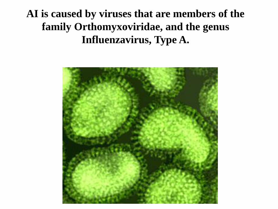
Birds may show cyanosis and edema of the skin,
particularly of the combs, wattles, and periocular areas.

edema and bleeding of the shanks and feet may also be
observed.

Post-mortem Lesions
• Lesions are variable in distribution and severity, dependinggreatly on host species, pathogenicity, and secondaryinfections.
• Birds infected with HPNAI that die peracutely may not showany gross lesions.
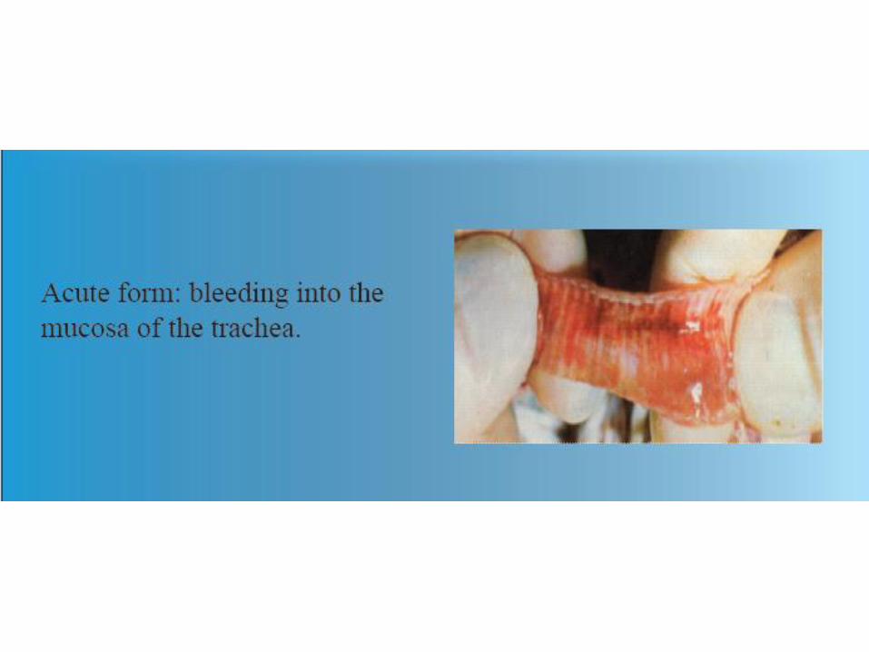
• In chickens infected with the acute to subacute form,significant gross lesions are usually observed
• lesions are more pronounced and are distributed throughout theupper respiratory tract and include exudates.
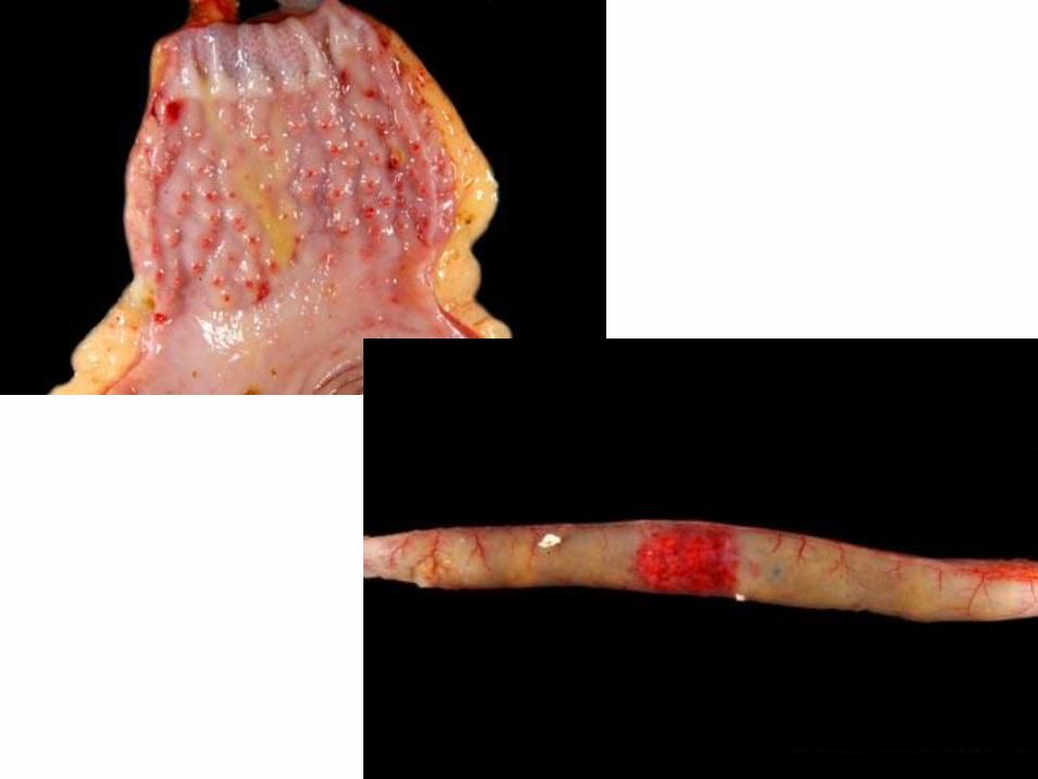
• The tracheal mucosa may be edematous with congestion and hemorrhages.
• The infraorbital sinuses may be swollen and nasal dischargemay be present.
• The ovaries may be hemorrhagic with ova involution anddegeneration.

crop


Prevention and Control
The recommended strategy for controlling AI is eradication.
• This requires 5 components including:
1) biosecurity practices
• controlling human traffic, quarantining birds before introduction, proper cleaning and disinfection of facilities, keeping healthy birds away from contact with sick birds and wild birds, and incubating eggs only from clean flocks
2) increasing host resistance through vaccination
• Inactivated vaccines have been shown to be effective but are fairly expensive
Infectious bronchitis(IB)
• Cause
• Corona-virus is the causal agent. Several different serotypes of IB virus are known to exist.
• Transmission
• The virus is transmitted from bird to bird through the airborne route. The virus can also be transmitted via the air between chicken houses and even from farm to farm.
• Species affected
• Only chickens are susceptible to IB virus.
• Clinical signs
• In young chicks IB virus infection causes a cheesy exudate in the bifurcation of the bronchi
• In older birds IB does not cause mortality.
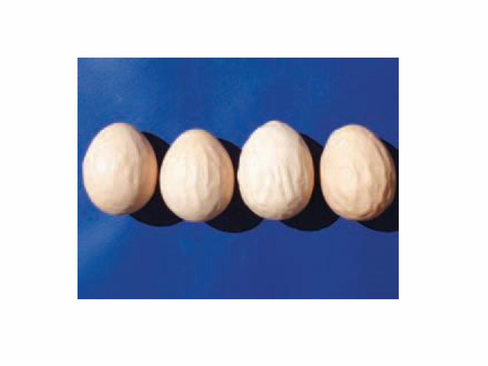
• Egg production will decrease dramatically, deformed eggs with wrinkled shells will often be laid

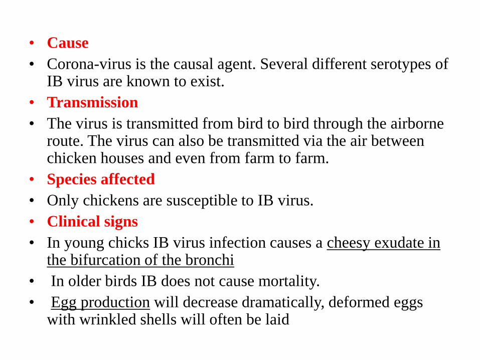
Respiratory symptoms


• Internal lesions
• Mucus and redness in tracheas, froth in airsacs
in older chickens.
• In young chicks a yellow cheesy plug at the
tracheal bifurcation is indicative of IB
infection.


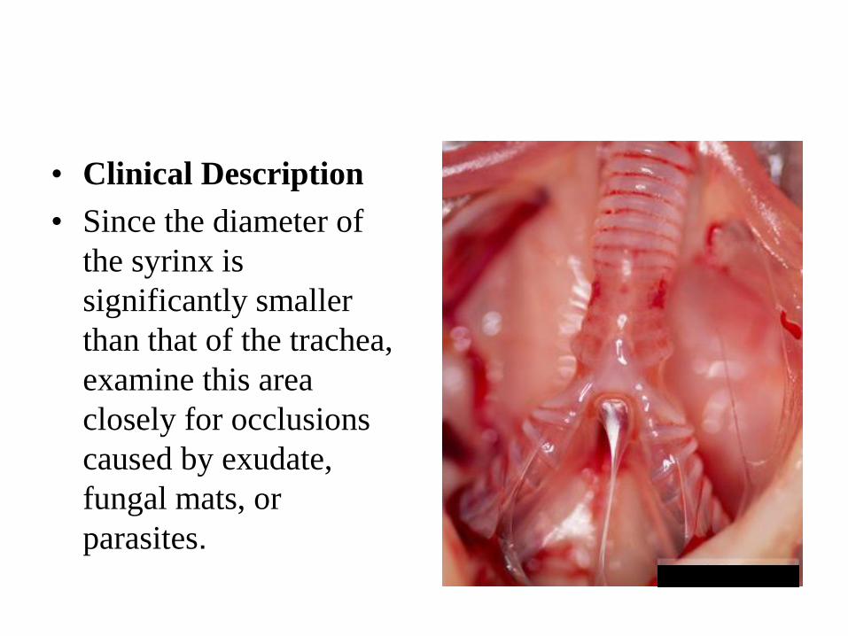
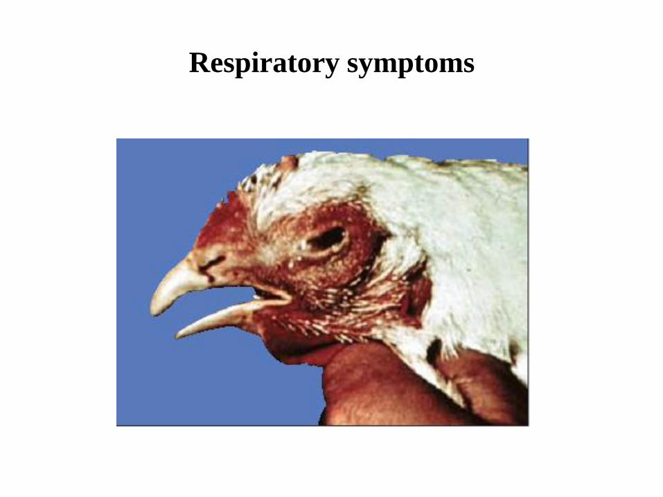
• Clinical Description
• Since the diameter of
the syrinx is
significantly smaller
than that of the trachea,
examine this area
closely for occlusions
caused by exudate,
fungal mats, or
parasites.
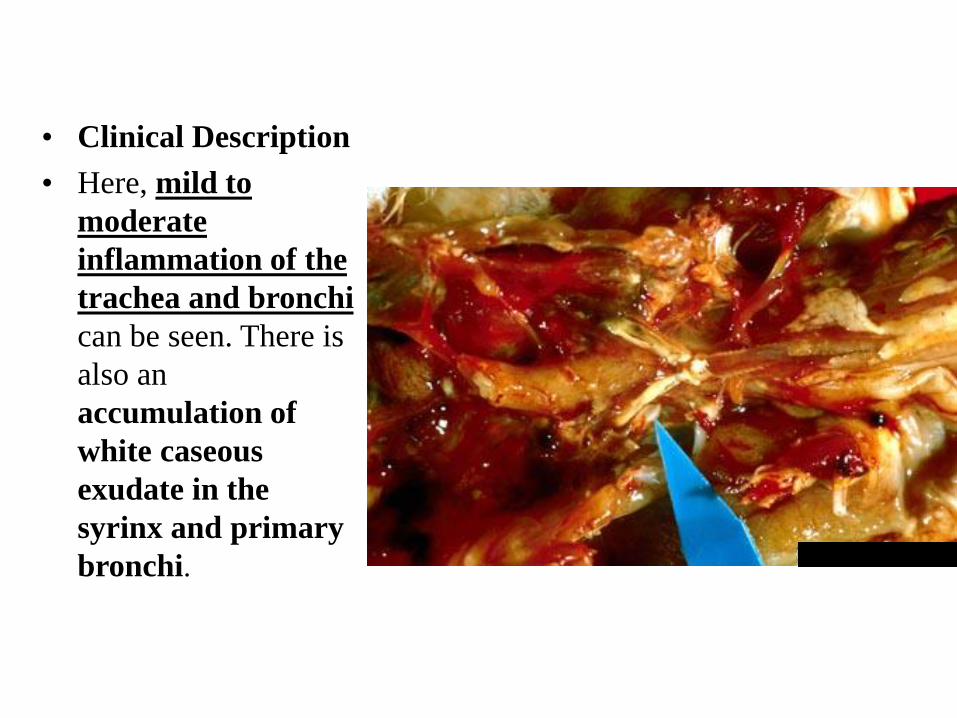
• Clinical Description
• Here, mild to
moderate
inflammation of the
trachea and bronchi
can be seen. There is
also an
accumulation of
white caseous
exudate in the
syrinx and primary
bronchi.

• Clinical Description
• This image shows plugs of
exudate in the lumen of
the trachea and bronchi.

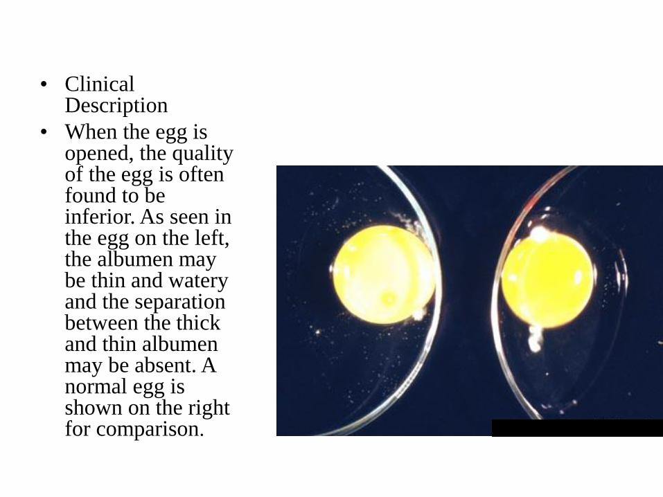
• Clinical Description
• When the egg is opened, the quality of the egg is often found to be inferior. As seen in the egg on the left, the albumen may be thin and watery and the separation between the thick and thin albumen may be absent. A normal egg is shown on the right for comparison.


• Diagnosis
• There are three main factors to be considered in order to arrive at a diagnosis:
• a. The clinical picture including post-mortem findings in the flock.
• b. Isolation of the virus in the laboratory.
• c. A rising antibody titre when the serum is tested against a known strain of bronchitis virus.
• Treatment and control
• There is no treatment for infectious bronchitis.
• Secondary bacterial infections may be prevented by, or treated with antibiotics.
• Prevention by vaccination is the best method to control IB.
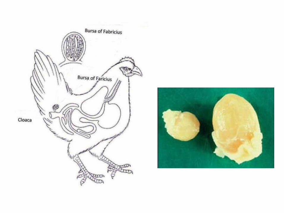
Gumboro disease
Infectious Bursal Disease
(IBD)
• Cause
• The disease is caused by a Birnavirus of serotype 1.
• Virus strains can be divided in classical and variant strains.
• The virus is very stable and is difficult to eradicate from an infected farm.
• Transmission
• IBD virus is very infectious and spreads easily from bird to bird by way of droppings.
• Infected clothing and equipment are means of transmission between farms.
• Species affected
• Chickens and turkeys appear to be natural hosts.
• Clinical signs
• Clinical IBD occurs usually between 3 and 6 weeks of age.
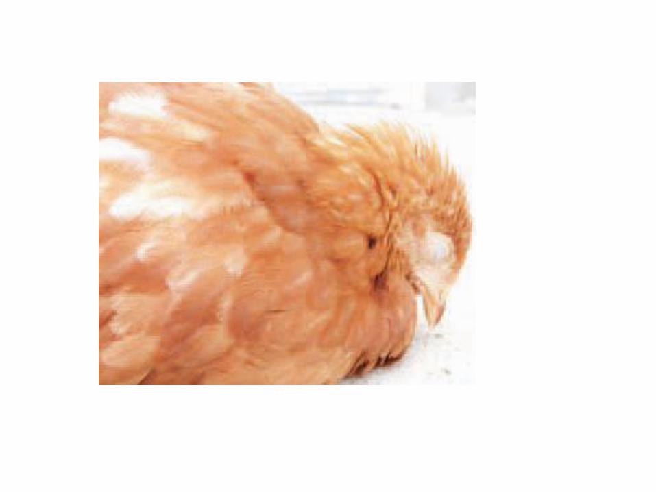
• Affected birds are listless and depressed.
• Mortality varies.
• Usually new cases of IBD have a mortality rate ofabout 5 to10% but can be as high as 60% dependingon the pathogenicity of the strain involved.
• The immunosuppressive effect of the IBD virus isnow a more economic importance in that the immunesystem of the bird is damaged.
• In broilers this form of the disease results in badperformance with lower weight gains and higher feedconversion ratios.

Affected birds are listless and depressed
• Diagnosis
• In acute cases the bursa of Fabricius is enlarged andgelatinous, sometimes even bloody.
• Muscle haemorrhages and pale kidneys can be seen.
•


• Infection by variant strains is usuallyaccompanied by a fast bursal atrophy (in 24-48hours) without the typical signs of Gumborodisease.
• Also in chronic cases the bursa is smaller thannormal (atrophy)
• Histopathological examination
• serology
• virus isolation.
• Treatment and control
• No treatment is available for IBD.
• Vaccination of parent breeders and/or young chicks is the best means of control.
• The induction of a high maternal immunity in the progeny of vaccinated breeders, together with the vaccination of the offspring is the most effective approach to successful IBD control.

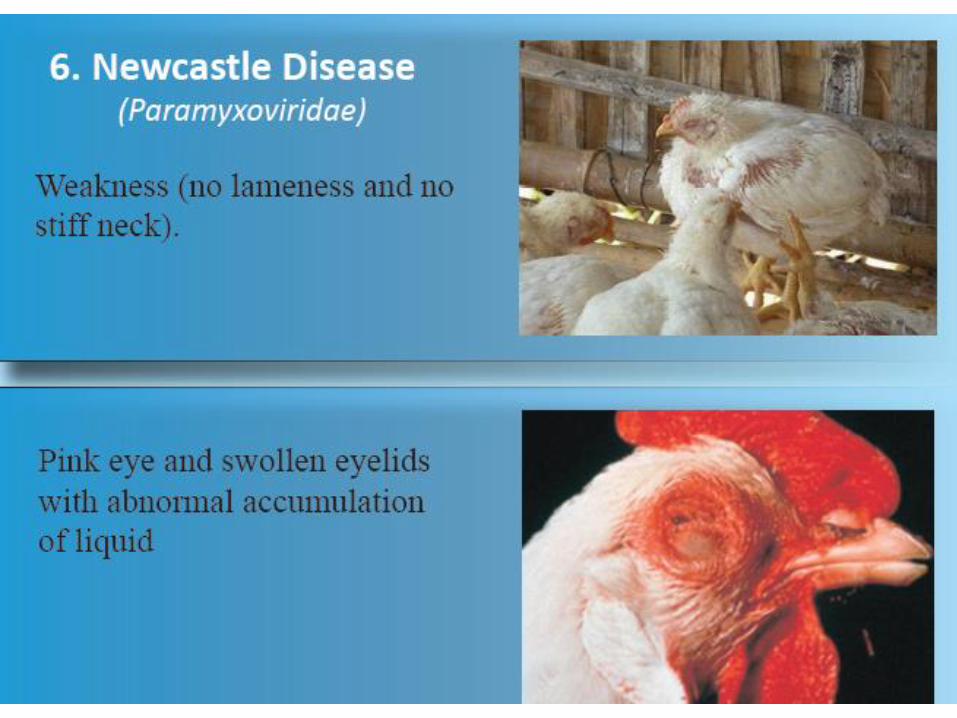
Newcastle disease

• Cause
• Newcastle disease is caused by a paramyxovirus.
• ND virus has
• mild strains (lentogenic),
• medium strains (mesogenic)
• virulent strains (velogenic).
• The strains used for live vaccines are mainly lentogenic.
• Transmission
• Newcastle disease virus is highly contagious through infected droppings and respiratory discharge between birds.
• Spread between farms is by infected equipment, trucks, personnel, wild birds or air.
• Species affected
• Chickens and turkeys. Most species of the birds

• Clinical signs
• Newcastle disease causes high mortality with depression and death in 3 to 5 days as major signs.
• Affected chickens do not always exhibit respiratory or nervous signs.
• Mesogenic strains cause typical signs of respiratory distress.
• nervous signs, such as paralysis or twisted necks (torticollis)
• Egg production will decrease 30 to 50 % or more, returning to normal levels in about 2 weeks.
• Eggs may have thin shells and eggs without shells may also be
• found.
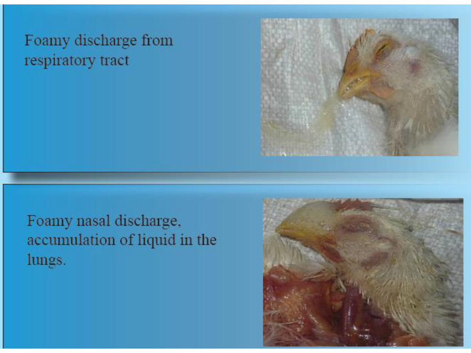


• Internal lesions
• Inflamed tracheas, pneumonia, and/or froth in
the airsacs are the main lesions.
• Haemorrhagic lesions are observed in the
proventriculus and the intestines.


• Diagnosis
• Is made by virus isolation from tracheal or cloacal swabs together with blood testing to demonstrate high antibody levels.
• Treatment and control
• There is no treatment for Newcastle disease.
• Vaccination against ND with live and/or inactivated (killed) adjuvant
• vaccines is the only reliable control method.

Bacterial disease
Chronic respiratory Disease
(CRD)

• Cause
• The underlying cause of CRD is Mycoplasma gallisepticum (Mg).
• The condition is frequently triggered by respiratory virusessuch as ND and IB and subsequently complicated by bacterialinvasion.
• The main agents involved in the infection are Mycoplasma gallisepticum and E. coli.
• Stress caused by moving the birds, by debeaking or otheroperations or other unfavorable conditions e.g. cold or badventilation, make the birds more susceptible.
• Transmission
• The main problem is that parent birds infected withMycoplasma gallisepticum can transmit the organism throughthe egg to their offspring.
• In addition, infection can occur by contact or by airborne dustor droplets.
• Species affected
• Chickens and turkeys.

• Clinical signs
• Young chickens (broiler chicks or layer pullets) will show respiratory distress
• The birds frequently show a lack of appetite, decreased weight gain and increased feed conversion ratios.
• In adult birds the most common symptoms are sneezing, coughing and general signs of respiratory congestion.
• In laying birds a drop of egg production between 20-30 % can occur.
• CRD does not normally cause an alarming number of deaths..
• Internal lesions
• A reddish inflamed trachea and/or cheesy exudate in airsacs especially in complicated cases (e.g. with secondary E. coli infections) are observed.
• In mild Mg infections the only lesion might be slight mucus in trachea and a cloudy or light froth in the airsacs.
• Diagnosis
• blood testing of chickens
• post-mortem examination
• isolating the causative Mg organism


• Differential diagnosis
• Respiratory virus infection (Newcastle disease or infectious bronchitis) with secondary infection (E. coli, etc.) can give similar lesions.
• Treatment
• Treatment of Mg-infected chickens or turkeys with suitable antibiotics or chemotherapeutics has been found to be of economic value.
• control by medication or vaccination and eradication of Mg infections
• Fertile eggs from infected birds can be treated with antibiotics such as tylosin to eliminate the Mycoplasma gallisepticum organisms.
• Methods used are the injection of fertile eggs or egg dipping. Blood serum testing of breeder chickens for Mg antibodies has become a routine to test flocks for a Mg infection.
ESCHERICHIA COLI
INFECTIONS
• Escherichia coli infections are widely distributed among poultry of all ages and categories.
• They are primarily related to poor hygienicconditions, neglected technologicalrequirements or to respiratory andimmunosuppressive diseases.
• A common sequel of navel infections is local or diffuse peritonitis .

• Omphalitis (navel infection)
• It is characterized with reddening and tissue
oedema in the umbilical region .

The delayed absorption of the yolk sac is a prerequisite for E. coli infections and peritonitis.

• Salpingitis (inflammation of the oviduct). Salpingites due to E. coli infections could be also observed in growing birds.
• The oviduct is dilated, with thinned wall and filled with caseous exudate all along its length .

• Salpingites are among the commonest causes for death in layer hens. E. coli penetrates from the cloaca via an ascendant route.
• Egg yolk peritonitis in a layer hen consequently to E. coli salpingitis. The chickens could be hatched with a latent infection, when E. coli is present in ovaries and the oviduct. In these instances, the infection could turn into an overt infection under the influence of some stress factors or lesions
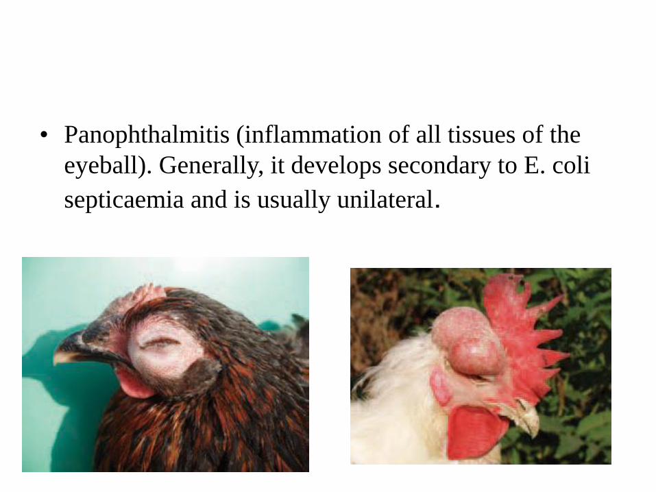
• Panophthalmitis (inflammation of all tissues of the
eyeball). Generally, it develops secondary to E. coli
septicaemia and is usually unilateral.

• E. coli septicaemia of a respiratory origin. In such cases, the respiratory mucosa damaged by infectious and non-infections agents (ND viruses including vaccinal strains, IB, mycoplasmae, high ammonia levels) is the entrance door of the E. coli infection. The lesions are principally observed in the respiratory tract (trachea, lungs and air sacs), but some adjacent serous coats (pericardium, peritoneum) are also affected and thus, the picture of a typical serofibrinous polyserositis is produced .

Pericarditis
and
perihepatitis

Related Documents