Importance of polyunsaturated acyl chains in chlorpromazine interaction with phosphatidylserines: A 13 C and 31 P solid-state NMR study B Song Chen b , Anja Underhaug Gjerde a , Holm Holmsen a , Willy Nerdal b, * a Department of Biomedicine and Molecular Biology, University of Bergen, Norway b Department of Chemistry, University of Bergen, Allegaten 41, N-5007 Bergen, Norway Received 8 April 2005; received in revised form 10 May 2005; accepted 10 May 2005 Available online 25 May 2005 Abstract The polyunsaturated fatty acid docosahexaenoic acid (DHA, c22:6, n-3) is found at a level of about 50% in the phospholipids of neuronal tissue membranes and appears to be crucial to human health. Dipalmitoyl phosphatidylcholine (DPPC, 16:0/16:0 PC), 1-palmitoyl-2-oleoyl phosphatidylserine (POPS) and the DHA containing 1-stearaoyl-2-docosahexenoyl phosphatidylserine (SDPS) were used to make DPPC (60%)/POPS (29%)/SDPS (11%) bilayers with and without 10 mol% chlorpromazine (CPZ), a cationic, amphiphilic phenothiazine. The T 1 relaxation measurements make it clear that the saturated acyl chains carbons (palmitic, stearic and most of the oleic chain) and the choline head group are not affected by CPZ addition. The observed increased signal intensity and T 1 -values of DHA indicate reduced mobility of C 4 and C 5 due to CPZ binding. 31 P NMR spectra confirm that CPZ binding to the phosphatidylserines in the bilayer enhances phospholipid head group mobility. D 2005 Elsevier B.V. All rights reserved. Keywords: 13 C NMR; 31 P NMR; DPPC/SDPS/POPS; DPPC/SDPS/POPS bilayers; Chlorpromazine HCl interaction 1. Introduction The effects of phospholipid acyl chain length and degree of unsaturation on bilayer thickness is well documented [1] and so is the effect of bilayer thickness on membrane enzyme activity [2]. The polyunsaturated fatty acid docosahexaenoic acid (DHA, c22:6, n-3) is found at a level of about 50% in the phospholipids of neuronal tissue membranes and appears to be crucial to human health [3,4]. Despite this cruciality, only sparse information has been gathered on DHA’s physical function(s) in the membrane. Findings on the conformational changes of rhodopsin (the MI-to-MII transition) suggest that phospholipid membranes with polyunsaturated acyl chains promote these conformational changes of rhodopsin [5]. DHA has been modelled by molecular mechanics methods and suggested to have a rigid and ordered structure [6–8]. Contrary to the results of these modelling studies, DHA with its long run of double-bonded carbons separated by a single methylene group has been found in a compressibility study [9] to have high flexibility and minimal sensitivity to temperature in that DHA showed to be the most easily compressed acyl chain, when compared with saturated (stearoyl) and monounsaturated (oleic) acyl chains in phospholipids with choline head group. The importance of the specific phospholipid head group is illustrated by the membrane protein topology and activity- 0301-4622/$ - see front matter D 2005 Elsevier B.V. All rights reserved. doi:10.1016/j.bpc.2005.05.002 Abbreviations: CPZ, Chlorpromazine; CSA, Chemical Shift Aniso- tropy; DMPC, Dimyristoyl phosphatidylcholine (14:0/14:0 PC); DMPE, Dimyristoyl phosphatidylethanolamine (16:0/16:0 PE); DPPC, Dipalmitoyl phosphatidylcholine(16:0/16:0 PC); HPLC, High pressure liquid chroma- tography; PA, Phosphatidic acid; PBPS, Bovine brain phosphatidylserine; POPS, 1-Palmitoyl-2-oleoyl phosphatidylserine (16:0/18:1 (n-9) PS); SDPS, 1-Stearaoyl-2-docosahexenoyl phosphatidylserine (18:0/22:6 (n-3) PS); PC, Phosphatidylcholine; PI, Phosphatidylinositol; PKC, Protein kinase C; PLA2, Phospholipase A 2 ; PS, Phosphatidylserine; Tc, Transition temperature. i This work was supported by EU BIOMED 2 grant EC BMH4-97-2609 from the European Union (EU) (no 149115/310), grants from the Norwegian Research Council (NFR) and from the Blix foundation. * Corresponding author. Tel.: +47 55 583353; fax: +47 55 589400. E-mail address: [email protected] (W. Nerdal). Biophysical Chemistry 117 (2005) 101 – 109 http://www.elsevier.com/locate/biophyschem

Welcome message from author
This document is posted to help you gain knowledge. Please leave a comment to let me know what you think about it! Share it to your friends and learn new things together.
Transcript
http://www.elsevier.com/locate/biophyschem
Biophysical Chemistry
Importance of polyunsaturated acyl chains in chlorpromazine interaction
with phosphatidylserines: A 13C and 31P solid-state NMR studyB
Song Chenb, Anja Underhaug Gjerdea, Holm Holmsena, Willy Nerdalb,*
aDepartment of Biomedicine and Molecular Biology, University of Bergen, NorwaybDepartment of Chemistry, University of Bergen, Allegaten 41, N-5007 Bergen, Norway
Received 8 April 2005; received in revised form 10 May 2005; accepted 10 May 2005
Available online 25 May 2005
Abstract
The polyunsaturated fatty acid docosahexaenoic acid (DHA, c22:6, n-3) is found at a level of about 50% in the phospholipids of neuronal
tissue membranes and appears to be crucial to human health. Dipalmitoyl phosphatidylcholine (DPPC, 16:0/16:0 PC), 1-palmitoyl-2-oleoyl
phosphatidylserine (POPS) and the DHA containing 1-stearaoyl-2-docosahexenoyl phosphatidylserine (SDPS) were used to make DPPC
(60%)/POPS (29%)/SDPS (11%) bilayers with and without 10 mol% chlorpromazine (CPZ), a cationic, amphiphilic phenothiazine. The T1
relaxation measurements make it clear that the saturated acyl chains carbons (palmitic, stearic and most of the oleic chain) and the choline
head group are not affected by CPZ addition. The observed increased signal intensity and T1-values of DHA indicate reduced mobility of C4
and C5 due to CPZ binding. 31P NMR spectra confirm that CPZ binding to the phosphatidylserines in the bilayer enhances phospholipid head
group mobility.
D 2005 Elsevier B.V. All rights reserved.
Keywords: 13C NMR; 31P NMR; DPPC/SDPS/POPS; DPPC/SDPS/POPS bilayers; Chlorpromazine HCl interaction
1. Introduction
The effects of phospholipid acyl chain length and degree
of unsaturation on bilayer thickness is well documented [1]
and so is the effect of bilayer thickness on membrane enzyme
activity [2]. The polyunsaturated fatty acid docosahexaenoic
0301-4622/$ - see front matter D 2005 Elsevier B.V. All rights reserved.
doi:10.1016/j.bpc.2005.05.002
Abbreviations: CPZ, Chlorpromazine; CSA, Chemical Shift Aniso-
tropy; DMPC, Dimyristoyl phosphatidylcholine (14:0/14:0 PC); DMPE,
Dimyristoyl phosphatidylethanolamine (16:0/16:0 PE); DPPC, Dipalmitoyl
phosphatidylcholine(16:0/16:0 PC); HPLC, High pressure liquid chroma-
tography; PA, Phosphatidic acid; PBPS, Bovine brain phosphatidylserine;
POPS, 1-Palmitoyl-2-oleoyl phosphatidylserine (16:0/18:1 (n-9) PS);
SDPS, 1-Stearaoyl-2-docosahexenoyl phosphatidylserine (18:0/22:6 (n-3)
PS); PC, Phosphatidylcholine; PI, Phosphatidylinositol; PKC, Protein
kinase C; PLA2, Phospholipase A2; PS, Phosphatidylserine; Tc, Transition
temperature.i This work was supported by EU BIOMED 2 grant EC BMH4-97-2609
from the European Union (EU) (no 149115/310), grants from the
Norwegian Research Council (NFR) and from the Blix foundation.
* Corresponding author. Tel.: +47 55 583353; fax: +47 55 589400.
E-mail address: [email protected] (W. Nerdal).
acid (DHA, c22:6, n-3) is found at a level of about 50% in the
phospholipids of neuronal tissue membranes and appears to
be crucial to human health [3,4]. Despite this cruciality, only
sparse information has been gathered on DHA’s physical
function(s) in the membrane. Findings on the conformational
changes of rhodopsin (the MI-to-MII transition) suggest that
phospholipid membranes with polyunsaturated acyl chains
promote these conformational changes of rhodopsin [5].
DHA has been modelled by molecular mechanics methods
and suggested to have a rigid and ordered structure [6–8].
Contrary to the results of these modelling studies, DHAwith
its long run of double-bonded carbons separated by a single
methylene group has been found in a compressibility study
[9] to have high flexibility and minimal sensitivity to
temperature in that DHA showed to be the most easily
compressed acyl chain, when compared with saturated
(stearoyl) and monounsaturated (oleic) acyl chains in
phospholipids with choline head group.
The importance of the specific phospholipid head group
is illustrated by the membrane protein topology and activity-
117 (2005) 101 – 109

S. Chen et al. / Biophysical Chemistry 117 (2005) 101–109102
determining properties of glycerophospholipids with anionic
head groups [10], these phospholipids alter the structure of
human recombinant prion protein associated with mem-
branes in living cells [11]. Influence of lipid composition on
membrane protein activities has recently been reviewed by
Lee [12].
The above observations indicate that the activity of
membrane-bound proteins can be influenced by the lipid
composition of the membranes. Thus, it is possible that
perturbation of lipid organization in a bilayer by amphiphilic
molecules will influence the activity of such proteins even
without direct interaction between the protein and the
amphiphile. Chlorpromazine, a cationic, amphiphilic phe-
nothiazine, has been found to interact preferentially with
bilayers containing phospholipids with a high proportion of
phosphatidylserines and highly unsaturated acyl chains [13].
Furthermore, CPZ has been found to slightly increase lipid
order when the bilayer is above the gel to liquid crystalline
phase transition temperature, Tc, and decrease lipid order
when the bilayer is below Tc [14].
Membrane perturbation with CPZ and other amphiphils
induces a host of genes in both bacteria and mammalian
cells (reviewed in [15]). It is, thus, possible that CPZ’s
reported/claimed antagonistic effect on the D2-receptor is
partially due to perturbation by CPZ of the membrane that
contains the receptor. In micromolar concentration CPZ
causes large increases in the mean molecular areas in
monolayers of acidic phospholipids, whereas no such
molecular area increase is found for the neutral glycer-
ophospholipids in monolayers [16]. Similar findings by us
[17], using magic angle spinning solid state 13C NMR on
bilayer samples with partial hydration (12 H2O per
phospholipid), showed that CPZ had low or no interaction
on the acyl packing of liposomes made of phospholipids
without a net negative head group charge and with saturated
acyl chains, such as palmitoyl (DPPC) and myristoyl
(DMPC), while it caused a large (5–15 ppm) shift to higher
ppm values of ¨30% of the acyl chain carbon resonances in
liposomes composed of pig brain PS (PBPS) and DPPC. PS
is a major anionic phospholipid in mammalian cell
membranes like peripheral and central nervous system
myelin and PBPS was subjected to CPZ interaction studies
as PBPS bilayer and in a mixture with DPPC as a DPPC (60
mol%)/PBPS (40 mol%) bilayer. This pig brain PS
contained molecular species of phospholipids with the
following acyl chains: two major molecular species 18:0–
18:1 (49%) and 18:0–22:6 (28%), and five minor molecular
species each in the 3–7% range, of which two are known
16:0–22:6 (6%) and 18:0–20:4 (3%).
Recently [13], we have studied the interaction on fully
hydrated (30 H2O per phospholipid) DPPC (60%)/PBPS
(40%) bilayers above the gel to liquid crystalline phase
transition temperature, Tc. In this recent study on a
DPPC(60%)/PBPS(40%) bilayer and with CPZ added
(DPPC(54%)/PBPS(36%)/CPZ(10%)), the Tcs were found
to be about the same, 303.5 and 305.8 K, respectively. With
this acyl chain composition of pig brain PS (18:0–18:1
(49%), 18:0–22:6 (28%), 16:0–22:6 (6%) and 18:0–20:4
(3%)), the sample composition can be outlined as a DPPC
(60%)/SOPS (20%)/SDPS (11%)/OTHER (9%). Compared
with the sample of this work DPPC (60%)/POPS (29%)/
SDPS (11%), the samples differ in the amounts of
polyunsaturated PS (11% SDPS and 9% OTHER versus
11% SDPS of this study) and monounsaturated PS (20%
SOPS versus 29% POPS of this work). (We have carried out
solid-state NMR experiments on pure SOPS and pure POPS
bilayers with CPZ added and found the CPZ interaction to
be negligible for both of these monounsaturated phospha-
tidylserines.) On the basis of the amount of unsaturated acyl
chains, it is reasonable to expect the Tcs of samples used in
the work presented here to be comparable with the Tcs of
303.5–305.8 K of the previous study.
A general feature of the phosphatidylserine 31P static
NMR spectra is a large chemical shielding anisotropy (CSA)
(the CSA is generally larger for serine than for choline and
ehanolamine head groups). The CSA appears to be
influenced by the chemical nature of the fatty acyl chains
[13]. Furthermore, the similarities of the static shielding
tensor of phosphatidylserine and -choline taken together
with the somewhat larger CSA for phosphatidylserines,
suggest that the phosphatidylserine phosphate moiety differs
conformationally or motionally from the phosphatidylcho-
line phosphate moiety [18,19]. This can be accounted for by
greater rigidity of the phosphatidylserine head group than
the phosphatidylcholine head group. This rigidity suppos-
edly results from electrostatic interactions and/or hydrogen
bonding between or within the phosphatidylserine head
groups. Thus, dilution of negatively charged PBPS with
neutral DPPC removes some of this interaction and will
allow greater freedom of motion of the phosphatidylserine
head group. The gel to liquid crystalline phase transition of
a phospholipid bilayer upon increase in temperature is
accompanied by several structural changes in the lipid
molecules. The principal change is the trans-gauche
isomerization in the saturated carbons in the acyl chains
and the average number of gauche conformers can be
related to bilayer thickness.
In our previous study [13] we deduced from our analysis
of the composition of molecular species in PBPS that it must
have been SDPS in the PBPS that caused the main, strong
interaction with CPZ since POPS and SOPS showed
negligible interaction with CPZ. In the present study, we
have investigated phospholipid acyl chain unsaturation
effect on CPZ bilayer interaction further by employing
fully hydrated (30 H2O per phospholipid) and authentic
DPPC(60%)/POPS(29%)/SDPS(11%) and DPPC(54%)/
POPS(26%)/SDPS(10%)/CPZ(10%) bilayers both below
and above the gel to liquid crystalline phase transition
temperature, Tc. The biologically abundant phosphatidyl-
serines, POPS and SDPS, where the POPS species has its
unsaturated sn-2 acyl chain bond at C9\C10, and the DHA
containing SDPS species, with the 6 unsaturated acyl chain
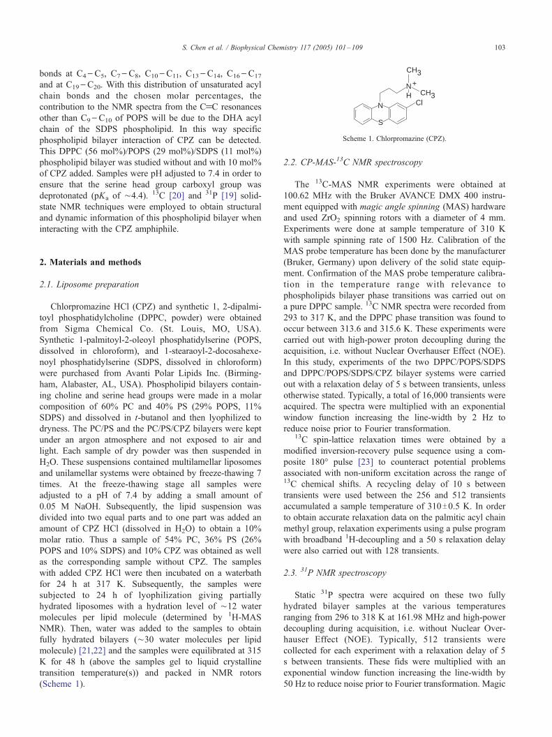
N
S
NH CH3
CH3
Cl
+
Scheme 1. Chlorpromazine (CPZ).
S. Chen et al. / Biophysical Chemistry 117 (2005) 101–109 103
bonds at C4\C5, C7\C8, C10\C11, C13\C14, C16\C17
and at C19\C20. With this distribution of unsaturated acyl
chain bonds and the chosen molar percentages, the
contribution to the NMR spectra from the CfC resonances
other than C9\C10 of POPS will be due to the DHA acyl
chain of the SDPS phospholipid. In this way specific
phospholipid bilayer interaction of CPZ can be detected.
This DPPC (56 mol%)/POPS (29 mol%)/SDPS (11 mol%)
phospholipid bilayer was studied without and with 10 mol%
of CPZ added. Samples were pH adjusted to 7.4 in order to
ensure that the serine head group carboxyl group was
deprotonated (pKa of ¨4.4). 13C [20] and 31P [19] solid-
state NMR techniques were employed to obtain structural
and dynamic information of this phospholipid bilayer when
interacting with the CPZ amphiphile.
2. Materials and methods
2.1. Liposome preparation
Chlorpromazine HCl (CPZ) and synthetic 1, 2-dipalmi-
toyl phosphatidylcholine (DPPC, powder) were obtained
from Sigma Chemical Co. (St. Louis, MO, USA).
Synthetic 1-palmitoyl-2-oleoyl phosphatidylserine (POPS,
dissolved in chloroform), and 1-stearaoyl-2-docosahexe-
noyl phosphatidylserine (SDPS, dissolved in chloroform)
were purchased from Avanti Polar Lipids Inc. (Birming-
ham, Alabaster, AL, USA). Phospholipid bilayers contain-
ing choline and serine head groups were made in a molar
composition of 60% PC and 40% PS (29% POPS, 11%
SDPS) and dissolved in t-butanol and then lyophilized to
dryness. The PC/PS and the PC/PS/CPZ bilayers were kept
under an argon atmosphere and not exposed to air and
light. Each sample of dry powder was then suspended in
H2O. These suspensions contained multilamellar liposomes
and unilamellar systems were obtained by freeze-thawing 7
times. At the freeze-thawing stage all samples were
adjusted to a pH of 7.4 by adding a small amount of
0.05 M NaOH. Subsequently, the lipid suspension was
divided into two equal parts and to one part was added an
amount of CPZ HCl (dissolved in H2O) to obtain a 10%
molar ratio. Thus a sample of 54% PC, 36% PS (26%
POPS and 10% SDPS) and 10% CPZ was obtained as well
as the corresponding sample without CPZ. The samples
with added CPZ HCl were then incubated on a waterbath
for 24 h at 317 K. Subsequently, the samples were
subjected to 24 h of lyophilization giving partially
hydrated liposomes with a hydration level of ¨12 water
molecules per lipid molecule (determined by 1H-MAS
NMR). Then, water was added to the samples to obtain
fully hydrated bilayers (¨30 water molecules per lipid
molecule) [21,22] and the samples were equilibrated at 315
K for 48 h (above the samples gel to liquid crystalline
transition temperature(s)) and packed in NMR rotors
(Scheme 1).
2.2. CP-MAS-13C NMR spectroscopy
The 13C-MAS NMR experiments were obtained at
100.62 MHz with the Bruker AVANCE DMX 400 instru-
ment equipped with magic angle spinning (MAS) hardware
and used ZrO2 spinning rotors with a diameter of 4 mm.
Experiments were done at sample temperature of 310 K
with sample spinning rate of 1500 Hz. Calibration of the
MAS probe temperature has been done by the manufacturer
(Bruker, Germany) upon delivery of the solid state equip-
ment. Confirmation of the MAS probe temperature calibra-
tion in the temperature range with relevance to
phospholipids bilayer phase transitions was carried out on
a pure DPPC sample. 13C NMR spectra were recorded from
293 to 317 K, and the DPPC phase transition was found to
occur between 313.6 and 315.6 K. These experiments were
carried out with high-power proton decoupling during the
acquisition, i.e. without Nuclear Overhauser Effect (NOE).
In this study, experiments of the two DPPC/POPS/SDPS
and DPPC/POPS/SDPS/CPZ bilayer systems were carried
out with a relaxation delay of 5 s between transients, unless
otherwise stated. Typically, a total of 16,000 transients were
acquired. The spectra were multiplied with an exponential
window function increasing the line-width by 2 Hz to
reduce noise prior to Fourier transformation.13C spin-lattice relaxation times were obtained by a
modified inversion-recovery pulse sequence using a com-
posite 180- pulse [23] to counteract potential problems
associated with non-uniform excitation across the range of13C chemical shifts. A recycling delay of 10 s between
transients were used between the 256 and 512 transients
accumulated a sample temperature of 310T0.5 K. In order
to obtain accurate relaxation data on the palmitic acyl chain
methyl group, relaxation experiments using a pulse program
with broadband 1H-decoupling and a 50 s relaxation delay
were also carried out with 128 transients.
2.3. 31P NMR spectroscopy
Static 31P spectra were acquired on these two fully
hydrated bilayer samples at the various temperatures
ranging from 296 to 318 K at 161.98 MHz and high-power
decoupling during acquisition, i.e. without Nuclear Over-
hauser Effect (NOE). Typically, 512 transients were
collected for each experiment with a relaxation delay of 5
s between transients. These fids were multiplied with an
exponential window function increasing the line-width by
50 Hz to reduce noise prior to Fourier transformation. Magic

Serine : CH - CH - NH|CO
2
2
3_
D E+
O || CH - O - C - R2
A CH - O - C - R1 || O
2
O
B
S. Chen et al. / Biophysical Chemistry 117 (2005) 101–109104
angle spinning 31P experiments (T1 measurements) were
carried out with a rotor spinning speed of 2 KHz. These fids
of 64 transients were Fourier transformed without apodiza-
tion in order to keep spectral resolution. 31P relaxation data
were obtained with 1H-cross-polarization at temperatures
from 296 K to 318 K, and with rotor spinning speed of 2
kHz. Typically 512 transients were accumulated.
B
HDCA
G E
I
Choline : CH - CH - N ( CH )2 3
G H+
2 3CH - O - P - O - Serine/Choline2
||
|O
C
I
3. Results
The 13C magic angle spinning (MAS) spectra of bilayer
samples DPPC/POPS/SDPS and DPPC/POPS/SDPS/CPZ
in the liquid crystalline phase were recorded at a temper-
ature of 310 K and are presented as spectral regions in Figs.
1–4 where the top spectrum shows the phospholipid sample
with 10% CPZ and the bottom spectrum the corresponding
sample without CPZ. Fig. 1 shows the DPPC, POPS and
SDPS acyl chain sp3 carbon resonances in the 12–38 ppm
region. The two spectra (Fig. 1, top and bottom) are
dominated by the palmitic (DPPC and POPS) as well as the
oleic (POPS) molecular species. The molar composition of
the samples cause the palmitic (16:0) acyl chain resonances
to give ¨75% of the peak intensities in this spectral region,
whereas the contribution from the SDPS species in this
35 15202530ppm
2
14
4-7,12,13
8 3
1615
11*
Fig. 1. Methylene and methyl carbon resonance region (12–38 ppm) of
samples DPPC(60%)/POPS(29%)/SDPS(11%) (bottom spectrum) and
DPPC(54%)/POPS(26%)/SDPS(10%)/CPZ(10%) (top spectrum). Spectra
are acquired at 310 K (samples are in liquid crystalline phase). The samples
molar composition cause the palmitic (16:0) acyl chain resonances to
dominate (¨ 75% of the peak intensities) in this spectral region. Thus, only
the palmitic carbon resonances are assigned in the two spectra. An asterisk
‘‘*’’ indicate a possible DHA resonance. See the text for details.
5570 65 60
ppm
Fig. 2. Top: Structural formula of the glycerol moiety and the two
phospholipids head groups, serine and choline, with the corresponding
assignment letters used in the spectra. Bottom: Phospholipid head group
and glycerol carbon resonance region (52 – 73 ppm) of samples
DPPC(60%)/POPS(29%)/SDPS(11%) (bottom spectrum) and
DPPC(54%)/POPS(26%)/SDPS(10%)/CPZ(10%) (top spectrum). Spectra
are acquired at 310 K (samples are in liquid crystalline phase). The molar
composition makes a PC/PS ratio of 1.5. See the text for details.
spectral region is 10% (5% from each of the 18:0 and 22:6
acyl chains). Thus, only the palmitic carbon resonances are
assigned, see Fig. 1 (some of these peaks contain
contribution from carbon resonances of other acyl chains
than palmitoyl chains). The phospholipid choline and serine
head group carbon resonances as well as the glycerol moiety
resonances appear in the 52–73 ppm spectral region—see
Fig. 2. Of these resonances, only the choline head group
resonances come from a single molecular species, the DPPC
molecule. The serine resonances come from two PS species,
POPS and SDPS, and the molar composition gives a PC/PS
peak ratio of 1.5. The three glycerol resonances will be
composed of the three phospholipid species in the two
samples, DPPC, POPS and SDPS.
From the T1 data presented in Table 1, one finds that the
carbon T1 values of the choline head group are not affected

Table 113C spin-lattice relaxation times T1 (s) at 310 K DPPC/POPS/SDPS and
DPPC/POPS/SDPS/CPZ bilayers
Carbon DPPC/POPS/SDPS DPPC/POPS/SDPS/CPZ
CfO 2.04 2.15
CO2� 1.51 0.61
POPS CfC:C9 (Ca) 0.75 1.83
C10 (Ba) 1.32 1.03
DHA CfC:(Ea) 0.34 1.16
(Fa) 1.04 0.99
(Ga) 0.75 2.67
Glycerol carbon:
sn-1 0.12 0.31
sn-2 0.28 0.33
sn-3 0.13 0.15
Serine carbon:
a 0.27 0.07
h 1.44 0.77
Choline carbon:
a 0.34 0.30
h 0.26 0.22
CH3 0.32 0.29
Palmitic carbon:
2 0.55 0.36
3 0.68 0.52
4–14 0.72 0.60
15 3.51 2.12
16 5.03 5.33
Oleic carbon:
8, 11 0.55 0.61
a Peaks labeled in Fig. 3.
130 125135ppm
* A
B
C
DEFG
H *
Fig. 3. Double bonded acyl chain carbon resonance region (125–135 ppm)
of samples DPPC(60%)/POPS(29%)/SDPS(11%) (bottom spectrum) and
DPPC(54%)/POPS(26%)/SDPS(10%)/CPZ(10%) (top spectrum). Spectra
are acquired at 310 K (samples are in liquid crystalline phase). The molar
composition of the samples makes the oleic(18:1)/DHA(22:6) ratio of 2.5.
This causes the total oleic(CfC)/DHA(CfC) peak ratio to be 0.4. See the
text for details.
S. Chen et al. / Biophysical Chemistry 117 (2005) 101–109 105
by the addition of CPZ, whereas the serine head group T1
values show a reduction in presence of CPZ. The glycerol
carbon T1 values, on the other hand, demonstrate a diverse
effect of CPZ. The sn-1 glycerol carbon display an increased
T1 value due to CPZ in contrast to both the sn-2 and sn-3
glycerol carbons (where the POPS and SDPS unsaturated
acyl chain and the phosphate and head group are attached,
respectively) that have T1 values unaffected by CPZ.
Fig. 3 shows the 125–135 ppm region where the CfCresonances of the acyl chains of samples DPPC/POPS/
SDPS and DPPC/POPS/SDPS/CPZ are found. The molar
composition of the samples makes the oleic(18:1)/
DHA(22:6) acyl chain ratio 2.5. The oleic(18:1) acyl chain
of POPS double bond (at C9\C10) and the six double bonds
of DHA of SDPS (double bonds at C4\C5, C7\C8,
C10\C11, C13\C14, C16\C17,C19\C20) makes the car-
bon–carbon double bond ratio between POPS and SDPS to
be 1/6. Consequently, the observed sp2 carbon resonances in
the 13C NMR spectra can be expected to be close to the
described 1/6 ratio multiplied by the species percentages of
the samples. Thus, samples with and without CPZ has a
POPS/SDPS acyl chain CfC ratio of (0.29�1 double
bond)/(0.11�6 double bonds) or approximately 0.4.
Comparison of the CfC resonances with/without CPZ
(see Fig. 3) shows a pronounced intensity change of some of
these, the peaks at 127–129 ppm, upon CPZ interaction.
The crowded spectral region displayed in Fig. 3 pose an
obstacle to a complete resonance assignment. However, in a
recent solid-state NMR where 1H–13C two-dimensional
cross-polarization experiments were employed [24], the
investigators managed to firmly assign DHA’s C19 and C20
to 126.8 and 131.3 ppm, respectively. Thus, peak A is
assigned to resonance C20 and peak H to resonance C19—
see Fig. 3. The remaining CfC resonances of DHA
(C4\C5, C7\C8, C10\C11, C13\C14, C16\C17) are
located between 127.4 and 128.4 ppm and could not be
individually assigned. In an early study on CfC resonance
assignment and estimation of chemical shifts Gunstone et al.
[25] showed that in monoenoic acyl chains, like the oleic
chain of POPS, the C10 resonance would come at a higher
chemical shift than the C9, they found 130.02 and 129.78
ppm, respectively. Based on our own previous work, on the
signal intensities of these resonances (Fig. 3) and on POPS
and the described higher chemical shift of C10 of the
C9fC10 pair, peaks B and C in Fig. 3 can be assigned to the
oleic acyl chain of POPS where resonance B corresponds to
C10 and resonance C to C9 of POPS. These two CfCresonances from the middle of POPS’s acyl chain, display
almost no changes in intensity and T1 values (Table 1) when
CPZ is added (T1 values for peaks B, C, E, F and G in Fig. 3
could be determined).
As evident in Fig. 3 there is no intensity change of
DHA’s resonances C19 and C20 upon addition of CPZ (peaks
A and H, respectively). Furthermore, peaks E and G (Fig. 3
and Table 1) display a marked increase in T1 value when
CPZ is present (peak F has approximately similar T1 values
without and with CPZ). Thus, the part of SDPS’s DHA acyl
chain that are affected by the presence of CPZ is the part
close to the polar region of the bilayer, as demonstrated by
the intensity and T1 value increase of these resonances.
175 170ppm
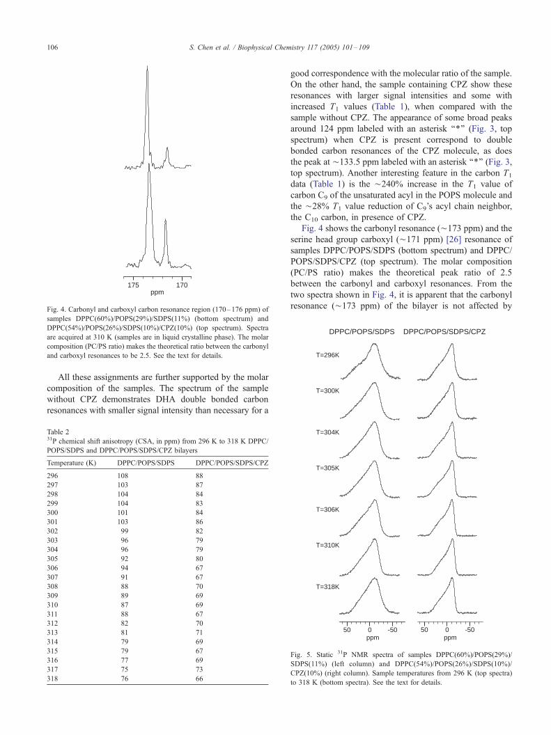
Fig. 4. Carbonyl and carboxyl carbon resonance region (170–176 ppm) of
samples DPPC(60%)/POPS(29%)/SDPS(11%) (bottom spectrum) and
DPPC(54%)/POPS(26%)/SDPS(10%)/CPZ(10%) (top spectrum). Spectra
are acquired at 310 K (samples are in liquid crystalline phase). The molar
composition (PC/PS ratio) makes the theoretical ratio between the carbonyl
and carboxyl resonances to be 2.5. See the text for details.
T=300K
T=296K
DPPC/POPS/SDPS/CPZDPPC/POPS/SDPS
S. Chen et al. / Biophysical Chemistry 117 (2005) 101–109106
All these assignments are further supported by the molar
composition of the samples. The spectrum of the sample
without CPZ demonstrates DHA double bonded carbon
resonances with smaller signal intensity than necessary for a
Table 231P chemical shift anisotropy (CSA, in ppm) from 296 K to 318 K DPPC/
POPS/SDPS and DPPC/POPS/SDPS/CPZ bilayers
Temperature (K) DPPC/POPS/SDPS DPPC/POPS/SDPS/CPZ
296 108 88
297 103 87
298 104 84
299 104 83
300 101 84
301 103 86
302 99 82
303 96 79
304 96 79
305 92 80
306 94 67
307 91 67
308 88 70
309 89 69
310 87 69
311 88 67
312 82 70
313 81 71
314 79 69
315 79 67
316 77 69
317 75 73
318 76 66
good correspondence with the molecular ratio of the sample.
On the other hand, the sample containing CPZ show these
resonances with larger signal intensities and some with
increased T1 values (Table 1), when compared with the
sample without CPZ. The appearance of some broad peaks
around 124 ppm labeled with an asterisk ‘‘*’’ (Fig. 3, top
spectrum) when CPZ is present correspond to double
bonded carbon resonances of the CPZ molecule, as does
the peak at ¨133.5 ppm labeled with an asterisk ‘‘*’’ (Fig. 3,
top spectrum). Another interesting feature in the carbon T1
data (Table 1) is the ¨240% increase in the T1 value of
carbon C9 of the unsaturated acyl in the POPS molecule and
the ¨28% T1 value reduction of C9’s acyl chain neighbor,
the C10 carbon, in presence of CPZ.
Fig. 4 shows the carbonyl resonance (¨173 ppm) and the
serine head group carboxyl (¨171 ppm) [26] resonance of
samples DPPC/POPS/SDPS (bottom spectrum) and DPPC/
POPS/SDPS/CPZ (top spectrum). The molar composition
(PC/PS ratio) makes the theoretical peak ratio of 2.5
between the carbonyl and carboxyl resonances. From the
two spectra shown in Fig. 4, it is apparent that the carbonyl
resonance (¨173 ppm) of the bilayer is not affected by
T=318K
T=310K
T=306K
T=305K
T=304K
ppm50 0 -50
ppm50 0 -50
Fig. 5. Static 31P NMR spectra of samples DPPC(60%)/POPS(29%)/
SDPS(11%) (left column) and DPPC(54%)/POPS(26%)/SDPS(10%)/
CPZ(10%) (right column). Sample temperatures from 296 K (top spectra)
to 318 K (bottom spectra). See the text for details.

Table 331P T1 values (s) from 296 K to 318 K DPPC/POPS/SDPS and DPPC/
POPS/SDPS/CPZ bilayers
Temperature (K) DPPC/POPS/SDPS DPPC/POPS/SDPS/CPZ
PC PS PC PS CPZ–PS
296 0.57 0.53 0.77 0.64 0.60
298 0.54 0.52 0.65 0.64 0.60
300 0.57 0.54 0.64 0.58 0.62
302 0.51 0.50 0.57 0.51 0.51
304 0.55 0.51 0.61 0.52 0.49
305 0.51 0.49 0.52 0.50 0.51
306 0.54 0.49 0.51 0.53 0.53
308 0.53 0.50 0.52 0.52 0.49
310 0.57 0.47 0.49 0.49 0.42
312 0.55 0.51 0.51 0.48 0.42
314 0.55 0.51 0.49 0.47 0.41
316 0.57 0.57 0.46 0.46 0.45
318 0.58 0.52 0.50 0.48 0.46
T=296K
T=298K
T=302K
T=310K
T=316K
T=306K
PS
PC
CPZ/PS
DPPC/POPS/SDPS DPPC/POPS/SDPS/CPZ
S. Chen et al. / Biophysical Chemistry 117 (2005) 101–109 107
addition of CPZ. A corresponding comparison of the serine
head group carboxyl resonance (¨ 171 ppm), on the other
hand, makes it evident that the 10% CPZ reduces the
carboxyl resonance intensity by about 2/3 and the T1 (Table
1) value by 40% (from 1.15 to 0.61 s). The corresponding
T1 values for the carbonyl resonance is about unchanged in
presence of CPZ.
In general, the 31P CSA data presented in Table 2 and
Fig. 5 show that the sample without CPZ has a higher CSA
than when CPZ is added over the whole temperature range
measured (296–310 K). The CSA of the sample without
CPZ (the DPPC/POPS/SDPS sample) displays a fairly
steady decrease in CSA value as temperature increases. In
addition to a general decrease in CSA value upon temper-
ature increase, the CPZ containing sample (the DPPC/
POPS/SDPS/CPZ sample) displays a sudden drop in CSA
of 13 ppm from 305 to 306 K. Thus, the CPZ containing
sample displays this sudden reduction in CSA at a sample
temperature of about 305.5 K, in correspondence with the
main melting (transition) temperature displayed by this kind
of phospholipid sample. The 31P T1 values are measured at
the central band of the MAS spectra, and presented in Table
3. In Fig. 6 the three central band peaks are displayed at
several of the temperatures investigated. They can be
assigned [13,27] to the three molecular species: PC, PS
and CPZ–PS complex. Both the PC and the PS species
show similar T1 values with and without CPZ and the CPZ–
PS complex shows a T1 similar to the PC and the PS
species—see Table 3.
4 2 0 -4-2ppm
4 2 0 -4-2ppm
Fig. 6. 31P MAS spectra of samples DPPC(60%)/POPS(29%)/SDPS(11%)
(right column) and DPPC(54%)/POPS(26%)/SDPS(10%)/CPZ(10%) (left
column) between sample temperatures 296 and 318 K. Note sudden
decrease in CSA of CPZ containing sample at 305 K. See the text for
details.
4. Discussion
The observed intensity decrease of the glycerol carbon
resonances of the DPPC/SDPS/CPZ sample (Fig. 2) when
compared with the DPPC/SDPS sample is most pronounced
for the sn-3 carbon, i.e. the glycerol carbon where the
phosphorus and head group are attached. A similar signal
intensity decrease/line broadening is observed for the serine
carboxyl resonance of the DPPC/SDPS/CPZ sample when
compared with the DPPC/SDPS sample. An explanation for
these observations can be found in the possibility of an
altered transverse relaxation of dipolar coupled spins under
radiofrequency irradiation (decoupling) [28]. In such a case
destructive interference effects cause line broadening due to
(molecular) motion interfering with the coherent modulation
from radiofrequency decoupling. Even carbons without
directly attached protons (such as carbonyl and carboxyl
carbons) can to some extent experience these effects when
coupled to other nearby protons. Furthermore, dipolar
interactions are expected to be weak for nonprotonated sp2
carbons and the main line broadening mechanism will be the
chemical shift anisotropy (CSA). (Protonated sp2 carbons of
the acyl chains’ olefinic double bonds will experience both
the described line broadening mechanisms [28].)
With the possibility of such effects (as described above)
complicating the spectral interpretations the 13C T1 data
obtained on the DDPC/POPS/SDPS and DDPC/POPS/

Scheme 2. Molecular model of chlorpromazine (CPZ) interaction with a 1-
stearoyl-2-docosahexanoylserine (SDPS) molecule. The CPZ molecule
(right) is positioned with its positive charge on the nitrogen atom (labelled
N) on the end of CPS’s acyl chain. This positive charge is in the vicinity of
the phosphate’s (labelled P) negative charge in the SDPS molecule (left).
Both acyl chains of the SDPS molecule have sp3 carbon dihedral angles of
60- (liquid crystalline state). The molecular model suggests that the carbons
C4 and C5 of DHA (sn-2 position) will be affected by an interdigitating
CPZ molecule.
S. Chen et al. / Biophysical Chemistry 117 (2005) 101–109108
SDPS/CPZ samples (of this work) are of great value. This is
even more so due to the simpler molecular species makeup
of the DDPC/POPS/SDPS sample of this work when
compared with the higher molecular species complexity of
our previous work [13], where we employed pig brain PS
(PBPS). For example, the CfC region of the 13C spectra
displayed in Fig. 3, the C9 and C10 resonances of POPS
could be assigned, so that the remaining CfC resonances
are known to belong to SDPS’s sn-2 attached DHA acyl
chain. Of these latter CfC resonances, the C19 and C20 of
DHA could be firmly assigned, and all the remaining
unassigned CfC resonances are then known to belong to the
DHA acyl chain, namely the C4, C5, C7, C8, C10, C11, C13,
C14, C16 and C17 carbon resonances.
Binding of CPZ to phospholipids can be followed in the13C spectra where serine head group carbon resonances show
increased intensity when CPZ is added to the DPPC/POPS/
SDPS bilayer. This result is contrary to the results found in
our previous work [13] on a PC/PS/CPZ sample where the
PS component is an extract from pig brain and composed as
follows: The PS composition of PBPS used has been
determined to mainly contain these components: 18:0–
18:1 (49%), 18:0–22:6 (28%), 16:0–22:6 (6%) and 18:0–
20:4 (3%). Thus, this can be described as a DPPC(60%)/
SOPS(20%)/SDPS(11%)/OTHER (9%) sample. Effects of
an altered transverse relaxation of dipolar coupled spins
under radiofrequency irradiation (decoupling) has been
described earlier [28] and the possibility of such effects
producing potentially confusing changes in signal intensities
when CPZ is present, make the 13C T1 measurements on both
DPPC/POPS/SDPS and DPPC/POPS/SDPS/CPZ samples
important. (Unfortunately, we did not carry out 13C T1
experiments on the pig brain PS samples (PBPS) of our
previous work, this kind of sample has a higher molecular
species complexity than the samples of this work.)
In a previous study [17] addition of CPZ to partially
hydrated DPPC/PBPS bilayer (¨ 12 H2O/phospholipid) ¨
30% of the main acyl chain carbon resonances in the 13C
NMR spectra were shifted down field by 5–15 ppm,
demonstrating CPZ interdigitation among the phospholipid
acyl chains. The fully hydrated DPPC/POPS/SDPS bilayer
of this study showed no such down field shift of acyl chain
resonances in the 13C NMR spectra when CPZ was present.
The lower mobility of the phospholipids in partially hydrated
bilayers when compared with the fully hydrated bilayers of
this study is evident from the broader line shapes in the 13C
NMR spectra of the partially hydrated bilayers [17]. Thus, a
less dense molecular packing of the phospholipids in fully
hydrated bilayers would presumably not make interdigitated
CPZ molecules come in close enough contact with acyl chain
carbons to perturb the p-orbitals of these carbons, and
consequently, a 5–15 ppm shift to higher ppm values of
these acyl chain carbon resonances is not observed in the
fully hydrated bilayers of this study.
Phospholipid head group (and phosphate) motion and an
altered motion caused by an interacting amphiphile like
CPZ in a bilayer will give static 31P NMR spectra that differ
in chemical shift anisotropy (CSA). In the static 31P NMR
spectra of DPPC/POPS/SDPS/and DPPC/POPS/SDPS/CPZ
(Fig. 5 and Table 2), the former demonstrate a CSA that is
10–17 ppm larger than the latter over a quite large
temperature range covering the gel to liquid crystalline
phase transition temperature. Thus, the presence of CPZ
causes an enhancement of the phospholipid head group
mobility. A separate (new) 31P chemical shift for the CPZ-
phosphate is observed when CPZ binds to the phosphate of
phosphatidylserine bilayers as demonstrated in the 31P NMR
spectra of the DPPC/POPS/SDPS and DPPC/POPS/SDPS/
CPZ bilayers. In the previous study of 60%/40% DPPC/
PBPS bilayer the bulky choline head groups imposes
conformational restrictions [13,29] on the CPZ-phosphate
complex and also promote CPZ-carboxyl binding which
was not observed for the ‘‘all serine’’ head group samples
and therefore seem less favoured.
The T1 relaxation measurements make it clear that the
unsaturated acyl chain carbons (palmitic, stearic and most of
the oleic chain) do not change in mobilities upon CPZ
addition (these carbons have similar T1 values (Table 1)
without and with CPZ in the bilayer). The unsaturated
carbons of the DHA acyl chain, on the other hand, display a
2–3 times increase in T1 value with CPZ present, i.e. these
unsaturated carbons experience a decreased mobility when
CPZ is present in the bilayer. The choline head group carbon
resonances, two of the glycerol carbons (the sn-2 and sn-3
carbons) and the carbonyl resonance display no change in
T1 upon CPZ addition. The serine head group carbon
resonances (the Ca, Ch and CO2�) display a 2–3 times
reduction in the T1 value upon CPZ addition, possibly due
to an increased mobility of these carbons (the phospholipids
are in the slow motion regime at the relaxation measurement
temperature). The 31P relaxation measurements show that all
three head group components (PC, PS and CPZ–PS) do not

S. Chen et al. / Biophysical Chemistry 117 (2005) 101–109 109
vary by any significant amount as function of sample
temperature, only the CPZ–PS component is to some
degree temperature sensitive below the main phase tran-
sition temperature. The PC and the PS components display
very similar 31P relaxation in both bilayer samples, i.e. with
and without CPZ present.
In addition to the importance of the phospholipid head
group, also the degree of phospholipid acyl chain
unsaturation will determine [30] part of the CPZ inter-
action with the bilayer. The observed increased signal
intensity of CfC SDPS’s DHA acyl chain carbon
resonances and an increase in the corresponding T1-values
for two (out of three that could be measured) of the CfCpeaks where the C4 and C5 resonances reside and, thus, the
reduced mobility of C4 and C5 appear to be due to CPZ
binding. A molecular model of the CPZ interaction with a
1-stearoyl-2-docosahexanoylserine (SDPS) molecule gen-
erated by the Titan software (Wavefunction, Irvine, CA) is
presented in Scheme 2. In this model a CPZ is located
with its positive charge (acyl chain nitrogen) in the vicinity
of a SDPS’s phosphate group negative charge. SDPS’s acyl
chains have the sp3 carbon dihedral angles of 60- (liquid
crystalline state). Even though the actual conformation(s)
of the DHA’s acyl chain (in the sn-2 position) may differ
somewhat from the conformation displayed in Scheme 2,
the model suggests that CPZ interdigitated in such a
bilayer will have an effect on both carbons C4 and C5 of
the DHA.
References
[1] A.M. Stinson, R.D. Wiegand, R.E. Anderson, Fatty acid and molecular
species compositions of phospholipids and diacylglycerols, Exp. Eye
Res. 52 (1991) 213–218.
[2] R.B. Genniss, J.L. Strominger, Activation of C55-isoprenoid alcohol
phosphokinase from Staphylococcus aureus: I. Activation by phospho-
lipids and fatty acids, J. Biol. Chem. 251 (1976) 1264–1282.
[3] E.E. Birch, D.G. Birch, D.R. Hoffman, R. Uauy, Dietary essential fatty
acid supply and visual acuity development, Invest. Ophthamol. Vis.
Sci. 33 (1992) 3242–3253.
[4] N. Salem Jr., Omega-3 fatty acids: molecular and biochemical aspects,
in: G.A. Spiller , J. Scala (Eds.), New Protective Roles for Selected
Nutrients, Alan R. Liss, New York, 1989, pp. 109–228.
[5] B.J. Litman, D.C. Mitchell, A role for phospholipid polyunsaturation in
modulating protein function, Lipids 31 (1996) S193–S197 (Suppl.).
[6] K.R. Applegate, J.A. Glomset, Computer-based modeling of the
conformation and packing properties of docosahexaenoic acid, J. Lipid
Res. 27 (1986) 658–680.
[7] K.R. Applegate, J.A. Glomset, Effect of acyl chain unsaturation on the
conformation of model diacylglycerols: a computer modeling study, J.
Lipid Res. 32 (1991) 635–644.
[8] K.R. Applegate, J.A. Glomset, Effect of acyl chain unsaturation on the
packing of model diacylglycerols in simulated monolayers, J. Lipid
Res. 32 (1991) 645–655.
[9] B.W. Koening, H.H. Strey, K. Gawrisch, Membrane lateral compres-
sibility determined by NMR and X-ray diffraction. Effect of acyl chain
polyunsaturation, Biophys. J. 73 (1997) 1954–1966.
[10] W. van Klompenburg, I.M. Nilsson, G. von Heijne, B. de Kruijff,
Anionic phospholipids are determinants of membrane protein top-
ology, EMBO J. 16 (1997) 4261–4266.
[11] M. Morillas, W. Swietnicki, P. Gambetti, W.E. Surewicz, Membrane
environment alters the conformational structure of the recombinant
human prion protein, J. Biol. Chem. 274 (1999) 36859–36865.
[12] A.G. Lee, Lipid–protein interactions in biological membranes: a
structural perspective, Biochim. Biophys. Acta 1612 (2003) 1–40.
[13] A.U. Gjerde, H. Holmsen, W. Nerdal, Chlorpromazine interaction
with phosphatidylserines: A 13C and 31P solid-state NMR study,
Biochim. Biophys. Acta 1682 (2004) 28–37.
[14] A. Wisniewska, A. Wolnicka-Glubisz, ESR studies on the effect of
cholesterol on chlorpromazine interaction with saturated and unsatu-
rated liposome membranes, Biophys. Chem. 111 (2004) 43–52.
[15] L. Vigh, B. Maresca, J.L. Harwood, Does the membrane’s physical
state control the expression of heat shock and other genes?, TIBS 23
(1998) 369–374.
[16] A.V. Agasøster, L.M. Tungodden, D. Cejka, E. Bakstad, L. Sydnes, H.
Holmsen, Chlorpromazine-induced increase in dipalmitoylphosphati-
dylserine surface area in monolayers at room temperature, Biochem.
Pharmacol. 61 (2001) 817–825.
[17] W. Nerdal, S.A. Gundersen, V. Thorsen, H. Høiland, H. Holmsen,
Chlorpromazine interaction with glycerophospholipid liposomes
studied by magic angle spinning solid state 13C-NMR and differential
scanning calorimetry, Biochim. Biophys. Acta 1464 (2000) 165–175.
[18] S.J. Kohler, M.P. Klein, Orientation and dynamics of phospholipid
head groups in bilayers and membranes determined from nuclear
magnetic resonance chemical shielding tensors, Biochemistry 16
(1977) 519–526.
[19] J.L. Browning, J. Seelig, Bilayers of phosphatidylserine: a deuterium
and phosphorus nuclear magnetic resonance study, Biochemistry 19
(1980) 1262–1270.
[20] W.-g. Wu, L.-M. Chi, Comparisons of lipid dynamics and packing in
fully interdigitated monoarachidoylphosphatidylcholine and non-
interdigitated dipalmitoylphosphatidylcholine bilayers: cross polar-
ization/magic angle spinning 13C-NMR studies, Bichim. Biophys.
Acta 1026 (1990) 225–235.
[21] M.J. Janiak, D.M. Small, G.G. Shipley, Temperature and composi-
tional dependence of the structure of hydrated dimyristoyl lecithin, J.
Biol. Chem. 254 (1979) 6068–6078.
[22] D.M. Small, Observations on lecithin. Phase equilibria and structure
of dry and hydrated egg lecithin, J. Lipid Res. 8 (1967) 551–557.
[23] M. Levitt, Symmetrical composite pulse sequences for NMR
population inversion: I. Compensation of radiofrequency field
inhomogeneity, J. Magn. Reson. 48 (1982) 234–264.
[24] S. Everts, J.H. Davis, 1H and 13C NMR of multilamellar
dispersions of polyunsaturated (22:6) phospholipids, Biophys. J. 79
(2000) 885–897.
[25] F.D. Gunstone, M.R. Pollard, C.M. Scrimgeour, H.S. Vedanayagam,
Fatty acids: Part 50. Nuclear magnetic resonance studies of olefinic
fatty acids and esters, Chem. Phys. Lipids 18 (1977) 115–129.
[26] D.L. Holwerda, P.D. Ellis, R.E. Wuthier, Carbon-13 and Phosphorus-
31 nuclear magnetic resonance studies on interaction of calcium with
phosphatidylserine, Biochemistry 20 (1981) 418–428.
[27] T.J.T. Pinheiro, A. Watts, Resolution of individual lipids in mixed
phospholipid membranes and specific lipid–cytochrome c interac-
tions by magic-angle spinning solid-state phosphorus-31 NMR,
Biochemistry 33 (1994) 2459–2467.
[28] F. Adebodun, J. Chung, B. Montez, E. Oldfield, X. Shan,
Spectroscopic studies of lipids and biological membranes: carbon-
13 and proton magic-angle sample-spinning nuclear magnetic
resonance study of glycolipid–water systems, Biochemistry 31
(1992) 4502–4509.
[29] P.R. Cullis, B. de Kruyff, R.E. Richards, Factors affecting the motion
of the polar head group in phospholipid bilayers. A 31P NMR study of
unsonicated phosphatidylcholine liposomes, Biochim. Biophys. Acta
426 (1976) 433–446.
[30] L.L. Holte, F. Separovic, K. Gawrisch, Nuclear magnetic resonance
investigation of hydrocarbon chain packing in bilayers of polyunsa-
turated phospholipids, Lipids 31 (1996) 199–203.
Related Documents