ORIGINAL ARTICLE Importance of amino acid composition to improve skin collagen protein synthesis rates in UV-irradiated mice Hitoshi Murakami • Kazutaka Shimbo • Yoshiko Inoue • Yoshinobu Takino • Hisamine Kobayashi Received: 16 May 2011 / Accepted: 8 August 2011 / Published online: 23 August 2011 Ó The Author(s) 2011. This article is published with open access at Springerlink.com Abstract Skin collagen metabolism abnormalities induced by ultraviolet (UV) radiation are the major causes of skin photoaging. It has been shown that the one-time exposure of UV irradiation decreases procollagen mRNA expression in dermis and that chronic UV irradiation decreases collagen amounts and induces wrinkle formation. Amino acids are generally known to regulate protein metabolism. Therefore, we investigated the effects of UV irradiation and various orally administered amino acids on skin collagen synthesis rates. Groups of 4–5 male, 8-week-old HR-1 hairless mice were irradiated with UVB (66 mJ/cm 2 ) twice every other day, then fasted for 16 h. The fractional synthesis rate (FSR; %/h) of skin tropocollagen was evaluated by incorporating L- [ring- 2 H 5 ]-phenylalanine. We confirmed that the FSR of dermal tropocollagen decreased after UVB irradiation. The FSR of dermal tropocollagen was measured 30 min after a single oral administration of amino acids (1 g/kg) to groups of 5–16 UVB-irradiated mice. Branched-chain amino acids (BCAA, 1.34 ± 0.32), arginine (Arg, 1.66 ± 0.39), gluta- mine (Gln, 1.75 ± 0.60), and proline (Pro, 1.48 ± 0.26) did not increase the FSR of skin tropocollagen compared with distilled water, which was used as a control (1.56 ± 0.30). However, essential amino acids mixtures (BCAA ? Arg ? Gln, BCAA ? Gln, and BCAA ? Pro) significantly increased the FSR (2.07 ± 0.58, 2.04 ± 0.54, 2.01 ± 0.50 and 2.07 ± 0.59, respectively). This result suggests that combinations of BCAA and glutamine or proline are important for restoring dermal collagen protein synthesis impaired by UV irradiation. Keywords Amino acids Á Skin collagen Á Protein synthesis rate Á UV-irradiated rat Abbreviations UV Ultraviolet FSR Fractional synthesis rate AP-1 Activator protein 1 TGF-b Transforming growth factor-beta EGF Epidermal growth factor IL1 Interleukin 1 TNF-a Tumor necrosis factor-alpha mTOR Mammalian target of rapamycin p70S6K 70 kDa ribosomal protein S6 kinase 4E-BP1 Eukaryotic initiation factor-binding protein 1 GAPP Glutamate-dependent protein phosphatase BCAA Branched-chain amino acids Leu Leucine Ile Isoleucine Val Valine Gln Glutamine Arg Arginine Pro Proline EAA Essential amino acids Glu Glutamate DW Distilled water H. Murakami (&) Á K. Shimbo Á Y. Inoue Á H. Kobayashi Frontier Research Laboratories, Institute for Innovation, Ajinomoto Co., Inc, 1-1 Suzuki-cho, Kawasaki 210-8681, Japan e-mail: [email protected] Y. Takino Research Institute for Bioscience Products and Fine Chemicals, Ajinomoto Co., Inc, 1-1 Suzuki-cho, Kawasaki 210-8681, Japan 123 Amino Acids (2012) 42:2481–2489 DOI 10.1007/s00726-011-1059-z

Welcome message from author
This document is posted to help you gain knowledge. Please leave a comment to let me know what you think about it! Share it to your friends and learn new things together.
Transcript
ORIGINAL ARTICLE
Importance of amino acid composition to improve skin collagenprotein synthesis rates in UV-irradiated mice
Hitoshi Murakami • Kazutaka Shimbo •
Yoshiko Inoue • Yoshinobu Takino •
Hisamine Kobayashi
Received: 16 May 2011 / Accepted: 8 August 2011 / Published online: 23 August 2011
� The Author(s) 2011. This article is published with open access at Springerlink.com
Abstract Skin collagen metabolism abnormalities induced
by ultraviolet (UV) radiation are the major causes of skin
photoaging. It has been shown that the one-time exposure of
UV irradiation decreases procollagen mRNA expression in
dermis and that chronic UV irradiation decreases collagen
amounts and induces wrinkle formation. Amino acids are
generally known to regulate protein metabolism. Therefore,
we investigated the effects of UV irradiation and various
orally administered amino acids on skin collagen synthesis
rates. Groups of 4–5 male, 8-week-old HR-1 hairless mice
were irradiated with UVB (66 mJ/cm2) twice every other
day, then fasted for 16 h. The fractional synthesis rate (FSR;
%/h) of skin tropocollagen was evaluated by incorporating L-
[ring-2H5]-phenylalanine. We confirmed that the FSR of
dermal tropocollagen decreased after UVB irradiation. The
FSR of dermal tropocollagen was measured 30 min after a
single oral administration of amino acids (1 g/kg) to groups
of 5–16 UVB-irradiated mice. Branched-chain amino acids
(BCAA, 1.34 ± 0.32), arginine (Arg, 1.66 ± 0.39), gluta-
mine (Gln, 1.75 ± 0.60), and proline (Pro, 1.48 ± 0.26) did
not increase the FSR of skin tropocollagen compared with
distilled water, which was used as a control (1.56 ± 0.30).
However, essential amino acids mixtures (BCAA ? Arg ?
Gln, BCAA ? Gln, and BCAA ? Pro) significantly
increased the FSR (2.07 ± 0.58, 2.04 ± 0.54, 2.01 ± 0.50
and 2.07 ± 0.59, respectively). This result suggests that
combinations of BCAA and glutamine or proline are
important for restoring dermal collagen protein synthesis
impaired by UV irradiation.
Keywords Amino acids � Skin collagen �Protein synthesis rate � UV-irradiated rat
Abbreviations
UV Ultraviolet
FSR Fractional synthesis rate
AP-1 Activator protein 1
TGF-b Transforming growth factor-beta
EGF Epidermal growth factor
IL1 Interleukin 1
TNF-a Tumor necrosis factor-alpha
mTOR Mammalian target of rapamycin
p70S6K 70 kDa ribosomal protein S6 kinase
4E-BP1 Eukaryotic initiation factor-binding protein 1
GAPP Glutamate-dependent protein phosphatase
BCAA Branched-chain amino acids
Leu Leucine
Ile Isoleucine
Val Valine
Gln Glutamine
Arg Arginine
Pro Proline
EAA Essential amino acids
Glu Glutamate
DW Distilled water
H. Murakami (&) � K. Shimbo � Y. Inoue � H. Kobayashi
Frontier Research Laboratories, Institute for Innovation,
Ajinomoto Co., Inc, 1-1 Suzuki-cho,
Kawasaki 210-8681, Japan
e-mail: [email protected]
Y. Takino
Research Institute for Bioscience Products and Fine Chemicals,
Ajinomoto Co., Inc, 1-1 Suzuki-cho,
Kawasaki 210-8681, Japan
123
Amino Acids (2012) 42:2481–2489
DOI 10.1007/s00726-011-1059-z
Introduction
Skin aging, especially wrinkling and sagging, is induced
by several factors, including ultraviolet (UV) irradiation,
dryness, chemical stimulation, malnutrition, and exposure
to activated oxygen species (Rittie and Fisher 2002). In
particular, UV radiation is a potent agent of skin aging, and
many reports suggest that chronic UV irradiation damages
the skin protein and induces wrinkle formation in humans
and animals (Boyer et al. 1992; Fisher et al. 2000, 2001;
Rittie and Fisher 2002; Takema et al. 1996). Dermal col-
lagen is a major component of skin dermis and is necessary
to maintain skin structure. UV irradiation stimulates sev-
eral factors, such as AP-1, TGF-b, EGF, IL1, and TNF-a,
that affect collagen metabolism. Fischer has reported that
in humans, procollagen mRNA levels are decreased and
matrix metalloprotease mRNA levels are increased by
single UV irradiation (Fisher 2005; Fisher et al. 2000,
2001). Takema found that in mice, chronic UV irradiation
decreases the dermal collagen protein, resulting in wrinkle
formation (Takema et al. 1996). These articles indicated
that the decrease in dermal collagen protein resulting from
chronic UV stimulation is one of the main causes of skin
aging (Rittie and Fisher 2002). Cellular protein levels are
regulated by protein turnover processes, such as protein
synthesis and breakdown. However, there has been little
study of the impact of UV irradiation on dermal collagen
protein synthesis rates. To maintain steady dermal collagen
levels, it is important to correct changes in the protein
turnover rate induced by UV irradiation.
Amino acids are protein substrates and regulators of
protein metabolism and are highly safe for humans. In an in
vitro study, Bellon et al. (1995, 1987) found that glutamine
increases procollagen mRNA levels and collagen content,
and suggested that de novo proline synthesis from gluta-
mine is important for collagen synthesis. Proline and its
precursors, glutamate and pyrroline-5-carboxylate, increase
collagen synthesis in human fibroblast cells (Karna et al.
2001). Some amino acids, such as arginine (Shi et al. 2003;
Stechmiller et al. 2005) and ornithine (Shi et al. 2002), and
amino acid mixtures (Badiu et al. 2010; Corsetti et al.
2010) enhance wound healing in rats. Zhang also indicated
that leucine supplementation has an anabolic effect on
protein metabolism in skin wounds in rabbits (Zhang et al.
2004). However, few studies have focused on amino acids’
ability to restore dermal collagen synthesis after UV irra-
diation. UV irradiation and wounds provoke different
healing responses (Fisher 2005; Johnstone and Farley
2005).
Consequently, the present study was performed to
investigate the effects of UVB irradiation on the FSR of
mouse skin collagen and to investigate which amino acids
can correct these FSR changes.
Methods
Animals
This study was approved by the Institutional Animal Care
and Use Committee of Ajinomoto Co., INC. Nine-week-
old male HR-1 hairless mice (Sankyo lab service Co.
Japan) were housed in a temperature-controlled room with
a 12-hour light and dark cycle. The animals were given
standard commercial chow (CR-F1, Charles River, Japan)
and water ad libitum.
UV irradiation
UVB radiation was generated with a bank of six sun lamps
(FL20S-E-30/DMR, 20W, peak emission near 305 nm;
Toshiba Medical Supply, Tokyo, Japan). The minimal
erythema dose (MED) determined 24 h after UV irradia-
tion was 66 mJ/cm2.
Experimental design
The first experiment investigated the effect of UVB irra-
diation on the FSR of skin tropocollagen. Mice (four or five
in each group) were irradiated with UVB (66 mJ/cm2) on
the dorsal skin one, two, three, or four times at a one-day
intervals. The FSR was evaluated using the flooding dose
method described by Garlick and McNurlan (1998). After
16 h of fasting after the last UV irradiation, the mice were
injected in the tail veins with flooding doses of phenylal-
anine (1.5 mmol/kg body weight) containing L-[ring-2H5]-
phenylalanine (50 mol percent excess, Cambridge isotope,
Cambridge, MA). The mice were killed by decapitation
5 min after the phenylalanine injection. Blood was then
collected from the necks, and the dorsal skins were
removed. Subcutaneous skin fat was immediately removed,
and the dermis was frozen in liquid nitrogen and stored at
-80�C. Blood was separated from plasma by centrifuga-
tion at 3,000g for 15 min at 4�C, and the plasma was stored
at -80�C.
The second experiment investigated the effect of orally
administered amino acids on the skin tropocollagen FSR of
UV-irradiated mice. The mice’s dorsal skins were irradi-
ated with UVB (66 mJ/cm2) twice every other day. After
16 h of fasting, different amino acid solutions (1 g/ml/kg
body weight) were orally administered by gastric tube to
groups of 5–16 mice. The amino acid amount in the
solutions was an amount commonly used in animal
experiments to investigate acute amino acid effects (Farges
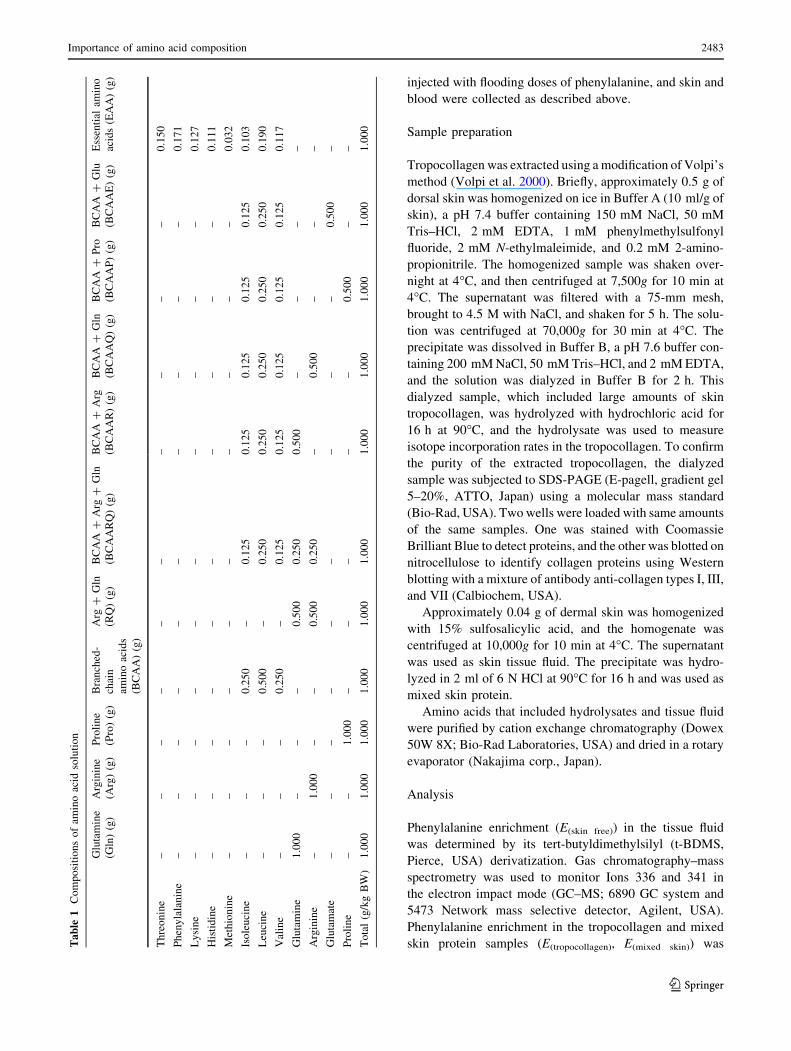
et al. 1999; Smriga and Torii 2003). The compositions of
the solutions are shown in Table 1 (all amino acids were
manufactured by Ajinomoto Co., Inc.). Twenty-five min-
utes after the solutions were administered, the mice were
2482 H. Murakami et al.
123
injected with flooding doses of phenylalanine, and skin and
blood were collected as described above.
Sample preparation
Tropocollagen was extracted using a modification of Volpi’s
method (Volpi et al. 2000). Briefly, approximately 0.5 g of
dorsal skin was homogenized on ice in Buffer A (10 ml/g of
skin), a pH 7.4 buffer containing 150 mM NaCl, 50 mM
Tris–HCl, 2 mM EDTA, 1 mM phenylmethylsulfonyl
fluoride, 2 mM N-ethylmaleimide, and 0.2 mM 2-amino-
propionitrile. The homogenized sample was shaken over-
night at 4�C, and then centrifuged at 7,500g for 10 min at
4�C. The supernatant was filtered with a 75-mm mesh,
brought to 4.5 M with NaCl, and shaken for 5 h. The solu-
tion was centrifuged at 70,000g for 30 min at 4�C. The
precipitate was dissolved in Buffer B, a pH 7.6 buffer con-
taining 200 mM NaCl, 50 mM Tris–HCl, and 2 mM EDTA,
and the solution was dialyzed in Buffer B for 2 h. This
dialyzed sample, which included large amounts of skin
tropocollagen, was hydrolyzed with hydrochloric acid for
16 h at 90�C, and the hydrolysate was used to measure
isotope incorporation rates in the tropocollagen. To confirm
the purity of the extracted tropocollagen, the dialyzed
sample was subjected to SDS-PAGE (E-pagell, gradient gel
5–20%, ATTO, Japan) using a molecular mass standard
(Bio-Rad, USA). Two wells were loaded with same amounts
of the same samples. One was stained with Coomassie
Brilliant Blue to detect proteins, and the other was blotted on
nitrocellulose to identify collagen proteins using Western
blotting with a mixture of antibody anti-collagen types I, III,
and VII (Calbiochem, USA).
Approximately 0.04 g of dermal skin was homogenized
with 15% sulfosalicylic acid, and the homogenate was
centrifuged at 10,000g for 10 min at 4�C. The supernatant
was used as skin tissue fluid. The precipitate was hydro-
lyzed in 2 ml of 6 N HCl at 90�C for 16 h and was used as
mixed skin protein.
Amino acids that included hydrolysates and tissue fluid
were purified by cation exchange chromatography (Dowex
50W 8X; Bio-Rad Laboratories, USA) and dried in a rotary
evaporator (Nakajima corp., Japan).
Analysis
Phenylalanine enrichment (E(skin free)) in the tissue fluid
was determined by its tert-butyldimethylsilyl (t-BDMS,
Pierce, USA) derivatization. Gas chromatography–mass
spectrometry was used to monitor Ions 336 and 341 in
the electron impact mode (GC–MS; 6890 GC system and
5473 Network mass selective detector, Agilent, USA).
Phenylalanine enrichment in the tropocollagen and mixed
skin protein samples (E(tropocollagen), E(mixed skin)) wasTa
ble
1C
om
po
siti
on
so
fam
ino
acid
solu
tio
n
Glu
tam
ine
(Gln
)(g
)
Arg
inin
e
(Arg
)(g
)
Pro
lin
e
(Pro
)(g
)
Bra
nch
ed-
chai
n
amin
oac
ids
(BC
AA
)(g
)
Arg
?G
ln
(RQ
)(g
)
BC
AA
?A
rg?
Gln
(BC
AA
RQ
)(g
)
BC
AA
?A
rg
(BC
AA
R)
(g)
BC
AA
?G
ln
(BC
AA
Q)
(g)
BC
AA
?P
ro
(BC
AA
P)
(g)
BC
AA
?G
lu
(BC
AA
E)
(g)
Ess
enti
alam
ino
acid
s(E
AA
)(g
)
Th
reo
nin
e–
––
––
––
––
–0
.15
0
Ph
eny
lala
nin
e–
––
––
––
––
–0
.17
1
Ly
sin
e–
––
––
––
––
–0
.12
7
His
tid
ine
––
––
––
––
––
0.1
11
Met
hio
nin
e–
––
––
––
––
–0
.03
2
Iso
leu
cin
e–
––
0.2
50
–0
.12
50
.12
50
.12
50
.12
50
.12
50
.10
3
Leu
cin
e–
––
0.5
00
–0
.25
00
.25
00
.25
00
.25
00
.25
00
.19
0
Val
ine
––
–0
.25
0–
0.1
25
0.1
25
0.1
25
0.1
25
0.1
25
0.1
17
Glu
tam
ine
1.0
00
––
–0
.50
00
.25
00
.50
0–
––
–
Arg
inin
e–
1.0
00
––
0.5
00
0.2
50
–0
.50
0–
––
Glu
tam
ate
––
––
––
––
–0
.50
0–
Pro
lin
e–
–1
.00
0–
––
––
0.5
00
––
To
tal
(g/k
gB
W)
1.0
00
1.0
00
1.0
00
1.0
00
1.0
00
1.0
00
1.0
00
1.0
00
1.0
00
1.0
00
1.0
00
Importance of amino acid composition 2483
123
determined by measuring their AQC-detergent (Waters,
USA) derivatization using liquid chromatography–mass
spectrometry to monitor ions 336 and 341 in the first MS
and 171 in the second MS (LC–MS/MS; Prominence
HPLC system, Shimazu, Japan and API 3200, Applied
Biosystems, USA). Plasma insulin concentrations were
measured using a commercial ELISA-kit (Morinaga Insti-
tute Biological Science, Japan), and amino acid concen-
trations were measured with an automatic amino acid
analyzer (L-8500, Hitachi, Japan).
Calculation and statistics
The FSR of the skin tropocollagen and mixed skin protein
was calculated with the precursor-product model. The
precursor represented the free phenylalanine enrichment in
the skin tissue fluid, and the product represented the
enrichment of the phenylalanine-incorporated skin tropo-
collagen or mixed skin protein. The FSR was calculated as
FSR (%/h) = E (tropocollagen or mixed skin)/(E (skin free) 9 t) 9
100, where t represents the time interval between phenyl-
alanine injection and sampling.
Values are presented as means ± SD. Comparisons with
the control group (given distilled water, DW) were made
via Dunnett’s test after ANOVA for multiple comparison
(JMP, SAS Institute, Cary, NC, USA). Values of P \ 0.05
were considered significant.
Results
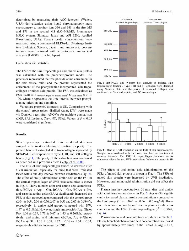
Skin tropocollagen extracted from the dorsal skin was
assayed with Western blotting to confirm its purity. The
protein bands of extracted skin tropocollagen separated by
SDS-PAGE corresponded to Type I, III, and VII collagen
bands (Fig. 1). The purity of the extraction was confirmed
as described in a previous article (Volpi et al. 2000).
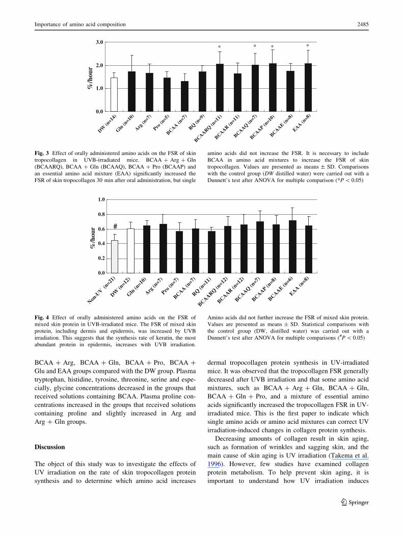
The FSR of skin tropocollagen tended to decrease after
UVB irradiation, especially for mice that were irradiated
twice with a one-day interval between irradiations (Fig. 2).
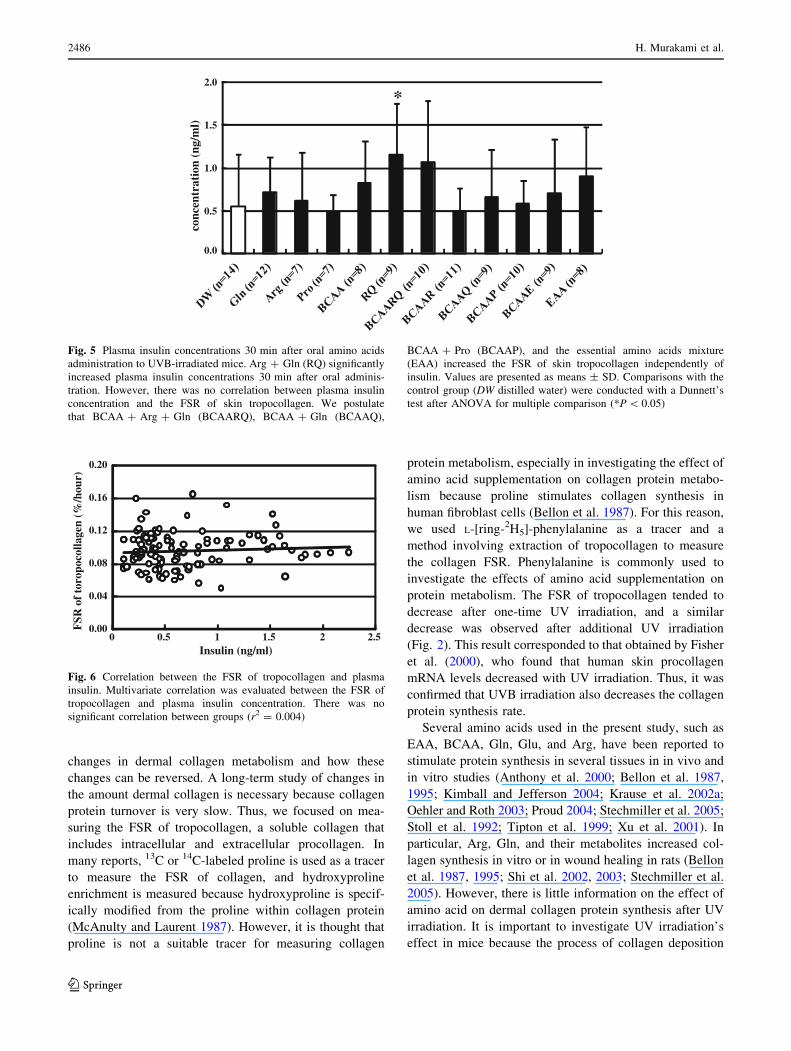
The effect of orally administered amino acid on the FSR in
the dermal tropocollagen of UVB-irradiated mice is shown
in Fig. 3. Thirty minutes after oral amino acid administra-
tion, BCAA ? Arg ? Gln, BCAA ? Gln, BCAA ? Pro,
and essential amino acids (EAA) significantly increased the
FSR of skin tropocollagen compared with the control group
(2.04 ± 0.54, 2.01 ± 0.50, 2.07 ± 0.59 and 2.07 ± 0.58%/h,
respectively, in amino acid groups compared with DW,
1.47 ± 0.21%/h). However, single amino acids (Arg, Gln or
Pro: 1.66 ± 0.39, 1.73 ± 0.67 or 1.45 ± 0.26%/h, respec-
tively) and amino acid mixtures (BCAA, Arg ? Gln or
BCAA ? Glu: 1.30 ± 0.32, 1.72 ± 0.26 or 1.74 ± 0.34,
respectively) did not increase the FSR.
The effect of oral amino acid administration on the
FSRs of mixed skin protein is shown in Fig. 4. The FSRs of
mixed skin protein were increased by UVB irradiation.
However, oral amino acid administration did not affect the
FSRs.
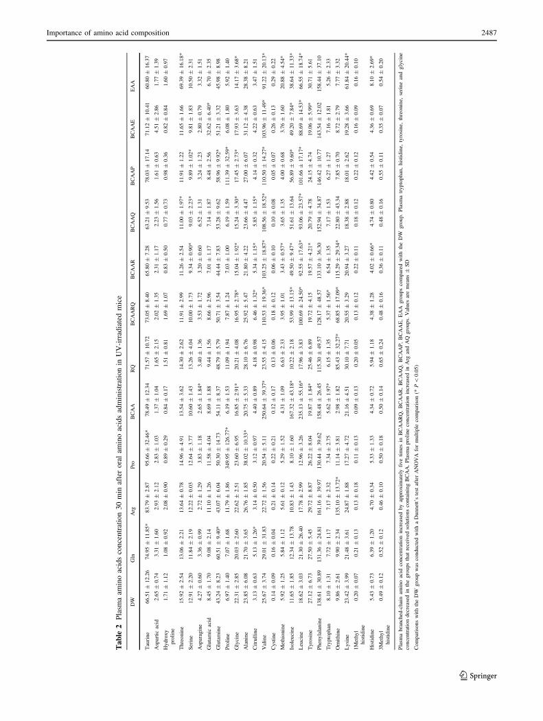
Plasma insulin concentrations 30 min after oral amino
acid administration are shown in Fig. 5. Arg ? Gln signifi-
cantly increased plasma insulin concentrations compared to
the DW group (1.14 ± 0.61 vs. 0.56 ± 0.6 nag/ml). How-
ever, there was no correlation between plasma insulin con-
centration and the FSR of skin tropocollagen (r2 = 0.0049;
Fig. 6).
Plasma amino acid concentrations are shown in Table 2.
Plasma branched-chain amino acid concentrations increased
by approximately five times in the BCAA ? Arg ? Gln,
Western-BlotStandard Tropocollagen
SDS-PAGEStandard Tropocollagen
105k Da
250 k Da
75 k Da
50 k Da
35k Da
160 k Da
Fig. 1 SDS-PAGE and Western blot analysis of isolated skin
tropocollagen fractions. Type I, III and VII collagen were identified
using Western blot, and the purity of extracted collagen was
confirmed. sd Standard protein, and TP tropocollagen
0.0
1.0
2.0
3.0
4.0
non-UV 1 time 2 times 3 times 4 times
%/h
our
Fig. 2 Effect of UVB irradiation on the FSR of skin tropocollagen.
Samples were irradiated with UVB one, two, three, or four times at
one-day intervals. The FSR of tropocollagen decreased to its
minimum value after two UVB irradiations. Values are means ± SD
2484 H. Murakami et al.
123
BCAA ? Arg, BCAA ? Gln, BCAA ? Pro, BCAA ?
Glu and EAA groups compared with the DW group. Plasma
tryptophan, histidine, tyrosine, threonine, serine and espe-
cially, glycine concentrations decreased in the groups that
received solutions containing BCAA. Plasma proline con-
centrations increased in the groups that received solutions
containing proline and slightly increased in Arg and
Arg ? Gln groups.
Discussion
The object of this study was to investigate the effects of
UV irradiation on the rate of skin tropocollagen protein
synthesis and to determine which amino acid increases
dermal tropocollagen protein synthesis in UV-irradiated
mice. It was observed that the tropocollagen FSR generally
decreased after UVB irradiation and that some amino acid
mixtures, such as BCAA ? Arg ? Gln, BCAA ? Gln,
BCAA ? Gln ? Pro, and a mixture of essential amino
acids significantly increased the tropocollagen FSR in UV-
irradiated mice. This is the first paper to indicate which
single amino acids or amino acid mixtures can correct UV
irradiation-induced changes in collagen protein synthesis.
Decreasing amounts of collagen result in skin aging,
such as formation of wrinkles and sagging skin, and the
main cause of skin aging is UV irradiation (Takema et al.
1996). However, few studies have examined collagen
protein metabolism. To help prevent skin aging, it is
important to understand how UV irradiation induces
0.0
1.0
2.0
3.0
DW(n
=14)
Gln (n
=10)
Arg (n
=7)
Pro(n
=5)
BCAA (n=7)
RQ (n=9)
BCAARQ (n=11
)
BCAAR (n=11
)
BCAAQ (n=7)
BCAAP (n=10
)
BCAAE (n=8)
EAA (n=8)
%/h
our
* * **
Fig. 3 Effect of orally administered amino acids on the FSR of skin
tropocollagen in UVB-irradiated mice. BCAA ? Arg ? Gln
(BCAARQ), BCAA ? Gln (BCAAQ), BCAA ? Pro (BCAAP) and
an essential amino acid mixture (EAA) significantly increased the
FSR of skin tropocollagen 30 min after oral administration, but single
amino acids did not increase the FSR. It is necessary to include
BCAA in amino acid mixtures to increase the FSR of skin
tropocollagen. Values are presented as means ± SD. Comparisons
with the control group (DW distilled water) were carried out with a
Dunnett’s test after ANOVA for multiple comparison (*P \ 0.05)
0.0
0.2
0.4
0.6
0.8
1.0
Non-U
V (n=21
)
DW (n
=12)
Gln (n
=10)
Arg (n
=7)
Pro (n
=7)
BCAA (n=7)
RQ (n=11
)
BCAARQ (n=12
)
BCAAR (n=12
)
BCAAQ (n=7)
BCAAP (n=8)
BCAAE(n
=6)
EAA (n=8)
%/h
our #
Fig. 4 Effect of orally administered amino acids on the FSR of
mixed skin protein in UVB-irradiated mice. The FSR of mixed skin
protein, including dermis and epidermis, was increased by UVB
irradiation. This suggests that the synthesis rate of keratin, the most
abundant protein in epidermis, increases with UVB irradiation.
Amino acids did not further increase the FSR of mixed skin protein.
Values are presented as means ± SD. Statistical comparisons with
the control group (DW, distilled water) was carried out with a
Dunnett’s test after ANOVA for multiple comparisons (#P \ 0.05)
Importance of amino acid composition 2485
123
changes in dermal collagen metabolism and how these
changes can be reversed. A long-term study of changes in
the amount dermal collagen is necessary because collagen
protein turnover is very slow. Thus, we focused on mea-
suring the FSR of tropocollagen, a soluble collagen that
includes intracellular and extracellular procollagen. In
many reports, 13C or 14C-labeled proline is used as a tracer
to measure the FSR of collagen, and hydroxyproline
enrichment is measured because hydroxyproline is specif-
ically modified from the proline within collagen protein
(McAnulty and Laurent 1987). However, it is thought that
proline is not a suitable tracer for measuring collagen
protein metabolism, especially in investigating the effect of
amino acid supplementation on collagen protein metabo-
lism because proline stimulates collagen synthesis in
human fibroblast cells (Bellon et al. 1987). For this reason,
we used L-[ring-2H5]-phenylalanine as a tracer and a
method involving extraction of tropocollagen to measure
the collagen FSR. Phenylalanine is commonly used to
investigate the effects of amino acid supplementation on
protein metabolism. The FSR of tropocollagen tended to
decrease after one-time UV irradiation, and a similar
decrease was observed after additional UV irradiation
(Fig. 2). This result corresponded to that obtained by Fisher
et al. (2000), who found that human skin procollagen
mRNA levels decreased with UV irradiation. Thus, it was
confirmed that UVB irradiation also decreases the collagen
protein synthesis rate.
Several amino acids used in the present study, such as
EAA, BCAA, Gln, Glu, and Arg, have been reported to
stimulate protein synthesis in several tissues in in vivo and
in vitro studies (Anthony et al. 2000; Bellon et al. 1987,
1995; Kimball and Jefferson 2004; Krause et al. 2002a;
Oehler and Roth 2003; Proud 2004; Stechmiller et al. 2005;
Stoll et al. 1992; Tipton et al. 1999; Xu et al. 2001). In
particular, Arg, Gln, and their metabolites increased col-
lagen synthesis in vitro or in wound healing in rats (Bellon
et al. 1987, 1995; Shi et al. 2002, 2003; Stechmiller et al.
2005). However, there is little information on the effect of
amino acid on dermal collagen protein synthesis after UV
irradiation. It is important to investigate UV irradiation’s
effect in mice because the process of collagen deposition
0.0
0.5
1.0
1.5
2.0
conc
entr
atio
n (n
g/m
l)
*
Fig. 5 Plasma insulin concentrations 30 min after oral amino acids
administration to UVB-irradiated mice. Arg ? Gln (RQ) significantly
increased plasma insulin concentrations 30 min after oral adminis-
tration. However, there was no correlation between plasma insulin
concentration and the FSR of skin tropocollagen. We postulate
that BCAA ? Arg ? Gln (BCAARQ), BCAA ? Gln (BCAAQ),
BCAA ? Pro (BCAAP), and the essential amino acids mixture
(EAA) increased the FSR of skin tropocollagen independently of
insulin. Values are presented as means ± SD. Comparisons with the
control group (DW distilled water) were conducted with a Dunnett’s
test after ANOVA for multiple comparison (*P \ 0.05)
0.00
0.04
0.08
0.12
0.16
0.20
0 0.5 1 1.5 2 2.5
FSR
of
toro
poco
llage
n (%
/hou
r)
Insulin (ng/ml)
Fig. 6 Correlation between the FSR of tropocollagen and plasma
insulin. Multivariate correlation was evaluated between the FSR of
tropocollagen and plasma insulin concentration. There was no
significant correlation between groups (r2 = 0.004)
2486 H. Murakami et al.
123
Ta
ble
2P
lasm
aam
ino
acid
sco
nce
ntr
atio
n3
0m
inaf
ter
ora
lam
ino
acid
sad
min
istr
atio
nin
UV
-irr
adia
ted
mic
e
DW
Gln
Arg
Pro
BC
AA
RQ
BC
AA
RQ
BC
AA
RB
CA
AQ
BC
AA
PB
CA
AE
EA
A
Tau
rine
66.5
1±
12.2
674.9
5±
11.8
5*
83.7
9±
2.8
795.6
6±
32.4
6*
78.4
9±
12.3
471.5
7±
10.7
273.0
5±
8.4
065.8
0±
7.2
863.2
1±
9.5
378.0
3±
17.1
471.1
2±
10.4
160.8
0±
16.3
7
Asp
arti
cac
id2.6
5±
0.7
43.3
1±
1.6
02.9
3±
2.1
22.8
3±
1.0
31.3
7±
1.0
41.6
5±
2.1
52.0
2±
1.3
52.3
1±
1.1
72.2
3±
1.5
61.6
1±
0.6
34.5
1±
2.8
61.7
7±
1.3
9
Hydro
xy
pro
line
1.7
1±
1.1
21.0
8±
0.9
22.0
8±
0.9
00.8
9±
0.2
90.8
4±
0.1
71.5
1±
0.8
11.6
9±
1.0
70.8
3±
0.5
00.7
7±
0.7
30.9
8±
0.3
60.8
2±
0.8
41.6
0±
0.9
7
Thre
onin
e15.9
2±
2.5
413.0
6±
2.2
113.6
4±
0.7
814.9
6±
4.9
113.5
4±
3.6
214.3
0±
2.6
211.9
1±
2.9
911.2
6±
2.5
411.0
0±
1.9
7*
11.9
1±
1.2
211.6
5±
1.6
669.3
9±
16.1
8*
Ser
ine
12.9
1±
2.2
011.8
4±
2.1
912.2
2±
0.0
312.6
4±
3.7
710.6
0±
1.4
313.2
6±
4.0
410.0
0±
1.7
39.3
4±
0.9
0*
9.0
3±
2.2
3*
9.8
9±
1.0
2*
9.8
1±
1.8
310.5
0±
2.3
1
Asp
arag
ine
4.2
7±
0.6
03.3
6±
0.9
92.7
2±
1.2
93.8
3±
1.1
82.6
5±
1.8
4*
3.4
0±
1.3
63.5
3±
1.7
23.2
0±
0.6
06.5
2±
1.3
13.2
4±
1.2
32.8
0±
0.7
93.3
2±
1.5
1
Glu
tam
icac
id8.4
5±
1.7
09.0
8±
2.1
411.1
0±
1.2
611.5
8±
4.0
48.6
9±
1.8
89.4
4±
1.5
68.6
6±
2.9
67.0
1±
1.1
77.1
4±
1.8
78.4
8±
2.5
632.6
2±
6.4
0*
6.7
0±
2.3
5
Glu
tam
ine
43.2
4±
8.2
360.5
1±
9.4
0*
43.0
7±
6.0
450.3
0±
14.7
354.1
1±
8.3
748.7
9±
5.7
950.7
1±
3.5
444.4
4±
7.8
353.2
8±
9.6
258.9
6±
9.9
2*
51.2
1±
3.3
245.9
8±
8.9
8
Pro
line
6.9
7±
1.4
07.0
7±
1.6
811.7
9±
1.8
6349.9
5±
126.7
7*
6.1
9±
1.5
311.0
9±
1.9
47.8
7±
1.2
47.0
3±
1.0
06.1
9±
1.5
9111.3
9±
32.5
9*
6.0
8±
1.8
05.9
2±
1.4
0
Gly
cine
22.3
1±
2.8
520.0
3±
2.6
022.6
2±
2.5
123.6
0±
6.9
516.8
5±
2.9
1*
20.2
1±
4.0
816.9
5±
2.7
8*
15.0
4±
1.9
2*
15.2
4±
3.3
0*
17.4
5±
2.7
3*
17.9
3±
3.6
314.1
7±
3.6
8*
Ala
nin
e23.8
5±
6.0
821.7
0±
3.6
526.7
6±
1.8
538.0
2±
10.3
3*
20.7
5±
5.3
328.1
0±
6.7
625.9
2±
5.4
721.8
0±
4.2
223.6
6±
4.4
727.0
0±
6.0
731.1
2±
4.3
828.3
8±
8.2
1
Cit
rull
ine
3.1
3±
0.6
35.1
3±
1.2
6*
3.1
4±
0.5
03.1
2±
0.9
74.4
0±
0.8
94.1
8±
0.9
86.4
6±
1.3
2*
5.3
4±
1.1
5*
5.8
5±
1.1
5*
4.1
4±
0.3
24.2
2±
0.6
33.4
7±
1.5
1
Val
ine
25.6
7±
3.7
429.0
1±
31.8
322.7
2±
1.5
620.5
4±
5.1
1250.6
4±
39.3
7*
23.5
5±
4.1
5110.5
3±
19.3
6*
103.2
5±
18.8
7*
108.5
6±
18.5
2*
110.5
0±
14.2
7*
103.9
6±
11.4
9*
91.2
2±
20.1
3*
Cyst
ine
0.1
4±
0.0
90.1
6±
0.0
40.2
1±
0.1
40.2
2±
0.2
10.1
2±
0.1
70.1
3±
0.0
60.1
8±
0.1
20.0
6±
0.1
00.1
0±
0.0
80.0
5±
0.0
70.2
6±
0.1
30.2
9±
0.2
2
Met
hio
nin
e5.9
2±
1.2
55.8
4±
1.1
25.6
1±
0.1
25.2
9±
1.5
24.3
1±
1.0
96.6
3±
2.3
33.9
5±
1.0
13.4
3±
0.5
7*
3.6
5±
1.3
54.0
0±
0.6
83.7
6±
1.6
020.8
8±
4.5
4*
Isole
uci
ne
11.6
5±
1.8
512.3
4±
13.7
810.8
3±
1.4
38.1
0±
1.6
0167.3
2±
43.1
8*
10.2
2±
2.1
853.9
9±
13.1
5*
49.5
0±
9.4
7*
51.6
1±
13.6
456.8
9±
9.6
0*
49.2
0±
7.8
4*
38.6
4±
11.3
3*
Leu
cine
18.6
2±
3.0
321.3
0±
26.4
017.7
8±
2.9
912.9
6±
3.2
6235.1
3±
55.1
6*
17.9
6±
3.8
3100.6
9±
24.5
0*
92.5
5±
17.6
3*
93.0
6±
23.5
7*
101.6
6±
17.1
7*
88.6
9±
14.5
3*
66.5
5±
18.7
4*
Tyro
sine
27.1
2±
6.7
327.5
0±
5.4
529.7
2±
8.8
726.2
2±
8.0
419.8
7±
1.8
4*
25.4
6±
6.8
919.7
2±
4.1
519.5
7±
4.2
1*
20.7
9±
4.7
824.1
5±
4.7
419.0
6±
5.9
9*
30.7
1±
5.6
1
Phen
yla
lanin
e138.6
1±
30.8
9131.3
6±
24.8
1161.1
6±
39.9
7130.4
4±
39.6
2158.4
8±
26.4
5115.3
0±
49.5
7128.1
7±
48.5
7133.1
0±
36.3
0152.9
4±
34.8
7146.4
2±
10.7
7143.5
4±
12.0
2158.4
4±
37.1
0
Try
pto
phan
8.1
0±
1.3
17.7
2±
1.1
77.1
7±
2.3
27.3
4±
2.7
55.6
2±
1.9
7*
6.1
5±
1.3
55.3
7±
1.5
6*
6.5
4±
1.3
57.1
7±
1.5
36.2
7±
1.2
77.1
6±
1.8
15.2
6±
2.3
3
Orn
ithin
e9.8
6±
2.6
19.9
0±
2.3
4135.1
0±
13.7
2*
11.1
4±
3.8
12.9
8±
1.8
285.4
3±
32.2
7*
68.8
5±
17.0
9*
115.2
9±
29.3
4*
22.8
0±
43.3
47.8
5±
0.7
08.7
2±
2.7
97.7
7±
3.3
2
Lysi
ne
23.4
2±
3.9
921.4
8±
3.6
124.8
7±
1.8
817.2
7±
4.7
221.1
6±
4.5
130.1
0±
7.7
120.5
5±
3.2
920.9
4±
3.2
718.3
8±
2.8
818.0
1±
2.6
219.2
8±
3.6
661.8
4±
20.4
4*
1M
ethyl
his
tidin
e
0.2
0±
0.0
70.2
1±
0.1
30.1
3±
0.1
80.1
1±
0.1
30.0
9±
0.1
30.2
0±
0.0
50.1
3±
0.1
20.2
2±
0.1
10.1
8±
0.1
20.2
2±
0.1
20.1
6±
0.0
90.1
6±
0.1
0
His
tidin
e5.4
3±
0.7
36.3
9±
1.2
04.7
0±
0.3
45.3
3±
1.3
34.3
4±
0.7
25.9
4±
1.1
84.3
8±
1.2
84.0
2±
0.6
6*
4.7
4±
0.8
04.4
2±
0.5
44.3
6±
0.6
98.1
0±
2.6
9*
3M
ethyl
his
tidin
e
0.4
9±
0.1
20.5
2±
0.1
20.4
6±
0.1
00.5
0±
0.1
80.5
0±
0.1
40.6
5±
0.2
40.4
8±
0.1
60.3
6±
0.1
10.4
8±
0.1
60.5
5±
0.1
10.3
5±
0.0
70.5
4±
0.2
0
Pla
sma
bra
nch
ed-c
hai
nam
ino
acid
conce
ntr
atio
nin
crea
sed
by
appro
xim
atel
yfi
ve
tim
esin
BC
AA
RQ
,B
CA
AR
,B
CA
AQ
,B
CA
AP
,B
CA
AE
,E
AA
gro
ups
com
par
edw
ith
the
DW
gro
up.
Pla
sma
trypto
phan
,his
tidin
e,ty
rosi
ne,
thre
onin
e,se
rine
and
gly
cine
conce
ntr
atio
ndec
reas
edin
the
gro
ups
that
rece
ived
solu
tions
conta
inin
gB
CA
A.
Pla
sma
pro
line
conce
ntr
atio
nin
crea
sed
inA
rgan
dA
Qgro
ups.
Val
ues
are
mea
ns
±S
D
Com
par
isons
wit
hth
eD
Wgro
up
was
conduct
edw
ith
aD
unnet
t’s
test
afte
rA
NO
VA
for
mult
iple
com
par
ison
(*P
\0.0
5)
Importance of amino acid composition 2487
123
differs between UV irradiation and wounds (Fisher 2005;
Johnstone and Farley 2005). Some amino acid mixtures
containing BCAA, such as EAA, BCAA ? Arg ? Gln,
BCAA ? Gln, and BCAA ? Pro, significantly increased
the FSR of tropocollagen (Fig. 3). BCAA in particular, but
also leucine and its metabolites, modulate mammalian
targets of rapamycin (mTOR) and stimulate phosphoryla-
tion of the 70-kDa ribosomal protein S6 kinase (p70S6K)
and eukaryotic initiation factor-binding protein-1 (4E-BP1),
initiating translation and transcription in protein synthesis
(Anthony et al. 2000; Kimball and Jefferson 2004; Meijer
2003; Proud 2004; Xu et al. 2001). However, BCAA and
BCAA ? Glu did not increase the FSR of dermal tropo-
collagen (Fig. 3). In addition, other amino acids, such as
Arg, Gln, Pro, Arg ? Gln, also did not increase the FSR
(Fig. 3). This result indicates that while BCAA is impor-
tant for skin tropocollagen synthesis, other specific amino
acids, such as Gln or Pro, are also necessary to stimulate
dermal tropocollagen synthesis.
In an in vitro study, Bellon and Karna showed that Gln
and its metabolites (glutamate, pyrroline-5-carboxylate,
arginine) increase collagen synthesis and suggested that de
novo synthesized proline is important for collagen syn-
thesis (Bellon et al. 1995, 1987; Karna et al. 2001). In the
present study, the FSR of tropocollagen did not increase
when amino acids containing precursors of proline (Gln,
Arg, BCAA ? Glu and Arg ? Gln) were administered, but
did increase with an amino acid mixture containing exog-
enous proline (BCAA ? Pro). In addition, plasma proline
concentrations were not increased by collagen synthesis-
stimulating amino acid mixtures containing proline pre-
cursors (BCAA ? Gln, BCAA ? Gln ? Arg), but were
slightly increased by Arg ? Gln and Arg, neither of which
stimulated the tropocollagen FSR. These results indicate
that de novo proline synthesis is not the main cause of
increased collagen protein synthesis. Proline constitutes
one-third of collagen protein’s amino acid residues. How-
ever, there is little information proline supplementation’s
effect on dermal skin collagen synthesis. Further study is
needed to understand the mechanism underlying the effect
of BCAA ? Pro on the FSR of tropocollagen.
Xu found that the combination of leucine and glutamine
synergistically stimulates the activity of S6K in pancreatic
beta cells. Some reports indicate that glutamine regulates
protein synthesis (Xu et al. 2001). For example, glutamine
restores energy metabolism into cells (Krause et al. 2002b),
increases cell swelling (Oehler and Roth 2003), and acti-
vates GAPP (glutamate dependent protein phosphatase,
which correlates with mTOR activation) (Krause et al.
2002a; Stoll et al. 1992). Arginine also improves wound
healing by increasing collagen synthesis; the mechanism
of this effect is thought to be the stimulation of growth
hormone secretion and nitric oxide synthesis (Stechmiller
et al. 2005). Williams reported that an orally administered
amino acid mixture consisting of metabolites of leucine
(b-hydroxy-b-methylbutyrate), arginine, and glutamine
increased the collagen content in subcutaneously implanted
tubes in healthy volunteers (Williams et al. 2002). In
addition, Corsetti reported that amino acid mixtures that
included leucine, proline, lysine, and glycine improved
wound healing associated with the modulation of nitric
oxidate synthase and transforming growth factor-b1
(Corsetti et al. 2010). Dioguardi (2008) also reported that
collagen synthesis is efficiently maintained only when
specific amino acids are continuously available and present
in a specific ratio. Therefore, a possible explanation of our
findings is that BCAA ? Gln and BCAA ? Gln ? Arg
synergistically stimulate dermal tropocollagen protein
synthesis using each amino acid’s individual effects on
protein synthesis. In addition, insulin is a powerful protein
synthesis stimulator, and leucine and arginine stimulate
insulin secretion. However, insulin was not the main cause
of the increase of the dermal tropocollagen FSR by the
amino acid mixtures, because there was no correlation
between insulin and the tropocollagen FSR in any group
(r2 = 0.034; Fig. 6). EAA is known to increase the FSR of
skeletal muscle protein (Tipton et al. 1999). In the present
study, EAA was the only amino acid observed to increase
the FSR of skeletal muscle protein (data not shown). Thus,
the tropocollagen FSR improvement resulting from EAA is
associated with the improvement of whole-body protein
metabolism.
In conclusion, UVB irradiation decreased the FSR of
skin tropocollagen, while BCAA ? Arg ? Gln, BCAA ?
Gln, BCAA ? Pro, and EAA increased the FSR of skin
tropocollagen independently of insulin. However, single
amino acids and BCAA did not increase the FSR. It should
be noted that combinations of specific amino acids, espe-
cially BCAA ? Gln or BCAA ? Pro are vital in stimu-
lating the FSR of skin tropocollagen independently of
insulin. However, the dermal collagen protein synthesis
stimulation mechanism of these amino acids mixtures is
unclear. Further study is necessary to understand the
mechanism of the increase tropocollagen FSR by these
amino acids.
Acknowledgments We thank Takashi Suzuki, Sayo Inoue, and
Shinobu Tanimoto for their assistance with the experimental trial.
Conflict of interest The authors declare that they have no conflict
of interest.
Open Access This article is distributed under the terms of the
Creative Commons Attribution Noncommercial License which per-
mits any noncommercial use, distribution, and reproduction in any
medium, provided the original author(s) and source are credited.
2488 H. Murakami et al.
123
References
Anthony JC, Anthony TG, Kimball SR, Vary TC, Jefferson LS (2000)
Orally administered leucine stimulates protein synthesis in
skeletal muscle of postabsorptive rats in association with
increased eIF4F formation. J Nutr 130:139–145
Badiu DL, Luque R, Dumitrescu E, Craciun A, Dinca D (2010)
Amino acids from Mytilus galloprovincialis (L.) and Rapanavenosa molluscs accelerate skin wounds healing via enhance-
ment of dermal and epidermal neoformation. Protein J 29:81–92
Bellon G, Monboisse JC, Randoux A, Borel JP (1987) Effects of
preformed proline and proline amino acid precursors (including
glutamine) on collagen synthesis in human fibroblast cultures.
Biochim Biophys Acta 930:39–47
Bellon G, Chaqour B, Wegrowski Y, Monboisse JC, Borel JP (1995)
Glutamine increases collagen gene transcription in cultured
human fibroblasts. Biochim Biophys Acta 1268:311–323
Boyer B, Fourtanier A, Kern P, Labat-Robert J (1992) UVA- and
UVB-induced changes in collagen and fibronectin biosynthesis
in the skin of hairless mice. J Photochem Photobiol B
14:247–259
Corsetti G, D’Antona G, Dioguardi FS, Rezzani R (2010) Topical
application of dressing with amino acids improves cutaneous
wound healing in aged rats. Acta Histochem (Germany)
112(5):497–507
Dioguardi FS (2008) Nutrition and skin Collagen integrity: a
dominant role for amino acids. Clin Dermatol (United States)
26(6):636–640
Farges MC, Berard MP, Raul F, Cezard JP, Joly B, Davot P, Vasson
MP, Cynober L (1999) Oral administration of a glutamine-
enriched diet before or after endotoxin challenge in aged rats has
limited effects. J Nutr 129:1799–1806
Fisher GJ (2005) The pathophysiology of photoaging of the skin.
Cutis 75:5–8 (discussion 8–9)
Fisher GJ, Datta S, Wang Z, Li XY, Quan T, Chung JH, Kang S,
Voorhees JJ (2000) c-Jun-dependent inhibition of cutaneous
procollagen transcription following ultraviolet irradiation is
reversed by all-trans retinoic acid. J Clin Invest 106:663–670
Fisher GJ, Choi HC, Bata-Csorgo Z, Shao Y, Datta S, Wang ZQ,
Kang S, Voorhees JJ (2001) Ultraviolet irradiation increases
matrix metalloproteinase-8 protein in human skin in vivo.
J Invest Dermatol 117:219–226
Garlick PJ, McNurlan MA (1998) Measurement of protein synthesis
in human tissues by the flooding method. Curr Opin Clin Nutr
Metab Care 1:455–460
Johnstone CC, Farley A (2005) The physiological basics of wound
healing. Nurs Stand 19:59–65 (quiz 66)
Karna E, Miltyk W, Wolczynski S, Palka JA (2001) The potential
mechanism for glutamine-induced collagen biosynthesis in
cultured human skin fibroblasts. Comp Biochem Physiol B
Biochem Mol Biol 130:23–32
Kimball SR, Jefferson LS (2004) Regulation of global and specific
mRNA translation by oral administration of branched-chain
amino acids. Biochem Biophys Res Commun 313:423–427
Krause U, Bertrand L, Hue L (2002a) Control of p70 ribosomal
protein S6 kinase and acetyl-CoA carboxylase by AMP-activated
protein kinase and protein phosphatases in isolated hepatocytes.
Eur J Biochem 269:3751–3759
Krause U, Bertrand L, Maisin L, Rosa M, Hue L (2002b) Signalling
pathways and combinatory effects of insulin and amino acids in
isolated rat hepatocytes. Eur J Biochem 269:3742–3750
McAnulty RJ, Laurent GJ (1987) Collagen synthesis and degradation
in vivo. Evidence for rapid rates of collagen turnover with
extensive degradation of newly synthesized collagen in tissues of
the adult rat. Coll Relat Res 7:93–104
Meijer AJ (2003) Amino acids as regulators and components of
nonproteinogenic pathways. J Nutr 133:2057S–2062S
Oehler R, Roth E (2003) Regulative capacity of glutamine. Curr Opin
Clin Nutr Metab Care 6:277–282
Proud CG (2004) mTOR-mediated regulation of translation factors by
amino acids. Biochem Biophys Res Commun 313:429–436
Rittie L, Fisher GJ (2002) UV-light-induced signal cascades and skin
aging. Ageing Res Rev 1:705–720
Shi HP, Fishel RS, Efron DT, Williams JZ, Fishel MH, Barbul A
(2002) Effect of supplemental ornithine on wound healing.
J Surg Res (United States) 106(2):299–302
Shi HP, Most D, Efron DT, Witte MB, Barbul A (2003) SupplementalL-arginine enhances wound healing in diabetic rats. Wound
Repair Regen (United States) 11(3):198–203
Smriga M, Torii K (2003) L-Lysine acts like a partial serotonin
receptor 4 antagonist and inhibits serotonin-mediated intestinal
pathologies and anxiety in rats. Proc Natl Acad Sci USA (United
States) 100(26):15370–15375
Stechmiller JK, Childress B, Cowan L (2005) Arginine supplemen-
tation and wound healing. Nutr Clin Pract 20:52–61
Stoll B, Gerok W, Lang F, Haussinger D (1992) Liver cell volume
and protein synthesis. Biochem J 287(Pt 1):217–222
Takema Y, Hattori M, Aizawa K (1996) The relationship between
quantitative changes in collagen and formation of wrinkles on
hairless mouse skin after chronic UV irradiation. J Dermatol Sci
12:56–63
Tipton KD, Gurkin BE, Matin S, Wolfe RR (1999) Nonessential
amino acids are not necessary to stimulate net muscle protein
synthesis in healthy volunteers. J Nutr Biochem 10:89–95
Volpi E, Jeschke MG, Herndon DN, Wolfe RR (2000) Measurement
of skin protein breakdown in a rat model. Am J Physiol
Endocrinol Metab 279:E900–E906
Williams, JZ, Abumrad, N, Barbul, A (2002) Effect of a specialized
amino acid mixture on human collagen deposition. Ann Surg
236:369–374 (discussion 374–365)
Xu G, Kwon G, Cruz WS, Marshall CA, McDaniel ML (2001)
Metabolic regulation by leucine of translation initiation through
the mTOR-signaling pathway by pancreatic beta-cells. Diabetes
50:353–360
Zhang XJ, Chinkes DL, Wolfe RR (2004) Leucine supplementation
has an anabolic effect on proteins in rabbit skin wound and
muscle. J Nutr 134:3313–3318
Importance of amino acid composition 2489
123
Related Documents