RESEARCH Open Access Implant impression accuracy of parallel and non-parallel implants: a comparative in-vitro analysis of open and closed tray techniques Motaz S. Osman 1 , Hassan M. Ziada 2 , Neamat H. Abubakr 3* and Ahmed M. Suliman 4 Abstract Background: The outcome of the evaluation of impression techniques accuracy may improve the selection criteria for an ideal technique. The aim was to evaluate the accuracy of the open and closed tray techniques for implant impressions, in a partially edentulous maxilla, replaced with a three-unit fixed partial denture, as well as to assess the effect of implants parallelism on accuracy. Material and methods: This is an experimental in vitro study to evaluate impressions accuracy of a simulated area restored with an implant retained FPD, using the open and closed tray implant impression techniques. The effect of implant position angulation, parallelism, and implant systems (Straumann, SIC Invent, Osstem) was also evaluated. Three custom-made acrylic resin test models were prepared with two parallel and two non-parallel implants, on either side of a maxillary arch. One hundred and ninety-two impressions were made using monophase VPS impression material. Their master casts were obtained and evaluated for the horizontal and vertical discrepancy. The casts were scanned using a model scanner. The distances between the two reference points were measured. Results: The Straumann and SIC Invent implants showed no statistically significant differences (Mann-Whitney U test), regarding accuracy for both the open and closed tray impression techniques (P = 0.667 and P = 0.472). There were no significant differences for the parallel and non-parallel implants (P = 0.323 and P = 0.814), respectively, while the Osstem system showed statistically significant differences for both the open and closed tray impression techniques (P = 0.035) and between the parallel and non-parallel implants (P = 0.045). For the vertical discrepancies, significant differences were detected (chi-square test) between the open and closed tray impression techniques (P = 0.037). Conclusions: Within the limitations of this study, there were generally no significant differences between open and closed, although better results were obtained for the open tray techniques. On the use of the non-parallel implants, the open tray technique provided a better result than the closed tray technique. Keywords: Impression accuracy, Parallel implants, Nonparallel implants, Open tray technique, Closed tray technique * Correspondence: [email protected] 3 Department of Clinical Science, School of Dental Medicine, University of Nevada, 1001 Shadow Lane, Suite 248, MS 7415, Las Vegas, Nevada 89106, USA Full list of author information is available at the end of the article International Journal of Implant Dentistry © The Author(s). 2019 Open Access This article is distributed under the terms of the Creative Commons Attribution 4.0 International License (http://creativecommons.org/licenses/by/4.0/), which permits unrestricted use, distribution, and reproduction in any medium, provided you give appropriate credit to the original author(s) and the source, provide a link to the Creative Commons license, and indicate if changes were made. Osman et al. International Journal of Implant Dentistry (2019) 5:4 https://doi.org/10.1186/s40729-019-0159-5

Welcome message from author
This document is posted to help you gain knowledge. Please leave a comment to let me know what you think about it! Share it to your friends and learn new things together.
Transcript
RESEARCH Open Access
Implant impression accuracy of parallel andnon-parallel implants: a comparativein-vitro analysis of open and closed traytechniquesMotaz S. Osman1, Hassan M. Ziada2, Neamat H. Abubakr3* and Ahmed M. Suliman4
Abstract
Background: The outcome of the evaluation of impression techniques accuracy may improve the selection criteriafor an ideal technique. The aim was to evaluate the accuracy of the open and closed tray techniques for implantimpressions, in a partially edentulous maxilla, replaced with a three-unit fixed partial denture, as well as to assessthe effect of implants parallelism on accuracy.
Material and methods: This is an experimental in vitro study to evaluate impressions accuracy of a simulated arearestored with an implant retained FPD, using the open and closed tray implant impression techniques. The effect ofimplant position angulation, parallelism, and implant systems (Straumann, SIC Invent, Osstem) was also evaluated.Three custom-made acrylic resin test models were prepared with two parallel and two non-parallel implants, oneither side of a maxillary arch. One hundred and ninety-two impressions were made using monophase VPSimpression material. Their master casts were obtained and evaluated for the horizontal and vertical discrepancy.The casts were scanned using a model scanner. The distances between the two reference points were measured.
Results: The Straumann and SIC Invent implants showed no statistically significant differences (Mann-Whitney U test),regarding accuracy for both the open and closed tray impression techniques (P = 0.667 and P = 0.472). There were nosignificant differences for the parallel and non-parallel implants (P = 0.323 and P = 0.814), respectively, while the Osstemsystem showed statistically significant differences for both the open and closed tray impression techniques (P = 0.035)and between the parallel and non-parallel implants (P = 0.045). For the vertical discrepancies, significant differenceswere detected (chi-square test) between the open and closed tray impression techniques (P = 0.037).
Conclusions: Within the limitations of this study, there were generally no significant differences between open andclosed, although better results were obtained for the open tray techniques. On the use of the non-parallel implants,the open tray technique provided a better result than the closed tray technique.
Keywords: Impression accuracy, Parallel implants, Nonparallel implants, Open tray technique, Closed tray technique
* Correspondence: [email protected] of Clinical Science, School of Dental Medicine, University ofNevada, 1001 Shadow Lane, Suite 248, MS 7415, Las Vegas, Nevada 89106,USAFull list of author information is available at the end of the article
International Journal ofImplant Dentistry
© The Author(s). 2019 Open Access This article is distributed under the terms of the Creative Commons Attribution 4.0International License (http://creativecommons.org/licenses/by/4.0/), which permits unrestricted use, distribution, andreproduction in any medium, provided you give appropriate credit to the original author(s) and the source, provide a link tothe Creative Commons license, and indicate if changes were made.
Osman et al. International Journal of Implant Dentistry (2019) 5:4 https://doi.org/10.1186/s40729-019-0159-5
IntroductionA three-dimensionally accurate impression is apre-requisite for implant restorations since there is nointervening periodontal ligament at the implant-boneinterface to compensate for any inaccuracies [1, 2]. Nu-merous factors impact on implant impression accuracy,including the technique, the materials used, and thenumber of implants, as well as the parallelism of the im-plants or abutments. Impression inaccuracies impactnegatively on the precision fit of the restoration [1, 3].Consequently, mechanical complications may arise, suchas screw or abutments loosening fracture of the pros-thetic components or the implant. Marginal or verticaldiscrepancies may also develop, increasing plaque accu-mulation, which may also negatively impact on the softand hard tissues around the implant [1, 3].The open and closed tray impression techniques are
both advocated, each has advantages and disadvantages.Non-parallel implants may strain the impression duringtray removal, due to the significant force required for itswithdrawal, which compromises accuracy [1]. Regardlessof the technique and the number of implants or parallel-ism, the use of a verification device would be advisable,to ensure a clinically passive metal framework fit [4].There is no evidence of the superiority of one impres-
sion technique or material over others. Nonetheless, anaccurate impression is fundamental, in achieving a pas-sive prosthetic fit and the long-term serviceability. Inthis regard, Mpikos et al. [5] found neither the open northe closed tray techniques influenced the accuracy ofimpressions of multiple implants. However, they foundimplant parallelism had a significant impact on impres-sion accuracy, particularly in implants with internal con-nections. Conrad et al. [2] reported that the averageangle of error and the magnitude of distortion betweenthe closed and open tray techniques were not signifi-cantly different. However, Alexander Hazboun et al. [6]found neither the open or closed tray techniques nor theimplant angulations (0, 15, and 30 degrees) had any sig-nificant effect on impression accuracy.It is generally difficult to detect vertical or marginal
discrepancies clinically. In this regard, restorations areconsidered “passive,” if they do not create any staticloading within the prosthetic system or bone. Occlusalinconsistency increases the incidence of mechanicalcomplications, i.e., screw loosening and/or prosthesisand implant fracture [1, 2], since unlike natural teeth,osseointegrated implants do not have periodontal liga-ments to compensate for any occlusal inconsistencies.Furthermore, superstructure misfits with vertical dis-crepancies may increase the plaque accumulation, nega-tively impacting on soft and/or hard tissues aroundimplants. However, such biologic complications on boneand tissues around implants are still controversial [1, 2].
We hypothesize that making an impression in a closedor open technique for two implants in a maxillary Ken-nedy class III for restoration with an implant retainedFPD, regardless of parallelism, would have no impact onaccuracy. The aim of this study is, therefore, to evaluatethe accuracy of the open and closed tray implant im-pression techniques, in a Kennedy class III partiallyedentulous jaws (to be restored with an implant retainedFPD), and the effect of implant angulation on impressionaccuracy.
Methods and materialsThe present in vitro investigation was conducted to evalu-ate the accuracy of the open and closed tray impressiontechniques and the effect of parallelism/angulation of twoimplants for constructing an implant-retained FPD, in aKennedy class III partially edentulous maxilla.The sample calculated was based on data from a previ-
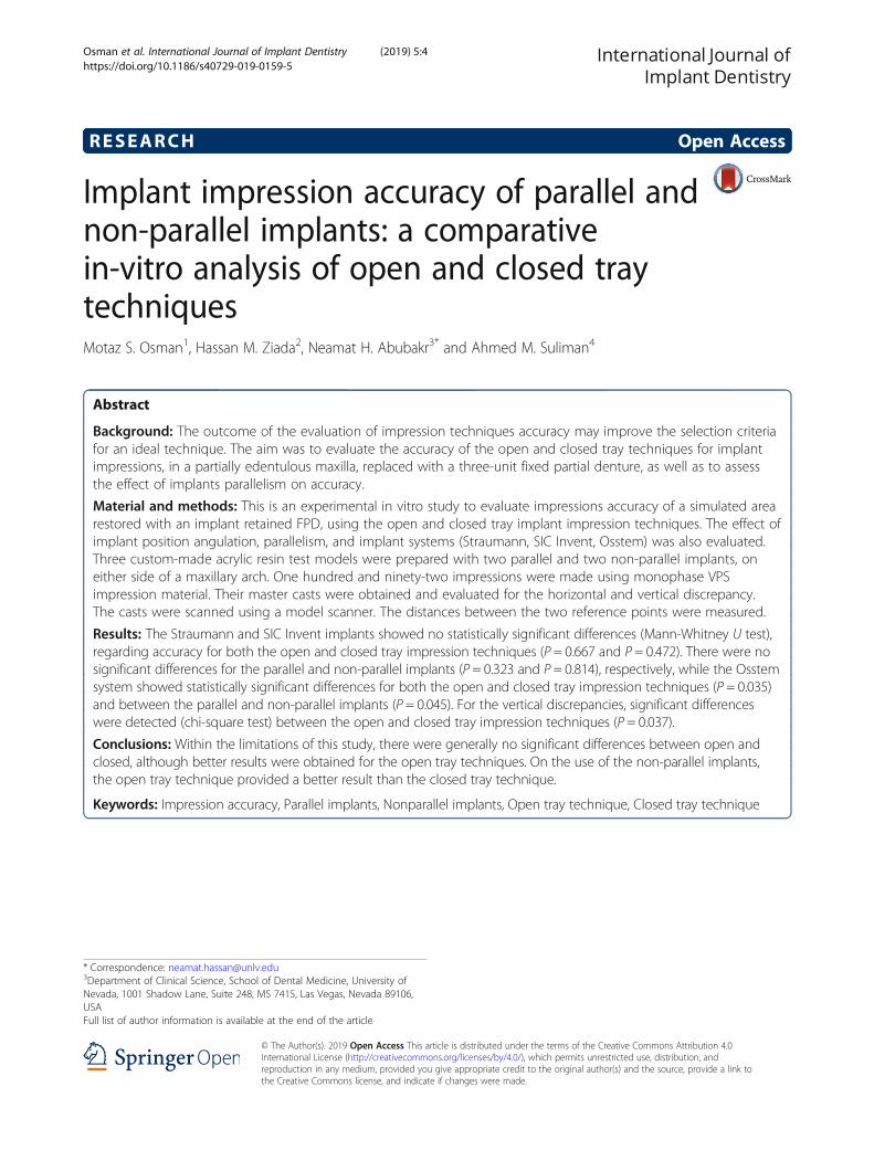
ous study [7]. A sample that would produce the powerfor analysis for this study was derived as 13, for each im-pression technique, for the three-implant systems. It wasincreased to 16, to allow for potential error during prep-aration. Accordingly, 192 impressions were made, in-cluding 96 parallel and 96 non-parallels, and figureshows the sample distribution (Fig. 1).Three custom-made acrylic resin models (each model
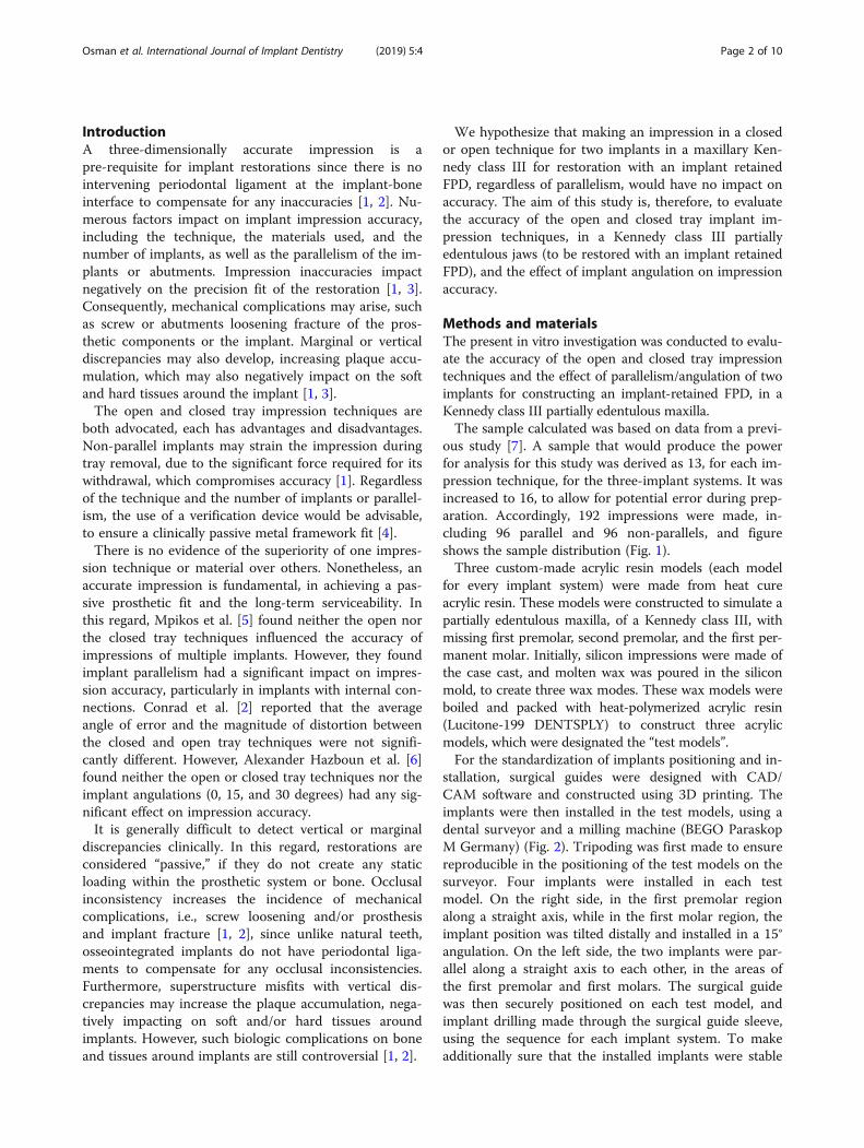
for every implant system) were made from heat cureacrylic resin. These models were constructed to simulate apartially edentulous maxilla, of a Kennedy class III, withmissing first premolar, second premolar, and the first per-manent molar. Initially, silicon impressions were made ofthe case cast, and molten wax was poured in the siliconmold, to create three wax modes. These wax models wereboiled and packed with heat-polymerized acrylic resin(Lucitone-199 DENTSPLY) to construct three acrylicmodels, which were designated the “test models”.For the standardization of implants positioning and in-
stallation, surgical guides were designed with CAD/CAM software and constructed using 3D printing. Theimplants were then installed in the test models, using adental surveyor and a milling machine (BEGO ParaskopM Germany) (Fig. 2). Tripoding was first made to ensurereproducible in the positioning of the test models on thesurveyor. Four implants were installed in each testmodel. On the right side, in the first premolar regionalong a straight axis, while in the first molar region, theimplant position was tilted distally and installed in a 15°angulation. On the left side, the two implants were par-allel along a straight axis to each other, in the areas ofthe first premolar and first molars. The surgical guidewas then securely positioned on each test model, andimplant drilling made through the surgical guide sleeve,using the sequence for each implant system. To makeadditionally sure that the installed implants were stable
Osman et al. International Journal of Implant Dentistry (2019) 5:4 Page 2 of 10
within the models, they were cemented using luting ad-hesive resin cement (Multilink/Ivoclar Vivadent) [8].The first test model had the Straumann Implant Sys-
tem (Straumann AG, Basel, Switzerland), with implantfixtures diameter of 4.1 mm and a length of 10.0 mm.
The second test model had the SIC Invent Implant Sys-tem (SIC invent Deutschland GmbH, Germany) with animplant fixture diameter of 4.0 mm and a length of 11.5mm. The third test model had the Osstem Implant Sys-tem (426-5, Gasan-dong, Geumcheon-gu, Seoul, Korea)
Sample Size Distribution (192)
Parallel (96)
Open Tray Technique (48)
Straumann
SIC (16)
Osstem (16)
Closed Tray Technique (48)
Straumann (16)
SIC (16)
Osstem (16)
Non-Parallel (96)
Open Tray Technique (48)
Straumann (16)
SIC (16)
Osstem (16)
Closed Tray Technique (48
Straumann (16)
SIC (16)
Osstem (16)
Fig. 1 Flowchart of the sample distribution
Fig. 2 Dental surveyor for the standardization of drilling, angulations, and implant installments
Osman et al. International Journal of Implant Dentistry (2019) 5:4 Page 3 of 10
with an implant fixture diameter of 4.0 mm and a lengthof 11.5 mm.
Horizontal measurementsThe three test models were scanned using ahigh-resolution dental scanner (Activity 885 Smart Op-tics - Sensortechnik GmbH, Bochum, Germany) [9]. Thedigital horizontal distances between the two implants onthe parallel and non-parallel implants sites were mea-sured from center to center of implant fixtures using thesoftware (exocad-Dental CAD). The distance on theright side between the implants at first premolar site(along axis) and the first molar site (distally angulated,15°) was assigned as distance 1 (D1), and the distance onthe left side between first premolar and first molar
(parallel implants) sites were designated as distance 2(D2). These measurements were recorded as the testmodel baseline reference measurements. Each testmodel was then stabilized on the table, the impres-sion copings inserted and secured to their implantfixtures, and the impressions were made (VirtualMonophase vinyl polysiloxane - Ivoclar VivadentAG.), and repeated for every implant system. The im-pressions were checked to fulfill the evaluation cri-teria described by Lee and Gallucci [10]. The criteriainclude the following:
� There should be an exact imprint and reproductionof the implant areas.
� The impression copings should not be displacedfrom the impression.
Fig. 3 CAD/CAM verification jig
a b c
Fig. 4 a Stereomicroscope used. b No vertical/marginal discrepancy. c Presence of vertical and marginal discrepancy
Osman et al. International Journal of Implant Dentistry (2019) 5:4 Page 4 of 10
� There should be no voids in the occlusal, buccal,lingual, and interproximal surfaces of theneighboring teeth.
� The impression material should not be separatedfrom the custom tray.
Any impression not meeting any of these criteriawas repeated.The impression copings were then reinserted and
secured in their corresponding implant analogs,poured using type IV dental stone (Elite Rock, Zher-mack, Italy). The casts were separated after 45 min,according to the manufacturer’s instruction. Theywere then stored at room temperature for 24 h beforethe second horizontal measurements made [7]. Forthese second horizontal distance measurements, themaster casts were scanned, similar to the test models(Activity 885 smart optics), and the D1 and D2 weremeasured using software exocad-Dental CAD. Themeasurements were recorded and used to comparethe horizontal distances measurements between thetest models and the cast digital measurements, forevery technique and each implant system.
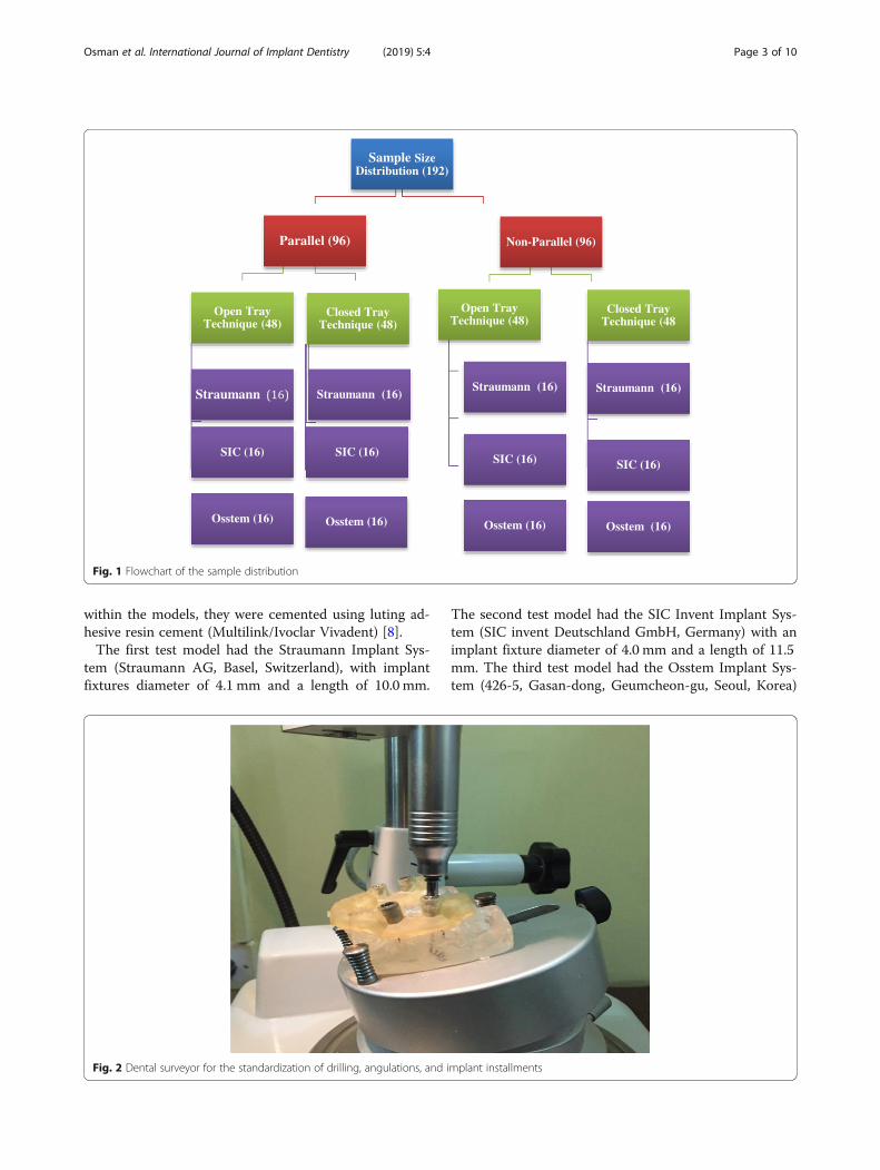
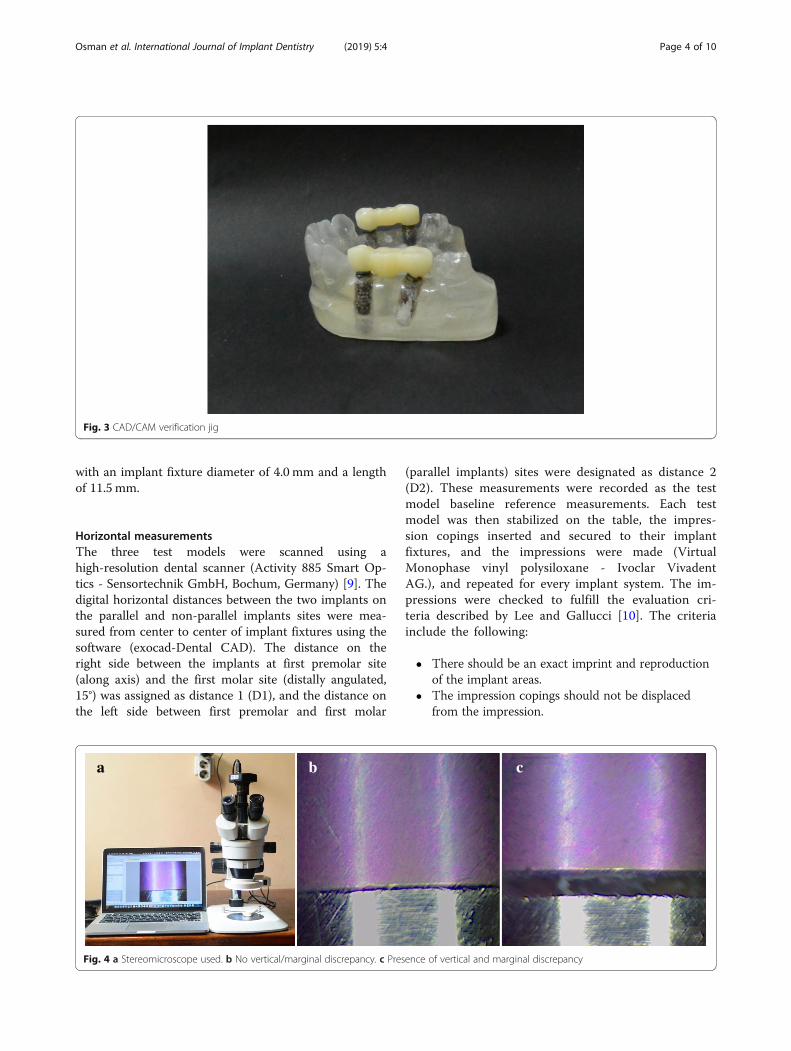
The vertical evaluationThe stone casts were later sectioned to a base of 20mm, tofacilitate placement under a stereomicroscope. Verificationjigs were initially fabricated from CAD/CAM acrylic resinblocks (BILKIM PMMA blank for CAD-CAM applications14mm -A2 color-Turkey) on the test models (Fig. 3). Theseverification jigs were made to evaluate vertical discrepancieson the master casts under a stereomicroscope.Tightening the screws on the laboratory implant analogs,
on the sectioned master cast, retained the jigs [11]. Thepresence or absence of the vertical discrepancies on the sec-tioned casts was then evaluated under the stereomicroscope(AmScop14370, Myford Road, #150, Irvine, CA 92606 USA)at × 50 magnifications and related data recorded (Fig. 4a–c).
Statistical analysisData were tabulated and statistically analyzed using IBMSPSS Statistics software version 22. The data collectedfrom the three-implant systems were classified and usedto compare the effects of implant impression techniquesand parallelism. The p value was set at p ≤ 0.05 andregarded as statistically significant.
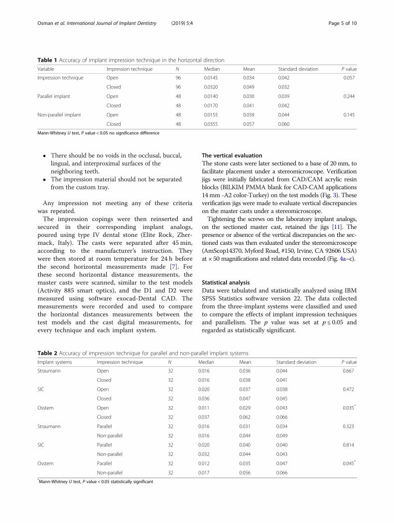
Table 1 Accuracy of implant impression technique in the horizontal direction
Variable Impression technique N Median Mean Standard deviation P value
Impression technique Open 96 0.0145 0.034 0.042 0.057
Closed 96 0.0320 0.049 0.032
Parallel implant Open 48 0.0140 0.030 0.039 0.244
Closed 48 0.0170 0.041 0.042
Non-parallel implant Open 48 0.0155 0.039 0.044 0.145
Closed 48 0.0355 0.057 0.060
Mann-Whitney U test, P value < 0.05 no significance difference
Table 2 Accuracy of impression technique for parallel and non-parallel implant systems
Implant systems Impression technique N Median Mean Standard deviation P value
Straumann Open 32 0.016 0.036 0.044 0.667
Closed 32 0.016 0.038 0.041
SIC Open 32 0.020 0.037 0.038 0.472
Closed 32 0.036 0.047 0.045
Osstem Open 32 0.011 0.029 0.043 0.035*
Closed 32 0.037 0.062 0.066
Straumann Parallel 32 0.016 0.031 0.034 0.323
Non-parallel 32 0.016 0.044 0.049
SIC Parallel 32 0.020 0.040 0.040 0.814
Non-parallel 32 0.032 0.044 0.043
Osstem Parallel 32 0.012 0.035 0.047 0.045*
Non-parallel 32 0.017 0.056 0.066*Mann-Whitney U test, P value < 0.05 statistically significant
Osman et al. International Journal of Implant Dentistry (2019) 5:4 Page 5 of 10
ResultsOne hundred ninety-two impressions, including 96 par-allel and 96 non-parallel implants were made for thethree test models with the three implant systems. Therewere no statistically significant differences (Mann-Whit-ney U test) in impression accuracy between the openand closed tray techniques (P = 0.057). For the paralleland non-parallel implants, there were also no statisticallysignificant differences between the two techniques(Mann-Whitney U test, P = 0.244, P = 0.145) (Table 1).For the implant system, the Straumann and SIC Invent
implants showed no statistically significant differences(Mann-Whitney U test), in the open and closed tray im-pression techniques (P = 0.667 and P = 0.472, respect-ively), while the Osstem system showed statisticallysignificant differences between the two impression tech-niques (P = 0.035) (Table 2).There was also no statistically significant differences
(Mann-Whitney U test) in impression accuracy betweenthe parallel and non-parallel implants, for the Straumann
and SIC Invent implant systems (P = 0.323 and P = 0.814,respectively). There were, however, significant differencesbetween the parallel and non-parallel implants for theOsstem implant system (P = 0.045*) (Table 2).For the open tray technique, significant differences
were observed (Mann-Whitney U test) between paralleland non-parallel implants for the Ostem implant system(P value 0.0166*), while no significant differences in thisregard for the Straumann (P value 0.926) and SIC Inventimplant systems (P value 0.999) (Table 3).In the closed tray technique, no significant differences
between parallel and non-parallel implants observed forthe three implant systems (Mann-Whitney U test):Ostem (P value 0.423), Straumann (P value 0.196), andSIC Invent implant system (P value 0.616) (Table 3).For vertical discrepancies evaluations, significant differ-
ences were detected (chi-square test) between the openand closed tray impression techniques (P = 0.037*). Forthe parallel and non-parallel implants evaluations, therewere no significant differences between the open and
Table 3 Accuracy of open and closed tray impression techniques for both parallel and non-parallel implant systems
Impression technique Implant system Angulations Count Median Mean SD P value
Open tray Straumann Parallel 16 0.017 0.028 0.031 0.926
Non-parallel 16 0.015 0.045 0.054
SIC Invent Parallel 16 0.019 0.040 0.045 0.999
Non-parallel 16 0.020 0.034 0.030
Osstem Parallel 16 0.008 0.021 0.038 0.0166*
Non-parallel 16 0.016 0.037 0.047
Closed tray Strauman Parallel 16 0.015 0.034 0.037 0.196
Non-parallel 16 0.016 0.043 0.045
SIC Invent Parallel 16 0.028 0.040 0.036 0.616
Non-parallel 16 0.041 0.053 0.053
Osstem Parallel 16 0.026 0.049 0.052 0.423
Non-parallel 16 0.039 0.075 0.078*Mann-Whitney U test, P value < 0.05 statistically significant
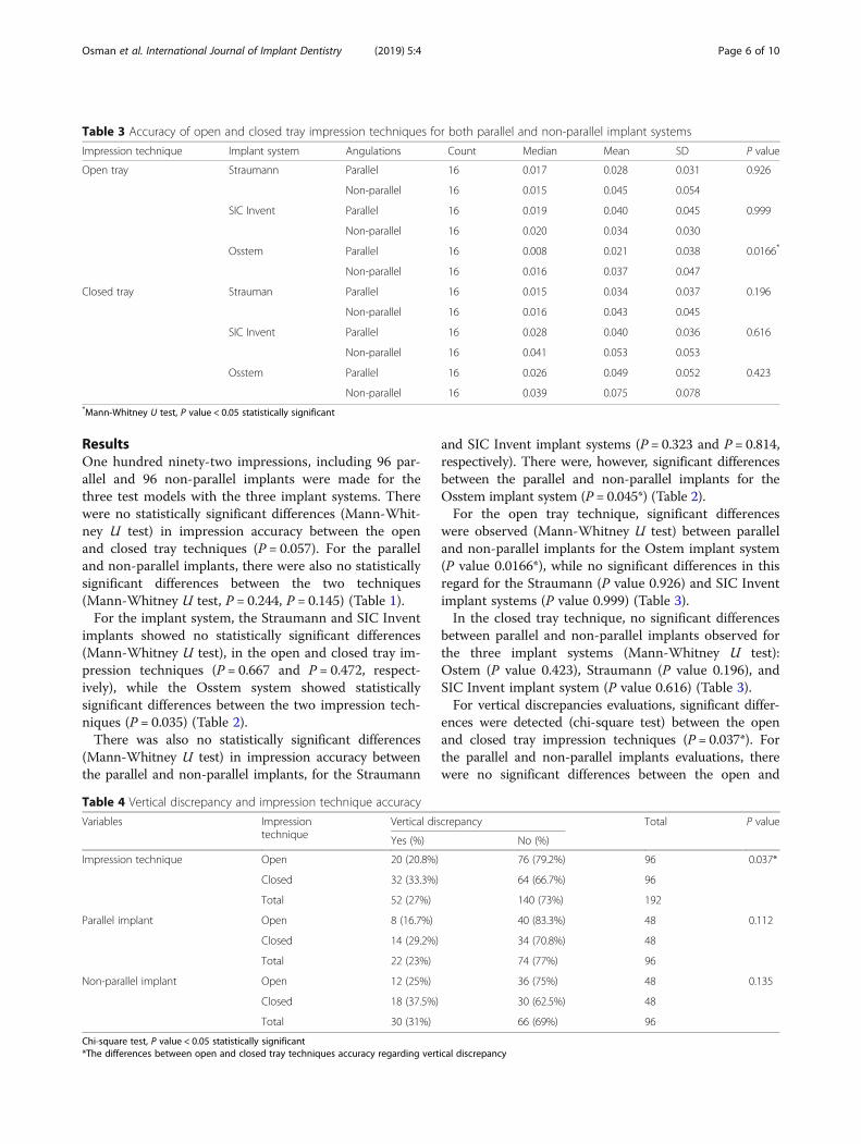
Table 4 Vertical discrepancy and impression technique accuracy
Variables Impressiontechnique
Vertical discrepancy Total P value
Yes (%) No (%)
Impression technique Open 20 (20.8%) 76 (79.2%) 96 0.037*
Closed 32 (33.3%) 64 (66.7%) 96
Total 52 (27%) 140 (73%) 192
Parallel implant Open 8 (16.7%) 40 (83.3%) 48 0.112
Closed 14 (29.2%) 34 (70.8%) 48
Total 22 (23%) 74 (77%) 96
Non-parallel implant Open 12 (25%) 36 (75%) 48 0.135
Closed 18 (37.5%) 30 (62.5%) 48
Total 30 (31%) 66 (69%) 96
Chi-square test, P value < 0.05 statistically significant*The differences between open and closed tray techniques accuracy regarding vertical discrepancy
Osman et al. International Journal of Implant Dentistry (2019) 5:4 Page 6 of 10
closed tray impression techniques (P = 0.112, P = 0.135)(Table 4).Regarding the vertical discrepancy for open tray im-
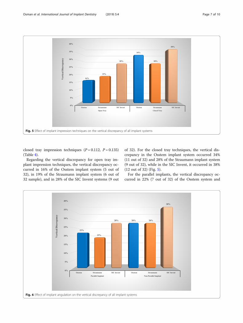
plant impression techniques, the vertical discrepancy oc-curred in 16% of the Osstem implant system (5 out of32), in 19% of the Straumann implant system (6 out of32 sample), and in 28% of the SIC Invent systems (9 out
of 32). For the closed tray techniques, the vertical dis-crepancy in the Osstem implant system occurred 34%(11 out of 32) and 28% of the Straumann implant system(9 out of 32), while in the SIC Invent, it occurred in 38%(12 out of 32) (Fig. 5).For the parallel implants, the vertical discrepancy oc-
curred in 22% (7 out of 32) of the Osstem system and
Fig. 5 Effect of implant impression techniques on the vertical discrepancy of all implant systems
Fig. 6 Effect of implant angulation on the vertical discrepancy of all implant systems
Osman et al. International Journal of Implant Dentistry (2019) 5:4 Page 7 of 10
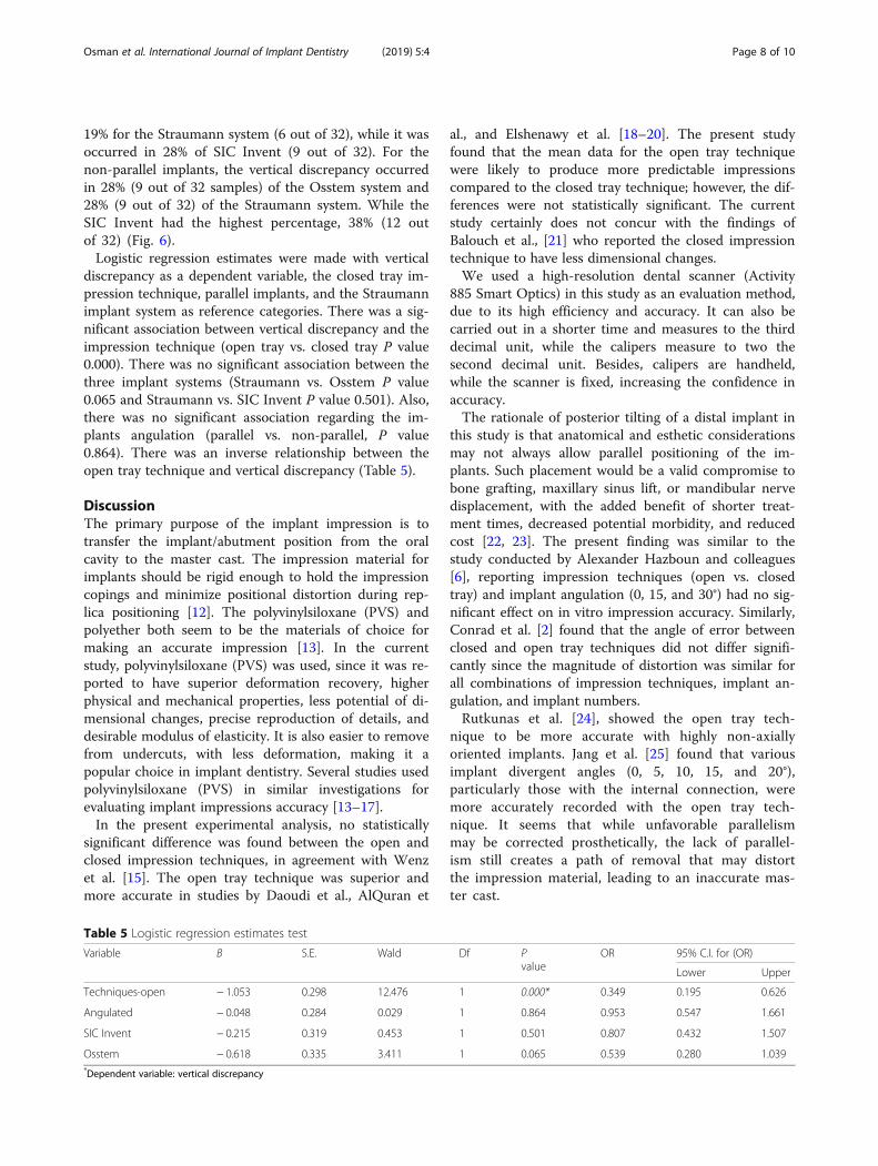
19% for the Straumann system (6 out of 32), while it wasoccurred in 28% of SIC Invent (9 out of 32). For thenon-parallel implants, the vertical discrepancy occurredin 28% (9 out of 32 samples) of the Osstem system and28% (9 out of 32) of the Straumann system. While theSIC Invent had the highest percentage, 38% (12 outof 32) (Fig. 6).Logistic regression estimates were made with vertical
discrepancy as a dependent variable, the closed tray im-pression technique, parallel implants, and the Straumannimplant system as reference categories. There was a sig-nificant association between vertical discrepancy and theimpression technique (open tray vs. closed tray P value0.000). There was no significant association between thethree implant systems (Straumann vs. Osstem P value0.065 and Straumann vs. SIC Invent P value 0.501). Also,there was no significant association regarding the im-plants angulation (parallel vs. non-parallel, P value0.864). There was an inverse relationship between theopen tray technique and vertical discrepancy (Table 5).
DiscussionThe primary purpose of the implant impression is totransfer the implant/abutment position from the oralcavity to the master cast. The impression material forimplants should be rigid enough to hold the impressioncopings and minimize positional distortion during rep-lica positioning [12]. The polyvinylsiloxane (PVS) andpolyether both seem to be the materials of choice formaking an accurate impression [13]. In the currentstudy, polyvinylsiloxane (PVS) was used, since it was re-ported to have superior deformation recovery, higherphysical and mechanical properties, less potential of di-mensional changes, precise reproduction of details, anddesirable modulus of elasticity. It is also easier to removefrom undercuts, with less deformation, making it apopular choice in implant dentistry. Several studies usedpolyvinylsiloxane (PVS) in similar investigations forevaluating implant impressions accuracy [13–17].In the present experimental analysis, no statistically
significant difference was found between the open andclosed impression techniques, in agreement with Wenzet al. [15]. The open tray technique was superior andmore accurate in studies by Daoudi et al., AlQuran et
al., and Elshenawy et al. [18–20]. The present studyfound that the mean data for the open tray techniquewere likely to produce more predictable impressionscompared to the closed tray technique; however, the dif-ferences were not statistically significant. The currentstudy certainly does not concur with the findings ofBalouch et al., [21] who reported the closed impressiontechnique to have less dimensional changes.We used a high-resolution dental scanner (Activity
885 Smart Optics) in this study as an evaluation method,due to its high efficiency and accuracy. It can also becarried out in a shorter time and measures to the thirddecimal unit, while the calipers measure to two thesecond decimal unit. Besides, calipers are handheld,while the scanner is fixed, increasing the confidence inaccuracy.The rationale of posterior tilting of a distal implant in
this study is that anatomical and esthetic considerationsmay not always allow parallel positioning of the im-plants. Such placement would be a valid compromise tobone grafting, maxillary sinus lift, or mandibular nervedisplacement, with the added benefit of shorter treat-ment times, decreased potential morbidity, and reducedcost [22, 23]. The present finding was similar to thestudy conducted by Alexander Hazboun and colleagues[6], reporting impression techniques (open vs. closedtray) and implant angulation (0, 15, and 30°) had no sig-nificant effect on in vitro impression accuracy. Similarly,Conrad et al. [2] found that the angle of error betweenclosed and open tray techniques did not differ signifi-cantly since the magnitude of distortion was similar forall combinations of impression techniques, implant an-gulation, and implant numbers.Rutkunas et al. [24], showed the open tray tech-
nique to be more accurate with highly non-axiallyoriented implants. Jang et al. [25] found that variousimplant divergent angles (0, 5, 10, 15, and 20°),particularly those with the internal connection, weremore accurately recorded with the open tray tech-nique. It seems that while unfavorable parallelismmay be corrected prosthetically, the lack of parallel-ism still creates a path of removal that may distortthe impression material, leading to an inaccurate mas-ter cast.
Table 5 Logistic regression estimates test
Variable B S.E. Wald Df Pvalue
OR 95% C.I. for (OR)
Lower Upper
Techniques-open − 1.053 0.298 12.476 1 0.000* 0.349 0.195 0.626
Angulated − 0.048 0.284 0.029 1 0.864 0.953 0.547 1.661
SIC Invent − 0.215 0.319 0.453 1 0.501 0.807 0.432 1.507
Osstem − 0.618 0.335 3.411 1 0.065 0.539 0.280 1.039*Dependent variable: vertical discrepancy
Osman et al. International Journal of Implant Dentistry (2019) 5:4 Page 8 of 10
A limitation of this study is the lack of three-dimen-sional evaluation and analysis. Hence, in thistwo-dimensional evaluation and analysis, some informa-tion may have been lost during the assessment. Also,using computer-aided design/computer-assisted manu-facture (CAD/CAM) and three-dimensional (3D) opticaldigitization may have led to different results. Neverthe-less, it still would be considered a simple and a percep-tive means of evaluating the accuracy of varyingimpression techniques [23]. Therefore, further studiesshould examine the effect of multiple implant positions,with various angulations and depths, and with variousimpression materials. Another limitation is that being anin vitro study, it is not clear if data from this studywould be similar in the clinical setting. The authors arecurrently analyzing data on the outcome of the two im-pression techniques in a clinical study.
ConclusionWithin the limitation of this study, the open and closedtray implant impression techniques showed a similar levelof accuracy. For the non-parallel implants, the open traytechnique provided a better result than the closed traytechnique. The open tray impression technique exhibitedthe least horizontal and vertical discrepancies for theStraumann, SIC Invent, and Osstem implant systems.
AcknowledgementsNot applicable.
FundingNot applicable.
Availability of data and materialsThe authors declare that they have full control on all data and materials ofthis study.
Authors’ contributionsMO carried out the sample preparation, measurements, data collection, anddrafting of the manuscript. HZ performed the analysis and interpretation ofdata participated in drafting and preparing of the manuscript. NAparticipated in the conception and design of the study and participated inthe preparation and revision of the manuscript. AS participated in the designof the study and discussion of the results. All authors read and approved thefinal manuscript.
Ethics approval and consent to participateNot applicable
Consent for publicationNot applicable.
Competing interestsMotaz Osman, Hassan Ziada, Neamat Abubakr, and Ahmed Suliman declarethat they have no competing interests.
Publisher’s NoteSpringer Nature remains neutral with regard to jurisdictional claims inpublished maps and institutional affiliations.
Author details1Department of Oral rehabilitation, Faculty of Dentistry, University ofKhartoum, Khartoum, Sudan. 2Department of Clinical Science, School ofDental Medicine, University of Nevada, Las Vegas, Nevada, USA. 3Departmentof Clinical Science, School of Dental Medicine, University of Nevada, 1001Shadow Lane, Suite 248, MS 7415, Las Vegas, Nevada 89106, USA.4Department of Oral Maxillofacial Surgery, University of Khartoum, Khartoum,Sudan.
Received: 4 December 2018 Accepted: 6 January 2019
References1. Sorrentino R, Gherlone EF, Calesini G, Zarone F. Effect of implant angulation,
connection length, and impression material on the dimensional accuracy ofimplant impressions: an in vitro comparative study. Clin Implant Dent RelatRes. 2010;12(Suppl 1):e63–76.
2. Conrad HJ, Pesun IJ, DeLong R, Hodges JS. Accuracy of two impressiontechniques with angulated implants. J Prosthet Dent. 2007;97:349–56.
3. Lee H, So JS, Hochstedler JL, Ercoli C. The accuracy of implant impressions:a systematic review. J Prosthet Dent. 2008;100:285–91.
4. Lin WS, Harris BT, Metz MJ, Morton D. A technique for verifying andcorrecting a milled polyurethane definitive cast for nonsegmental implantrestoration in an edentulous jaw. J Prosthet Dent. 2014;112:658–62.
5. Mpikos P, Kafantaris N, Tortopidis D, Galanis C, Kaisarlis G, Koidis P. Theeffect of impression technique and implant angulation on the impressionaccuracy of external- and internal-connection implants. Int J Oral MaxillofacImplants. 2012;27:1422–8.
6. Alexander Hazboun GB, Masri R, Romberg E, Kempler J, Driscoll CF. Effect ofimplant angulation and impression technique on impressions ofNobelActive implants. J Prosthet Dent. 2015;113:425–31.
7. Herbst D, Nel JC, Driessen CH, Becker PJ. Evaluation of impression accuracyfor osseointegrated implant supported superstructures. J Prosthet Dent.2000;83:555–61.
8. Shim JS, Ryu JJ, Shin SW, Lee JY. Effects of implant angulation andimpression coping type on the dimensional accuracy of impressions.Implant Dent. 2015;24:726–9.
9. Technical data sheet: Activity 885, Smart Optics Sensortechnik GmbH,Bochum, Germany. https://www.smartoptics.de/en/dental/dental-scan/.
10. Lee SJ, Gallucci GO. Digital vs. conventional implant impressions: efficiencyoutcomes. Clin Oral Implants Res. 2013;24:111–5.
11. Papaspyridakos P, Lal K, White GS, Weber HP, Gallucci GO. Effect of splintedand nonsplinted impression techniques on the accuracy of fit of fixedimplant prostheses in edentulous patients: a comparative study. Int J OralMaxillofac Implants. 2011;26:1267–72.
12. Gokcen-Rohlig B, Ongul D, Sancakli E, Sermet B. Comparative evaluation ofthe effects of implant position, impression material, and tray type onimplant impression accuracy. Implant Dent. 2014;23:283–8.
13. Prithviraj DR, Pujari M, Garg P, Shruthi D. Accuracy of the implantimpression obtained from different impression materials and techniques:review. J Clin Exp Dent. 2011;3:106–11.
14. Sabouhi M, Bajoghli F, Dakhilalian M, Beygi A, Abolhasani M. Effects ofimpression coping design, impression technique, and dental undercuts onthe accuracy of implant impressions assessed by 3-dimensional opticalscanning: an in vitro study. Implant Dent. 2016;25:238–46.
15. Wenz HJ, Hertrampf K. Accuracy of impressions and casts using differentimplant impression techniques in a multi-implant system with an internalhex connection. Int J Oral Maxillofac Implants. 2008;23:39–47.
16. Hamalian TA, Nasr E, Chidiac JJ. Impression materials in fixedprosthodontics: influence of choice on clinical procedure. J Prosthodont.2011;20:153–60.
17. Gallucci GO, Papaspyridakos P, Ashy LM, Kim GE, Brady NJ, Weber HP. Clinicalaccuracy outcomes of closed-tray and open-tray implant impressiontechniques for partially edentulous patients. Int J Prosthodont. 2011;24:469–72.
18. Daoudi MF, Setchell DJ, Searson LJ. An evaluation of three implant levelimpression techniques for single tooth implant. Eur J Prosthodont RestorDent. 2004;12:9–14.
19. Al Quran FA, Rashdan BA, Zomar AA, Weiner S. Passive fit and accuracyof three dental implant impression techniques. Quintessence Int. 2012;43:119–25.
Osman et al. International Journal of Implant Dentistry (2019) 5:4 Page 9 of 10

20. Elshenawy EA, Alam-Eldein AM, Abd Elfatah FA. Cast accuracy obtainedfrom different impression techniques at different implant angulations(in vitro study). Int J. Implant Dent. 2018;4:9.
21. Balouch F, Jalalian E, Nikkheslat M, Ghavamian R, Toopchi S, Jallalian F, et al.Comparison of dimensional accuracy between open-tray and closed-trayimplant impression technique in 15 degrees angled implants. J Dent.2013;14:96–102.
22. Papaspyridakos P, Chen CJ, Gallucci GO, Doukoudakis A, Weber HP,Chronopoulos V. Accuracy of implant impressions for partially andcompletely edentulous patients: a systematic review. Int J Oral MaxillofacImplants. 2014;29:836–45.
23. Kim JH, Kim KR, Kim S. Critical appraisal of implant impression accuracies: asystematic review. J Prosthet Dent. 2015;114:185–92.
24. Rutkunas V, Sveikata K, Savickas R. Effects of implant angulation, materialselection, and impression technique on impression accuracy: a preliminarylaboratory study. Int J Prosthodont. 2012;25:512–5.
25. Jang HK, Kim S, Shim JS, Lee KW, Moon HS. Accuracy of impressions forinternal-connection implant prostheses with various divergent angles.Int J Oral Maxillofac Implants. 2011;26:1011–5.
Osman et al. International Journal of Implant Dentistry (2019) 5:4 Page 10 of 10
Related Documents