Proc. Natl. Acad. Sci. USA Vol. 95, pp. 13612–13617, November 1998 Cell Biology Impairing follicle-stimulating hormone (FSH) signaling in vivo: Targeted disruption of the FSH receptor leads to aberrant gametogenesis and hormonal imbalance ANDRE ´E DIERICH* ² , M. RAM SAIRAM ²‡§ ,LUCIA MONACO*, GIAN MARIA FIMIA*, ANNE GANSMULLER*, MARIANNE LEMEUR*, AND PAOLO SASSONE-CORSI* § *Institut de Ge ´ne ´tique et de Biologie Mole ´culaire et Cellulaire, Centre National de la Recherche Scientifique, Institut National de la Sante ´ et de la Recherche Me ´dicale, Universite ´ Louis Pasteur, B. P. 163, 67404 Illkirch, Strasbourg, France; and ‡ Molecular Reproduction Research Laboratory, Clinical Research Institute, 110 Pine Avenue W., Montreal PQ H2W 1R7, Que ´bec, Canada Communicated by Pierre Chambon, Institut de Ge ´ne ´tique et de Biologie Mole ´culaire et Cellulaire, Strasbourg, France, September 14, 1998 (received for review August 21, 1998) ABSTRACT Pituitary gonadotropins follicle-stimulating hormone (FSH) and luteinizing hormone stimulate the gonads by regulating germ cell proliferation and differentiation. FSH receptors (FSH-Rs) are localized to testicular Sertoli cells and ovarian granulosa cells and are coupled to activation of the adenylyl cyclase and other signaling pathways. Activation of FSH-Rs is considered essential for folliculogenesis in the female and spermatogenesis in the male. We have generated mice lacking FSH-R by homologous recombination. FSH-R- deficient males are fertile but display small testes and partial spermatogenic failure. Thus, although FSH signaling is not essential for initiating spermatogenesis, it appears to be required for adequate viability and motility of the sperms. FSH-R-deficient females display thin uteri and small ovaries and are sterile because of a block in folliculogenesis before antral follicle formation. Although the expression of marker genes is only moderately altered in FSH-R 2y2 mice, drastic sex-specific changes are observed in the levels of various hormones. The anterior lobe of the pituitary gland in females is enlarged and reveals a larger number of FSH- and thyroid- stimulating hormone (TSH)-positive cells. The phenotype of FSH-R 2y2 mice is reminiscent of human hypergonadotropic ovarian dysgenesis and infertility. The pituitary glycoprotein hormone follicle-stimulating hor- mone (FSH) plays an essential role in mammalian reproduc- tion (1) through interaction with a specific receptor (2). The FSH receptor (FSH-R) is a G-protein-coupled, seven- transmembrane receptor linked to the adenylyl cyclase or other pathways (3–5). FSH-R activation initiates a cascade of intracellular events leading to modification of cell response (6–8) and receptor desensitization (3). In the male, the FSH-R is expressed exclusively in Sertoli cells (9, 10). FSH signaling is considered essential for pubertal initiation of spermatogenesis and maintenance of normal sperm production in the adult (3). This function is thought to be exerted via intimate contacts between Sertoli cells and differentiating spermatogonia (11). Upon FSH and testoster- one stimulation, Sertoli cells provide a critical support re- quired for germ cell differentiation (12). In the female, the FSH-R is expressed in granulosa cells and is thought to tightly regulate the various phases of follicle maturation in response to periodic pituitary FSH release (3, 10, 13). During the reproductive life only a fraction of follicles undergo differentiation, whereas more than 99% enter a degenerative process called atresia. The onset of FSH stimu- lation at puberty reduces apoptosis, enhances proliferation, and induces follicular maturation leading to ovulation (3, 13). The critical role played by FSH signaling is illustrated by the phenotype of mice carrying a targeted mutation in the FSHb subunit gene (14) and by the effect of FSH-R mutations in humans (15, 16). An inactivating mutation (Ala-189–Val) found in females with pure ovarian dysgenesis leads to a disease characterized by normal karyotype, high gonadotro- pins, and streaky gonads associated with primary amenorrhea (13, 17). This mutation lies in the extracellular domain and is thought to modify protein folding (17). Importantly, males with the same mutation display various degrees of spermato- genic failure, lack of azoospermia, or absolute infertility (18). Thus, the same inactivating mutation differentially influ- ences reproduction in males or females. One activating mu- tation has been found in a male hypophysectomized patient who, under testosterone therapy, was unexpectedly fertile in spite of undetectable gonadotropin levels (16). This heterozy- gous Asp-567–Gly substitution in the third intracytoplasmic loop leads to constitutively increased cAMP levels indepen- dently of FSH stimulation (16). We have generated mutant mice for the FSH-R by homol- ogous recombination. The conserved organization of the human, mouse, and rat FSH-R genes has been established (19–22). Mutant males display small testes, partial spermato- genic failure, and reduced fertility. FSH signaling is not essential for initiating spermatogenesis, but is required to sustain adequate viability and motility of the sperms. The phenotype of mutant females is much more severe. They display thin uteri and small ovaries and are sterile because of a block in folliculogenesis before antral follicle formation. Drastic changes in hormone levels prompted us to analyze the pituitary anatomy. There is a moderate, but significant, en- largement in the anterior lobe accompanied by a drastic increase of FSH-positive cells. These animals constitute a model to study the physiological link between gonads and pituitary, hypergonadotropic ovarian dysgenesis, and infertil- ity. MATERIALS AND METHODS Generation of FSH-R 2y2 Mice. An 11-kb genomic frag- ment covering exon I and a large part of intron I of the FSH-R gene was isolated from a 129SV phage library. A 7-kb XbaI– SacI fragment was subcloned for subsequent insertions. A EcoRV fragment (2420 to 1228) was replaced by a PGK-Neo The publication costs of this article were defrayed in part by page charge payment. This article must therefore be hereby marked ‘‘advertisement’’ in accordance with 18 U.S.C. §1734 solely to indicate this fact. © 1998 by The National Academy of Sciences 0027-8424y98y9513612-6$2.00y0 PNAS is available online at www.pnas.org. Abbreviations: FSH, follicle-stimulating hormone; FSH-R, FSH re- ceptor; LH, luteinizing hormone; TSH, thyroid-stimulating hormone. ² A.D. and M.R.S. contributed equally to this work. § To whom reprint requests should be addressed. e-mail: Sairamm@ IRCM.UMontreal.Ca or [email protected]. 13612

Welcome message from author
This document is posted to help you gain knowledge. Please leave a comment to let me know what you think about it! Share it to your friends and learn new things together.
Transcript

Proc. Natl. Acad. Sci. USAVol. 95, pp. 13612–13617, November 1998Cell Biology
Impairing follicle-stimulating hormone (FSH) signaling in vivo:Targeted disruption of the FSH receptor leads to aberrantgametogenesis and hormonal imbalance
ANDREE DIERICH*†, M. RAM SAIRAM†‡§, LUCIA MONACO*, GIAN MARIA FIMIA*, ANNE GANSMULLER*,MARIANNE LEMEUR*, AND PAOLO SASSONE-CORSI*§
*Institut de Genetique et de Biologie Moleculaire et Cellulaire, Centre National de la Recherche Scientifique, Institut National de la Sante et de la RechercheMedicale, Universite Louis Pasteur, B. P. 163, 67404 Illkirch, Strasbourg, France; and ‡Molecular Reproduction Research Laboratory, Clinical Research Institute,110 Pine Avenue W., Montreal PQ H2W 1R7, Quebec, Canada
Communicated by Pierre Chambon, Institut de Genetique et de Biologie Moleculaire et Cellulaire, Strasbourg, France, September 14, 1998 (receivedfor review August 21, 1998)
ABSTRACT Pituitary gonadotropins follicle-stimulatinghormone (FSH) and luteinizing hormone stimulate the gonadsby regulating germ cell proliferation and differentiation. FSHreceptors (FSH-Rs) are localized to testicular Sertoli cells andovarian granulosa cells and are coupled to activation of theadenylyl cyclase and other signaling pathways. Activation ofFSH-Rs is considered essential for folliculogenesis in thefemale and spermatogenesis in the male. We have generatedmice lacking FSH-R by homologous recombination. FSH-R-deficient males are fertile but display small testes and partialspermatogenic failure. Thus, although FSH signaling is notessential for initiating spermatogenesis, it appears to berequired for adequate viability and motility of the sperms.FSH-R-deficient females display thin uteri and small ovariesand are sterile because of a block in folliculogenesis beforeantral follicle formation. Although the expression of markergenes is only moderately altered in FSH-R 2y2 mice, drasticsex-specific changes are observed in the levels of varioushormones. The anterior lobe of the pituitary gland in femalesis enlarged and reveals a larger number of FSH- and thyroid-stimulating hormone (TSH)-positive cells. The phenotype ofFSH-R 2y2 mice is reminiscent of human hypergonadotropicovarian dysgenesis and infertility.
The pituitary glycoprotein hormone follicle-stimulating hor-mone (FSH) plays an essential role in mammalian reproduc-tion (1) through interaction with a specific receptor (2). TheFSH receptor (FSH-R) is a G-protein-coupled, seven-transmembrane receptor linked to the adenylyl cyclase orother pathways (3–5). FSH-R activation initiates a cascade ofintracellular events leading to modification of cell response(6–8) and receptor desensitization (3).
In the male, the FSH-R is expressed exclusively in Sertolicells (9, 10). FSH signaling is considered essential for pubertalinitiation of spermatogenesis and maintenance of normalsperm production in the adult (3). This function is thought tobe exerted via intimate contacts between Sertoli cells anddifferentiating spermatogonia (11). Upon FSH and testoster-one stimulation, Sertoli cells provide a critical support re-quired for germ cell differentiation (12).
In the female, the FSH-R is expressed in granulosa cells andis thought to tightly regulate the various phases of folliclematuration in response to periodic pituitary FSH release (3,10, 13). During the reproductive life only a fraction of folliclesundergo differentiation, whereas more than 99% enter adegenerative process called atresia. The onset of FSH stimu-
lation at puberty reduces apoptosis, enhances proliferation,and induces follicular maturation leading to ovulation (3, 13).
The critical role played by FSH signaling is illustrated by thephenotype of mice carrying a targeted mutation in the FSHbsubunit gene (14) and by the effect of FSH-R mutations inhumans (15, 16). An inactivating mutation (Ala-189–Val)found in females with pure ovarian dysgenesis leads to adisease characterized by normal karyotype, high gonadotro-pins, and streaky gonads associated with primary amenorrhea(13, 17). This mutation lies in the extracellular domain and isthought to modify protein folding (17). Importantly, maleswith the same mutation display various degrees of spermato-genic failure, lack of azoospermia, or absolute infertility (18).
Thus, the same inactivating mutation differentially influ-ences reproduction in males or females. One activating mu-tation has been found in a male hypophysectomized patientwho, under testosterone therapy, was unexpectedly fertile inspite of undetectable gonadotropin levels (16). This heterozy-gous Asp-567–Gly substitution in the third intracytoplasmicloop leads to constitutively increased cAMP levels indepen-dently of FSH stimulation (16).
We have generated mutant mice for the FSH-R by homol-ogous recombination. The conserved organization of thehuman, mouse, and rat FSH-R genes has been established(19–22). Mutant males display small testes, partial spermato-genic failure, and reduced fertility. FSH signaling is notessential for initiating spermatogenesis, but is required tosustain adequate viability and motility of the sperms. Thephenotype of mutant females is much more severe. Theydisplay thin uteri and small ovaries and are sterile because ofa block in folliculogenesis before antral follicle formation.Drastic changes in hormone levels prompted us to analyze thepituitary anatomy. There is a moderate, but significant, en-largement in the anterior lobe accompanied by a drasticincrease of FSH-positive cells. These animals constitute amodel to study the physiological link between gonads andpituitary, hypergonadotropic ovarian dysgenesis, and infertil-ity.
MATERIALS AND METHODS
Generation of FSH-R 2y2 Mice. An 11-kb genomic frag-ment covering exon I and a large part of intron I of the FSH-Rgene was isolated from a 129SV phage library. A 7-kb XbaI–SacI fragment was subcloned for subsequent insertions. AEcoRV fragment (2420 to 1228) was replaced by a PGK-Neo
The publication costs of this article were defrayed in part by page chargepayment. This article must therefore be hereby marked ‘‘advertisement’’ inaccordance with 18 U.S.C. §1734 solely to indicate this fact.
© 1998 by The National Academy of Sciences 0027-8424y98y9513612-6$2.00y0PNAS is available online at www.pnas.org.
Abbreviations: FSH, follicle-stimulating hormone; FSH-R, FSH re-ceptor; LH, luteinizing hormone; TSH, thyroid-stimulating hormone.†A.D. and M.R.S. contributed equally to this work.§To whom reprint requests should be addressed. e-mail: [email protected] or [email protected].
13612
cassette with an internal EcoRI site. A GTI-GTII promoter-driven herpes simplex virus thymidine kinase cassette wasinserted at the 59 SalI site. Embryonic stem cells (107) wereelectroporated with targeting plasmid at 400 V and 125 mF.Two mutant clones were injected in C57BLy6 blastocysts. Tenmale chimeras were bred with C57BLy6 females, and germ-line transmission was obtained with three of them.
Histological Analysis. Sections fixed in Bouin solution wereprepared and stained (hematoxylinyeosin) by using standardprocedures. Analysis of pituitary sections was performed asdescribed (23). For the analysis of the estrous cycle vaginalsmears were stained with hematoxylinyeosin as described (23).
FSH Binding and Hormone Assays. Total homogenates andcrude membranes from testes were assayed for FSH binding.Aliquots of 400 mg were incubated at 23°C with 5 ngyml of125I-FSH (NENyDuPont) in 50 mM TriszHCl, 0.5% BSA.Nonspecific binding was assessed in the presence of 50 mgymlof cold ovine FSH (Sigma). Bound hormone was separated bycentrifugation at 3,000 3 g.
Serum FSH, luteinizing hormone (LH), inhibin, and testos-terone levels were determined by RIA using Amersham andPeninsula assay systems according to the manufacturers’ pro-cedures. Intracellular testosterone levels were measured intestis homogenates after ether extraction.
RNA and Protein Analyses. RNA preparation, Northern,reverse transcription–PCR, in situ, and RNase-protection anal-yses were according to standard methods (24). Poly(A)1 RNAwas prepared by using the Pharmacia purification kit. Equalloading and RNA quality were confirmed with a G6PD probe.
Tissues were homogenized in Laemmli buffer. For Westernanalysis the following antibodies were as indicated by themanufacturer: a polyclonal antiprolactin (National Institutesof Health); a mouse monoclonal antiprolactin receptor (In-terchim, Montlucon, France); a rat monoclonal anticyclin D2(Santa Cruz Biotechnology); and a rat polyclonal raisedagainst the regulatory subunit RIIb of the cAMP-dependentprotein kinase (gift of K. Tasken, University of Olso, Norway).
RESULTSHomologous Recombination of the Mouse FSH-R Gene. The
single gene encoding the FSH-R generates a variety of alter-
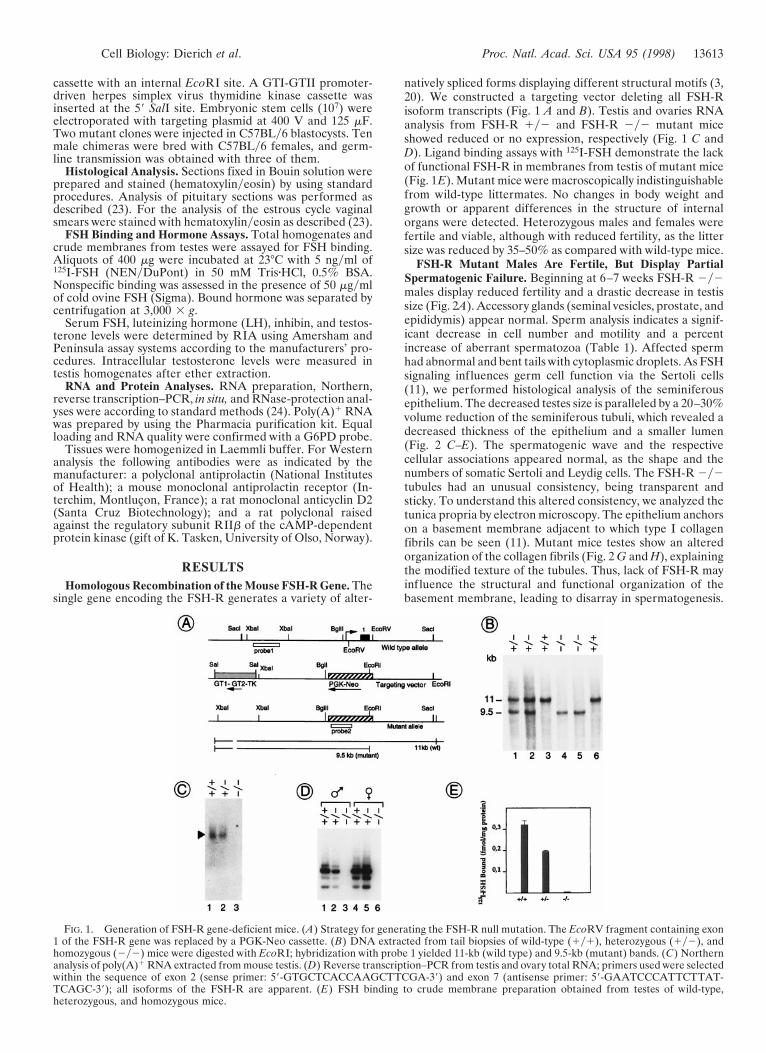
natively spliced forms displaying different structural motifs (3,20). We constructed a targeting vector deleting all FSH-Risoform transcripts (Fig. 1 A and B). Testis and ovaries RNAanalysis from FSH-R 1y2 and FSH-R 2y2 mutant miceshowed reduced or no expression, respectively (Fig. 1 C andD). Ligand binding assays with 125I-FSH demonstrate the lackof functional FSH-R in membranes from testis of mutant mice(Fig. 1E). Mutant mice were macroscopically indistinguishablefrom wild-type littermates. No changes in body weight andgrowth or apparent differences in the structure of internalorgans were detected. Heterozygous males and females werefertile and viable, although with reduced fertility, as the littersize was reduced by 35–50% as compared with wild-type mice.
FSH-R Mutant Males Are Fertile, But Display PartialSpermatogenic Failure. Beginning at 6–7 weeks FSH-R 2y2males display reduced fertility and a drastic decrease in testissize (Fig. 2A). Accessory glands (seminal vesicles, prostate, andepididymis) appear normal. Sperm analysis indicates a signif-icant decrease in cell number and motility and a percentincrease of aberrant spermatozoa (Table 1). Affected spermhad abnormal and bent tails with cytoplasmic droplets. As FSHsignaling influences germ cell function via the Sertoli cells(11), we performed histological analysis of the seminiferousepithelium. The decreased testes size is paralleled by a 20–30%volume reduction of the seminiferous tubuli, which revealed adecreased thickness of the epithelium and a smaller lumen(Fig. 2 C–E). The spermatogenic wave and the respectivecellular associations appeared normal, as the shape and thenumbers of somatic Sertoli and Leydig cells. The FSH-R 2y2tubules had an unusual consistency, being transparent andsticky. To understand this altered consistency, we analyzed thetunica propria by electron microscopy. The epithelium anchorson a basement membrane adjacent to which type I collagenfibrils can be seen (11). Mutant mice testes show an alteredorganization of the collagen fibrils (Fig. 2 G and H), explainingthe modified texture of the tubules. Thus, lack of FSH-R mayinfluence the structural and functional organization of thebasement membrane, leading to disarray in spermatogenesis.
FIG. 1. Generation of FSH-R gene-deficient mice. (A) Strategy for generating the FSH-R null mutation. The EcoRV fragment containing exon1 of the FSH-R gene was replaced by a PGK-Neo cassette. (B) DNA extracted from tail biopsies of wild-type (1y1), heterozygous (1y2), andhomozygous (2y2) mice were digested with EcoRI; hybridization with probe 1 yielded 11-kb (wild type) and 9.5-kb (mutant) bands. (C) Northernanalysis of poly(A)1 RNA extracted from mouse testis. (D) Reverse transcription–PCR from testis and ovary total RNA; primers used were selectedwithin the sequence of exon 2 (sense primer: 59-GTGCTCACCAAGCTTCGA-39) and exon 7 (antisense primer: 59-GAATCCCATTCTTAT-TCAGC-39); all isoforms of the FSH-R are apparent. (E) FSH binding to crude membrane preparation obtained from testes of wild-type,heterozygous, and homozygous mice.
Cell Biology: Dierich et al. Proc. Natl. Acad. Sci. USA 95 (1998) 13613
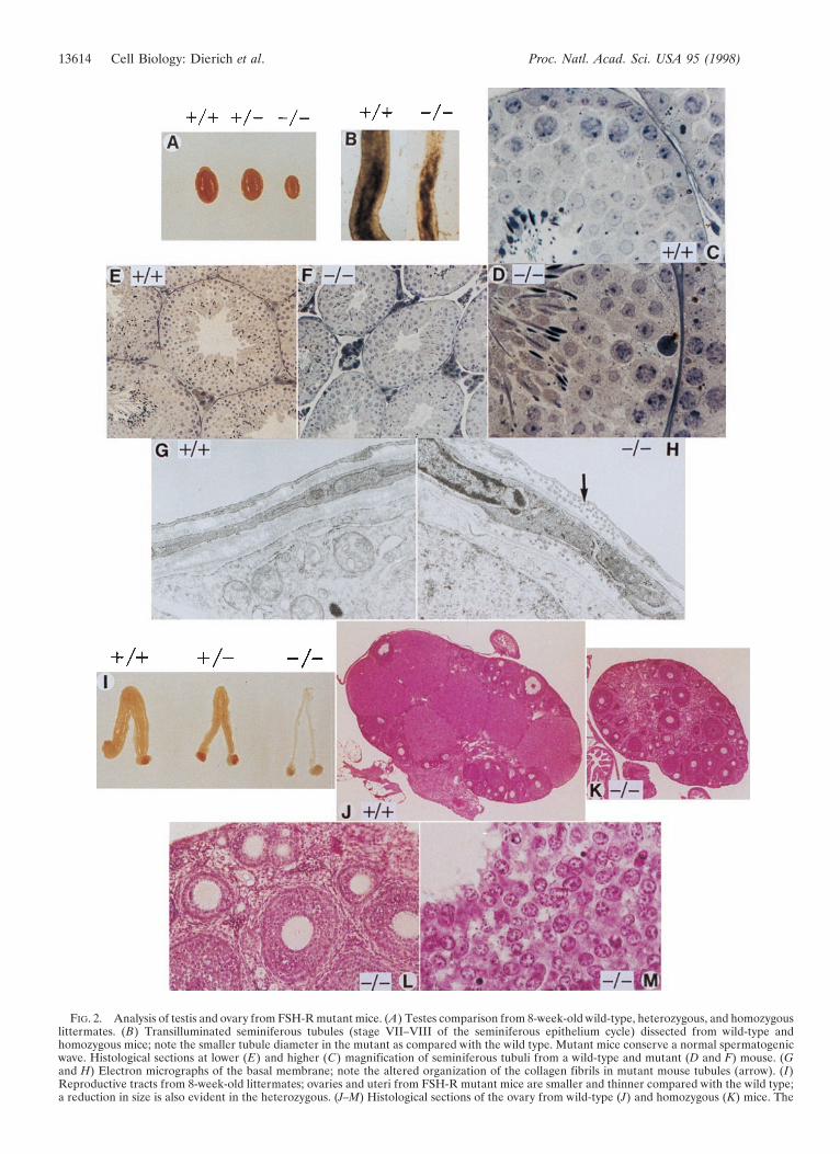
FIG. 2. Analysis of testis and ovary from FSH-R mutant mice. (A) Testes comparison from 8-week-old wild-type, heterozygous, and homozygouslittermates. (B) Transilluminated seminiferous tubules (stage VII–VIII of the seminiferous epithelium cycle) dissected from wild-type andhomozygous mice; note the smaller tubule diameter in the mutant as compared with the wild type. Mutant mice conserve a normal spermatogenicwave. Histological sections at lower (E) and higher (C) magnification of seminiferous tubuli from a wild-type and mutant (D and F) mouse. (Gand H) Electron micrographs of the basal membrane; note the altered organization of the collagen fibrils in mutant mouse tubules (arrow). (I)Reproductive tracts from 8-week-old littermates; ovaries and uteri from FSH-R mutant mice are smaller and thinner compared with the wild type;a reduction in size is also evident in the heterozygous. (J–M) Histological sections of the ovary from wild-type (J) and homozygous (K) mice. The
13614 Cell Biology: Dierich et al. Proc. Natl. Acad. Sci. USA 95 (1998)

FSH-R 2y2 Females Are Sterile. In contrast to the males,the mutant females are infertile. Ovaries and uteri frommutant mice appeared strikingly smaller and thinner as com-pared with wild-type littermates (Fig. 2I). FSH-R 1y2 fe-males displayed an intermediate phenotype. In contrast to thenormal stages of folliculogenesis and the presence of corporalutea, ovaries from FSH-R 2y2 females lacked corpora luteaand mature Graafian follicles (Fig. 2 J–M). Mutant micedemonstrate only primordial, primary, and secondary folliclesand an apparently normal number and location of granulosaand thecal cells at these stages. Thus, the decreased size of theovary in the FSH-R 2y2 females is caused by the lack of largeGraafian follicles and by the absence of corpora lutea causedby ovulatory failure. Hence, the infertility of mutant femalesarises from a block in follicular maturation.
Effects on Gene Expression. The coordinated expression ofseveral genes at defined stages of sperm and follicular devel-opment has been described (13, 25). Surprisingly, the expres-sion of most of these genes is only marginally altered in bothmale and female mutants as compared with wild-type litter-mates (Fig. 3). Relevant examples are represented by cAMP-responsive element modulator (CREM) in males and cyclin D2in females. CREM expression is under FSH regulation duringspermatogenesis (26), while targeted disruption of the geneblocks spermiogenesis and increases germ cells apoptosis (24).Cyclin D2 expression is FSH responsive, and mutation of thegene blocks granulosa cell proliferation, causing sterility (27).The reasons for the unaltered expression of these genes in theFSH-R mutants are unclear. It is plausible that alternativesignaling routes exist that could insure the function of somekey genes as a compensatory mechanism.
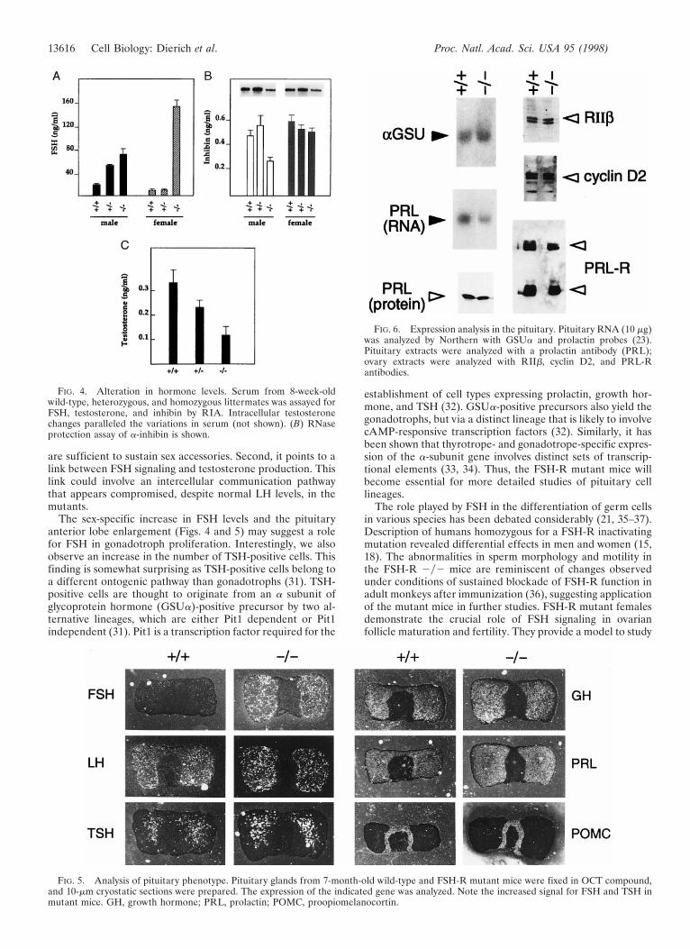
Drastic Alterations in Hormonal Levels. Regulation of thehypothalamic-pituitary axis is controlled by a feedback loopsystem operating from the gonads to the pituitary (3). To studyhow this control may be altered in the absence of FSHsignaling, we measured FSH and LH levels in males andfemales. FSH levels increased by 3-fold in FSH-R 2y2 adultmales, whereas there is a drastic increase of at least 15-fold inmutant females. Interestingly, although heterozygous malesshow an increase with respect to wild-type controls, thisincrease is not observed in the females (Fig. 4A). LH andLH-receptor levels appeared unchanged in male and femalemutants (not shown). The robust change in FSH levels sug-gested that the feedback control exerted by inhibin might bealtered. Regulated a-inhibin is responsible for gonadal cellproliferation and differentiation (27). Although a-inhibinrates drop to about 50% of the wild type in the mutant males,no significant changes are detected in the females (Fig. 4B). Asthe most important increase in FSH levels is observed inmutant females, it appears that inhibin production from othersites is possible in the absence of FSH signaling and thatinhibin-mediated retroinhibition is not essential for FSH-modulated synthesis.
Testosterone levels in mutant males decrease to about 35%of the wild type, with intermediate values in the heterozygous(Fig. 4C). Comparative analyses of the estrous cycle of normaland mutant mice (ref. 23; not shown) showed that a rhythmicperiodicity with a recognizable estrous every 4 days is presentin both populations.
Analysis of the Pituitary Phenotype. The dramatic increaseof FSH levels observed in the female mutants and the directlink between gonads and pituitary function prompted us tostudy the pituitary gland anatomy of the FSH-R mutants. Weanalyzed pituitary glands from mice of both sexes and ofvarious ages (1 week to 1 year). No anatomical abnormalitiesare present in the males. On the contrary, in all adult femalesstarting from 4 months of age there is a moderate, butsignificant, enlargement of the anterior lobe (Fig. 5). Histo-logical analysis (23) revealed a reproducible 20% increase inthe size of the anterior lobe in the mutants, whereas theintermediate and posterior lobes were unaltered. This enlarge-ment appears related to an increase in the number of cells andnot of their size.
Expression analysis by in situ hybridization of pituitaryhormones genes reveal a drastic increase in FSH-positive cellsin the mutants and a moderate increase in thyroid-stimulatinghormone (TSH)-positive cells (Fig. 5). Expression levels anddistribution of other hormones appear normal. Analysis ofother genes involved in pituitary physiology revealed no majordifferences (Fig. 6).
DISCUSSION
Targeted disruption of the FSH-R gene brings critical infor-mation to the study of the role played by FSH signaling inmaintaining spermatogenesis and fertility. Mutation of theFSHb subunit gene leads to a similar, but slightly less severe,phenotype (14, 28). Mutation of the FSHb subunit gene maynot fully interfere with FSH-R-dependent signaling because ofthe existence of possible compensatory mechanisms (1). In-teresting is the complete block of gametogenesis in both malesand females mice lacking the glycoprotein hormone a-subunit(29, 30). We have selectively perturbed signaling in Sertoli andgranulosa cells, yet allowed evaluation of the peripheral hor-monal response.
FSH and testosterone levels change in the FSH-R mutants.The magnitude of these changes is sex specific, as FSH levelsincrease 15-fold in the female and only 3- to 4-fold in the male.The drop in testosterone in the males is remarkable (Fig. 4C),but only moderately affects fertility. This result is interestingfor several reasons. First, it shows that lower testosterone levels
FIG. 3. Analysis of gene expression. Gonadal-specific gene expres-sion evaluated by reverse transcription–PCR using 1-mg total RNAaliquots from 8-week-old wild-type, heterozygous, and homozygousmice. The identity of the amplified fragments was confirmed bySouthern analysis hybridizing with internal probes.
decreased ovary size in the FSH-R 2y2 females is evident. There are no Graafian follicles nor corpora lutea. (L and M) Higher magnificationof detailed structures in the FSH-R 2y2 ovary. Primary and secondary follicles appear normal (L), as well as the granulosa cells composition andorganization (M). Magnifications: C and D 3370; E and F 3111; G and H 318,000; J and K 310; L 396; M 3370.
Table 1. Sperm count and motility
1y1 2y2
Spermatozoaymouse 5.6 3 106 3.6 3 106
Motility, % moving spermatozoa 62 47Aberrant structure, % 18 47
Sperm cells were collected from the epididymis and counted with anhemocytometer. Aberrant sperm cells had abnormal and bent tailswith cytoplasmic droplets. Sperm motility was assessed by using amicroscope connected to a video camera.
Cell Biology: Dierich et al. Proc. Natl. Acad. Sci. USA 95 (1998) 13615
are sufficient to sustain sex accessories. Second, it points to alink between FSH signaling and testosterone production. Thislink could involve an intercellular communication pathwaythat appears compromised, despite normal LH levels, in themutants.
The sex-specific increase in FSH levels and the pituitaryanterior lobe enlargement (Figs. 4 and 5) may suggest a rolefor FSH in gonadotroph proliferation. Interestingly, we alsoobserve an increase in the number of TSH-positive cells. Thisfinding is somewhat surprising as TSH-positive cells belong toa different ontogenic pathway than gonadotrophs (31). TSH-positive cells are thought to originate from an a subunit ofglycoprotein hormone (GSUa)-positive precursor by two al-ternative lineages, which are either Pit1 dependent or Pit1independent (31). Pit1 is a transcription factor required for the
establishment of cell types expressing prolactin, growth hor-mone, and TSH (32). GSUa-positive precursors also yield thegonadotrophs, but via a distinct lineage that is likely to involvecAMP-responsive transcription factors (32). Similarly, it hasbeen shown that thyrotrope- and gonadotrope-specific expres-sion of the a-subunit gene involves distinct sets of transcrip-tional elements (33, 34). Thus, the FSH-R mutant mice willbecome essential for more detailed studies of pituitary celllineages.
The role played by FSH in the differentiation of germ cellsin various species has been debated considerably (21, 35–37).Description of humans homozygous for a FSH-R inactivatingmutation revealed differential effects in men and women (15,18). The abnormalities in sperm morphology and motility inthe FSH-R 2y2 mice are reminiscent of changes observedunder conditions of sustained blockade of FSH-R function inadult monkeys after immunization (36), suggesting applicationof the mutant mice in further studies. FSH-R mutant femalesdemonstrate the crucial role of FSH signaling in ovarianfollicle maturation and fertility. They provide a model to study
FIG. 5. Analysis of pituitary phenotype. Pituitary glands from 7-month-old wild-type and FSH-R mutant mice were fixed in OCT compound,and 10-mm cryostatic sections were prepared. The expression of the indicated gene was analyzed. Note the increased signal for FSH and TSH inmutant mice. GH, growth hormone; PRL, prolactin; POMC, proopiomelanocortin.
FIG. 6. Expression analysis in the pituitary. Pituitary RNA (10 mg)was analyzed by Northern with GSUa and prolactin probes (23).Pituitary extracts were analyzed with a prolactin antibody (PRL);ovary extracts were analyzed with RIIb, cyclin D2, and PRL-Rantibodies.
FIG. 4. Alteration in hormone levels. Serum from 8-week-oldwild-type, heterozygous, and homozygous littermates was assayed forFSH, testosterone, and inhibin by RIA. Intracellular testosteronechanges paralleled the variations in serum (not shown). (B) RNaseprotection assay of a-inhibin is shown.
13616 Cell Biology: Dierich et al. Proc. Natl. Acad. Sci. USA 95 (1998)

hypergonadotropic-hypogonadism, which is a hereditary syn-drome characterized by failure of follicular development, lackof ovarian response, and elevated levels of circulating gonad-otropins (15). Thus, our genetically altered mice mimic meno-pausal conditions in women, arguing for their potential utilityin human health.
We thank P. Chambon, E. Borrelli, E. Lalli, V.S.R. Subbarayan, andM. Chretien for interest and support; E. Borrelli, P. Mellon, and K.Tasken for reagents; and E. Blondelle, J. M. Garnier, and E. Heitz fortechnical help. M.R.S. was supported by a visiting scientist award of theFond de la Recherche Scientifique du Quebec-Institut National de laSante et de la Recherche Medicale exchange program while on leavefrom Institut de Recherches Cliniques de Montreal. L.M. was sup-ported by Fondation pour la Recherche Medicale. G.M.F. is supportedby the European Community. This work was funded by the CentreNational de la Recherche Scientifique, Institut National de la Sante etde la Recherche Medicale, Centre Hospitalier Universitaire Regional,Fondation pour la Recherche Medicale, Medical Research CouncilCanada, and Association pour la Recherche sur le Cancer.
1. Ulloa-Aguirre, A., Midgley, A. R., Beitins, I. Z. & Padmanabhan,V. (1995) Endocr. Rev. 16, 765–787.
2. Sprengel, R., Braun, T., Nikolics, K., Segaloff, D. & Seeburg,P. H. (1990) Mol. Endocrinol. 4, 525–530.
3. Simoni, M., Gromoll, J. & Nieschlag, E. (1997) Endocr. Rev. 18,739–773.
4. Grasso, P. & Reichert, L. E. (1990) Endocrinology 128, 949–956.5. Sairam, M. R., Jiang, L. G., Yarney, T. A. & Khan, H. (1996)
Biochem. Biophys. Res. Comm. 226, 717–722.6. Blok, L. J., Mecknebach, P., Trapman, J., Themmen, A. P. N.,
Brinkmann, A. O. & Grootegoed, J. A. (1989) Mol. Cell. Endo-crinol. 63, 269–271.
7. Monaco, L., Foulkes, N. S. & Sassone-Corsi, P. (1995) Proc. Natl.Acad. Sci. USA 92, 10673–10677.
8. Stegtenhorst-Eegdeman, K., Post, M., Baarends, M., Themmen,A. & Grootegoed, J. A. (1995) Mol. Cell. Endocrinol. 108,115–124.
9. Kangasniemi, M., Kaipia, A., Toppari, J., Perheentupa, A.,Huhtaniemi, I. & Parvinen, M. (1990) J. Androl. 11, 336–343.
10. Ranniki, A. S., Zhang, F. P. & Huhtaniemi, I. (1995) Mol. Cell.Endocrinol. 107, 199–208.
11. Jegou, B. (1993) Int. Rev. Cytol. 147, 25–96.12. Parvinen, M. (1982) Endocr. Rev. 3, 404–417.13. Richards, J. S. (1994) Endocr. Rev. 15, 725–751.14. Rajendrakumar, T., Wang, Y., Lu, N. & Matzuk, M. M. (1997)
Nat. Genet. 15, 201–204.
15. Aittomaki, K., Herva, R., Stenman, U., Juntunen, K., Ylostalo,P., Hovata, O. & de la Chapelle, A. (1996) J. Clin. Endocrinol.Metab. 81, 3722–3726.
16. Gromoll, J., Simoni, M. & Nieschlag, E. (1996) J. Clin. Endocri-nol. Metab. 81, 1367–1370.
17. Aittomaki, K., Dieguez Lucena, J. L., Pakarinen, P., Sistonen, P.,Tapanainen, J., Lehvaslaiho, H., Reyes Engel, A., Nieschlag, E.,Huhtaniemi, I. & de la Chapelle, A. (1995) Cell 82, 959–968.
18. Tapanainen, J. S., Aittomaki, K., Min, J., Vaskivuo, T. &Huhtaniemi, I. L. (1997) Nat. Genet. 15, 205–206.
19. Heckert, L. L., Daley, I. J. & Griswold, M. D. (1992) Mol.Endocrinol. 6, 70–80.
20. O’Shaughnessy, P., Marsh, P. & Dudley, K. (1994) Mol. Cell.Endocrinol. 101, 197–201.
21. Gromoll, J., Pekel, E. & Nieschlag, E. (1996) Genomics 35,308–311.
22. Huhtaniemi, I. T., Eskola, V., Pakarinen, P., Matikainen, T. &Sprengel, R. (1992) Mol. Cell. Endocrinol. 88, 55–66.
23. Saiardi, A., Bozzi, Y., Baik, J. H. & Borrelli, E. (1997) Neuron 19,114–127.
24. Nantel, F., Monaco, L., Foulkes, N. S., Masquilier, D., LeMeur,M., Henriksen, K., Dierich, A., Parvinen, M. & Sassone-Corsi, P.(1996) Nature (London) 380, 159–162.
25. Sassone-Corsi, P. (1997) Cell 88, 163–166.26. Foulkes, N. S., Schlotter, F., Pevet, P. & Sassone-Corsi, P. (1993)
Nature (London) 362, 264–267.27. Sicinsky, P., Donaher, J., Geng, Y., Parker, S., Gardner, H., Park,
M. Y., Robker, R. L., Richards, J. S., McGinnis, L., Biggers, J.,et al. (1996) Nature (London) 384, 470–474.
28. Philip M., Arbelle, J. E., Seger, Y. & Parvari, R. (1998) N. Engl.J. Med. 338, 1729–1732.
29. Kendall, S. K., Samuleson, L. C., Saunders, T. L., Wood, R. I. &Camper, S. A. (1995) Genes Dev. 9, 2007–2019.
30. Markkula, M. & Huhtaniemi, I. (1996) Rev. Reprod. 1, 97–106.31. Borrelli, E. (1994) Trends Genet. 10, 222–224.32. Voss, J. W. & Rosenfeld, M. G. (1992) Cell 70, 527–530.33. Hammernik, D., Keri, R., Clay, C., Clay, J., Sherman, G., Sawyer,
R., Nett, T. & Nilson, J. (1992) Mol. Endocrinol. 6, 1745–1755.34. Kendall, S. K., Gordon, D. F., Birkmeier, T. S., Petrey, D.,
Sarapura, V. D., O’Shea, K. S., Wood, W. M., Lloyd, R. V.,Ridgway, E. C. & Camper, S. A. (1994) Mol. Endocrinol. 8,1420–1433.
35. Lerchl, A., Sotiviadon, S., Behre, H., Pierce, J., Weinbauer, G. &Nieschlag, E. (1993) Biol. Reprod. 49, 1108–1117.
36. Moudgal, N. R., Sairam, M. R., Krishnamurthy, H. N., Sridhar,S., Krishnamurthy, M. & Khan, H. (1997) Endocrinology 138,3065–3068.
37. Mougdal, N. R. & Sairam, M. R. (1998) Hum. Reprod. 13,916–919.
Cell Biology: Dierich et al. Proc. Natl. Acad. Sci. USA 95 (1998) 13617

Commentary. In the article “Lignification of plant cell walls:Impact of genetic manipulation” by Hans-Joachim G. Jung andWeiting Ni, which appeared in number 22, October 27, 1998,of Proc. Natl. Acad. Sci. USA (95, 12742–12743), the authorsrequest that the following corrections be noted. It was acci-dentally stated that the studies by Kajita et al. (1) and Lee etal. (2) dealt with cinnamoyl-CoA reductase modified plantswhen in fact they concerned 4-coumarate:coenzyme A ligase(4CL) transgenic plants. Lignin concentration was reduced bydown-regulation of 4CL activity in both studies (1, 2). In asubsequent article, Kajita et al. (3) reported a negligibledecrease in lignin concentration and a decreased syringyl-to-guaiacyl ratio for lignin composition of a sense-suppressed4CL transgenic tobacco line. Kajita et al. (1) rather than Kajitaet al. (3) was inadvertently cited when this later report wascontrasted with the large decreases in lignin concentration andan increased syringyl-to-guaiacyl lignin ratio for anti-sensesuppressed 4CL Arabidopsis transgenics (2). The authors apol-ogize for the confusion these errors have created for readersof their Commentary and to the authors of the cited work formisrepresenting their research.
1. Kajita, S., Katayama, Y. & Omori, S. (1996) Plant Cell Physiol. 37,957–965.
2. Lee, D., Meyer, K., Chapple, C. & Douglas, C. J. (1997) Plant Cell9, 1985–1998.
3. Kajita, S., Hishiyama, S., Tomimura, Y., Katayama, Y. & Omori,S. (1997) Plant Physiol. 114, 871–879.
Biochemistry. In the article “Requirement of GM2 ganglio-side activator for phospholipase D activation” by Shun-ichiNakamura, Toshihiro Akisue, Hitoshi Jinnai, TomohiroHitomi, Sukumar Sakar, Noriko Miwa, Taro Okada, Kimi-hisa Yoshida, Shun’ichi Kuroda, Ushio Kikkawa, and Yasu-tomi Nishizuka, which appeared in number 21, October 13,1998, of Proc. Natl. Acad. Sci. USA (95, 12249–12253), theauthors would like to note that the position of Figs. 3 and 4were transposed. The correct figures and their legends arereproduced below.
Cell Biology. In the article “Impairing follicle-stimulatinghormone (FSH) signaling in vivo: Targeted disruption of theFSH receptor leads to aberrant gametogenesis and hormonalimbalance” by Andree Dierich, M. Ram Sairam, Lucia Mo-naco, Gian Maria Fimia, Anne Gansmuller, Marianne LeMeurand Paolo Sassone-Corsi, which appeared in number 23,November 10, 1998, of Proc. Natl. Acad. Sci. USA (95, 13612–13617), the authors request that the following correction benoted: In Fig. 2 appearing on page 13614, the genotypeidentification for testicular histology in panels C and D wereshown reversed. The correct identification is 2y2 for panel Cand 1y1 for panel D. The fifth sentence of the figure legendshould read as follows: “Histological sections at lower (E) andhigher (D) magnification of the seminiferous tubuli from awild-type and mutant (F and C) mouse.”
Cell Biology. In the article “Efficient construction of a largenonimmune phage antibody library: The production of high-affinity human single-chain antibodies to protein antigens” byMichael D. Sheets, Peter Amersdorfer, Ricarda Finnem, PeterSargent, Ericka Lindqvist, Robert Schier, Grete Hemingsen,Cindy Wong, John C. Gerhart, and James D. Marks, whichappeared in number 11, May 26, 1998, of Proc. Natl. Acad. Sci.USA (95, 6157–6162), the following correction should benoted. The fifth author’s name was spelled incorrectly. Thecorrect spelling is Ericka Lindquist. In addition, her depart-ment affiliation is also incorrect. Ericka Lindquist’s affiliationshould be “Program in Infectious Diseases, School of PublicHealth, University of California, Berkeley, CA 94720.”
FIG. 3. Enhancement by PLD activator of enzymatic conversion ofGM2 to GM3 ganglioside catalyzed by b-hexosaminidase A. PurifiedPLD activator was loaded on a Superdex 200 column (Fig. 1). Eachfraction was assayed for the ability to stimulate enzymatic conversionof GM2 to GM3 ganglioside catalyzed by b-hexosaminidase A. PLDactivation also is plotted in the same figure. F, PLD activity; E, GM3formation.
FIG. 4. Stimulation of PLD by GM2 ganglioside activator orheat-stable PLD activator. (A) Stimulation of PLD by various amountsof purified GM2 ganglioside activator or by heat-stable PLD activator.F and E, with GM2 ganglioside activator; ■ and h, with heat-stablePLD activator; F and ■, with 200 nM ARF; E and h, without ARF.(B) Time course of PLD reaction. F, with 56 nM GM2 gangliosideactivator and 200 nM ARF; ■, with 56 nM heat-stable PLD activatorand 200 nM ARF; E, with 200 nM ARF alone.
Corrections Proc. Natl. Acad. Sci. USA 96 (1999) 795
Related Documents