The Journal of Experimental Medicine ARTICLE JEM © The Rockefeller University Press $8.00 Vol. 203, No. 10, October 2, 2006 2293–2303 www.jem.org/cgi/doi/10.1084/jem.20060921 2293 Presentation of glycosphingolipid (GSL) anti- gens via highly conserved, β2-microglobulin– associated CD1d molecules has recently been shown to exert important regulatory immune functions in mouse and man (1, 2). These ef- fects are mediated by invariant natural killer T cells (iNKT cells), a highly specialized subset of CD1d-restricted T lymphocytes that are characterized by their use of an invariant TCR α chain and expression of markers specific for NK cells (3). Human and mouse CD1d molecules can bind a diverse range of endogenous lipid ligands, including GSLs (4). An endogenous globoside, isoglobotrihexosylceramide (iGb3), has recently been shown to activate both human and mouse iNKT cells (5, 6). Synthetic α-galactosylceramide (α-GalCer), originally isolated from a marine sponge, is widely used for in vitro and in vivo experiments of iNKT cell function for its ability to stimulate iNKT cells in a CD1d- and TCR-dependent manner. Studies in germ-free mice, CD1d-deficient mice (CD1d −/− ), and with human cord blood indicate that iNKT cells are positively selected via endogenous antigens (7, 8). For presenta- tion to iNKT cells, these antigens are loaded in an apparently saposin-dependent process onto CD1d molecules within the late endosome/ lysosome (9, 10). Late endocytic and lysosomal compart- ments are the major intracellular sites of enzy- matic GSL degradation. Inherited deficiencies of lysosomal hydrolases or one of their cofactors give rise to human sphingolipid storage diseases, which are characterized by the intracellular Impaired selection of invariant natural killer T cells in diverse mouse models of glycosphingolipid lysosomal storage diseases Stephan D. Gadola, 1 Jonathan D. Silk, 1 Aruna Jeans, 2 Petr A. Illarionov, 3 Mariolina Salio, 1 Gurdyal S. Besra, 3 Raymond Dwek, 2 Terry D. Butters, 2 Frances M. Platt, 2 and Vincenzo Cerundolo 1 1 Weatherall Institute of Molecular Medicine, Tumour Immunology Group, John Radcliffe Hospital, Oxford OX3 9DS, England, UK 2 Glycobiology Institute, Department of Biochemistry, University of Oxford, Oxford OX1 3QU, England, UK 3 School of Biosciences, University of Birmingham, Birmingham B15 2TT, England, UK Glycolipid ligands for invariant natural killer T cells (iNKT cells) are loaded onto CD1d molecules in the late endosome/lysosome. Accumulation of glycosphingolipids (GSLs) in lysosomal storage diseases could potentially influence endogenous and exogenous lipid loading and/or presentation and, thus, affect iNKT cell selection or function. The percent- ages and frequency of iNKT cells were reduced in multiple mouse models of lysosomal GSL storage disease, irrespective of the specific genetic defect or lipid species stored. Reduced numbers of iNKT cells resulted in the absence of cytokine production in response to -galactosylceramide (-GalCer) and reduced iNKT cell–mediated lysis of wild-type targets loaded with -GalCer. The reduction in iNKT cells did not result from defective ex- pression of CD1d or a lack of antigen-presenting cells. Although H-2 restricted CD4 + T cell responses were generally unaffected, processing of a lysosome-dependent analogue of -GalCer was impaired in all the strains of mice tested. These data suggest that GSL stor- age may result in alterations in thymic selection of iNKT cells caused by impaired presenta- tion of selecting ligands. CORRESPONDENCE Vincenzo Cerundolo: vincenzo.cerundolo@ imm.ox.ac.uk Abbreviations used: α-GalCer, α-galactosylceramide; BMDC, bone marrow–derived DC; Gal(1→2)GalCer, galactosyl(α1→2) galactosylce- ramide; GSL, glycosphingolipid; iGb3, isoglobotrihexosylce- ramide; iNKT cells, invariant natural killer T cells; LOTS, late-onset Tay-Sachs disease; NPC1, Niemann-Pick disease type C1. S.D. Gadola, J.D. Silk, and A. Jeans contributed equally to this work. S.D. Gadola’s current address is Clinic for Rheumatology and Clinical Immunology/Allergology, University Hospital of Bern, CH-3010 Bern, Switzerland. F.M. Platt and A. Jeans’s current address is Dept. of Pharmacol- ogy, University of Oxford, Oxford OX1 3QT, England, UK. The online version of this article contains supplemental material.

Welcome message from author
This document is posted to help you gain knowledge. Please leave a comment to let me know what you think about it! Share it to your friends and learn new things together.
Transcript

The
Journ
al o
f Exp
erim
enta
l M
edic
ine
ARTICLE
JEM © The Rockefeller University Press $8.00
Vol. 203, No. 10, October 2, 2006 2293–2303 www.jem.org/cgi/doi/10.1084/jem.20060921
2293
Presentation of glycosphingolipid (GSL) anti-gens via highly conserved, β2-microglobulin–associated CD1d molecules has recently been shown to exert important regulatory immune functions in mouse and man (1, 2). These ef-fects are mediated by invariant natural killer T cells (iNKT cells), a highly specialized subset of CD1d-restricted T lymphocytes that are characterized by their use of an invariant TCR α chain and expression of markers specifi c for NK cells (3).
Human and mouse CD1d molecules can bind a diverse range of endogenous lipid ligands, including GSLs (4). An endogenous
globoside, isoglobotrihexosylceramide (iGb3), has recently been shown to activate both human and mouse iNKT cells (5, 6). Synthetic α-galactosylceramide (α-GalCer), originally isolated from a marine sponge, is widely used for in vitro and in vivo experiments of iNKT cell function for its ability to stimulate iNKT cells in a CD1d- and TCR-dependent manner. Studies in germ-free mice, CD1d-defi cient mice (CD1d−/−), and with human cord blood indicate that iNKT cells are positively selected via endogenous antigens (7, 8). For presenta-tion to iNKT cells, these antigens are loaded in an apparently saposin-dependent process onto CD1d molecules within the late endosome/lysosome (9, 10).
Late endocytic and lysosomal compart-ments are the major intracellular sites of enzy-matic GSL degradation. Inherited defi ciencies of lysosomal hydrolases or one of their cofactors give rise to human sphingolipid storage diseases, which are characterized by the intracellular
Impaired selection of invariant natural killer T cells in diverse mouse models of glycosphingolipid lysosomal storage diseases
Stephan D. Gadola,1 Jonathan D. Silk,1 Aruna Jeans,2 Petr A. Illarionov,3 Mariolina Salio,1 Gurdyal S. Besra,3 Raymond Dwek,2 Terry D. Butters,2 Frances M. Platt,2 and Vincenzo Cerundolo1
1Weatherall Institute of Molecular Medicine, Tumour Immunology Group, John Radcliffe Hospital, Oxford OX3 9DS,
England, UK2Glycobiology Institute, Department of Biochemistry, University of Oxford, Oxford OX1 3QU, England, UK3School of Biosciences, University of Birmingham, Birmingham B15 2TT, England, UK
Glycolipid ligands for invariant natural killer T cells (iNKT cells) are loaded onto CD1d
molecules in the late endosome/lysosome. Accumulation of glycosphingolipids (GSLs) in
lysosomal storage diseases could potentially infl uence endogenous and exogenous lipid
loading and/or presentation and, thus, affect iNKT cell selection or function. The percent-
ages and frequency of iNKT cells were reduced in multiple mouse models of lysosomal
GSL storage disease, irrespective of the specifi c genetic defect or lipid species stored.
Reduced numbers of iNKT cells resulted in the absence of cytokine production in response
to 𝛂-galactosylceramide (𝛂-GalCer) and reduced iNKT cell–mediated lysis of wild-type
targets loaded with 𝛂-GalCer. The reduction in iNKT cells did not result from defective ex-
pression of CD1d or a lack of antigen-presenting cells. Although H-2 restricted CD4+ T cell
responses were generally unaffected, processing of a lysosome-dependent analogue of
𝛂-GalCer was impaired in all the strains of mice tested. These data suggest that GSL stor-
age may result in alterations in thymic selection of iNKT cells caused by impaired presenta-
tion of selecting ligands.
CORRESPONDENCE
Vincenzo Cerundolo:
vincenzo.cerundolo@
imm.ox.ac.uk
Abbreviations used: α-GalCer,
α-galactosylceramide; BMDC,
bone marrow–derived DC;
Gal(1→2)GalCer,
galactosyl(α1→2) galactosylce-
ramide; GSL, glycosphingolipid;
iGb3, isoglobotrihexosylce-
ramide; iNKT cells, invariant
natural killer T cells; LOTS,
late-onset Tay-Sachs disease;
NPC1, Niemann-Pick disease
type C1.
S.D. Gadola, J.D. Silk, and A. Jeans contributed equally to
this work.
S.D. Gadola’s current address is Clinic for Rheumatology and
Clinical Immunology/Allergology, University Hospital of
Bern, CH-3010 Bern, Switzerland.
F.M. Platt and A. Jeans’s current address is Dept. of Pharmacol-
ogy, University of Oxford, Oxford OX1 3QT, England, UK.
The online version of this article contains supplemental material.

2294 REDUCTION OF INKT CELLS IN GLYCOSPHINGOLIPIDOSES | Gadola et al.
accumulation of storage lipids within lysosomes (11, 12). We reasoned that loading of endogenous iNKT cell ligands onto CD1d within late endocytic compartments could be defec-tive in lysosomal GSL storage disease. This could arise as a result of the entrapment of endogenous ligands within membranous cytoplasmic storage bodies or as a result of these ligands being out-competed by storage lipids for binding to CD1d. To test this hypothesis, we have studied the frequency, function, and selection of iNKT cells in multiple independent mouse models of GSL storage. These include the GM1 gangliosidosis model (defi cient in β- galactosidase) and a Niemann-Pick disease type C1 (NPC1) model. NPC1 does not result from mutations in a lysosomal enzyme or cofactor but from defects in the NPC1 transmembrane protein of the late endosome/lysosome, which is currently of unknown function. In NPC1, there is a block in the egress of cholesterol and some sphingolipids from the late endosome (13). These diseases, therefore, have radically diff erent etiologies and display disease- specifi c patterns of GSL storage (14). In this paper we show that iNKT cells are defi cient in GSL storage models irre-spective of the disease-specifi c defect, and we discuss potential mechanisms.
RESULTS
Defi ciency of iNKT cells in diverse mouse models of GSL
storage disease
Selection of iNKT cells occurs through interaction with CD1d molecules presenting endogenous GSLs (15). The ab-errant accumulation of GSLs in the lysosome in storage dis-eases therefore has the potential to negatively aff ect this process. We have investigated several GSL storage disease models that diff er in terms of their primary etiology and store GSLs from diff erent branches of the GSL catabolic pathway (Table I) (14).
We determined the percentage of iNKT cells in the thymus, spleen, and liver of diff erent GSL storage mice and controls using CD1d tetramers loaded with the synthetic iNKT cell ligand α-GalCer. The organ-specifi c fl ow cytom-etry data are summarized for all models tested in Fig. 1 and Fig. S1 (available at http://jem.org/cgi/content/full/jem.20060921/DC1). In the mouse models of Sandhoff , GM1 gangliosidosis, Fabry, late-onset Tay-Sachs disease (LOTS), and NPC1, there was a signifi cant reduction in the percent-
age of iNKT cells across all organs tested (ranging from a 50 to 90% reduction, depending on the model and organ; Fig. 1). Tay-Sachs mice showed a reduction in liver but not in the spleen or thymus. This mouse model has low levels of GM2 and GA2 storage relative to the other models and a normal life expectancy (Table I). We confi rmed these results by examining iNKT cells as a percentage of total lympho-cytes and the total cell number from the spleen and thymus from Sandhoff , GM1 gangliosidosis, NPC1, and Fabry mice. These data show a reduction in the iNKT cells as both a per-centage of total lymphocytes and as absolute numbers in each model tested (Fig. S1). In contrast, the NPC1+/+ thymic iNKT cell population was larger than that of other control animals. The percentage of iNKT cells detected in the liver of NPC1 control mice (NPC1+/+), which are on a BALB/c background, was reduced relative to the controls of the other models that are on a C57BL/6 background. However, there
Figure 1. The percentage of iNKT cells is reduced in the majority
of mouse models of GSL storage disease tested. Splenocytes, thymo-
cytes, and purifi ed liver lymphocytes from homozygous Sandhoff, NPC1,
Fabry, Tay-Sachs, LOTS, and GM1 gangliosidosis mice were analyzed by
fl ow cytometry for the presence of iNKT cells. Cells were stained with CD3
and α-GalCer/CD1d tetramer. The percentage of CD3+/tetramer+ cells
(mean ± SE; n = 4–8 animals/group) from homozygous GSL storage dis-
ease animals were compared with those from control animals. Statistical
signifi cance between GSL storage disease and controls (using the t test) is
indicated. *, P ≤ 0.05. NT, not tested.
Table I. Mouse models of human GSL storage disease
Disease Lifespan Defect Major storage GSLs
Tay-Sachs 2 yr β-hexosaminidase A Minor storage of GM2 and GA2aLOTS 2 yr β-hexosaminidase A GM2 and GA2
Sandhoff 4–5 mo β-hexosaminidase A/B GM2, GA2, and globoside
Fabry 2 yr α-galactosidase A Gb3
GM1 gangliosidosis 7–9 mo β-galactosidase GM1 and GA1bNPC1 2.5 mo NPC1 protein GM2, GM3, GlcCer, and LacCer
aLOTS mice become symptomatic at 6–12 mo but live to 2 yr (reference 32). The Tay-Sachs mice typically remain healthy until 2 yr.bNPC1 is a transmembrane protein of the late endosome/lysosome, not a catabolic enzyme, in contrast with the enzyme defi ciencies that characterize the other diseases.

JEM VOL. 203, October 2, 2006 2295
ARTICLE
was no diff erence in terms of absolute iNKT cell numbers relative to control strains (Fig. S1). Additionally, a bias to-ward a CD4+ phenotype was observed in intrasplenic iNKT cells of the genetically identical Tay-Sachs and LOTS mice but not in the genetic background control mice (unpublished data).
Functional responses to 𝛂-GalCer are abrogated in GSL
storage mice, whereas CD1d levels are generally unaffected
To determine whether the in vivo function of iNKT cells is diminished in GSL storage mice, we examined the iNKT-dependent response to injection of α-GalCer. Injection of α-GalCer in mice rapidly induces secretion of several diff erent cytokines, including IL-4 and IFN-γ, into the serum (16). Although both IL-4 and IFN-γ showed normal profi les in control mice treated with α-GalCer, Sandhoff and NPC1 mice (as well as Fabry mice; unpublished data) failed to pro-duce detectable levels of either cytokine (Fig. 2). Further-more, in vivo cytotoxicity assays (17) demonstrated that the elimination of α-GalCer–pulsed and C20:2 analogue–pulsed wild-type targets by iNKT cells were severely reduced in the Sandhoff homozygote compared with control recipients (Fig. 3, B and C). These data are consistent with the low frequency of iNKT cells detected in these mouse models and indicate that residual iNKT cells are capable of antigen-specifi c lysis in vivo. Owing to surface loading of C20:2 onto CD1d (18), this ligand is effi ciently presented by splenocytes, which re-sults in an increase in killing effi ciency observed in both wild-type and Sandhoff mice. Consistent with these observations, we demonstrated that residual iNKT cells from Sandhoff and GM1 gangliosidosis mice were capable of releasing IFN-γ in ELISPOT assays when stimulated by α-GalCer–pulsed bone marrow–derived DCs (BMDCs) from wild-type mice (Fig. S2, available at http://jem.org/cgi/content/full/jem.20060921/DC1). These data were confi rmed by ELISA (unpublished data). Given these data, it is unlikely that the
Figure 2. Cytokine release in response to injection of 𝛂-GalCer is
completely abrogated in GSL storage disease mice. Homozygous
Sandhoff and NPC1, wild-type, and CD1d−/− control mice were injected
i.v with 1 μg α-GalCer or vehicle. Blood samples were taken at 0, 2, 6 and
24 h, and the serum was analyzed by capture ELISA for the presence of
IFN-γ (A) and IL-4 (B). The data represent the mean ± SE (n = 3–4
mice/group).
Figure 3. In vivo killing of 𝛂-GalCer–pulsed targets is reduced in
GSL storage disease mice. (A) Schematic representation of the experi-
ment. Splenocytes from wild-type donors were pulsed with α-GalCer or
the C20:2 analogue and labeled with CFSE. Vehicle-pulsed targets
(CMTMR labeled) were used in each case as internal controls. Target cells
were injected i.v. into iNKT−/−, wild-type, or Sandhoff homozygote recipi-
ents, and specifi c lysis was measured by fl ow cytometry after 48 h. Target
cells were pulsed with 0.05 (black shaded), 0.5 (open), or 5 (gray shaded)
μg/ml α-GalCer or C20:2 analogue. Splenocytes from wild-type donors
were pulsed with α-GalCer (B) or the C20:2 analogue (C). The data repre-
sent the mean percent specifi c lysis (±SE; n = 3–5 mice/group) and were
calculated by the formula described in Materials and methods.

2296 REDUCTION OF INKT CELLS IN GLYCOSPHINGOLIPIDOSES | Gadola et al.
residual iNKT cells have an inherent functional defect, but additional experiments need to be done to address this ques-tion more completely.
To further investigate the possible reasons for the reduced iNKT cell frequencies in GSL storage mice, we measured the cell surface CD1d expression on thymocytes and splenic B cells in GSL storage models and age-matched controls. Cell surface expression of CD1d molecules, particularly in the thymus, was not substantially altered in storage disease mice
(Fig. 4, A and B; and not depicted). The only exceptions were a 40% reduction in the thymus and a 20% reduction in the spleen of NPC1 mice, and a small increase in the thymus in Fabry mice (Fig. 4, A and B).
To determine whether storage disease mice had a gen-eralized defect in APC numbers that could contribute to the iNKT cell defi ciency, we analyzed B cell (unpublished data), macrophage, and DC frequencies in these animals (Table II). There was a small increase in the splenic macrophage
Figure 4. Cell surface expression of CD1d in GSL storage disease
mice is unaffected, with the exception of NPC1 mice. Cell surface
expression of CD1d on the surface of thymocytes and peripheral B cells
(gating on CD19+ CD1dint splenocytes) from Sandhoff, NPC1, GM1 gan-
gliosidosis, Fabry, and control mice was assessed by fl ow cytometry.
(A) Relative percentage expression (compared with controls) of CD1d cell
surface level (mean ± SE; n = 3–5 mice/group) comparing homozygous and
control mice. (B) Representative histogram overlays showing the level of
CD1d expression on CD1d−/− (dotted line histogram), homozygote (dashed
line histogram), and wild-type control (fi lled gray histogram) thymocytes.
Table II. Percentages of APCs within different disease models
Spleen Thymus Liver
Macrophage DC Macrophage DC Macrophage DC
C57BL/6 8.2 ± 0.5 2.5 ± 0.1 0.71 ± 0.04 0.51 ± 0.03 11.6 ± 1.9 3.1 ± 0.4
Sandhoff a11.3 ± 0.8 a1.6 ± 0.2 0.74 ± 0.1 0.6 ± 0.02 8.7 ± 1 3.6 ± 0.5
Tay-Sachs 9.2 ± 1.2 a1.7 ± 0.1 0.66 ± 0.03 a0.64 ± 0.03 8.1 ± 0.8 a1.5 ± 0.4
GM1 gangliosidosis a10.2 ± 0.4 a1.9 ± 0.1 0.95 ± 0.1 0.61 ± 0.05 9.7 ± 1 2.8 ± 0.4
Fabry a10.5 ± 0.6 2.3 ± 1.1 a0.54 ± 0.02 0.61 ± 0.05 14 ± 2.7 3.8 ± 0.5
NPC1+/+ 4.9 ± 0.2 1.01 ± 0.02 0.44 ± 0.02 0.61 ± 0.02 12.5 ± 2.02 1.8 ± 0.2
NPC1 a4 ± 0.1 1.17 ± 0.07 a0.55 ± 0.02 a1.1 ± 0.13 9.22 ± 0.24 a3.26 ± 0.005
Cell suspensions from the various tissues were stained with antibodies against CD11c and CD68 to detect DCs and macrophages, respectively.aStatistical signifi cance between GSL storage disease and controls using the t test (P ≤ 0.05).

JEM VOL. 203, October 2, 2006 2297
ARTICLE
population in Sandhoff , GM1 gangliosidosis, and Fabry mice, whereas the DCs were slightly reduced in Sandhoff and GM1 gangliosidosis mice. In the thymus, there were only subtle diff erences in APC frequencies between GSL storage models and controls (Table II). The percentage of cortical thymo-cytes was generally unaff ected in younger Sandhoff , GM1 gangliosidosis, and NPC1 mice, whereas a loss in iNKT cells in both Sandhoff and NPC1 mice was already observed at this age (unpublished data). Therefore, we conclude that the defi ciency of iNKT cells in lysosomal GSL storage diseases is not caused by defective CD1d expression in either the thy-mus or periphery or a lack of CD1d+ APCs.
Peptide antigen processing and presentation by MHC
class II molecules are not affected by GSL storage
Processing and presentation of peptides onto MHC class II molecules is dependent on functional late endosomes/lyso-somes (19). Therefore, lipid storage has the potential to dis-rupt these processes. We tested the capacity of Sandhoff mice to generate functional CD4+ T cell responses to a model an-tigen, OVA. The CD4+ T cell responses against two diff er-ent I-Ab–binding OVA peptides, OVA265-280 (Fig. 5 A) and OVA323-339 (not depicted), were measured in an IFN-γ ELISPOT assay in Sandhoff and control mice. Mice were either 6 wk (before onset of neurological signs) or 10 wk of age (when the animals have a neurodegenerative phenotype). Strong OVA-specifi c responses of similar magnitude in Sand-hoff mice (6 and 10 wk of age) and age-matched controls were detected after prime-boost immunization with OVA.
These data rule out any impairment of OVA processing and presentation via MHC class II molecules. Although both Fabry (Fig. 5 B) (20) and GM1 gangliosidosis mice made class II–restricted responses, NPC1 mice had a reduced ability to generate anti-OVA responses (not depicted). Furthermore, the proportion of thymic CD4+ T cells in Sandhoff and con-trol mice were similar, which was consistent with unimpaired H-2–dependent thymic selection (unpublished data).
GSL storage affects positive selection of iNKT cells
Diff erent maturation stages of iNKT cells have recently been defi ned in mice (21–23). After positive selection (24), α- GalCer/CD1d–specifi c thymocytes bearing the invariant TCR α chain (TCR Vα14-Jα18) sequentially express CD44 and NK1.1 molecules, respectively. To investigate whether GSL storage aff ects thymic development of iNKT cells, we analyzed the populations of iNKT cells at diff erent stages of
Figure 5. CD4+ T cell responses are unaffected in GSL storage
disease mice. (A) Sandhoff mice and controls (6 and 10 wk old) and
(B) Fabry mice were primed with CFA/OVA s.c. and boosted with UV-
inactivated vaccinia virus encoding full-length OVA i.v. 1 wk after boosting,
splenocytes were isolated, and an overnight IFN-γ ELISPOT assay was
performed using the I-Ab binding peptide OVA265-280. Data represent the
mean ± SE of the number of IFN-γ–producing spots per 106 cells from
naive controls and prime-boosted mice (n = 3–5 mice/group).
Figure 6. Selection and maturation of iNKT cells is compromised
in the thymus of Sandhoff and GM1 gangliosidosis mice. Four-color
fl ow cytometry was performed on thymocytes from Sandhoff, GM1 gan-
gliosidosis, and age-matched control mice. Cells were stained with NK1.1
and CD44 antibodies, a pan–TCR Vβ antibody, and an α-GalCer/CD1d
tetramer. The maturation phenotype was analyzed by gating on CD3+/
tetramer+ cells. (A) The percentage of thymic iNKT cells of the immature
CD44−/NK1.1−, semimature CD44+/NK1.1−, or mature CD44+/NK1.1+ phe-
notype as a percentage of α-GalCer/CD1d tetramer+/Vβ+ cells. (B) Total
number of iNKT cells at three different stages of maturation (CD44−/
NK1.1−, CD44+/NK1.1−, and CD44+/NK1.1+) from homozygous Sandhoff
or GM1 gangliosidosis mice and controls are shown. Data represent one
of two independent experiments (mean ± SE; n = 3–5 mice/group).

2298 REDUCTION OF INKT CELLS IN GLYCOSPHINGOLIPIDOSES | Gadola et al.
thymic maturation in Sandhoff and GM1 gangliosidosis mice and controls (Fig. 6 A). The diff erent iNKT cell populations observed in age-matched control mice were similar to previous reported data (25). The majority of thymic iNKT cells were of the “mature” CD44+/NK1.1+ phenotype, with subpopulations of the “semimature” CD44+/NK1.1− and “ immature” CD44−/NK1.1− cells. All three maturation stages could be clearly identifi ed in both strains of mice (percent of gated tetramer+/TCR+ cells; Fig. 6 A). How-ever, both Sandhoff and GM1 gangliosidosis mice exhibited a striking reduction in absolute numbers of α-GalCer/CD1d– specifi c thymocytes at all three stages of development studied (Fig. 6 B), culminating in the most signifi cant reduction at the mature CD44+/NK1.1+ stage. These results are consistent with impaired thymic-positive selection of iNKT cells caused by GSL storage.
To further analyze whether thymocytes, necessary for the positive selection of iNKT cells (15), display inappro-priate GSL accumulation in Sandhoff mice, we performed quantitative analysis of the levels of GSL storage in total thymus. As shown in Table III, the levels of GM2 and GA2 were signifi cantly elevated in 10–12-d-old Sandhoff thy-mus compared with age-matched controls, whereas the level of GM3 was reduced. As expected, the levels of GM1a, GM1b, and GA1, not substrates for β-hexosaminidase A/B, were unchanged.
It is known that macrophages accumulate considerable levels of GSLs within late endosomes/lysosomes in Sandhoff disease (unpublished data). Therefore, it was possible that the storage of GSLs observed in the thymus (Table III) was caused by resident macrophages and not thymocytes. To determine whether GSLs are also stored in thymocytes, we examined the size/number of lysosomes in thymocytes using Lyso-Tracker staining. (Table III). We found that the relative fl uo-rescence intensity of staining was signifi cantly elevated in Sandhoff thymocytes compared with wild-type controls. An increase in the number and/or size of lysosomes within these cells suggests that thymocytes are storing GSLs. Collectively, these data suggest that accumulation of GSLs within late endosomes/lysosomes in thymocytes may impair selection of iNKT cells in Sandhoff mice.
Impaired presentation of exogenous ligands by GSL
storage APCs
To further examine the mechanism for the loss of iNKT cells in the diff erent GSL storage disease mice, we performed ex-periments using α-GalCer and an analogue that requires en-dosomal processing to stimulate iNKT cells (5, 20). This was done to determine whether APCs from GSL storage disease mice had a defect in the capacity to load CD1d with ligands in the lysosome.
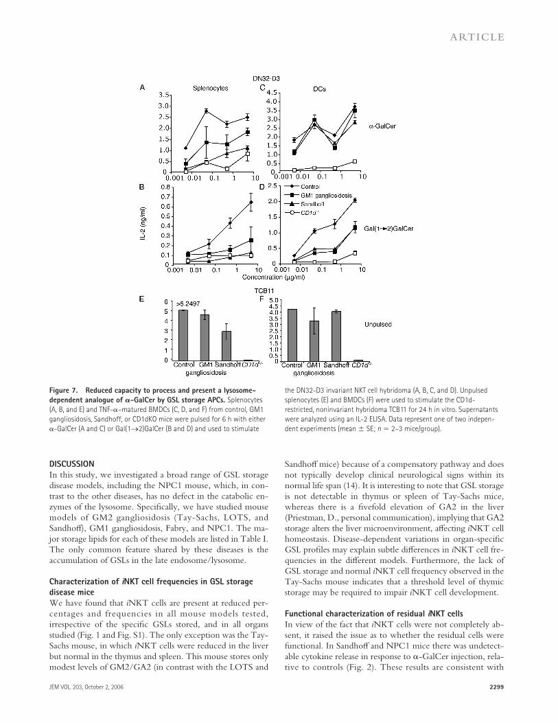
Sandhoff , GM1 gangliosidosis, Fabry, or NPC1 spleno-cytes or BMDCs were pulsed with α-GalCer (Fig. 7, A and C; and not depicted) and used to stimulate an iNKT cell hy-bridoma (DN32-D3), or left unpulsed and used to stimulate a control, CD1d-restricted, noninvariant NKT cell hybrid-oma (TCB11; Fig. 7, E and F). Splenocytes from both Sand-hoff and GM1 gangliosidosis mice appeared to be defective in presenting α-GalCer to the DN32 hybridoma compared with wild-type controls (Fig. 7 A). Interestingly, the defect in ability to present α-GalCer observed with Sandhoff and GM1 gangliosidosis splenocytes is not apparent when the APCs used were cultured BMDCs (Fig. 7 C).
We also examined the capacity of APCs from GSL stor-age disease mice to process and present an analogue of α- GalCer that is strictly dependent on lysosomal processing, galactosyl(α1→2) galactosylceramide (Gal(1→2)GalCer) (5, 20). This analogue has a second galactose group linked to the primary galactose of α-GalCer and requires processing by α-galactosidase (defi cient in Fabry disease) within the lyso-some, before recognition in the context of CD1d can occur. It has previously been shown that Fabry mice are unable to process and present Gal(1→2)GalCer (20).
These experiments were performed using splenocytes (Fig. 7 B) or BMDCs (Fig. 7 D) from Sandhoff and GM1 gangliosidosis mice, as well as NPC1 mice (unpublished data). The response of the DN32 hybridoma to spleno-cytes or BMDCs from Sandhoff and GM1 gangliosidosis mice pulsed with Gal(1→2)GalCer was reduced compared with wild-type controls but was greater than that seen with CD1d−/− APCs. Similarly, NPC1 mice had a dra-matically reduced capacity to present Gal(1→2)GalCer (unpublished data).
Table III. Biochemical and cell biological consequences of GSL storage in the thymus of the Sandhoff disease mouse
Control Sandhoff Fold elevation
HPLC analysis of GSLs aGM1a 28.3 ± 2.9 24.8 ± 2.1 0.89aGM1b 150.3 ± 24 133.7 ± 15.8 0.89aGA1 121.7 ± 12.6 118.6 ± 12.8 0.97aGM2 8.3 ± 1.5 b17.9 ± 3.8 2.15aGA2 34 ± 4.89 b166.9 ± 24.8 4.91aGM3 5.8 ± 1.05 b2.14 ± 0.75 0.36
Flow cytometry cLysoTracker 100 ± 5.3 b211.3 ± 21.3 2.11
aLevels of GSL species in total thymic extract from 10–12-day-old Sandhoff and control mice (pg/mg protein), as determined by HPLC (reference 41).bStatistical signifi cance using the t test (P ≤ 0.05).cA relative measure of the total acidic late endosomal/lysosomal compartment was made using LysoTracker staining. Flow cytometry was used to analyze LysoTracker-stained
thymocytes (relative fl uorescence intensity), gating on the lymphoid population using forward and side scatter parameters.
JEM VOL. 203, October 2, 2006 2299
ARTICLE
DISCUSSION
In this study, we investigated a broad range of GSL storage disease models, including the NPC1 mouse, which, in con-trast to the other diseases, has no defect in the catabolic en-zymes of the lysosome. Specifi cally, we have studied mouse models of GM2 gangliosidosis (Tay-Sachs, LOTS, and Sandhoff ), GM1 gangliosidosis, Fabry, and NPC1. The ma-jor storage lipids for each of these models are listed in Table I. The only common feature shared by these diseases is the accumulation of GSLs in the late endosome/lysosome.
Characterization of iNKT cell frequencies in GSL storage
disease mice
We have found that iNKT cells are present at reduced per-centages and frequencies in all mouse models tested, irrespective of the specifi c GSLs stored, and in all organs studied (Fig. 1 and Fig. S1). The only exception was the Tay-Sachs mouse, in which iNKT cells were reduced in the liver but normal in the thymus and spleen. This mouse stores only modest levels of GM2/GA2 (in contrast with the LOTS and
Sandhoff mice) because of a compensatory pathway and does not typically develop clinical neurological signs within its normal life span (14). It is interesting to note that GSL storage is not detectable in thymus or spleen of Tay-Sachs mice, whereas there is a fi vefold elevation of GA2 in the liver (Priestman, D., personal communication), implying that GA2 storage alters the liver microenvironment, aff ecting iNKT cell homeostasis. Disease-dependent variations in organ-specifi c GSL profi les may explain subtle diff erences in iNKT cell fre-quencies in the diff erent models. Furthermore, the lack of GSL storage and normal iNKT cell frequency observed in the Tay-Sachs mouse indicates that a threshold level of thymic storage may be required to impair iNKT cell development.
Functional characterization of residual iNKT cells
In view of the fact that iNKT cells were not completely ab-sent, it raised the issue as to whether the residual cells were functional. In Sandhoff and NPC1 mice there was undetect-able cytokine release in response to α-GalCer injection, rela-tive to controls (Fig. 2). These results are consistent with
Figure 7. Reduced capacity to process and present a lysosome-
dependent analogue of 𝛂-GalCer by GSL storage APCs. Splenocytes
(A, B, and E) and TNF-α–matured BMDCs (C, D, and F) from control, GM1
gangliosidosis, Sandhoff, or CD1dKO mice were pulsed for 6 h with either
α-GalCer (A and C) or Gal(1→2)GalCer (B and D) and used to stimulate
the DN32-D3 invariant NKT cell hybridoma (A, B, C, and D). Unpulsed
splenocytes (E) and BMDCs (F) were used to stimulate the CD1d-
restricted, noninvariant hybridoma TCB11 for 24 h in vitro. Supernatants
were analyzed using an IL-2 ELISA. Data represent one of two indepen-
dent experiments (mean ± SE; n = 2–3 mice/group).

2300 REDUCTION OF INKT CELLS IN GLYCOSPHINGOLIPIDOSES | Gadola et al.
previously published papers (5, 26) demonstrating a reduc-tion in iNKT cell frequencies in Sandhoff and NPC1 mice. The complete absence of a cytokine response in the storage disease mice suggests that if the residual iNKT cells are func-tional, the levels of cytokines released are below the detec-tion limit of the assays. Alternatively, they may be defective and incapable of responding. In addition, GSL storage disease APCs may ineffi ciently present α-GalCer. To investigate the functional capacity of the residual iNKT cells, in vivo killing of wild-type α-GalCer–pulsed and C20:2 analogue–pulsed (18) targets was assessed in Sandhoff mice (Fig. 3, B and C). As predicted, the specifi c lysis was reduced (as a result of the reduction in iNKT cells). However, there was detectable killing (Fig. 3 C) and IFN-γ production (Fig. S2), suggesting that the residual iNKT cells are capable of responding to antigen-pulsed wild-type targets. To address the function of residual iNKT cells, it would be useful to use intracellular cytokine staining on individual cells. However, in our hands this technique was insuffi ciently sensitive for this purpose (unpublished data). Whether residual iNKT cells in Sandhoff or GM1 gangliosidosis mice are defective—compared with controls—on a per cell basis remains thus unclear.
The role of CD1d expression
In agreement with published studies of the Sandhoff and pro-saposin knockout mouse models (5, 27), levels of cell surface CD1d were generally unchanged in GSL storage disease mice (Fig. 4). Of note, the level of CD1d was reduced on thymo-cytes and B cells in the NPC1 mouse (Fig. 4). These data suggest that the complex pattern of storage lipids in NPC1 and/or the block in traffi cking from the late endosome that occurs in this disease partially suppresses cell surface CD1d expression. However, as all other mouse models tested did not exhibit reduced CD1d expression, it is unlikely that the decreased CD1d levels in NPC1 mice are entirely responsible for the reduction in iNKT cells.
Processing and presentation within the lysosome
Although presentation of peptides by MHC class II molecules is unaff ected in the diff erent disease mouse models (with the exception of NPC1 ; Fig. 5), processing and presentation of endogenous or exogenous GSL antigens may be aff ected by GSL storage. To test whether presentation of exogenous GSLs was compromised, we used APCs from Sandhoff , GM1 gangliosidosis, NPC1, and control mice—loaded with either α-GalCer or an analogue, Gal(1→2)GalCer (20)—to stimu-late a Vα14+ iNKT cell hybridoma (27). In these experi-ments, CD1d presentation of both α-GalCer (partially dependent on lysosomal loading) and Gal(1→2)GalCer (strictly dependent on lysosomal processing and loading) (20) was impaired in splenocytes from Sandhoff , GM1 gangliosi-dosis (Fig. 7, A and B), and NPC1 mice (not depicted). The response of the TCB11 control hybridoma was similar be-tween mouse strains (Fig. 7, E and F).
Experiments performed using Sandhoff - and GM1 gan-gliosidosis–cultured BMDCs confi rmed reduced presentation
of Gal(1→2)GalCer, as compared with presentation by con-trol BMDCs (Fig. 7 D). In contrast with splenocytes, BM-DCs from Sandhoff and GM1 gangliosidosis mice were capable of effi ciently presenting α-GalCer (Fig. 7, A and C). Collectively, these data suggest that the accumulation of GSL in the lysosome can impair lysosomal processing and/or load-ing of CD1d ligands for presentation to iNKT cells. In addi-tion, the defect in sphingolipid traffi cking observed in all models (28) may contribute to the reduction in exogenous ligand presentation. GSL storage by diff erent GSL storage disease model BMDCs was confi rmed by their elevation in LysoTracker staining (unpublished data).
The diff erence observed between splenocytes and BMDCs in their capacity to present exogenous α-GalCer could be explained by several factors. For example, it may refl ect increased glycolipid antigen uptake/processing by an enriched population of DCs compared with splenocytes, which contains CD1d-positive macrophages and B cells as well as DCs. TNF-α–induced maturation of BMDCs could further enhance uptake and presentation of glycolipids. Alter-natively, cultured BMDCs and splenocytes may diff er with regard to the degree of GSL accumulation within lysosomes.
Interestingly, previously published data (5) did not fi nd any such defect in the capacity to process and present Gal(1→2)GalCer by Sandhoff APCs. It is possible that tech-nical diff erences in the experiments—such as the age of the mice and, consequently, the degree of GSL accumulation—may aff ect the results obtained. A more detailed analysis of the eff ects of age and degree of accumulation on the ability to present Gal(1→2)GalCer is currently being undertaken.
Impaired iNKT cell thymic selection in GSL storage mice
Processing and presentation of endogenous iNKT cell select-ing ligands may also be aff ected by GSL storage. The reduc-tion of iNKT cells in GSL storage mice might therefore be caused by a reduced capacity to positively select iNKT cells during thymic development. A reduction of iNKT cells at all developmental stages was observed in the thymus of young adult Sandhoff and GM1 gangliosidosis mice (Fig. 6). Inter-estingly, we found a signifi cant reduction in the frequency of iNKT cells in Sandhoff mice at postnatal day 12 (unpublished data), when appreciable levels of GSL storage are already de-tected (Table III), suggesting that accumulation of GSLs in thymocytes may lead to a decreased capacity to positively select iNKT cells.
Loss of iNKT cells in mouse models characterized by lysosomal GSL accumulation has previously been described. One report (20) proposed that the loss of iNKT cells in Fabry mice was indicative of a requirement for α-galactosidase (the enzyme defi cient in Fabry disease) for the generation of glycolipid ligands recognized by iNKT cells. Another group observed a reduction in iNKT cells in Sandhoff mice and concluded that there was an essential requirement for β- hexosaminidase A/B (the enzymes defi cient in Sandhoff mice) to generate the endogenous lipid necessary for selec-tion of iNKT cells (5). Of the candidate substrates tested,

JEM VOL. 203, October 2, 2006 2301
ARTICLE
iGb3 was proposed to be the selecting ligand based upon its activity in vitro. Likewise, recent studies with prosaposin- defi cient mice revealed a similar reduction in iNKT cell fre-quencies, suggesting an essential role for saposins in the loading of CD1d (27). Interestingly, a paper was recently published supporting our observations of a reduction of iNKT cells in NPC1 mice (26).
Alternative hypothesis and potential mechanisms
Our data obtained from a range of diff erent GSL storage models, including GM1 gangliosidosis and NPC1, support the hypothesis that lysosomal lipid storage has an indepen-dent, nonspecifi c negative eff ect on iNKT cell selection. In NPC1 mice, reduced iNKT cell frequencies may be caused by a combined eff ect of GSL storage, impaired GSL traffi ck-ing, and decreased CD1d expression (26). This hypothesis could also explain the fi nding that iNKT cells are reduced in Fabry mice, where the defi ciency in α-galactosidase A should lead to lysosomal accumulation of iGb3, the recently identi-fi ed candidate endogenous ligand for iNKT cells.
We propose an alternative model to explain iNKT cell loss in the disparate mouse models investigated in this study and others (5, 20, 26, 27). The storage of any GSL above a certain threshold in the late endosome/lysosome, irrespective of its structure, could impair iNKT cell development. It is the storage per se rather than a specifi c enzyme defect that results in iNKT cell defi ciency. This model is also consistent with the iNKT cell reduction observed in the saposin-null mice because GSLs accumulate as a result of the saposin defi ciency. In addition, lack of lipid solubilization (29) mediated by sa-posins may further impair the loading of CD1d with endoge-nous iNKT cell selecting ligands. Therefore, saposin-defi cient mice suff er from the combined eff ects of GSL storage and reduced GSL loading. This may also explain why residual iNKT cells are detectable in the Fabry, Sandhoff , GM1 gan-gliosidosis, and NPC1 mice (GSL storage), whereas they are undetectable in the saposin-defi cient animals (GSL storage and loading defect).
There are several potential mechanisms to envision for how GSL storage aff ects CD1d loading. The fi rst would be that endogenous GSLs normally presented by CD1d become trapped within the storage bodies in the diseased cells and consequently are not loaded onto CD1d. A second hypothe-sis would be that the high levels of storage GSLs outnumber natural ligands within the late endosome/lysosome and, therefore, out-compete the endogenous lipids for binding to CD1d. Both of these models are consistent with a threshold level of storage being required to disrupt iNKT cell develop-ment. This is supported by the intermediate iNKT cell loss exhibited by the Tay-Sachs mouse (Fig. 1) that has subpatho-logical levels of GSL storage (Table I). Alternatively, GSL storage may indirectly infl uence iNKT cell selection. The defect in sphingolipid traffi cking (27) observed in all GSL storage disease models may disrupt the cellular distribution of the selecting ligands, reducing their presentation by CD1d and iNKT cell selection. The proposed mechanisms by which
GSL storage disrupts iNKT cell selection are not mutually exclusive and, therefore, we cannot rule out that this also plays a role.
It will be interesting to determine whether comparable iNKT cell defi ciencies occur in patients with glycosphingo-lipidoses. Although these studies are currently in progress, a recent report has described that Gaucher patients (who pos-sess a defect in the lysosomal enzyme glucocerebrosidase) treated with enzyme replacement therapy showed a modest increase in the proportion of Vα24+ cells in the peripheral CD4+ and CD8+ T cell pool (30). Should the fi ndings of our study translate to the human disorders, iNKT cell defi ciency may be a contributing factor to the clinical heterogeneity characteristic of these diseases. It is possible that this may have consequences for host immune responses by reducing the effi cacy of presentation of bacterial or endogenous GSLs.
In summary, storage of multiple GSLs with disparate structures, resulting from unique etiologies, causes defective iNKT cell selection. Our data suggest general mechanisms by which lysosomal storage of GSLs, irrespective of the underly-ing gene defect, can disrupt the presentation of endogenous ligands by CD1d.
MATERIALS AND METHODSAnimals. The following mouse models of GSL storage (C57BL/6 back-
ground) were maintained and genotyped according to published methods:
Tay-Sachs hexa−/− (31); LOTS hexa−/− (32); Sandhoff hexb−/− (33); Fabry
α-galA−/− (34); and GM1 gangliosidosis bgal−/− (35). Each mouse strain had
been backcrossed at least eight times before use. LOTS mice are female
Tay-Sachs mice that have been repeatedly bred before 6 mo of age (32).
Tay-Sachs (nonbred) mice are asymptomatic because of the presence of a
bypass pathway (the combined eff ects of sialidase and hexosaminidase B).
Pregnancy induces down-regulation of components of the bypass pathway,
causing higher levels of storage relative to the Tay-Sachs mouse and clinical
presentation in 100% of LOTS mice. NPC1 mice (13) are on a BALB/c
background. Also used were mice lacking the Jα18 TCR gene segment (36),
which were devoid of Vα14 iNKT cells while having other lymphoid cell
lineages intact (iNKT−/− mice), and CD1d knockout mice (CD1d−/−) (37),
which were also devoid of Vα14 iNKT cells. Heterozygote littermates and
age-matched C57BL/6 or NPC1+/+ mice, as appropriate, were used as con-
trols. All mice were maintained in the Biological Services Unit, Department
of Biochemistry, University of Oxford and used according to established
University of Oxford institutional guidelines under the authority of a UK
Home Offi ce project license.
Cell preparations. Intrahepatic mononuclear cells were separated from
mouse livers according to the following protocol: livers were cut into small
pieces using a scalpel, passed through a 100-mm metal mesh fi lter, washed
twice with PBS, layered over Ficoll-Hypaque gradients, and centrifuged at
2,000 rpm at room temperature for 20 min. An analogous procedure was
used to separate splenic and thymic mononuclear cells. Before staining for
FACS, both intrahepatic and splenic mononuclear cells were incubated for
10 min at room temperature with 20 μg of unconjugated anti-FcR antibody
(BD Biosciences). For all FACS staining experiments, three to fi ve animals
per group were used.
Flow cytometry. Cell suspensions were stained according to published
methods (38). In brief, 106 cells in 50 μl FACS buff er (PBS containing 1%
bovine serum albumin and 0.02 M sodium azide) were incubated on ice for
30 min with monoclonal antibodies (all from BD Biosciences) or α-GalCer/
CD1d tetramer (39), followed by two washes in FACS buff er. The anti-
bodies used were R-phycoerythrin (RPE)–conjugated rat anti–mouse CD1d

2302 REDUCTION OF INKT CELLS IN GLYCOSPHINGOLIPIDOSES | Gadola et al.
(CD1.1, Ly-38), FITC-conjugated rat anti–mouse CD19 (1D3), hamster
anti–mouse CD11c (HL3), and CD68. Allophycocyanin-conjugated strep-
tavidin (Phykolink) and RPE-conjugated Extraavidin (Sigma-Aldrich) were
used for the generation of CD1d tetramers. CD1d tetramers were generated
as previously described (39). Propidium iodide was used to gate out dead
cells. Quantitation of binding sites was performed using fl uorescent micro-
bead standards according to published methods (38). To analyze the matura-
tion phenotype of iNKT thymocytes, cells were stained with CD1d/α-GalCer
tetramer–PE, NK1.1-PerCP, pan Vβ–FITC, and CD44-allophycocyanin
(BD Biosciences). To analyze lysosomes, 106 cells from 10–12-d-old Sand-
hoff and control thymi were stained in 200 μl 200 nM LysoTracker-Green
(Invitrogen) for 10 min at room temperature. After washing, the cells were
analyzed by fl ow cytometry gating on thymocytes by forward and side scat-
ter. Percentages of APCs were analyzed by staining cells from diff erent tis-
sues with CD11c and CD68 antibodies.
Cytokine assays. Animals were injected i.v. with 1 μg α-GalCer (dissolved
in a vehicle solution of 0.5% Tween 20/PBS) or vehicle diluted in PBS.
Blood samples were collected at the time points indicated in the fi gures, and
serum levels of IL-4 and IFN-γ were determined using cytokine-specifi c
capture ELISAs. Antibodies used for ELISAs were obtained from eBiosci-
ence and Pierce Chemical Co.
In vivo cytotoxicity assay. Target splenocytes were pulsed with 5, 0.5,
and 0.05 μg/ml α-GalCer or C20:2 analogue for 2 h at 37°C and labeled
with diff erent concentrations (1.65, 0.3, and 0.07 nM) of CFSE (Invitrogen),
as previously described (17). A control vehicle-pulsed population was labeled
with 10 μM chloromethyl-benzoyl-aminotetramethyl-rhodamine (CMTMR;
Invitrogen). An equal mixture of the four populations of splenocytes was in-
jected i.v. at 107 cells/mouse into Sandhoff homozygote mice, age-matched
littermate controls, and iNKT−/− mice (n = 3–5 mice/group). 48 h after in-
jection, blood samples were examined for the presence of fl uorescent cells by
fl ow cytometry (FACSCalibur; BD Biosciences). Data are expressed as per-
cent survival of α-GalCer– or analogue-pulsed splenocytes compared with
unpulsed controls. Mean percent specifi c lysis (±SEM) of α-GalCer–pulsed
splenocytes compared with unpulsed controls was calculated by the follow-
ing formula: 100 − ([pulsed/unpulsed] × 100).
Monitoring MHC class II–restricted T cell responses. Sandhoff ho-
mozygotes (6 and 10 wk of age) or other GSL storage mice and age-matched
controls were immunized s.c. with 100 μg chicken OVA protein (Sigma-
Aldrich) in CFA (Sigma-Aldrich). 11 d later, the mice were injected i.v.
with 2 × 10⁶ PFU/mouse UV-inactivated recombinant vaccinia virus en-
coding full-length chicken OVA. After 7 d, immune responses were assessed
by performing ELISPOT using a mouse IFN-γ ELISPOT kit (Mabtech) and
10 μM of two diff erent I-Ab–restricted OVA peptides (synthesized in house),
OVA265-280 (T E W T S S N V M E E R K I K V ) and OVA323-339 (I S Q A V H A A H A E-
I N E A G R ), according to the manufacturer’s protocol.
Analysis of GSL storage. Thymi from 10–12-d-old Sandhoff mice and
controls were homogenized in 0.5 ml distilled water, and GSLs were ex-
tracted using the Svernnerholm method (40). The equivalent of 200 μg
protein was treated with ceramide glycanase (Calbiochem-Novabiochem).
Released glycans were labeled with anthranillic acid (Sigma-Aldrich) and
analyzed by HPLC as described previously (41).
Processing and presentation of 𝛂-GalCer and analogues by GSL
storage disease APCs. APCs used were either BMDCs, cultured for 7 d
in the presence of 20 ng/ml GM-CSF/IL-4 (PeproTech) and matured from
day 6 of culture with 10 ng/ml TNF-α (PeproTech) or splenocytes. APCs
were pulsed with α-GalCer, Gal(1→2)GalCer, or vehicle for 6 h, washed,
and 5 × 10⁵ splenocytes or 5 × 10⁴ BMDCs were added to 96-well fl atbot-
tom plates. The DN32-D3 iNKT cell hybridoma or the control, CD1d-
restricted, non–iNKT cell hybridoma (TCB11) from A. Bendelac (University
of Chicago, Chicago, IL) and S. Porcelli (Albert Einstein School of Medicine,
New York, NY) were added (5 × 10⁴) to each well. Cells were cultured
for 18–24 h at 37°C, and the supernatant was analyzed for the presence of
IL-2 by ELISA (antibodies used were anti–mouse IL-2 JES6-1A12 and
biotinylated JES6-5H4; eBioscience).
Online supplemental material. In Fig. S1, lymphocyte preparations
from the spleen and thymus were generated, and cells were counted. Lym-
phocytes were stained for iNKT cells as described in Flow cytometry, and
samples were analyzed. The cell number and frequency of iNKT cells as a
percentage of total lymphocytes were then used to calculate the number of
tissue iNKT cells.
In Fig. S2, the ability of iNKT cells from storage disorder mice to
mount a cytokine response was assayed by performing an overnight IFN-γ
ELISPOT using T cell–enriched splenocytes incubated with BMDCs loaded
with α-GalCer. Splenocyte cell preparations were enriched for T cells by in-
cubating for 1 h at 37°C, adhering out the macrophages. BMDCs derived
from wild-type mice were pulsed for 3 h with 5 μg/ml α-GalCer. Online
supplemental material is available at http://www.jem.org/cgi/content/full/
jem.20060921/DC1.
The authors wish to thank Dawn Shepherd, Andrea Tarlton, David Smith, and Denise
Jelfs for excellent technical support; Dr. David Neville for HPLC analysis of GSL
oligosaccharides; and Professor Albert Bendelac, Dr. Dapeng Zhou, and Professor
Steve Porcelli for the kind gift of the hybridomas.
This work was funded by the Swiss National Science Foundation and Max
Cloetta Foundation (S.D. Gadola), the Medical Research Council (grant G0400421 to
V. Cerundolo and a studentship to A. Jeans), the Wellcome Trust (grant 072021/
Z/03/Z), the Lister Institute for Preventive Medicine (G.S. Besra and F.M. Platt), the
Glycobiology Institute, the University of Oxford (T.D. Butters and F.M. Platt) and
Cancer Research UK (grant C399-A2291), and the Cancer Research Institute.
The authors have no confl icting fi nancial interests.
Submitted: 12 May 2006
Accepted: 23 August 2006
REFERENCES 1. Taniguchi, M., M. Harada, S. Kojo, T. Nakayama, and H. Wakao.
2003. The regulatory role of Vα14 NKT cells in innate and acquired immune response. Annu. Rev. Immunol. 21:483–513.
2. Brigl, M., and M.B. Brenner. 2004. CD1: antigen presentation and T cell function. Annu. Rev. Immunol. 22:817–890.
3. Bendelac, A. 1995. CD1: presenting unusual antigens to unusual T lymphocytes. Science. 269:185–186.
4. Naidenko, O.V., J.K. Maher, W.A. Ernst, T. Sakai, R.L. Modlin, and M. Kronenberg. 1999. Binding and antigen presentation of ceramide-containing glycolipids by soluble mouse and human CD1d molecules. J. Exp. Med. 190:1069–1080.
5. Zhou, D., J. Mattner, C. Cantu III, N. Schrantz, N. Yin, Y. Gao, Y. Sagiv, K. Hudspeth, Y.P. Wu, T. Yamashita, et al. 2004. Lysosomal glycosphingolipid recognition by NKT cells. Science. 306:1786–1789.
6. Mattner, J., K.L. Debord, N. Ismail, R.D. Goff , C. Cantu III, D. Zhou, P. Saint-Mezard, V. Wang, Y. Gao, N. Yin, et al. 2005. Exogenous and endogenous glycolipid antigens activate NKT cells during microbial infections. Nature. 434:525–529.
7. Park, S.H., K. Benlagha, D. Lee, E. Balish, and A. Bendelac. 2000. Unaltered phenotype, tissue distribution and function of Vα14(+) NKT cells in germ-free mice. Eur. J. Immunol. 30:620–625.
8. van Der Vliet, H.J., N. Nishi, T.D. de Gruijl, B.M. von Blomberg, A.J. van den Eertwegh, H.M. Pinedo, G. Giaccone, and R.J. Scheper. 2000. Human natural killer T cells acquire a memory-activated phenotype before birth. Blood. 95:2440–2442.
9. Jayawardena-Wolf, J., K. Benlagha, Y.H. Chiu, R. Mehr, and A. Bendelac. 2001. CD1d endosomal traffi cking is independently regulated by an intrinsic CD1d-encoded tyrosine motif and by the invariant chain. Immunity. 15:897–908.
10. Chiu, Y.H., S.H. Park, K. Benlagha, C. Forestier, J. Jayawardena-Wolf, P.B. Savage, L. Teyton, and A. Bendelac. 2002. Multiple defects in antigen presentation and T cell development by mice expressing cyto-plasmic tail-truncated CD1d. Nat. Immunol. 3:55–60.

JEM VOL. 203, October 2, 2006 2303
ARTICLE
11. Schuette, C.G., T. Doering, T. Kolter, and K. Sandhoff . 1999. The gly-cosphingolipidoses-from disease to basic principles of metabolism. Biol. Chem. 380:759–766.
12. Jeyakumar, M., T.D. Butters, R.A. Dwek, and F.M. Platt. 2002. Glycosphingolipid lysosomal storage diseases: therapy and pathogenesis. Neuropathol. Appl. Neurobiol. 28:343–357.
13. Loftus, S.K., J.A. Morris, E.D. Carstea, J.Z. Gu, C. Cummings, A. Brown, J. Ellison, K. Ohno, M.A. Rosenfeld, D.A. Tagle, et al. 1997. Murine model of Niemann-Pick C disease: mutation in a cholesterol homeostasis gene. Science. 277:232–235.
14. Hopwood, J.J., A.C. Crawley, and R.M. Taylor. 2004. Spontaneous and engineered mammalian storage disease models. In Lysosomal Disorders of the Brain: Recent Advances in Molecular and Cellular Pathogenesis and Treatment. F. Platt and S. Walkley, editors. Oxford University Press, Oxford. 257–289.
15. Bendelac, A. 1995. Positive selection of mouse NK1+ T cells by CD1-expressing cortical thymocytes. J. Exp. Med. 182:2091–2096.
16. Hermans, I.F., J.D. Silk, U. Gileadi, M. Salio, B. Mathew, G. Ritter, R. Schmidt, A.L. Harris, L. Old, and V. Cerundolo. 2003. NKT cells en-hance CD4+ and CD8+ T cell responses to soluble antigen in vivo through direct interaction with dendritic cells. J. Immunol. 171:5140–5147.
17. Hermans, I.F., J.D. Silk, J. Yang, M.J. Palmowski, U. Gileadi, C. McCarthy, M. Salio, F. Ronchese, and V. Cerundolo. 2004. The VITAL assay: a versatile fl uorometric technique for assessing CTL- and NKT-mediated cytotoxicity against multiple targets in vitro and in vivo. J. Immunol. Methods. 285:25–40.
18. Yu, K.O., J.S. Im, A. Molano, Y. Dutronc, P.A. Illarionov, C. Forestier, N. Fujiwara, I. Arias, S. Miyake, T. Yamamura, et al. 2005. Modulation of CD1d-restricted NKT cell responses by using N-acyl variants of α-galactosylceramides. Proc. Natl. Acad. Sci. USA. 102:3383–3388.
19. Wolf, P.R., and H.L. Ploegh. 1995. How MHC class II molecules ac-quire peptide cargo: biosynthesis and traffi cking through the endocytic pathway. Annu. Rev. Cell Dev. Biol. 11:267–306.
20. Prigozy, T.I., O. Naidenko, P. Qasba, D. Elewaut, L. Brossay, A. Khurana, T. Natori, Y. Koezuka, A. Kulkarni, and M. Kronenberg. 2001. Glycolipid antigen processing for presentation by CD1d mol-ecules. Science. 291:664–667.
21. Benlagha, K., T. Kyin, A. Beavis, L. Teyton, and A. Bendelac. 2002. A thymic precursor to the NK T cell lineage. Science. 296:553–555.
22. Pellicci, D.G., K.J. Hammond, A.P. Uldrich, A.G. Baxter, M.J. Smyth, and D.I. Godfrey. 2002. A natural killer T (NKT) cell developmental pathway involving a thymus-dependent NK1.1−CD4+ CD1d-dependent precursor stage. J. Exp. Med. 195:835–844.
23. Benlagha, K., D.G. Wei, J. Veiga, L. Teyton, and A. Bendelac. 2005. Characterization of the early stages of thymic NKT cell development. J. Exp. Med. 202:485–492.
24. Gapin, L., J.L. Matsuda, C.D. Surh, and M. Kronenberg. 2001. NKT cells derive from double-positive thymocytes that are positively selected by CD1d. Nat. Immunol. 2:971–978.
25. Matsuda, J.L., L. Gapin, S. Sidobre, W.C. Kieper, J.T. Tan, R. Ceredig, C.D. Surh, and M. Kronenberg. 2002. Homeostasis of Vα14i NKT cells. Nat. Immunol. 3:966–974.
26. Sagiv, Y., K. Hudspeth, J. Mattner, N. Schrantz, R.K. Stern, D. Zhou, P.B. Savage, L. Teyton, and A. Bendelac. 2006. Cutting edge: impaired glycosphingolipid traffi cking and NKT cell development in mice lack-ing Niemann-Pick type C1 protein. J. Immunol. 177:26–30.
27. Zhou, D., C. Cantu III, Y. Sagiv, N. Schrantz, A.B. Kulkarni, X. Qi, D.J. Mahuran, C.R. Morales, G.A. Grabowski, K. Benlagha, et al. 2004. Editing of CD1d-bound lipid antigens by endosomal lipid transfer proteins. Science. 303:523–527.
28. Chen, C.S., M.C. Patterson, C.L. Wheatley, J.F. O’Brien, and R.E. Pagano. 1999. Broad screening test for sphingolipid-storage diseases. Lancet. 354:901–905.
29. Kolter, T., and K. Sandhoff . 2005. Principles of lysosomal membrane digestion: stimulation of sphingolipid degradation by sphingolipid acti-vator proteins and anionic lysosomal lipids. Annu. Rev. Cell Dev. Biol. 21:81–103.
30. Balreira, A., L. Lacerda, C.S. Miranda, and F.A. Arosa. 2005. Evidence for a link between sphingolipid metabolism and expression of CD1d and MHC-class II: monocytes from Gaucher disease patients as a model. Br. J. Haematol. 129:667–676.
31. Yamanaka, S., M.D. Johnson, A. Grinberg, H. Westphal, J.N. Crawley, M. Taniike, K. Suzuki, and R.L. Proia. 1994. Targeted disruption of the Hexa gene results in mice with biochemical and pathologic features of Tay-Sachs disease. Proc. Natl. Acad. Sci. USA. 91:9975–9979.
32. Jeyakumar, M., D. Smith, E. Eliott-Smith, M. Cortina-Borja, G. Reinkensmeier, T.D. Butters, T. Lemm, K. Sandhoff , V.H. Perry, R.A. Dwek, and F.M. Platt. 2002. An inducible mouse model of late onset Tay-Sachs disease. Neurobiol. Dis. 10:201–210.
33. Sango, K., M.P. McDonald, J.N. Crawley, M.L. Mack, C.J. Tiff t, E. Skop, C.M. Starr, A. Hoff mann, K. Sandhoff , K. Suzuki, and R.L. Proia. 1996. Mice lacking both subunits of lysosomal β-hexosaminidase display gangliosidosis and mucopolysaccharidosis. Nat. Genet. 14:348–352.
34. Ohshima, T., G.J. Murray, W.D. Swaim, G. Longenecker, J.M. Quirk, C.O. Cardarelli, Y. Sugimoto, I. Pastan, M.M. Gottesman, R.O. Brady, and A.B. Kulkarni. 1997. α-Galactosidase A defi cient mice: a model of Fabry disease. Proc. Natl. Acad. Sci. USA. 94:2540–2544.
35. Hahn, C.N., M. del Pilar Martin, M. Schroder, M.T. Vanier, Y. Hara, K. Suzuki, and A. d’Azzo. 1997. Generalized CNS disease and mas-sive GM1-ganglioside accumulation in mice defective in lysosomal acid β-galactosidase. Hum. Mol. Genet. 6:205–211.
36. Cui, J., T. Shin, T. Kawano, H. Sato, E. Kondo, I. Toura, Y. Kaneko, H. Koseki, M. Kanno, and M. Taniguchi. 1997. Requirement for Vα14 NKT cells in IL-12-mediated rejection of tumors. Science. 278:1623–1626.
37. Mendiratta, S.K., W.D. Martin, S. Hong, A. Boesteanu, S. Joyce, and L. Van Kaer. 1997. CD1d1 mutant mice are defi cient in natural T cells that promptly produce IL-4. Immunity. 6:469–477.
38. Platt, F.M., G.B. Karlsson, and G.S. Jacob. 1992. Modulation of cell- surface transferrin receptor by the imino sugar N-butyldeoxynojir imycin. Eur. J. Biochem. 208:187–193.
39. Karadimitris, A., S. Gadola, M. Altamirano, D. Brown, A. Woolfson, P. Klenerman, J.L. Chen, Y. Koezuka, I.A. Roberts, D.A. Price, et al. 2001. Human CD1d-glycolipid tetramers generated by in vitro oxidative refolding chromatography. Proc. Natl. Acad. Sci. USA. 98:3294–3298.
40. Svennerholm, L., and P. Fredman. 1980. A procedure for the quantita-tive isolation of brain gangliosides. Biochim. Biophys. Acta. 617:97–109.
41. Neville, D.C., V. Coquard, D.A. Priestman, D.J. te Vruchte, D.J. Sillence, R.A. Dwek, F.M. Platt, and T.D. Butters. 2004. Analysis of fl uorescently labeled glycosphingolipid-derived oligosaccharides fol-lowing ceramide glycanase digestion and anthranilic acid labeling. Anal. Biochem.331:275–282.
Related Documents