Impaired recognition of body expressions in the behavioral variant of frontotemporal dementia Jan Van den Stock 1,a, *, François-Laurent De Winter 1,a , Beatrice de Gelder 1,2 , Janaki Raman Rangarajan 3 , Gert Cypers 4 , Frederik Maes 3 , Stefan Sunaert 5 , Karolien Goffin 6 , Rik Vandenberghe 7 , Mathieu Vandenbulcke 1 1 Laboratory for Translational Neuropsychiatry, Research Group Psychiatry, Department of Neurosciences, KU Leuven & Old Age Psychiatry, University Hospitals Leuven, Leuven, Belgium 2 Cognitive Neuroscience, Faculty of Psychology and Neuroscience, Maastricht University, Maastricht, the Netherlands 3 ESAT/PSI, Department of Electrical Engineering, KU Leuven & iMinds, Medical IT Department, University Hospitals Leuven, Leuven, Belgium 4 Department of Neurology, Onze-Lieve-Vrouwziekenhuis Aalst- Asse-Ninove, Aalst, Belgium 1

Welcome message from author
This document is posted to help you gain knowledge. Please leave a comment to let me know what you think about it! Share it to your friends and learn new things together.
Transcript

Impaired recognition of body expressions in the
behavioral variant of frontotemporal dementia
Jan Van den Stock 1,a, *, François-Laurent De Winter 1,a , Beatrice
de Gelder 1,2 , Janaki Raman Rangarajan3, Gert Cypers4, Frederik
Maes3, Stefan Sunaert5, Karolien Goffin6, Rik Vandenberghe7,
Mathieu Vandenbulcke1
1Laboratory for Translational Neuropsychiatry, Research Group
Psychiatry, Department of Neurosciences, KU Leuven & Old Age
Psychiatry, University Hospitals Leuven, Leuven, Belgium
2Cognitive Neuroscience, Faculty of Psychology and
Neuroscience, Maastricht University, Maastricht, the
Netherlands
3ESAT/PSI, Department of Electrical Engineering, KU Leuven &
iMinds, Medical IT Department, University Hospitals Leuven,
Leuven, Belgium
4Department of Neurology, Onze-Lieve-Vrouwziekenhuis Aalst-
Asse-Ninove, Aalst, Belgium1

5Department of Imaging and Pathology, KU Leuven & Department of
Radiology, University Hospitals Leuven, Leuven, Belgium
6Department of Imaging and Pathology, KU Leuven & Division of
Nuclear Medicine and Molecular Imaging, University Hospitals
Leuven, Leuven, Belgium
7Laboratory for Cognitive Neurology, Department of
Neurosciences, KU Leuven & Neurology, University Hospitals
Leuven, Leuven, Belgium
aequally contributing authors
*Correspondence:
Jan Van den Stock
University Hospitals Leuven,
Research Group Psychiatry, O&N 2, bus 1027
Herestraat 49
3000 Leuven
Belgium
2

Highlights
We examine recognition of body emotion, face emotion and face
identity in bvFTD
Results show deficits on the emotion tasks, but not on the identity
task
Body and face emotion recognition deficits are correlated.
IFG volume correlates with body and face emotion recognition in
bvFTD
Imaging and behavior results support supra-modal emotion recognition
deficit in bvFTD
3

Abstract
Progressive deterioration of social cognition and emotion
processing are core symptoms of the behavioral variant of
frontotemporal dementia (bvFTD). Here we investigate whether
bvFTD is also associated with impaired recognition of static
(Experiment 1) and dynamic (Experiment 2) bodily expressions.
In addition, we compared body expression processing with
processing of static (Experiment 3) and dynamic (Experiment 4)
facial expressions, as well as with face identity processing
(Experiment 5). The results reveal that bvFTD is associated
with impaired recognition of static and dynamic bodily and
facial expressions, while identity processing was intact. No
differential impairments were observed regarding motion (static
vs. dynamic) or category (body vs. face). Within the bvFTD
group, we observed a significant partial correlation between
body and face expression recognition, when controlling for
performance on the identity task. Voxel-Based Morphometry (VBM)
analysis revealed that body emotion recognition was positively
associated with grey matter volume in a region of the inferior
4

frontal gyrus (pars orbitalis/triangularis). The results are in
line with a supramodal emotion recognition deficit in bvFTD.
Keywords: behavioral variant frontotemporal dementia; emotion;
body; inferior frontal gyrus; supramodal emotion recognition
5

1. Introduction
Fronto-temporal lobar degeneration (FTLD) is a
neurodegenerative disorder that has a profound impact on
personality and cognition. It is among the most frequent
manifestations of early-onset dementia . Clinical phenotypes of
FTLD include language variants and a behavioral variant
(bvFTD). bvFTD is primarily characterized by deterioration of
social behavior including loss of empathy and changes in
personality . It is associated with early atrophy of medio-
frontal cortex, anterior temporal cortex and striatum . The
loss of empathy has been related to impaired comprehension of
emotional expressions displayed by others . Studies
investigating emotion recognition in bvFTD have primarily
focused on perception of facial expressions. The results point
to a deficit in recognizing facial expressions with a negative
valence, while processing of positive facial expressions seems
relatively preserved. Other between-emotion effects have been
reported inconsistently .
Several studies have investigated the underlying pattern of
atrophy by correlating regional grey matter volume with emotion
6

recognition performance. The results indicate the involvement
of a distributed network including the amygdala , orbito-
frontal cortex , temporal pole and insula .
Interestingly, these regions have also been associated with
perception of both static and dynamic whole body expressions of
emotion in normal subjects . It has been documented that whole
body expressions convey reliable emotional cues, even when the
face is not visible . To our knowledge, no study has addressed
recognition of emotional body expressions in bvFTD. Our aim in
the present study was to explore whether the deficits in
emotion recognition that have been documented in the face
modality extend to body expressions in bvFTD. Our second aim
was to investigate the influence of motion by comparing static
with dynamic body expressions and the influence of category by
comparing bodily with facial expressions . As perception of
dynamic bodies is associated with more activation in fronto-
temporal and subcortical areas compared to static bodies, we
anticipate a larger impairment for recognizing dynamic stimuli.
Similarly, perceiving bodies activates more subcortical and
temporal areas than perceiving faces, with the important
exception of the amygdala, which is more activated by faces
7

than by bodies . The third purpose of the study was to evaluate
the hypothesis of a supra-modal emotion recognition deficit in
bvFTD, which emerges from the overlap between the atrophic
topography characteristic for early bvFTD and the functional
neuro-anatomy of supra-modal emotion processing, specifically
in medial prefrontal cortex and the temporal poles . In
summary, the present study addresses categorical and motion
effects of emotion recognition in bvFTD. The purpose is to
investigate whether the visual emotion recognition deficit in
bvFTD is category specific (and hence a ‘conditional’ visual
emotion recognition deficit) in nature. Compared to facial
expressions, body expressions convey more information regarding
adaptive action , which may influence recognition performance.
Similarly, dynamic stimuli are more naturalistic and contain
temporal information that may provide recognition cues . To
evaluate the emotion specificity of the results, we included a
control task consisting of identity recognition.
2. Material and Methods
The study was conducted in accordance with the Declaration of
Helsinki and included written informed consent from all
8
participants. Ethical approval for the study was provided by
the Ethical Committee of University Hospitals Leuven.
2.1. Participants
A total of 26 consecutive bvFTD patients were recruited. Six of
these patients could not be included in the study since no
experimental data could be acquired due to a lack of
cooperation and/or agitation. The remaining 20 were recruited
via the Memory Clinic (N=6) and Old Age Psychiatry Departement
of University Hospitals Leuven (N=8) and the Neurology
Department of Onze-Lieve-Vrouwziekenhuis Aalst-Asse-Ninove
(N=6). All patients were evaluated via clinical assessment,
neuropsychological testing and structural MRI. In addition,
[18]Fluorodeoxyglucose Positron Emission Tomography (FDG-PET)
was performed in all but two patients. Two patients fulfilled
the revised diagnostic criteria of ‘behavioural variant FTD
with definite FTLD Pathology’, based on a C9orf72 pathogenetic
mutation, while the other 18 patients fulfilled the criteria
for ‘Probable bvFTD’ . In none of the patients, language
difficulty was the most prominent clinical feature.
Furthermore, in none of the patients, aphasia was the most
prominent deficit at symptom onset and during the initial phase
9

of the disease. These phenotypes are not in line with the
current inclusion criteria for primary progressive aphasia
(PPA) . Patients were included after clinical judgment deemed
them able to successfully undergo an experimental scanning
session.
The control group was recruited through advertisements in local
newspapers. Twenty control subjects participated in the
behavioral and imaging experimental procedures, including
neuropsychological examination. Exclusion criteria were present
or past neurological or psychiatric disorders including
substance abuse as well as significant systemic comorbidities
or use of medication susceptible to affect the central nervous
system. MRI scanning of all participants was performed on the
same scanner. Demographic data and neuropsychological test
results of all participants are presented in Table 1.
Table 1. Demographic and neuropsychological test results. MMSE
= Mini-Mental-State Examination; RAVLT = Rey Auditory Verbal
Learning Test; A1-A5 = the sum of scores on trials A1 to A5 of
the RAVLT; Recognition = the recognition score constitutes the
difference between the number of correct hits and false hits on
10

the recognition trial; BNT = Boston Naming Test; AVF = Animal
Verbal Fluency; TMT = Trail Making Test; BORB = Birmingham
Object Recognition Battery; RCPMT = Raven Colored Progressive
Matrices Test; AAT = Aachen Aphasia Test. $(N=19); %(N=17); £
(N=15)
bvFTD (N=20) Controls (N=20)
t
(χ2)
p
Age
65.7 (8.7) 66.6 (6.1) 0.3
85
.703
Sex
(M/F
) 12/8 12/8 0.0
00
1.000
MMSE
26.7$ (1.5) 29.2 (0.6) 6.7
73
.001
RAVL
T
A1-A5
27.4$ (9.1) 50.8 (7.3) 8.9
08
.001
% recall
54.1$ (31.5) 80.9 (17.4) 3.2
67
.003
Recognitio
n
6.0$ (7.4) 14.0 (1.3) 4.6
22
.001
11

BNT
40.2$ (13.0) 54.4 (2.9) 4.6
55
.001
AVF
15.1$ (5.7) 22.1 (5.8) 3.8
62
.001
TMT
A (secs)
63.5$ (42.7) 32.5 (9.4) 3.0
99
.006
B (secs)
193.0£
(141.2)
89.8 (42.3) 2.7
42
.015
BORB
Length
87.6% (7.3) 90.7 (4.5) 1.2
62
.218
Size
85.5% (6.9) 88.9 (6.3) 1.5
77
.126
Orientatio
n
81.4% (9.1) 86.1 (6.0) 1.8
45
.074
RCPMT 16.4$ (3.9) 20.8 (2.8) 3.9
99
.001
AAT
Comprehens
ion
93. 9$
(12.3)
109.5 (5.3) 5.0
93
.001
The individual demographic and neuropsychological data of the
patients, including a detailed overview of the diagnostic
12

criteria they fulfilled, are presented in supplementary
TableS1.
2.2. Experiment 1: static body emotion matching
The stimuli and procedure have been described in detail
elsewhere . In short, the experiment consisted of a two-
alternative forced choice simultaneous match to sample
procedure. A stimulus consisted of a sample picture presented
at the top and target and distracter underneath (see Figure 1
for an example). One of the two bottom pictures (i.e. the
target) expressed the same emotion as the picture on top (i.e.
the sample). The Experiment consisted of 24 trials (4 emotions
(anger, fear, happiness, and sadness x 3 distracters/emotion x
2 genders). Stimulus presentation time was unlimited, but
participants were instructed to respond as accurately and as
fast as possible. Participants were instructed to indicate by a
button press whether the left or right bottom picture displayed
the same expression as the one on top.
2.3. Experiment 2: dynamic body emotion matching
Stimuli were constructed from 90 validated 2 second video clips
of emotional whole body expressions (15 anger, 15 disgust, 15
13

fear, 15 happy, 15 sad, 15 neutral), taken from our own
database . The procedure was analogous to Experiment 1, with
the exception that the three clips in a stimulus looped until
the participant responded (with a maximum of 10 repeats). The
experiment consisted of 30 trials (6 emotions x 5 distracters
per emotion).
2.4. Experiment 3: static face emotion matching
Frontal view pictures of emotional expressions (anger,
happiness, disgust, fear, sadness and surprise) from the
Karolinska Directed Emotional Faces set were validated on
emotion recognition in a pilot study. One hundred and eighty
pictures were similarly categorized by at least 15 of the 20
participants (75%) and were selected for the experiment. The
procedure was analogous to Experiment 1. The experiment
consisted of 60 trials (6 emotions x 5 distracters/emotion x 2
gender).
2.5. Experiment 4: dynamic face emotion matching
Stimuli were constructed from 90 validated video clips (2s) of
emotional facial expressions (15 anger, 15 disgust, 15 fear, 15
happy, 15 sad, 15 neutral) of 6 professional male actors, taken
14

from our own database . The procedure was analogous to
Experiment 2. The experiment consisted of 30 trials (6 emotions
x 5 distracters/emotion).
2.6. Experiment 5 (control experiment): face identity
matching
The stimuli and procedure have been described in detail
elsewhere . In short, a stimulus consisted of a picture
displaying a front view of a face presented on top, with 2
pictures displaying ¾ views of a face presented below. One of
the bottom faces showed the same identity as the one on top.
The Experiment consisted of 32 trials. The procedure was
analogous to Experiment 1, with the exception that the task
consisted of identity matching as opposed to emotion matching.
Trials in which the reaction time differed more than three
standard deviations from the subject-specific mean reaction
time were defined as outliers. These trials were excluded from
further analysis. All subsequent analyses are performed on
accuracy data. To test for normality of the data, Shapiro-Wilk
tests were performed on the total score of every experiment and
on the appropriate combined scores of experiments. This
15

revealed that normality could not be assumed in any of the
variables (p<.021). Box-Cox transformations did not
sufficiently optimize the skewness of the data to a normal
distribution. Therefore, we performed non-parametric
Independent-Samples Mann-Whitney U tests on the variables of
interest to investigate group differences. For every
experiment, group differences were evaluated on the total score
as well as on the performance for positive expressions to
investigate whether any emotion recognition deficit was
restricted to negative emotions. In addition, the average
performance on the negative emotions was subtracted from the
average performance of the positive emotion(s) and group
differences were examined on this difference-score to
investigate whether any emotion recognition deficit was
disproportional regarding the valence of the emotions. Finally,
group differences on the average performance on the negative
emotions were investigated.
16
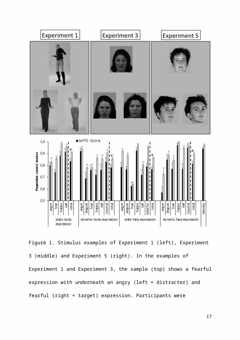
Figure 1. Stimulus examples of Experiment 1 (left), Experiment
3 (middle) and Experiment 5 (right). In the examples of
Experiment 1 and Experiment 3, the sample (top) shows a fearful
expression with underneath an angry (left = distracter) and
fearful (right = target) expression. Participants were
17

instructed to match the bottom expression to the one on top. In
Experiment 5, all pictures show a neutral expression.
Participants were instructed to match the bottom identity to
the one on top. The sample (top) shows a frontal view with
underneath ¾ views of the same (left = target) and a different
(right = distractor) identity. The bottom bar-chart displays
the behavioral results as a function of group, Experiment and
emotion (for illustrative purposes). Error bars represent 1
SEM. * = p<.05.
2.7. Magnetic Resonance Imaging and analysis
A high-resolution T1-weighted anatomical image (voxel
size=0.98x0.98x1.20 mm3) was acquired on 3T Philips Achieva
system equipped with a 32 channel head coil using a 3D turbo
field echo sequence (TR = 9.6ms; TE = 4.6ms; matrix
size=256x256; 182 slices). Analysis of local grey matter (GM)
volume was performed with SPM8 (Wellcome Trust Centre for
Neuroimaging, UCL, London, United Kingdom) within MatLab R2008a
(Mathworks, Natick, MA). Preprocessing included image
segmentation, spatial normalization, modulation and smoothing.
Segmentation was performed using SPM8’s unified segmentation
routine in combination with in-house developed algorithms to
18

address suboptimal segmentation results in the most atrophic
regions, primarily the right temporal pole. Next, the images
were spatially normalized by creating a customized group-
specific template using SPM8’s DARTEL routine and warping each
of the individual GM segmentations onto this template. The
warped GM segmentations were modulated to account for local
shape differences and smoothed using a Gaussian kernel of 8 mm
at FWHM. To investigate regional group differences in grey
matter volume, we performed a two samples t-test on the grey
matter voxels (p<.001, minimal cluster size=100 voxels).
The GM maps were subsequently used in a regression analysis in
which body expression recognition performance was entered as
covariate in order to investigate correlations between
performance and voxel-wise GM volume (p<.001, minimal cluster
size = 100 voxels). As the primary focus of the present study
was to gain insight into recognition of bodily expression in
bvFTD and its associated structural neuro-anatomy, rather than
into bodily expression recognition per se, we opted to confine
the regression analysis to the patient group and hence not to
combine it with the data from the control group. Although this
does not benefit statistical power, it excludes contamination
19

of the results by non-bvFTD data. While the alternative
approach has proven valuable , the current method provides
complementary evidence to it as well as to region of interest
analyses .
3. Results
3.1. VBM group comparison
Two patient’s T1 images were not included in the analysis due
to excessive motion. The two samples t-test (p<.001, minimal
cluster size=100 voxels) revealed a large bilateral cluster
covering the anterior half of the temporal lobes, insula,
ventral striatum and orbitofrontal cortex, consistent with
previous studies (Figure 2).
20

Figure 2. Atrophic topography of patient group. Statistical map
(p<.001) of group differences in grey matter volume,
represented on coronal slices from posterior (top left) to
anterior (bottom right) (Controls>bvFTD). Numbers refer to MNI
Y-coordinates. Color coding refers to t-values.
3.2. Behavioral results
Behavioral results are displayed in Figure 1.
3.2.1. Experiment 1: static body emotion matching
Nineteen outlier trials were detected (1.8%, maximum/subject:
2). There was a significant group difference on the total score
(p=.026), on the score for matching happy expressions (p=.046)
and on the average score for matching negative expressions
(p=.046) but not on the difference between negative and
positive expressions (p=.289).
3.2.2. Experiment 2: dynamic body emotion matching
One control subject did not take part in Experiment 2. Twelve
outliers were detected (1.0%, maximum/subject: 1). There was a
significant group difference on the total score (p=.028) and on
the score for matching happy expressions (p=.015) but not on
the difference between negative and positive expressions 21

(p=.101) nor on the average score for matching negative
expressions (p=.120).
3.2.3. Experiment 3: static face emotion matching
Forty-seven outliers were detected (1.9%, maximum/subject: 3).
There was a significant group difference on the total score
(p<.001), on the average score for matching happy and surprised
static facial expressions (p<.001) and on the average score for
matching negative expressions (p=.004), but not on the
difference between negative and positive expressions (p=.414).
3.2.4. Experiment 4: dynamic face emotion matching
One control subject did not take part in Experiment 4. Fourteen
outliers were detected (1.3%, maximum/subject: 1). There was a
significant group difference on the total score (p=.007) but
not on the score for matching happy dynamic facial expressions
(p=.428). The difference between negative and positive
expressions was significant (p=.038), as was the average score
on the negative expressions (p=.006).
3.2.5. Experiment 5 (control experiment): face identity
matching
22

One patient did not take part in Experiment 5. Twenty-three
outliers were detected (1.8%, maximum/subject: 1). There was no
significant group difference on the total score (p=.184).
3.2.6. Between and across experiments analysis
We investigated whether the deficit in matching body
expressions was proportional to the deficit in matching facial
expressions. For this purpose, the average performance on
matching facial expressions (static and dynamic) was subtracted
from the average performance on matching body expressions. The
resulting difference did not show a significant group effect
(p=.620). Similarly, to compare the deficit for matching static
and dynamic expressions, the average performance on matching
static expressions (faces and bodies) was subtracted from the
average performance on dynamic expressions. Again, this
variable showed no significant group difference (p=.857).
In addition, to investigate the association between categories
(across motion conditions) in the bvFTD group, we computed the
partial correlation coefficient between the score on the body
and on the face (averaged over static and dynamic) emotion
tasks factoring out the score on the identity task. This
23

revealed a significant correlation (r=.670, p=.002, see Figure
3).
Figure 3. Scatterplot displaying the partial correlation
between body and face emotion matching controlling for identity
matching, i.e. the unstandardized residual following linear
regression of body emotion matching to identity matching (Y-
axis) as a function of the unstandardized residual following
linear regression of face emotion matching to identity matching
(X-axis).
3.3. Imaging results
To investigate the structural neuro-anatomy of body emotion
recognition in bvFTD, the average score of Experiments 1 and 2
24

(static and dynamic body emotion matching) were entered as
covariate in the regression model. Age did not correlate with
the average score (r=.106; p=.655) and was hence not included
as a nuisance variable. The score on the identity matching
Experiment was included as a nuisance variable. Body expression
matching performance correlated significantly with GM volume in
the left inferior frontal gyrus (IFG) pars
orbitalis/triangularis (285 voxels, MNI coordinates of peak
voxel: -41; 26; 3; see Figure 4). To investigate whether the
normality assumption was fulfilled for this result, we
performed a post-hoc Shapiro Wilk test on the unstandardized
residuals of the linear regression. This revealed no
significant outcome (p=.744), supporting the validity of the
result. To investigate the specificity of these results, we
computed the partial correlation between the GM volume in this
region and face expression recognition performance (average of
static and dynamic), controlling for identity recognition
performance. This revealed a significant correlation (r=.637,
p<.008).
25

As a supplementary analysis, we investigated the neural
correlates of perceiving static and dynamic expressions as well
as facial expressions (Supplementary Materials).
Figure 4. Association between matching body expressions and
regional grey matter volume. The top panels display statistical
maps following regression of the body matching score to grey
matter volume (p<.001, minimal cluster size=100 voxels) with
performance on the identity matching task as nuisance variable.
For comparison purposes, the regional atrophy is also displayed
(p<.001). The scatterplot at the right displays the partial
correlation between matching body expressions and the grey
26

matter volume of the cluster in the inferior frontal gyrus
(GMvol IFG), factoring out matching, i.e. the unstandardized
residual following linear regression of body emotion matching
to identity matching (Y-axis) as a function of the
unstandardized residual following linear regression of IFG grey
matter volume to identity matching (X-axis). The bottom panel
displays a zoomed view of the green-delineated part of the top
panel picture. Color bars indicate t-values.
4. Discussion
The main goal of the study was to investigate recognition of
bodily expressions in bvFTD. Based on the clinical phenotype of
bvFTD but also on the overlap between the atrophic topography
and the functional neuro-anatomy of perceiving body
expressions, we hypothesized a deficit in bvFTD. We recruited a
sample with a minor global cognitive deterioration (as
evidenced by an average MMSE-score above 26) and displaying the
expected anterior temporal and orbito-frontal atrophy.
4.1. Behavioral results
The behavioral experiments consisted of a forced choice
procedure with only two alternatives to minimize decision
27

options and executive task demands. Furthermore, a simultaneous
match-to-sample task was administered, as this requires less
semantic/word finding processing, which can also be impaired in
bvFTD as compared to for example a verbal categorization
task .
The results provide support for the hypothesis, as the bvFTD
group displayed a body recognition impairment. This result
extends previous reports of impaired recognition of emotion
cues conveyed by faces , voices and music . On the other hand,
the bvFTD group in the present study was not impaired on an
identity matching task which matched the cognitive task demands
of the emotion matching tasks, suggesting that the impairment
was specific for emotions. The intact identity processing we
observed here contrasts with recent evidence for impaired
identity processing in bvFTD . This discrepancy might be
explained by two factors. First, Kumfor et al. made use of an
identity discrimination task with facial stimuli containing
only the inner face, i.e. with identifying features like hair
and ears removed. Secondly, the identity processing task in
Kumfor et al. consisted of emotional mixed with neutral
stimuli. Although the emotional information was task
28

irrelevant, there is accumulating evidence that facial emotion
and facial identity processing mutually influence each other .
These methodological differences may account for the
discrepancy with the present results showing intact matching of
neutral whole face identities.
Secondly, to investigate the role of dynamic information
conveyed by the body expressions, we investigated recognition
of both static (Experiment 1) and dynamic (Experiment 2) body
expressions. Similarly, to compare bodies with faces, we
included static (Experiment 3) and dynamic (Experiment 4)
facial expression recognition tasks. The results did not reveal
a disproportionate deficit according to motion or category
condition.
Furthermore, we tested the hypothesis that the emotion
recognition deficit in bvFTD only applies to negative emotions,
as has been reported in previous studies with facial
expressions . While happy faces are typically among the easiest
emotions to recognize, this is not the case for body
expressions . Happy faces are quite prototypical and are easily
differentiated from other emotions, particularly in the lower
half of the face , while (faceless) body expressions typically
29

involve raising of the arms. This latter features is also
typically for several negative body expressions like fear and
anger. The present results do not support a valence based
dissociation in emotion recognition impairment, as we did not
observe a disproportionate deficit for negative emotions in
three of the four emotion Experiments. The only support for a
valence specific impairment was observed in the dynamic face
Experiment (Experiment 4). However, all control subjects
performed flawlessly in the happy condition, so this ceiling
effect may conceal a latent group difference in this condition.
Interestingly, when controlled for cognitive task demands,
there was a significant correlation between the performance on
the body and face emotion tasks, independent of motion
information. This indicates that the emotion recognition
deficit in bvFTD applies similarly for faces and bodies. This
observation is in line with a previous study in which an
association between facial and vocal emotion recognition was
reported in the frontal variant of FTD .
However, as the behavioral results were not normally
distributed, we made use of non-parametric tests. This does not
30

allow controlling for neuropsychological variables, like MMSE
score.
The study results have a clinical relevance, particularly
related to the diagnosis. Deficits in social cognition are an
important diagnostic domain in addition to standard clinical
neuropsychological testing involving attention, memory,
language and visuospatial functioning. There is currently
little consensus regarding the optimal tool to assess social
cognition in general and emotion recognition in particular. The
present results suggest that recognition of bodily expressions
may provide a valuable measure to evaluate social cognition
abilities.
Imaging results
4.1.1. Controls vs patients
Comparing GM volumes between the control and bvFTD group
revealed reduced GM volume in the anterior temporal lobes,
orbitofrontal cortex, insula, dorsolateral prefrontal cortex
and striatum. This atrophic topography is largely in accordance
with previous reports Within-patient group results
31

We did not include the control group in the regression analysis
nor did we use pre-defined regions of interest in order to
provide complementary results to previous studies . The results
from the regression analysis revealed an association between
recognition of body expressions and grey matter volume in the
IFG (pars orbitalis/triangularis). The IFG has also been
associated with perceiving emotions from bodies in normal
subjects , but also with emotion processing from faces and
scenes . The cluster in the IFG we observe here was also
associated with face expression recognition, in line with
previous reports in FLTD . In addition, there is evidence that
the IFG is involved in recognition of emotions from music in
bvFTD . These combined findings reveal an association between
the structural integrity of the IFG and emotion recognition
deficits in multiple stimulus categories in bvFTD. Furthermore,
the involvement of the region in the IFG we observe here has
been reported in other neurodegenerative disorders like
Alzheimer’s disease. A recent fMRI study reported reduced
activation (compared to controls) in the IFG when viewing
emotional vs neutral faces .
32

There is evidence that the recognition of emotions shows both
psychological and neuro-anatomical overlap with the experience
of emotions . The IFG has been particularly associated with
both experience and perception of emotions. Furthermore,
activation in the IFG during emotion perception is positively
associated with trait empathy . The present findings are in
line with these notions, namely that recognition of emotion
involves motor regions to understand the emotional state of
others and that this is related to empathy, which is primarily
affected in bvFTD.
However, it is remarkable that the cluster falls largely
outside the atrophic region, similar to a previous study . This
may suggest that symptom manifestation is not by default
directly related to structural degeneration of an associated
area, as revealed through MRI. In addition, there is evidence
that the temporal poles constitute an amodal hub in storing
semantic knowledge about emotions, operating through
connectivity with primary and association cortices . Our
results are therefore in line with the notion that it is
primarily the degeneration of the combination of temporal poles
with IFG that influences symptom severity.
33

Finally, some limitations of the study should be noted. As we
did not include a clinical control group, there is no evidence
that the present results obtain specifically for bvFTD. There
is conflicting evidence regarding the degree of emotion
recognition impairments between FTLD and other
neurodegenerative disorders like Alzheimer’s disease (AD).
While some studies have reported a larger impairment in FTLD ,
other have reported equally large deficits . However, the
latter study provided evidence that emotion recognition
deficits are primarily associated with language impairments in
SD as opposed to perceptual impairments in bvFTD and AD. Future
studies can investigate whether emotion recognition deficits
are observed already at the detection stage, or only emerge
when the task is to discriminate emotions. In addition to the
matching approach that we used here, it would be informative to
investigate whether a similar impairment is present when the
task is to select or categorize emotions and how performance
differs from other neurodegenerative syndromes like AD. This
may provide cues regarding the involvement of the specific
emotion processing deficits in a recently proposed liability
spectrum . Secondly, our clinical sample showed a primarily
34

anterior temporal atrophic topography. It cannot be ruled out
that the cooperative and motivational demands of the study
resulted in an inclusion bias favoring temporal dominant
variants . In fact, 6 patients were invited and agreed to
participate in the study, but could not be included because of
insufficient cooperation or agitation, similar to a previous
study . In addition to comparisons with other neurodegenerative
disorders, it would be informative to compare emotion
recognition in bvFTD as a function of neuro-anatomic phenotype.
In conclusion, the present findings reveal that bvFTD is
characterized by a deficit in recognizing both static and
dynamic body expressions. Furthermore, the emotion recognition
deficit was proportional regarding both category (faces
compared to bodies) and motion (static compared to dynamic). We
also observed a significant correlation between body and face
emotion recognition, compatible with a supra-modal emotion
recognition deficit.
Acknowledgements
35

We are particularly grateful to the patients for their
cooperative participation in the study. In addition, we thank
Dr. Marc Van Orshoven, Dr. Marleen Vieren and Dr. Miriam
Bouckaert at the Neurology Department of OLV-Hospitals Aalst
for their cooperation, Dr. Wim Van der Elst for statistical
advice and Sherihane Bensemmane for technical support. The
authors declare no competing financial interests. J.V.d.S. is a
post-doctoral researcher supported by FWO-Vlaanderen
(1.5.072.13N) and Foundation for Alzheimer Research (SAO-FRA
P#14013). The funding sources had no role in the study design;
in the collection, analysis and interpretation of data; in the
writing of the report; and in the decision to submit the
article for publication.
36

References
Aviezer, H., Trope, Y., & Todorov, A. (2012). Body cues, not facial
expressions, discriminate between intense positive and negative
emotions. Science, 338(6111), 1225-1229. doi:
10.1126/science.1224313
Bastiaansen, J. A., Thioux, M., & Keysers, C. (2009). Evidence for
mirror systems in emotions. Philos Trans R Soc Lond B Biol Sci, 364(1528),
2391-2404. doi: 10.1098/rstb.2009.0058
Bertoux, M., Volle, E., Funkiewiez, A., de Souza, L. C., Leclercq,
D., & Dubois, B. (2012). Social Cognition and Emotional
Assessment (SEA) is a marker of medial and orbital frontal
functions: a voxel-based morphometry study in behavioral
variant of frontotemporal degeneration. J Int Neuropsychol Soc, 18(6),
972-985. doi: 10.1017/S1355617712001300
Bowers, D., Blonder, L. X., & Heilman, K. M. (1999). The Florida affect
battery Gainsville, Florida: Center for Neuropsychological
Studies.
Calder, A. J., Young, A. W., Keane, J., & Dean, M. (2000).
Configural information in facial expression perception. J Exp
Psychol Hum Percept Perform, 26(2), 527-551.
37

Chen, W., Lander, K., & Liu, C. H. (2011). Matching faces with
emotional expressions. Front Psychol, 2, 206. doi:
10.3389/fpsyg.2011.00206
Couto, B., Manes, F., Montanes, P., Matallana, D., Reyes, P.,
Velasquez, M., . . . Ibanez, A. (2013). Structural neuroimaging
of social cognition in progressive non-fluent aphasia and
behavioral variant of frontotemporal dementia. Front Hum Neurosci,
7, 467. doi: 10.3389/fnhum.2013.00467
de Gelder, B. (2006). Towards the neurobiology of emotional body
language. Nature Reviews Neuroscience, 7(3), 242-249.
de Gelder, B. (2009). Why bodies? Twelve reasons for including
bodily expressions in affective neuroscience. Philos Trans R Soc
Lond B Biol Sci, 364(1535), 3475-3484. doi: 364/1535/3475
[pii]10.1098/rstb.2009.0190
de Gelder, B., Snyder, J., Greve, D., Gerard, G., & Hadjikhani, N.
(2004). Fear fosters flight: a mechanism for fear contagion
when perceiving emotion expressed by a whole body. Proceedings of
the National Academy of Sciences, U.S.A., 101(47), 16701-16706.
de Gelder, B., & Van den Stock, J. (2010). Moving and being moved.
The relative importance of dynamical information for residual
face processing in clinical populations and brain damaged
patients. In C. Curio, H. H. Bülthoff, & M. A. Giese (Eds.),
38

Dynamic faces: Insights from experiments and computation (pp. 161-173): MIT
Press.
de Gelder, B., & Van den Stock, J. (2011). The Bodily Expressive
Action Stimulus Test (BEAST). Construction and Validation of a
Stimulus Basis for Measuring Perception of Whole Body
Expression of Emotions. Front Psychol, 2, 181. doi:
10.3389/fpsyg.2011.00181
de Gelder, B., Van den Stock, J., Meeren, H. K., Sinke, C. B., Kret,
M. E., & Tamietto, M. (2010). Standing up for the body. Recent
progress in uncovering the networks involved in the perception
of bodies and bodily expressions. Neurosci Biobehav Rev, 34(4), 513-
527. doi: 10.1016/j.neubiorev.2009.10.008
De Winter, F. L., Zhu, Q., Van den Stock, J., Nelissen, K., Peeters,
R., de Gelder, B., . . . Vandenbulcke, M. (2015).
Lateralization for dynamic facial expressions in human superior
temporal sulcus. Neuroimage, 106, 340-352. doi:
10.1016/j.neuroimage.2014.11.020
Diehl-Schmid, J., Onur, O. A., Kuhn, J., Gruppe, T., & Drzezga, A.
(2014). Imaging frontotemporal lobar degeneration. Curr Neurol
Neurosci Rep, 14(10), 489. doi: 10.1007/s11910-014-0489-x
Diehl-Schmid, J., Pohl, C., Ruprecht, C., Wagenpfeil, S., Foerstl,
H., & Kurz, A. (2007). The Ekman 60 Faces Test as a diagnostic
39

instrument in frontotemporal dementia. Arch Clin Neuropsychol, 22(4),
459-464. doi: 10.1016/j.acn.2007.01.024
Fernandez-Duque, D., & Black, S. E. (2005). Impaired recognition of
negative facial emotions in patients with frontotemporal
dementia. Neuropsychologia, 43(11), 1673-1687. doi:
10.1016/j.neuropsychologia.2005.01.005
Gallegos, D. R., & Tranel, D. (2005). Positive facial affect
facilitates the identification of famous faces. Brain Lang, 93(3),
338-348. doi: 10.1016/j.bandl.2004.11.001
Gorno-Tempini, M. L., Hillis, A. E., Weintraub, S., Kertesz, A.,
Mendez, M., Cappa, S. F., . . . Grossman, M. (2011).
Classification of primary progressive aphasia and its variants.
Neurology, 76(11), 1006-1014. doi: 10.1212/WNL.0b013e31821103e6
Grezes, J., Pichon, S., & de Gelder, B. (2007). Perceiving fear in
dynamic body expressions. Neuroimage, 35(2), 959-967.
Guo, C. C., Gorno-Tempini, M. L., Gesierich, B., Henry, M.,
Trujillo, A., Shany-Ur, T., . . . Seeley, W. W. (2013).
Anterior temporal lobe degeneration produces widespread
network-driven dysfunction. Brain, 136(Pt 10), 2979-2991. doi:
10.1093/brain/awt222
Hsieh, S., Hodges, J. R., & Piguet, O. (2013). Recognition of
positive vocalizations is impaired in behavioral-variant
40

frontotemporal dementia. J Int Neuropsychol Soc, 19(4), 483-487. doi:
10.1017/S1355617712001592
Huis in 't Veld, E., Van den Stock, J., & de Gelder, B. (2012).
Configuration perception and face memory, and face context
effects in developmental prosopagnosia. Cogn Neuropsychol, 29(5-6),
464-481. doi: 10.1080/02643294.2012.732051
Jabbi, M., Swart, M., & Keysers, C. (2007). Empathy for positive and
negative emotions in the gustatory cortex. Neuroimage, 34(4),
1744-1753. doi: 10.1016/j.neuroimage.2006.10.032
Kaufmann, J. M., & Schweinberger, S. R. (2004). Expression
influences the recognition of familiar faces. Perception, 33(4),
399-408.
Keane, J., Calder, A. J., Hodges, J. R., & Young, A. W. (2002). Face
and emotion processing in frontal variant frontotemporal
dementia. Neuropsychologia, 40(6), 655-665.
Kret, M. E., Pichon, S., Grezes, J., & de Gelder, B. (2011a). Men
fear other men most: gender specific brain activations in
perceiving threat from dynamic faces and bodies - an FMRI
study. Front Psychol, 2, 3. doi: 10.3389/fpsyg.2011.00003
Kret, M. E., Pichon, S., Grezes, J., & de Gelder, B. (2011b).
Similarities and differences in perceiving threat from dynamic
faces and bodies. An fMRI study. Neuroimage, 54(2), 1755-1762.
doi: 10.1016/j.neuroimage.2010.08.012
41

Kret, M. E., & Ploeger, A. (2015). Emotion processing deficits: A
liability spectrum providing insight into comorbidity of mental
disorders. Neurosci Biobehav Rev, 52, 153-171. doi:
10.1016/j.neubiorev.2015.02.011
Kumfor, F., Hutchings, R., Irish, M., Hodges, J. R., Rhodes, G.,
Palermo, R., & Piguet, O. (2015). Do I know you? Examining face
and object memory in frontotemporal dementia. Neuropsychologia, 71,
101-111. doi: 10.1016/j.neuropsychologia.2015.03.020
Kumfor, F., Irish, M., Hodges, J. R., & Piguet, O. (2013). Discrete
Neural Correlates for the Recognition of Negative Emotions:
Insights from Frontotemporal Dementia. PLoS One, 8(6), e67457.
doi: 10.1371/journal.pone.0067457
Kumfor, F., Irish, M., Leyton, C., Miller, L., Lah, S., Devenney,
E., . . . Piguet, O. (2014). Tracking the progression of social
cognition in neurodegenerative disorders. J Neurol Neurosurg
Psychiatry, 85(10), 1076-1083. doi: 10.1136/jnnp-2013-307098
Kumfor, F., & Piguet, O. (2012). Disturbance of emotion processing
in frontotemporal dementia: a synthesis of cognitive and
neuroimaging findings. Neuropsychol Rev, 22(3), 280-297. doi:
10.1007/s11065-012-9201-6
Lamm, C., Decety, J., & Singer, T. (2011). Meta-analytic evidence
for common and distinct neural networks associated with
42

directly experienced pain and empathy for pain. Neuroimage,
54(3), 2492-2502. doi: 10.1016/j.neuroimage.2010.10.014
Lavenu, I., Pasquier, F., Lebert, F., Petit, H., & Van der Linden,
M. (1999). Perception of emotion in frontotemporal dementia and
Alzheimer disease. Alzheimer Dis Assoc Disord, 13(2), 96-101.
Lee, T. M., Sun, D., Leung, M. K., Chu, L. W., & Keysers, C. (2013).
Neural activities during affective processing in people with
Alzheimer's disease. Neurobiol Aging, 34(3), 706-715. doi:
10.1016/j.neurobiolaging.2012.06.018
Levy, Y., & Bentin, S. (2008). Interactive processes in matching
identity and expressions of unfamiliar faces: evidence for
mutual facilitation effects. Perception, 37(6), 915-930.
Lough, S., Kipps, C. M., Treise, C., Watson, P., Blair, J. R., &
Hodges, J. R. (2006). Social reasoning, emotion and empathy in
frontotemporal dementia. Neuropsychologia, 44(6), 950-958. doi:
10.1016/j.neuropsychologia.2005.08.009
Lundqvist, D., Flykt, A., & Öhman, A. (1998). The Karolinska Directed
Emotional Faces - KDEF. Stockholm: Karolinska Institutet.
Mercy, L., Hodges, J. R., Dawson, K., Barker, R. A., & Brayne, C.
(2008). Incidence of early-onset dementias in Cambridgeshire,
United Kingdom. Neurology, 71(19), 1496-1499. doi:
10.1212/01.wnl.0000334277.16896.fa
43

Miller, L. A., Hsieh, S., Lah, S., Savage, S., Hodges, J. R., &
Piguet, O. (2012). One size does not fit all: face emotion
processing impairments in semantic dementia, behavioural-
variant frontotemporal dementia and Alzheimer's disease are
mediated by distinct cognitive deficits. Behav Neurol, 25(1), 53-
60. doi: 10.3233/BEN-2012-0349
Omar, R., Henley, S. M., Bartlett, J. W., Hailstone, J. C., Gordon,
E., Sauter, D. A., . . . Warren, J. D. (2011). The structural
neuroanatomy of music emotion recognition: evidence from
frontotemporal lobar degeneration. Neuroimage, 56(3), 1814-1821.
doi: 10.1016/j.neuroimage.2011.03.002
Omar, R., Rohrer, J. D., Hailstone, J. C., & Warren, J. D. (2011).
Structural neuroanatomy of face processing in frontotemporal
lobar degeneration. J Neurol Neurosurg Psychiatry, 82(12), 1341-1343.
doi: 10.1136/jnnp.2010.227983
Peelen, M. V., Atkinson, A. P., & Vuilleumier, P. (2010). Supramodal
representations of perceived emotions in the human brain. J
Neurosci, 30(30), 10127-10134. doi: 10.1523/JNEUROSCI.2161-10.2010
Pichon, S., de Gelder, B., & Grezes, J. (2008). Emotional modulation
of visual and motor areas by dynamic body expressions of anger.
Soc Neurosci, 3(3-4), 199-212.
Piguet, O., Hornberger, M., Mioshi, E., & Hodges, J. R. (2011).
Behavioural-variant frontotemporal dementia: diagnosis,
44

clinical staging, and management. Lancet Neurol, 10(2), 162-172.
doi: 10.1016/S1474-4422(10)70299-4
Rankin, K. P., Kramer, J. H., & Miller, B. L. (2005). Patterns of
cognitive and emotional empathy in frontotemporal lobar
degeneration. Cogn Behav Neurol, 18(1), 28-36.
Rascovsky, K., Hodges, J. R., Knopman, D., Mendez, M. F., Kramer, J.
H., Neuhaus, J., . . . Miller, B. L. (2011). Sensitivity of
revised diagnostic criteria for the behavioural variant of
frontotemporal dementia. Brain, 134(Pt 9), 2456-2477. doi:
10.1093/brain/awr179
Rosen, H. J., Perry, R. J., Murphy, J., Kramer, J. H., Mychack, P.,
Schuff, N., . . . Miller, B. L. (2002). Emotion comprehension
in the temporal variant of frontotemporal dementia. Brain, 125(Pt
10), 2286-2295.
Rosen, H. J., Wilson, M. R., Schauer, G. F., Allison, S., Gorno-
Tempini, M. L., Pace-Savitsky, C., . . . Miller, B. L. (2006).
Neuroanatomical correlates of impaired recognition of emotion
in dementia. Neuropsychologia, 44(3), 365-373. doi:
10.1016/j.neuropsychologia.2005.06.012
Sabatinelli, D., Fortune, E. E., Li, Q., Siddiqui, A., Krafft, C.,
Oliver, W. T., . . . Jeffries, J. (2011). Emotional perception:
meta-analyses of face and natural scene processing. Neuroimage,
45

54(3), 2524-2533. doi: S1053-8119(10)01303-0 [pii]
10.1016/j.neuroimage.2010.10.011
Seeley, W. W., Crawford, R., Rascovsky, K., Kramer, J. H., Weiner,
M., Miller, B. L., & Gorno-Tempini, M. L. (2008). Frontal
paralimbic network atrophy in very mild behavioral variant
frontotemporal dementia. Arch Neurol, 65(2), 249-255. doi:
10.1001/archneurol.2007.38
Smith, M. L., Cottrell, G. W., Gosselin, F., & Schyns, P. G. (2005).
Transmitting and decoding facial expressions. Psychological Science,
16(3), 184-189. doi: 10.1111/j.0956-7976.2005.00801.x
Snowden, J. S., Austin, N. A., Sembi, S., Thompson, J. C., Craufurd,
D., & Neary, D. (2008). Emotion recognition in Huntington's
disease and frontotemporal dementia. Neuropsychologia, 46(11),
2638-2649. doi: 10.1016/j.neuropsychologia.2008.04.018
van de Riet, W. A., Grezes, J., & de Gelder, B. (2009). Specific and
common brain regions involved in the perception of faces and
bodies and the representation of their emotional expressions.
Soc Neurosci, 4(2), 101-120. doi: 909175842 [pii]
10.1080/17470910701865367
Van den Stock, J., & de Gelder, B. (2012). Emotional information in
body and background hampers recognition memory for faces.
Neurobiol Learn Mem, 97(3), 321-325. doi: 10.1016/j.nlm.2012.01.007
46

Van den Stock, J., & de Gelder, B. (2014). Face identity matching is
influenced by emotions conveyed by face and body. Front Hum
Neurosci, 8, 53. doi: 10.3389/fnhum.2014.00053
Van den Stock, J., Righart, R., & de Gelder, B. (2007). Body
expressions influence recognition of emotions in the face and
voice. Emotion, 7(3), 487-494. doi: 10.1037/1528-3542.7.3.487
Van den Stock, J., van de Riet, W. A., Righart, R., & de Gelder, B.
(2008). Neural correlates of perceiving emotional faces and
bodies in developmental prosopagnosia: an event-related fMRI-
study. PLoS One, 3(9), e3195. doi: 10.1371/journal.pone.0003195
Virani, K., Jesso, S., Kertesz, A., Mitchell, D., & Finger, E.
(2013). Functional neural correlates of emotional expression
processing deficits in behavioural variant frontotemporal
dementia. J Psychiatry Neurosci, 38(3), 174-182. doi:
10.1503/jpn.120008
Whitwell, J. L., Przybelski, S. A., Weigand, S. D., Ivnik, R. J.,
Vemuri, P., Gunter, J. L., . . . Josephs, K. A. (2009).
Distinct anatomical subtypes of the behavioural variant of
frontotemporal dementia: a cluster analysis study. Brain, 132(Pt
11), 2932-2946. doi: 10.1093/brain/awp232
Wicker, B., Keysers, C., Plailly, J., Royet, J. P., Gallese, V., &
Rizzolatti, G. (2003). Both of us disgusted in My insula: the
47

common neural basis of seeing and feeling disgust. Neuron, 40(3),
655-664.
Zhu, Q., Nelissen, K., Van den Stock, J., De Winter, F. L., Pauwels,
K., de Gelder, B., . . . Vandenbulcke, M. (2013). Dissimilar
processing of emotional facial expressions in human and monkey
temporal cortex. Neuroimage, 66, 402-411.
48

Supplementary Materials
1) Individual demographic, clinical and neuropsychological scores in the patient group.
Table S1. Case summaries of demographic and behavioral data. Duration=disease duration based on
heteroanamnesis; MMSE=Mini-Mental State Examination; A1-A5= sum of scores on trials A1 to A5 of
the RAVLT (Rey’s Auditory Verbal Learning Test); %Recall=score on trial A7 (delayed
recall)/(maximum of trials A1 to A5) of the RAVLT; Recog= correct hits – false hits on trial A8
(recognition) of the RAVLT; TMT = Trail Making Test; AVF=Animal Verbal Fluency (1 minute); RCPMT=
Raven’s Colored Progressive Matrices Test (sets A & B); Compr= Score on Comprehension subtest of
the Aachen Aphasia Test; BORB=Birmingham Object Recognition Battery; Le=Length matching; Si=Size
matching; Or=Orientation matching; DiagnCrit_A=Diagnostic Criterium (Rascovsky, Hodges et al.
2011); A=Early behavioral disinhibition; B=Early apathy or inertia; C=Early loss of sympathy or empathy;
D=Early perseverative, stereotyped or compulsive/ritualistic behavior; E=Hyperorality and dietary changes;
49

F=Neuropsychological profile: executive/generation deficits with relative sparing of memory and
visuospatial functions.
1 2 3 4 5 6 7 8 9 10 11 12 13 14 15 16 17 18 19 20
age 56
,3
58
,9
82
,4
48
,7
69
,2
63
,6
76
,6
61
,9
63
,2
55
,0
73
,0
67
,2
67
,0
77
,6
54
,5
70
,0
71
,2
59
,1
73
,7
65
,0
sex ♂ ♂ ♀ ♂ ♂ ♂ ♂ ♂ ♀ ♀ ♀ ♀ ♂ ♂ ♂ ♀ ♂ ♀ ♀ ♂
duration
(y)1 2 4 1 1 2 3 2 4 2 3 2 1 1 2 3 4 2 2 2
MMSE 27 na 26 28 27 27 24 25 26 28 24 26 27 30 25 28 28 28 26 27
A1-A5 18 na 29 11 26 26 15 21 29 47 27 38 37 39 26 26 32 26 15 32
%Recall50 na 75 25 13 38 0 29 75 83 71 80 67 90 33 44
12
514 50 67
Recog -2 na 8 5 4 11 2 -6 12 14 - 13 7 15 -5 13 11 8 5 10
50

11
TMT A56 na 84 56 40 29
21
848 53 25 53
10
674 51 43 43 44 31 83 70
TMT Bna na
34
0
30
786 92 na na
59
778 na
25
8
25
0
13
7
12
5
12
8
10
466
18
8
14
0
AVF 13 na 20 14 21 10 4 8 18 20 8 20 13 10 20 16 17 22 9 23
RCPMT 17 na 12 16 18 21 14 17 11 10 14 19 17 19 21 13 17 21 12 24
Compr85 na
10
098 98 67 90
10
0
10
795 73
10
082
10
0
10
6
10
1
10
9
10
473 96
BNT 50 na 41 49 36 35 34 56 48 46 14 44 34 55 45 48 51 45 9 23
BORB_Le83 na 89 na 83 90 80 80 87 73 Na 97
10
093 93 93 87 97 80 83
BORB_Si 80 na 70 na 93 87 73 80 80 87 Na 93 93 90 87 87 90 90 83 90
BORB_Or 83 na 77 na 88 83 53 70 83 83 na 80 87 83 93 87 87 87 73 87
DiagnCri 1 1 1 1 1 1 1 1 1 1 1 1 1 1 1 1 0 1 1 1
51

t_A
DiagnCri
t_B0 1 1 0 1 1 1 1 1 1 1 1 1 1 1 1 1 1 1 0
DiagnCri
t_C1 1 1 1 1 1 1 1 1 1 1 1 1 1 1 1 1 1 1 1
DiagnCri
t_D1 1 0 1 0 0 0 0 0 0 1 1 0 0 0 0 0 1 0 1
DiagnCri
t_E1 0 0 0 0 0 1 0 1 1 0 1 1 0 0 1 0 0 0 1
DiagnCri
t_F1 0 0 1 0 1 1 0 1 1 0 1 1 1 0 1 1 0 1 1
52

1) Supplementary analysis: Regression of matching facial expressions to regional grey matter volume.
We performed a regression analysis on the grey matter volume with the average performance on the face emotion recognition tasks as predictor and performance on the control task as a confounding variable. This revealed no significant results at the threshold of p<.001, minimal cluster size=100 voxels.
2) Supplementary analysis: Regression of matching static and dynamic expression to regional grey matter volume.
To investigate the neural correlates of static emotion matching, we performed a multiple linear regression analysis with the average performance on the static emotion matching tasks as covariate. We included the average performance on the dynamic emotion matching task as a confounding variable. To investigate the neural correlates of dynamic emotion matching, we performed the analogous analysis, where the average performance on the static tasks was included as a confounding variable (p<.001, minimal cluster size=100 voxels). There were no significant results for the correlation between matching dynamic expressions and regional grey matter volume, controlledfor matching static expressions. Two frontal clusters were significantly associated with the performance for matching static expressions, controlled for matching dynamic expressions. The left cluster was located in the inferior frontal gyrus (IFG; 412 voxels, MNI coordinates of peak voxel: -43; 23; -2) and the right cluster in the inferior frontal sulcus (IFS; 300 voxels, MNI coordinates of peak voxel: 37; 21;23). To investigate whether the normality assumption was fulfilled for these results, we performed post-hoc Shapiro Wilktests on the unstandardized residuals of the linear regression.This revealed no significant outcome for both clusters (p=.754 for the left cluster and p=.967 for the right cluster), supporting the validity of the results.
53

Figure S1. Association between matching static expressions and regional grey matter volume. The top panels display statisticalmaps following regression of the matching static expressions score to grey matter volume (p<.001, minimal cluster size=100 voxels) with performance on the matching dynamic expression tasks as nuisance variable. For comparison purposes, the regional atrophy is also displayed (p<.001, minimal cluster size=100 voxels). The scatterplot on the left displays the partial correlation between matching static expressions and thegrey matter volume of the cluster in the left inferior frontal gyrus (GMvol IFG), factoring out the influence of matching dynamic expressions, i.e. the unstandardized residual followinglinear regression of static emotion matching to dynamic emotionmatching (Y-axis) as a function of the unstandardized residual following linear regression of IFG cluster grey matter volume to dynamic emotion matching (X-axis). The scatterplot on the right displays the partial correlation between matching static expressions and the grey matter volume of the cluster in the right inferior frontal sulcus (GMvol IFS), factoring out the influence of matching dynamic expressions, i.e. the unstandardized residual following linear regression of static emotion matching to dynamic emotion matching (Y-axis) as a function of the unstandardized residual following linear regression of IFS cluster grey matter volume to dynamic emotionmatching (X-axis). Color bars indicate t-values.
54

55
Related Documents