RESEARCH PAPERImpaired neoangiogenesis in b 2 –adrenoceptor gene-deficient mice: restoration by intravascular human b 2 –adrenoceptor gene transfer and role of NFkB and CREB transcription factors Michele Ciccarelli 1,2 *, Daniela Sorriento 1 *, Ersilia Cipolletta 1 , Gaetano Santulli 1 , Anna Fusco 1 , Rui-Hai Zhou 2 , Andrea D. Eckhart 2 , Karsten Peppel 2 , Walter J. Koch 2 , Bruno Trimarco 1 and Guido Iaccarino 1 1 Dipartimento di Medicina Clinica, Scienze Cardiovascolari ed Immunologiche, Università Federico II, Napoli, Italy, and 2 Center for Translational Medicine, Thomas Jefferson University, Philadelphia, PA, USA Correspondence Guido Iaccarino, Medicina Clinica, Scienze Cardiovascolari ed Immunologiche, Federico II University, Via Pansini 5; Edificio 2, 80131 Naples, Italy. E-mail: [email protected] ---------------------------------------------------------------- *These Authors contributed equally to this paper. ---------------------------------------------------------------- Keywords adrenergic signalling; gene therapy; ischaemic hindlimb; NFkB activity ---------------------------------------------------------------- Received 7 May 2010 Revised 22 September 2010 Accepted 25 September 2010 BACKGROUND AND PURPOSE There is much evidence supporting the role of b2-adrenoceptors (b2AR) in angiogenesis but the mechanisms underlying their effects have not been elucidated. Hence, we studied post-ischaemic angiogenesis in the hindlimb (HL) of b2AR knock-out mice (b2AR-/-) in vivo and explored possible molecular mechanisms in vitro. EXPERIMENTAL APPROACH Femoral artery resection (FAR) was performed in wild-type and b2AR-/- mice and adaptive responses to chronic HL ischaemia were explored; blood flow was measured by ultrasound and perfusion of dyed beads, bone rarefaction, muscle fibrosis and skin thickness were evaluated by immunoflourescence and morphometric analysis. Intrafemoral delivery of an adenovirus encoding the human b2AR (ADb2AR) was used to reinstate b2ARs in b2AR-/- mice. Molecular mechanisms were investigated in mouse-derived aortic endothelial cells (EC) in vitro, focusing on NFkB activation and transcriptional activity. RESULTS Angiogenesis was severely impaired in b2AR-/- mice subjected to FAR, but was restored by gene therapy with ADb2AR. The proangiogenic responses to a variety of stimuli were impaired in b2AR-/- EC in vitro. Moreover, removal of b2ARs impaired the activation of NFkB, a transcription factor that promotes angiogenesis; neither isoprenaline (stimulates bARs) nor TNFa induced NFkB activation in b2AR -/- EC. Interestingly, cAMP response element binding protein (CREB), a transcription factor that counter regulates NFkB, was constitutively increased in b2AR -/- ECs. ADb2AR administration restored b2AR membrane density, reduced CREB activity and reinstated the NFkB response to isoprenaline and TNFa. CONCLUSIONS AND IMPLICATIONS Our results suggest that b2ARs control angiogenesis through the tight regulation of nuclear transcriptional activity. Abbreviations ADb2AR, gene therapy with adenovirus encoding the human b2AR; ADU, arbitrary densitometry units; b2AR, b2 adrenoceptor; b2AR-/-, b2AR knock-out mice; BF, blood flow; CREB, cAMP response element binding protein; EC, endothelial cell; US, ultrasound; VEGF, vascular endothelial growth factor BJP British Journal of Pharmacology DOI:10.1111/j.1476-5381.2010.01078.x www.brjpharmacol.org 712 British Journal of Pharmacology (2011) 162 712–721 © 2011 The Authors British Journal of Pharmacology © 2011 The British Pharmacological Society

Welcome message from author
This document is posted to help you gain knowledge. Please leave a comment to let me know what you think about it! Share it to your friends and learn new things together.
Transcript
RESEARCH PAPERbph_1078 712..721
Impaired neoangiogenesisin b2–adrenoceptorgene-deficient mice:restoration by intravascularhuman b2–adrenoceptorgene transfer and role ofNFkB and CREBtranscription factorsMichele Ciccarelli1,2*, Daniela Sorriento1*, Ersilia Cipolletta1,Gaetano Santulli1, Anna Fusco1, Rui-Hai Zhou2, Andrea D. Eckhart2,Karsten Peppel2, Walter J. Koch2, Bruno Trimarco1 and Guido Iaccarino1
1Dipartimento di Medicina Clinica, Scienze Cardiovascolari ed Immunologiche, Università
Federico II, Napoli, Italy, and 2Center for Translational Medicine, Thomas Jefferson University,
Philadelphia, PA, USA
CorrespondenceGuido Iaccarino, MedicinaClinica, Scienze Cardiovascolaried Immunologiche, Federico IIUniversity, Via Pansini 5; Edificio2, 80131 Naples, Italy. E-mail:guiaccar@unina.it----------------------------------------------------------------
*These Authors contributedequally to this paper.----------------------------------------------------------------
Keywordsadrenergic signalling; genetherapy; ischaemic hindlimb;NFkB activity----------------------------------------------------------------
Received7 May 2010Revised22 September 2010Accepted25 September 2010
BACKGROUND AND PURPOSEThere is much evidence supporting the role of b2-adrenoceptors (b2AR) in angiogenesis but the mechanisms underlying theireffects have not been elucidated. Hence, we studied post-ischaemic angiogenesis in the hindlimb (HL) of b2AR knock-out mice(b2AR-/-) in vivo and explored possible molecular mechanisms in vitro.
EXPERIMENTAL APPROACHFemoral artery resection (FAR) was performed in wild-type and b2AR-/- mice and adaptive responses to chronic HL ischaemiawere explored; blood flow was measured by ultrasound and perfusion of dyed beads, bone rarefaction, muscle fibrosis andskin thickness were evaluated by immunoflourescence and morphometric analysis. Intrafemoral delivery of an adenovirusencoding the human b2AR (ADb2AR) was used to reinstate b2ARs in b2AR-/- mice. Molecular mechanisms were investigated inmouse-derived aortic endothelial cells (EC) in vitro, focusing on NFkB activation and transcriptional activity.
RESULTSAngiogenesis was severely impaired in b2AR-/- mice subjected to FAR, but was restored by gene therapy with ADb2AR. Theproangiogenic responses to a variety of stimuli were impaired in b2AR-/- EC in vitro. Moreover, removal of b2ARs impaired theactivation of NFkB, a transcription factor that promotes angiogenesis; neither isoprenaline (stimulates bARs) nor TNFa inducedNFkB activation in b2AR-/- EC. Interestingly, cAMP response element binding protein (CREB), a transcription factor thatcounter regulates NFkB, was constitutively increased in b2AR-/- ECs. ADb2AR administration restored b2AR membrane density,reduced CREB activity and reinstated the NFkB response to isoprenaline and TNFa.
CONCLUSIONS AND IMPLICATIONSOur results suggest that b2ARs control angiogenesis through the tight regulation of nuclear transcriptional activity.
AbbreviationsADb2AR, gene therapy with adenovirus encoding the human b2AR; ADU, arbitrary densitometry units; b2AR, b2
adrenoceptor; b2AR-/-, b2AR knock-out mice; BF, blood flow; CREB, cAMP response element binding protein; EC,endothelial cell; US, ultrasound; VEGF, vascular endothelial growth factor
BJP British Journal ofPharmacology
DOI:10.1111/j.1476-5381.2010.01078.xwww.brjpharmacol.org
712 British Journal of Pharmacology (2011) 162 712–721 © 2011 The AuthorsBritish Journal of Pharmacology © 2011 The British Pharmacological Society
Introduction
Little is known about the role of the b2-adrenoceptor (b2AR) inthe vasculature. Recently, our group and others have begun toelucidate the mechanisms of b2AR control of vascular func-tions (Lembo et al., 1997; Ferro et al., 1999) and showed thatthis receptor can activate eNOS in an Akt-dependent mannerand induce release of nitric oxide (NO) at the endothelium(Iaccarino et al., 2002; 2004). We have also shown that theadenoviral-mediated endothelial overexpression of the b2ARregulates post-ischaemic angiogenesis (Iaccarino et al., 2005),to the extent that b2ARs can correct impaired angiogenesis inanimal models of cardiovascular disease such as the sponta-neously hypertensive rat (Iaccarino et al., 2002; 2005). Themechanisms underlying b2AR-regulated angiogenesis havestill not been elucidated. One possible explanation is thatreceptors such as the b2AR are able to regulate the transcrip-tional activity of the cell, in order to promote the release ofproangiogenic cytokines such as vascular endothelial growthfactor (VEGF). Indeed, b2AR overexpression results in VEGFaccumulation in the culture medium of endothelial cells.VEGF has a transcriptional regulation that is controlled bythe hypoxia responsive transcription factor-1 (HIF-1), andalso by NFkB, which is known to be activated downstream bytumour necrosis factor receptors (TNF-R) superfamily-members and some G protein-coupled, seven transmembrane(7TM) receptors (GPCRs) (Ye, 2001).
The role of NFkB in angiogenesis is well established, andis linked to its ability to regulate inflammatory cytokine pro-duction in many cellular types; these include the endothe-lium, infiltrating macrophages, vascular smooth muscle cellsand pericytes, and skeletal muscle cells. In this context, recep-tor and transcription factors are major players, allowing thefinely tuned response of different cell types. Therefore, NFkBrepresents a particularly apt subject for our investigation onthe signal transduction mechanisms underlying the proan-giogenic effects of b2ARs.
The b2AR knock-out (b2AR-/-) mouse model has alreadyproved useful to unveil specific molecular features of the b2AR(Chruscinski et al., 1999; Shenoy et al., 2006). We thereforeexploited such a model to investigate the adaptive responsesto chronic hindlimb (HL) ischaemia in a b2AR-negative(b2AR-/-) situation. Furthermore, using adenoviral-mediatedgene transfer, we reconstituted membrane b2AR expression inthe ischaemic HL. In vitro, we explored the possible mecha-nisms accounting for b2AR’s beneficial effects on angio-genesis, focusing particularly on NFkB activation andtranscriptional activity.
Methods
Mouse strain and surgical proceduresPreviously described b2AR-/- and b2AR+/+ mice (age 14–18weeks) were used in this study (Chruscinski et al., 1999).Founders were provided by courtesy of Brian Kobilka, Stan-ford University, CA. Mice were bred in heterozygousity andhomozygous b2AR-/- or b2AR+/+ male littermates were usedas the study and control population. All procedures wereapproved by the Thomas Jefferson University Institutional
Animal Care and Use Committee and the Federico II Univer-sity Ethical Committee for Animal Research. Mice were anaes-thetized with a mixture of ketamine (100 mg·kg-1) andxylazine (3 mg·kg-1) and the right common femoral arterywas isolated and removed. In a group of b2AR-/- mice (n =20), we placed a silastic catheter into the femoral artery distalto the resection, through which a solution containing1011 tvp of either an adenovirus encoding for LacZ or thehuman b2AR gene was infused into the HL and allowed toremain there for 30 min while the saphenous vein was tem-porarily occluded (Santulli et al., 2009a). Afterwards, the viruswas removed through the catheter, the common femoralartery removed and the wound closed in layers. With thismanoeuvre, we ascertained that endothelial cells, vascularsmooth muscle cells and skeletal myocytes of the hindlimbexpress the transgene carried by the viral vector, as foundpreviously (Santulli et al., 2009a). Mice were checked daily forfur loss, skin lesions (blistering), necrosis, self-inflicted ampu-tations of the ischaemic hindlimb. Surgical aftercare and dis-tress surveillance were performed according to institution’sguidelines.
Blood flow determinationBlood flow (BF) in the posterior tibial artery of ischaemic andnon-ischaemic HL was evaluated by ultrasound (US) (using aVisualSONICS VeVo 770 imaging system with a 710 MHzscanhead) in isofluorane-anaesthetized mice (2% v·v-1) imme-diately after surgery and at days 3, 7, 10 and 14 thereafter. Wemeasured maximal velocity (Vmax) and maximal diameter ofthe vessel. After calculation of the vessel area, BF was calcu-lated using the formula: BF = Vmax/vessel area (Santulli et al.,2009a). Data are expressed as ischaemic to non-ischaemicratio. Fourteen days after surgery, mice were anaesthetized asabove, and a PE 10 catheter was placed into the abdominalaorta through the left common carotid, as previouslydescribed (Iaccarino et al., 2002). Maximal vasodilatation wasobtained by administration of nitroglycerin (2 mg i.a.) fol-lowed by injection of 3 ¥ 106 orange-dyed beads (15 mmdiameter, Triton Technologies, San Diego, CA, USA). Animalswere then killed by cervical dislocation, samples of the gas-trocnemius muscle from the ischaemic and non-ischaemicHL were collected and frozen with liquid nitrogen and storedat -80°C. Next, the samples were homogenized and digestedaccording to manufacturer protocol; the beads were collectedand suspended in DMTF. The release of dye was assessed bylight absorption at 450 nm (Santulli et al., 2009a). Data areexpressed as ischaemic to non-ischaemic muscle ratio.
Immunofluorescence and morphologyAt 14 days, we used B mode US for morphological analysis ofthe ischaemic and controlateral hindlimbs. In particular, weevaluated bone rarefaction, muscle fibrosis and skin thick-ness, all processes that are associated with HL ischaemia (San-tulli et al., 2009b).
The anterior tibial muscle was isolated and harvested forimmuonostaining as described previously (Zhou et al., 2003).Specimens were fixed in 4% paraformaldehyde and thenembedded in paraffin. A series of cross-sections (6 mm) wereobtained. Rat anti-CD31 antibody (1:50, BD Pharmingen, CA)and rabbit anti-von Willebrand (vW) factor (1:50, DAKO,
BJPb2AR and neoangiogenesis
British Journal of Pharmacology (2011) 162 712–721 713
Carpinteria, CA) were used as primary antibodies for doublestaining of endothelial cells. As a negative control, normal ratand rabbit IgG were used instead of the primary antibody.The primary antibodies were recognized by Alexa Fluor 594goat anti-rat (Green) and Alexa Fluor 488 goat anti-rabbit(Blue) secondary antibodies (1:100), respectively (MolecularProbes, Eugene, Oregan). Nuclei were counterstained withVECTASHIELD mounting medium with DAPI (Red) (Vector,Burlingame, CA). Immunofluorescence was visualized undera fluorescence microscope (Olympus IX71, Olympus, CenterValley, CA, USA) and the number of capillaries per 20 fieldswas measured on each section by two independent operators(M.C., R.H.Z.), blinded to treatment. Another series of tissuesections were stained with haematoxylin/eosin (H&E) formorphological analysis.
bAR radioligand bindingMembrane fractions were obtained from quadricep musclehomogenates by centrifugation as previously described (Iac-carino et al., 1998). Total receptor density was assessed by bARradioligand binding studies using the non-selective bARantagonist [125I]-cyanopindolol (125I-CYP), as described previ-ously (Iaccarino et al., 2001b). The percentage of b2ARs wascalculated from the high affinity binding subpopulationusing GraphPad Prism.
Adenoviral constructsWe used adenoviral vectors encoding for the human wild-type b2AR gene (Adb2AR) and the LacZ (control virus) aspreviously described (Iaccarino et al., 2002; 2005; Ciccarelliet al., 2007).
Cell cultureAortic endothelial cells (ECs) from b2AR-/- and b2AR+/+ micewere isolated as previously described (Iaccarino et al., 2002).Vessels were cut into rings, placed on matrigel, incubated inDMEM supplemented with 20% FBS and EC growth supple-ment (10 mg · 100 mL-1), and incubated at 37°C in 5% CO2.After 7 days, aortic rings were removed, and the ECs remain-ing on matrigel were expanded in DMEM containing10% FBS.
Western blottingCells were deprived of serum overnight, exposed to agonistsand lysed in RIPA/SDS buffer (50 mmol·L-1 Tris-HCl, pH 7.5,150 mmol·L-1 NaCl, 1% NP-40, 0.25% deoxycholate,9.4 mg·50 mL-1 sodium orthovanadate, 20% SDS). Proteinconcentration was determined using a BSA assay kit (Pierce,Thermoscientific, Rockford, IL, USA). IkBa was immunopre-cipitated from total lysates with anti-IkBa antibody andprotein A/G agarose. Immunocomplexes or total lysates wereelectrophoresed by SDS/PAGE and transferred to a nitrocellu-lose filter. Total IkBa and b2AR were visualized by specificantibodies (Santacruz, Santa Cruz, CA, USA), anti-rabbithorseradish peroxidase–conjugated secondary antibody (San-tacruz) and standard chemiluminescence (Pierce). Autorad-iographies were then digitalized and densitometryquantification performed using dedicated software(ImageQuant, GE HealthCare, Milano, Italy). Data arepresented as arbitrary densitometry units (ADU) after
normalization for actin. In other experiments, cells wereinfected with Adb2AR at a rate of 20:1.
VEGF quantificationAortic ECs from b2AR-/- and b2AR+/+ mice were deprived ofserum overnight and then stimulated with isoprenaline (Iso)for 6 h. Culture medium was collected and VEGF was immu-noprecipitated with anti-VEGF antibody (Santacruz) andprotein A/G agarose (Santacruz). After being extensivelywashed, the immunocomplexes were electrophoresed bySDS/PAGE and transferred to nitrocellulose; VEGF was visu-alized by specific antibody (Santacruz), anti-rabbit HRP-conjugated secondary antibody (Santacruz) and standardchemiluminescence (Pierce). For our analysis, we examinedthe Western blot band corresponding to VEGF 164 isoform.
Cell transfection and luciferase assayTransient transfection was performed using Lipofectamine2000 (Invitrogen, Paisley, UK) according to manufacturer’sinstruction. Aortic ECs from b2AR-/- and b2AR+/+ mice weretransfected with plasmid expression vectors coding cAMPresponse element binding protein (CREB) and IkB plasmids(Sorriento et al., 2009; 2010) used for signal transductionstudies, or with a kB-luciferase reporter and b-galactosidasefor NFkB activity. In this latter case, 24 h after transfection,cells were deprived of serum overnight and stimulated withTNFa (20 ng·mL-1), as positive control, and Iso (10-7 mol·L-1)for 1, 3 and 6 h. Lysates were analysed using the luciferaseassay system with reporter lysis buffer from Promega andmeasured in a b-counter. Relative luciferase activity was nor-malized against the co-expressed b-galactosidase activity toovercome variations in transfection efficiency betweensamples. In other experiments, cells were stimulated with Iso(10-7 mol·L-1) or TNFa (20 ng·mL-1) for 3 h (Santulli et al.,2009b).
Tube formation assayWhen plated on matrigel (Becton Dickinson, Bedford, MA,USA), ECs organize themselves into a network-like structure,resembling sinusoids of immature vessels (Ciccarelli et al.,2008). Six-well multidishes were coated with growth factor-reduced matrigel (10 mg·mL-1) according to the manufactur-er’s instructions. Control and b2AR-/- ECs (2 ¥ 105) wereincubated at 37°C for 12 h in 1 mL of DMEM medium. Tubeformation was defined as a structure exhibiting a length fourtimes its width. Network formation was observed using aninverted phase-contrast microscope (ZEISS). Representativefields were taken, and the average of the total number ofcomplete tubes formed by cells was counted in 15 randomfields by two independent investigators (D.S and E.C.).
RT PCRTotal RNA was isolated from ECs deprived of serum overnightor mouse hindlimb muscle using Trizol reagent (Invitrogen)and cDNA was synthesized by means of Thermo-ScriptRT-PCR System (Invitrogen), following the manufacturer’sinstruction. After reverse transcription, real-time quantitativepolymerase chain reaction (RT-PCR) was performed with theSYBR Green Real Time PCR master mix kit (Applied Biosys-tems, Carlsbad, CA, USA). The reaction was visualized with
BJP M Ciccarelli et al.
714 British Journal of Pharmacology (2011) 162 712–721
SYBR Green Analysis (Applied Biosystem) software on aStepOne thermocycler (Applied Biosystem). Primers forVEGF-165 and GAPDH gene analysis were as previouslydescribed (Sorriento et al., 2009).
Statistical analysisData are presented as mean � SEM. Each experiment wasperformed from three to five times. P values were calculatedby Student’s t-test or two-way ANOVA as appropriate. For dis-tribution statistics, the chi-squared test was performed.
The nomenclature conforms to the British Journal ofPharmacology’s ‘Guide to Receptors and Channels’ (Alex-ander et al., 2009).
Results
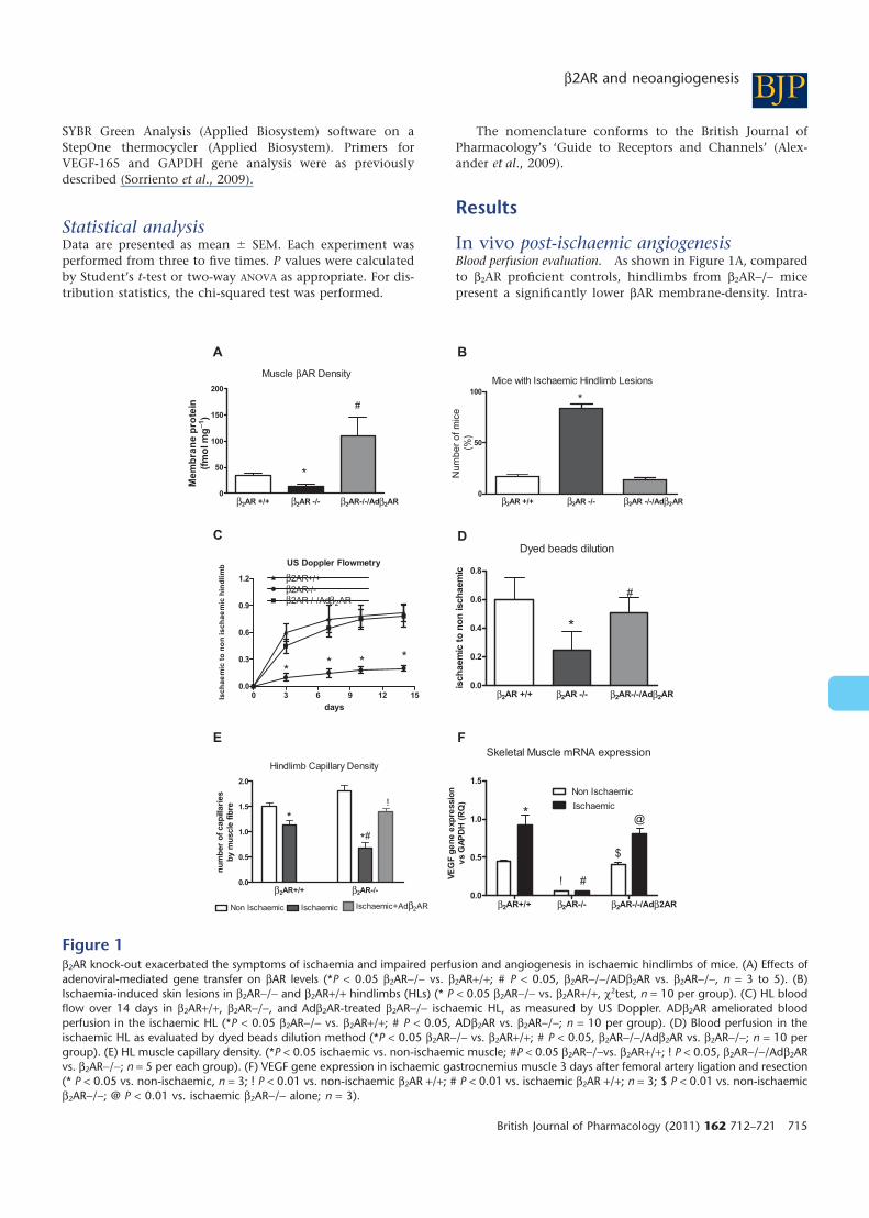
In vivo post-ischaemic angiogenesisBlood perfusion evaluation. As shown in Figure 1A, comparedto b2AR proficient controls, hindlimbs from b2AR-/- micepresent a significantly lower bAR membrane-density. Intra-
β2AR +/+ β2AR -/- β2AR-/-/Ad 2AR0
50
100
150
200
*
#
Muscle βAR Density
Me
mb
ran
e p
rote
in
(fm
ol m
g−1
)
β2AR +/+ β2AR -/- β2AR -/-/Ad 2AR0
50
100
Num
ber
of m
ice
(%)
*
Mice with Ischaemic Hindlimb Lesions
US Doppler Flowmetry
0 3 6 9 12 150.0
0.3
0.6
0.9
1.2
β2AR-/-β2AR-/-/Ad 2AR
** * *
β2AR+/+
days
Isc
ha
em
ic t
o n
on
is
ch
ae
mic
hin
dli
mb
A B
C D
E
β2AR +/+ β2AR -/- β2AR-/-/Ad 2AR0.0
0.2
0.4
0.6
0.8
*
Dyed beads dilution
#
isch
aem
ic t
o n
on
isch
aem
ic
β2AR+/+ 2AR-/-0.0
0.5
1.0
1.5
2.0
*#
*!
Hindlimb Capillary Density
Ischaemic+Adβ2ARNon Ischaemic Ischaemic
nu
mb
er
of
cap
illari
es
by m
uscle
fib
re
β2AR+/+ β2AR-/- β2AR-/-/Adβ2AR0.0
0.5
1.0
1.5
Non Ischaemic
Ιschaemic*
#!
$
@
VE
GF
ge
ne
exp
ressio
nv
s G
AP
DH
(R
Q)
Skeletal Muscle mRNA expressionF
β β
β
β
β
Figure 1b2AR knock-out exacerbated the symptoms of ischaemia and impaired perfusion and angiogenesis in ischaemic hindlimbs of mice. (A) Effects ofadenoviral-mediated gene transfer on bAR levels (*P < 0.05 b2AR-/- vs. b2AR+/+; # P < 0.05, b2AR-/-/ADb2AR vs. b2AR-/-, n = 3 to 5). (B)Ischaemia-induced skin lesions in b2AR-/- and b2AR+/+ hindlimbs (HLs) (* P < 0.05 b2AR-/- vs. b2AR+/+, c2test, n = 10 per group). (C) HL bloodflow over 14 days in b2AR+/+, b2AR-/-, and Adb2AR-treated b2AR-/- ischaemic HL, as measured by US Doppler. ADb2AR ameliorated bloodperfusion in the ischaemic HL (*P < 0.05 b2AR-/- vs. b2AR+/+; # P < 0.05, ADb2AR vs. b2AR-/-; n = 10 per group). (D) Blood perfusion in theischaemic HL as evaluated by dyed beads dilution method (*P < 0.05 b2AR-/- vs. b2AR+/+; # P < 0.05, b2AR-/-/Adb2AR vs. b2AR-/-; n = 10 pergroup). (E) HL muscle capillary density. (*P < 0.05 ischaemic vs. non-ischaemic muscle; #P < 0.05 b2AR-/-vs. b2AR+/+; ! P < 0.05, b2AR-/-/Adb2ARvs. b2AR-/-; n = 5 per each group). (F) VEGF gene expression in ischaemic gastrocnemius muscle 3 days after femoral artery ligation and resection(* P < 0.05 vs. non-ischaemic, n = 3; ! P < 0.01 vs. non-ischaemic b2AR +/+; # P < 0.01 vs. ischaemic b2AR +/+; n = 3; $ P < 0.01 vs. non-ischaemicb2AR-/-; @ P < 0.01 vs. ischaemic b2AR-/- alone; n = 3).
BJPb2AR and neoangiogenesis
British Journal of Pharmacology (2011) 162 712–721 715
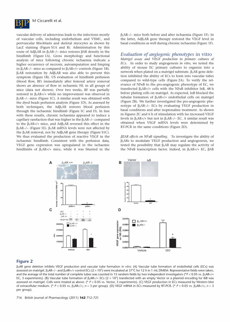
vascular delivery of adenovirus leads to the infections mostlyof vascular cells, including endothelium and VSMC, andperivascular fibroblasts and skeletal myocytes as shown byLacZ staining (Figure S1A and B). Administration by thisroute of Adb2AR in b2AR-/- mice restores bAR density in thehindlimb (Figure 1A). Gross morphology and functionalanalysis of mice following chronic ischaemia indicate ahigher occurrence of necrosis, autoamputation and limpingin b2AR-/- mice as compared to b2AR+/+ controls (Figure 1B).b2AR restoration by Adb2AR was also able to prevent thissymptom (Figure 1B). US evaluation of hindlimb perfusion(blood flow, BF) immediately after femoral artery removalshows an absence of flow in ischaemic HL in all groups ofmice (data not shown). Over two weeks, BF was partiallyrestored in b2AR+/+ while no improvement was observed inb2AR-/- mice (Figure 1C). A similar result was obtained withthe dyed beads perfusion analysis (Figure 1D). As assessed byboth techniques, the Adb2AR restores blood perfusionthrough the ischaemic hindlimb (Figure 1C and D). In linewith these results, chronic ischaemia appeared to induce acapillary rarefaction that was higher in the b2AR-/- comparedto the b2AR+/+ mice, and Adb2AR reversed this effect in theb2AR-/- (Figure 1E). b1AR mRNA levels were not affected bythe b2AR removal, nor by Adb2AR gene therapy (Figure S1C).We thus evaluated the production of reactive VEGF in theischaemic hindlimb. Consistent with the perfusion data,VEGF gene expression was upregulated in the ischaemichindlimbs of b2AR+/+ mice, while it was blunted in the
b2AR-/- mice both before and after ischaemia (Figure 1F). Inthe latter, Adb2AR gene therapy restored the VEGF level inbasal conditions as well during chronic ischaemia (Figure 1F).
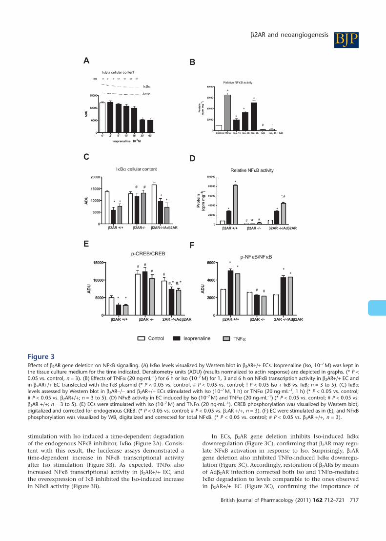
Evaluation of angiogenic phenotypes in vitroMatrigel assay and VEGF production in primary cultures ofECs. In order to study angiogenesis in vitro, we tested theability of mouse EC primary cultures to organize into anetwork when plated on a matrigel substrate. b2AR gene dele-tion inhibited the ability of ECs to form into vascular tubescompared to wild-type cells (Figure 2A). To verify the rel-evance of NFkB to the pro-angiogenic phenotype of EC, wetransfected b2AR+/+ cells with the NFkB inhibitor IkB, 48 hbefore plating cells on matrigel. As expected, IkB blocked thetubular formation of b2AR+/+ endothelial cells on matrigel(Figure 2B). We further investigated the pro-angiogenic phe-notype of b2AR-/- ECs by evaluating VEGF production inbasal conditions and after isoprenaline treatment. As shownin Figures 2C and 6 h of stimulation with Iso increased VEGFlevels in b2AR+/+ but not in b2AR-/- EC. A similar result wasobtained when VEGF mRNA levels were determined byRT-PCR in the same conditions (Figure 2D).
b2AR effects on NFkB signalling. To investigate the ability ofb2ARs to modulate VEGF production and angiogenesis, wetested the possibility that b2AR may regulate the activity ofthe NFkB transcription factor. Indeed, in b2AR+/+ EC, bAR
b2AR+/+ b2AR-/-0
10
20
30
*
Nu
mb
er
of
bra
nch
es/1
0000 u
m2
Vector IkB0
10
20
30
40
50
*
Nu
mb
er
of
bra
nc
he
s/1
00
0 m
m2
Vehicle ISO
0
125000
250000
375000
500000 2AR +/+2AR -/-
VEGF164
ISO
2AR -/-
2AR +/+
--+
-
+-
+
-+
++-
AD
U *
C
0.000
0.002
0.004
0.006
0.008
0.010
2AR-/-2 AR+/+
*
Vehicle ISO
VE
GF
gen
e e
xp
ressio
n
vs 1
8S
(R
Q)
Db
bb
bb
β2AR -/-β2AR +/+Vector IkB
BA
Figure 2b2AR gene deletion inhibits VEGF production and vascular tube formation in vitro. (A) Vascular tube formation of endothelial cells (ECs) wasassessed on matrigel. b2AR-/- and b2AR+/+ control ECs (2 ¥ 105) were incubated at 37°C for 12 h in 1 mL DMEM. Representative fields were taken,and the average of the total number of complete tubes was counted in 15 random fields by two independent investigators (*P < 0.05 vs. b2AR+/+EC, 3 experiments). (B) Vascular tube formation of b2AR+/+ ECs (2 ¥ 105) transfected with an empty Vector or a plasmid encoding for IkB wasassessed on matrigel. Cells were treated as above. (* P < 0.05 vs. Vector, 3 experiments). (C) VEGF production in ECs measured by Western blotof extracellular medium. (* P < 0.05 vs. b2AR+/+; n = 3 per group). (D) VEGF mRNA in ECs measured by RT-PCR. (* P < 0.05 vs. b2AR+/+; n = 3per group).
BJP M Ciccarelli et al.
716 British Journal of Pharmacology (2011) 162 712–721
stimulation with Iso induced a time-dependent degradationof the endogenous NFkB inhibitor, IkBa (Figure 3A). Consis-tent with this result, the luciferase assays demonstrated atime-dependent increase in NFkB transcriptional activityafter Iso stimulation (Figure 3B). As expected, TNFa alsoincreased NFkB transcriptional activity in b2AR+/+ EC, andthe overexpression of IkB inhibited the Iso-induced increasein NFkB activity (Figure 3B).
In ECs, b2AR gene deletion inhibits Iso-induced IkBadownregulation (Figure 3C), confirming that b2AR may regu-late NFkB activation in response to Iso. Surprisingly, b2ARgene deletion also inhibited TNFa-induced IkBa downregu-lation (Figure 3C). Accordingly, restoration of b2ARs by meansof Adb2AR infection corrected both Iso and TNFa–mediatedIkBa degradation to levels comparable to the ones observedin b2AR+/+ EC (Figure 3C), confirming the importance of
0' 2' 5' 10' 15' 30' 60'0
6000
12000
18000
* *
IκBα
IκBα
cellular content
0' 2' 5' 10' 15' 30' 60'ISO
Actin
Isoprenaline, 10-7
M
AD
U
Control TNFa Iso, 1h Iso, 3h Iso, 6h IkB Iso, 3h + IkB0
20000
40000
60000
80000
*
Relative NFκB activity
*
*
*
!#
Pro
tein
(cp
m m
g−1
)
Pro
tein
(cp
m m
g−1
)
b2AR +/+ b2AR-/- -/-/Adb2AR0
5000
10000
15000
20000
IκBα cellular content
* * * *
##
AD
U
b2AR +/+ b2AR -/- b2AR -/-/Adb2AR0
20000
40000
60000
80000
100000
# # #
*
*
*,#
*
Relative NFκB activity
b2AR +/+ b2AR -/- 2AR -/-/Adb2AR0
5000
10000
15000
p-CREB/CREB
#
#
#
#,*#,*
***
#
AD
U
D
F
B
C
E
A
b2AR +/+ b2AR -/- 2AR -/-/Adb2AR0
2000
4000
6000
p-NFκB/NFκB
# #
**
**
AD
U
Control Isoprenaline TNFα
b2AR
Figure 3Effects of b2AR gene deletion on NFkB signalling. (A) IkBa levels visualized by Western blot in b2AR+/+ ECs. Isoprenaline (Iso, 10-7 M) was kept inthe tissue culture medium for the time indicated. Densitometry units (ADU) (results normalized to actin response) are depicted in graphs. (* P <0.05 vs. control, n = 3). (B) Effects of TNFa (20 ng·mL-1) for 6 h or Iso (10-7 M) for 1, 3 and 6 h on NFkB transcription activity in b2AR+/+ EC andin b2AR+/+ EC transfected with the IkB plasmid (* P < 0.05 vs. control, # P < 0.05 vs. control; ! P < 0.05 Iso + IkB vs. IkB; n = 3 to 5). (C) IkBalevels assessed by Western blot in b2AR-/- and b2AR+/+ ECs stimulated with Iso (10-7 M, 1 h) or TNFa (20 ng·mL-1, 1 h) (* P < 0.05 vs. control;# P < 0.05 vs. b2AR+/+; n = 3 to 5). (D) NFkB activity in EC induced by Iso (10-7 M) and TNFa (20 ng·mL-1) (* P < 0.05 vs. control; # P < 0.05 vs.b2AR +/+; n = 3 to 5). (E) ECs were stimulated with Iso (10-7 M) and TNFa (20 ng·mL-1). CREB phosphorylation was visualized by Western blot,digitalized and corrected for endogenous CREB. (* P < 0.05 vs. control; # P < 0.05 vs. b2AR +/+, n = 3). (F) EC were stimulated as in (E), and NFkBphosphorylation was visualized by WB, digitalized and corrected for total NFkB. (* P < 0.05 vs. control; # P < 0.05 vs. b2AR +/+, n = 3).
BJPb2AR and neoangiogenesis
British Journal of Pharmacology (2011) 162 712–721 717
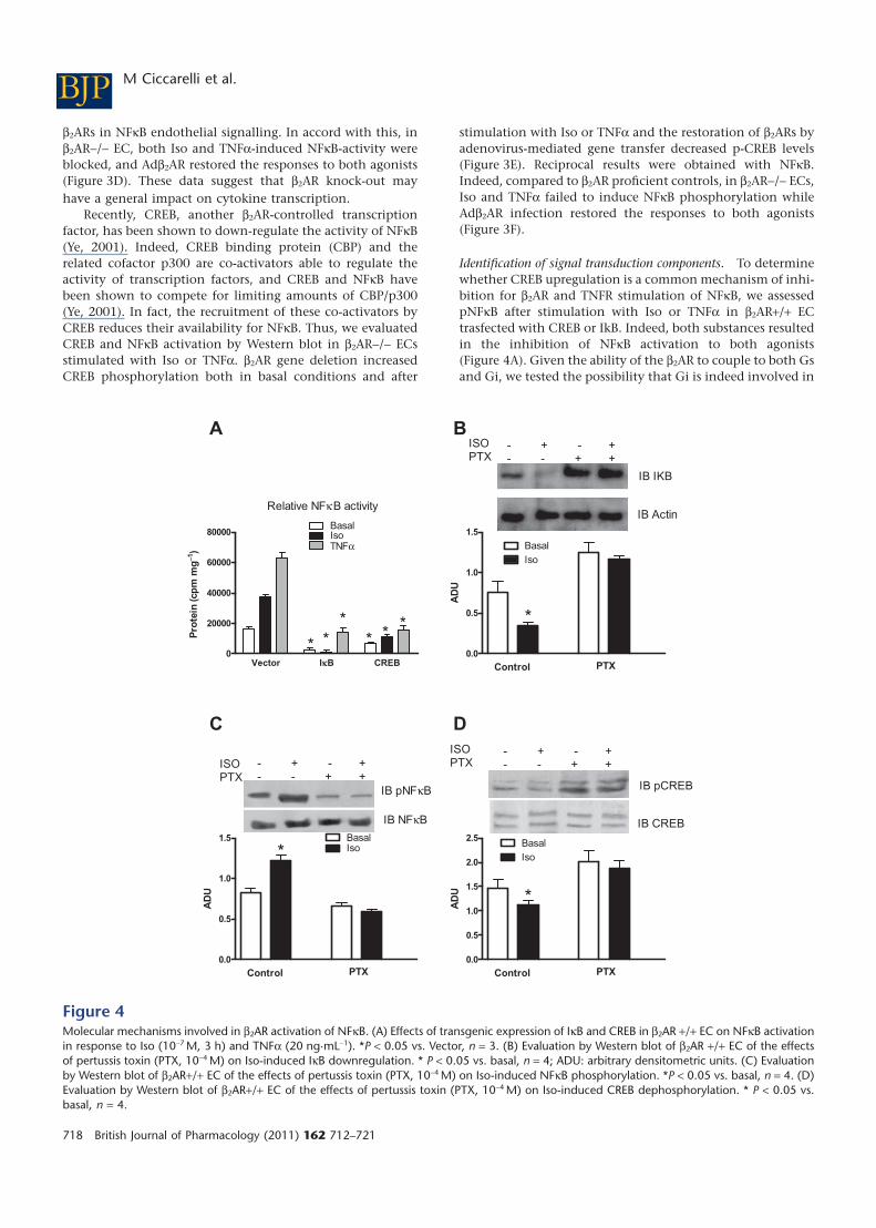
b2ARs in NFkB endothelial signalling. In accord with this, inb2AR-/- EC, both Iso and TNFa-induced NFkB-activity wereblocked, and Adb2AR restored the responses to both agonists(Figure 3D). These data suggest that b2AR knock-out mayhave a general impact on cytokine transcription.
Recently, CREB, another b2AR-controlled transcriptionfactor, has been shown to down-regulate the activity of NFkB(Ye, 2001). Indeed, CREB binding protein (CBP) and therelated cofactor p300 are co-activators able to regulate theactivity of transcription factors, and CREB and NFkB havebeen shown to compete for limiting amounts of CBP/p300(Ye, 2001). In fact, the recruitment of these co-activators byCREB reduces their availability for NFkB. Thus, we evaluatedCREB and NFkB activation by Western blot in b2AR-/- ECsstimulated with Iso or TNFa. b2AR gene deletion increasedCREB phosphorylation both in basal conditions and after
stimulation with Iso or TNFa and the restoration of b2ARs byadenovirus-mediated gene transfer decreased p-CREB levels(Figure 3E). Reciprocal results were obtained with NFkB.Indeed, compared to b2AR proficient controls, in b2AR-/- ECs,Iso and TNFa failed to induce NFkB phosphorylation whileAdb2AR infection restored the responses to both agonists(Figure 3F).
Identification of signal transduction components. To determinewhether CREB upregulation is a common mechanism of inhi-bition for b2AR and TNFR stimulation of NFkB, we assessedpNFkB after stimulation with Iso or TNFa in b2AR+/+ ECtrasfected with CREB or IkB. Indeed, both substances resultedin the inhibition of NFkB activation to both agonists(Figure 4A). Given the ability of the b2AR to couple to both Gsand Gi, we tested the possibility that Gi is indeed involved in
Vector IkB CREB0
20000
40000
60000
80000BasalIsoTNFα
Relative NFκB activity
**
* ** *
0.0
0.5
1.0
1.5
IB IKB
IB Actin
Control PTX
ISOPTX
- + - + - - + +
*
BasalIso
AD
U
0.0
0.5
1.0
1.5
IB pNFκB
IB NFκB
Control PTX
ISOPTX
- + - + - - + +
*BasalIso
AD
U
0.0
0.5
1.0
1.5
2.0
2.5
Control PTX
ISOPTX
- + - + - - + +
IB pCREB
IB CREB
BasalIso
*
AD
U
B
D
A
C
Pro
tein
(cp
m m
g−1
)
Figure 4Molecular mechanisms involved in b2AR activation of NFkB. (A) Effects of transgenic expression of IkB and CREB in b2AR +/+ EC on NFkB activationin response to Iso (10-7 M, 3 h) and TNFa (20 ng·mL-1). *P < 0.05 vs. Vector, n = 3. (B) Evaluation by Western blot of b2AR +/+ EC of the effectsof pertussis toxin (PTX, 10-4 M) on Iso-induced IkB downregulation. * P < 0.05 vs. basal, n = 4; ADU: arbitrary densitometric units. (C) Evaluationby Western blot of b2AR+/+ EC of the effects of pertussis toxin (PTX, 10-4 M) on Iso-induced NFkB phosphorylation. *P < 0.05 vs. basal, n = 4. (D)Evaluation by Western blot of b2AR+/+ EC of the effects of pertussis toxin (PTX, 10-4 M) on Iso-induced CREB dephosphorylation. * P < 0.05 vs.basal, n = 4.
BJP M Ciccarelli et al.
718 British Journal of Pharmacology (2011) 162 712–721
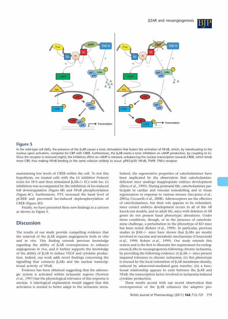
maintaining low levels of CREB within the cell. To test thishypothesis, we treated cells with the Gi inhibitor Pertussistoxin for 18 h and then stimulated b2AR+/+ ECs with Iso. Giinhibition was accompanied by the inhibition of Iso-inducedIkB downregulation (Figure 4B) and NFkB phosphorylation(Figure 4C). Furthermore, PTX increased the basal level ofpCREB and prevented Iso-induced dephosphorylation ofCREB (Figure 4D).
Finally, we have presented these new findings in a cartoonas shown in Figure 5.
Discussion
The results of our study provide compelling evidence thatthe removal of the b2AR impairs angiogenesis both in vitroand in vivo. This finding extends previous knowledgeregarding the ability of b2AR overexpression to enhanceangiogenesis in vivo, and it further supports the knowledgeof the ability of b2AR to induce VEGF and cytokine produc-tion. Indeed, our work adds novel findings concerning thesignalling that connects b2ARs and the nuclear transcrip-tional activity of NFkB.
Evidence has been obtained suggesting that the adrener-gic system is activated within ischaemic regions (Newtonet al., 1997) but the physiological relevance of this response isunclear. A teleological explanation would suggest that thisactivation is needed to better adapt to the ischaemic stress.
Indeed, the regenerative properties of catecholamines havebeen implicated by the observation that catecholamine-deficient mice undergo inappropriate embryo development(Zhou et al., 1995). During postnatal life, catecholamines par-ticipate in cardiac and vascular remodelling and in tissueregeneration in response to various stresses (Iaccarino et al.,2001a; Ciccarelli et al., 2008). Adrenoceptors are the effectorsof catecholamines, but their role appears to be redundant,since correct embryo development occurs in all of the ARknock-out models, and in adult life, mice with deletion of ARgenes do not present basal phenotypic alterations. Understress conditions, though, or in the presence of catechola-mine challenge, a perturbation in the phenotype of KO micehas been noted (Rohrer et al., 1999). In particular, previousstudies in bAR-/- mice have shown that b2ARs are mostlyinvolved in vascular and metabolic mechanisms (Chruscinskiet al., 1999; Rohrer et al., 1999). Our study extends thisnotion and is the first to illustrate the requirement for endog-enous b2ARs in neoangiogenesis following chronic ischaemia,by providing the following evidence: (i) b2AR-/- mice presentimpaired tolerance to chronic ischaemia; (ii) this phenotypeis rescued by the local restoration of b2AR membrane-density,induced by adenoviral-mediated gene transfer; (iii) a func-tional relationship appears to exist between the b2AR andNFkB, the transcription factor involved in ischaemia-inducedcytokine production.
These results accord with our recent observation thatoverexpression of the b2AR enhances the adaptive pro-
Figure 5In the wild-type cell (left), the presence of the b2AR causes a tonic stimulation that fosters the activation of NFkB, which, by translocating to thenucleus upon activation, competes for CBP with CREB. Furthermore, the b2AR exerts a tonic inhibition on cAMP production, by coupling to Gi.Once the receptor is removed (right), the inhibitory effect on cAMP is released, unbalancing the nuclear transcription towards CREB, which bindsmore CBP, thus making NFkB binding to the same cofactor unlikely to occur. pP65/p50: NFkB; TNFR: TNFa receptor.
BJPb2AR and neoangiogenesis
British Journal of Pharmacology (2011) 162 712–721 719
angiogenic response to ischaemia (Iaccarino et al., 2005).Importantly, the present results add to the hypothesis thatother endogenously expressed bAR subtypes cannot under-take this role of b2ARs in ischaemic neo-angiogenesis. Inti-mate differences in the signalling capabilities of this receptormake it unique, and probably this is the reason for our find-ings. Removal of this receptor alters the intracellular signaltransduction pathways to such an extent that the trascrip-tional status of the cell is modified. Our in vitro study revealedthat angiogenesis is impaired in b2AR-/- EC, probably due tothe impaired production of cytokines such as VEGF. The maintranscription factors modulating VEGF expression arehypoxia-inducible factor-1a (HIF-1a) (Marti et al., 2000) andNFkB (Kiriakidis et al., 2003). HIF-1a activates the transcrip-tion of target genes in response to hypoxia and is inactive inbasal conditions due to ubiquitination and degradation.NFkB, in contrast, can be activated following stimulation ofdifferent receptors. The ability of GPCRs to activate this tran-scription factor has been established in different cell types,mainly in the immune system. With regard to the bARs, theevidence showing that bAR agonists are able to induce NFkBactivation is controversial. In monocytes, Farmer and Pugin(2000) showed that a number of bAR agonists had inhibitoryeffects on LPS-induced TNFa and IL-8 production, and pro-posed that cAMP through PKA is the mediator of such inhi-bition (Farmer and Pugin, 2000). In contrast, Chandrasekaret al. (2004) showed that in cardiac-derived ECs, Iso inducesNFkB promoter activity in a b2AR-dependent manner. Theseauthors concluded that b2ARs induce NFkB activation in acAMP-independent manner, through the activation of Gi,Pi3K, Akt and IKK with IkBa degradation. Our results accordwith the latter findings, since in b2AR+/+ EC, Iso induced IkBadegradation and enhanced NFkB transcriptional activity in atime-dependent manner. Furthermore, our data indicate thatthe physical presence of b2ARs is needed to activate NFkB;indeed, b2AR gene deletion inhibited NFkB activity inresponse to both GPCRs and TNF-Rs. Gas and Gai signallingpathways have opposite effects on NFkB: Gs-dependent sig-nalling, induces PKA-dependent CREB activation and conse-quently inhibition of NFkB activity (Ye, 2001). In contrast, Giactivates NFkB by inhibiting/removing IkB. Our hypothesis isthat the lack of b2ARs that have the ability to couple to Gitilts the balance towards Gs-dependent signalling, whichwould lead to PKA-induced CREB activation and CBP/p300recruitment, making the latter unavailable for NFkB activa-tion (Figure 5). Indeed, validation of such a hypothesisderives from our studies with the pertussis toxin, showing therelevance of Gi for b2AR-induced activation of NFkB. Irrespec-tive of this result, the observation that regardless of itsmechanism of action b2AR influenced not only isoprenaline,but also TNFa-dependent activation of NFkB is strongly sug-gestive of a pivotal role played by b2ARs in the instruction ofsignals required for the fine-tuning of NFkB transcriptionalactivity. We have performed our experiments in the endot-helial cells, but it is most likely that the b2AR can regulateNFkB activity also in other cell types. In particular it would beinteresting to evaluate the skeletal muscle, which expresses alarge number of b2ARs and is also an important source ofVEGF.
In conclusion, our data indicate that the b2AR is impor-tant in mediating production of key pro-angiogenic cytok-
ines, such as VEGF. The impairment of the signaltransduction of this receptor results in imparment of angio-genesis in response to chronic ischaemia. Our data add anovel piece to the puzzling paradigm of those pathophysi-ological conditions that are characterized by increased adr-energic neural drive, impaired b adrenoceptor signalling andimpaired organ function such as myocardial ischaemia.Under these conditions, the reduction of b2ARs signalling isdetrimental not only for the cardiac function but also for thedevelopment of an adequate compensatory neoangiogenesis,which would worsen the blood supply to the ischaemic heartand accelerate the progression of myocardial dysfunction.The use of therapeutic strategies aimed at improving bARsignalling, may therefore achieve double efficacy, by amelio-rating myocardial function and by hastening compensatoryangiogenesis.
Acknowledgements
Fundings to GI from Italian Ministry of Research, PRIN20074MSWYW.
Conflict of interest
The authors declare that they have no competing financialinterests.
ReferencesAlexander SPH, Mathie A, Peters JA (2009). Guide to Receptors andChannels (GRAC), 4th Edition. Br J Pharmacol 158 (Suppl. 1):S1–254.
Chandrasekar B, Marelli-Berg FM, Tone M, Bysani S, Prabhu SD,Murray DR (2004). Beta-adrenergic stimulation inducesinterleukin-18 expression via beta2-AR, PI3K, Akt, IKK, andNF-kappaB. Biochem Biophys Res Commun 319: 304–311.
Chruscinski AJ, Rohrer DK, Schauble E, Desai KH, Bernstein D,Kobilka BK (1999). Targeted disruption of the beta2 adrenergicreceptor gene. J Biol Chem 274: 16694–16700.
Ciccarelli M, Cipolletta E, Santulli G, Campanile A, Pumiglia K,Cervero P et al. (2007). Endothelial beta2 adrenergic signaling toAKT: role of Gi and SRC. Cell Signal 19: 1949–1955.
Ciccarelli M, Santulli G, Campanile A, Galasso G, Cervero P,Altobelli GG et al. (2008). Endothelial alpha1-adrenoceptorsregulate neo-angiogenesis. Br J Pharmacol 153: 936–946.
Farmer P, Pugin J (2000). Beta-adrenergic agonists exert their‘anti-inflammatory’ effects in monocytic cells through theIkappaB/NF-kappaB pathway. Am J Physiol Lung Cell Mol Physiol279: L675–L682.
Ferro A, Queen LR, Priest RM, Xu B, Ritter JM, Poston L et al.(1999). Activation of nitric oxide synthase by beta 2-adrenoceptorsin human umbilical vein endothelium in vitro. Br J Pharmacol 126:1872–1880.
Iaccarino G, Tomhave ED, Lefkowitz RJ, Koch WJ (1998). Reciprocalin vivo regulation of myocardial G protein-coupled receptor kinaseexpression by beta-adrenergic receptor stimulation and blockade.Circulation 98: 1783–1789.
BJP M Ciccarelli et al.
720 British Journal of Pharmacology (2011) 162 712–721
Iaccarino G, Barbato E, Cipoletta E, Fiorillo A, Trimarco B (2001a).Role of the sympathetic nervous system in cardiac remodeling inhypertension. Clin Exp Hypertens 23: 35–43.
Iaccarino G, Barbato E, Cipolleta E, Esposito A, Fiorillo A, Koch WJet al. (2001b). Cardiac betaARK1 upregulation induced by chronicsalt deprivation in rats. Hypertension 38: 255–260.
Iaccarino G, Cipolletta E, Fiorillo A, Annecchiarico M, Ciccarelli M,Cimini V et al. (2002). Beta(2)-adrenergic receptor gene delivery tothe endothelium corrects impaired adrenergic vasorelaxation inhypertension. Circulation 106: 349–355.
Iaccarino G, Ciccarelli M, Sorriento D, Cipolletta E, Cerullo V,Iovino GL et al. (2004). AKT participates in endothelial dysfunctionin hypertension. Circulation 109: 2587–2593.
Iaccarino G, Ciccarelli M, Sorriento D, Galasso G, Campanile A,Santulli G et al. (2005). Ischemic neoangiogenesis enhanced bybeta2-adrenergic receptor overexpression: a novel role for theendothelial adrenergic system. Circ Res 97: 1182–1189.
Kiriakidis S, Andreakos E, Monaco C, Foxwell B, Feldmann M,Paleolog E (2003). VEGF expression in human macrophages isNF-kappaB-dependent: studies using adenoviruses expressing theendogenous NF-kappaB inhibitor IkappaBalpha and akinase-defective form of the IkappaB kinase 2. J Cell Sci 116:665–674.
Lembo G, Iaccarino G, Vecchione C, Barbato E, Izzo R, Fontana Det al. (1997). Insulin modulation of an endothelial nitric oxidecomponent present in the alpha2- and beta-adrenergic responses inhuman forearm. J Clin Invest 100: 2007–2014.
Marti HJ, Bernaudin M, Bellail A, Schoch H, Euler M, Petit E et al.(2000). Hypoxia-induced vascular endothelial growth factorexpression precedes neovascularization after cerebral ischemia. Am JPathol 156: 965–976.
Newton GE, Adelman AG, Lima VC, Seidelin PH, Schampaert E,Parker JD (1997). Cardiac sympathetic activity in response to acutemyocardial ischemia. Am J Physiol 272: H2079–2084.
Rohrer DK, Chruscinski A, Schauble EH, Bernstein D, Kobilka BK(1999). Cardiovascular and metabolic alterations in mice lackingboth beta1- and beta2-adrenergic receptors. J Biol Chem 274:16701–16708.
Santulli G, Ciccarelli M, Palumbo G, Campanile A, Galasso G,Ziaco B et al. (2009a). In vivo properties of the proangiogenicpeptide QK. J Transl Med 7: 41.
Santulli G, Cipolletta E, Campanile A, Maione S, Trimarco V,Marino M et al. (2009b). Deletion of the CaMK4 Gene in MiceDetermines a Hypertensive Phenotype. Circulation 120: S613–S613.
Shenoy SK, Drake MT, Nelson CD, Houtz DA, Xiao K, Madabushi Set al. (2006). beta-arrestin-dependent, G protein-independentERK1/2 activation by the beta2 adrenergic receptor. J Biol Chem281: 1261–1273.
Sorriento D, Campanile A, Santulli G, Leggiero E, Pastore L,Trimarco B et al. (2009). A new synthetic protein, TAT-RH, inhibitstumor growth through the regulation of NFkappaB activity. MolCancer 8: 97.
Sorriento D, Santulli G, Fusco A, Anastasio A, Trimarco B,Iaccarino G (2010). Intracardiac injection of AdGRK5-NT reducesleft ventricular hypertrophy by inhibiting NF-{kappa}B-Dependenthypertrophic gene expression. Hypertension 56: 696–704.
Ye RD (2001). Regulation of nuclear factor kappaB activation byG-protein-coupled receptors. J Leukoc Biol 70: 839–848.
Zhou QY, Quaife CJ, Palmiter RD (1995). Targeted disruption of thetyrosine hydroxylase gene reveals that catecholamines are requiredfor mouse fetal development. Nature 374: 640–643.
Zhou RH, Lee TS, Tsou TC, Rannou F, Li YS, Chien S et al. (2003).Stent implantation activates Akt in the vessel wall: role ofmechanical stretch in vascular smooth muscle cells. ArteriosclerThromb Vasc Biol 23: 2015–2020.
Supporting information
Additional Supporting Information may be found in theonline version of this article:
Figure S1 Efficient gene delivery via femoral artery catheter-ization (A-B). Recombinant LacZ Adenovirus was delivered tothe ischemic hindlimb via catheterization as described in theMethods section and results in gene transduction of capillar-ies (A) as well as perivascular muscle fibers (B). Muscle b1ARmRNA levels measured by RT-PCR (C). Hindlimb musclesprocessed as described in methods showing unmodified b1ARmRNA levels among b2AR +/+, b2AR-/- and Adb2AR treatedmice.
Please note: Wiley-Blackwell are not responsible for thecontent or functionality of any supporting materials suppliedby the authors. Any queries (other than missing material)should be directed to the corresponding author for the article.
BJPb2AR and neoangiogenesis
British Journal of Pharmacology (2011) 162 712–721 721
Related Documents