Brain (1999), 122, 567–579 Impaired modulation of quadriceps tendon jerk reflex during spastic gait: differences between spinal and cerebral lesions Michael Faist, 1 Matthias Ertel, 1 Wiltrud Berger 1 and Volker Dietz 2 1 Department of Clinical Neurology and Neurophysiology, Correspondence to: Dr Michael Faist, Department of University of Freiburg, Germany and 2 Swiss Paraplegic Clinical Neurology and Neurophysiology, University of Centre, University Hospital Balgrist, Zu ¨rich, Switzerland Freiburg, Breisacherstr. 64, D-79106-Freiburg, Germany E-mail: faist@nz11.ukl.uni-freiburg.de Summary In healthy subjects, functionally appropriate modulation of short latency leg muscle reflexes occurs during gait. This modulation has been ascribed, in part, to changes in presynaptic inhibition of Ia afferents. The changes in modulation of quadriceps tendon jerk reflexes during gait of healthy subjects were compared with those of hemi- or paraparetic spastic patients. The spasticity was due to unilateral cerebral infarction or traumatic spinal cord injury, respectively. The modulation of the quadriceps femoris tendon jerk reflex at 16 phases of the step cycle was studied. The reflex responses obtained during treadmill walking were compared with control values obtained during gait-mimicking standing postures with corresponding levels of voluntary muscle contraction and knee angles. In healthy subjects the size of the reflexes was profoundly modulated and was generally depressed Keywords: tendon-tap reflexes; gait; spasticity; hemiparesis; paraparesis Abbreviation: MI 5 modulation index Introduction During normal human gait short latency leg muscle reflexes (H-reflex and stretch/tendon jerk reflex) have been shown to be profoundly modulated throughout the step cycle (Capaday and Stein, 1986, 1987; Crenna and Frigo, 1987; Llewellyn et al., 1987; Dietz et al., 1990a, b; Yang et al., 1991a; Sinkjaer et al., 1996b; van de Crommert et al., 1996). However, this modulation is not merely a reflection of the changes in motor neuron excitability associated with the strength of the electromyographic (EMG) activity. Part of the overall effect of reflex modulation may be due to changes in presynaptic inhibition of Ia afferents (Morin et al., 1982; Capaday and Stein, 1986; Dietz et al., 1990a, b; for review,see Stein, 1995). Indeed, recent studies have suggested that there are rhythmic changes in presynaptic inhibition of Ia terminals projecting onto the soleus muscle during gait (Faist et al., 1996a). © Oxford University Press 1999 throughout the step cycle. In patients with spinal lesion the reflex depression during gait was almost removed and was associated with weak or no modulation during the step cycle. In patients with cerebral lesion there was less depression of the reflex size associated with a reduced reflex modulation on the affected side compared with healthy subjects. On the ‘unaffected’ side of these patients reflex modulation was similar to that of healthy subjects, but the reflex size during gait was not significantly different from standing control values. These observations suggest that the mechanisms responsible for the depression of reflex size and the modulation normally seen during gait in healthy subjects are impaired to different extents in spasticity of spinal or cerebral origin, possibly due to the unilateral preservation of fibre tracts in hemiparesis. As the modulation of reflexes is considered to be of functional importance during normal locomotion, defective reflex behaviour may contribute to gait disorders. In fact, impaired modulation of soleus short latency reflexes has been described in patients with spastic gait resulting from multiple sclerosis or spinal cord injury (Sinkjaer et al., 1995, 1996a). It was concluded that this impairment may contribute to the functional deficit of spastic patients and that a deficient spinal processing of segmental and/or supraspinal input onto the motor neuron pool might be responsible for the dysregulation (Yang et al., 1991b). However, for the resting muscle there is increasing evidence indicating that the mechanisms underlying the impaired reflex behaviour in spasticity depend on the site of the lesion. For example, in patients with spinal cord injury (Faist et al., 1994) or multiple sclerosis (Nielsen et al., 1995), presynaptic inhibition of Ia afferents is reduced Downloaded from https://academic.oup.com/brain/article-abstract/122/3/567/528058 by guest on 04 April 2019

Welcome message from author
This document is posted to help you gain knowledge. Please leave a comment to let me know what you think about it! Share it to your friends and learn new things together.
Transcript

Brain (1999),122,567–579
Impaired modulation of quadriceps tendon jerkreflex during spastic gait: differences betweenspinal and cerebral lesionsMichael Faist,1 Matthias Ertel,1 Wiltrud Berger1 and Volker Dietz2
1Department of Clinical Neurology and Neurophysiology, Correspondence to: Dr Michael Faist, Department ofUniversity of Freiburg, Germany and2Swiss Paraplegic Clinical Neurology and Neurophysiology, University ofCentre, University Hospital Balgrist, Zu¨rich, Switzerland Freiburg, Breisacherstr. 64, D-79106-Freiburg, Germany
E-mail: [email protected]
SummaryIn healthy subjects, functionally appropriate modulationof short latency leg muscle reflexes occurs during gait.This modulation has been ascribed, in part, to changesin presynaptic inhibition of Ia afferents. The changes inmodulation of quadriceps tendon jerk reflexes during gaitof healthy subjects were compared with those of hemi-or paraparetic spastic patients. The spasticity was due tounilateral cerebral infarction or traumatic spinal cordinjury, respectively. The modulation of the quadricepsfemoris tendon jerk reflex at 16 phases of the stepcycle was studied. The reflex responses obtained duringtreadmill walking were compared with control valuesobtained during gait-mimicking standing postures withcorresponding levels of voluntary muscle contraction andknee angles. In healthy subjects the size of the reflexeswas profoundly modulated and was generally depressed
Keywords: tendon-tap reflexes; gait; spasticity; hemiparesis; paraparesis
Abbreviation : MI 5 modulation index
IntroductionDuring normal human gait short latency leg muscle reflexes(H-reflex and stretch/tendon jerk reflex) have been shown tobe profoundly modulated throughout the step cycle (Capadayand Stein, 1986, 1987; Crenna and Frigo, 1987; Llewellynet al., 1987; Dietz et al., 1990a, b; Yang et al., 1991a;Sinkjaer et al., 1996b; van de Crommertet al., 1996).However, this modulation is not merely a reflection of thechanges in motor neuron excitability associated with thestrength of the electromyographic (EMG) activity. Part ofthe overall effect of reflex modulation may be due to changesin presynaptic inhibition of Ia afferents (Morinet al., 1982;Capaday and Stein, 1986; Dietzet al., 1990a, b; for review,seeStein, 1995). Indeed, recent studies have suggested that thereare rhythmic changes in presynaptic inhibition of Ia terminalsprojecting onto the soleus muscle during gait (Faistet al.,1996a).
© Oxford University Press 1999
throughout the step cycle. In patients with spinal lesionthe reflex depression during gait was almost removed andwas associated with weak or no modulation during thestep cycle. In patients with cerebral lesion there was lessdepression of the reflex size associated with a reducedreflex modulation on the affected side compared withhealthy subjects. On the ‘unaffected’ side of these patientsreflex modulation was similar to that of healthy subjects,but the reflex size during gait was not significantlydifferent from standing control values. These observationssuggest that the mechanisms responsible for thedepression of reflex size and the modulation normallyseen during gait in healthy subjects are impaired todifferent extents in spasticity of spinal or cerebral origin,possibly due to the unilateral preservation of fibre tractsin hemiparesis.
As the modulation of reflexes is considered to be offunctional importance during normal locomotion, defectivereflex behaviour may contribute to gait disorders. In fact,impaired modulation of soleus short latency reflexes has beendescribed in patients with spastic gait resulting from multiplesclerosis or spinal cord injury (Sinkjaeret al., 1995, 1996a).It was concluded that this impairment may contribute to thefunctional deficit of spastic patients and that a deficient spinalprocessing of segmental and/or supraspinal input onto themotor neuron pool might be responsible for the dysregulation(Yang et al., 1991b). However, for theresting muscle thereis increasing evidence indicating that the mechanismsunderlying the impaired reflex behaviour in spasticity dependon the site of the lesion. For example, in patients with spinalcord injury (Faistet al., 1994) or multiple sclerosis (Nielsenet al., 1995), presynaptic inhibition of Ia afferents is reduced
Dow
nloaded from https://academ
ic.oup.com/brain/article-abstract/122/3/567/528058 by guest on 04 April 2019

568 M. Faist et al.
compared with healthy subjects, whereas it is unchangedfollowing ischaemic stroke (Faistet al., 1994). Recurrentinhibition in the resting muscle is unchanged followingcerebral lesions (Katz and Pierrot-Deseilligny, 1982), butincreased following spinal lesions (Shefneret al., 1992)and decreased in patients with amyotrophic lateral sclerosis(Raynor and Shefner, 1994). In addition, autogenic Ibinhibition in the resting muscle has been found to be reducedin patients with hemiparesis (Delwaide and Oliver, 1988),but seems to be unchanged after spinal lesion (Downeset al., 1995). These findings may represent pathophysiologicaldifferences between spasticity of cerebral or spinal origin,which may also result in different therapeutic approachesbeing considered. In this context it seems noteworthy thatthe patients described in the various studies presented similarsymptoms of spasticity and that there was no clear correlationbetween the impairment assessed by the clinical examinationand any of the spinal reflex mechanisms described above.
The above studies were performed under resting conditions.Therefore, it remains unclear as to how far the reflexbehaviour is defective during movement. As it is only thespastic movement disorder that hampers the patient, it shoulditself be the focus of any therapeutic treatment. The aim ofthe present study was to investigate differences in themodulation of the quadriceps tendon jerk reflex duringgaitin a relatively homogeneous population of spastic patients.Comparison was made between patients with spasticityresulting from spinal cord (paraparesis) or unilateralischaemic cerebral (hemiparesis) lesions. Some of the findingspresented here have been published in abstract form (Faistet al., 1996b).
Patients and methodsPatientsThe ethical committees of the Universities of Freiburg andZurich approved the protocol and the patients gave informedconsent before recordings were taken from 12 healthy subjectsaged 19–34 years (mean6 SD: 24.96 4.1) and 28 spasticpatients aged 16–68 years (48.36 14.6). All patients werediagnosed as having moderate spasticity due either toincomplete traumatic lesion of the cervical or thoracic spinalcord (n 5 12) or unilateral cerebral stroke in the area of themiddle cerebral artery (n 5 16). All patients presentedexaggerated stretch reflexes and/or increased muscle tone asassessed by the Ashworth scale (Ashworth, 1964). The mainclinical parameters of the 17 patients (eight with hemiparesisand nine with paraparesis), who were studied with the fullprotocol, are shown in Table 1.
Experimental paradigmEach patient was required to walk, with the aid of a handrail,on a treadmill for at least 2–3 min at a speed of at least0.5 km/h. Only 17 patients were able to perform the full
experimental protocol during standing and gait. Controlexperiments were also performed in 12 healthy subjectswalking at a speed of 1.5 km/h (approximately the averagespeed of the patients).
Quadriceps tendon jerk reflexes were recorded during levelsplit-belt treadmill locomotion at a comfortable pace for thepatients (mean: 1.28 km/h, range 0.5–2.0 km/h). Tendon jerkreflexes were evoked by a 90 g hammer with an arc radiusof 11 cm (see Dietzet al., 1990a). The hammer was drivenby a revolving field magnetic motor (power 120 W) fixed tothe anterior part of the calf (total weight 950 g). The hammerwas accelerated by the motor up to a constant angular velocityof 800o/s. A potentiometer located at the motor’s axis ofrotation indicated the movement of the hammer and alsoindicated the time of impact of the hammer on the tendon.Force measuring platforms under each belt of the treadmillwere connected to a circuit which triggered the hammermotor at a given delay after heel contact. At least 10 tendonreflexes were elicited during 16 different phases of the stepcycle with a 4 s inter-stimulus interval. As a control thesubjects produced various levels of controlled tonicquadriceps activity at different knee joint angles duringstanding. The change in activity was achieved by alteringthe body loading on the leg under examination. Visualfeedback of the ongoing EMG activity enabled the subjectsto keep the quadriceps EMG activity constant. The positionthus adopted corresponded to those of the various phases ofthe step cycle.
Depending on the patients’ walking ability 6–16 differentcontrol conditions of standing, each including 10 tendonreflexes, were recorded. These control trials were interspersedwith gait trials. Extended periods of rest were allowedbetween trials in order to avoid fatigue and to enable mostof the patients to perform the full experimental protocol. Inall patients both legs were examined on the same day. In thehealthy control group only the right leg was examined.
Data recordingThe EMG activity of the quadriceps was recorded by surfaceelectrodes placed 3 cm apart over the belly of the rectusfemoris muscle about 15–20 cm above the patella. EMGactivity was also obtained from the biceps femoris, the tibialisanterior and the soleus. A sampling rate of 5 kHz was usedand signals were filtered from 3 Hz to 1 kHz. Knee and hipjoint movements were recorded using potentiometers asdescribed earlier (Dietzet al., 1990a). Reflex responses wereassessed as rectified and averaged EMG activity triggered bythe tendon tap. To quantify the net reflex response, thecorresponding rectified and averaged background EMGrecorded during the step cycles without stimuli wassubtracted. The reflex EMG signal was integrated over 25 msafter a latency of 26–33 ms following hammer impact. Theduration of the EMG responses was typically ~25 ms. Thelatency and waveform of the EMG responses obtained
Dow
nloaded from https://academ
ic.oup.com/brain/article-abstract/122/3/567/528058 by guest on 04 April 2019

Tendon jerk reflex during spastic gait 569
Table 1 Clinical and neurological features
Patient Age Lesion Duration Tonus* Treatment† Walking Reflex MI‡ Background(years) (months) velocity (km/h) EMG MI‡
1 48 Cerebral 108 1 C 2 93 392 43 Cerebral 7 1 0 2.5 74 93 38 Cerebral 4 3 0 0.6 88 44 57 Cerebral 1 1 0 1.5 79 905 63 Cerebral 5 2 0 1.5 71 786 48 Cerebral 168 3 DS 0.6 34 687 68 Cerebral 25 3 0 1 75 948 39 Cerebral 96 4 DS 0.5 98 949 55 Spinal 26 2 C 0.7 20 65
10 55 Spinal 4 2 0 1.3 53 3211 67 Spinal 144 2 C 0.8 55 2912 54 Spinal 13 3 B 1.5 99 9913 31 Spinal 13 2 0 0.8 59 8614 60 Spinal 48 2 0 1.7 81 9715 20 Spinal 10 3 0 2 72 3916 24 Spinal 7 1 0 1.2 41 6817 16 Spinal 5 3 B 0.5 78 52
*The degree of spasticity as assessed by Ashworth scale.†0 5 none, C5 Clonazepam, DS5 Dantrolene Sodium, B5 Baclofen;‡MI 5 Modulation Index
indicated that they were indeed typical short latency reflexesevoked by the tendon jerk.
Assessment of reflex modulation during the stepcycleIn order to allow inter-individual comparison of reflexmodulation during walking, the reflexes were normalized tothe mean reflex response obtained in all 16 phases of thestep cycle. For the assessment of the influence of EMGbackground activity on reflex modulation, the reflexes elicitedduring walking were compared with those elicited during thecontrol trials, i.e. the reflex responses obtained at a similarlevel of EMG activity and knee joint angle during standing.For each phase of the step cycle the closest control conditionwas selected for comparison. Only differences of,20% inthe rectified background EMG activity of the quadricepsand ,10o for the knee joint angle were considered to becomparable.
For each of the 16 phases of the step cycle the mean reflexsize was calculated for each of the three groups of subjects(i.e. healthy subjects and patients with either paraparesis orhemiparesis). Statistical significance for the modulation interms of the time-course of changes in reflex size during thestep cycle and the reflex depression during gait comparedwith standing were evaluated by repeated measurementsanalysis of variance. After adjustment of the data for theirskewness by adding a constant and calculating the logarithmswe first investigated differencesbetweensubject groups andtwo effects (time-course and gait versus standing)withinsubject groups. For simplification of the model, the 16 phaseswere reduced to stance phase (mean of the first eight phases)and swing phase (mean of the last five phases) and thetransition from stance to swing was omitted. Secondly, we
tested for an interaction between the time-course of the reflexchanges and the reflex depression during gait comparedwith standing control. Thirdly, we carried out a three-wayinteraction performed to test the differences in time-courseand reflex depression among groups of subjects. In the mainmodel healthy subjects, paraparetic patients and the affectedside of hemiparetic patients were included. As separatequestions the affected and the unaffected side of hemipareticpatients were compared in a paired model and the unaffectedside was also compared with healthy subjects.
To illustrate the degree of modulation during the step cyclein individual subjects the modulation index (MI) (accordingto Yang et al., 1991a) was calculated using the formula ofSinkjaeret al. (1996b):
MI 5 (max. reflex – min. reflex) * 100 / max. reflex
where max. reflexand min. reflexwere the maximum andminimum mean reflex size obtained during the stance andswing phases, respectively. The modulation index for thebackground EMG activity given by the integral of thequadriceps EMG activity in the corresponding 16 phases ofthe step cycle was calculated for each subject. Mean valuesand standard errors of the mean (SEM) were calculated foreach of the three populations (i.e. healthy subjects andpatients with either paraparesis or hemiparesis). Analysis ofvariance was performed and the significance of the differencebetween results obtained in the different populations wasexamined using the Scheffe´ test.
ResultsReflex changes during the step cycle of gaitHealthy subjectsFigure 1A and B show recordings taken from a healthysubject. Figure 1A displays the net reflex quadriceps EMG
Dow
nloaded from https://academ
ic.oup.com/brain/article-abstract/122/3/567/528058 by guest on 04 April 2019

570 M. Faist et al.
Fig. 1 Individual examples of reflex modulation during gait.A, Rectified and averaged (n 5 10) net reflex responses, i.e. withbackground EMG subtracted at 16 phases throughout the step cycle. The arrow and the vertical line indicate the time of hammer impactand the filled and open vertical bars the stance and swing phases, respectively.B, Quantitative values of the quadriceps tendon reflexesduring the step cycle from an individual healthy subject (same subject as inA). The percentage of the step cycle at the time of thehammer impact was calculated from heel contact. Circles represent the EMG integral of the reflex responses obtained during gait,triangles represent the control reflexes obtained at a similar level of EMG activity during standing. The top trace shows the knee jointangle, the two bottom traces show the rectified EMG of the quadriceps and the treadmill force applied by the subject.C andD, E andF, andG andH, The corresponding data to that described in A and B of a patient with paraparesis, the affected side ofa patient with hemiparesis and the unaffected side of the same hemiparetic patient, respectively.
activity (i.e. after subtraction of background EMG activity)at different times during the step cycle. Figure 1B illustratesthe quantitative values of the reflex during gait and duringthe corresponding gait-mimicking standing control togetherwith the biomechanical parameters (knee joint angle andtreadmill force) as well as the rectified and averaged
quadriceps background EMG activity over one step cycle. Itis evident that there was modulation of both the net quadricepsreflex response and backgound EMG activity during the stepcycle. Little or no reflex activity was observed during thelate stance phase or at any time during the swing phase.These effects are seen more clearly in Fig. 2A and B which
Dow
nloaded from https://academ
ic.oup.com/brain/article-abstract/122/3/567/528058 by guest on 04 April 2019

Tendon jerk reflex during spastic gait 571
Fig. 2 Quantified values of the normalized quadriceps tendonreflex during gait and standing. (A) Modulation of the tendon jerkreflex during gait and standing (control) at corresponding kneeangles and voluntary EMG activity. To allow intersubjectcomparison, the reflex integrals were expressed as the percentageof the mean reflex observed during gait. Each symbol representsthe mean of 12 healthy subjects. Vertical bars indicate one SEM.(B) Modulation of the integrated background quadriceps EMGactivity during the step cycle. The background EMG values werenormalized to the mean EMG value obtained during gait. Verticalbars indicate one SEM of the stance phase.
summarize the results obtained from all 12 healthy subjects.The size of the reflex responses was significantly reducedduring the whole step cycle during gait compared with thoserecorded under gait-mimicking control conditions (Fig. 2A).These differences were especially so during the late stanceand swing phases. Although the reflex modulation was similarin all subjects, a large inter-individual variability was observedas indicated by the large SEM.
Patients with spinal lesionsIn patients with paraparesis, the modulation of both the reflexresponse and the background EMG during the step cycle wasmarkedly reduced compared with healthy subjects. A typicalexample of the reflex modulation of one patient is shown inFig. 1C and D. There was a large reflex response at allphases of the step cycle with the greatest responses being inthe early and late stance phase and also during the swingphase. Figure 3 summarizes the results obtained from allnine patients with paraparesis. It is clear from Fig. 3A thatthere was no depression of the reflex response during thelate stance or swing phases compared with the gait-mimicking
Fig. 3 Quantified values obtained from the group of patients withparaparesis (n 5 9). For details see legend to Fig. 2.
controls (cf. Fig. 2A for healthy subjects). Furthermore,compared with healthy subjects the background EMG (Fig.3B) was less well modulated during the step cycle (cf. Figs2B and 3B). Essentially similar results were obtained forboth legs in patients with paraparesis. The mean data shownin Fig. 3 was taken from the more affected leg of the patients.If both legs were similarly affected then the data from theright leg was taken.
Patients with cerebral lesion—affected sideFigure 4 shows the results obtained from the affected leg ofall eight patients with hemiparesis. Results from one subjectare presented in Fig. 1E and F. It is evident that there wassome modulation of the reflex response at different phasesof the step cycle (Fig. 1F). Furthermore, compared withhealthy subjects the reflexes in the swing phase were lessdepressed with respect to the gait-mimicking control (cf. Fig.2A and Fig. 4A). However, compared with parapareticpatients a reflex depression was present with respect to thecontrol condition (cf. Fig. 3A and Fig. 4A). There was stillsome modulation of background EMG activity in somehemiparetic patients (Fig. 4B).
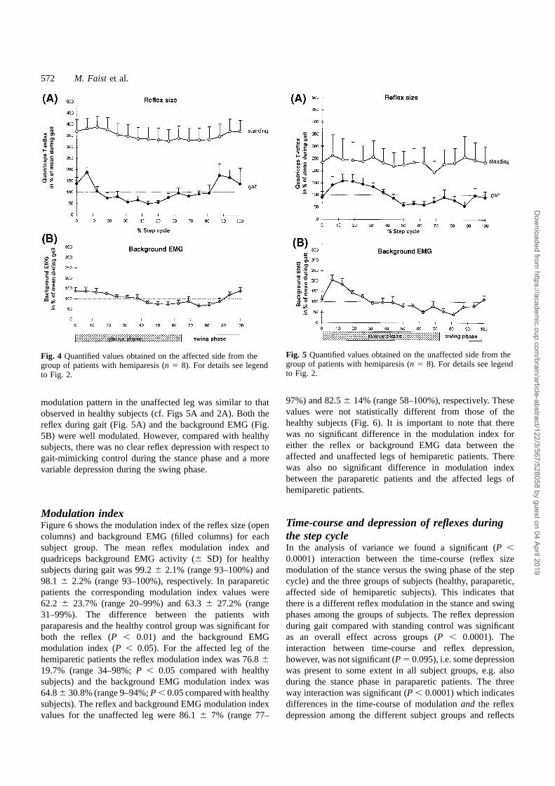
Patients with cerebral lesion—unaffected sideSummarized data from the unaffected leg of hemipareticpatients is shown in Fig. 5. Individual recordings from onesuch patient are shown in Fig. 1G and H. The reflex
Dow
nloaded from https://academ
ic.oup.com/brain/article-abstract/122/3/567/528058 by guest on 04 April 2019
572 M. Faist et al.
Fig. 4 Quantified values obtained on the affected side from thegroup of patients with hemiparesis (n 5 8). For details see legendto Fig. 2.
modulation pattern in the unaffected leg was similar to thatobserved in healthy subjects (cf. Figs 5A and 2A). Both thereflex during gait (Fig. 5A) and the background EMG (Fig.5B) were well modulated. However, compared with healthysubjects, there was no clear reflex depression with respect togait-mimicking control during the stance phase and a morevariable depression during the swing phase.
Modulation indexFigure 6 shows the modulation index of the reflex size (opencolumns) and background EMG (filled columns) for eachsubject group. The mean reflex modulation index andquadriceps background EMG activity (6 SD) for healthysubjects during gait was 99.26 2.1% (range 93–100%) and98.1 6 2.2% (range 93–100%), respectively. In parapareticpatients the corresponding modulation index values were62.2 6 23.7% (range 20–99%) and 63.36 27.2% (range31–99%). The difference between the patients withparaparesis and the healthy control group was significant forboth the reflex (P , 0.01) and the background EMGmodulation index (P , 0.05). For the affected leg of thehemiparetic patients the reflex modulation index was 76.8619.7% (range 34–98%;P , 0.05 compared with healthysubjects) and the background EMG modulation index was64.86 30.8% (range 9–94%;P , 0.05 compared with healthysubjects). The reflex and background EMG modulation indexvalues for the unaffected leg were 86.16 7% (range 77–
Fig. 5 Quantified values obtained on the unaffected side from thegroup of patients with hemiparesis (n 5 8). For details see legendto Fig. 2.
97%) and 82.56 14% (range 58–100%), respectively. Thesevalues were not statistically different from those of thehealthy subjects (Fig. 6). It is important to note that therewas no significant difference in the modulation index foreither the reflex or background EMG data between theaffected and unaffected legs of hemiparetic patients. Therewas also no significant difference in modulation indexbetween the paraparetic patients and the affected legs ofhemiparetic patients.
Time-course and depression of reflexes duringthe step cycleIn the analysis of variance we found a significant (P ,0.0001) interaction between the time-course (reflex sizemodulation of the stance versus the swing phase of the stepcycle) and the three groups of subjects (healthy, paraparetic,affected side of hemiparetic subjects). This indicates thatthere is a different reflex modulation in the stance and swingphases among the groups of subjects. The reflex depressionduring gait compared with standing control was significantas an overall effect across groups (P , 0.0001). Theinteraction between time-course and reflex depression,however, was not significant (P 5 0.095), i.e. some depressionwas present to some extent in all subject groups, e.g. alsoduring the stance phase in paraparetic patients. The threeway interaction was significant (P , 0.0001) which indicatesdifferences in the time-course of modulationand the reflexdepression among the different subject groups and reflects
Dow
nloaded from https://academ
ic.oup.com/brain/article-abstract/122/3/567/528058 by guest on 04 April 2019

Tendon jerk reflex during spastic gait 573
Fig. 6 Modulation indices in the different subject groups. Meanvalues for each population are displayed. Open columns representthe modulation index for the reflex, closed columns themodulation index for the background EMG. Vertical bars indicateSEM. Significance levels were calculated using the Scheffe´ test(*P , 0.05; **P , 0.01; ns5 not significant).
the lack of reflex depression during the swing phase of theparaparetic subjects. The direct comparison between each ofthe three subject groups revealed significant differences forhealthy versus paraparetic subjects (P , 0.0001), healthyversus hemiparetic subjects (P , 0.05) and paraparetic versushemiparetic subjects (P , 0.01).
As a separate question the unaffected side of hemipareticpatients was compared with the affected side in a paireddesign. A significant difference was present between the twosides of hemiparetic patients as a general effect (P , 0.05)and for the three-way interaction (P , 0.05) while the two-way interactions were not significant. Finally, the unaffectedside was compared with healthy control subjects. Theinteraction of the time-course and the two groups wassignificant (P , 0.01). The reflex depression was significantas an overall effect across both groups (P , 0.0001) but theinteraction of depression and groups and also the three-wayinteraction only showed a trend (P 5 0.069 andP 5 0.056,respectively).
Reflex size and background activity: differencesbetween patients with cerebral and spinallesionsFigure 7 shows relative (part A) and absolute (part B) valuesof reflex size during an entire step cycle (as an average ofthe reflex size in the 16 phases of the step cycle) in the threegroups of subjects. When the reflex size during gait wasrelated to the standing condition (Fig. 7A) the relative reflexsize was largest in the group of patients with a spinal lesion.A significant reflex depression was seen in both the groupof healthy subjects (P , 0.001) and on the affected leg of
Fig. 7 Quantified values of the mean reflex size during a stepcycle (i.e. average of reflex size over the 16 phases of the stepcycle) of the three groups of subjects. InA the reflex size duringgait is normalized to the value of reflex size obtained in thestanding condition. InB the absolute values of the integratedbackground EMG (closed columns) and reflex size during gait(open columns) and standing (hatched columns) are displayed forthe three groups of subjects. Vertical bars indicate one standarddeviation. Significance levels were calculated by the Scheffe´ test(** P , 0.01; ***P , 0.001).
the patients with a cerebral lesion (P , 0.01). A moredetailed analysis is given in Fig. 7B where the absolute(integrated) EMG values for the entire step cycle arepresented.
Compared with healthy subjects, the reflex size was largestin patients with a spinal lesion (gaitP , 0.01; standingP ,0.05) but not significantly different from both legs of patientswith hemiparesis. Compared with patients with a spinallesion, the reflex size in patients with a cerebral lesion wassignificantly smaller on the affected side (gaitP , 0.05;standingP , 0.05) and the unaffected side (gaitP , 0.01;reflexP , 0.05), although the background EMG did not differsignificantly between the groups. There was no difference inthe reflex size during gait or standing between both sides ofthe hemiparetic patients. Corresponding to the results shown
Dow
nloaded from https://academ
ic.oup.com/brain/article-abstract/122/3/567/528058 by guest on 04 April 2019

574 M. Faist et al.
Fig. 8 Gait reflex in relation to the standing control for the different groups of subjects throughout the step cycle. Each symbolrepresents the mean of all subjects in the respective group in the respective phase of the step cycle. Vertical bars indicate one SEM.
in Fig. 7A, a significant difference between gait and controlreflex was present only in healthy subjects and the affectedside of patients with hemiparesis.
Reflex size during gait with respect to controlvaluesFigure 8 illustrates for the subject groups the mean rectusfemoris tendon jerk reflexes obtained during the 16 phasesof gait expressed as a percentage of the gait-mimickingcontrol value during standing. As in Fig. 7A, a reflexdepression was present for healthy subjects and thehemiparetic patients throughout the step cycle. In theparaparetic patients a lack of depression during the swingphase was most remarkable.
Background activity in other major leg musclesduring gaitIn the present study for biceps femoris, tibialis anterior andsoleus muscles, no relationship between background EMGactivity and rectus femoris reflex size was seen during gait.The background EMG activity of the biceps femoris inhealthy subjects showed maximal activity in the late swingand early stance phases. In the paraparetic patients and on
the affected side of hemiparetic patients biceps femoris EMGwas largest during late swing and the first half of stancephase. A less pronounced modulation was seen on theunaffected side. The strongest modulation of backgroundEMG occurred in the patient groups—similar to healthysubjects—in the tibialis anterior during swing and in thesoleus in the midstance.
DiscussionThe aim of this study was to evaluate the changes in behaviourof quadriceps short latency reflexes during spastic gait.While recent studies have investigated the changes in soleus/gastrocnemius reflex behaviour in spastic patients (Yanget al., 1991a; Sinkjaeret al., 1995, 1996b), the present study,to our knowledge, is the first to investigate quadriceps reflexmodulation in patients with either spinal or supraspinallesions. Two questions were addressed. (i) Are theredifferences between spasticity of spinal or cerebral origin inthe modulation or depression of short latency reflexes duringlocomotion? This possibility has been extrapolated fromresting conditions (see Faistet al., 1994). (ii) Is the reflexmodulation on the clinically ‘unaffected’ side in patients withhemiparesis different from that in healthy subjects? This has
Dow
nloaded from https://academ
ic.oup.com/brain/article-abstract/122/3/567/528058 by guest on 04 April 2019

Tendon jerk reflex during spastic gait 575
been proposed previously based upon arm reflexes studiedunder passive conditions (Thilmannet al., 1990).
The main results obtained in this study were as follows.(i) Differences in the depression of the short latency tendonjerk reflex were revealed between patients with paraparesisand the affected leg in patients with hemiparesis. (ii) Reflexdepression and modulation were maximally reduced inpatients with paraparesis compared with healthy subjects.(iii) There was a reduced reflex modulation on the affectedside of patients with hemiparesis compared to healthysubjects. (iv) An impaired reflex depression can be assumedon the clinically unaffected side of patients with hemiparesis.
Validity of the comparisonThe differences in reflex modulation pattern during the stepcycle between the groups of subjects were not due to analtered motor neuronal excitability because the time-courseof the background EMG activity was changed in a similarfashion in the spastic legs of patients with either paraparesisor hemiparesis (see Figs 3 and 4). To allow comparison ofreflex size between different subjects the reflex size wasnormalized to the mean reflex during gait. To assess theinfluence of background EMG activity all subjects weretested at corresponding levels of muscle contraction duringstanding. It was evident that the alteration of the reflexmodulation pattern and the reflex size were related to thesite of the lesion (i.e. spinal or supraspinal, see Fig. 8).
It was possible that biomechanical changes during thedifferent step phases contributed to the modulation of thereflex size observed. In an attempt to negate these effects anumber of conditions were imposed. The impact of thehammer at the tendon occurred at a constant angular velocityin all step phases (see Dietzet al., 1990b). Differences inbackground EMG activity and thus differences in the tensionof the tendon were taken into account by the control condition.The different knee joint angles were also comparable betweengait and standing. Nevertheless, it was not possible to quantifyforce or tension exerted by tendons in this control condition.Therefore, we are aware that this control is less than ideal,although it represents the best possible approach to performthese types of experiments in spastic patients.
The asymmetric gait pattern of patients with hemiparesiscompared with the patients with paraparesis could also havebeen responsible for some of the differences observed.Furthermore, changes in the gamma drive to muscle spindlesas well as changes in presynaptic inhibition of group Iaafferents or other spinal mechanisms (see below) may alsohave accounted for the differences. Although the mechanicalconditions should have been similar in all three groups ofsubjects, especially during the swing phase, the strongestdifferences in reflex responses were observed during swing.In spite of the fact that the afferent stimulus of the H-reflexbypasses the muscle spindles, the quadriceps (Dietzet al.,1990a) and soleus (Llewllynet al., 1987; M. Faist andW. Berger, unpublished observations) tendon tap reflexes in
healthy subjects were modulated in a similar way during gaitas were the H-reflexes (quadriceps: Dietzet al., 1990b;soleus: Faistet al., 1996a, their Fig. 5B). Although somedifferences were reported between the modulation of soleusstretch reflexes (Sinkjaeret al., 1996a) and H-reflexes(Capaday and Stein, 1986) regarding the depression duringgait, all described a maximum in the late stance phase.Furthermore, not all studies showed a depression in all phasesof the step cycle for the H-reflex of the soleus (cf. Faistet al., 1996a, their Fig. 5B) or the quadriceps (Dietzet al.,1990b). This indicates that there is no dramatic change inthe gamma drive to the respective muscle spindles in healthysubjects during locomotion. Data available for the ankle jointin spinal lesion-induced spasticity showed that the modulationof both soleus H-reflexes (Yanget al., 1991a) and stretchreflexes (Sinkjaeret al., 1996b) is severely disturbed duringlocomotion, i.e. the modulation index is reduced and themaximum reflex is not found during midstance. Forhemiparetic patients soleus H-reflexes and tendon tap reflexesare modulated in parallel during gait in a similar fashion asin healthy subjects (M. Faist and W. Berger, unpublishedobservations). This would argue against the assumption thatan altered gamma drive is responsible for the changesobserved in patients with paraparesis and hemiparesis. Finally,it could be argued that the reduced modulation found in theparaparetic patients might be due to a ceiling effect, i.e. thata saturation of reflex amplitude occurred with the consequencethat the reflex became more or less insensitive to anymodulatory effect during gait. This is unlikely for thefollowing reasons. (i) Reflexes in paraparetic patients weresmaller during the stance phase than in the swing phasewhich indicates that the reflexes during the stance phasewere not yet maximal and thus still susceptible to modulation.(ii) Although the reflex size was smaller in hemipareticcompared with paraparetic patients, the modulation indexwas reduced in both patient groups.
Differences between patients with paraparesisand hemiparesisThe present results for quadriceps tendon tap reflexmodulation in patients with paraparesis agree with previousstudies for the ankle joint which have suggested thatmodulation of both the soleus H and stretch reflexes duringgait is reduced (Yanget al., 1991a; Sinkjaer et al., 1995,1996b). It is therefore conceivable that for paraparetic patientsa similarly impaired quadriceps H-reflex modulation may bepresent. To the best of our knowledge only one studypublished for hemiparetic patients has investigated soleusH-reflexes during gait in a small sample of three patientsafter head injury. A partly preserved modulation was presentat least in the one subject whose data were presentedseparately (Yanget al., 1991a, their Fig. 6). However, dueto the small number of subjects, no significant differenceswere found compared with spinal cord injured patients.
Dow
nloaded from https://academ
ic.oup.com/brain/article-abstract/122/3/567/528058 by guest on 04 April 2019

576 M. Faist et al.
In the present study, the clinical signs of spasticity weresimilar in the two patient groups, but the reflex modulationand size clearly differed compared with control conditionsduring standing. In contrast to paraparetic patients there wasa partly preserved reflex depression on theaffectedside ofhemiparetic patients while on theunaffected side nosignificant reflex depression with respect to standing wasseen, although the direct statistical comparison only showeda trend. This observation that also the clinically unaffectedside of patients with hemiparesis may not be ‘normal’ fitswith the behaviour of elbow and ankle joint stretch reflexesin passive muscles of hemiparetic patients (Thilmannet al.,1990; Thilmann and Fellows, 1991).
There exists little correlation between impaired stretchreflex mechanisms and the clinical symptoms of spasticity(see O’Dwyeret al., 1996) or spastic movement disorder(Dietz et al., 1981, 1994, 1995). To date only reciprocal Iainhibition was shown to be decreased in spasticity of bothspinal and cerebral origin (Artiedaet al., 1991; Croneet al., 1994; Boormanet al., 1996). This decrease has beencorrelated with clinical signs of spasticity in patients withhemiparesis (Okuma and Lee, 1996). In contrast to thesemore recent data an increase of reciprocal inhibition fromankle dorsi- to plantarflexors (Boormanet al., 1991) andfrom plantar- to dorsiflexors (Ashby and Wiens, 1989) wasfound in patients with incomplete spinal lesion.
Depending on the site of the lesion, there are disturbancesof various other spinal reflex mechanisms which maycontribute to the hyperexcitability of short latency stretchreflexes (see Introduction). However, in neither of the latterinvestigations was a correlation found between theimpairment of spinal reflex mechanisms and clinical signsof spasticity. Furthermore, the present observations indicatethat pathophysiological differences in reflex behaviourbetween spasticity of spinal or cerebral origin are not reflectedin the clinical examination. The difference in the reflexbehaviour between patients with spinal and cerebral lesionsfound in this study indicates that some common features ofspasticity, such as increased muscle tone, may developindependently from hyperactive short latency stretch reflexes.A similar suggestion has been proposed from studies on theresponse of spastic passive (O’Dwyeret al., 1996) and activemuscles (Dietzet al., 1981; Bergeret al., 1984, 1988; Ibrahimet al., 1993) to stretch. If disturbances in spinal reflexmechanisms do have a causal relationship to the spasticmovement disorder then their differential impairment couldreflect pathophysiological differences between spasticity ofspinal or cerebral origin. This, in turn, could have importantconsequences for the therapeutic approach that is chosen (seeFunctional considerations).
Mechanisms contributing to the differences inreflex modulationPart of the net modulation of short latency reflexes duringthe step cycle has been attributed to rhythmic changes of
presynaptic inhibition of Ia afferents (see Introduction). Asthis study investigated tendon jerk reflexes the contributionfrom presynaptic inhibition or, alternatively, of gamma-motorneuron activity to the observed reflex modulation cannotbe determined. The present investigation included a gait-mimicking standing control condition not included in previousstudies. A rough matching of the strength of the EMG activityand leg geometry could be achieved by using such a controlcondition. However, one has to admit that the tonic quadricepsactivity during standing is physiologically different from thatduring the step cycle even with the same level of EMGactivity. The quadriceps tendon reflex was significantlysmaller during gait of healthy subjects than during the controlcondition. A similar depression during gait compared withstanding has been reported for the soleus and the vastusmedialis muscle H-reflexes (Capaday and Stein, 1986; Brookeet al., 1991). This supports the hypothesis that in healthysubjects, changes in presynaptic inhibition contribute to thenet modulation of the tendon reflex during gait.
Compared with healthy subjects reflex depression wasalmost removed in patients with spinal cord lesion duringgait, in relation to standing. In the latter patients the netreflex size (absolute values) was also strongly enhancedduring locomotion and during the control condition, while itwas less enhanced in relation to the background EMG onthe affected side and not significantly changed on theunaffected side in patients with cerebral lesions, comparedwith healthy subjects. These findings are compatible withthe hypothesis that in patients with paraparesis, presynapticinhibition of quadriceps Ia afferents is disturbed during gait,an effect also observed in soleus Ia afferents (Yanget al.,1991a). A similar differential effect of presynaptic inhibitionbetween patients with paraparesis and hemiparesis has beendescribed under resting conditions (Faistet al., 1994). In thepresent study the average age in the patient group was higherthan in the control group. As presynaptic inhibition mightincrease with human ageing (Moritaet al., 1995) one wouldexpect a decrease in reflex size in the patient group. However,reflexes were rather enhanced especially in the parapareticpatients.
Alternative explanations for the impaired reflex behaviourmight be as follows. (i) Descending tract lesions might exerta differential effect on low- and high-threshold motor neurons,thus producing a compression of the range of functionalthresholds in the motor neuron pool and thereby an increasein the input–output relationship (‘recruitment gain’) of thereflex (Kernell and Hultborn, 1990). As a result the same Iaafferent input would excite more motor neurons than inhealthy subjects and thus increase the reflex in spasticpatients. However, in this case one would expect that onlythe reflex level is enhanced, but that the rhythmic reflexmodulation during the step cycle remains preserved whichwas not the case in the paraparetic patients. (ii) Homo-synaptic post-activation depression is probably related to areduced transmitter release (Hultbornet al., 1996), which isdecreased in spasticity (Nielsen and Hultborn, 1993). As the
Dow
nloaded from https://academ
ic.oup.com/brain/article-abstract/122/3/567/528058 by guest on 04 April 2019

Tendon jerk reflex during spastic gait 577
time course of this depression is in the range of 10 s itshould not alter the reflex modulation in a muscle which isrhythmically active during gait. More serious problems mightoccur if a resting condition is compared with voluntarycontraction. (iii) Widespread disturbances of other spinalinterneuronal mechanisms might occur secondary to adefective control of Renshaw cell activity (Mazzochio andRossi, 1997). At present no data are available on changes inrecurrent inhibition contributing to reflex changes in spasticgait. In the resting muscle recurrent inhibition was estimatedto be unchanged or increased following cerebral lesions (Katzand Pierrot-Deseilligny, 1982). During postural or voluntarycontractions in most patients a defective supraspinal controlof Renshaw cell excitability was found which was interpretedas causing difficulties in grading the strength of a muscularcontraction and regulating reciprocal Ia inhibition accordingto the requirements of a voluntary contraction. (iv) The lackof reflex depression during the swing phase might be due toa decrease in reciprocal inhibition especially in the anklemuscles. To our knowledge no experimental data are availableconcerning the effect of reciprocal inhibition on themodulation of tendon jerk reflexes during gait. In healthysubjects the biceps femoris is most active at the end of theswing and in the early stance phase. In all patient groups thebackground activity of the biceps femoris was also largest atthe end of the swing and early stance so that the lack ofreflex depression during the entire swing phase cannot beexplained exclusively by reduced antagonistic activity. (v)Finally, group II inhibition was found to be decreased inspinal spasticity (Erikssonet al., 1996) and could contributeto the reflex changes in spastic patients.
It cannot be decided from our experiments which one orwhich combination of mechanisms established above isresponsible for our observations. Irrespective of themechanisms contributing to the reflex changes, the differencebetween patients with paraparesis and hemiparesis observedhere may be due to compensation by novel ipsilateral motorpathways with abnormal branching on brainstem/spinal levelas is suggested in hemiplegic cerebral palsy (Carret al., 1993).Another explanation is bypassing the lesioned pyramidal tractby polysynaptic corticoreticulospinal connections. This viewis supported by the observation that suprathresholdstimulation of the affected hemisphere in hemiplegic patientscould evoke a bilateral response (Frieset al., 1991). Theextent to which peripheral or central inputs are responsiblefor the modulation of group Ia input during locomotion hasnot yet been determined (see Yanget al., 1991b; Yangand Whelan, 1993; Misiaszeket al., 1995; Stein, 1995).Alternatively, the differences may be due to a change inproprioceptive feedback mechanisms. As described recently,rhythmic peripheral feedback is induced by locomotion whichmay contribute to the rhythmic reflex modulation seen inhealthy subjects (Fung and Barbeau, 1994; Misiaszeket al.,1995). Theoretically, afferent information arising from theunaffected side of patients with hemiparesis may be sufficientto preserve part of the rhythmic reflex modulation. As both
sides are affected in paraparetic patients the reflex modulationwould be expected to be more severely disturbed.
Functional considerationsThe modulation of short latency reflexes of the triceps suraeis considered to be of importance during gait in intact humans(Capaday and Stein, 1986, 1987; Crenna and Frigo, 1987).Quadriceps reflex modulation has been interpreted as beingnecessary to allow the yielding of the knee during the stancephase (Dietzet al., 1990a, b). In addition, a reflex depressionof extensor stretch reflexes during swing is functionallyappropriate, as passive stretch of the quadriceps and thetriceps surae muscles without eliciting a reflex response isnecessary for adequate foot clearance during the swing phaseof gait. In spasticity of spinal origin this mechanism isdefective and stretch reflexes are elicited during this phaseof the step cycle. This may contribute to the spastic movementdisorder of such patients. The action of most antispasticdrugs (e.g. Baclofen and Clonazepam) is directed towardsreducing the reflex excitability in spastic patients (cf. Dietzand Young, 1996). However, in the light of the presentexperiments this approach would be pointless in patientswith hemiparesis as they show a largely preserved reflexdepression during gait and no functional benefit would beexpected by additional depression of reflex activity.
AcknowledgementsThe authors wish to thank Mrs U. Ro¨mmelt and Mr F. Pfisterfor their excellent technical assistance, Professor E. Pierrot-Deseilligny and Dr I. Gibson for valuable editorial help andProfessor J. Schulte-Mo¨nting, R. Rossner and Th. Erni forstatistical analysis. This work was supported by grantsfrom Deutsche Forschungsgemeinschaft (Be 936/4–1),Bundesministerium fu¨r Bildung und Forschung (01KL9402),the Swiss National Foundation (Grant No. 31–53526.98) andfrom the Schweizer Bankgesellschaft on behalf of a client.
ReferencesArtieda J, Quesada P, Obeso JA. Reciprocal inhibition betweenforearm muscles in spastic hemiplegia. Neurology 1991; 41: 286–9.
Ashby P, Wiens M. Reciprocal inhibition following lesions of thespinal cord in man. J Physiol (Lond) 1989; 414: 145–57.
Ashworth B. Preliminary trial of carisoprodol in multiple sclerosis.Practitioner 1964; 192: 540–2.
Berger W, Horstmann G, Dietz V. Tension development and muscleactivation in the leg during gait in spastic hemi-paresis: independenceof muscle hypertonia and exaggerated stretch reflexes. J NeurolNeurosurg Psychiatry 1984; 47: 1029–33.
Berger W, Horstmann G, Dietz V. Spastic paresis: impaired spinalreflexes and intact motor programs. J Neurol Neurosurg Psychiatry1988; 51: 568–71.
Dow
nloaded from https://academ
ic.oup.com/brain/article-abstract/122/3/567/528058 by guest on 04 April 2019

578 M. Faist et al.
Boorman G, Hulliger M, Lee RG, Tako K, Tanaka R. ReciprocalIa inhibition in patients with spinal spasticity. Neurosci Lett 1991;127: 57–60.
Boorman GI, Lee RG, Becker WJ, Windhorst UR. Impaired ‘naturalreciprocal inhibition’ in patients with spasticity due to incompletespinal cord injury. Electroencephalogr Clin Neurophysiol 1996; 101:84–92.
Brooke JD, Collins DF, Boucher S, McIlroy WE. Modulation ofhuman short latency reflexes between standing and walking. BrainRes 1991; 548: 172–8.
Capaday C, Stein RB. Amplitude modulation of the soleus H-reflexin the human during walking and standing. J Neurosci 1986; 6:1308–13.
Capaday C, Stein RB. Difference in the amplitude of the humansoleus H reflex during walking and running. J Physiol (Lond) 1987;392: 513–22.
Carr LJ, Harrison LM, Evans AL, Stephens JA. Patterns of centralmotor reorganization in hemiplegic cerebral palsy. Brain 1993; 116:1223–47.
Crenna P, Frigo C. Excitability of the soleus H-reflex arc duringwalking and stepping in man. Exp Brain Res 1987; 66: 49–60.
Crommert HW van de, Faist M, Berger W, Duysens J. Bicepsfemoris tendon jerk reflexes are enhanced at the end of the swingphase in humans. Brain Res 1996; 734: 341–4.
Crone C, Nielsen J, Petersen N, Ballegaard M, Hultborn H.Disynaptic reciprocal inhibition of ankle extensors in spastic patients.Brain 1994; 117: 1161–8.
Delwaide PJ, Oliver E. Short-latency autogenic inhibition (IBinhibition) in human spasticity. J Neurol Neurosurg Psychiatry 1988;51: 1546–50.
Dietz V, Young RR. The syndrome of spastic paresis. In: Brandt T,Caplan LR, Dichgans J, Diener HC, Kennard C, editors. Neurologicaldisorders. Course and Treatment. San Diego: Academic Press; 1996.p. 861–71.
Dietz V, Quintern J, Berger W. Electrophysiological studies of gaitin spasticity and rigidity: evidence that altered mechanical propertiesof muscle contribute to hypertonia. Brain 1981; 104: 431–49.
Dietz V, Discher M, Faist M, Trippel M. Amplitude modulation ofthe human quadriceps tendon jerk reflex during gait. Exp Brain Res1990a; 82: 211–3.
Dietz V, Faist M, Pierrot-Deseilligny E. Amplitude modulation ofthe quadriceps H-reflex in the human during the early stance phaseof gait. Exp Brain Res 1990b; 79: 221–4.
Dietz V, Colombo G, Jensen L. Locomotor activity in spinal man.Lancet 1994; 344: 1260–3.
Dietz V, Colombo G, Jensen L, Baumgartner L. Locomotor capacityof spinal cord in paraplegic patients [see comments]. Ann Neurol1995; 37: 574–82. Comment in: Ann Neurol 1995; 37: 555–6.
Downes L, Ashby P, Bugaresti J. Reflex effects from Golgi tendonorgan (Ib) afferents are unchanged after spinal cord lesions inhumans. Neurology 1995; 45: 1720–4.
Eriksson J, Olausson B, Jankowska E. Antispastic effects of L-dopa.Exp Brain Res 1996; 111: 296–304.
Faist M, Mazevet D, Dietz V, Pierrot-Deseilligny E. A quantitativeassessment of presynaptic inhibition of Ia afferents in spastics:differences in hemiplegics and paraplegics. Brain 1994; 117:1449–55.
Faist M, Dietz V, Pierrot-Deseilligny E. Modulation, probablypresynaptic in origin, of monosynaptic Ia excitation during humangait. Exp Brain Res 1996a; 109: 441–9.
Faist M, Ertel M, Berger W. Modulation of the quadriceps tendonjerk reflex during gait in hemispastic patients [abstract].Electroencephalogr Clin Neurophysiol 1996b; 99: 367.
Fries W, Danek A, Witt TN. Motor responses after transcranialelectrical stimulation of cerebral hemispheres with a degeneratedpyramidal tract. Ann Neurol 1991; 29: 646–50.
Fung J, Barbeau H. Effects of conditioning cutaneomuscularstimulation on the soleus H-reflex in normal and spastic pareticsubjects during walking and standing. J Neurophysiol 1994; 72:2090–104.
Hultborn H, Illert M, Nielsen J, Paul A, Ballegaard M, Wiese H.On the mechanism of the post-activation depression of the H-reflexin human subjects. Exp Brain Res 1996; 108: 450–62.
Ibrahim IK, Berger W, Trippel M, Dietz V. Stretch-inducedelectromyographic activity and torque in spastic elbow muscles.Differential modulation of reflex activity in passive and active motortasks. Brain 1993; 116: 971–89.
Katz R, Pierrot-Deseilligny E. Recurrent inhibition of alpha-motorneurons in patients with upper motor neuron lesions. Brain 1982;105: 103–24.
Kernell D, Hultborn H. Synaptic effects on recruitment gain: amechanism of importance for the input-output relations of motorneurone pools? Brain Res 1990; 507: 176–9.
Llewellyn M, Prochazka A, Vincent S. Transmission of humantendon jerk reflexes during stance and gait. J Physiol (Lond) 1987;382: 82P.
Mazzocchio R, Rossi A. Involvement of spinal recurrent inhibitionin spasticity. Further insight into the regulation of Renshaw cellactivity. Brain 1997; 120: 991–1003.
Misiaszek JE, Barclay JK, Brooke JD. Inhibition of canine Hreflexes during locomotor-like rotation about the knee arises frommuscle mechanoreceptors in quadriceps. J Neurophysiol 1995; 73:2499–506.
Morin C, Katz R, Mazie`res L, Pierrot-Deseilligny E. Comparisonof soleus H reflex facilitation at the onset of soleus contractionsproduced voluntarily and during the stance phase of human gait.Neurosci Lett 1982; 33: 47–53.
Morita H, Shindo M, Yanagawa S, Yoshida T, Momoi H, YanagisawaN. Progressive decrease in heteronymous monosynaptic Iafacilitation with human ageing. Exp Brain Res 1995; 104: 167–70.
Dow
nloaded from https://academ
ic.oup.com/brain/article-abstract/122/3/567/528058 by guest on 04 April 2019

Tendon jerk reflex during spastic gait 579
Nielsen J, Hultborn H. Regulated properties of notoneurons andprimary afferents: new aspects on possible spinal mechanismsunderlying spasticity. In: Thilman AF, Burke DJ, Rymer WZ,editors. Spasticity: mechanisms and management. Berlin: SpringerVerlag; 1993. p.177–92.
Nielsen J, Petersen N, Crone C. Changes in transmission acrosssynapses of Ia afferents in spastic patients. Brain 1995; 118:995–1004.
O’Dwyer NJ, Ada LM, Neilson PD. Spasticity and musclecontracture following stroke. Brain 1996; 119: 1737–49.
Okuma Y, Lee RG. Reciprocal inhibition in hemiplegia: correlationwith clinical features and recovery. Can J Neurol Sci 1996; 23: 15–23.
Raynor EM, Shefner JM. Recurrent inhibition is decreased inpatients with amyotrophic lateral sclerosis. Neurology 1994; 44:2148–53.
Shefner JM, Berman SA, Sarkarati M, Young RR. Recurrentinhibition is increased in patients with spinal cord injury. Neurology1992; 42: 2162–8.
Sinkjaer T, Toft E, Hansen HJ. H-reflex modulation during gait inmultiple sclerosis patients with spasticity. Acta Neurol Scand 1995;91: 239–46.
Sinkjaer T, Andersen JB, Larsen B. Soleus stretch reflex modulationduring gait in humans. J Neurophysiol 1996a; 76: 1112–20.
Sinkjaer T, Andersen JB, Nielsen JF. Impaired stretch reflex andjoint torque modulation during spastic gait in multiple scleorsispatients. J Neurol 1996b; 243: 566–74.
Stein RB. Presynaptic inhibition in humans. [Review]. ProgNeurobiol 1995; 47: 533–44.
Thilmann AF, Fellows SJ. The time-course of bilateral changes inthe reflex excitability of relaxed triceps surae muscle in humanhemiparetic spasticity. J Neurol 1991; 238: 293–8.
Thilmann AF, Fellows SJ, Garms E. Pathological stretch reflexeson the ‘good’ side of hemiparetic patients. J Neurol NeurosurgPsychiatry 1990; 53: 208–14.
Yang JF, Whelan PJ. Neural mechanisms that contribute to cyclicalmodulation of the soleus H-reflex in walking in humans. Exp BrainRes 1993; 95: 547–56.
Yang JF, Fung J, Edamura M, Blunt R, Stein RB, Barbeau H.H-reflex modulation during walking in spastic paretic subjects.Can J Neurol Sci 1991a; 18: 443–52.
Yang JF, Stein RB, James KB. Contribution of peripheral afferentsto the activation of the soleus muscle during walking in humans.Exp Brain Res 1991b; 87: 679–87.
Received September 4, 1998. Accepted October 12, 1998
Dow
nloaded from https://academ
ic.oup.com/brain/article-abstract/122/3/567/528058 by guest on 04 April 2019
Related Documents


