Impaired Cross-Modal Inhibition in Alzheimer Disease Alexander Drzezga 1* , Timo Grimmer 2 , Martin Peller 1,3 , Marc Wermke 1 , Hartwig Siebner 3 , Josef P. Rauschecker 4 , Markus Schwaiger 1 , Alexander Kurz 2 1 Department of Nuclear Medicine, Technische Universita ¨t Mu ¨ nchen, Munich, Germany, 2 Department of Psychiatry and Psychotherapy, Technische Universita ¨t Mu ¨ nchen, Munich, Germany 3 Department of Neurology, Christian-Albrechts-Universita ¨t, Kiel, Germany, 4 Department of Physiology and Biophysics, Georgetown University Medical Center, Washington, District of Columbia, United States of America Competing Interests: The authors have declared that no competing interests exist. Author Contributions: AD, MS, and AK designed the study. AD, TG, MP, MW, and HS analyzed the data. TG, MP, and MW enrolled patients. AD, TG, HS, JPR, and AK contributed to writing the paper. Academic Editor: Gary Small, University of California at Los Angeles, United States of America Citation: Drzezga A, Grimmer T, Peller M, Wermke M, Siebner H, et al. (2005) Impaired cross-modal inhibition in Alzheimer disease. PLoS Med 2(10): e288. Received: October 18, 2004 Accepted: July 20, 2005 Published: September 20, 2005 DOI: 10.1371/journal.pmed.0020288 Copyright: Ó 2005 Drzezga et al. This is an open-access article distributed under the terms of the Creative Commons Attribution License, which permits unrestricted use, distribution, and reproduction in any medium, provided the original work is properly cited. Abbreviations: A1, primary auditory cortex; AD, Alzheimer disease; BA, Brodmann area; CI, confidence interval; FDR, false discovery rate; MCI, mild cognitive impairment; MMSE, mini mental state examination; rCBF, regional cerebral blood flow; TGH, transverse gyrus of Heschl; VR, virtual reality *To whom correspondence should be addressed. E-mail: a.drzezga@lrz. tum.de ABSTRACT Background Successful cognitive performance depends not only on the activation of specific neuronal networks but also on selective suppression of task-irrelevant modalities, i.e., deactivation of non-required cerebral regions. This ability to suppress the activation of specific brain regions has, to our knowledge, never been systematically evaluated in patients with Alzheimer disease (AD). The aim of the current study was to evaluate both cerebral activation and deactivation in (1) healthy volunteers, (2) patients with mild cognitive impairment (MCI) who are at risk for AD, and (3) patients with moderate AD during active navigation, representing a cognitive task typically affected in AD. Methods and Findings Changes in regional cerebral blood flow (rCBF) were assessed with PET imaging during an active navigation task in a 3D virtual-reality environment. The task was based on visual cues exclusively; no auditory cues were provided. Age-matched groups of healthy individuals, patients with MCI, and patients with AD were examined. Specific differences in the activation patterns were observed in the three groups, with stronger activation of cerebellar portions and visual association cortex in controls and stronger activation of primary visual and frontal cortical areas in patients with MCI and AD. Highly significant bilateral decrease of rCBF in task- irrelevant auditory cortical regions was detected in healthy individuals during performance of the task. This rCBF decrease was interpreted as a cross-modal inhibitory effect. It was diminished in patients with MCI and completely absent in patients with AD. A regression analysis across all individuals revealed a clear positive relation between cognitive status (mini mental state examination score) and the extent of auditory cortical deactivation. Conclusion During active navigation, a high level of movement automation and an involvement of higher-order cerebral association functions were observed in healthy controls. Conversely, in patients with MCI and AD, increased cognitive effort and attention towards movement planning, as well as stronger involvement of lower-order cerebral systems, was found. Successful cognitive performance in healthy individuals is associated with deactivation of task- irrelevant cerebral regions, whereas the development of AD appears to be characterized by a progressive impairment of cross-modal cerebral deactivation functions. These changes may cause the generally decreased ability of patients with AD to direct attention primarily to the relevant cognitive modality. PLoS Medicine | www.plosmedicine.org October 2005 | Volume 2 | Issue 10 | e288 0986 Open access, freely available online P L o S MEDICINE

Welcome message from author
This document is posted to help you gain knowledge. Please leave a comment to let me know what you think about it! Share it to your friends and learn new things together.
Transcript
Impaired Cross-Modal Inhibition in AlzheimerDiseaseAlexander Drzezga
1*, Timo Grimmer
2, Martin Peller
1,3, Marc Wermke
1, Hartwig Siebner
3, Josef P. Rauschecker
4,
Markus Schwaiger1
, Alexander Kurz2
1 Department of Nuclear Medicine, Technische Universitat Munchen, Munich, Germany, 2 Department of Psychiatry and Psychotherapy, Technische Universitat Munchen,
Munich, Germany 3 Department of Neurology, Christian-Albrechts-Universitat, Kiel, Germany, 4 Department of Physiology and Biophysics, Georgetown University Medical
Center, Washington, District of Columbia, United States of America
Competing Interests: The authorshave declared that no competinginterests exist.
Author Contributions: AD, MS, andAK designed the study. AD, TG, MP,MW, and HS analyzed the data. TG,MP, and MW enrolled patients. AD,TG, HS, JPR, and AK contributed towriting the paper.
Academic Editor: Gary Small,University of California at LosAngeles, United States of America
Citation: Drzezga A, Grimmer T,Peller M, Wermke M, Siebner H, et al.(2005) Impaired cross-modalinhibition in Alzheimer disease. PLoSMed 2(10): e288.
Received: October 18, 2004Accepted: July 20, 2005Published: September 20, 2005
DOI:10.1371/journal.pmed.0020288
Copyright: � 2005 Drzezga et al.This is an open-access articledistributed under the terms of theCreative Commons AttributionLicense, which permits unrestricteduse, distribution, and reproductionin any medium, provided theoriginal work is properly cited.
Abbreviations: A1, primary auditorycortex; AD, Alzheimer disease; BA,Brodmann area; CI, confidenceinterval; FDR, false discovery rate;MCI, mild cognitive impairment;MMSE, mini mental stateexamination; rCBF, regional cerebralblood flow; TGH, transverse gyrus ofHeschl; VR, virtual reality
*To whom correspondence shouldbe addressed. E-mail: [email protected]
A B S T R A C TBackground
Successful cognitive performance depends not only on the activation of specific neuronalnetworks but also on selective suppression of task-irrelevant modalities, i.e., deactivation ofnon-required cerebral regions. This ability to suppress the activation of specific brain regionshas, to our knowledge, never been systematically evaluated in patients with Alzheimer disease(AD). The aim of the current study was to evaluate both cerebral activation and deactivation in(1) healthy volunteers, (2) patients with mild cognitive impairment (MCI) who are at risk for AD,and (3) patients with moderate AD during active navigation, representing a cognitive tasktypically affected in AD.
Methods and Findings
Changes in regional cerebral blood flow (rCBF) were assessed with PET imaging during anactive navigation task in a 3D virtual-reality environment. The task was based on visual cuesexclusively; no auditory cues were provided. Age-matched groups of healthy individuals,patients with MCI, and patients with AD were examined. Specific differences in the activationpatterns were observed in the three groups, with stronger activation of cerebellar portions andvisual association cortex in controls and stronger activation of primary visual and frontalcortical areas in patients with MCI and AD. Highly significant bilateral decrease of rCBF in task-irrelevant auditory cortical regions was detected in healthy individuals during performance ofthe task. This rCBF decrease was interpreted as a cross-modal inhibitory effect. It wasdiminished in patients with MCI and completely absent in patients with AD. A regressionanalysis across all individuals revealed a clear positive relation between cognitive status (minimental state examination score) and the extent of auditory cortical deactivation.
Conclusion
During active navigation, a high level of movement automation and an involvement ofhigher-order cerebral association functions were observed in healthy controls. Conversely, inpatients with MCI and AD, increased cognitive effort and attention towards movementplanning, as well as stronger involvement of lower-order cerebral systems, was found.Successful cognitive performance in healthy individuals is associated with deactivation of task-irrelevant cerebral regions, whereas the development of AD appears to be characterized by aprogressive impairment of cross-modal cerebral deactivation functions. These changes maycause the generally decreased ability of patients with AD to direct attention primarily to therelevant cognitive modality.
PLoS Medicine | www.plosmedicine.org October 2005 | Volume 2 | Issue 10 | e2880986
Open access, freely available online PLoSMEDICINE
Introduction
It is a common hypothesis that information processingcapacities of the brain are generally limited. Therefore, thesecapacities must be focused on the relevant sensory featuresand modalities. Modality-dependent selective attentionmechanisms not only rely on the activation of specificneuronal networks, but also on selective suppression oftask-irrelevant modalities, i.e., inhibition of less involvedcortical areas [1]. This mechanism has been referred to as‘‘cross-modal’’ inhibition. The underlying hypothesis issupported by several studies using neuroimaging tools.Particularly, cross-modal auditory/visual deactivation hasbeen demonstrated repeatedly in healthy individuals [2–4].
In patients with Alzheimer disease (AD), multiple atten-tion- and perception-related cognitive deficits are well-known. Functional neuroimaging studies offer a chance toevaluate the functional correlates of these deficits and toidentify compensatory strategies. In some studies, changes incerebral activation patterns have already been demonstratedin patients with AD [5,6]. However, to our knowledge, it hasnever been evaluated whether altered cerebral inhibitoryprocesses are also involved in the cognitive deficits typicallypresent in neurodegenerative disorders. Spatial navigation isamong the first cognitive functions to be impaired in AD,leading to severe limitations in independent living. Percep-tual and attention-related functions are particularly essentialfor this complex cognitive process, and previous neuro-imaging studies have demonstrated extended activation ofmulti-modal cerebral systems in healthy individuals duringnavigational tasks [7]. However, information on functionalcerebral changes during navigation in patients with AD isextremely limited.
In the present study, we examined changes of regionalcerebral blood flow (rCBF) during active navigation in termsof increases (activation) and decreases (deactivation). Weincluded healthy volunteers, patients with early AD, andpatients with mild cognitive impairment (MCI). Patients withMCI were included because they represent a risk populationfor AD [8]. We selected a navigation task based on visual cuesin a specially designed 3D virtual-reality (VR) environment, inorder to simulate a real-life situation. All individuals had tonavigate from a predefined starting point to a destination,and performance was measured in terms of time required toreach the end point. The purpose of the study was to identifydifferences between healthy individuals and the patientgroups regarding cerebral activation and, particularly,cerebral deactivation during navigation.
Methods
ParticipantsParticipants were recruited prospectively at the university
outpatient clinic for memory research. Prior to starting theactual study protocol, all participants underwent an extensiveevaluation including the following: (1) F18 FDG PET imaging(Siemens ECAT HRþPET scanner, CTI, Knoxville, Tennessee,United States) and subsequent data analysis (NEUROSTAT,University of Michigan, Ann Arbor, Michigan, United States)to assess cerebral metabolic patterns [9,10]; (2) structural MRI(1.5 Siemens ‘‘Magnetom Symphony’’) for exclusion ofanatomic abnormalities, vascular pathology, or major atro-
phy; and (3) extended neuropsychological examination(CERAD-NP [Consortium to Establish a Registry for Alz-heimer’s Disease Neuropsychological battery], CDR [clinicaldementia rating], GDS [geriatric depression scale], and ADL[activities of daily living]). Subsequently, participants weresubdivided into three groups: (1) healthy volunteers, (2)patients with MCI, and (3) patients with moderate AD. Thissubdivision was performed according to established diagnos-tic criteria regarding neuropsychology, clinical examinationand F-18 FDG PET results [9]. Based on the results of aprevious study, patients with MCI but without any character-istic abnormalities in F-18 FDG PET and healthy participantswho did show suspicious PET findings were excluded [9].Using this approach, we attempted to enrich the MCIpopulation with high-risk patients, in order to homogenizethe groups and to exclude ‘‘healthy’’ controls with apparentbrain pathology. Furthermore, participants with other neuro-logical or psychiatric disorders or on medication withpossible psychotropic effects were excluded. All participantswere right-handed (assessed by the Edinburgh inventory;[11]). The study protocol was approved by the ethicscommittee of the Technische Universitat Munchen and theradiation protection authorities.
MaterialsAn IDL-based VR environment computer system was
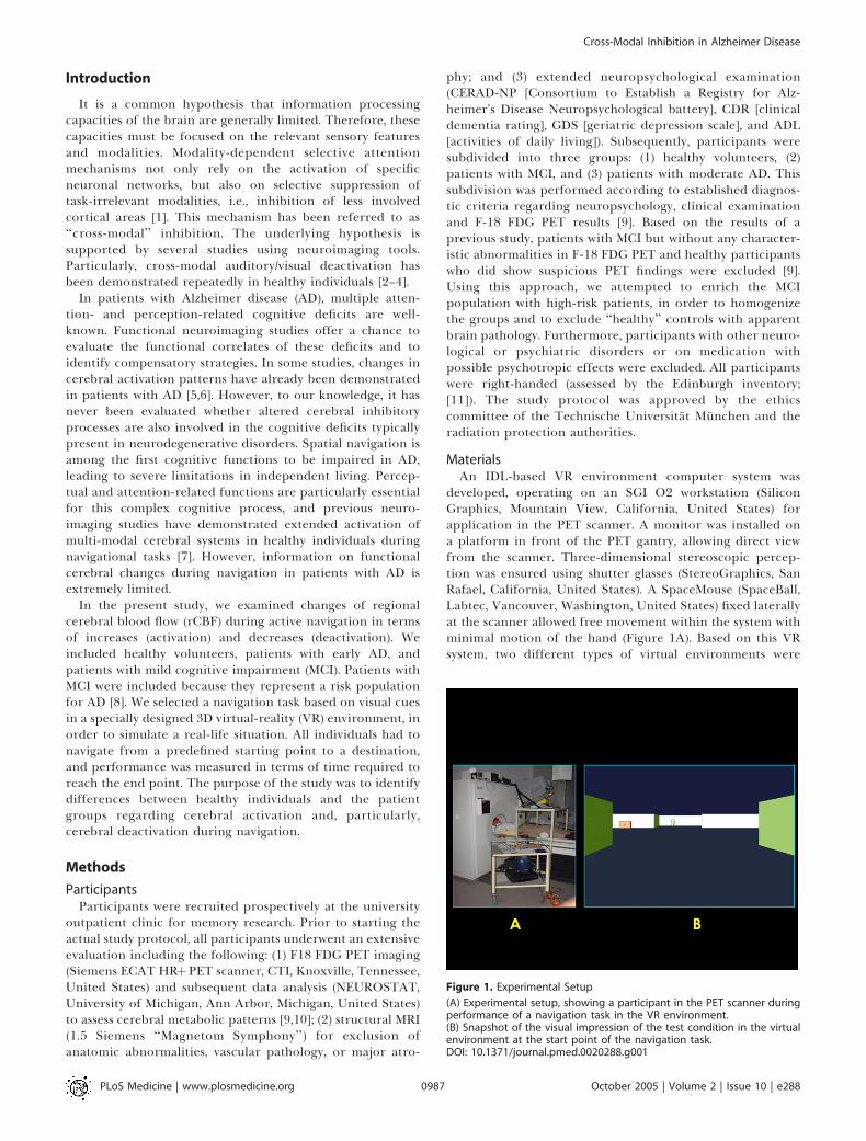
developed, operating on an SGI O2 workstation (SiliconGraphics, Mountain View, California, United States) forapplication in the PET scanner. A monitor was installed ona platform in front of the PET gantry, allowing direct viewfrom the scanner. Three-dimensional stereoscopic percep-tion was ensured using shutter glasses (StereoGraphics, SanRafael, California, United States). A SpaceMouse (SpaceBall,Labtec, Vancouver, Washington, United States) fixed laterallyat the scanner allowed free movement within the system withminimal motion of the hand (Figure 1A). Based on this VRsystem, two different types of virtual environments were
Figure 1. Experimental Setup
(A) Experimental setup, showing a participant in the PET scanner duringperformance of a navigation task in the VR environment.(B) Snapshot of the visual impression of the test condition in the virtualenvironment at the start point of the navigation task.DOI: 10.1371/journal.pmed.0020288.g001
PLoS Medicine | www.plosmedicine.org October 2005 | Volume 2 | Issue 10 | e2880987
Cross-Modal Inhibition in Alzheimer Disease
designed: (1) the control condition, constituted by a never-ending rectangular pathway without predetermined start orend points or turn-offs, and (2) the test condition, consistingof a trail across a sequence of four rooms in a complex spatialarrangement with additional exits, enabling false turn-offsinto blind rooms (Figure 1B). Both conditions resembled theinside of a simple building. A plain design was selected andthe general features of the environment (color, texture, andsize) were kept identical to ensure an overall comparablevisual impression and to avoid unnecessary distraction of theparticipants and contamination of the cognitive process ofnavigation. The entire system was evaluated and provedapplicable in patients with AD in previous experiments [12].
ProcedureBefore the actual start of the scans, all participants were
trained in the virtual environment outside of the scanner inorder to familiarize them with the levels and handling of thesystem. Participants who were not able to perceive the virtualenvironment three-dimensionally or to operate the naviga-tion system properly were excluded from the study. Theperformance of all participants was also observed during theentire PET examination by a study coordinator, to ensurecorrect transaction of every single scan. Participants per-formed the task in complete silence; the study coordinatorwas quietly watching the patient during the entire examina-tion to report potential contamination with noise and tosupervise the patient without the need of verbal interaction.Patients were instructed not to talk during the examinationexcept in case of emergency. In the control condition,participants were instructed to steer along the never-endingpathway, thus no actual navigation was required but visualand motor demands were similar to the test condition. In thetest condition, participants were asked to find their way froma start point to a predefined destination point. The twodifferent conditions of the VR environment were presentedin the scanner in a randomized order (43 control conditionand 83 test condition). During the test conditions, perform-ance (in terms of time required to reach the destination) wasrecorded.
O-15 Water PET AcquisitionA Siemens ECAT HRþ PET scanner was used for O-15 PET
measurements (3D mode; total axial field of view, 15.5 cm).For each scan, 350 MBq of O-15 water was injected in a slowintravenous bolus after start of the VR paradigm using aninfusion pump. Data acquisition was triggered by the peak ofcount rate. Each scan lasted 50 s for the measurement ofrCBF. Twelve scans were performed in each participant.Attenuation-corrected data were reconstructed (63 slices; 1283 128 pixel matrix; pixel size, 2.0 mm; plane separation, 2.42mm).
Data AnalysisStatistical parametric mapping software (SPM 99, Well-
come Department of Cognitive Neurology, London, UnitedKingdom) was used for image realignment, transformationinto standard stereotactic space, smoothing, and statisticalanalysis, resulting in 26 planes (pixel size, 2 3 2 mm;interplane distance, 4 mm), as previously described [13].
The effect of global differences in cerebral blood flow wasremoved by treating global activity as a confounding variable
and using participant-specific scaling to a nominal grandmean global activity of 50 ml/100 g/min. Thus, data wereadjusted for the global mean (rCBF), taking into account therepetitions across participants. The adjusted voxel valueswere then used for further statistical analysis [14].The statistical analysis was performed according to the
general linear model and the theory of Gaussian fields at eachand every voxel using a mixed-effects model [13,15,16]. Theresulting statistical parametric maps based on the t statisticwere subsequently transformed into normally distributedstatistical parametric (Z) maps [13].A network of expected cortical activations was predefined
based on previous publications on cerebral activation duringvisual spatial processing and active navigation [7,17–19]. Thisnetwork included cerebral regions belonging to the ventraland dorsal streams of the visual system, such as primary andsecondary visual cortex, inferotemporal cortex, ventrolateralprefrontal cortex, posterior parietal cortex, and dorsolateralprefrontal cortex. In addition, cerebral regions traditionallylinked to spatial orientation were included in the network,such as the hippocampus (allocentric mapping, recognitionof landmarks), posterior and superior parietal cortex (spatialattention, egocentric mapping, and optic flow processing; seeabove), and posterior cingulate cortex (spatial orientation).Finally, as the task required active movement, we expectedactivation of motor-associated systems such as premotorcortex, as well as supplementary and primary motor cortex(associated with movement planning and execution, respec-tively). Stereotactic coordinates of these anatomic regionswere selected according to corresponding Brodmann areas(BAs) in the atlas of Talairach and Tournoux [20].As mentioned previously, we expected to find cross-modal
deactivation of auditory cortical areas. Therefore, as forcerebral activations, a network regarding deactivations waspredefined based on previous data on the extent of auditorycortical areas [21,22] and on information about cross-modaldeactivation of auditory areas during visually dominatedtasks [2,23]. This network included BAs 41, 42, 21, and 22 inboth hemispheres, representing primary auditory cortex (A1)and adjacent belt areas that have been linked to auditoryassociative functions [22,24]. Corresponding to formerstudies, analysis was mainly restricted to these areas [25].Regarding the interindividual and interhemispheric varia-bility of the location of the auditory cortical areas, we definedthree-dimensional volumes independently for each hemi-sphere representing the probabilistic location of the primaryauditory cortex according to Penhune et al. [21]. Thesevolumes were transferred into the stereotactic referencesystem, to allow for correct anatomical assignment of theresults. Within the predefined expected cortical activationand deactivation networks, all statistical results were based ona single-voxel z-threshold corresponding to p , 0.001,uncorrected for multiple comparisons. To our knowledge,no conclusive previous information on cerebral deactivationduring visually guided navigation exists. Thus, only voxelssurviving false discovery rate (FDR) correction for the entirevolume at p , 0.05 were accepted in the statistical analysis ofrCBF decrease between conditions within groups, in order toavoid false-positive results [26].First the rCBF changes between the control and test
conditions were examined in each of the different groups(control individuals, patients with MCI, and patients with
PLoS Medicine | www.plosmedicine.org October 2005 | Volume 2 | Issue 10 | e2880988
Cross-Modal Inhibition in Alzheimer Disease
AD). For the assessment of differences of rCBF changesbetween the groups, a ‘‘difference of differences’’ analysis wasused. In all group analyses, the rating of navigation perform-ance of participants (time required for passage from start toend point of the labyrinth) was used as a covariate of nointerest, to diminish the possible effects due to individualdifferences of performance or subjective experience of theparadigm. Finally, a voxel-based linear regression analysis ofcognitive performance (as measured with the mini mentalstate examination [MMSE]) and regional cerebral deactiva-tion was carried out in the entire population, containing theparticipants from all three groups. We generated subtractionimages of the individual patients (control condition) and putthese images in relation to the MMSE scores of the patients,using a ‘‘covariate only’’ analysis. This analysis tested forvoxels in the brain in which task-related deactivations showeda significant linear relationship with the MMSE scores. An apriori hypothesis for the location of the probable linearrelation was defined for this analysis, based on the previouslyidentified deactivation foci, and an uncorrected threshold ofp , 0.001 was applied. We limited the linear regressionanalysis to clusters within the A1 (transverse gyrus of Heschl[TGH]). Additionally, a small volume correction was per-formed within a sphere of 20-mm radius, centered on themaximum deactivation in the TGH. The results of all analyses(maxima of the activation foci) were reported with therespective standard stereotactic coordinates according toTalairach and Tournoux [20].
Results
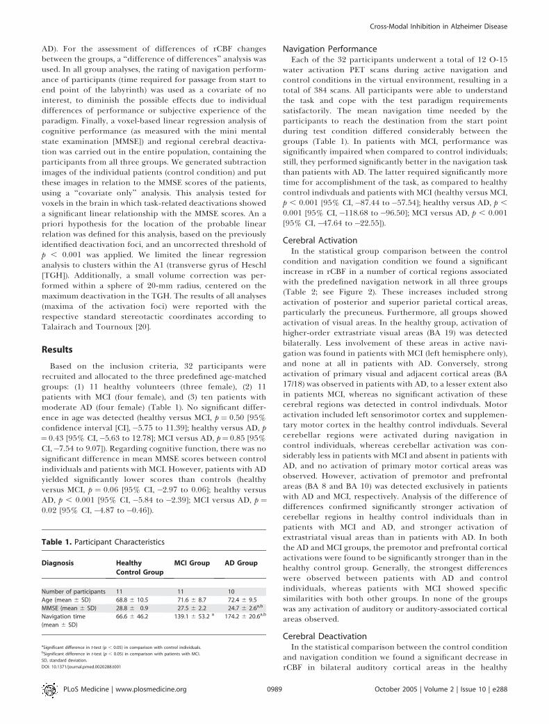
Based on the inclusion criteria, 32 participants wererecruited and allocated to the three predefined age-matchedgroups: (1) 11 healthy volunteers (three female), (2) 11patients with MCI (four female), and (3) ten patients withmoderate AD (four female) (Table 1). No significant differ-ence in age was detected (healthy versus MCI, p ¼ 0.50 [95%confidence interval [CI],�5.75 to 11.39]; healthy versus AD, p¼ 0.43 [95% CI,�5.63 to 12.78]; MCI versus AD, p¼ 0.85 [95%CI,�7.54 to 9.07]). Regarding cognitive function, there was nosignificant difference in mean MMSE scores between controlindividuals and patients with MCI. However, patients with ADyielded significantly lower scores than controls (healthyversus MCI, p ¼ 0.06 [95% CI, �2.97 to 0.06]; healthy versusAD, p , 0.001 [95% CI, �5.84 to �2.39]; MCI versus AD, p ¼0.02 [95% CI, �4.87 to �0.46]).
Navigation PerformanceEach of the 32 participants underwent a total of 12 O-15
water activation PET scans during active navigation andcontrol conditions in the virtual environment, resulting in atotal of 384 scans. All participants were able to understandthe task and cope with the test paradigm requirementssatisfactorily. The mean navigation time needed by theparticipants to reach the destination from the start pointduring test condition differed considerably between thegroups (Table 1). In patients with MCI, performance wassignificantly impaired when compared to control individuals;still, they performed significantly better in the navigation taskthan patients with AD. The latter required significantly moretime for accomplishment of the task, as compared to healthycontrol indivduals and patients with MCI (healthy versus MCI,p , 0.001 [95% CI,�87.44 to�57.54]; healthy versus AD, p ,
0.001 [95% CI,�118.68 to�96.50]; MCI versus AD, p , 0.001[95% CI,�47.64 to �22.55]).
Cerebral ActivationIn the statistical group comparison between the control
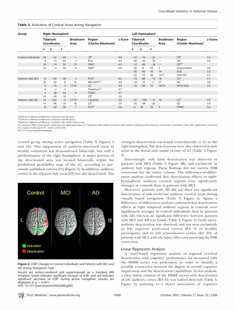
condition and navigation condition we found a significantincrease in rCBF in a number of cortical regions associatedwith the predefined navigation network in all three groups(Table 2; see Figure 2). These increases included strongactivation of posterior and superior parietal cortical areas,particularly the precuneus. Furthermore, all groups showedactivation of visual areas. In the healthy group, activation ofhigher-order extrastriate visual areas (BA 19) was detectedbilaterally. Less involvement of these areas in active navi-gation was found in patients with MCI (left hemisphere only),and none at all in patients with AD. Conversely, strongactivation of primary visual and adjacent cortical areas (BA17/18) was observed in patients with AD, to a lesser extent alsoin patients MCI, whereas no significant activation of thesecerebral regions was detected in control indivduals. Motoractivation included left sensorimotor cortex and supplemen-tary motor cortex in the healthy control indivduals. Severalcerebellar regions were activated during navigation incontrol individuals, whereas cerebellar activation was con-siderably less in patients with MCI and absent in patients withAD, and no activation of primary motor cortical areas wasobserved. However, activation of premotor and prefrontalareas (BA 8 and BA 10) was detected exclusively in patientswith AD and MCI, respectively. Analysis of the difference ofdifferences confirmed significantly stronger activation ofcerebellar regions in healthy control individuals than inpatients with MCI and AD, and stronger activation ofextrastriatal visual areas than in patients with AD. In boththe AD and MCI groups, the premotor and prefrontal corticalactivations were found to be significantly stronger than in thehealthy control group. Generally, the strongest differenceswere observed between patients with AD and controlindividuals, whereas patients with MCI showed specificsimilarities with both other groups. In none of the groupswas any activation of auditory or auditory-associated corticalareas observed.
Cerebral DeactivationIn the statistical comparison between the control condition
and navigation condition we found a significant decrease inrCBF in bilateral auditory cortical areas in the healthy
Table 1. Participant Characteristics
Diagnosis Healthy
Control Group
MCI Group AD Group
Number of participants 11 11 10
Age (mean 6 SD) 68.8 6 10.5 71.6 6 8.7 72.4 6 9.5
MMSE (mean 6 SD) 28.8 6 0.9 27.5 6 2.2 24.7 6 2.6a,b
Navigation time
(mean 6 SD)
66.6 6 46.2 139.1 6 53.2 a 174.2 6 20.6a,b
aSignificant difference in t-test (p , 0.05) in comparison with control individuals.bSignificant difference in t-test (p , 0.05) in comparison with patients with MCI.
SD, standard deviation.
DOI: 10.1371/journal.pmed.0020288.t001
PLoS Medicine | www.plosmedicine.org October 2005 | Volume 2 | Issue 10 | e2880989
Cross-Modal Inhibition in Alzheimer Disease
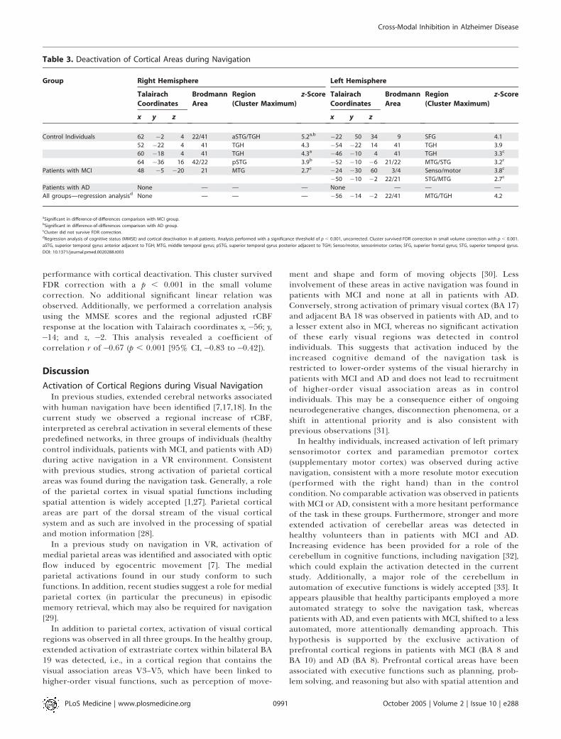
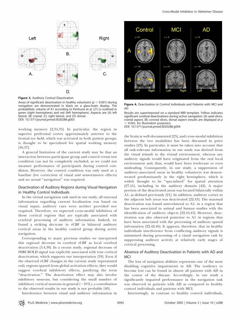
control group during active navigation (Table 3; Figures 3and 4A). This suppression of auditory-associated areas inhealthy volunteers was demonstrated bilaterally, but with apredominance of the right hemisphere. A major portion ofthe deactivated area was located bilaterally within thepredefined probability map of the A1, according to pre-viously published criteria [21] (Figure 3). In addition, auditorycortex in the adjacent belt areas [22] was also deactivated. The
strongest deactivation was found ventrolaterally to A1 in theright hemisphere, but deactivations were also observed in beltareas in the dorsal and caudal vicinity of A1 (Table 3; Figure3).Interestingly, only faint deactivation was observed in
patients with MCI (Table 3; Figure 4B), and exclusively inauditory belt regions. These findings did not survive FDRcorrection for the entire volume. The difference-of-differ-ences analysis confirmed that deactivation effects in right-hemispheric auditory cortical regions were significantlystronger in controls than in patients with MCI.Moreover, patients with AD did not show any significant
deactivation of task-irrelevant auditory cortical areas duringvisually based navigation (Table 3; Figure 4). Again, adifference-of-differences analysis confirmed that deactivationeffects in right temporal auditory regions in controls weresignificantly stronger in control individuals than in patientswith AD, whereas no significant difference between patientswith MCI and AD was found (Table 3; Figure 4). Little extra-auditory deactivation was observed, and was seen exclusivelyin left superior prefrontal cortex (BA 9) of healthyparticipants and in left sensorimotor cortex (BA 3/4) ofpatients with MCI, with the latter effect not surviving the FDRcorrection.
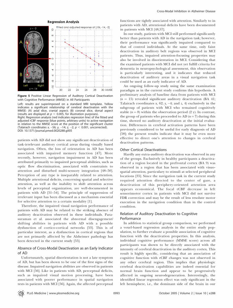
Linear Regression AnalysisA voxel-based regression analysis of regional cerebral
deactivation with cognitive performance (as measured withthe MMSE score) was performed, in order to identify apossible connection between the degree of overall cognitiveimpairment and the deactivation capabilities. In this analysis,a clear linear relation of the MMSE scores with deactivationof left auditory cortex (BA 41) was indeed detected (Table 3;Figure 5), pointing to a direct association of cognitive
Table 2. Activation of Cortical Areas during Navigation
Group Right Hemisphere Left Hemisphere
Talairach
Coordinates
Brodmann
Area
Region
(Cluster Maximum)
z-Score Talairach
Coordinates
Brodmann
Area
Region
(Cluster Maximum)
z-Score
x y z x y z
Control Individuals 24 �42 �30 — CBa 4.6 �52 �70 �22 — CBa 5.2
2 �72 54 7 PCU 4.4 �38 �42 �30 — CBa 4.4
30 �74 32 19 SOGa 4.3 �10 �80 �34 — CBa,b
22 4 54 6 SMAa 4.0 �30 �8 56 4 Senso/motor 3.6
�38 �80 34 19 SOG 3.5
�32 �74 46 19/7 SOG/SPL 3.3
Patients with MCI 6 �64 48 7 PCUa 4.2 �18 �80 �10 18 LGa 4.1
28 62 8 10 MFG/SFGa,c 3.9 �18 �34 �2 27 HG 3.8
0 �76 2 17/18 LG 3.9 �18 �88 16 18/19 MOG/SOG 3.6
6 �2 2 — Thalamusa,c 3.7
6 28 54 8 FSMGc 3.7
24 �40 �18 — CBa 3.6
Patients with AD 16 �102 �4 17/18 Calcarine 3.8 �22 �100 �12 18 LGb 4.4
16 �96 �16 18 LGc 3.6 �30 �94 �20 � CB 3.9
8 �48 58 7 PCUb 3.6 �4 34 52 8 FSMGc 3.4
aSignificant in difference-of-differences comparison with AD group.bSignificant in difference-of-differences comparison with MCI group.cSignificant in difference-of-differences comparison with healthy control group.
CB, cerebellum; FSMG, frontal superior medial gyrus; HG, hippocampal gyrus; LG, lingual gyrus; MFG, middle frontal gyrus; MOG, middle occipital gyrus; PCU, precuneus; Senso/motor, sensorimotor cortex; SMA, supplementary motor area;
SOG, superior occipital gyrus; SPL, superior parietal lobe.
DOI: 10.1371/journal.pmed.0020288.t002
Figure 2. rCBF Changes in Control Individuals and Patients with MCI and
AD during Navigation Task
Results are surface-rendered and superimposed on a standard MRItemplate. Green indicates significant increase of rCBF, and red indicatessignificant decrease of rCBF during active navigation (results aredisplayed at p , 0.001).DOI: 10.1371/journal.pmed.0020288.g002
PLoS Medicine | www.plosmedicine.org October 2005 | Volume 2 | Issue 10 | e2880990
Cross-Modal Inhibition in Alzheimer Disease
performance with cortical deactivation. This cluster survivedFDR correction with a p , 0.001 in the small volumecorrection. No additional significant linear relation wasobserved. Additionally, we performed a correlation analysisusing the MMSE scores and the regional adjusted rCBFresponse at the location with Talairach coordinates x, �56; y,�14; and z, �2. This analysis revealed a coefficient ofcorrelation r of �0.67 (p , 0.001 [95% CI,�0.83 to �0.42]).
Discussion
Activation of Cortical Regions during Visual NavigationIn previous studies, extended cerebral networks associated
with human navigation have been identified [7,17,18]. In thecurrent study we observed a regional increase of rCBF,interpreted as cerebral activation in several elements of thesepredefined networks, in three groups of individuals (healthycontrol individuals, patients with MCI, and patients with AD)during active navigation in a VR environment. Consistentwith previous studies, strong activation of parietal corticalareas was found during the navigation task. Generally, a roleof the parietal cortex in visual spatial functions includingspatial attention is widely accepted [1,27]. Parietal corticalareas are part of the dorsal stream of the visual corticalsystem and as such are involved in the processing of spatialand motion information [28].
In a previous study on navigation in VR, activation ofmedial parietal areas was identified and associated with opticflow induced by egocentric movement [7]. The medialparietal activations found in our study conform to suchfunctions. In addition, recent studies suggest a role for medialparietal cortex (in particular the precuneus) in episodicmemory retrieval, which may also be required for navigation[29].
In addition to parietal cortex, activation of visual corticalregions was observed in all three groups. In the healthy group,extended activation of extrastriate cortex within bilateral BA19 was detected, i.e., in a cortical region that contains thevisual association areas V3–V5, which have been linked tohigher-order visual functions, such as perception of move-
ment and shape and form of moving objects [30]. Lessinvolvement of these areas in active navigation was found inpatients with MCI and none at all in patients with AD.Conversely, strong activation of primary visual cortex (BA 17)and adjacent BA 18 was observed in patients with AD, and toa lesser extent also in MCI, whereas no significant activationof these early visual regions was detected in controlindividuals. This suggests that activation induced by theincreased cognitive demand of the navigation task isrestricted to lower-order systems of the visual hierarchy inpatients with MCI and AD and does not lead to recruitmentof higher-order visual association areas as in controlindividuals. This may be a consequence either of ongoingneurodegenerative changes, disconnection phenomena, or ashift in attentional priority and is also consistent withprevious observations [31].In healthy individuals, increased activation of left primary
sensorimotor cortex and paramedian premotor cortex(supplementary motor cortex) was observed during activenavigation, consistent with a more resolute motor execution(performed with the right hand) than in the controlcondition. No comparable activation was observed in patientswith MCI or AD, consistent with a more hesitant performanceof the task in these groups. Furthermore, stronger and moreextended activation of cerebellar areas was detected inhealthy volunteers than in patients with MCI and AD.Increasing evidence has been provided for a role of thecerebellum in cognitive functions, including navigation [32],which could explain the activation detected in the currentstudy. Additionally, a major role of the cerebellum inautomation of executive functions is widely accepted [33]. Itappears plausible that healthy participants employed a moreautomated strategy to solve the navigation task, whereaspatients with AD, and even patients with MCI, shifted to a lessautomated, more attentionally demanding approach. Thishypothesis is supported by the exclusive activation ofprefrontal cortical regions in patients with MCI (BA 8 andBA 10) and AD (BA 8). Prefrontal cortical areas have beenassociated with executive functions such as planning, prob-lem solving, and reasoning but also with spatial attention and
Table 3. Deactivation of Cortical Areas during Navigation
Group Right Hemisphere Left Hemisphere
Talairach
Coordinates
Brodmann
Area
Region
(Cluster Maximum)
z-Score Talairach
Coordinates
Brodmann
Area
Region
(Cluster Maximum)
z-Score
x y z x y z
Control Individuals 62 �2 4 22/41 aSTG/TGH 5.2a,b �22 50 34 9 SFG 4.1
52 �22 4 41 TGH 4.3 �54 �22 14 41 TGH 3.9
60 �18 4 41 TGH 4.3a �46 �10 4 41 TGH 3.3c
64 �36 16 42/22 pSTG 3.9b �52 �10 �6 21/22 MTG/STG 3.2c
Patients with MCI 48 �5 �20 21 MTG 2.7c �24 �30 60 3/4 Senso/motor 3.8c
�50 �10 �2 22/21 STG/MTG 2.7c
Patients with AD None — — — None — — —
All groups—regression analysisd None — — — �56 �14 �2 22/41 MTG/TGH 4.2
aSignificant in difference-of-differences comparison with MCI group.bSignificant in difference-of-differences comparison with AD group.cCluster did not survive FDR correction.dRegression analysis of cognitive status (MMSE) and cortical deactivation in all patients. Analysis performed with a significance threshold of p , 0.001, uncorrected. Cluster survived FDR correction in small volume correction with p , 0.001.
aSTG, superior temporal gyrus anterior adjacent to TGH; MTG, middle temporal gyrus; pSTG, superior temporal gyrus posterior adjacent to TGH; Senso/motor, sensorimotor cortex; SFG, superior frontal gyrus; STG, superior temporal gyrus.
DOI: 10.1371/journal.pmed.0020288.t003
PLoS Medicine | www.plosmedicine.org October 2005 | Volume 2 | Issue 10 | e2880991
Cross-Modal Inhibition in Alzheimer Disease
working memory [2,34,35]. In particular, the region insuperior prefrontal cortex approximately anterior to thefrontal eye field, which was activated in both patient groups,is thought to be specialized for spatial working memory[36,37].
A general limitation of the current study may be that aninteraction between participant group and control versus testcondition can not be completely excluded, as we could notmeasure performance of participants during control con-dition. However, the control condition was only used as abaseline (for correction of visual and sensorimotor effects)and no actual ‘‘navigation’’ was required.
Deactivation of Auditory Regions during Visual Navigationin Healthy Control Individuals
In the virtual navigation task used in our study, all externalinformation regarding current localization was based onvisual input; auditory cues were neither provided norrequired. Therefore, we expected cross-modal inhibition ofthose cortical regions that are typically associated withcerebral processing of auditory information. Indeed, wefound a striking decrease in rCBF in bilateral auditorycortical areas in the healthy control group during activenavigation.
Corresponding to many previous studies we interpretedthis regional decrease in cerebral rCBF as local cerebraldeactivation [3,4,38]. In a recent study, regional decrease offMRI BOLD signal was explicitly associated with true corticaldeactivation, which supports our interpretation [39]. Even ifthe observed rCBF changes in the current study representedonly regions spared from global activation effects, they wouldsuggest cerebral inhibitory effects, justifying the term‘‘deactivation.’’ The deactivation effect may also involveinhibitory neurons, but considering the small number ofinhibitory cortical neurons in general (;10%), a contributionto the observed results in our study is not probable [40].
Interference between visual and auditory information in
the brain is well-documented [25], and cross-modal inhibitionbetween the two modalities has been discussed in priorstudies [23]. In particular, it must be taken into account thatall task-relevant information in our study was derived fromthe visual stimuli in the virtual environment, whereas anyauditory signals would have originated from the real localenvironment and, thus, would have been irrelevant or evenmisleading. Consequently, in our study, a suppression ofauditory-associated areas in healthy volunteers was demon-strated predominantly in the right hemisphere, which iswidely thought to be ‘‘specialized’’ for spatial attention[27,41], including in the auditory domain [42]. A majorportion of the deactivated areas was located bilaterally withinA1, as defined previously [21]. In addition, auditory cortex inthe adjacent belt areas was deactivated [22,43]. The maximaldeactivation was found anterolateral to A1, in a region thathas been associated in animal and human studies with theidentification of auditory objects [22,44,45]. However, deac-tivation was also observed posterior to A1 in regions thathave been associated with the processing of auditory spatialinformation [22,42,46]. It appears, therefore, that in healthyindividuals interference from conflicting auditory signals isminimized during processing of a visual navigation task bysuppressing auditory activity at relatively early stages ofcortical processing.
Absence of Auditory Deactivation in Patients with AD andMCIThe loss of navigation abilities represents one of the most
disabling cognitive impairments in AD. The tendency tobecome lost can be found in almost all patients with AD inthe course of the disease. Accordingly, in our study asignificantly impaired performance in the navigation taskwas observed in patients with AD as compared to healthycontrol individuals and patients with MCI.Interestingly, in contrast to healthy control individuals,
Figure 3. Auditory Cortical Deactivation
Areas of significant deactivation in healthy volunteers (p , 0.001) duringnavigation are demonstrated in black on a glass-brain display. Theprobabilistic volume of A1 according to Penhune et al. [21] is outlined ingreen (right hemisphere) and red (left hemisphere). Aspects are (A) leftlateral, (B) cranial, (C) right lateral, and (D) dorsal.DOI: 10.1371/journal.pmed.0020288.g003
Figure 4. Deactivation in Control Individuals and Patients with MCI and
AD
Results are superimposed on a standard MRI template. Yellow indicatessignificant cerebral deactivations during active navigation: (A) axial slices,cranial aspect; (B) coronal slices, dorsal aspect (results are displayed at p, 0.005, for illustration purposes).DOI: 10.1371/journal.pmed.0020288.g004
PLoS Medicine | www.plosmedicine.org October 2005 | Volume 2 | Issue 10 | e2880992
Cross-Modal Inhibition in Alzheimer Disease
patients with AD did not show any significant deactivation oftask-irrelevant auditory cortical areas during visually basednavigation. Often, the loss of orientation in AD has beenassociated with impaired memory function [47]. Morerecently, however, navigation impairment in AD has beenattributed primarily to impaired perceptual abilities, such asoptic flow discrimination, associated with constraints toattention and disturbed multi-sensory integration [48–50].Perception of any type is inseparably related to attention.Multiple attentional deficits, concerning spatial and selectiveattention, as well as the inability to shift attention acrosslevels of perceptual organization, are well-documented inpatients with AD [51–54]. The principle of suppression ofirrelevant input has been discussed as a mechanism essentialfor selective attention to a certain modality [1].
Therefore, the impaired visual navigation performance ofpatients with AD may be related to the striking absence ofauditory deactivation observed in these individuals. Para-suraman et al. associated the abnormal disengagement/shifting abilities in patients with AD with a possibledysfunction of cortico-cortical networks [53]. This is ofparticular interest, as a dysfunction in cortical regions thatare not primarily affected by the Alzheimer pathology hasbeen detected in the current study [55].
Absence of Cross-Modal Deactivation as an Early Indicatorof AD
Unfortunately, spatial disorientation is not a late symptomof AD, but has been shown to be one of the first signs of thedisease. Impaired navigation abilities are observed in patientswith MCI [56]. Like in patients with AD, perceptual deficits,such as impaired visual motion processing, have beenassociated with poorer performance in spatial navigationtests in patients with MCI [56]. Again, the affected perceptual
functions are tightly associated with attention. Similarly to inpatients with AD, attentional deficits have been documentedin patients with MCI [48,57].In our study, patients with MCI still performed significantly
better than patients with AD in the navigation task; however,their performance was significantly impaired compared tothat of control individuals. At the same time, only faintdeactivation in auditory belt regions was observed in MCIpatients. Thus, impaired attention-focusing properties mayalso be involved in disorientation in MCI. Considering thatthe examined patients with MCI did not yet fulfill criteria fordementia in neuropsychological assessment, this observationis particularly interesting, and it indicates that reduceddeactivation of auditory areas in a visual navigation taskcould be used as an early indicator of AD.An ongoing follow-up study using the same examination
paradigm as in the current study confirms this hypothesis. Apreliminary analysis of baseline data from patients with MCI(n ¼ 15) revealed significant auditory deactivation (BA 41;Talairach coordinates x, 62; y, �4; and z, 4) exclusively in thesubgroup of patients with MCI who remained cognitivelystable (n¼ 8) within the observation period (2 y). In contrast,the group of patients who proceeded to AD (n¼7) during thistime, showed no auditory deactivation at the initial evalua-tion. Differences in cerebral activation patterns have beenpreviously considered to be useful for early diagnosis of AD[58]; the present results indicate that it may be even moreeffective to direct one’s attention to changes in cerebraldeactivation patterns.
Other Cortical DeactivationsHardly any extra-auditory deactivation was observed in any
of the groups. Exclusively in healthy participants a deactiva-tion of a region located in the prefrontal cortex (BA 9) wasobserved in a region that has been associated with visuo-spatial attention, particulary to stimuli at selected peripherallocations [35]. Since the navigation task in the current studyrequired attention directed to the center of gaze, adeactivation of this periphery-oriented attention areaappears economical. The focal rCBF decrease in leftsensorimotor cortex in patients with MCI did not surviveFDR correction and may be the result of less resolute motorexecution in the navigation condition than in the controlcondition.
Relation of Auditory Deactivation to CognitivePerformanceIn addition to statistical group comparison, we performed
a voxel-based regression analysis in the entire study pop-ulation, to further evaluate a possible association of cognitivefunction with the deactivation properties. In this analysis,individual cognitive performance (MMSE score) across allparticipants was shown to be directly associated with theintensity of cerebral deactivation in the auditory cortex. Thisresult is highly specific, considering that an association ofcognitive function with rCBF changes was not observed inany other cerebral region. This implies that physiologiccerebral deactivation capabilities are indeed essential fornormal brain function and appear to be progressivelyaffected in ongoing neurodegeneration. Interestingly, theidentified linear regression effect was only significant in theleft hemisphere, i.e., the dominant side of the brain in our
Figure 5 Positive Linear Regression of Auditory Cortical Deactivation
with Cognitive Performance (MMSE) in All Participants
Left: results are superimposed on a standard MRI template. Yellowindicates a significant relationship of cerebral deactivation with theMMSE: (A) axial slice, cranial aspect; (B) coronal slice, dorsal aspect(results are displayed at p , 0.005, for illustration purposes).Right: Regression analysis (red indicates regression line) of the fitted andadjusted rCBF response (blue points, arbitrary units) to active navigationin relation to the MMSE score at the position of the significant cluster(Talairach coordinates x,�56; y, �14; z,�2; p , 0.001, uncorrected).DOI: 10.1371/journal.pmed.0020288.g005
PLoS Medicine | www.plosmedicine.org October 2005 | Volume 2 | Issue 10 | e2880993
Cross-Modal Inhibition in Alzheimer Disease
purely right-handed population. Thus, the changes indeactivation functions in relation to overall cognitive impair-ment may be most clearly expressed in the dominanthemisphere.
Impaired cortical inhibitory capabilities in patients withAD appear plausible for two reasons. First, a strongervulnerability of the neocortical inhibitory system has oftenbeen suggested. Second, recent studies were able to demon-strate cortical disinhibition in AD and could relate it to acholinergic deficit [59,60].
ConclusionsThe results of this study indicate that altered deactivation
patterns must be taken into account in activation studiescomparing effects in patient groups versus control individ-uals, in order to avoid misinterpretation of differences. Wehave demonstrated that navigation based on visual cuesinduces a deactivation of auditory cortical areas in healthyindividuals. These deactivation effects are impaired inpatients with MCI and absent in patients with AD, pointingto a progressive inability to tune out irrelevant input and tofocus attention on the task-relevant modalities. Thus, theorientation disability in the outside world seen in patientswith AD may in fact be partially based on the inability toselectively orient spatial attention to task-relevant internalrepresentations of perceptual stimuli.
Acknowledgments
We thank Brigitte Dzewas and Coletta Kruschke for their technicalassistance and the radiochemistry group for their reliable supply ofradiopharmaceuticals. We are obliged to Denise Lee for the verycareful review of the manuscript and to Wolfgang Kloiber fortechnical support. Furthermore, we thank Dr. Stephan Nekolla andDr. Istvan Nagy for their essential contribution to the development ofthe VR environment computer system. This study has been supportedin part by a grant for clinical research from the Kommission furklinische Forschung (KKF), Technische Universitat Munchen (AD),and by a Research Award from the Alexander-von-HumboldtFoundation (JPR). The funders had no role in study design, datacollection and analysis, decision to publish, or preparation of themanuscript.
References1. Posner MI, Petersen SE (1990) The attention system of the human brain.
Annu Rev Neurosci 13: 25–42.2. Haxby JV, Horwitz B, Ungerleider LG, Maisog JM, Pietrini P, et al. (1994)
The functional organization of human extrastriate cortex: A PET-rCBFstudy of selective attention to faces and locations. J Neurosci 14: 6336–6353.
3. Kawashima R, O’Sullivan BT, Roland PE (1995) Positron-emissiontomography studies of cross-modality inhibition in selective attentionaltasks: Closing the ‘‘mind’s eye’’. Proc Natl Acad Sci U S A 92: 5969–5972.
4. Laurienti PJ, Burdette JH, Wallace MT, Yen YF, Field AS, et al. (2002)Deactivation of sensory-specific cortex by cross-modal stimuli. J CognNeurosci 14: 420–429.
5. Bookheimer SY, Strojwas MH, Cohen MS, Saunders AM, Pericak-VanceMA, et al. (2000) Patterns of brain activation in people at risk forAlzheimer’s disease. N Engl J Med 343: 450–456.
6. Pietrini P, Furey ML, Alexander GE, Mentis MJ, Dani A, et al. (1999)Association between brain functional failure and dementia severity inAlzheimer’s disease: Resting versus stimulation PET study. Am J Psychiatry156: 470–473.
7. Maguire EA, Burgess N, Donnett JG, Frackowiak RS, Frith CD, et al. (1998)Knowing where and getting there: A human navigation network. Science280: 921–924.
8. Petersen RC, Smith GE, Waring SC, Ivnik RJ, Tangalos EG, et al. (1999) Mildcognitive impairment. Cinical characterization and outcome. Arch Neurol56: 303–308.
9. Drzezga A, Lautenschlager N, Siebner H, Riemenschneider M, Willoch F, etal. (2003) Cerebral metabolic changes accompanying conversion of mildcognitive impairment into Alzheimer’s disease: A PET follow-up study. EurJ Nucl Med Mol Imaging 30: 1104–1113.
10. Minoshima S, Frey KA, Koeppe RA, Foster NL, Kuhl DE (1995) A diagnostic
approach in Alzheimer’s disease using three-dimensional stereotacticsurface projections of fluorine-18–FDG PET. J Nucl Med 36: 1238–1248.
11. Oldfield RC (1971) The assessment and analysis of handedness: TheEdinburgh inventory. Neuropsychologia 9: 97–113.
12. Drzezga A, Grimmer T, Peller M, Wermke M, Nekolla S, et al. (2003)Functional reorganization of cerebral activation in patients with MCI andAlzheimer’s disease during navigation in a virtual reality environment[abstract]. In: 9th International Conference on Functional Mapping of theHuman Brain; 2003 June 19–22; New York, New York, United States.Neuroimage 19: CD-ROM.
13. Friston KJ, Holmes AP, Worsley KJ, Poline JP, Frith CD, et al. (1995)Statistical parametric mapping in functional imaging: A general linearapproach. Hum Brain Mapp 2: 189–210.
14. Friston KJ, Frith CD, Liddle PF, Dolan RJ, Lammertsma AA, et al. (1990)The relationship between global and local changes in PET scans. J CerebBlood Flow Metab 10: 458–466.
15. Friston KJ, Frith CD, Liddle PF, Frackowiak RS (1991) Comparingfunctional (PET) images: The assessment of significant change. J CerebBlood Flow Metab 11: 690–699.
16. Worsley KJ, Evans AC, Marrett S, Neelin P (1992) A three-dimensionalstatistical analysis for CBF activation studies in human brain. J Cereb BloodFlow Metab 12: 900–918.
17. Hartley T, Maguire EA, Spiers HJ, Burgess N (2003) The well-worn routeand the path less traveled: Distinct neural bases of route following andwayfinding in humans. Neuron 37: 877–888.
18. Maguire EA, Burgess N, O’Keefe J (1999) Human spatial navigation:Cognitive maps, sexual dimorphism, and neural substrates. Curr OpinNeurobiol 9: 171–177.
19. Kawashima R, Imaizumi S, Mori K, Okada K, Goto R, et al. (1999) Selectivevisual and auditory attention toward utterances—A PET study. Neuro-image 10: 209–215.
20. Talairach J, Tournoux P (1988) Co-planar stereotaxic atlas of the humanbrain. New York: Thieme. 132 p.
21. Penhune VB, Zatorre RJ, MacDonald JD, Evans AC (1996) Interhemisphericanatomical differences in human primary auditory cortex: Probabilisticmapping and volume measurement from magnetic resonance scans. CerebCortex 6: 661–672.
22. Rauschecker JP, Tian B (2000) Mechanisms and streams for processing of‘‘what’’ and ‘‘where’’ in auditory cortex. Proc Natl Acad Sci U S A 97:11800–11806.
23. Shulman GL, Corbetta M, Buckner RL, Raichle ME, Fiez JA, et al. (1997)Top-down modulation of early sensory cortex. Cereb Cortex 7: 193–206.
24. Kaas JH, Hackett TA (2000) Subdivisions of auditory cortex and processingstreams in primates. Proc Natl Acad Sci U S A 97: 11793–11799.
25. Finney EM, Fine I, Dobkins KR (2001) Visual stimuli activate auditorycortex in the deaf. Nat Neurosci 4: 1171–1173.
26. Genovese CR, Lazar NA, Nichols T (2002) Thresholding of statistical mapsin functional neuroimaging using the false discovery rate. Neuroimage 15:870–878.
27. Mesulam MM (1999) Spatial attention and neglect: Parietal, frontal andcingulate contributions to the mental representation and attentionaltargeting of salient extrapersonal events. Philos Trans R Soc Lond B BiolSci 354: 1325–1346.
28. Ungerleider LG, Mishkin M (1982) Two cortical visual systems. GoodaleMA, Mansfield RJW, editors. Analysis of visual behavior. Cambridge: MITPress. pp. 549–586
29. Krause BJ, Schmidt D, Mottaghy FM, Taylor J, Halsband U, et al. (1999)Episodic retrieval activates the precuneus irrespective of the imagerycontent of word pair associates. A PET study. Brain 122: 255–263.
30. Zeki S, Watson JD, Lueck CJ, Friston KJ, Kennard C, et al. (1991) A directdemonstration of functional specialization in human visual cortex. JNeurosci 11: 641–649.
31. Rapoport SI (1991) Positron emission tomography in Alzheimer’s disease inrelation to disease pathogenesis: A critical review. Cerebrovasc BrainMetab Rev 3: 297–335.
32. Rondi-Reig L, Burguiere E (2004) Is the cerebellum ready for navigation?Prog Brain Res 148: 199–212.
33. Lang CE, Bastian AJ (2002) Cerebellar damage impairs automaticity of arecently practiced movement. J Neurophysiol 87: 1336–1347.
34. Goldman-Rakic PS (1996) The prefrontal landscape: Implications offunctional architecture for understanding human mentation and thecentral executive. Philos Trans R Soc Lond B Biol Sci 351: 1445–1453.
35. Corbetta M, Miezin FM, Shulman GL, Petersen SE (1993) A PET study ofvisuospatial attention. J Neurosci 13: 1202–1226.
36. Paus T (1996) Location and function of the human frontal eye-field: Aselective review. Neuropsychologia 34: 475–483.
37. Courtney SM, Petit L, Maisog JM, Ungerleider LG, Haxby JV (1998) An areaspecialized for spatial working memory in human frontal cortex. Science279: 1347–1351.
38. Born AP, Law I, Lund TE, Rostrup E, Hanson LG, et al. (2002) Corticaldeactivation induced by visual stimulation in human slow-wave sleep.Neuroimage 17: 1325–1335.
39. Czisch M, Wehrle R, Kaufmann C, Wetter TC, Holsboer F, et al. (2004)Functional MRI during sleep: BOLD signal decreases and their electro-physiological correlates. Eur J Neurosci 20: 566–574.
PLoS Medicine | www.plosmedicine.org October 2005 | Volume 2 | Issue 10 | e2880994
Cross-Modal Inhibition in Alzheimer Disease
40. Peters A, Kara DA, Harriman KM (1985) The neuronal composition of area17 of rat visual cortex. III. Numerical considerations. J Comp Neurol 238:263–274.
41. Mapstone M, Weintraub S, Nowinski C, Kaptanoglu G, Gitelman DR, et al.(2003) Cerebral hemispheric specialization for spatial attention: Spatialdistribution of search-related eye fixations in the absence of neglect.Neuropsychologia 41: 1396–1409.
42. Zatorre RJ, Bouffard M, Ahad P, Belin P (2002) Where is ‘where’ in thehuman auditory cortex? Nat Neurosci 5: 905–909.
43. Rauschecker JP, Tian B, Hauser M (1995) Processing of complex sounds inthe macaque nonprimary auditory cortex. Science 268: 111–114.
44. Zatorre RJ, Bouffard M, Belin P (2004) Sensitivity to auditory objectfeatures in human temporal neocortex. J Neurosci 24: 3637–3642.
45. Maeder PP, Meuli RA, Adriani M, Bellmann A, Fornari E, et al. (2001)Distinct pathways involved in sound recognition and localization: A humanfMRI study. Neuroimage 14: 802–816.
46. Tian B, Reser D, Durham A, Kustov A, Rauschecker JP (2001) Functionalspecialization in rhesus monkey auditory cortex. Science 292: 290–293.
47. Henderson VW, Mack W, Williams BW (1989) Spatial disorientation inAlzheimer’s disease. Arch Neurol 46: 391–394.
48. Rizzo M, Anderson SW, Dawson J, Myers R, Ball K (2000) Visual attentionimpairments in Alzheimer’s disease. Neurology 54: 1954–1959.
49. Kavcic V, Duffy CJ (2003) Attentional dynamics and visual perception:Mechanisms of spatial disorientation in Alzheimer’s disease. Brain 126:1173–1181.
50. Golob EJ, Miranda GG, Johnson JK, Starr A (2001) Sensory corticalinteractions in aging, mild cognitive impairment, and Alzheimer’s disease.Neurobiol Aging 22: 755–763.
51. Filoteo JV, Delis DC, Massman PJ, Demadura T, Butters N, et al. (1992)Directed and divided attention in Alzheimer’s disease: Impairment inshifting of attention to global and local stimuli. J Clin Exp Neuropsychol 14:871–883.
52. Mohr E, Cox C, Williams J, Chase TN, Fedio P (1990) Impairment of centralauditory function in Alzheimer’s disease. J Clin Exp Neuropsychol 12: 235–246.
53. Parasuraman R, Greenwood PM, Haxby JV, Grady CL (1992) Visuospatialattention in dementia of the Alzheimer type. Brain 115: 711–733.
54. Baddeley AD, Baddeley HA, Bucks RS, Wilcock GK (2001) Attentionalcontrol in Alzheimer’s disease. Brain 124: 1492–1508.
55. Kurylo DD, Corkin S, Allard T, Zatorre RJ, Growdon JH (1993) Auditoryfunction in Alzheimer’s disease. Neurology 43: 1893–1899.
56. Mapstone M, Steffenella TM, Duffy CJ (2003) A visuospatial variant of mildcognitive impairment: Getting lost between aging and AD. Neurology 60:802–808.
57. Arnaiz E, Almkvist O (2003) Neuropsychological features of mild cognitiveimpairment and preclinical Alzheimer’s disease. Acta Neurol Scand Suppl179: 34–41.
58. Wagner AD (2000) Early detection of Alzheimer’s disease: An fMRI markerfor people at risk? Nat Neurosci 3: 973–974.
59. Luhmann HJ, Heinemann U (1992) Hypoxia-induced functional alterationsin adult rat neocortex. J Neurophysiol 67: 798–811.
60. Liepert J, Bar KJ, Meske U, Weiller C (2001) Motor cortex disinhibition inAlzheimer’s disease. Clin Neurophysiol 112: 1436–1441.
Patient Summary
Background Problems with finding one’s way are one of the early signsof Alzheimer disease (AD). Researchers can measure how good peopleare at ‘‘spatial orientation’’ by asking them to solve navigation tasks onthe computer. These are similar to virtual-reality video games in whichone needs to find one’s way based on a set of cues. Using brain imagingtechnology, scientists can then observe what is going on in the brains ofpeople while they solve such navigation tasks.
Why Was This Study Done? Scientists used to think of AD as mostly adisease of memory loss, and loss of orientation was considered to bemostly a memory problem. More recently, however, navigation problemshave been linked to problems with perception and with payingattention. In this context, the researchers wanted to compare the brainactivities in people with and without AD while they completed suchnavigation tasks. They were looking for which areas of the brain wereactivated, and also whether others (which had nothing to do with thenavigation task) were deactivated. The idea was that maybe to do thetask well, individuals need to both activate relevant areas of the brainand also deactivate others to be able to focus overall brain activity onprocesses that would help with the task at hand.
What Did the Researchers Do and Find? They studied brain activity inthree groups of people while the individuals solved a navigation task.The first group consisted of 11 healthy individuals, the second of tenindividuals with mild cognitive impairment, and the third of 11individuals with AD. The cues that guided them through the navigationtask were exclusively visual, and all of the participants were able tounderstand the cues. They found that the activation patterns weredifferent between the three groups. The healthy individuals showedmore activity in parts of the brain thought to be involved in processingvisual cues, whereas the other two groups showed stronger activation inthe parts involved with visual perception. Moreover, the researchersfound differences in the deactivation patterns, mostly in the area of thebrain that deals with sound and hearing. In healthy individuals theseareas shut down to some extent during the visual navigation task, butthis effect was diminished in individuals with mild cognitive impairment,and individuals with AD showed no signs of deactivation in these ‘‘task-irrelevant’’ areas.
What Does This Mean? This study suggests that in addition to difficultyintegrating complex information, inability to focus activity in the relevantparts of the brain might be contributing to some of the orientationproblems in patients with AD. It is not clear whether cognitive therapycould strengthen the ability to ‘‘focus one’s brain,’’ but it seems an ideaworth pursuing. The researchers also suggest that reduced deactivationof auditory areas in visual navigation tasks might be useful as an earlyindicator of AD, and they are currently doing studies to test this idea.
Additional Online Information The following Web sites containinformation on AD in general and describe some of the changes in thebrains of patients with AD.Alzheimer Research Forum:http://www.alzforum.org/home.aspUS National Institute on Aging:http://www.nia.nih.gov/Alzheimer’s Disease Education and Referral Center (search for ‘‘brainimaging’’):http://www.alzheimers.org/pubs/adfact.htmlAlzheimer’s Association (search for ‘‘brain imaging’’):http://www.alz.org/US National Institute of Neurological Disorders and Stroke’s Web pageon AD:h t t p : / / w w w . n i n d s . n i h . g o v / d i s o r d e r s / a l z h e i m e r s d i s e a s e /alzheimersdisease.htm
PLoS Medicine | www.plosmedicine.org October 2005 | Volume 2 | Issue 10 | e2880995
Cross-Modal Inhibition in Alzheimer Disease
Related Documents


![Impaired angiogenesis and tumor development by inhibition ... · Mitotic kinesins have been studied in the context of cell division almost exclusively in tumor cells [12]. Beside](https://static.cupdf.com/doc/110x72/5fd6f4e990feee195b1ec4cf/impaired-angiogenesis-and-tumor-development-by-inhibition-mitotic-kinesins-have.jpg)