Impaired Angiogenesis during Fracture Healing in GPCR Kinase 2 Interacting Protein-1 (GIT1) Knock Out Mice Guoyong Yin 1,3. , Tzong-Jen Sheu 2. , Prashanthi Menon 1 , Jinjiang Pang 1 , Hsin-Chiu Ho 2 , Shanshan Shi 2 , Chao Xie 2 , Elaine Smolock 1 , Chen Yan 1 , Michael J. Zuscik 2 , Bradford C. Berk 1 * 1 Aab Cardiovascular Research Institute and the Department of Medicine, University of Rochester Medical Center, Rochester, New York, United States of America, 2 Center for Musculoskeletal Research and the Department of Orthopaedics and Rehabilitation, University of Rochester Medical Center, Rochester, New York, United States of America, 3 Orthopaedic Department, The First Affiliated Hospital of Nanjing Medical University, Jiangsu, China Abstract G protein coupled receptor kinase 2 (GRK2) interacting protein-1 (GIT1), is a scaffold protein that plays an important role in angiogenesis and osteoclast activity. We have previously demonstrated that GIT1 knockout (GIT1 KO) mice have impaired angiogenesis and dysregulated osteoclast podosome formation leading to a reduction in the bone resorbing ability of these cells. Since both angiogenesis and osteoclast-mediated bone remodeling are involved in the fracture healing process, we hypothesized that GIT1 participates in the normal progression of repair following bone injury. In the present study, comparison of fracture healing in wild type (WT) and GIT1 KO mice revealed altered healing in mice with loss of GIT1 function. Alcian blue staining of fracture callus indicated a persistence of cartilagenous matrix in day 21 callus samples from GIT1 KO mice which was temporally correlated with increased type 2 collagen immunostaining. GIT1 KO mice also showed a decrease in chondrocyte proliferation and apoptosis at days 7 and 14, as determined by PCNA and TUNEL staining. Vascular microcomputed tomography analysis of callus samples at days 7, 14 and 21 revealed decreased blood vessel volume, number, and connection density in GIT1 KO mice compared to WT controls. Correlating with this, VEGF-A, phospho-VEGFR2 and PECAM1 (CD31) were decreased in GIT1 KO mice, indicating reduced angiogenesis with loss of GIT1. Finally, calluses from GIT1 KO mice displayed a reduced number of tartrate resistant acid phosphatase-positive osteoclasts at days 14 and 21. Collectively, these results indicate that GIT1 is an important signaling participant in fracture healing, with gene ablation leading to reduced callus vascularity and reduced osteoclast number in the healing callus. Citation: Yin G, Sheu T-J, Menon P, Pang J, Ho H-C, et al. (2014) Impaired Angiogenesis during Fracture Healing in GPCR Kinase 2 Interacting Protein-1 (GIT1) Knock Out Mice. PLoS ONE 9(2): e89127. doi:10.1371/journal.pone.0089127 Editor: Luc Malaval, INSERM U1059/LBTO, Universite ´ Jean Monnet, France Received November 13, 2012; Accepted January 21, 2014; Published February 19, 2014 Copyright: ß 2014 Yin et al. This is an open-access article distributed under the terms of the Creative Commons Attribution License, which permits unrestricted use, distribution, and reproduction in any medium, provided the original author and source are credited. Funding: This work was supported by NIH/NHLBI R01 HL063462 (to BCB), National Natural Science Foundation of China Grant #81271988 (to GY), NIH/NIAMS P50 AR054041-5471 (to MJZ) and NIH/NIAMS P30 AR061307. The funders had no role in study design, data collection and analysis, decision to publish, or preparation of the manuscript. Competing Interests: The authors have declared that no competing interests exist. * E-mail: [email protected] . These authors contributed equally to this work. Introduction Fracture healing is a complex process involving an early inflammatory phase, recruitment, expansion and differentiation of mesenchymal cells, and production of cartilage and bone matrix in a temporally regulated manner [1–3]. After fracture, the repair process begins with hematoma formation and an inflammatory response [2]. In this early inflammatory phase, lack of blood vessels causes a regional hypoxic environment leading to the formation of a cartilagenous template that initiates a process of differentiation that recapitulates endochondral ossification [4]. Included are the proliferation and differentiation of mesenchymal progenitor cells into chondrocytes [1,5] which facilitate deposition of extracellular matrix components at the fracture site resulting in the formation of the transient soft callus [4]. In an initial remodeling phase, the avascular cartilagenous callus is converted into a vascularized and mineralized tissue that is remodeled by osteoclasts during an initial cartilage resorption phase [6], and then later in a bone remodeling phase that sculpts the healed skeletal element into the anatomically appropriate shape [7–9]. The importance of vascular invasion during endochondral bone formation has been established, with defects in bone vasculature having been reported in osteoporosis and rickets [10]. Thus, not surprisingly, during skeletal repair, neoangiogenesis driven by vascular endothelial growth factor (VEGF) is required to support nutrient and oxygen transport, with tissue oxygenation being required for osteogenic differentiation [11–16]. Further suggesting the need for this angiogenic cascade of events in the repair process, pharmacologic inhibition of angiogenesis has been shown to impair fracture healing by reducing/delaying callus mineralization [17]. G protein coupled receptor kinase 2 (GRK2) interacting protein 1 (GIT1) was originally identified by its binding to GRK2 and its effects on adrenergic receptor endocytosis [18]. GIT1 has five functional domains, including a zinc finger domain responsible for ARF-GAP activity, three ankyrin repeats, a Spa2 homology domain (SHD), a synaptic localization domain (SLD), and a conserved carboxyl-terminal region that interacts with paxillin (PBS) [19]. Through these domains, GIT1 interacts with diverse proteins including ARF6, MEK, phospholipase C-c (PLCc), p21- activated kinase (PAK)-interacting exchange factor (PIX) and PLOS ONE | www.plosone.org 1 February 2014 | Volume 9 | Issue 2 | e89127

Welcome message from author
This document is posted to help you gain knowledge. Please leave a comment to let me know what you think about it! Share it to your friends and learn new things together.
Transcript

Impaired Angiogenesis during Fracture Healing in GPCRKinase 2 Interacting Protein-1 (GIT1) Knock Out MiceGuoyong Yin1,3., Tzong-Jen Sheu2., Prashanthi Menon1, Jinjiang Pang1, Hsin-Chiu Ho2, Shanshan Shi2,
Chao Xie2, Elaine Smolock1, Chen Yan1, Michael J. Zuscik2, Bradford C. Berk1*
1 Aab Cardiovascular Research Institute and the Department of Medicine, University of Rochester Medical Center, Rochester, New York, United States of America, 2 Center
for Musculoskeletal Research and the Department of Orthopaedics and Rehabilitation, University of Rochester Medical Center, Rochester, New York, United States of
America, 3 Orthopaedic Department, The First Affiliated Hospital of Nanjing Medical University, Jiangsu, China
Abstract
G protein coupled receptor kinase 2 (GRK2) interacting protein-1 (GIT1), is a scaffold protein that plays an important role inangiogenesis and osteoclast activity. We have previously demonstrated that GIT1 knockout (GIT1 KO) mice have impairedangiogenesis and dysregulated osteoclast podosome formation leading to a reduction in the bone resorbing ability of thesecells. Since both angiogenesis and osteoclast-mediated bone remodeling are involved in the fracture healing process, wehypothesized that GIT1 participates in the normal progression of repair following bone injury. In the present study,comparison of fracture healing in wild type (WT) and GIT1 KO mice revealed altered healing in mice with loss of GIT1function. Alcian blue staining of fracture callus indicated a persistence of cartilagenous matrix in day 21 callus samples fromGIT1 KO mice which was temporally correlated with increased type 2 collagen immunostaining. GIT1 KO mice also showed adecrease in chondrocyte proliferation and apoptosis at days 7 and 14, as determined by PCNA and TUNEL staining. Vascularmicrocomputed tomography analysis of callus samples at days 7, 14 and 21 revealed decreased blood vessel volume,number, and connection density in GIT1 KO mice compared to WT controls. Correlating with this, VEGF-A, phospho-VEGFR2and PECAM1 (CD31) were decreased in GIT1 KO mice, indicating reduced angiogenesis with loss of GIT1. Finally, callusesfrom GIT1 KO mice displayed a reduced number of tartrate resistant acid phosphatase-positive osteoclasts at days 14 and21. Collectively, these results indicate that GIT1 is an important signaling participant in fracture healing, with gene ablationleading to reduced callus vascularity and reduced osteoclast number in the healing callus.
Citation: Yin G, Sheu T-J, Menon P, Pang J, Ho H-C, et al. (2014) Impaired Angiogenesis during Fracture Healing in GPCR Kinase 2 Interacting Protein-1 (GIT1)Knock Out Mice. PLoS ONE 9(2): e89127. doi:10.1371/journal.pone.0089127
Editor: Luc Malaval, INSERM U1059/LBTO, Universite Jean Monnet, France
Received November 13, 2012; Accepted January 21, 2014; Published February 19, 2014
Copyright: � 2014 Yin et al. This is an open-access article distributed under the terms of the Creative Commons Attribution License, which permits unrestricteduse, distribution, and reproduction in any medium, provided the original author and source are credited.
Funding: This work was supported by NIH/NHLBI R01 HL063462 (to BCB), National Natural Science Foundation of China Grant #81271988 (to GY), NIH/NIAMSP50 AR054041-5471 (to MJZ) and NIH/NIAMS P30 AR061307. The funders had no role in study design, data collection and analysis, decision to publish, orpreparation of the manuscript.
Competing Interests: The authors have declared that no competing interests exist.
* E-mail: [email protected]
. These authors contributed equally to this work.
Introduction
Fracture healing is a complex process involving an early
inflammatory phase, recruitment, expansion and differentiation of
mesenchymal cells, and production of cartilage and bone matrix in
a temporally regulated manner [1–3]. After fracture, the repair
process begins with hematoma formation and an inflammatory
response [2]. In this early inflammatory phase, lack of blood
vessels causes a regional hypoxic environment leading to the
formation of a cartilagenous template that initiates a process of
differentiation that recapitulates endochondral ossification [4].
Included are the proliferation and differentiation of mesenchymal
progenitor cells into chondrocytes [1,5] which facilitate deposition
of extracellular matrix components at the fracture site resulting in
the formation of the transient soft callus [4]. In an initial
remodeling phase, the avascular cartilagenous callus is converted
into a vascularized and mineralized tissue that is remodeled by
osteoclasts during an initial cartilage resorption phase [6], and
then later in a bone remodeling phase that sculpts the healed
skeletal element into the anatomically appropriate shape [7–9].
The importance of vascular invasion during endochondral bone
formation has been established, with defects in bone vasculature
having been reported in osteoporosis and rickets [10]. Thus, not
surprisingly, during skeletal repair, neoangiogenesis driven by
vascular endothelial growth factor (VEGF) is required to support
nutrient and oxygen transport, with tissue oxygenation being
required for osteogenic differentiation [11–16]. Further suggesting
the need for this angiogenic cascade of events in the repair process,
pharmacologic inhibition of angiogenesis has been shown to
impair fracture healing by reducing/delaying callus mineralization
[17].
G protein coupled receptor kinase 2 (GRK2) interacting protein
1 (GIT1) was originally identified by its binding to GRK2 and its
effects on adrenergic receptor endocytosis [18]. GIT1 has five
functional domains, including a zinc finger domain responsible for
ARF-GAP activity, three ankyrin repeats, a Spa2 homology
domain (SHD), a synaptic localization domain (SLD), and a
conserved carboxyl-terminal region that interacts with paxillin
(PBS) [19]. Through these domains, GIT1 interacts with diverse
proteins including ARF6, MEK, phospholipase C-c (PLCc), p21-
activated kinase (PAK)-interacting exchange factor (PIX) and
PLOS ONE | www.plosone.org 1 February 2014 | Volume 9 | Issue 2 | e89127

paxillin [20,21]. GIT1 has diverse biological functions, which we
have shown to include a critical role in pulmonary vascular
development by regulating VEGF induced PLCc and ERK1/2
activation [22]. GIT1 is also upregulated in atherosclerotic plaques
and regulates endothelial cell and vascular smooth muscle cell
migration [23]. Recently, we identified an important role of GIT1
in bone physiology based on its regulation of osteoclast sealing
zone formation, a critical step required for the function of this cell
[24]. Based on the observations that GIT1 plays an important role
in angiogenesis and osteoclast function, both of which are critical
for bone repair, we hypothesized that GIT1 is an important
molecular player in the bone healing process.
In the present study, we established that loss of GIT1 alters the
fracture healing process. Homozygous GIT1 knockout (GIT1 KO)
mice display a persistence of cartilagenous callus evidenced by
preservation of Alcian Blue staining and type 2 collagen content.
Chondrocyte differentiation was impacted by loss of GIT1, with
PCNA and TUNEL staining revealing decreased proliferation and
apoptosis of chondrocytes. We used microcomputed tomography
(microCT) to investigate overall callus volume and vascular
parameters and discovered that GIT1 KO mice have reduced
vessel volume and vessel number, which correlated with decreased
expression of VEGF-A and VEGFR2. We also examined osteoclst
numbers in the callus, and document a reduced number of TRAP-
stained cells during the remodeling phases of healing in mice with
loss of GIT1 function. Overall, findings documented in this report
establish that GIT1 is important for the normal progression of the
bone healing program via effects on callus vascularization and
osteoclast number, improtant determinants for callus mineraliza-
tion and remodeling respectively.
Materials and Methods
Ethics StatementTo ensure the humane treatment of mice in this study, all
experiments involving mice were performed with the approval and
supervision of the University Committee on Animal Resources, the
AALAC-, OLAW- and USDA-approved IACUC at the Univer-
sity of Rochester Medical Center.
AnimalsHomozygous GIT1 knockout (GIT1 KO) mice were generated
on the C57/BL6 background as described in Pang et al [22].
Chimeric mice generated were backcrossed for more than 7
generations. GIT1 KO mice at age of 10–12 weeks were used for
femoral fracture with WT littermates used as controls. It is
important to note that because of the high rate of perinatal
lethality in GIT1 KO mice due to a pulmonary defect [22] and
because of sensitivity to anesthesia, we were limited to an
experimental strategy that included only 3 KO mice at each of
3 harvest time points post-fracture: 7, 14 and 21 days.
Mouse Femur Fracture ModelFemur fractures in mice were performed as described in Xie
et al [25]. Briefly, mice were anesthetized using a mixture of
ketamine and xylazine delivered via intraperitoneal injection. The
skin and the underlying tissues over the left knee were incised. A
25-gauge needle was inserted through the patellar tendon and into
the medullary canal of the femur. A mid-diaphyseal fracture was
created via three-point bending using an Einhorn device [1]. After
fracture, 0.5 mg/kg buprenorphine (Abbott labs, North Chicago,
Illinois) was administered subcutaneously to each mouse daily for 3
days to control pain. Radiographs were obtained on 7, 14, and 21
days under anesthesia using a Faxitron X-ray system (Faxitron X-
ray, Wheeling, Illinois).
Quantitative Real Time PCR (qPCR)Fracture calluses from WT mice (n = 4) per time point (7, 14, 21
days) were carefully excised from the lower limb. The soft tissue
surrounding the calluses was removed and the samples were flash-
frozen in liquid N2. Frozen samples were placed in a Tissuelyser
(Qiagen, Venlo, Netherlands) along with 1 mL of TRIzol
(Invitrogen, Carlesbad, CA). Homogenization was performed
using a frequency of 30 Hz for a time of 3 minutes. The samples
were checked for adequate disruption and the mRNA was purified
according to the TRIzol System protocol. The concentration of
stock mRNA was determined in triplicate using a Nanodrop
photospectrometer. The mRNA was diluted in RNase-free water
and aliquoted into working dilutions of 0.5 mg/mL. A cDNA
library was synthesized using 0.5 mg of callus mRNA by a
commercially available reverse transcription kit (Invitrogen,
Carlesbad, CA). qPCR analyses were performed using murine-
specific primers for GIT1 and GAPDH. The qPCR reactions were
performed using SyberGreen (ABgene, Rochester, NY) in a
RotorGene real time PCR machine (Corbett Research, Carlsbad,
CA). GIT1 expression was normalized using GAPDH expression
as an internal control.
Microcomputed Tomography ImagingMicrocomputed tomography imaging (microCT) was per-
formed to assess mineralized callus volume and vascularity as we
have previously described [25–28]. Vascular networks at the
cortical bone junction and around the fractures were examined
using microCT analysis combined with perfusion of a lead
chromate based contrast agent [29]. Briefly, Microfil MV-122
(Flow Tech, Inc., Carver, Massachusetts) contrast media, a
radiopaque silicone rubber compound containing lead chromate,
was perfused via the heart along with 4% paraformaldehyde
following an initial vascular flush with heparinized saline. After
perfusion, the fractured femur was removed and scanned using a
microCT imaging system (VivaCT 40; Scanco Medical AG,
Basserdorf, Switzerland) at resolution of 10.5 mm to image bone
and vasculature. The samples were subsequently decalcified for 21
Figure 1. GIT1 mRNA is expressed during fracture healing. WTmice were administered femur fractures and fractured femora wereharvested for isolation of mRNA from the callus at 7, 14, and 21 dayspost-injury. qPCR was performed to examine the profile of GIT1expression. Bars represent mean GIT1 expression level relative toGAPDH +/2 SEM (N = 4, *p,0.05).doi:10.1371/journal.pone.0089127.g001
Fracture Healing Is Impaired in GIT1 Knockout Mice
PLOS ONE | www.plosone.org 2 February 2014 | Volume 9 | Issue 2 | e89127

days using a 10% EDTA solution. After complete decalcification,
the samples were scanned again to image only vascularization
within the callus. By registering the 2-D slices before and after
decalcification, contour lines were drawn to define a VOI that
only included the vasculature in or immediately adjacent to the
fracture callus itself. The reconstructed scan images were globally
thresholded based on intensity values to render 3-D images of the
vasculature in new bone callus, excluding the vessels in the
surrounding tissues. Three-dimensional morphometric analysis,
based on direct distance transform methods, was subsequently
performed on the 3-D images using algorithms that are commonly
used to model trabecular bone morphology. This facilitated
quantification and analysis of vascular network morphology
including vessel volume, number, spacing, and connection density.
It should be noted that all measurements of the vascular network
were constrained by the limit of resolution of the scanner (10 mm)
and the permeability of the networks to the viscous Microfil.
Careful heparinization and consistent application of perfusion
conditions were established to ensure comparability between mice.
Histology and HistomorphometryFractured femora harvested for microCT analysis were
processed for histology as previously described [30]. Briefly,
femora were disarticulated at the knee and hip, denuded of soft
tissue, and fixed at RT in 10% NBF for 72 hours. After three
washes in phosphate buffered saline (PBS), fixed femora were
decalcified in 10% EDTA for 7 to 14 days. Tissues were then
processed using a Tissue-Tek VIP 6 tissue processor (Sakura
Finetek USA, Inc., Torrance, CA, USA) and embedded in
paraffin. Serial 3 mm thick sagittal sections were obtained from a
60 mm region spanning the center of the fracture callus. Three
sections, each separated by approximately 25 to 50 mm, were
stained with Alcian Blue Hematoxylin/Orange G and histomor-
phometric analysis was performed using a point counting method
as described previously [31]. Briefly, blinded sections were
analyzed using a standardized eyepiece grid under the 106objective to determine the percent of total callus area composed of
cartilage and woven bone. Cartilage was defined as tissue with
positive Alcian Blue stain. Woven bone was counted whenever a
trabecular structure was observed, regardless of staining. Cortical
bone and internal (i.e. intramedullary) callus were not included in
these analyses. At each intersection of a horizontal and vertical
grid line, the identity of the underlying tissue was determined, with
the outcomes for every intersection documented and counted.
Based on the number of counted intersections on each slide, the
relative area of each tissue type as a percentage of the callus area
(i.e. grid intersections that fell within the callus domain) was
calculated.
ImmunohistochemistryPreviosly published methods were employed for immunohisto-
chemical analysis of PCNA and VEGFR2 expression [32] and
type 2 collagen expression [33]. Briefly, for either antigen, sections
were incubated at 60uC for 30 minutes, followed by deparaffiniza-
tion in xylene and hydration in gradient ethanol. Sections were
permeabilized in PBS containing 0.2% Triton X-100 (Sigma, St.
Louis, MO) for 10 min, washed in PBS, blocked with 3% H2O2 in
methanol for 30 minutes, and 3% goat serum in PBS for 30
minutes. Sections were then incubated with primary antibodies
overnight at 4uC. The next day, sections were washed with PBS
and incubated with biotinylated secondary antibodies (Vector
Laboratories, Burlingame, CA) in blocking buffer for 30 minutes.
After washing with PBS, ABC reagent (Vector Laboratories) was
added for 30 minutes and sections were washed again before
detection with AEC reagent (Vector Laboratories). Negative
controls were performed on adjacent sections by omitting primary
antibodies. All sections were analyzed using an Olympus VS120
Whole Slide Imager and quantification was performed using the
automated Visiopharm Integrator System (Visiopharm, Hoer-
sholm, Denmark) and its associated software.
ImmunofluorescenceSlides from WT and GIT1 KO femure fractures were
deparaffinized in xylene, rehydrated in graded ethanol, and rinsed
in distilled, deionized H2O as described above. For PECAM1
(CD31) detection, slides were boiled for 15 minutes in citrate
buffer (Zymed Laboratories, San Francisco, CA), cooled for 20
minutes, and washed in PBS for 5 minutes. Slides were then
blocked with normal goat serum containing 5% BSA in PBS for
30–60 minutes and incubated overnight with primary antibodies
diluted 1:100 in blocking buffer. After two rinses with PBS, the
sections were incubated in the dark for 1 hour at 37uC with rabbit
secondary antibody diluted 1:500 in normal rabbit serum. Slides
were rinsed in PBS before the addition of Topro3 nuclear stain
(Invitrogen, Grand Island, NY) and then mounted with Prolong
Gold mounting media (Invitrogen). Confocal images were
captured using Zeiss LSM 510 Axioskop 2 microscope (Zeiss
Microimaging, Thornwood, NY) and analyzed with Zen 2007
software (Zeiss, San Diego, CA).
Osteoclast QuantificationThree sections per callus were stained for tartrate-resistant acid
phosphatase (TRAP) using a previously described method [34].
Briefly, after deparaffinization and rehydration with distilled
water, sections were incubated at 37uC for 25 minutes in a
solution of anhydrous sodium acetate (Sigma), L-(+) tartaric acid
(Sigma), glacial acetic acid, fast red violet LB salt (Sigma), naphthol
AS-MX phosphate (Sigma), ethylene glycol monoethyl ether
(Sigma), and distilled water. Sections were rinsed in distilled water,
counterstained with hematoxylin for 10 seconds and then placed
in ammonia water for 5 seconds. Quantification was completed
using the 106 objective and Osteomeasure software (Osteo-
Metrics, Inc., Decatur, GA) to contour bone perimeter within the
callus and identify osteoclast number as a percentage of covered
bone surface perimeter and as a number of cells per mm of bone
surface. Osteoclasts were defined as multi-nucleated, TRAP-
positive cells seated on a bone surface.
Statistical AnalysisAll values are expressed as mean +/2 SEM. Statistical
differences between groups were detected using either ANOVA
(when .2 experimental groups were compared) or two-tailed
unpaired Student’s t tests (when only 2 experimental groups were
compared). A p-value less than 0.05 (p,0.05) was considered
significant.
Results
GIT1 is Expressed during Fracture HealingTo establish that GIT1 is expressed and regulated during the
fracture healing process, qPCR was performed on mRNA isolated
from the healing callus of WT mice at days 7, 14 and 21 post-
fracture. Consistent with previously published work identifying
GIT1 function in vascular tissue and osteoclasts, GIT1 is
upregulated significantly by day 14 and remains highly-expressed
at day 21 (Fig. 1). These timepoints correspond to callus
revascularization, cartilage remodeling and woven bone remod-
eling in the temporal progression of healing. These results set the
Fracture Healing Is Impaired in GIT1 Knockout Mice
PLOS ONE | www.plosone.org 3 February 2014 | Volume 9 | Issue 2 | e89127

stage for study of bone healing in mice in the context of GIT1 loss-
of-function.
Fracture Healing is Impaired in GIT1 KO MiceTo begin investigating the role of GIT1 in the fracture healing
process, we induced femoral fractures in WT and GIT1 KO mice
and assessed the healing process by radiographical evaluation and
microCT analysis of mineralized callus volume. At day 14, loss of
radiolucency at the fracture site in WT mice suggested the normal
pacing of repair (Fig. 2A), while the healing process was delayed in
GIT1 KO mice (Fig. 2B, red arrow). Representative microCT
reconstructions confirm persistence of disjunction in GIT1 KO
Figure 2. Disjunction persists at 14 days post-fracture in GIT1 KO mice. Femur fractures were induced in 10-week-old WT and GIT1 KO mice.Fractured femora were harvested for analysis at 7, 14, and 21 days post-injury. Radiographs obtained at the 14 day time point consistently revealedradiolucency in GIT1 KO calluses (B, red arrow) compared with calluses from WT mice (A, yellow arrow). This was supported by microCT analysis,which revealed lack of bridging mineral in GIT1 KOs (D, red arrows) compared to a connected shell of mineral in WT controls (C). Furtherquantification of callus geometry via microCT indicated that there were no differences in mineralized callus volume between WT and GIT1 KO mice(E). Bars represent mean callus volume (mm3) +/2 SEM (N = 3, *p,0.05).doi:10.1371/journal.pone.0089127.g002
Fracture Healing Is Impaired in GIT1 Knockout Mice
PLOS ONE | www.plosone.org 4 February 2014 | Volume 9 | Issue 2 | e89127

Figure 3. Cartilage persists and woven bone callus is delayed in GIT1 KO mice. Histological analysis of fracture callus cartilage content wasperformed via Alcian Blue Hematoxylin/Orange G staining. Representative stains of calluses from WT and GIT1 KO mice at 1, 2 and 3 weeks post-fracture are displayed (A–F). Red arrows denote Alcian Blue-stained cartilagenous matrix and asterisks denote mineralized woven bone.Histomorphometry was performed on triplicate sections from multiple mice, with % Cartilage Area (G) and % Bone Area (H) quantified. Bars represent% Area (cartilage or bone) +/2 SEM (*p,0.05, N = 3).doi:10.1371/journal.pone.0089127.g003
Fracture Healing Is Impaired in GIT1 Knockout Mice
PLOS ONE | www.plosone.org 5 February 2014 | Volume 9 | Issue 2 | e89127

mice (Fig. 2D, red arrows indicating disjunction) compared to WT
mice (Fig. 2C) at 14 days post-fracture, although quantification of
mineralized callus volume in cohorts of animals failed to achieve
significance at any timepoint, only revealing a trend toward a
decrease (Fig. 2E).
Chondrocyte Maturation is Delayed in Fracture Callus ofGIT1 KO Mice
Alcian Blue was used to stain extracellular matrix surrounding
chondrocytes within the fracture callus, with representative
sections depicted (Fig. 3A–F). At days 7 and 14 post-fracture,
cartilage matrix content in WT mice (Fig. 3A and 3C) was similar
to that in GIT1 KO mice (Fig. 3B and 3D). However, unlike in
WT mice (Fig. 3E), cartilaginous callus in GIT1 KO mice was still
present at day 21 (red arrows, Fig. 3F). Histomorphometric
quantification of callus cartilage and bone content confirmed that
at 21 days post-fracture, GIT1 KO mice had persistent cartilage
(Fig. 3G) that was at the expense of woven bone (Fig. 3H).
To further examine the cartilage phenotype, we performed
COL2A1 immunohistochemistry in WT and GIT1 KO mice at 7,
14 and 21 days post-fracture. Representative stained sections are
displayed in Fig. 4, with GIT1 KO mice trending toward
persistence of COL2A1 at 14 and 21 days (Fig. 4D and 4F)
compared to matched sections from WT mice (Fig. 4C and 4E).
These results suggest that cartilage persists in the fracture callus of
GIT1 KO mice at the expense of woven bone formation.
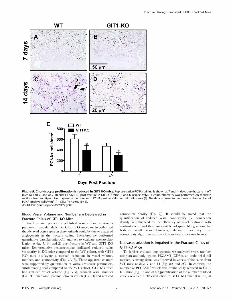
Chondrocyte Proliferation and Apoptosis is Reduced inFracture Callus of GIT1 KO Mice
To explore the possible association between persistent cartila-
genous callus and changes in chondrocyte proliferation and
apoptosis, we performed PCNA immunostaining and TUNEL
immunofluorescence respectively. At days 7 and 14, PCNA-
positive cells were more abundant in the cartilaginous soft callus of
WT mice (Fig. 5A and 5C) relative to calluses from GIT1 KO
mice (Fig. 5B and 5D). Histomorphometry confirmed this,
establishing that GIT1 KO chondrocytes were less proliferative
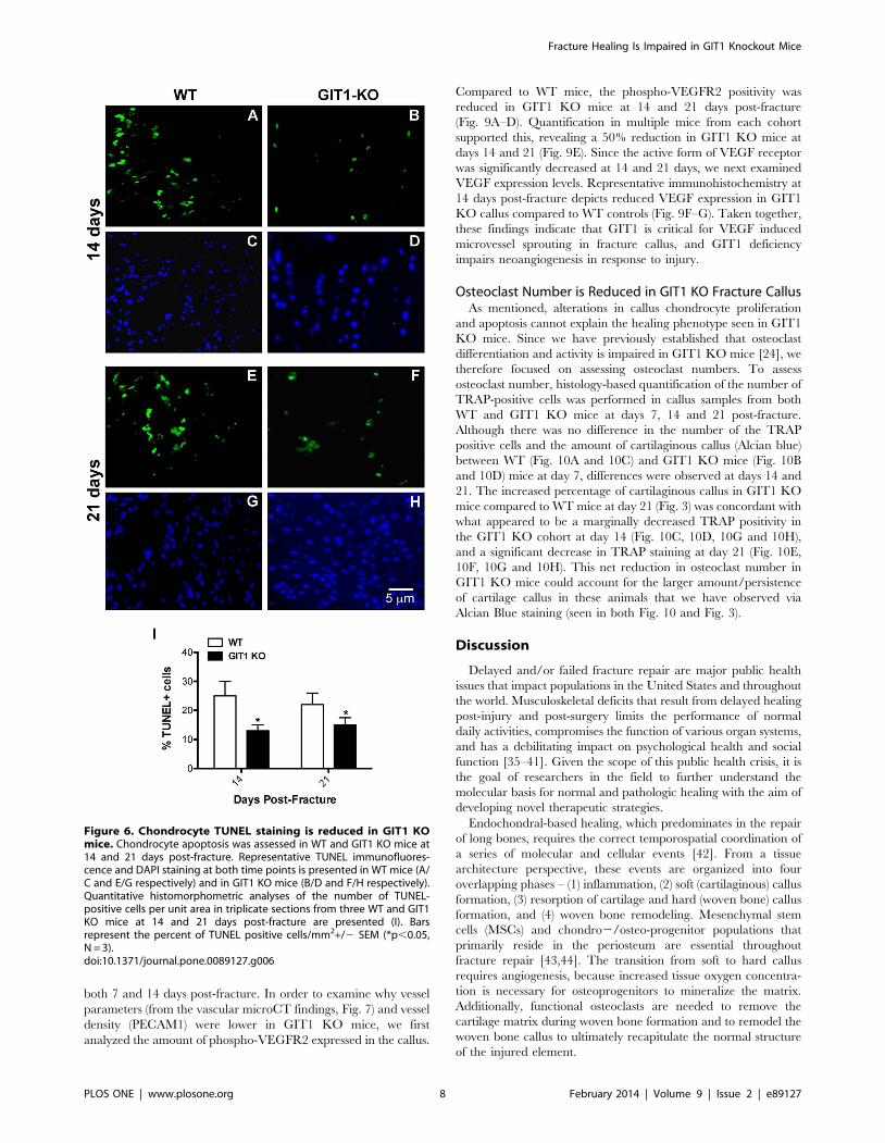
at 7 and 14 days post fracture (Fig. 5E). Regarding apoptosis,
representative TUNEL-stained sections reveal that the percentage
of TUNEL-positive cells (relative to DAPI) was reduced in GIT1
KO chondrocytes at both day 14 and 21 post-fracture (Fig. 6A–H).
Again, this was confirmed quantitatively (Fig. 6I), indicating that
GIT1 KO chondrocytes are less apoptotic during the time period
of healing that normally involves remodeling of the cartilagenous
callus (i.e. conversion to bone) and associated programmed death
of chondrocytes. While GIT1 appears to have a role in modulating
proliferation and death of chondrocytes, it is not clear how these
combined phenotypes (reduced mitosis and death in the GIT1 KO
group) account for the net persistence of the cartilagenous callus
and the delay in its conversion to woven bone.
Figure 4. Type 2 collagen-containing matrix persists in GIT1 KO mice. Tissue sections cut from WT and GIT1 KO mice were analyzed forCOL2A1 content using an immunohistochemistry approach. Representative stains at 7, 14 and 21 days post-fracture are depicted, with asterisksdenoting areas within the callus at 2 and 3 weeks post-fracture in GIT1 KO mice (D and F respectively) that have more robust/persistent staining.doi:10.1371/journal.pone.0089127.g004
Fracture Healing Is Impaired in GIT1 Knockout Mice
PLOS ONE | www.plosone.org 6 February 2014 | Volume 9 | Issue 2 | e89127
Blood Vessel Volume and Number are Decreased inFracture Callus of GIT1 KO Mice
Based on our previously published results demonstrating a
pulmonary vascular deficit in GIT1 KO mice, we hypothesized
that delayed bone repair in these animals could be due to impaired
angiogenesis in the fracture callus. Therefore, we performed
quantitative vascular microCT analyses to evaluate neovascular-
ization at day 7, 14, and 21 post-fracture in WT and GIT1 KO
mice. Representative reconstructions indicated reduced callus
vascularity in KO mice compared to the WT cohort, with GIT1
KO mice displaying a marked reduction in vessel volume,
number, and connectivity (Fig. 7A–F). These apparent changes
were supported by quantifation of various vascular parameters,
substantiating that compared to the WT cohort, GIT KO mice
had reduced vessel volume (Fig. 7G), reduced vessel number
(Fig. 7H), increased spacing between vessels (Fig. 7I) and reduced
connection density (Fig. 7J). It should be noted that the
quantification of reduced vessel connectivity (i.e. connection
density) is influenced by the efficiency of vessel perfusion with
contrast agent, and there may not be adequate filling in vascular
beds with smaller vessel diameters, reducing the accuracy of the
connectivity algorithm and conclusions that are drawn from it.
Neovascularization is Impaired in the Fracture Callus ofGIT1 KO Mice
To further evaluate angiogenesis, we analyzed vessel number
using an antibody against PECAM1 (CD31), an endothelial cell
marker. A strong signal was observed in vessels of the callus from
WT mice at days 7 and 14 (Fig. 8A and 8C). In contrast, the
number of PECAM1+ vessels was dramatically reduced in GIT1
KO mice (Fig. 8B and 8D). Quantification of the number of blood
vessels revealed a 60% reduction in GIT1 KO mice (Fig. 8E) at
Figure 5. Chondrocyte proliferation is reduced in GIT1 KO mice. Representative PCNA staining is shown at 7 and 14 days post-fracture in WTmice (A and C) and at 7 (B) and 14 days (D) post-fracture in GIT1 KO mice (B and D respectively). Histomorphometry was performed on triplicatesections from multiple mice to quantify the number of PCNA-positive cells per unit callus area (E). The data is presented as mean of the number ofPCNA positive cells/mm2+/2 SEM (*p,0.05, N = 3).doi:10.1371/journal.pone.0089127.g005
Fracture Healing Is Impaired in GIT1 Knockout Mice
PLOS ONE | www.plosone.org 7 February 2014 | Volume 9 | Issue 2 | e89127
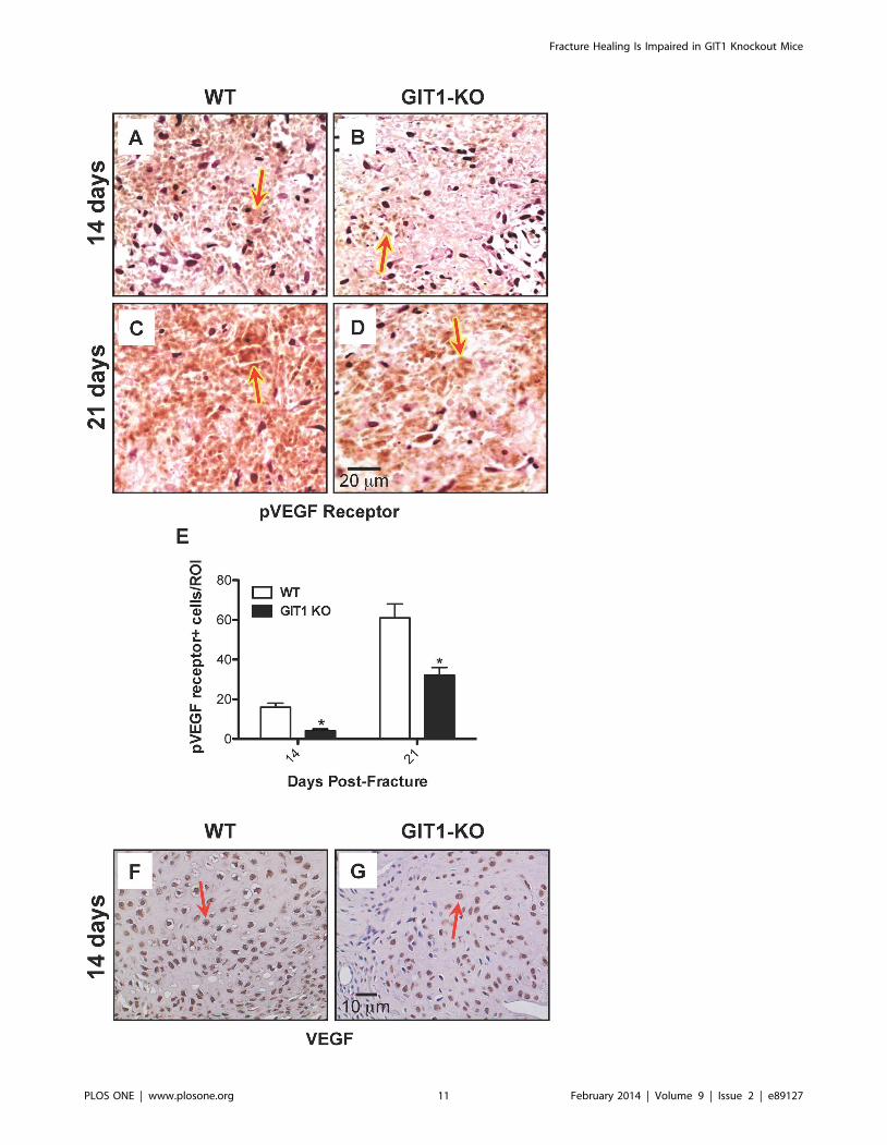
both 7 and 14 days post-fracture. In order to examine why vessel
parameters (from the vascular microCT findings, Fig. 7) and vessel
density (PECAM1) were lower in GIT1 KO mice, we first
analyzed the amount of phospho-VEGFR2 expressed in the callus.
Compared to WT mice, the phospho-VEGFR2 positivity was
reduced in GIT1 KO mice at 14 and 21 days post-fracture
(Fig. 9A–D). Quantification in multiple mice from each cohort
supported this, revealing a 50% reduction in GIT1 KO mice at
days 14 and 21 (Fig. 9E). Since the active form of VEGF receptor
was significantly decreased at 14 and 21 days, we next examined
VEGF expression levels. Representative immunohistochemistry at
14 days post-fracture depicts reduced VEGF expression in GIT1
KO callus compared to WT controls (Fig. 9F–G). Taken together,
these findings indicate that GIT1 is critical for VEGF induced
microvessel sprouting in fracture callus, and GIT1 deficiency
impairs neoangiogenesis in response to injury.
Osteoclast Number is Reduced in GIT1 KO Fracture CallusAs mentioned, alterations in callus chondrocyte proliferation
and apoptosis cannot explain the healing phenotype seen in GIT1
KO mice. Since we have previously established that osteoclast
differentiation and activity is impaired in GIT1 KO mice [24], we
therefore focused on assessing osteoclast numbers. To assess
osteoclast number, histology-based quantification of the number of
TRAP-positive cells was performed in callus samples from both
WT and GIT1 KO mice at days 7, 14 and 21 post-fracture.
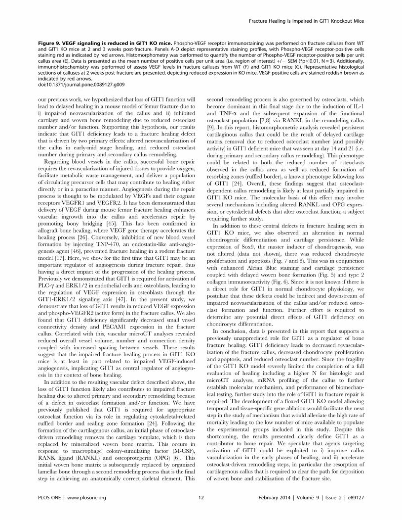
Although there was no difference in the number of the TRAP
positive cells and the amount of cartilaginous callus (Alcian blue)
between WT (Fig. 10A and 10C) and GIT1 KO mice (Fig. 10B
and 10D) mice at day 7, differences were observed at days 14 and
21. The increased percentage of cartilaginous callus in GIT1 KO
mice compared to WT mice at day 21 (Fig. 3) was concordant with
what appeared to be a marginally decreased TRAP positivity in
the GIT1 KO cohort at day 14 (Fig. 10C, 10D, 10G and 10H),
and a significant decrease in TRAP staining at day 21 (Fig. 10E,
10F, 10G and 10H). This net reduction in osteoclast number in
GIT1 KO mice could account for the larger amount/persistence
of cartilage callus in these animals that we have observed via
Alcian Blue staining (seen in both Fig. 10 and Fig. 3).
Discussion
Delayed and/or failed fracture repair are major public health
issues that impact populations in the United States and throughout
the world. Musculoskeletal deficits that result from delayed healing
post-injury and post-surgery limits the performance of normal
daily activities, compromises the function of various organ systems,
and has a debilitating impact on psychological health and social
function [35–41]. Given the scope of this public health crisis, it is
the goal of researchers in the field to further understand the
molecular basis for normal and pathologic healing with the aim of
developing novel therapeutic strategies.
Endochondral-based healing, which predominates in the repair
of long bones, requires the correct temporospatial coordination of
a series of molecular and cellular events [42]. From a tissue
architecture perspective, these events are organized into four
overlapping phases – (1) inflammation, (2) soft (cartilaginous) callus
formation, (3) resorption of cartilage and hard (woven bone) callus
formation, and (4) woven bone remodeling. Mesenchymal stem
cells (MSCs) and chondro2/osteo-progenitor populations that
primarily reside in the periosteum are essential throughout
fracture repair [43,44]. The transition from soft to hard callus
requires angiogenesis, because increased tissue oxygen concentra-
tion is necessary for osteoprogenitors to mineralize the matrix.
Additionally, functional osteoclasts are needed to remove the
cartilage matrix during woven bone formation and to remodel the
woven bone callus to ultimately recapitulate the normal structure
of the injured element.
Figure 6. Chondrocyte TUNEL staining is reduced in GIT1 KOmice. Chondrocyte apoptosis was assessed in WT and GIT1 KO mice at14 and 21 days post-fracture. Representative TUNEL immunofluores-cence and DAPI staining at both time points is presented in WT mice (A/C and E/G respectively) and in GIT1 KO mice (B/D and F/H respectively).Quantitative histomorphometric analyses of the number of TUNEL-positive cells per unit area in triplicate sections from three WT and GIT1KO mice at 14 and 21 days post-fracture are presented (I). Barsrepresent the percent of TUNEL positive cells/mm2+/2 SEM (*p,0.05,N = 3).doi:10.1371/journal.pone.0089127.g006
Fracture Healing Is Impaired in GIT1 Knockout Mice
PLOS ONE | www.plosone.org 8 February 2014 | Volume 9 | Issue 2 | e89127

Figure 7. Fracture callus vascularity is reduced in GIT1 KO mice. To visualize and quantify callus vascularity, WT and GIT1 KO mice wereperfused with lead chromate microfilm perfusion reagent. Harvested femora were decalcified and representative vascular microCT reconstructions
Fracture Healing Is Impaired in GIT1 Knockout Mice
PLOS ONE | www.plosone.org 9 February 2014 | Volume 9 | Issue 2 | e89127

Our group has recently documented a critical role for GIT1 in
pulmonary vascular development [22] and in the formation of
functional osteoclasts via regulation of sealing zone formation [24].
Given these unique roles for GIT1, both critical for normal tissue
morphogenesis in fracture repair, we became interested in the
function of this protein during the bone healing process. Based on
from each experimental group at 7, 14 and 21 days post-fracture are presented. Reduced vascularity in GIT1 KO mice (A, C, E) compared to WT controlmice (B, D, F) was evident at all time points. Quantification of callus vascular parameters, including Vessel Volume (G), Vessel Number (H), VesselSpacing (I) and Connection Density (J) supported these findings, with GIT1 KO mice possessing reduced callus vessel volume, vessel number andconnection density and increased space between vessels compared to callus from WT mice. Bars represent mean for each value +/2 SEM (N = 3, *p,0.05).doi:10.1371/journal.pone.0089127.g007
Figure 8. PECAM1+ blood vessel number is reduced in GIT1 KO mice. Representative PECAM1 immunofluorescence is presented at 7 and 14days post-fracture in WT mice (A and C) and GIT12/2 mice (B and D). Histomorphometry was performed to quantify the average number of positively-stained blood vessels present in each field of view on each section analyzed. Three sections (from 3 levels within each callus, 25–50 mm apart) wereviewed using the 106objective, with counts being collected from 3 fields of view in each section. All counts from each callus (9 fields total) wereaveraged. Vessel counting using this approach confirmed the immunofluorescence in panels A–D, with WT calluses possessing between 2 and 3-foldmore PECAM1+ vessels than GIT1 KO calluses at both time points (E). Bars represent mean number of PECAM1+ vessels/field +/2 SEM (*p,0.01,N = 3).doi:10.1371/journal.pone.0089127.g008
Fracture Healing Is Impaired in GIT1 Knockout Mice
PLOS ONE | www.plosone.org 10 February 2014 | Volume 9 | Issue 2 | e89127
Fracture Healing Is Impaired in GIT1 Knockout Mice
PLOS ONE | www.plosone.org 11 February 2014 | Volume 9 | Issue 2 | e89127
our previous work, we hypothesized that loss of GIT1 function will
lead to delayed healing in a mouse model of femur fracture due to
i) impaired neovascularization of the callus and ii) inhibited
cartilage and woven bone remodeling due to reduced osteoclast
number and/or function. Supporting this hypothesis, our results
indicate that GIT1 deficiency leads to a fracture healing defect
that is driven by two primary effects: altered neovascularization of
the callus in early-mid stage healing, and reduced osteoclast
number during primary and secondary callus remodeling.
Regarding blood vessels in the callus, successful bone repair
requires the revascularization of injured tissues to provide oxygen,
facilitate metabolic waste management, and deliver a population
of circulating precursor cells that may contribute to healing either
directly or in a paracrine manner. Angiogenesis during the repair
process is thought to be modulated by VEGFs and their cognate
receptors VEGFR1 and VEGFR2. It has been demonstrated that
delivery of VEGF during mouse femur fracture healing enhances
vascular ingrowth into the callus and accelerates repair by
promoting bony bridging [45]. This has been confirmed in
allograft bone healing, where VEGF gene therapy accelerates the
healing process [26]. Conversely, inhibition of new blood vessel
formation by injecting TNP-470, an endostatin-like anti-angio-
genesis agent [46], prevented fracture healing in a rodent fracture
model [17]. Here, we show for the first time that GIT1 may be an
important regulator of angiogenesis during fracture repair, thus
having a direct impact of the progression of the healing process.
Previously we demonstrated that GIT1 is required for activation of
PLC-c and ERK1/2 in endothelial cells and osteoblasts, leading to
the regulation of VEGF expression in osteoblasts through the
GIT1-ERK1/2 signaling axis [47]. In the present study, we
demonstrate that loss of GIT1 results in reduced VEGF expression
and phospho-VEGFR2 (active form) in the fracture callus. We also
found that GIT1 deficiency significantly decreased small vessel
connectivity density and PECAM1 expression in the fracture
callus. Correlated with this, vascular microCT analyses revealed
reduced overall vessel volume, number and connection density
coupled with increased spacing between vessels. These results
suggest that the impaired fracture healing process in GIT1 KO
mice is at least in part related to impaired VEGF-induced
angiogenesis, implicating GIT1 as central regulator of angiogen-
esis in the context of bone healing.
In addition to the resulting vascular defect described above, the
loss of GIT1 function likely also contributes to impaired fracture
healing due to altered primary and secondary remodeling because
of a defect in osteoclast formation and/or function. We have
previously published that GIT1 is required for appropriate
osteoclast function via its role in regulating cytoskeletal-related
ruffled border and sealing zone formation [24]. Following the
formation of the cartilagenous callus, an initial phase of osteoclast-
driven remodeling removes the cartilage template, which is then
replaced by mineralized woven bone matrix. This occurs in
response to macrophage colony-stimulating factor (M-CSF),
RANK ligand (RANKL) and osteoprotegerin (OPG) [6]. This
initial woven bone matrix is subsequently replaced by organized
lamellar bone through a second remodeling process that is the final
step in achieving an anatomically correct skeletal element. This
second remodeling process is also governed by osteoclasts, which
become dominant in this final stage due to the induction of IL-1
and TNF-a and the subsequent expansion of the functional
osteoclast population [7,8] via RANKL in the remodeling callus
[9]. In this report, histomorphometric analysis revealed persistent
cartilaginous callus that could be the result of delayed cartilage
matrix removal due to reduced osteoclast number (and possibly
activity) in GIT1 deficient mice that was seen at day 14 and 21 (i.e.
during primary and secondary callus remodeling). This phenotype
could be related to both the reduced number of osteoclasts
observed in the callus area as well as reduced formation of
resorbing zones (ruffled border), a known phenotype following loss
of GIT1 [24]. Overall, these findings suggest that osteoclast-
dependent callus remodeling is likely at least partially impaired in
GIT1 KO mice. The molecular basis of this effect may involve
several mechanisms including altered RANKL and OPG expres-
sion, or cytoskeletal defects that alter osteoclast function, a subject
requiring further study.
In addition to these central defects in fracture healing seen in
GIT1 KO mice, we also observed an alteration in normal
chondrogenic differentiation and cartilage persistence. While
expression of Sox9, the master inducer of chondrogenesis, was
not altered (data not shown), there was reduced chondrocyte
proliferation and apoptosis (Fig. 7 and 8). This was in conjunction
with enhanced Alcian Blue staining and cartilage persistence
coupled with delayed woven bone formation (Fig. 5) and type 2
collagen immunoreactivity (Fig. 6). Since it is not known if there is
a direct role for GIT1 in normal chondrocyte physiology, we
postulate that these defects could be indirect and downstream of
impaired neovascularization of the callus and/or reduced osteo-
clast formation and function. Further effort is required to
determine any potential direct effects of GIT1 deficiency on
chondrocyte differentiation.
In conclusion, data is presented in this report that supports a
previously unappreciated role for GIT1 as a regulator of bone
fracture healing. GIT1 deficiency leads to decreased revascular-
ization of the fracture callus, decreased chondrocyte proliferation
and apoptosis, and reduced osteoclast number. Since the fragility
of the GIT1 KO model severely limited the completion of a full
evaluation of healing including a higher N for histologic and
microCT analyses, mRNA profiling of the callus to further
establish molecular mechanism, and performance of biomechan-
ical testing, further study into the role of GIT1 in fracture repair is
required. The development of a floxed GIT1 KO model allowing
temporal and tissue-specific gene ablation would facilitate the next
step in the study of mechanism that would alleviate the high rate of
mortality leading to the low number of mice available to populate
the experimental groups included in this study. Despite this
shortcoming, the results presented clearly define GIT1 as a
contributor to bone repair. We speculate that agents targeting
activation of GIT1 could be exploited to i) improve callus
vascularization in the early phases of healing, and ii) accelerate
osteoclast-driven remodeling steps, in particular the resorption of
cartilagenous callus that is required to clear the path for deposition
of woven bone and stabilization of the fracture site.
Figure 9. VEGF signaling is reduced in GIT1 KO mice. Phospho-VEGF receptor immunostaining was performed on fracture calluses from WTand GIT1 KO mice at 2 and 3 weeks post-fracture. Panels A-D depict representative staining profiles, with Phospho-VEGF receptor-positive cellsstaining red as indicated by red arrows. Histomorphometry was performed to quantify the number of Phospho-VEGF receptor-positive cells per unitcallus area (E). Data is presented as the mean number of positive cells per unit area (i.e. region of interest) +/2 SEM (*p,0.01, N = 3). Additionally,immunohistochemistry was performed of assess VEGF levels in fracture calluses from WT (F) and GIT1 KO mice (G). Representative histologicalsections of calluses at 2 weeks post-fracture are presented, depicting reduced expression in KO mice. VEGF positive cells are stained reddish-brown asindicated by red arrows.doi:10.1371/journal.pone.0089127.g009
Fracture Healing Is Impaired in GIT1 Knockout Mice
PLOS ONE | www.plosone.org 12 February 2014 | Volume 9 | Issue 2 | e89127
Acknowledgments
We thank Dr. Tianfang Li and Dr. Yufeng Dong for helpful suggestions
and thoughtful discussion of data, and we thank Michael Thullen for
assistance with all microCT analysis of mineralized and vascular tissues.
Author Contributions
Conceived and designed the experiments: GY TJS MJZ BCB. Performed
the experiments: GY TJS. Analyzed the data: GY TJS PM JP HH SS CX
ES CY MJZ BCB. Contributed reagents/materials/analysis tools: GY TJS
ES MJZ BCB. Wrote the paper: GY TJS.
Figure 10. Osteoclast number is reduced in GIT1 KO mice. The presence of osteoclasts in the fracture callus of WT and GIT1 KO mice wasassessed at 7 (A and B respectively), 14 (C and D respectively) and 21 days (E and F respectively) post-fracture via TRAP staining. Histomorphometry toquantify percentage of osteoclast surface (G) and osteoclast number per unit bone surface (H) was also performed on triplicate sections from multiplemice, with bars representing the mean for each parameter +/2 SEM (*p,0.05, N = 3).doi:10.1371/journal.pone.0089127.g010
Fracture Healing Is Impaired in GIT1 Knockout Mice
PLOS ONE | www.plosone.org 13 February 2014 | Volume 9 | Issue 2 | e89127
References
1. Einhorn TA (1998) The cell and molecular biology of fracture healing.
ClinOrthop: S7–21.2. Gerstenfeld LC, Cullinane DM, Barnes GL, Graves DT, Einhorn TA (2003)
Fracture healing as a post-natal developmental process: molecular, spatial, andtemporal aspects of its regulation. JCell Biochem 88: 873–884.
3. Marsell R, Einhorn TA (2011) The biology of fracture healing. Injury 42: 551–
555.4. Thompson Z, Miclau T, Hu D, Helms JA (2002) A model for intramembranous
ossification during fracture healing. Journal of Orthopaedic Research 20: 1091–1098.
5. Li G, White G, Connolly C, Marsh D (2002) Cell proliferation and apoptosis
during fracture healing. J Bone Miner Res 17: 791–799.6. Kon T, Cho TJ, Aizawa T, Yamazaki M, Nooh N, et al. (2001) Expression of
osteoprotegerin, receptor activator of NF-kappaB ligand (osteoprotegerin ligand)and related proinflammatory cytokines during fracture healing. Journal of Bone
and Mineral Research 16: 1004–1014.7. Mountziaris PM, Mikos AG (2008) Modulation of the inflammatory response for
enhanced bone tissue regeneration. Tissue Eng Part B Rev 14: 179–186.
8. Ai-Aql ZS, Alagl AS, Graves DT, Gerstenfeld LC, Einhorn TA (2008)Molecular mechanisms controlling bone formation during fracture healing and
distraction osteogenesis. J Dent Res 87: 107–118.9. Gerstenfeld LC, Sacks DJ, Pelis M, Mason ZD, Graves DT, et al. (2009)
Comparison of effects of the bisphosphonate alendronate versus the RANKL
inhibitor denosumab on murine fracture healing. J Bone Miner Res 24: 196–208.
10. Maes C, Kobayashi T, Selig MK, Torrekens S, Roth SI, et al. (2010) Osteoblastprecursors, but not mature osteoblasts, move into developing and fractured
bones along with invading blood vessels. Dev Cell 19: 329–344.11. Pufe T, Wildemann B, Petersen W, Mentlein R, Raschke M, et al. (2002)
Quantitative measurement of the splice variants 120 and 164 of the angiogenic
peptide vascular endothelial growth factor in the time flow of fracture healing: astudy in the rat. Cell Tissue Res 309: 387–392.
12. Eckardt H, Bundgaard KG, Christensen KS, Lind M, Hansen ES, et al. (2003)Effects of locally applied vascular endothelial growth factor (VEGF) and VEGF-
inhibitor to the rabbit tibia during distraction osteogenesis. J Orthop Res 21:
335–340.13. Ferguson C, Alpern E, Miclau T, Helms JA (1999) Does adult fracture repair
recapitulate embryonic skeletal formation? Mechanisms of Development 87: 57–66.
14. Glowacki J (1998) Angiogenesis in fracture repair. Clin Orthop Relat Res: S82–89.
15. Gerber HP, Vu TH, Ryan AM, Kowalski J, Werb Z, et al. (1999) VEGF couples
hypertrophic cartilage remodeling, ossification and angiogenesis duringendochondral bone formation. NatMed 5: 623–628.
16. Athanasopoulos AN, Schneider D, Keiper T, Alt V, Pendurthi UR, et al. (2007)Vascular endothelial growth factor (VEGF)-induced up-regulation of CCN1 in
osteoblasts mediates proangiogenic activities in endothelial cells and promotes
fracture healing. Journal of Biological Chemistry 282: 26746–26753.17. Hausman MR, Schaffler MB, Majeska RJ (2001) Prevention of fracture healing
in rats by an inhibitor of angiogenesis. Bone 29: 560–564.18. Premont RT, Claing A, Vitale N, Freeman JL, Pitcher JA, et al. (1998) beta2-
Adrenergic receptor regulation by GIT1, a G protein-coupled receptor kinase-associated ADP ribosylation factor GTPase-activating protein. Proc Natl Acad
Sci U S A 95: 14082–14087.
19. Natarajan K, Yin G, Berk BC (2004) Scaffolds direct Src-specific signaling inresponse to angiotensin II: new roles for Cas and GIT1. Mol Pharmacol 65:
822–825.20. Haendeler J, Yin G, Hojo Y, Saito Y, Melaragno M, et al. (2003) GIT1 mediates
Src-dependent activation of phospholipase Cgamma by angiotensin II and
epidermal growth factor. J Biol Chem 278: 49936–49944.21. Yin G, Haendeler J, Yan C, Berk BC (2004) GIT1 functions as a scaffold for
MEK1-extracellular signal-regulated kinase 1 and 2 activation by angiotensin IIand epidermal growth factor. Mol Cell Biol 24: 875–885.
22. Pang J, Hoefen R, Pryhuber GS, Wang J, Yin G, et al. (2009) G-protein-coupled
receptor kinase interacting protein-1 is required for pulmonary vasculardevelopment. Circulation 119: 1524–1532.
23. Wang J, Taba Y, Pang J, Yin G, Yan C, et al. (2009) GIT1 mediates VEGF-induced podosome formation in endothelial cells: critical role for PLCgamma.
Arterioscler Thromb Vasc Biol 29: 202–208.24. Menon P, Yin G, Smolock EM, Zuscik MJ, Yan C, et al. (2010) GPCR kinase 2
interacting protein 1 (GIT1) regulates osteoclast function and bone mass. J Cell
Physiol 225: 777–785.
25. Xie C, Liang B, Xue M, Lin AS, Loiselle A, et al. (2009) Rescue of impaired
fracture healing in COX-22/2 mice via activation of prostaglandin E2 receptorsubtype 4. AmJPathol 175: 772–785.
26. Ito H, Koefoed M, Tiyapatanaputi P, Gromov K, Goater JJ, et al. (2005)
Remodeling of cortical bone allografts mediated by adherent rAAV-RANKLand VEGF gene therapy. NatMed 11: 291–297.
27. Zhang X, Xie C, Lin AS, Ito H, Awad H, et al. (2005) Periosteal progenitor cell
fate in segmental cortical bone graft transplantations: implications for functionaltissue engineering. Journal of Bone and Mineral Research 20: 2124–2137.
28. Dhillon RS, Xie C, Tyler W, Calvi LM, Awad HA, et al. (2013) PTH-enhanced
structural allograft healing is associated with decreased angiopoietin-2-mediatedarteriogenesis, mast cell accumulation, and fibrosis. J Bone Miner Res 28: 586–
597.
29. Duvall CL, Taylor WR, Weiss D, Guldberg RE (2004) Quantitativemicrocomputed tomography analysis of collateral vessel development after
ischemic injury. Am J Physiol Heart Circ Physiol 287: H302–310.
30. Zhang X, Schwarz EM, Young DA, Puzas JE, Rosier RN, et al. (2002)Cyclooxygenase-2 regulates mesenchymal cell differentiation into the osteoblast
lineage and is critically involved in bone repair. JClinInvest 109: 1405–1415.
31. Naik AA, Xie C, Zuscik MJ, Kingsley P, Schwarz EM, et al. (2009) ReducedCOX-2 expression in aged mice is associated with impaired fracture healing.
Journal of Bone and Mineral Research 24: 251–264.
32. Konishi H, Wu J, Cooke JP (2010) Chronic exposure to nicotine impairscholinergic angiogenesis. Vasc Med 15: 47–54.
33. Arasapam G, Scherer M, Cool JC, Foster BK, Xian CJ (2006) Roles of COX-2
and iNOS in the bony repair of the injured growth plate cartilage. J CellBiochem 99: 450–461.
34. Boyce BF, Xing L (2008) Functions of RANKL/RANK/OPG in bone modeling
and remodeling. Arch Biochem Biophys 473: 139–146.
35. Alarcon T, Gonzalez-Montalvo JI, Gotor P, Madero R, Otero A (2011)Activities of daily living after hip fracture: profile and rate of recovery during 2
years of follow-up. Osteoporos Int 22: 1609–1613.
36. Brenneman SK, Barrett-Connor E, Sajjan S, Markson LE, Siris ES (2006)Impact of recent fracture on health-related quality of life in postmenopausal
women. J Bone Miner Res 21: 809–816.
37. Ding R, McCarthy ML, Houseknecht E, Ziegfeld S, Knight VM, et al. (2006)The health-related quality of life of children with an extremity fracture: a one-
year follow-up study. J Pediatr Orthop 26: 157–163.
38. Giannoudis PV, Harwood PJ, Kontakis G, Allami M, Macdonald D, et al.(2009) Long-term quality of life in trauma patients following the full spectrum of
tibial injury (fasciotomy, closed fracture, grade IIIB/IIIC open fracture andamputation). Injury 40: 213–219.
39. Lonnroos E, Kautiainen H, Sund R, Karppi P, Hartikainen S, et al. (2009)
Utilization of inpatient care before and after hip fracture: a population-basedstudy. Osteoporos Int 20: 879–886.
40. Olerud P, Ahrengart L, Soderqvist A, Saving J, Tidermark J (2010) Quality of
life and functional outcome after a 2-part proximal humeral fracture: aprospective cohort study on 50 patients treated with a locking plate. J Shoulder
Elbow Surg 19: 814–822.
41. Silverman SL, Shen W, Minshall ME, Xie S, Moses KH (2007) Prevalence ofdepressive symptoms in postmenopausal women with low bone mineral density
and/or prevalent vertebral fracture: results from the Multiple Outcomes ofRaloxifene Evaluation (MORE) study. J Rheumatol 34: 140–144.
42. Schindeler A, McDonald MM, Bokko P, Little DG (2008) Bone remodeling
during fracture repair: The cellular picture. Semin Cell Dev Biol 19: 459–466.
43. Zhang X, Naik A, Xie C, Reynolds D, Palmer J, et al. (2005) Periosteal stem cellsare essential for bone revitalization and repair. JMusculoskeletNeuronalInteract
5: 360–362.
44. Zuscik MJ, O’Keefe RJ (2009) Skeletal Healing. In: Rosen CJ, editor. Primer onthe Metabolic Bone Diseases and Disorders of Mineral Metabolism: American
Society of Bone and Mineral Research. 61–65.
45. Street J, Bao M, deGuzman L, Bunting S, Peale FV Jr, et al. (2002) Vascularendothelial growth factor stimulates bone repair by promoting angiogenesis and
bone turnover. ProcNatlAcadSciUSA 99: 9656–9661.
46. Moulton KS, Heller E, Konerding MA, Flynn E, Palinski W, et al. (1999)Angiogenesis inhibitors endostatin or TNP-470 reduce intimal neovasculariza-
tion and plaque growth in apolipoprotein E-deficient mice. Circulation 99:1726–1732.
47. Rui Z, Li X, Fan J, Ren Y, Yuan Y, et al. (2012) GIT1Y321 phosphorylation is
required for ERK1/2- and PDGF-dependent VEGF secretion from osteoblaststo promote angiogenesis and bone healing. Int J Mol Med 30: 819–825.
Fracture Healing Is Impaired in GIT1 Knockout Mice
PLOS ONE | www.plosone.org 14 February 2014 | Volume 9 | Issue 2 | e89127
Related Documents