Impact of Iron Overload and Potential Benefit from Iron Chelation in Low-risk Myelodysplastic Syndrome Niraj Shenoy [1] Nishanth Vallumsetla [1] Eliezer Rachmilewitz [2] Amit Verma [1] Yelena Ginzburg [3]* [1] Albert Einstein College of Medicine, Bronx, NY [2] Edith Wolfson Medical Center, Holon, Israel [3] New York Blood Center, New York, NY *Address Correspondence to: Yelena Ginzburg, MD, Erythropoiesis Laboratory, LFKRI, New York Blood Center, 310 East 67 th Street, New York, NY 10065, Tel (212) 570-3463. Email: [email protected] Blood First Edition Paper, prepublished online June 12, 2014; DOI 10.1182/blood-2014-03-563221 Copyright © 2014 American Society of Hematology For personal use only. on April 23, 2016. by guest www.bloodjournal.org From

Welcome message from author
This document is posted to help you gain knowledge. Please leave a comment to let me know what you think about it! Share it to your friends and learn new things together.
Transcript

Impact of Iron Overload and Potential Benefit from Iron Chelation in Low-risk
Myelodysplastic Syndrome
Niraj Shenoy [1]
Nishanth Vallumsetla [1]
Eliezer Rachmilewitz [2]
Amit Verma [1]
Yelena Ginzburg [3]*
[1] Albert Einstein College of Medicine, Bronx, NY
[2] Edith Wolfson Medical Center, Holon, Israel
[3] New York Blood Center, New York, NY
*Address Correspondence to: Yelena Ginzburg, MD, Erythropoiesis Laboratory, LFKRI, New
York Blood Center, 310 East 67th Street, New York, NY 10065, Tel (212) 570-3463. Email:
Blood First Edition Paper, prepublished online June 12, 2014; DOI 10.1182/blood-2014-03-563221
Copyright © 2014 American Society of Hematology
For personal use only.on April 23, 2016. by guest www.bloodjournal.orgFrom
Abstract
Myelodysplastic syndromes (MDS) are a group of heterogeneous clonal bone marrow disorders
characterized by ineffective hematopoiesis, peripheral blood cytopenias, and potential for
malignant transformation. Lower / intermediate risk MDS are associated with longer survival and
high RBC transfusion requirements resulting in secondary iron overload. Recent data suggest that
markers of iron overload portend a relatively poor prognosis and retrospective analysis
demonstrates that iron chelation therapy is associated with prolonged survival in transfusion-
dependent MDS patients. New data provides concrete evidence of iron’s adverse effects on
erythroid precursors in vitro and in vivo. Renewed interest in the iron field was heralded by the
discovery of hepcidin, the main serum peptide hormone negative regulator of body iron. Evidence
from β-thalassemia suggests that regulation of hepcidin by erythropoiesis dominates regulation by
iron. Since iron overload develops in some MDS patients who do not require RBC transfusions, the
suppressive effect of ineffective erythropoiesis on hepcidin may also play a role in iron overload.
We anticipate that additional novel tools for measuring iron overload and a molecular mechanism-
driven description of MDS subtypes will provide a deeper understanding of how iron metabolism
and erythropoiesis intersect in MDS and improve clinical management of this patient population.
For personal use only.on April 23, 2016. by guest www.bloodjournal.orgFrom

Introduction
Diseases associated with iron deficiency and iron overload affect a large fraction of the
world’s population. Maintaining iron balance is critical for hemoglobin synthesis. No physiologic
mechanism for iron excretion exists in humans. Furthermore, diseases requiring regular red blood
cell (RBC) transfusions are hampered by iron overload which, if untreated, leads to significant
morbidity and mortality. The purpose of this review is to present the current state of knowledge
regarding the impact of iron overload and the benefits of iron chelation on survival and
erythropoiesis in patients with low-risk myelodysplastic syndrome (MDS).
Role of RBC transfusion in different MDS subtypes
MDS is a heterogeneous group of disorders characterized by ineffective hematopoiesis.
Multiple classifications are available to group MDS into prognostic subtypes based on clinical and
pathologic characteristics. The current dogma suggests that all members of these classifications
are clonal hematopoietic stem cell disorders with a progressively increasing propensity toward
transformation to acute leukemia. Refractory anemia (RA), refractory anemia with ringed
sideroblasts (RARS), and 5q- subtypes of the World Health Organization (WHO) classification and
the Low or Intermediate-1 subtypes of the International Prognostic Score System have a longer
median survival and the lowest rate of progression to acute leukemia. The common biological
characteristic of low-risk MDS includes a defect in hematopoietic stem and progenitor cell self-
renewal and differentiation, resulting in cytopenias.
Approximately 60–80% of patients with MDS experience symptomatic anemia and 80–90%
require RBC transfusions as supportive therapy.1,2 The WHO classification-based prognostic
scoring system defines RBC transfusion-dependence as requiring ≥1 RBC unit every 8 weeks
averaged over 4 months,3 and RBC transfusion-dependence correlates strongly with decreased
survival in MDS patients.4 The prolonged exposure to RBC transfusions makes low-risk MDS
patients most susceptible to iron overload and its clinical consequences.
For personal use only.on April 23, 2016. by guest www.bloodjournal.orgFrom
Iron Storage, Recycling, and Secondary Iron Overload
A unit of RBCs for transfusion contains 200-250 mg of iron released from hemoglobin as
the RBCs are cleared from circulation. Iron released from reticuloendothelial cells (i.e. bone
marrow and spleen macrophages and Kupffer cells in the liver) is bound for storage in cytosolic
ferritin or exported to plasma proteins (e.g. transferrin). Present on all cells involved in iron flows
(e.g. macrophages, duodenal enterocytes, and hepatocytes), ferroportin is the only known iron
exporter. In macrophages, ferroportin expression is transcriptionally regulated by heme5 and under
translational regulation (i.e. iron response elements on mRNA bound to iron response proteins) by
iron.5-8 However, ferroportin expression is mainly regulated at the cell surface where hepcidin, a
liver-derived peptide hormone, binds and leads to ferroportin internalization and degradation.9
Increased circulating hepcidin and consequent ferroportin degradation inhibit the absorption of
dietary iron in the duodenum and iron release from erythrophagocytosing macrophages (Figure 1),
making hepcidin the master negative regulator of iron homeostasis.10
Iron released into circulation binds transferrin with high affinity. Transferrin saturation is
calculated as a ratio of serum iron to total iron binding capacity, approximately 30% under normal
conditions. When transferrin’s iron binding capacity is exceeded, non-transferrin bound iron (NTBI)
is produced. NTBI is found after transferrin saturation exceeds 80%.11,12 Labile plasma iron is redox
active NTBI, permeates cell membranes, and causes cellular damage via production of reactive
oxygen species (ROS).13,14 ROS oxidize lipids, proteins, and nucleic acids, resulting in premature
apoptosis, cell death, tissue and organ damage (e.g. iron overload associated liver cirrhosis,
diabetes and other endocrinopathies, and cardiomyopathy), and, if untreated, death. Transfusion-
dependent β-thalassemia patients have a natural history of transfusion iron overload resulting in
significant morbidity and mortality.15-19 Evidence of iron overload in MDS patients is mounting.20-24
However, since MDS patients present in adulthood (when propensity for co-morbid diseases is
For personal use only.on April 23, 2016. by guest www.bloodjournal.orgFrom

high) and have a significantly shorter life expectancy, the impact of iron overload and benefit from
iron chelation may be somewhat different relative to β-thalassemia patients.25
Iron Requirement for and Hepcidin Regulation by Erythropoiesis
In circulation, 2.4 g of iron are present at all times (5 L blood volume x hematocrit of 48% =
2.4 L of RBCs = 2.4 g of iron). Approximately 20 mg of iron is required daily for hemoglobin
synthesis, the majority of which is recycled from senescent RBCs under the regulation of hepcidin
(Figure 1). Since hepcidin’s discovery in 2000, convincing evidence suggests that erythroid
suppression of hepcidin is a direct consequence of increased erythropoietic activity itself,
irrespective of anemia, hypoxia, and increased erythropoietin.26,27 The observed finding of
insufficient hepcidin (relative to the degree of iron overload) in diseases of expanded and/or
ineffective erythropoiesis supports the predicted existence of an “erythoid factor” regulating iron
metabolism.28
Multiple pieces of data suggest strongly that this “erythroid factor” is secreted by erythroid
precursors, functioning as a hormone to suppress hepcidin expression in the liver. Several
candidates have been proposed. Circulating growth differentiation factor 15 (GDF15) is increased
in patients with several congenital and acquired anemias and correlates with concurrent low
hepcidin.29 However, studies in phlebotomized mice30 and in MDS patients31 have shown poor
correlation between GDF15 and hepcidin. Thus, mechanisms of hepcidin suppression may be
disease-specific. Recently, a potential physiological regulator of hepcidin, erythroferrone (ERFE),
has been identified;32 the authors demonstrate the loss of hepcidin suppression after phlebotomy in
ERFE knockout mice, increased ERFE in mouse models of β-thalassemia, and relatively increased
hepcidin expression and decreased iron overload in β-thalassemic/ERFE knockout relative to β-
thalassemic mice.32 Circulating ERFE concentration in MDS patients and mouse models thereof
have not yet been evaluated.
For personal use only.on April 23, 2016. by guest www.bloodjournal.orgFrom

Hepcidin and Iron Overload in Different MDS Subtypes
Hepcidin levels are heterogeneous across different MDS subtypes, with a 10-fold difference
between the lowest (1.43 nM in RARS) and the highest (11.3 nM in RA with excess blasts)
(p = 0.003) concentrations. MDS subtypes remain independent predictors of hepcidin in multivariate
analyses (adjusted for serum ferritin (SF) and transfusion history), suggesting that the relationship
between erythropoiesis and iron metabolism differs between MDS subtypes.31
Another recent study reveals an increased hepcidin concentration in transfusion-dependent
MDS patients relative to controls.33 A strong positive correlation between hepcidin and SF (r =
0.976) is observed in those receiving <9 RBC units, no correlation in those receiving 9-24 RBC
units, and a negative correlation (r = -0.536) in those receiving >24 RBC units. Thus, despite the
dominant regulation of hepcidin by erythropoiesis, hepcidin remains sensitive to regulation by iron
in patients with relatively low RBC transfusion requirements; similar findings have been observed in
β-thalassemic mice.34 RBC transfusions de-repress circulating hepcidin but fall short of levels
expected in response to iron overload (as measured by SF); these findings reflect prior evidence in
transfusion-dependent β-thalassemia patients.35 Furthermore, hepcidin concentration is again
suppressed prior to the next RBC transfusion, suggesting that erythropoiesis-mediated hepcidin
suppression is proportional to the degree of erythroid expansion, highest immediately post-
transfusion to lowest immediately prior to the next transfusion (Figure 2).
RBC transfusions are the main source of progressive iron overload in transfusion-
dependent MDS patients. A person requiring 4 RBC units per month acquires approximately 9.6 g
of iron per year. Because maximal daily iron absorption is 4 mg (1.4 g per year), the amount of iron
acquired from RBC transfusion exceeds >6-fold that gained from gastrointestinal absorption. In
contrast to other iron-overload diseases, the literature presents only one case report of iron
overload in a transfusion-independent MDS patient with RARS,36 consistent with RARS having the
lowest hepcidin levels of all MDS sub-categories.
For personal use only.on April 23, 2016. by guest www.bloodjournal.orgFrom

Erythroid expansion and ineffective erythropoiesis suppress hepcidin and result in iron
overload in RARS patients, evidenced by their highest levels of NTBI relative to other MDS
subtypes.31 In RARS patients, the magnitude of ineffective erythropoiesis is higher than in MDS-RA
patients because mitochondrial iron trapping prevents normal iron incorporation into heme,
resulting in low tissue oxygen tension, higher endogenous erythropoietin levels, and hepcidin
suppression.31 SF3B1 gene mutations (resulting in aberrant RNA splicing) in MDS patients are
associated with ringed sideroblasts, low hepcidin, and parenchymal iron overload.37-39 Between 60
and 80% of RARS patients have the SF3B1 mutation.40 Furthermore, HFE gene polymorphisms
that predispose to iron overload are detected in up to 21% of MDS RARS patients, significantly
higher than in other MDS subtypes (9%).41,42 These data suggest that multiple factors, including
hepcidin, increase the burden of iron overload in RARS patients and that this MDS subtype may be
different from other low risk transfusion-dependent MDS patients in whom transfusional iron is the
main cause of iron overload.
Effects of iron overload on erythropoiesis
Iron chelation results in improved hemoglobin and reduced RBC transfusion
requirements43,44, suggesting that iron overload impedes erythropoiesis. However, the mechanisms
by which this occurs are not completely understood. Iron overload inhibits Burst Forming Unit
(BFU-E) colony formation and erythroblast differentiation of both murine and human hematopoietic
progenitors in vitro, and cells exposed to excess iron exhibit dysplastic changes with increased
intracellular ROS and decreased BCL-2 (anti-apoptotic gene) expression.45 Furthermore, the
addition of ferrous ammonium sulfate to peripheral blood mononuclear cells derived from MDS
patients resulted in increased ROS, oxidation of 8-oxoguanine, and an abnormal comet assay
consistent with DNA damage.46 Reduced BCL-2 and increased ROS in the cell together cause the
leakage of cytochrome C from the mitochondria into the cytoplasm, triggering apoptosis by
activated caspase 9.47
For personal use only.on April 23, 2016. by guest www.bloodjournal.orgFrom

MDS patients with elevated SF have significantly fewer BFU-Es,48 even when SF is
minimally elevated (>250 ug/L). Granulocyte Macrophage Colony Forming Units (CFU-GM)
showed no significant difference between the two groups, suggesting that erythroid progenitors are
more susceptible to iron overload than myeloid progenitors.48 Erythroid precursor susceptibility
likely results from impaired heme synthesis and mitochondrial iron trapping, observed in RARS
patients, and is reminiscent of primary diseases associated with impaired heme synthesis (e.g.
porphyria and congenital sideroblastic anemia). Because of this similarity and the known disease
exacerbation in the presence of iron overload and improvement with iron chelation, it is reasonable
to expect that iron depletion is beneficial for erythropoiesis in iron overload diseases.49 Data from
multiple sources including our laboratories reveals that iron restriction improves erythropoiesis in β-
thalassemia, a disease characterized by ineffective erythropoiesis similar to MDS.50-54 Recent data
suggests that in addition to causing relative iron deficiency and reversal of ineffective
erythropoiesis in mouse models of β-thalassemia, the extracellular domain of activin receptor IIa
results in ligand trapping of GDF11.55 Furthermore, the extracellular domain of activin receptor IIb
also results in ligand trapping of GDF11 and has been shown to reverse anemia and ineffective
erythropoiesis in a mouse model of MDS.56 Thus, the impact of iron overload on erythropoiesis
may be mediated through GDF11, a member of the transforming growth factor β (TGFβ)
superfamily.
Increased TGFβ has been implicated in the pathophysiology of MDS. In a recent study, the
authors report increased serum GDF11 concentration in MDS relative to age-matched controls.56
As signal transduction following TGFβ receptor binding results in the phosphorylation of Smad
proteins, Smad2 is heavily phosphorylated in MDS bone marrow progenitors and is found to be up-
regulated in CD34+ cell from MDS patients.57 Furthermore, Smad7, a negative regulator in the
Smad complex, is markedly decreased in MDS and leads to further increased TGFβ signal
For personal use only.on April 23, 2016. by guest www.bloodjournal.orgFrom

transduction (Figure 3).58 Consequently, more complete understanding of the importance of TGFβ
is beginning to emerge.
Comparison of available methods to measure iron overload
Various non-invasive methods of measuring iron overload exist, including serum ferritin
(SF), NTBI, labile plasma iron, and liver iron concentration (LIC) as determined by MRI R2* and
biomagnetic liver susceptometry, and cardiac T2* MRI. Invasive methods include liver and heart
biopsies. In general, ubiquitous access to non-invasive methods has replaced biopsy as the
standard method for measuring tissue iron concentrations in most centers. However, due to lack of
consensus on the timing and utility of measuring the degree of, screening for, and diagnosing iron
overload in MDS, the pros and cons of these markers and methods are discussed.
Serum ferritin (SF):
A study in transfusion naïve newly diagnosed MDS patients reveals that patients with SF
<500 ng/ml survive longer than those with SF >500 ng/ml (118.8 vs. 10.2 months, p=0.002),59 with
significantly longer leukemia-free survival. In addition, SF independently predicts survival and risk
of leukemia in MDS in multivariate analyses when transfusion dependence and iron overload are
used as corrected time-dependent covariates.60 These studies indicate that excess iron is
potentially harmful in MDS patients. While some authors have questioned the utility of SF because
of its increase in systemic inflammatory conditions as an acute phase reactant, others assert that
determining an individual’s SF/LIC ratio enables its use for further follow up and monitoring of iron
overload, especially when other markers of inflammation (e.g. CRP or ESR) are also assessed.61
Despite its limitation, SF remains a valuable tool for monitoring iron overload given its ubiquitous
availability, low cost, and well standardized measures. However, more direct measures of iron
stores are now available and required to establish diagnosis, estimate prognosis, and assess
impact of treatment in patients with complex iron overload syndromes, i.e. transfusion-dependent
MDS patients. These are discussed below.
For personal use only.on April 23, 2016. by guest www.bloodjournal.orgFrom

Liver iron concentration:
Liver iron concentration (LIC) is the reference standard to estimate body iron stores.62
Although normal LIC is estimated at 1.5-2 mg/g dry weight, high LIC (>15-20 mg/g dry weight)
correlates with hepatic dysfunction,21 hepatic fibrosis,63 and worse overall prognosis.64 As high
accuracy of non-invasive measures has been achieved, LIC is measured by non-invasive methods
(e.g. MRI), and biopsy use is secondary at most centers. Among the non-invasive methods, MRI is
widely available and less expensive. The MRI R2 and R2* techniques demonstrate an average
sensitivity of >85% and specificity of >92% and can be applied at any center with a reasonably
new MRI machine, providing a rapid and accurate method for estimating LIC to diagnose and
manage iron overload.65 MDS patients have a high prevalence of liver iron loading, with R2* >
158.7 Hz in 35/43 (81%) patients.24 In one study, liver R2* was positively correlated with RBC
transfusion frequency (r = 0.72, P < 0.0001) and SF (r = 0.53, P < 0.0001),24 but in another study,
liver T2* showed no correlation with RBC transfusion frequency (r = 0.19; P = 0.57) or with SF (r =
-0.09; P = 0.77).21 Chelated MDS patients had a trend toward lower LIC (median 5.9 mg/g dry
weight; range 3.0–9.3 mg/g dry weight) than unchelated MDS patients (median 9.5 mg/g dry
weight; range 3.0–14.4 mg/g dry weight); (P = 0.17).21 Although all patients received RBC
transfusions, not all those analyzed were low / int-1 risk MDS, and because some received iron
chelation and only a small number of patients was assessed in total, reliable conclusions regarding
the natural history of iron overload or effectiveness of iron chelation in low / int-1 risk MDS patients
is not possible in this study.
Non-transferrin bound iron and labile plasma iron:
NTBI, measured by spectrophotometry, is significantly higher in low-risk relative to high-risk
MDS.66 Tissue damage in iron overload diseases is thought to result from the increased uptake of
labile plasma iron leading to increased intracellular labile iron.67 Labile plasma iron > 0.4 μM is
highly correlated with iron overload.13 Labile plasma iron in β-thalassemia patients is undetectable
For personal use only.on April 23, 2016. by guest www.bloodjournal.orgFrom
during the course of deferoxamine infusion and peaks prior to the next infusion.68 These data
suggest that labile plasma iron levels may aid in determining the efficacy of iron chelation therapy
and provide more immediate evidence about iron overload to support decisions to change iron
chelation dosing. Several means of measuring labile plasma iron have been proposed, but few
laboratories have established an accurate and reproducible methodology at this time.
Cardiac T2* MRI:
Heart failure is the primary cause of death in severe iron overload.69,70 Cardiac T2* MRI
provides an accurate assessment of cardiac iron overload,71 correlating well with left ventricular
function72 but poorly with SF and LIC, indicating that iron loading and clearance are differently
regulated in the heart relative to the liver.71 Although cardiac complications account for the majority
of non-leukemic death in low-risk MDS patients,4 there is insufficient evidence that these
complications result from cardiac iron overload.21-23 Only one study using cardiac T2* MRI in MDS
patients found that, despite 13% on iron chelation therapy, 19% (8/43) of patients demonstrated
evidence of myocardial iron overload (R2* > 50 Hz) which correlates with longer transfusion
history.24 Although these retrospective studies include low-risk transfusion-dependent MDS
patients with significantly elevated SF and evidence of iron overload in the liver, most patients
evaluated were already receiving chelation therapy and had no symptoms or signs of heart failure.
Prospective evaluation of cardiac T2* MRI in low-risk transfusion-dependent MDS patients is
needed to clarify the relationship of frequent transfusion, systemic iron overload, and heart failure
in this patient population. (Table 1)
Taken together, evaluation of iron overload in MDS is complex and requires multiple
assessment methods to properly evaluate potential clinical risk and impact of therapy. SF is
relatively non-specific and, like transferrin saturation, is only clinically relevant for diagnosis of iron
overload at very high values (i.e. SF > 2500 ng/mL and transferrin saturation >70%). However,
using changes in these easily available methods to evaluate the efficacy of iron chelation remains
For personal use only.on April 23, 2016. by guest www.bloodjournal.orgFrom

appropriate. Finding labile plasma iron present in transfusion-dependent MDS patients is always
pathological, warranting iron chelation therapy. The endpoint goal of all therapy is to return patients
to undetectable labile plasma iron; 0.4 µM is the lower limit of detectable in the standardized
currently commercially available FeROS assay (Afferix Ltd, Ashkelon, Israel) previously used in
studies with iron overloaded patients.73 Elevated LIC (measured by liver T2* MRI) and below
normal cardiac T2* MRI strongly suggest parenchymal iron overload, warranting iron chelation
therapy when cardiac iron overload is identified even in mild forms (e.g. cardiac T2* < 20 msec)
because of the length of time it takes to remove iron from the heart. Trigger to initiate iron chelation
in response to LIC measurement is more flexible and although normal LIC is < 1.2 mg Fe / g dry
weight, iron chelation can be initiated when LIC reaches 3 mg Fe / g dry weight. As multiple novel
methods for measuring iron-related parameters become standardized and applied to MDS in larger
and prospective studies, a more robust understanding of differences between patients will further
aid clinicians managing iron overload in MDS patients.
Clinical Impact of Iron Chelation in MDS Patients
Iron overload reduction and survival benefit:
Uni- and multivariate analyses of MDS patients demonstrate that every 500 ug/L increase in
SF above 1000 ug/L is associated with a 1.36-fold increase in death.4 The increased mortality
extended to low-risk MDS patients with RA, RARS and 5q-, with a trend toward statistical
significance between SF and mortality in RCMD patients, and poor correlation in patients with RA
with excess blasts. Additional studies corroborate these findings.74 However, one recent
retrospective study showed that neither the number of RBC transfusions nor SF have a statistical
significance effect on overall survival in RARS patients.75 We anticipate that serum hepcidin,
particularly in RARS, and liver T2* MRI, more broadly in MDS, will provide more clarity to the
association between iron overload and overall survival.
For personal use only.on April 23, 2016. by guest www.bloodjournal.orgFrom

By demonstrating survival benefit, retrospective studies of iron overloaded MDS patients
receiving iron chelation therapy further support the premise that iron overload is an independent
predictor of mortality. A study in low-risk MDS demonstrates a significant difference in median
survival with iron-chelated vs. non-chelated patients (160 months vs. 40.1 months, respectively).
Significantly more low-risk MDS patients receiving iron chelation survive up to 4 years (80% versus
44%; p<0.03).76 Another study in low-risk MDS patients shows a median survival of 124 vs. 53
months in chelated and non-chelated group, respectively (p<0.0003).77 However, as in all
retrospective studies, selection bias is an important potential confounder.
Multiple studies suggest that iron chelation inhibits the consequences of iron overload in
MDS patients. Deferasirox (Exjade ®) decreases SF, labile plasma iron, and LIC in a dose-
dependent manner despite ongoing RBC transfusions78-80 in transfusion-dependent MDS
patients.81 Furthermore, treatment with deferasirox in iron-overloaded MDS patients increases
serum and urinary hepcidin, likely by reducing iron overload and the inhibitory effect of ineffective
erythropoiesis on hepcidin.82 Lastly, deferasirox also reduced cardiac iron concentration and
cardiac myocyte superoxide levels.83
The EPIC (Evaluation of Patients' Iron Chelation with Exjade) trial, the largest cohort of
MDS patients using deferasirox, demonstrates a significant decrease in median SF at 1 year, but
of significant concern is the 49% discontinuation rate due to renal and gastrointestinal side
effects.84 Additional studies corroborate this relatively low rate of compliance due to side
effects.85,86 An ongoing multicenter, randomized, double-blind, placebo controlled clinical trial,
TELESTO, to evaluate the effect of deferasirox on low-risk MDS patients with iron overload, is
underway. This trial intends to evaluate death and non-fatal events related to cardiac and liver
function,87 but, due to sustained difficulties with patient recruitment, the results may not be
sufficiently powered to definitively answer these questions.
For personal use only.on April 23, 2016. by guest www.bloodjournal.orgFrom

Prior to the approval of deferasirox, deferiprone (L1) was the only oral iron chelator
available for clinical use outside of the US; recent FDA approval of deferiprone has stimulated
discussion and data analysis on its relative effectiveness. Profound granulocytopenia has been
reported as the most severe adverse effect and thus limited the use of deferiprone to treat iron
overload in MDS patients.88-90 Furthermore, use of deferiprone in low risk MDS patients reveals the
greatest utility in patients with relatively low SF and RBC transfusion requirements.91 A
retrospective study comparing deferasirox with deferiprone in transfusion-dependent low risk MDS
patients with iron overload confirmed the superior efficacy and milder side effect profile of
deferasirox in this patient population.90 However, several important details are missing, namely the
degree of compliance with iron chelation therapy as well as the fraction of patients receiving the
indicated dose of deferiprone, 75 mg/kg/day, to determine deferiprone’s efficacy and properly
interpret these results.
Although no universally accepted guidelines currently exist, both NCCN and MDS
Foundation recommendations are available (Table II).92,93 The guidelines focus on avoiding harm
from iron overload in otherwise healthy “low risk” transfusion-dependent MDS patients with
elevated SF or evidence of organ iron overload (e.g. liver T2* MRI). The recommendations for
monitoring iron load and initiation of iron chelation include a baseline SF and liver T2* MRI and
then follow up studies (e.g. every 3-4 months in transfusion-dependent patients) based on the rate
of RBC transfusion. Although MRI is a well-established measure of organ iron deposition, it is not
yet routinely used the way we use elevated SF which by itself is insufficiently sensitive to inform
when to initiate or how to monitor therapeutic effectiveness in MDS patients.
These guidelines suggest that although benefit from iron chelation has not been clearly
delineated, a strong consensus on the need to avoid iron overload is evident and needs to be
balanced against potential side effects of iron chelation therapy. Deferoxamine, which because of
its short half-life needs to be given parenterally, is relatively inconvenient, negatively impacting
For personal use only.on April 23, 2016. by guest www.bloodjournal.orgFrom

compliance73 and is associated with injection site reactions as well as potential ocular and otic
toxicity requiring monitoring. Side effects of the oral iron chelators mentioned above have resulted
in cessation of treatment in a significant proportion of patients.85,88-90 Lastly, the benefit of iron
chelation on quality of life may be more difficult to assess in light of the multiple comorbidities in
many MDS patients.94 However, because transfusion dependence decreases quality of life,95-97
reversal of transfusion-induced iron overload or transfusion-dependence as a consequence of iron
chelation may result in quality of life improvements.
Iron chelation and improved erythropoiesis in MDS:
The question remains whether iron chelation prevents iron-induced ROS, consequent
oxidation of nucleotides (e.g. 8-oxoguanine), and resultant mutations.98 In vitro experiments in
trisomy 8 MDS patient cells demonstrate that iron chelation induces cell differentiation.99 In another
report, MDS patients’ peripheral blood mononuclear cells treated with ferrous ammonium sulfate
increased ROS and 8-oxoguanine oxidation and resulted in an abnormal comet assay consistent
with DNA damage; all these findings were reversed with the addition of iron chelator.46 In addition,
iron chelation induces differentiation of several human myeloid cell lines (e.g. HL60 and NB4) and
MDS patients’ peripheral blood-derived CD34+ cells. Taken together, iron chelation in MDS
patients ameliorates pathological effects of iron overload and consequent oxidative stress by
decreasing iron induced cytotoxicity, DNA damage, blocked differentiation in hematopoietic cells,
and possibly transformation to leukemia.
Iron chelation improves ineffective erythropoiesis in MDS patients. The majority of
desferoxamine (desferal ®; DFO) chelated MDS patients reduce RBC transfusion requirements by
50% and 46% became completely transfusion independent.100 Other studies in deferasirox
chelated MDS patients also observe reduced RBC transfusion requirements.101-103 In one study,
16% of patients achieved transfusion independence by 12 months with a median hemoglobin of 8
g. In this study, all patients reduced RBC transfusion frequency by 67% after 12 months of
For personal use only.on April 23, 2016. by guest www.bloodjournal.orgFrom
treatment.86 Most recently, chelation with deferasirox demonstrated a 1.5-1.8 g/dL increase in
hemoglobin despite a decrease in RBC transfusion requirements in patients with low-risk MDS.104
Deferasirox also reduced the oxidative stress within RBCs, platelets and neutrophils,
reduced intracellular ROS and lipid peroxidation, and increased intracellular reduced glutathione
stores.16 Similar results were independently presented by other groups.105
Conclusions and Recommendations
Iron overload in low-risk MDS is an important clinical problem resulting from RBC
transfusions to correct the anemia. Despite its potential impact, hepcidin appears to play a
relatively minor role in iron overload compared to frequent RBC transfusions in transfusion-
dependent MDS patients. However, in some transfusion-independent low-risk MDS patients (e.g.
RARS), increased erythropoietic activity results in hepcidin suppression and contributes to iron
loading. It may therefore be prudent to monitor transfusion-independent RARS patients for iron
overload.
The most appropriate direct method to diagnose iron overload is T2* MRI (i.e. liver and
heart). Furthermore, circulating NTBI and labile plasma iron concentration are becoming more
available and have been used in several clinical studies. Currently, the recommendation to initiate
iron chelation therapy in MDS patients is based on the total number of RBC transfusions and on
elevated SF in transfusion-dependent patients. Because different patients undergo relatively
different rates of parenchymal iron loading and different volume loss from blood sampling for
diagnostic testing, liver T2* MRI is the most reasonable and objective measure to evaluate iron
overload and would be reasonably assessed after 20 RBC units have been transfused. Although
insufficiently sensitive or specific to diagnose iron overload, changes in SF and transferrin
saturation are still useful measures of therapeutic efficacy of iron chelation in iron overloaded
patients.
For personal use only.on April 23, 2016. by guest www.bloodjournal.orgFrom

NCCN guidelines mention hemoglobin of 10 g/dL as reasonable and well accepted goal of
RBC transfusion in MDS (and in general),93 and transfusion-dependence is reasonably defined in
our view as requiring at least 2 RBC units per month for at least 1 year. This results in the delivery
of at least 24 RBC units and 6 g of iron. In our view, once 20 RBC units have been transfused,
even if less than 1 year has passed, evaluation for iron overload is warranted if the patient is
expected to continue chronic RBC transfusion therapy.
Evidence from managing patients with β-thalassemia suggests that it is appropriate to start
iron chelation when T2* MRI results are above the equivalent of LIC 3-4 mg/g dry weight106 and
certainly if any evidence of cardiac iron overload is identified. As chronically transfused iron
chelated patients with β-thalassemia are followed with annual T2* MRI to determine effectiveness
of therapy, need for dose adjustments, and chelation holidays,106 low-risk MDS patients who
receive chronic RBC transfusions can be managed similarly. Low-risk MDS patients with relatively
lower RBC transfusion requirements may be considered for T2* MRI after every additional 10-20
RBC units to evaluate the need to initiate iron chelation, determine effectiveness of therapy, or
need for dose adjustments. Once iron chelation therapy is initiated, tests to monitor associated
side effects should be performed. MDS patients on deferasirox should be assessed monthly for
serum creatinine, bilirubin, aminotransferases and complete blood count.106 Similar
recommendations are appropriate for MDS patients treated with deferiprone although insufficient
data makes practical recommendations premature.
The rationale for iron chelation therapy in MDS remains compelling but has not been tested
in prospective randomized studies. Considerable evidence suggests that iron chelation decreases
SF, LIC, and labile plasma iron. Data from multiple independent retrospective studies
demonstrates that iron chelation therapy results in a marked survival benefit in low-risk MDS
patients. Results of an ongoing double blind, randomized, placebo controlled trial are anticipated to
provide additional survival and morbidity data. However, in view of the serious adverse effects of
For personal use only.on April 23, 2016. by guest www.bloodjournal.orgFrom

iron overload, it is reasonable to offer iron chelation therapy to low-risk MDS patients at high risk
for developing iron overload.
Acknowledgements
The authors extend sincere appreciation to E Nemeth (UCLA) for evaluation and contribution to the
coherence of the figures.
Authorship
N.S. wrote and edited the article. N.V. wrote the article. E.R. edited the article and figures. A.V.
edited the article and figures. Y.G. wrote and edited the article and figures.
Conflict of Interest Disclosure
The authors have no conflict of interest to disclose.
For personal use only.on April 23, 2016. by guest www.bloodjournal.orgFrom

References
1.Hellstrom-Lindberg E. Management of anemia associated with myelodysplastic syndrome. Semin
Hematol. 2005;42(2 Suppl 1):S10-13.
2.Greenberg PL, Young NS, Gattermann N. Myelodysplastic syndromes. Hematology Am Soc
Hematol Educ Program. 2002:136-161.
3.Malcovati L, Germing U, Kuendgen A, et al. Time-dependent prognostic scoring system for
predicting survival and leukemic evolution in myelodysplastic syndromes. J Clin Oncol.
2007;25(23):3503-3510.
4.Malcovati L, Porta MG, Pascutto C, et al. Prognostic factors and life expectancy in
myelodysplastic syndromes classified according to WHO criteria: a basis for clinical decision
making. J Clin Oncol. 2005;23(30):7594-7603.
5.Delaby C, Pilard N, Puy H, Canonne-Hergaux F. Sequential regulation of ferroportin expression
after erythrophagocytosis in murine macrophages: early mRNA induction by haem, followed by
iron-dependent protein expression. Biochem J. 2008;411(1):123-131
6.Abboud S, Haile DJ. A novel mammalian iron-regulated protein involved in intracellular iron
metabolism. J Biol Chem. 2000;275(26):19906-19912.
7.McKie AT, Marciani P, Rolfs A, et al. A novel duodenal iron-regulated transporter,
IREG1,implicated in the basolateral transfer of iron to the circulation. Mol Cell. 2000;5(2):299-
309.
8.Huang Y, Liu DP, Wu L, et al. Proper developmental control of human globin genes reproduced
by transgenic mice containing a 160-kb BAC carrying the human beta-globin locus. Blood Cells
Mol Dis. 2000;26(6):598-610.
9.Nemeth E, Tuttle MS, Powelson J, et al. Hepcidin regulates cellular iron efflux by binding to
ferroportin and inducing its internalization. Science. 2004;306(5704):2090-2093.
10.Ganz T, Nemeth E. Hepcidin and disorders of iron metabolism. Annu Rev Med. 2011;62:347-
360.
11.von Bonsdorff L, Lindeberg E, Sahlstedt L, Lehto J, Parkkinen J. Bleomycin-detectable iron
assay for non-transferrin-bound iron in hematologic malignancies. Clin Chem. 2002;48(2):307-
314.
12.Piga A, Longo F, Duca L, et al. High nontransferrin bound iron levels and heart disease in
thalassemia major. Am J Hematol. 2009;84(1):29-33
13.Cabantchik ZI, Breuer W, Zanninelli G, Cianciulli P. LPI-labile plasma iron in iron overload. Best
Pract Res Clin Haematol. 2005;18(2):277-287.
For personal use only.on April 23, 2016. by guest www.bloodjournal.orgFrom

14.Pootrakul P, Breuer W, Sametband M, Sirankapracha P, Hershko C, Cabantchik ZI. Labile
plasma iron (LPI) as an indicator of chelatable plasma redox activity in iron-overloaded beta-
thalassemia/HbE patients treated with an oral chelator. Blood. 2004;104(5):1504-1510.
15.Ghoti H, Fibach E, Merkel D, Perez-Avraham G, Grisariu S, Rachmilewitz EA. Changes in
parameters of oxidative stress and free iron biomarkers during treatment with deferasirox in iron-
overloaded patients with myelodysplastic syndromes. Haematologica. 2010;95(8):1433-1434.
16.Giardina PJ, Grady RW. Chelation therapy in beta-thalassemia: an optimistic update. Semin
Hematol. 2001;38(4):360-366.
17.Hershko CM, Link GM, Konijn AM, Cabantchik ZI. Iron chelation therapy. Curr Hematol Rep.
2005;4(2):110-116.
18.Piga A, Longo F, Duca L, et al. High nontransferrin bound iron levels and heart disease in
thalassemia major. Am J Hematol. 2009;84(1):29-33.
19.Rund D, Rachmilewitz E. Beta-thalassemia. N Engl J Med. 2005;353(11):1135-1146.
20.Jensen PD, Jensen FT, Christensen T, Nielsen JL, Ellegaard J. Relationship between
hepatocellular injury and transfusional iron overload prior to and during iron chelation with
desferrioxamine: a study in adult patients with acquired anemias. Blood. 2003;101(1):91-96.
21.Chacko J, Pennell DJ, Tanner MA, et al. Myocardial iron loading by magnetic resonance
imaging T2* in good prognostic myelodysplastic syndrome patients on long-term blood
transfusions. Br J Haematol. 2007;138(5):587-593.
22.Konen E, Ghoti H, Goitein O, et al. No evidence for myocardial iron overload in multitransfused
patients with myelodysplastic syndrome using cardiac magnetic resonance T2 technique. Am J
Hematol. 2007;82(11):1013-1016.
23.Di Tucci AA, Matta G, Deplano S, et al. Myocardial iron overload assessment by T2* magnetic
resonance imaging in adult transfusion dependent patients with acquired anemias.
Haematologica. 2008;93(9):1385-1388.
24.Roy NB, Myerson S, Schuh AH, et al. Cardiac iron overload in transfusion-dependent patients
with myelodysplastic syndromes. Br J Haematol. 2011;154(4):521-524.
25,Leitch HA. Optimizing therapy for iron overload in the myelodysplastic syndromes: recent
developments. Drugs. 2011;71(2):155-177
26.Pak M, Lopez MA, Gabayan V, Ganz T, Rivera S. Suppression of hepcidin during anemia
requires erythropoietic activity. Blood. 2006;108(12):3730-3735.
27.Liu Q, Davidoff O, Niss K, Haase VH. Hypoxia-inducible factor regulates hepcidin via
erythropoietin-induced erythropoiesis. J Clin Invest. 2012;122(12):4635-4644.
28.Finch C. Regulators of iron balance in humans. Blood. 1994;84(6):1697-1702.
For personal use only.on April 23, 2016. by guest www.bloodjournal.orgFrom

29.Tanno T, Noel P, Miller JL. Growth differentiation factor 15 in erythroid health and disease. Curr
Opin Hematol. 2010;17(3):184-190.
30.Casanovas G, Spasic MV, Casu C, et al. The murine growth differentiation factor 15 is not
essential for systemic iron homeostasis in phlebotomized mice. Haematologica. 2013;98(3):444-
447.
31.Santini V, Girelli D, Sanna A, et al. Hepcidin levels and their determinants in different types of
myelodysplastic syndromes. PLoS One. 2011;6(8):e23109.
32.Kautz L, Jung G, Valore EV, Rivella S, Nemeth E, Ganz T. Identification of Erythroferrone as an
erythroid regulator of iron metabolism. 2014, accepted for publication in Nat Genetics.
33.Qin Y, Liu H, Ruan S, Cai YF, You XF, Song GQ. [Detection of Hepcidin in transfusion
dependent myelodysplastic syndrome patients and its clinical significance]. Zhonghua Xue Ye
Xue Za Zhi. 2011;32(11):758-761.
34.Gardenghi S, Marongiu MF, Ramos P, et al. Ineffective erythropoiesis in beta-thalassemia is
characterized by increased iron absorption mediated by down-regulation of hepcidin and up-
regulation of ferroportin. Blood. 2007;109(11):5027-5035.
35.Kearney SL, Nemeth E, Neufeld EJ, et al. Urinary hepcidin in congenital chronic anemias.
Pediatr Blood Cancer. 2007;48(1):57-63.
36.Ohashi H, Arita K, Suzuki Y, et al. Iron chelation therapy for a case of transfusion-independent
MDS-RARS with significant iron overload. Int J Hematol. 2013;97(1):151-153.
37.Visconte V, Rogers HJ, Singh J, et al. SF3B1 haploinsufficiency leads to formation of ring
sideroblasts in myelodysplastic syndromes. Blood. 2012;120(16):3173-3186.
38.Ambaglio I, Malcovati L, Papaemmanuil E, et al. Inappropriately low hepcidin levels in patients
with myelodysplastic syndrome carrying a somatic mutation of SF3B1. Haematologica.
2013;98(3):420-423.
39.Malcovati L, Papaemmanuil E, Bowen DT, et al. Clinical significance of SF3B1 mutations in
myelodysplastic syndromes and myelodysplastic/myeloproliferative neoplasms. Blood.
2011;118(24):6239-6246.
40.Gattermann N. SF3B1 and the riddle of the ring sideroblast. Blood. 2012;120(16):3167-3168.
41.Nearman ZP, Szpurka H, Serio B, et al. Hemochromatosis-associated gene mutations in
patients with myelodysplastic syndromes with refractory anemia with ringed sideroblasts. Am J
Hematol. 2007;82(12):1076-1079.
42.Valent P, Krieger O, Stauder R, et al. Iron overload in myelodysplastic syndromes (MDS) -
diagnosis, management, and response criteria: a proposal of the Austrian MDS platform. Eur J
Clin Invest. 2008;38(3):143-149.
For personal use only.on April 23, 2016. by guest www.bloodjournal.orgFrom

43.Oliva EN, Ronco F, Marino A, Alati C, Pratico G, Nobile F. Iron chelation therapy associated
with improvement of hematopoiesis in transfusion-dependent patients. Transfusion.2010; 50(7):
1568-1570
44.Badawi MA, Vickars LM, Chase JM, Leitch HA. Red blood cell transfusion independence
following the initiation of iron chelation therapy in myelodysplastic syndrome. Adv
Hematol.2010;2010:164045
45.Taoka K, Kumano K, Nakamura F, et al. The effect of iron overload and chelation on erythroid
differentiation. Int J Hematol. 2012;95(2):149-159.
46.Fibach E, Rachmilewitz EA. Selective toxicity towards myelodysplastic hematopoietic
progenitors - another rationale for iron chelation in MDS. Leuk Res. 2012;36(8):962-963.
47.Pan Z, Voehringer DW, Meyn RE. Analysis of redox regulation of cytochrome c-induced
apoptosis in a cell-free system. Cell Death Differ. 1999;6(7):683-688.
48.Hartmann J, Braulke F, Sinzig U, et al. Iron overload impairs proliferation of erythroid
progenitors cells (BFU-E) from patients with myelodysplastic syndromes. Leuk Res.
2013;37(3):327-332.
49.Camaschella C, Campanella A, De Falco L, et al. The human counterpart of zebrafish shiraz
shows sideroblastic-like microcytic anemia and iron overload. Blood 2007;110(4):1353-1358
50.Li H, Rybicki AC, Suzuka SM, et al. Transferrin therapy ameliorates disease in beta-
thalassemic mice. Nat Med. 2010;16(2):177-182.
51.Gardenghi S, Ramos P, Marongiu MF, et al. Hepcidin as a therapeutic tool to limit iron overload
and improve anemia in beta-thalassemic mice. J Clin Invest. 2010;120(12):4466-4477.
52.Nai A, Pagani A, Mandelli G, et al. Deletion of TMPRSS6 attenuates the phenotype in a mouse
model of beta-thalassemia. Blood. 2012;119(21):5021-5029.
53.Guo S, Casu C, Gardenghi S, et al. Reducing TMPRSS6 ameliorates hemochromatosis and
beta-thalassemia in mice. J Clin Invest. 2013;123(4):1531-1541.
54.Schmidt PJ, Toudjarska I, Sendamarai AK, et al. An RNAi therapeutic targeting Tmprss6
decreases iron overload in Hfe(-/-) mice and ameliorates anemia and iron overload in murine
beta-thalassemia intermedia. Blood. 2013;121(7):1200-1208.
55.Dussiot M MT, Fricot A, Chartier C, Negre O, Veiga J, Grapton D, Paubelle E, Payen E,
Beuzard Y, Leboulch P, Ribeil JA, Arlet JB, Coté F, Courtois G, Ginzburg YZ, Daniel TO,
Chopra R, Sung V, Hermine O, Moura IC. The activin receptor IIa ligand trap Sotatercept
corrects ineffective erythropoiesis in beta-thalassemia. Nat Med ( accepted for publication ).
2014.
56.Suragani RN, Cadena SM, Cawley SM, et al. Transforming growth factor-beta superfamily
For personal use only.on April 23, 2016. by guest www.bloodjournal.orgFrom

ligand trap ACE-536 corrects anemia by promoting late-stage erythropoiesis. Nat Med.
2014;20(4):408-414.
57.Zhou L, Nguyen AN, Sohal D, et al. Inhibition of the TGF-beta receptor I kinase promotes
hematopoiesis in MDS. Blood. 2008;112(8):3434-3443.
58.Zhou L, McMahon C, Bhagat T, et al. Reduced SMAD7 leads to overactivation of TGF-beta
signaling in MDS that can be reversed by a specific inhibitor of TGF-beta receptor I kinase.
Cancer Res. 2011;71(3):955-963.
59.Kikuchi S, Kobune M, Iyama S, et al. Prognostic significance of serum ferritin level at diagnosis
in myelodysplastic syndrome. Int J Hematol. 2012;95(5):527-534.
60.Sanz G, Nomdedeu B, Such E, et al. Independent impact of iron overload and transfusion
dependency on survival and leukemic evolution in patients with myelodysplastic syndrome.
Blood (ASH Annual Meeting Abstracts) 2008.
61.Fischer R, Harmatz PR. Non-invasive assessment of tissue iron overload. Hematology Am Soc
Hematol Educ Program. 2009:215-221.
62.Taher AT, Musallam KM, Inati A. Iron overload: consequences, assessment, and monitoring.
Hemoglobin. 2009;33 Suppl 1:S46-57.
63.Angelucci E, Baronciani D, Lucarelli G, et al. Needle liver biopsy in thalassaemia: analyses of
diagnostic accuracy and safety in 1184 consecutive biopsies. Br J Haematol. 1995;89(4):757-
761.
64.Telfer PT, Prestcott E, Holden S, Walker M, Hoffbrand AV, Wonke B. Hepatic iron concentration
combined with long-term monitoring of serum ferritin to predict complications of iron overload in
thalassaemia major. Br J Haematol. 2000;110(4):971-977.
65.Voskaridou E, Douskou M, Terpos E, et al. Magnetic resonance imaging in the evaluation of
iron overload in patients with beta thalassaemia and sickle cell disease. Br J Haematol.
2004;126(5):736-742.
66.Cortelezzi A, Cattaneo C, Cristiani S, et al. Non-transferrin-bound iron in myelodysplastic
syndromes: a marker of ineffective erythropoiesis? Hematol J. 2000;1(3):153-158.
67.Breuer W, Shvartsman M, Cabantchik ZI. Intracellular labile iron. Int J Biochem Cell Biol.
2008;40(3):350-354.
68.Esposito BP, Breuer W, Sirankapracha P, Pootrakul P, Hershko C, Cabantchik ZI. Labile
plasma iron in iron overload: redox activity and susceptibility to chelation. Blood.
2003;102(7):2670-2677.
69.Modell B, Khan M, Darlison M. Survival in beta-thalassaemia major in the UK: data from the UK
Thalassaemia Register. Lancet. 2000;355(9220):2051-2052.
For personal use only.on April 23, 2016. by guest www.bloodjournal.orgFrom

70.Telfer P, Coen PG, Christou S, et al. Survival of medically treated thalassemia patients in
Cyprus. Trends and risk factors over the period 1980-2004. Haematologica. 2006;91(9):1187-
1192.
71.Anderson LJ, Holden S, Davis B, et al. Cardiovascular T2-star (T2*) magnetic resonance for the
early diagnosis of myocardial iron overload. Eur Heart J. 2001;22(23):2171-2179.
72.Pennell DJ. T2* magnetic resonance and myocardial iron in thalassemia. Ann N Y Acad Sci.
2005;1054:373-378.
73.Armand P, Sainvil MM, Kim HT, et al. Pre-transplantation iron chelation in patients with MDS or
acute leukemia and iron overload undergoing myeloablative allo-SCT. Bone Marrow Transplant.
2013;48(1):146-147.
74.Takatoku M, Uchiyama T, Okamoto S, et al. Retrospective nationwide survey of Japanese
patients with transfusion-dependent MDS and aplastic anemia highlights the negative impact of
iron overload on morbidity/mortality. Eur J Haematol. 2007;78(6):487-494.
75.Chee CE, Steensma DP, Wu W, Hanson CA, Tefferi A. Neither serum ferritin nor the number of
red blood cell transfusions affect overall survival in refractory anemia with ringed sideroblasts.
Am J Hematol. 2008;83(8):611-613.
76.Leitch HA. Improving clinical outcome in patients with myelodysplastic syndrome and iron
overload using iron chelation therapy. Leuk Res. 2007;31 Suppl 3:S7-9.
77.Rose C, Brechignac S, Vassilief D, et al. Does iron chelation therapy improve survival in
regularly transfused lower risk MDS patients? A multicenter study by the GFM (Groupe
Francophone des Myelodysplasies). Leuk Res. 2010;34(7):864-870.
78.Metzgeroth G, Dinter D, Schultheis B, et al. Deferasirox in MDS patients with transfusion-
caused iron overload--a phase-II study. Ann Hematol. 2009;88(4):301-310.
79.Porter J, Galanello R, Saglio G, et al. Relative response of patients with myelodysplastic
syndromes and other transfusion-dependent anaemias to deferasirox (ICL670): a 1-yr
prospective study. Eur J Haematol. 2008;80(2):168-176.
80.Gattermann N, Finelli C, Porta MD, et al. Deferasirox in iron-overloaded patients with
transfusion-dependent myelodysplastic syndromes: Results from the large 1-year EPIC study.
Leuk Res. 2010;34(9):1143-1150.
81.List AF, Baer MR, Steensma DP, et al. Deferasirox reduces serum ferritin and labile plasma iron
in RBC transfusion-dependent patients with myelodysplastic syndrome. J Clin Oncol.
2012;30(17):2134-2139.
For personal use only.on April 23, 2016. by guest www.bloodjournal.orgFrom

82.Ghoti H, Fibach E, Westerman M, Gordana O, Ganz T, Rachmilewitz EA. Increased serum
hepcidin levels during treatment with deferasirox in iron-overloaded patients with
myelodysplastic syndrome. Br J Haematol. 2011;153(1):118-120.
83.Al-Rousan RM, Paturi S, Laurino JP, et al. Deferasirox removes cardiac iron and attenuates
oxidative stress in the iron-overloaded gerbil. Am J Hematol. 2009;84(9):565-570.
84.Cappellini MD, Porter J, El-Beshlawy A, et al. Tailoring iron chelation by iron intake and serum
ferritin: the prospective EPIC study of deferasirox in 1744 patients with transfusion-dependent
anemias. Haematologica. 2010;95(4):557-566.
85.Angelucci E, Santini V, Di Tucci A, et al. Deferasirox chelation therapy in transfusion-dependent
MDS patients: Final report from the Gimema MDS0306 Prospective Trial. Blood (ASH Annual
Meeting Abstracts) 2012.
86.Cancado R, Olivato MC, Bruniera P, et al. Two-year analysis of efficacy and safety of
deferasirox treatment for transfusional iron overload in sickle cell anemia patients. Acta
Haematol. 2012;128(2):113-118.
87.Steensma DP. The relevance of iron overload and the appropriateness of iron chelation
therapy for patients with myelodysplastic syndromes: a dialogue and debate. Curr Hematol
Malig Rep. 2011;6(2):136-144.
88.Jaeger M, Aul C, Sohngen D, et al. Iron overload in polytransfused patients with MDS. Use of
L1 for oral iron chelation.Drugs Today.1992;28:143-147
89.Al-Refaie FN, Wonke B, Hoffbrand AV. Deferiprone-associated myelotoxicity. Eur J Haematol.
1994;53(5):298-301
90.Cermak J, Jonasova A, Vondrakova J, Cervinek L, Belohlavkova P, Neuwirtova R. A
comparative study od deferasirox and deferiprone in the treatment of iron overload in patients
with myelodysplastic syndromes. Leuk Res. 2013;37(12):1612-1615
91.Cermak J, Jonasova A, Vondrakova J, et al. Efficacy and safety of administration of oral iron
chelator deferiprone in patients with early myelodysplastic syndrome. Hemoglobin 2011;35(3):
217-227
92.Bennett JM. Consensus statement on iron overload in myelodysplastic syndromes. Am J
Hematol. 2008;83(11):858-861
93.Gattermann N. Overview of guidelines on iron chelation therapy in patients with myelodysplastic
syndromes and transfusional iron overload. Int J Hematol. 2008;88(1):24-29.
94.Zipperer E, Pelz D, Nachtkamp K, et al. The hematopoietic stem cell transplantation comorbidity
index is of prognostic relevance for patients with myelodysplastic syndrome. Haematologica.
2009;94(5):729-732.
For personal use only.on April 23, 2016. by guest www.bloodjournal.orgFrom

95.Oliva EN, Dimitrov BD, Benedetto F, D'Angelo A, Nobile F. Hemoglobin level threshold for
cardiac remodeling and quality of life in myelodysplastic syndrome. Leuk Res.2005; 29(10):
1217-1219 .
96.Hellstrom-Lindberg E, Gulbrandsen N, Lindberg G, et al. A validated decision model for treating
the anaemia of myelodysplastic syndromes with erythropoietin + granulocyte colony-stimulating
factor: significant effects on quality of life. Br J Haematol. 2003;120(6):1037-1046.
97.Gabrilove J, Paquette R, Lyons RM, et al. Phase 2, single-arm trial to evaluate the effectiveness
of darbepoetin alfa for correcting anaemia in patients with myelodysplastic syndromes. Br J
Haematol. 2008;142(3):379-393.
98.Novotna B, Bagryantseva Y, Siskova M, Neuwirtova R. Oxidative DNA damage in bone
marrow cells of patients with low-risk myelodysplastic syndrome.Leuk Res. 2009;33(2):340-343
99.Callens C, Coulon S, Naudin J, et al. Targeting iron homeostasis induces cellular
differentiation and synergizes with differentiating agents in acute myeloid leukemia.
J Exp Med. 2010;207(4):731-750.
100.Jensen PD, Heickendorff L, Pedersen B, et al. The effect of iron chelation on haemopoiesis in
MDS patients with transfusional iron overload. Br J Haematol. 1996;94(2):288-299.
101.Messa E, Cilloni D, Messa F, Arruga F, Roetto A, Saglio G. Deferasirox treatment improved
the hemoglobin level and decreased transfusion requirements in four patients with the
myelodysplastic syndrome and primary myelofibrosis. Acta Haematol. 2008;120(2):70-74.
102.Capalbo S, Spinosa G, Franzese MG, Palumbo G. Early deferasirox treatment in a patient with
myelodysplastic syndrome results in a long-term reduction in transfusion requirements. Acta
Haematol. 2009;121(1):19-20.
103.Gattermann N, Finelli C, Della Porta M, et al. Hematologic responses to deferasirox therapy in
transfusion-dependent patients with myelodysplastic syndromes. Haematologica.
2012;97(9):1364-1371.
104.Improta S, Villa MR, Volpe A, et al. Transfusion-dependent low-risk myelodysplastic
patients receiving deferasirox: Long-term follow-up. Oncol Lett. 2013;6(6):1774-1778.
105.Saigo K, Kono M, Takagi Y, et al. Deferasirox reduces oxidative stress in patients with
transfusion dependency. J Clin Med Res. 2013;5(1):57-60.
106.Brittenham GM. Iron-chelating therapy for transfusional iron overload. N Engl J Med. 2011; 364(2): 146-156.
For personal use only.on April 23, 2016. by guest www.bloodjournal.orgFrom

Tables
Table I: Diagnostic parameters for multiple methods used to evaluate clinical iron overload. Method Normal
level Mild-moderate iron overload Severe iron
overload Serum ferritin (ng/ml) <400 1000-2500 >2500
Transferrin saturation (%) 20-40 55-70 in men; 50-70 in women >70 LIC (mg Fe / g dry weight) <1.2 3-15 >15 Labile plasma iron (µM) <0.4 >0.4 >0.4 Liver T2* MRI (msec) >6.3 <6.3 <1.4
Cardiac T2* MRI (msec) >20 8-20 <8 adapted from 62
Table II: Recommendations for management of iron overload in MDS patients. Characteristic NCCN 93 MDS Foundation92
Transfusion status
• Received >20 RBC transfusions • Continuing transfusions
Transfusion dependent, requiring 2 units/mo for >1 yr
Serum ferritin concentration
>2500 μg/L
>1000 μg/L
MDS risk category
IPSS: low or intermediate 1 risk • IPSS: low or intermediate 1 risk • WHO: RA, RARS and 5q-
Patient profile Candidate for allograft • Candidate for allograft • Need to preserve organ
function • Life expectancy > 1 yr without
comorbidities limiting progress
For personal use only.on April 23, 2016. by guest www.bloodjournal.orgFrom

Figure legends
Figure 1: Hepcidin regulation by erythropoiesis and its effects on iron efflux from cells
involved in iron metabolism. Hepcidin plays a central role in the maintenance of iron
homeostasis and regulating plasma iron concentrations by controlling ferroportin concentrations on
iron-exporting cells including duodenal enterocytes, recycling macrophages of the spleen and liver,
and hepatocytes (involved in iron storage). The bone marrow has the highest iron requirements for
hemoglobin synthesis and thus, increased erythropoietic activity suppresses hepcidin production.
Several potential candidate erythroid regulators of hepcidin (e.g. GDF15 and TWSG1) in β-
thalassemia have been reported. Recently, a non-disease specific mechanism has been proposed
(e.g. ERFE). (EPO = erythropoietin; Fe = iron; RBC = red blood cell; GDF15 = growth
differentiation factor 15; TWSG1 = twisted gastrulation 1; ERFE = erythroferrone)
Figure 2: Model effect of erythropoiesis on hepcidin expression between RBCs
transfusions. Ultimate hepcidin concentrations are the sum of effects from multiple regulators.
RBC transfusions both suppress endogenous erythropoiesis and ultimately result in the
accumulation of iron, released from transfused RBCs at the end of their life cycle. Thus, hepcidin is
initially de-repressed after RBC transfusion and progressively decreases to pre-transfusion levels,
mirroring Hb and endogenous epo concentrations (modified from data in 15). (TX = transfusion; Hb
= hemoglobin; epo = erythropoietin)
Figure 3: Model of cross-talk between erythropoiesis and iron metabolism involving TGFβ
family member GDF11. Erythroid precursor proliferation and differentiation is regulated in part by
multiple members of the TGFβ family. GDF11 binding to ActRII results in Smad 2,3
phosphorylation and leads to the expansion of erythroid precursors and suppresses differentiation,
For personal use only.on April 23, 2016. by guest www.bloodjournal.orgFrom
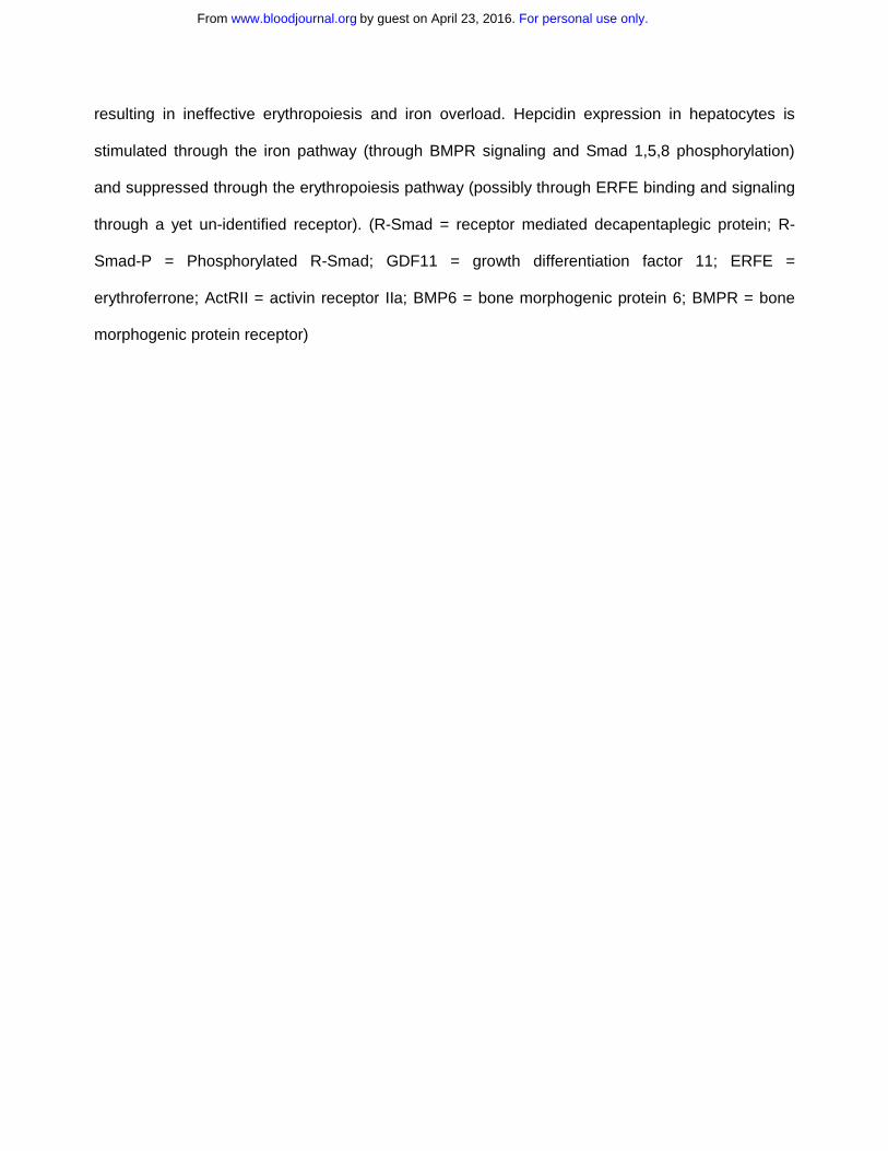
resulting in ineffective erythropoiesis and iron overload. Hepcidin expression in hepatocytes is
stimulated through the iron pathway (through BMPR signaling and Smad 1,5,8 phosphorylation)
and suppressed through the erythropoiesis pathway (possibly through ERFE binding and signaling
through a yet un-identified receptor). (R-Smad = receptor mediated decapentaplegic protein; R-
Smad-P = Phosphorylated R-Smad; GDF11 = growth differentiation factor 11; ERFE =
erythroferrone; ActRII = activin receptor IIa; BMP6 = bone morphogenic protein 6; BMPR = bone
morphogenic protein receptor)
For personal use only.on April 23, 2016. by guest www.bloodjournal.orgFrom

Erythropoiesis
Erythropoietic stimulation
(hemorrhage, phlebotomy, EPO)
Bone marrow
Increased iron availability
Liver
Erythroid factor
(e.g. GDF15, TWSG1, ERFE)
Hepcidin suppression
Fe
Ferroportin
Fe
Increases intestinal iron absorption
Fe FeFerroportin
Iron release from erythrophagocytosing
macrophages)
Figure 1: Hepcidin regulation by erythropoiesis and its effect on iron efflux from cells involved in iron metabolism.
Fe
For personal use only.
on April 23, 2016.
by guest
ww
w.bloodjournal.org
From

Figure 2: Model effect of erythropoiesis on hepcidin expression between RBCs transfusions.
Hb
epo
hepcidinTX TX
For personal use only.
on April 23, 2016.
by guest
ww
w.bloodjournal.org
From

R-SmadPSmad4 hepcidinhepatocyte
intracellular iron
R-SmadP
R-Smad
Smad7
BMPR
BMP6
differentiationproliferation
Smad4
erythroid precursor
?
?
Figure 3: Model of cross-talk between erythropoiesis and iron metabolism involving TGFβ member GDF11
ERFE
R-SmadP
R-Smad
Smad7
Smad4
R-SmadPSmad4 ?
BMP6
ActRII
GDF11ERFE
For personal use only.
on April 23, 2016.
by guest
ww
w.bloodjournal.org
From

doi:10.1182/blood-2014-03-563221Prepublished online June 12, 2014;
Niraj Shenoy, Nishanth Vallumsetla, Eliezer Rachmilewitz, Amit Verma and Yelena Ginzburg myelodysplastic syndromeImpact of iron overload and potential benefit from iron chelation in low-risk
http://www.bloodjournal.org/site/misc/rights.xhtml#repub_requestsInformation about reproducing this article in parts or in its entirety may be found online at:
http://www.bloodjournal.org/site/misc/rights.xhtml#reprintsInformation about ordering reprints may be found online at:
http://www.bloodjournal.org/site/subscriptions/index.xhtmlInformation about subscriptions and ASH membership may be found online at:
digital object identifier (DOIs) and date of initial publication. indexed by PubMed from initial publication. Citations to Advance online articles must include final publication). Advance online articles are citable and establish publication priority; they areappeared in the paper journal (edited, typeset versions may be posted when available prior to Advance online articles have been peer reviewed and accepted for publication but have not yet
Copyright 2011 by The American Society of Hematology; all rights reserved.Hematology, 2021 L St, NW, Suite 900, Washington DC 20036.Blood (print ISSN 0006-4971, online ISSN 1528-0020), is published weekly by the American Society of
For personal use only.on April 23, 2016. by guest www.bloodjournal.orgFrom
Related Documents