Review Impact of endocrine-disrupting compounds (EDCs) on female reproductive health Paul A. Fowler a,⇑ , Michelle Bellingham b , Kevin D. Sinclair c , Neil P. Evans b , Paola Pocar d , Bernd Fischer e , Kristina Schaedlich e , Juliane-Susanne Schmidt e , Maria R. Amezaga a , Siladitya Bhattacharya f , Stewart M. Rhind g , Peter J. O’Shaughnessy b a Division of Applied Medicine, Institute of Medical Sciences, Polwarth Building, University of Aberdeen, Foresterhill, Aberdeen AB25 2ZD, UK b Institute of Biodiversity, Animal Health and Comparative Medicine, College of Medical, Veterinary and Life Sciences, University of Glasgow, Glasgow G61 1QH, UK c School of Biosciences, University of Nottingham, Loughborough LE12 5RD, UK d Dipartimento di Patologia Animale, Igiene e Sanità Pubblica Veterinaria, University of Milano, Milano, Italy e Department of Anatomy and Cell Biology, Martin Luther University, Halle, Germany f Applied Health Sciences, Polwarth Building, University of Aberdeen, Foresterhill, Aberdeen AB25 2ZD, UK g The James Hutton Institute, Craigiebuckler, Aberdeen AB15 8QH, UK article info Article history: Available online 28 October 2011 Keywords: Female Reproduction Endocrine-disrupting compounds Development Ovary dysgenesis syndrome Pregnancy abstract Evidence is accumulating that environmental chemicals (ECs) including endocrine-disrupting com- pounds (EDCs) can alter female reproductive development, fertility and onset of menopause. While not as clearly defined as in the male, this set of abnormalities may constitute an Ovarian Dysgenesis Syn- drome with at least some origins of the syndrome arising during foetal development. ECs/EDCs have been shown to affect trophoblast and placental function, the female hypothalamo-pituitary–gonadal axis, onset of puberty and adult ovarian function. The effects of ECs/EDCs are complex, not least because it is emerging that low-level, ‘real-life’ mixtures of ECs/EDCs may carry significant biological potency. In addition, there is evidence that ECs/EDCs can alter the epigenome in a sexually dimorphic manner, which may lead to changes in the germ line and perhaps even to transgenerational effects. This review summa- rises the evidence for EC, including EDC, involvement in female reproductive dysfunction, it highlights potential mechanisms of EC action in the female and emphasises the need for further research into EC effects on female development and reproductive function. Ó 2011 Elsevier Ireland Ltd. All rights reserved. Contents 1. Introduction ......................................................................................................... 231 2. Are EDCs implicated in Ovarian Dysgenesis Syndrome (ODS)?................................................................. 232 3. EDC effects on trophoblast and placental function .......................................................................... 232 4. EDCs can disrupt the hypothalamo-pituitary–gonad (HPG) axis................................................................ 233 5. EDCs and the timing of puberty ......................................................................................... 235 6. Factors leading to gender differences in EDC exposure ....................................................................... 235 7. Evidence for epigenetic effects of EDCs ................................................................................... 236 8. Conclusions and perspective ............................................................................................ 237 Acknowledgements ................................................................................................... 237 References .......................................................................................................... 237 1. Introduction Environmental chemicals (ECs), including endocrine disrupting compounds (EDCs), comprise many different chemicals from a wide range of (primarily) anthropogenic, industrial, agricultural and domestic sources and in recent decades it has become 0303-7207/$ - see front matter Ó 2011 Elsevier Ireland Ltd. All rights reserved. doi:10.1016/j.mce.2011.10.021 ⇑ Corresponding author. Tel.: +44 0 1224 437528; fax: +44 01224 437465. E-mail address: [email protected] (P.A. Fowler). Molecular and Cellular Endocrinology 355 (2012) 231–239 Contents lists available at SciVerse ScienceDirect Molecular and Cellular Endocrinology journal homepage: www.elsevier.com/locate/mce

Welcome message from author
This document is posted to help you gain knowledge. Please leave a comment to let me know what you think about it! Share it to your friends and learn new things together.
Transcript
Molecular and Cellular Endocrinology 355 (2012) 231–239
Contents lists available at SciVerse ScienceDirect
Molecular and Cellular Endocrinology
journal homepage: www.elsevier .com/locate /mce
Review
Impact of endocrine-disrupting compounds (EDCs) on female reproductive health
Paul A. Fowler a,⇑, Michelle Bellingham b, Kevin D. Sinclair c, Neil P. Evans b, Paola Pocar d, Bernd Fischer e,Kristina Schaedlich e, Juliane-Susanne Schmidt e, Maria R. Amezaga a, Siladitya Bhattacharya f,Stewart M. Rhind g, Peter J. O’Shaughnessy b
a Division of Applied Medicine, Institute of Medical Sciences, Polwarth Building, University of Aberdeen, Foresterhill, Aberdeen AB25 2ZD, UKb Institute of Biodiversity, Animal Health and Comparative Medicine, College of Medical, Veterinary and Life Sciences, University of Glasgow, Glasgow G61 1QH, UKc School of Biosciences, University of Nottingham, Loughborough LE12 5RD, UKd Dipartimento di Patologia Animale, Igiene e Sanità Pubblica Veterinaria, University of Milano, Milano, Italye Department of Anatomy and Cell Biology, Martin Luther University, Halle, Germanyf Applied Health Sciences, Polwarth Building, University of Aberdeen, Foresterhill, Aberdeen AB25 2ZD, UKg The James Hutton Institute, Craigiebuckler, Aberdeen AB15 8QH, UK
a r t i c l e i n f o a b s t r a c t
Article history:Available online 28 October 2011
Keywords:FemaleReproductionEndocrine-disrupting compoundsDevelopmentOvary dysgenesis syndromePregnancy
0303-7207/$ - see front matter � 2011 Elsevier Irelandoi:10.1016/j.mce.2011.10.021
⇑ Corresponding author. Tel.: +44 0 1224 437528; fE-mail address: [email protected] (P.A. Fowle
Evidence is accumulating that environmental chemicals (ECs) including endocrine-disrupting com-pounds (EDCs) can alter female reproductive development, fertility and onset of menopause. While notas clearly defined as in the male, this set of abnormalities may constitute an Ovarian Dysgenesis Syn-drome with at least some origins of the syndrome arising during foetal development. ECs/EDCs have beenshown to affect trophoblast and placental function, the female hypothalamo-pituitary–gonadal axis,onset of puberty and adult ovarian function. The effects of ECs/EDCs are complex, not least because itis emerging that low-level, ‘real-life’ mixtures of ECs/EDCs may carry significant biological potency. Inaddition, there is evidence that ECs/EDCs can alter the epigenome in a sexually dimorphic manner, whichmay lead to changes in the germ line and perhaps even to transgenerational effects. This review summa-rises the evidence for EC, including EDC, involvement in female reproductive dysfunction, it highlightspotential mechanisms of EC action in the female and emphasises the need for further research into ECeffects on female development and reproductive function.
� 2011 Elsevier Ireland Ltd. All rights reserved.
Contents
1. Introduction . . . . . . . . . . . . . . . . . . . . . . . . . . . . . . . . . . . . . . . . . . . . . . . . . . . . . . . . . . . . . . . . . . . . . . . . . . . . . . . . . . . . . . . . . . . . . . . . . . . . . . . . . 2312. Are EDCs implicated in Ovarian Dysgenesis Syndrome (ODS)?. . . . . . . . . . . . . . . . . . . . . . . . . . . . . . . . . . . . . . . . . . . . . . . . . . . . . . . . . . . . . . . . . 2323. EDC effects on trophoblast and placental function . . . . . . . . . . . . . . . . . . . . . . . . . . . . . . . . . . . . . . . . . . . . . . . . . . . . . . . . . . . . . . . . . . . . . . . . . . 2324. EDCs can disrupt the hypothalamo-pituitary–gonad (HPG) axis. . . . . . . . . . . . . . . . . . . . . . . . . . . . . . . . . . . . . . . . . . . . . . . . . . . . . . . . . . . . . . . . 2335. EDCs and the timing of puberty . . . . . . . . . . . . . . . . . . . . . . . . . . . . . . . . . . . . . . . . . . . . . . . . . . . . . . . . . . . . . . . . . . . . . . . . . . . . . . . . . . . . . . . . . 2356. Factors leading to gender differences in EDC exposure . . . . . . . . . . . . . . . . . . . . . . . . . . . . . . . . . . . . . . . . . . . . . . . . . . . . . . . . . . . . . . . . . . . . . . . 2357. Evidence for epigenetic effects of EDCs . . . . . . . . . . . . . . . . . . . . . . . . . . . . . . . . . . . . . . . . . . . . . . . . . . . . . . . . . . . . . . . . . . . . . . . . . . . . . . . . . . . 2368. Conclusions and perspective . . . . . . . . . . . . . . . . . . . . . . . . . . . . . . . . . . . . . . . . . . . . . . . . . . . . . . . . . . . . . . . . . . . . . . . . . . . . . . . . . . . . . . . . . . . . 237
Acknowledgements . . . . . . . . . . . . . . . . . . . . . . . . . . . . . . . . . . . . . . . . . . . . . . . . . . . . . . . . . . . . . . . . . . . . . . . . . . . . . . . . . . . . . . . . . . . . . . . . . . . 237References . . . . . . . . . . . . . . . . . . . . . . . . . . . . . . . . . . . . . . . . . . . . . . . . . . . . . . . . . . . . . . . . . . . . . . . . . . . . . . . . . . . . . . . . . . . . . . . . . . . . . . . . . . 237
d Ltd. All rights reserved.
ax: +44 01224 437465.r).
1. Introduction
Environmental chemicals (ECs), including endocrine disruptingcompounds (EDCs), comprise many different chemicals from awide range of (primarily) anthropogenic, industrial, agriculturaland domestic sources and in recent decades it has become
232 P.A. Fowler et al. / Molecular and Cellular Endocrinology 355 (2012) 231–239
increasingly clear that they have the capacity to interfere with fe-male reproductive development and function in a wide range ofspecies including humans (Woodruff and Walker, 2008).
2. Are EDCs implicated in Ovarian Dysgenesis Syndrome (ODS)?
The term testicular dysgenesis syndrome (TDS) is used to de-scribe the phenotypic consequences of endocrine disruption inthe human male (Skakkebaek et al., 2001). These include reducedadult sperm counts and increased incidences of hypospadias (mal-formation of the urogenital tract), cryptorchidism (testis maldes-cent) and testicular cancer. TDS has a developmental origin andthe processes involved are being elucidated (Sharpe andSkakkebaek, 2008). There is, no evidence for such a tight set of in-ter-related developmental abnormalities inducing a comparablesyndrome in females. One phenotype suggestive of endocrine dis-ruption is the Mayer–Rokitansky–Kuster–Hauser (MRKH) syn-drome (Sultan et al., 2009). Multiple abnormalities are associatedwith this syndrome whose main characteristic is Müllerian ductaplasia (including uterine and vaginal abnormalities) in XX individ-uals with a female phenotype and primary amenorrhoea afterpuberty. Attempts to identify a genetic basis for MRKH syndromehave not been successful. Anti-Mullerian hormone (AMH), theAMH receptor, WT1, PAX2, CFTR and HOX genes, for instance, donot associate strongly with MRKH (e.g. (Burel et al., 2006)) andthe absence of a uterus and virilisation in women with WNT4 defi-ciency suggests that this is not a cause of MRKH but a distinct syn-drome in itself (Biason-Lauber and Konrad, 2008). Nevertheless,despite the lack of evidence for a female equivalent of TDS, bothoestrogenic and anti-androgenic signalling and chemicals can alsodisturb uterine and reproductive tract development in humans(e.g. (Smith and Taylor, 2007; Walters et al., 2009) and reviewedby (Crain et al., 2008)). So, what about the ovary and a unifying fe-male syndrome? While effects of MRKH syndrome can include theovary (e.g. (Al Omari et al., 2011)) this is rare, with the ovarynormally remaining functional. Despite similarities between testisand ovary development, the differences, such as in genetic pro-gramming (Cederroth et al., 2007), have marked implications forpotential mechanisms and consequences of EDC exposures. In theirrecent review Buck Louis et al. (2011) coined the term ‘‘OvarianDysgenesis Syndrome’’. They present a synthesis of the literaturethat supports the tentative use of the term, while highlighting someof the current knowledge deficits that need to be overcome. Wesupport the use of this term, ODS, as a loose umbrella concept thatmay include various combinations of other syndromes, such asMRKH and polycystic ovarian syndrome all of which may haveenvironmental components to their aetiology.
The effects of ECs on female fertility are mediated through di-verse, physiological systems, reflecting equally diverse mecha-nisms of action amongst the EC classes. Consequently, if effectsof ECs on fertility are to be identified, and associated mechanismsof action understood, it is imperative that the appropriate compo-nents of the reproductive system are fully characterised, includinggene expression and developmental processes. Effects of exposureto ECs, which may encompass some EDCs, include direct effects onphysiological mechanisms associated with gonadal developmentand function. Reproductive systems have been shown to beperturbed at multiple developmental stages; disrupted processesinclude hypothalamo-pituitary neuropeptide expression(Bellingham et al., 2009, 2010), ovarian follicle development andhealth (Fowler et al., 2008) in sheep exposed to pastures fertilisedusing sewage sludge, a by-product of waste water treatment con-taining thousands of ECs with known endocrine disrupting proper-ties. Many additional effects have also been reported in studiesinvolving a range of species; these include mitotic disorders,perturbed energy metabolism, reduced blastocyst development,
delayed implantation and increased embryo loss (Rhind et al.,2010a). Many individual disruptions, often associated with expo-sure to a single chemical, by themselves may be of little biologicalconsequence but it should be noted that humans and animals aretypically exposed to a mixture of ECs. These may act additively,increasing the effect exerted on a single physiological process, ormay initiate many, separate, disruptive effects in a single reproduc-tive cycle, regardless of the mechanisms involved, the combinedactions may have more severe reproductive consequences. Fur-thermore, there is evidence for xenochemical induction of epige-netic modification of genes involved in controlling femaleurogenital tract development, such as the HOX family, as shownin studies involving mice exposed to bisphenol A (BPA), in utero.(Smith and Taylor, 2007) (see Section 6). It should also be remem-bered that the effects of chemical exposure on reproductive devel-opment may be layered onto other programmed changes and arecent review has highlighted, in particular, the links between fe-male reproductive deficits, early life events and nutrition in bothfemales and males (Sloboda et al., 2011). Thus effects of EDCs haveto be seen in combination with effects of other factors that can dis-rupt normal foetal development and adult health (Skinner et al.,2010).
3. EDC effects on trophoblast and placental function
During early embryonic development, potential targets of EDCsinclude cell cleavage and differentiation, cell lineage determina-tion, methylation, implantation, maintenance of pregnancy andorganogenesis (Table 1). Polychlorinated biphenyls (PCBs), dioxins(such as TCDD) and phthalates (such as DEHP) can affect cell line-age formation and trophoblast function in blastocysts and thesechemicals are known to cross the placental barrier and to disturbembryonic development in humans (Myatt, 2006).
TCDD and coplanar PCBs are known to act on human endome-trial cells, via the arylhydrocarbon receptor (AhR), (Willing et al.,2011). The AhR-arylhydrocarbon receptor nuclear translocator het-erodimer (AhR-ARNT) is a typical transcription factor. AhR andARNT are expressed in mouse preimplantation embryos (Petersand Wiley, 1995) and in 3 day-old (days post coitum, dpc) morulaeand trophoblast cells of 4 dpc blastocysts in rabbits (Tscheudsch-ilsuren et al., 1999). Exposures to coplanar (PCB 77, 126, and169) and non-coplanar PCB (PCB 28, 52, 101, 118, 153, and 180)in low and high concentrations (0.1 ng or 1 lg/congener/mL med-ium) increase AhR signalling (AhR target genes include CYP1A1,CYP1B1) and expression of implantation-associated genes (VEGFR2and COX2) in rabbit 6 dpc blastocysts (Clausen et al., 2005), inter-fering with the cellular programmes that underlie early mamma-lian development. TCDD, via AhR activation, also decreases theexpression of the glucose transporter 1 (GLUT1 in the plasmamembrane in P19 mouse embryonic carcinoma cells (Tonacket al., 2007), disturbing glucose uptake in embryonic cells.Similarly, stress responses of human trophoblast-like JAR cellsare affected by TCDD treatment. Specific responses include signif-icantly increased levels of intracellular ROS and lipid peroxides, areduction in ATP content and mtDNA copy numbers as well ashigher rates of apoptosis (Chen et al., 2010).
Phthalates have also been shown to affect trophoblast cells inhuman placentae. For instance, higher maternal urinary concentra-tions of five phthalate metabolites (MEHP, MEOHP, MnBP, MiBO,MBzP) have been associated with a decreased expression of tro-phoblast differentiation markers (PPARc, AhR, hCG), leading toalterations in human placental development and function (Adibiet al., 2010). Interestingly, in rat HRP-1 trophoblast cells exposureto DEHP, MEHP and 2-ethylhexanoic acid also results in significantincreases in the concentrations of different lipid classes (Xu et al.,2006), leading to the conclusion that these chemicals might affect
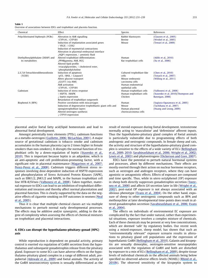
Table 1Overview of associations between EDCs and trophoblast and placenta function.
Chemical Effect Species References
Polychlorinated biphenyls (PCBs) Alteration in AhR signalling"CYP1A1, "CYP1B1Induction of implantation associated genes" VEGF, " COX2Induction of myometrial contractionsAlteration in placental-embryonal interface;AQP1 expression, " amniotic fluid
Rabbit blastocystsBovineMouse
(Clausen et al., 2005)(Wrobel et al., 2005)(Tewari et al., 2009)
Diethylhexylphthalate (DEHP) andits metabolites
Altered trophoblast differentiation:;PPARgamma, AhR, HCGAltered lipid profile"triacylglycerides, " cholesterol ester,"phosphatidylcholine
HumanRat trophoblast cells
(Adibi et al., 2010)(Xu et al., 2006)
2,3,7,8-Tetrachlorodibenzodioxin(TCDD)
Induction of apoptosis"p53, "BAX, " Caspase3Alters glucose transport;GLUT1 (via AhR)AhR activation"CYP1A1, "CYP1B1
Cultured trophoblast-likecellsMouse embryoniccarcinoma cellsHuman endometrialepithelial cells
(Chen et al., 2010)(Tonack et al., 2007)(Willing et al., 2011)
Cadmium Induction of stress response" HSP70, "MAPK; leptin expressionRetardation of trophoblast outgrowth
Human trophoblast cellsHuman trophoblast cellsChicken
(Valbonesi et al., 2008)(Stasenko et al. 2010)(Thompson andBannigan, 2008)
Bisphenol A (BPA) Positive correlation with miscarriagesInduction of degenerative trophoblastic giant cells andspongiotrophoblast layersAltered oestrogen synthesis; CYP19 expression
HumanMouseHuman placentalchoriocarcinoma cells
(Sugiura-Ogasawara et al., 2005)(Tachibana et al., 2007)(Huang and Leung, 2009)
P.A. Fowler et al. / Molecular and Cellular Endocrinology 355 (2012) 231–239 233
placental and/or foetal fatty acid/lipid homeostasis and lead toabnormal foetal development.
Amongst potentially toxic elements (PTEs), cadmium functionsas a metallo-oestrogen (Guillette, 2006; McLachlan, 2001). A majorsource of gestational cadmium is tobacco smoke and cadmiumaccumulates in the human placenta (up to 2 times higher in femalesmokers than non-smokers). It disrupts the normal function of tro-phoblast cells by a down-regulation of leptin (Stasenko et al.,2010). This is important because leptin is an adipokine, which isan anti-apoptotic and cell proliferation-promoting factor, with asignificant role in placental maintenance (Magarinos et al., 2007;Perez-Perez et al., 2008). Cadmium triggers different stress re-sponses involving dose-dependent induction of HSP70 expressionand phosphorylation of Stress Activated Protein Kinases (SAPK),such as ERK1/2, JNK1/2 and MAPK, in the human trophoblast cellline HTR-8/SVneo (Valbonesi et al., 2008). Taken together, mater-nal exposure to EDCs can lead to an inhibition of trophoblast differ-entiation and invasion and thereby affect normal placentation andplacental function. This is clearly supported by the data on the neg-ative effects of cigarette smoking on IVF outcomes in women (Nealet al., 2005).
Thus it is clear that multiple chemical classes act via multiplemechanisms to perturb normal early developmental processes.The effects may be additive and/or synergistic, adding to the de-gree of complexity when assessing the effects of chemical mixtureson trophoblast and placental interactions.
4. EDCs can disrupt the hypothalamo-pituitary–gonad (HPG)axis
While reproduction is dependent on gonadal activity, primarycontrol is exerted via regulation of GnRH secretion from the hypo-thalamus and subsequent gonadotrophin release from the pituitarygland. Exposure to some ECs can have adverse effects on the hypo-thalamo-pituitary gland complex in a range of different adult, pre-pubertal (Adewale et al., 2009) and foetal animals. The activity ofthe GnRH neurosecretory system is sexually differentiated as the
result of steroid exposure during foetal development, testosteronenormally acting to ‘masculinise’ and ‘defeminise’ afferent inputs.Thus the hypothalamo-pituitary gland complex of foetal animalsis particularly vulnerable due to programming effects of bothandrogenic and oestrogenic compounds. In female sheep and rats,the activity and structure of the hypothalamo-pituitary gland com-plex is sensitive to the effects of a wide variety of ECs (Bellinghamet al., 2009, 2010; Savabieasfahani et al., 2006; Wright et al., 2002;Lewis et al., 2003) and phytoestrogens (Dickerson and Gore, 2007).
EDCs have the potential to perturb natural hormonal systemsand processes, albeit by different mechanisms. Their effects aremostly exerted through their action on nuclear hormone receptors,such as oestrogen and androgen receptors, where they can haveagonistic or antagonistic effects. Effects of exposure are compoundand time specific. Thus, while in utero octylphenol (OP) exposurein sheep both directly suppresses gonadotrophin secretion (Swee-ney et al., 2000) and affects LH secretion later in life (Wright et al.,2002), post-natal OP exposure is not always associated with anobvious phenotype (Evans et al., 2004). On the other hand, expo-sure of sheep to other oestrogenic compounds such as BPA andmethoxychlor at later developmental time-points does result in al-tered gonadotrophin secretion (Savabieasfahani et al., 2006; Evanset al., 2004).
The effects on individuals of environmental EC exposure arecomplicated by the fact that under natural, rather than experimen-tal situations, exposure involves a complex mixture of chemicals.Each of these chemicals may be present at very low concentrations,which are deemed ‘safe’ by regulatory bodies. Our recent workusing a mixed-exposure, sheep model, has shown that such an‘‘environmentally relevant’’ exposure scenario results in altera-tions to pituitary gland cell populations and the expression ofhypothalamic GnRH (Bellingham et al., 2010). Galanin and kisspep-tin are sexually dimorphic, oestrogen-sensitive neuropeptidesassociated with the regulation of GnRH and both are disturbedby exposure to a mix of ECs (Bellingham et al., 2009), despite tissuelevels of individual chemicals in the affected animals being belowspecified no observed adverse effects levels (NOAEL) (Rhind et al.,2010b). The observed sensitivity of the kisspeptin system to
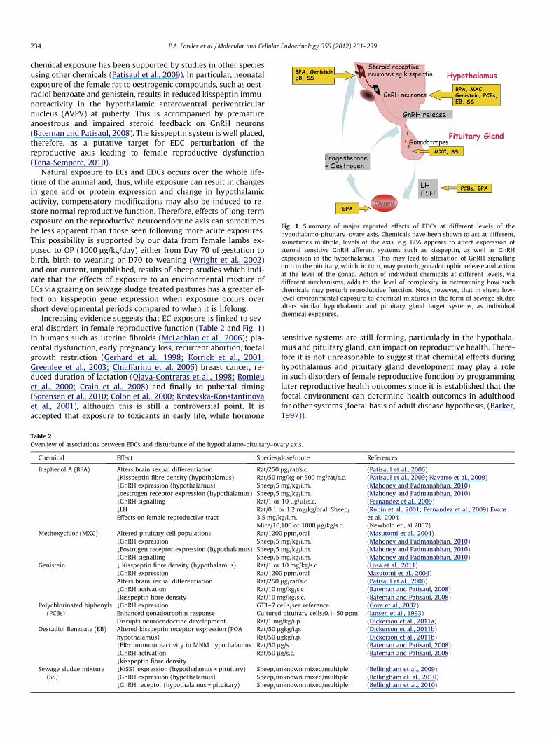
Fig. 1. Summary of major reported effects of EDCs at different levels of thehypothalamo-pituitary–ovary axis. Chemicals have been shown to act at different,sometimes multiple, levels of the axis, e.g. BPA appears to affect expression ofsteroid sensitive GnRH afferent systems such as kisspeptin, as well as GnRHexpression in the hypothalamus. This may lead to alteration of GnRH signallingonto to the pituitary, which, in turn, may perturb, gonadotrophin release and actionat the level of the gonad. Action of individual chemicals at different levels, viadifferent mechanisms, adds to the level of complexity in determining how suchchemicals may perturb reproductive function. Note, however, that in sheep low-level environmental exposure to chemical mixtures in the form of sewage sludgealters similar hypothalamic and pituitary gland target systems, as individualchemical exposures.
234 P.A. Fowler et al. / Molecular and Cellular Endocrinology 355 (2012) 231–239
chemical exposure has been supported by studies in other speciesusing other chemicals (Patisaul et al., 2009). In particular, neonatalexposure of the female rat to oestrogenic compounds, such as oest-radiol benzoate and genistein, results in reduced kisspeptin immu-noreactivity in the hypothalamic anteroventral periventricularnucleus (AVPV) at puberty. This is accompanied by prematureanoestrous and impaired steroid feedback on GnRH neurons(Bateman and Patisaul, 2008). The kisspeptin system is well placed,therefore, as a putative target for EDC perturbation of thereproductive axis leading to female reproductive dysfunction(Tena-Sempere, 2010).
Natural exposure to ECs and EDCs occurs over the whole life-time of the animal and, thus, while exposure can result in changesin gene and or protein expression and change in hypothalamicactivity, compensatory modifications may also be induced to re-store normal reproductive function. Therefore, effects of long-termexposure on the reproductive neuroendocrine axis can sometimesbe less apparent than those seen following more acute exposures.This possibility is supported by our data from female lambs ex-posed to OP (1000 lg/kg/day) either from Day 70 of gestation tobirth, birth to weaning or D70 to weaning (Wright et al., 2002)and our current, unpublished, results of sheep studies which indi-cate that the effects of exposure to an environmental mixture ofECs via grazing on sewage sludge treated pastures has a greater ef-fect on kisspeptin gene expression when exposure occurs overshort developmental periods compared to when it is lifelong.
Increasing evidence suggests that EC exposure is linked to sev-eral disorders in female reproductive function (Table 2 and Fig. 1)in humans such as uterine fibroids (McLachlan et al., 2006); pla-cental dysfunction, early pregnancy loss, recurrent abortion, foetalgrowth restriction (Gerhard et al., 1998; Korrick et al., 2001;Greenlee et al., 2003; Chiaffarino et al. 2006) breast cancer, re-duced duration of lactation (Olaya-Contreras et al., 1998; Romieuet al., 2000; Crain et al., 2008) and finally to pubertal timing(Sorensen et al., 2010; Colon et al., 2000; Krstevska-Konstantinovaet al., 2001), although this is still a controversial point. It isaccepted that exposure to toxicants in early life, while hormone
Table 2Overview of associations between EDCs and disturbance of the hypothalamo-pituitary–ov
Chemical Effect Species/
Bisphenol A (BPA) Alters brain sexual differentiation;Kisspeptin fibre density (hypothalamus);GnRH expression (hypothalamus);oestrogen receptor expression (hypothalamus);GnRH signalling;LHEffects on female reproductive tract
Rat/250Rat/50 mSheep/5Sheep/5Rat/1 orRat/0.13.5 mg/kMice/10
Methoxychlor (MXC) Altered pituitary cell populations;GnRH expression;Eostrogen receptor expression (hypothalamus);GnRH signalling
Rat/120Sheep/5Sheep/5Sheep/5
Genistein ; Kisspeptin fibre density (hypothalamus);GnRH expressionAlters brain sexual differentiation;GnRH activation;kisspeptin fibre density
Rat/1 orRat/120Rat/250Rat/10 mRat/10 m
Polychlorinated biphenyls(PCBs)
;GnRH expressionEnhanced gonadotrophin responseDisrupts neuroendocrine development
GT1–7 cCulturedRat/1 m
Oestadiol Benzoate (EB) Altered kisspeptin receptor expression (POAhypothalamus)"ERa immunoreactivity in MNM hypothalamus;GnRH activation;kisspeptin fibre density
Rat/50 lRat/50 lRat/50 lRat/50 l
Sewage sludge mixture(SS)
;KiSS1 expression (hypothalamus + pituitary);GnRH expression (hypothalamus);GnRH receptor (hypothalamus + pituitary)
Sheep/uSheep/uSheep/u
sensitive systems are still forming, particularly in the hypothala-mus and pituitary gland, can impact on reproductive health. There-fore it is not unreasonable to suggest that chemical effects duringhypothalamus and pituitary gland development may play a rolein such disorders of female reproductive function by programminglater reproductive health outcomes since it is established that thefoetal environment can determine health outcomes in adulthoodfor other systems (foetal basis of adult disease hypothesis, (Barker,1997)).
ary axis.
dose/route References
lg/rat/s.c.g/kg or 500 mg/rat/s.c.
mg/kg/i.m.mg/kg/i.m.10 lg/ll/s.c.
or 1.2 mg/kg/oral, Sheep/g/i.m.
,100 or 1000 lg/kg/s.c.
(Patisaul et al., 2006)(Patisaul et al., 2009; Navarro et al., 2009)(Mahoney and Padmanabhan, 2010)(Mahoney and Padmanabhan, 2010)(Fernandez et al., 2009)(Rubin et al., 2001; Fernandez et al., 2009) Evanset al., 2004(Newbold et., al 2007)
0 ppm/oralmg/kg/i.m.mg/kg/i.m.mg/kg/i.m.
(Masutomi et al., 2004)(Mahoney and Padmanabhan, 2010)(Mahoney and Padmanabhan, 2010)(Mahoney and Padmanabhan, 2010)
10 mg/kg/s.c0 ppm/orallg/rat/s.c.g/kg/s.cg/kg/s.c.
(Losa et al., 2011)Masutomi et al., 2004)(Patisaul et al., 2006)(Bateman and Patisaul, 2008)(Bateman and Patisaul, 2008)
ells/see referencepituitary cells/0.1–50 ppm
g/kg/i.p.
(Gore et al., 2002)(Jansen et al., 1993)(Dickerson et al., 2011a)
gkg/i.p.gkg/i.p.g/s.c.g/s.c.
(Dickerson et al., 2011b)(Dickerson et al., 2011b)(Bateman and Patisaul, 2008)(Bateman and Patisaul, 2008)
nknown mixed/multiplenknown mixed/multiplenknown mixed/multiple
(Bellingham et al., 2009)(Bellingham et. al., 2010)(Bellingham et al., 2010)
P.A. Fowler et al. / Molecular and Cellular Endocrinology 355 (2012) 231–239 235
5. EDCs and the timing of puberty
Puberty is a multifaceted developmental process that is underthe control of different hormonal regulatory mechanisms and, be-cause there can be significant social consequences of prematurepuberty, this section will focus on the human. Puberty is character-ised by activation of the hypothalamic-pituitary–gonadal axis, theappearance of secondary sexual characteristics and a growth spurt(Kakarla and Bradshaw, 2003). The onset of puberty begins late inchildhood and results in the individual’s transition period from anon-reproductive to a reproductive state. In girls, puberty is initi-ated by changes in the expression of hypothalamic neurotransmit-ters, which awaken the ovaries leading to oestradiol secretion. Ageat onset of puberty is determined by multiple genetic and environ-mental factors, including psychosocial and socio-economic condi-tions, nutrition and ethnicity (Parent et al., 2003). During thepast decades, secular trends of earlier age at onset of puberty havebeen reported, especially in girls, in both the US and Europe (Parentet al., 2003; Mul et al., 2001). One prominent hypothesis, devel-oped to explain the recent changes in puberty timing, is that expo-sure to EDCs during the prepubertal period, but also in earlyperinatal programming windows, may cause an early age of pub-erty (Den Hond et al., 2002; Herman-Giddens, 2006; Sharpe andSkakkebaek, 1993). Currently, most of the evidence implicatingEDCs in the dysregulation of pubertal development stems fromanimal experiments and in vitro studies. In humans, a large num-ber of cohort studies also suggest that exogenous compounds mayhave pronounced effects on pubertal timing although a conclusiverelationship between precocious puberty and environmentalagents has not yet been established, as summarised in Table 3.
A temporal trend toward premature thelarche in Puerto Ricowas noted during the early 1980s (Bongiovanni, 1983; Freni-Titul-aer et al., 1986; Saenz de Rodriguez et al., 1985). About 68% of thegirls with premature breast development had measurable circulat-ing levels of the phthalates [dimethyl, diethyl, dibutyl and di-(2-ethylhexyl)], compared with 17% of girls with normal thelarchetiming (Colon et al., 2000). The association between phthalateexposure and precocious puberty in female proposed by Colon isvery suggestive. However, interpretation of these results is stillcontroversial due to the very high levels of serum phthalates re-ported which may imply major analytical problems and possiblylaboratory contamination (for comments see: McKee, 2004). Inmore recent studies, association between increased serum levelsof DBP and DEHP and precocious puberty, in Chinese and Koreangirls, was also reported (Qiao et al., 2007; Lee et al., 2006), whereas
Table 3Human cohort studies investigating the potential relationship between endocrine disrupti
Compound Study area Exposure Subjects Main fi
Main studies supporting associations between early puberty and EDCsDDE Michigan (USA) Prenatal 151 Girls ReduceDDE Belgium Pubertal 41 Girls Serum
with pPBBs Michigan (USA) Prenatal & lactational 327 Girls High P
menarPhthalate Puerto Rico Pubertal 76 Girls Elevate
earlierPhyto-estrogens Turkey Pubertal 4 Girls Use of
premaZearalenone Tuscany (Italy) Prepubertal 32 Girls Serum
in girlsDDT/DDE Shanghai Prepubertal 466 Girls Higher
menar
Main studies finding no association between early puberty and EDCsDDE PCBs North Carolina (USA) Prenatal and lactational 316 Girls No effeDioxin Seveso (Italy) Prepubertal 282 Girls No effePCBs Michigan (USA) Prenatal and lactational 327 Girls No effeDDE Mohawk nation (USA) Prepubertal 138 Girls No ass
other investigators failed to demonstrate a link between phthalateexposure and age of sexual maturity (Lomenick et al., 2010). Thusmore studies are necessary to elucidate the possible links betweenphthalate exposure and the risk of early puberty. Detectable plas-ma levels of the DDT metabolite p,p0-DDE in children with preco-cious puberty immigrating from developing countries to Belgium,were strikingly different to the undetectable levels in Belgian-borngirls with idiopathic or organic precocious puberty. Since DDT isstill used in a number of developing countries, but banned inEurope, the data suggest a possible relationship between earlyexposure to this pesticide and premature menarche (Krstevska-Konstantinova et al., 2001). In-utero exposure to EDCs can alsoalter the growth of the mammary gland and the age of onset ofpuberty of the offspring many years after exposure (Wang et al.,2005). The effect of in utero exposure to polybrominated biphenyls(PBBs) on sexual maturation has been evaluated in US girls whosemothers were accidentally exposed through diet to a PBB flameretardant (FireMaster) (Blanck et al., 2000). Menarche and pubertalhair growth were significantly advanced in breastfed girls exposedto high levels of PBB in utero compared to breastfed girls exposedto lower levels of PBB in utero or girls who were not breastfed.
Precocious puberty has several physical, psychological, and so-cial consequences (reviewed in Golub et al., 2008). The early onsetof thelarche and/or menarche is positively associated with an earlydiagnosis of breast cancer in susceptible populations (Hamiltonand Mack, 2003) and adult obesity (Biro et al., 2003). From a socialpoint of view, changes in the timing of pubertal development mayinfluence the risk for substances abuse, antisocial behaviour, eatingdisorders and emotional stress (Patton and Viner, 2007).
6. Factors leading to gender differences in EDC exposure
The processes that determine tissue exposure to EDCs appear, atfirst sight, to be both simple and independent of gender. However,gender-related factors can affect exposure to EDCs, indirectly,through differences in the ecology of the two sexes. e.g. in manydeer species, males and females inhabit different geographic areasand/or exploit different feed resources and may therefore be ex-posed to subtly different EDC burdens. In mammals, gender differ-ences in physiology, especially those associated with the nutrientdemands of pregnancy and lactation may result in increased food(and possibly EC) consumption in some species (e.g. sheep; Footand Russel, 1979) and result in the mobilisation of fat reservesand associated release of stored pollutants (Bigsby et al., 1997);
ng agents and early onset of puberty in girls.
ndings References
d age of menarche by 1 year (Vasiliu et al., 2004)DDE was significantly elevated in girlsrecocious puberty
(Krstevska-Konstantinova et al., 2001)
BBs were associated with precociousche and earlier pubic hair stage
(Blanck et al. 2000)
d phthalate levels were associated withthelarche
(Colon et al., 2000)
Foeniculum vulgare was associated withture thelarche
(Turkyilmaz et al., 2008)
zearalenone was significantly elevatedwith precocious puberty
(Massart et al. 2008)
DDT/DDE associated with earlier age atche
(Ouyang et al., 2005)
ct on pubertal stages (Gladen et al., 2000)ct on age at menarche (Warner et al., 2004)ct on pubertal stages (Blanck et al., 2000)
ociation with age of menarche (Denham et al., 2005)
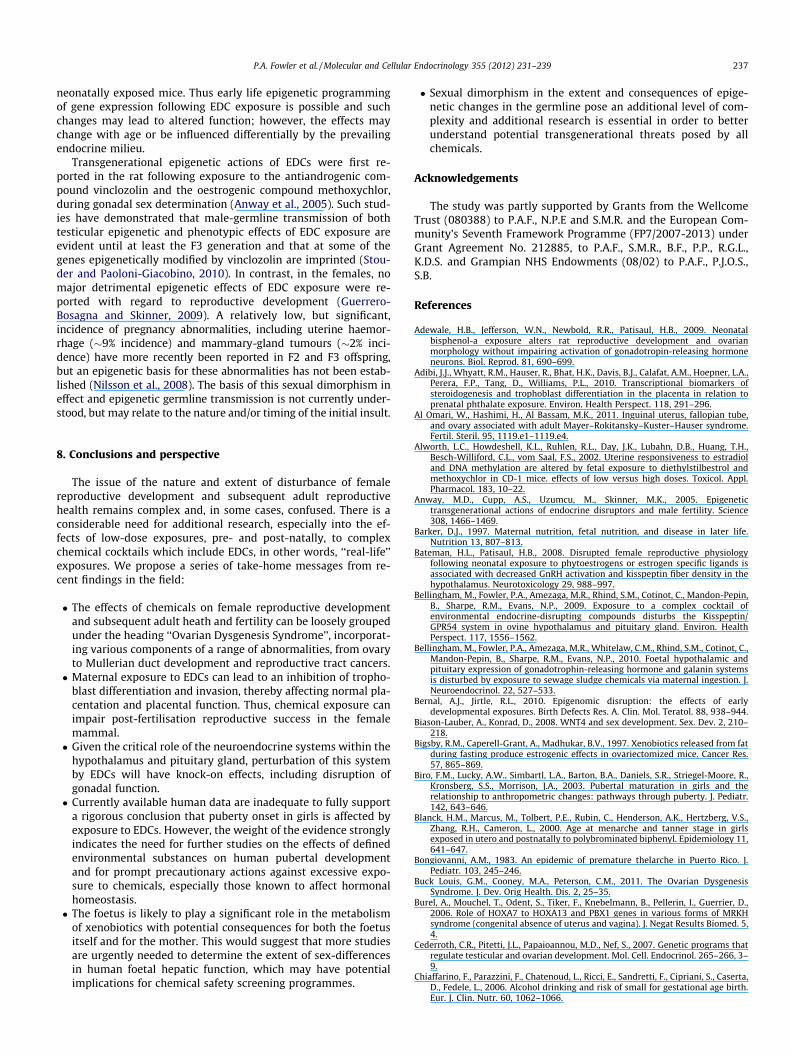
Fig. 2. Schematic summarising EDC actions across generations. If the mother isexposed to EDCs directly her fertility may be affected and, if she is pregnant, herfoetus will be directly exposed (F1, children). However, the germ cells in the foetuswill also be exposed and this may result in disturbance of both the F1 directly and ofthe F2 in the form of the F1 germ cells. For a true transgenerational effect however,the F3 generation (great-grandchildren) would have to show disruption withoutdirect exposure of the F1 and F2 generations, post-natally. Of course the F3generation is likely to be exposed to a different panel of chemicals and pollutantssince the profiles of different chemical classes change with time, production anduse.
236 P.A. Fowler et al. / Molecular and Cellular Endocrinology 355 (2012) 231–239
both effects may contribute to different chemical exposures be-tween males and females.
In addition to differences in exposure associated with ecologyor physiological state, there is also potential for gender differencesin post-exposure metabolism, which in turn affects target organexposure. Sex-based differences in the pharmacokinetics of EDCscan be attributable to sex differences in hormone profiles and asso-ciated effects on gastrointestinal motility and body composition,organ blood flow and renal clearance rates (Waxman and Hollo-way, 2009). However, differences in metabolism are consideredto be the primary determinants of sex differences in the processingof drugs and xenobiotics. These differences are a function of theactivities of many phase 1 and phase 2 liver enzymes, includingcytochrome P450s, sulfotransferases, glutathione transferases andUDP-glucuronosyltransferase and of complex interactions betweenthe relevant genes, hormonal signals and associated transcriptionfactors (Waxman and Holloway, 2009). While many of the princi-ples underlying sex differences in metabolism have been estab-lished through studies of pharmaceuticals, it has also beenshown in rats that sexually dimorphic metabolic responses occurin response to well known disrupting chemicals such as DDT (Sier-ra-Santoyo et al., 2000). Similarly, the dimorphic neuronal re-sponses to the herbicide atrazine shown in mice (Giusi et al.,2006), and dimorphic behavioural responses to PCBs observed inhumans (Ibanez et al., 2000), may be a function of underlying met-abolic differences.
In humans, foetal exposure to environmental toxicants is a ma-jor concern (Mattison, 2010). During development in the human,the basic structure of the liver is present by the end of the first tri-mester and several enzymes involved in drug metabolism are al-ready expressed at this stage (Koukouritaki et al., 2002; Stevenset al., 2003). About 70% of the blood going to the foetal liver comesdirectly from the placenta and the feto–maternal interface throughthe umbilical vein and the foetal liver is, therefore, exposed to highconcentrations of maternally-derived xenochemicals. It is likely,therefore, that the liver is both affected by xenobiotics and isimportant for their metabolism/activation from early in foetaldevelopment through to adult life. In foetal and neonatal develop-ment there is growing evidence that sex-dependent differences ex-ist in hepatic function. In rats, for example, foetal hepatic transcriptlevels can be affected by changes in maternal diet in a sexuallydimorphic manner (Kwong et al., 2007). More recently, in a studyof 55 normally-progressing, electively terminated, human foetuses,we have shown that there are significant sex-differences in the lev-els of mRNA transcripts encoding a number of hepatic enzymes inthe human foetal liver (O’Shaughnessy et al., 2011). The most likelycause of such foetal gender effects is the action of testicular andro-gen in males although differences in foetal growth rates cannot beexcluded. The contribution of foetal metabolism to overall pharma-cokinetics of xenobiotics and pharmaceuticals during pregnancy inthe human remains unclear. However, there is evidence from otherprimates that foetal levels of some metabolites exceed maternallevels (Garland et al., 2005) suggesting that for some ECs and EDCs,foetal toxicant burden are probably higher than those in themother.
Once puberty is reached, differences in the pattern of pituitaryGH release induce a clear sexual dimorphism in hepatic metabolicactivity. This is most apparent in rodents although less pronouncedGH-dependent sex differences in hepatic function are also seen inhumans (Waxman and Holloway, 2009). These endogenous sex dif-ferences can interact with ECs and EDCs in the human to alter pat-terns of liver metabolism. For example, CYP3A4 is the majorcatalyst of cytochrome P450 oxidative metabolism in the liver(responsible for metabolism of more than 60% of all drugs). It is ex-pressed at higher levels in women than in men and is induced inboth sexes by xenobiotics through action on the orphan nuclear
pregnane X receptor (PXR). PXR is a member of a larger group ofnuclear receptors which act to detect and activate a defensive re-sponse to xenobiotics and they can themselves show sex differ-ences in expression (Jalouli et al., 2003). Overall hepaticresponses to EDC exposure, therefore, are complex and likely to de-pend on age and sex as well as level and type of exposure.
7. Evidence for epigenetic effects of EDCs
Given the level of interest in epigenetic programming of earlydevelopment over the past decade, the paucity of primary dataon the effects of EDCs on epigenetic programming through thegermline, particularly the female germline, is perhaps surprising(Zama and Uzumcu, 2009, 2010; Bernal and Jirtle, 2010). This is acritical deficiency since such epigenetic changes may induce defi-cits or alterations in the developmental trajectory of unexposedgenerations, i.e. true transgenerational effects (Fig. 2). Studies todate, understandably, have largely been conducted in rodentsand have tended to focus on DNA methylation at specific regions(usually promoter associated CpG islands) in specific genes, orhave employed global DNA methylation assays that can providequantitative estimates of changes to DNA methylation but littlein the way of a mechanistic insight. Consequently, these data tendto be correlative in nature. That said, that there is sufficient evi-dence to support the notion that at least some of the actions ofEDCs on developmentally-important genes and associated pro-cesses are epigenetic in nature and there is also evidence of epige-netic transgenerational transmission of these effects.
By way of example, (Alworth et al., 2002) screened CpG islandloci in uteri of CD-1 mice exposed during foetal life to varyingdoses of diethylstilbestrol (DES). Their analysis showed that in re-sponse to both low and high doses of DES there was an increase inmethylation of a number of ribosomal genes, a state frequently ob-served in breast cancer. While the long term effects of hyperme-thylation were not characterised in that study, Tang et al. (2008)reported that in aged CD-1 mice neonatally exposed to DES (1 or1000 lg/kg) and the isoflavonoid phytoestrogen genistein(50 mg/kg), Nsbp1, which is involved in chromatin remodelling,was hypomethylated and over-expressed in the uteri of DES/GEN
P.A. Fowler et al. / Molecular and Cellular Endocrinology 355 (2012) 231–239 237
neonatally exposed mice. Thus early life epigenetic programmingof gene expression following EDC exposure is possible and suchchanges may lead to altered function; however, the effects maychange with age or be influenced differentially by the prevailingendocrine milieu.
Transgenerational epigenetic actions of EDCs were first re-ported in the rat following exposure to the antiandrogenic com-pound vinclozolin and the oestrogenic compound methoxychlor,during gonadal sex determination (Anway et al., 2005). Such stud-ies have demonstrated that male-germline transmission of bothtesticular epigenetic and phenotypic effects of EDC exposure areevident until at least the F3 generation and that at some of thegenes epigenetically modified by vinclozolin are imprinted (Stou-der and Paoloni-Giacobino, 2010). In contrast, in the females, nomajor detrimental epigenetic effects of EDC exposure were re-ported with regard to reproductive development (Guerrero-Bosagna and Skinner, 2009). A relatively low, but significant,incidence of pregnancy abnormalities, including uterine haemor-rhage (�9% incidence) and mammary-gland tumours (�2% inci-dence) have more recently been reported in F2 and F3 offspring,but an epigenetic basis for these abnormalities has not been estab-lished (Nilsson et al., 2008). The basis of this sexual dimorphism ineffect and epigenetic germline transmission is not currently under-stood, but may relate to the nature and/or timing of the initial insult.
8. Conclusions and perspective
The issue of the nature and extent of disturbance of femalereproductive development and subsequent adult reproductivehealth remains complex and, in some cases, confused. There is aconsiderable need for additional research, especially into the ef-fects of low-dose exposures, pre- and post-natally, to complexchemical cocktails which include EDCs, in other words, ‘‘real-life’’exposures. We propose a series of take-home messages from re-cent findings in the field:
� The effects of chemicals on female reproductive developmentand subsequent adult heath and fertility can be loosely groupedunder the heading ‘‘Ovarian Dysgenesis Syndrome’’, incorporat-ing various components of a range of abnormalities, from ovaryto Mullerian duct development and reproductive tract cancers.� Maternal exposure to EDCs can lead to an inhibition of tropho-
blast differentiation and invasion, thereby affecting normal pla-centation and placental function. Thus, chemical exposure canimpair post-fertilisation reproductive success in the femalemammal.� Given the critical role of the neuroendocrine systems within the
hypothalamus and pituitary gland, perturbation of this systemby EDCs will have knock-on effects, including disruption ofgonadal function.� Currently available human data are inadequate to fully support
a rigorous conclusion that puberty onset in girls is affected byexposure to EDCs. However, the weight of the evidence stronglyindicates the need for further studies on the effects of definedenvironmental substances on human pubertal developmentand for prompt precautionary actions against excessive expo-sure to chemicals, especially those known to affect hormonalhomeostasis.� The foetus is likely to play a significant role in the metabolism
of xenobiotics with potential consequences for both the foetusitself and for the mother. This would suggest that more studiesare urgently needed to determine the extent of sex-differencesin human foetal hepatic function, which may have potentialimplications for chemical safety screening programmes.
� Sexual dimorphism in the extent and consequences of epige-netic changes in the germline pose an additional level of com-plexity and additional research is essential in order to betterunderstand potential transgenerational threats posed by allchemicals.
Acknowledgements
The study was partly supported by Grants from the WellcomeTrust (080388) to P.A.F., N.P.E and S.M.R. and the European Com-munity’s Seventh Framework Programme (FP7/2007-2013) underGrant Agreement No. 212885, to P.A.F., S.M.R., B.F., P.P., R.G.L.,K.D.S. and Grampian NHS Endowments (08/02) to P.A.F., P.J.O.S.,S.B.
References
Adewale, H.B., Jefferson, W.N., Newbold, R.R., Patisaul, H.B., 2009. Neonatalbisphenol-a exposure alters rat reproductive development and ovarianmorphology without impairing activation of gonadotropin-releasing hormoneneurons. Biol. Reprod. 81, 690–699.
Adibi, J.J., Whyatt, R.M., Hauser, R., Bhat, H.K., Davis, B.J., Calafat, A.M., Hoepner, L.A.,Perera, F.P., Tang, D., Williams, P.L., 2010. Transcriptional biomarkers ofsteroidogenesis and trophoblast differentiation in the placenta in relation toprenatal phthalate exposure. Environ. Health Perspect. 118, 291–296.
Al Omari, W., Hashimi, H., Al Bassam, M.K., 2011. Inguinal uterus, fallopian tube,and ovary associated with adult Mayer–Rokitansky–Kuster–Hauser syndrome.Fertil. Steril. 95, 1119.e1–1119.e4.
Alworth, L.C., Howdeshell, K.L., Ruhlen, R.L., Day, J.K., Lubahn, D.B., Huang, T.H.,Besch-Williford, C.L., vom Saal, F.S., 2002. Uterine responsiveness to estradioland DNA methylation are altered by fetal exposure to diethylstilbestrol andmethoxychlor in CD-1 mice. effects of low versus high doses. Toxicol. Appl.Pharmacol. 183, 10–22.
Anway, M.D., Cupp, A.S., Uzumcu, M., Skinner, M.K., 2005. Epigenetictransgenerational actions of endocrine disruptors and male fertility. Science308, 1466–1469.
Barker, D.J., 1997. Maternal nutrition, fetal nutrition, and disease in later life.Nutrition 13, 807–813.
Bateman, H.L., Patisaul, H.B., 2008. Disrupted female reproductive physiologyfollowing neonatal exposure to phytoestrogens or estrogen specific ligands isassociated with decreased GnRH activation and kisspeptin fiber density in thehypothalamus. Neurotoxicology 29, 988–997.
Bellingham, M., Fowler, P.A., Amezaga, M.R., Rhind, S.M., Cotinot, C., Mandon-Pepin,B., Sharpe, R.M., Evans, N.P., 2009. Exposure to a complex cocktail ofenvironmental endocrine-disrupting compounds disturbs the Kisspeptin/GPR54 system in ovine hypothalamus and pituitary gland. Environ. HealthPerspect. 117, 1556–1562.
Bellingham, M., Fowler, P.A., Amezaga, M.R., Whitelaw, C.M., Rhind, S.M., Cotinot, C.,Mandon-Pepin, B., Sharpe, R.M., Evans, N.P., 2010. Foetal hypothalamic andpituitary expression of gonadotrophin-releasing hormone and galanin systemsis disturbed by exposure to sewage sludge chemicals via maternal ingestion. J.Neuroendocrinol. 22, 527–533.
Bernal, A.J., Jirtle, R.L., 2010. Epigenomic disruption: the effects of earlydevelopmental exposures. Birth Defects Res. A. Clin. Mol. Teratol. 88, 938–944.
Biason-Lauber, A., Konrad, D., 2008. WNT4 and sex development. Sex. Dev. 2, 210–218.
Bigsby, R.M., Caperell-Grant, A., Madhukar, B.V., 1997. Xenobiotics released from fatduring fasting produce estrogenic effects in ovariectomized mice. Cancer Res.57, 865–869.
Biro, F.M., Lucky, A.W., Simbartl, L.A., Barton, B.A., Daniels, S.R., Striegel-Moore, R.,Kronsberg, S.S., Morrison, J.A., 2003. Pubertal maturation in girls and therelationship to anthropometric changes: pathways through puberty. J. Pediatr.142, 643–646.
Blanck, H.M., Marcus, M., Tolbert, P.E., Rubin, C., Henderson, A.K., Hertzberg, V.S.,Zhang, R.H., Cameron, L., 2000. Age at menarche and tanner stage in girlsexposed in utero and postnatally to polybrominated biphenyl. Epidemiology 11,641–647.
Bongiovanni, A.M., 1983. An epidemic of premature thelarche in Puerto Rico. J.Pediatr. 103, 245–246.
Buck Louis, G.M., Cooney, M.A., Peterson, C.M., 2011. The Ovarian DysgenesisSyndrome. J. Dev. Orig Health. Dis. 2, 25–35.
Burel, A., Mouchel, T., Odent, S., Tiker, F., Knebelmann, B., Pellerin, I., Guerrier, D.,2006. Role of HOXA7 to HOXA13 and PBX1 genes in various forms of MRKHsyndrome (congenital absence of uterus and vagina). J. Negat Results Biomed. 5,4.
Cederroth, C.R., Pitetti, J.L., Papaioannou, M.D., Nef, S., 2007. Genetic programs thatregulate testicular and ovarian development. Mol. Cell. Endocrinol. 265–266, 3–9.
Chiaffarino, F., Parazzini, F., Chatenoud, L., Ricci, E., Sandretti, F., Cipriani, S., Caserta,D., Fedele, L., 2006. Alcohol drinking and risk of small for gestational age birth.Eur. J. Clin. Nutr. 60, 1062–1066.
238 P.A. Fowler et al. / Molecular and Cellular Endocrinology 355 (2012) 231–239
Chen, S.C., Liao, T.L., Wei, Y.H., Tzeng, C.R., Kao, S.H., 2010. Endocrine disruptor,dioxin (TCDD)-induced mitochondrial dysfunction and apoptosis in humantrophoblast-like JAR cells. Mol. Hum. Reprod. 16, 361–372.
Clausen, I., Kietz, S., Fischer, B., 2005. Lineage-specific effects of polychlorinatedbiphenyls (PCB) on gene expression in the rabbit blastocyst. Reprod. Toxicol. 20,47–56.
Colon, I., Caro, D., Bourdony, C.J., Rosario, O., 2000. Identification of phthalate estersin the serum of young Puerto Rican girls with premature breast development.Environ. Health Perspect. 108, 895–900.
Crain, D.A., Janssen, S.J., Edwards, T.M., Heindel, J., Ho, S.M., Hunt, P., Iguchi, T., Juul,A., McLachlan, J.A., Schwartz, J., Skakkebaek, N., Soto, A.M., Swan, S., Walker, C.,Woodruff, T.K., Woodruff, T.J., Giudice, L.C., Guillette Jr, L.J., 2008. Femalereproductive disorders: the roles of endocrine-disrupting compounds anddevelopmental timing. Fertil. Steril. 90, 911–940.
Den Hond, E., Roels, H.A., Hoppenbrouwers, K., Nawrot, T., Thijs, L., Vandermeulen,C., Winneke, G., Vanderschueren, D., Staessen, J.A., 2002. Sexual maturation inrelation to polychlorinated aromatic hydrocarbons: Sharpe and Skakkebaek’shypothesis revisited. Environ. Health Perspect. 110, 771–776.
Denham, M., Schell, L.M., Deane, G., Gallo, M.V., Ravenscroft, J., DeCaprio,A.P.Akwesasne Task Force on the Environment, 2005. Relationship of lead,mercury, mirex, dichlorodiphenyldichloroethylene, hexachlorobenzene, andpolychlorinated biphenyls to timing of menarche among Akwesasne Mohawkgirls. Pediatrics 115, e127–e134.
Dickerson, S.M., Gore, A.C., 2007. Estrogenic environmental endocrine-disruptingchemical effects on reproductive neuroendocrine function and dysfunctionacross the life cycle. Rev. Endocr. Metab. Disord. 8, 143–159.
Dickerson, S.M., Cunningham, S.L., Gore, A.C., 2011a. Prenatal PCBs disrupt earlyneuroendocrine development of the rat hypothalamus. Toxicol. Appl.Pharmacol. 252, 36–46.
Dickerson, S.M., Cunningham, S.L., Patisaul, H.B., Woller, M.J., Gore, A.C., 2011b.Endocrine disruption of brain sexual differentiation by developmental PCBexposure. Endocrinology 152, 581–594.
Evans, N.P., North, T., Dye, S., Sweeney, T., 2004. Differential effects of theendocrine-disrupting compounds bisphenol-A and octylphenol ongonadotropin secretion, in prepubertal ewe lambs. Domest. Anim. Endocrinol.26, 61–73.
Fernandez, M., Bianchi, M., Lux-Lantos, V., Libertun, C., 2009. Neonatal exposure tobisphenol a alters reproductive parameters and gonadotropin releasinghormone signaling in female rats. Environ. Health Perspect. 117, 757–762.
Foot, J.Z., Russel, A.J.F., 1979. The relationship in ewes between voluntary foodintake during pregnancy and forage intake during lactation and after weaning.Animal Prod. 28, 25–39.
Fowler, P.A., Dora, N.J., McFerran, H., Amezaga, M.R., Miller, D.W., Lea, R.G., Cash, P.,McNeilly, A.S., Evans, N.P., Cotinot, C., Sharpe, R.M., Rhind, S.M., 2008. In uteroexposure to low doses of environmental pollutants disrupts fetal ovariandevelopment in sheep. Mol. Hum. Reprod. 14, 269–280.
Freni-Titulaer, L.W., Cordero, J.F., Haddock, L., Lebron, G., Martinez, R., Mills, J.L.,1986. Premature thelarche in Puerto Rico. A search for environmental factors.Am. J. Dis. Child. 140, 1263–1267.
Garland, M., Abildskov, K.M., Kiu, T.W., Daniel, S.S., Stark, R.I., 2005. Thecontribution of fetal metabolism to the disposition of morphine. Drug Metab.Dispos. 33, 68–76.
Gerhard, I., Daniel, V., Link, S., Monga, B., Runnebaum, B., 1998. Chlorinatedhydrocarbons in women with repeated miscarriages. Environ. Health Perspect.106, 675–681.
Giusi, G., Facciolo, R.M., Canonaco, M., Alleva, E., Belloni, V., Dessi’-Fulgheri, F.,Santucci, D., 2006. The endocrine disruptor atrazine accounts for a dimorphicsomatostatinergic neuronal expression pattern in mice. Toxicol. Sci. 89, 257–264.
Gladen, B.C., Ragan, N.B., Rogan, W.J., 2000. Pubertal growth and development andprenatal and lactational exposure to polychlorinated biphenyls anddichlorodiphenyl dichloroethene. J. Pediatr. 136, 490–496.
Golub, M.S., Collman, G.W., Foster, P.M., Kimmel, C.A., Rajpert-De Meyts, E., Reiter,E.O., Sharpe, R.M., Skakkebaek, N.E., Toppari, J., 2008. Public health implicationsof altered puberty timing. Pediatrics 121 (Suppl. 3), S218–S230.
Gore, A.C., Wu, T.J., Oung, T., Lee, J.B., Woller, M.J., 2002. A novel mechanism forendocrine-disrupting effects of polychlorinated biphenyls: direct effects ongonadotropin-releasing hormone neurones. J. Neuroendocrinol. 14, 814–823.
Greenlee, A.R., Arbuckle, T.E., Chyou, P.H., 2003. Risk factors for female infertility inan agricultural region. Epidemiology 14, 429–436.
Guerrero-Bosagna, C.M., Skinner, M.K., 2009. Epigenetic transgenerational effects ofendocrine disruptors on male reproduction. Semin. Reprod. Med. 27, 403–408.
Guillette, L.J., Jr., 2006. Endocrine disrupting contaminants – beyond the dogma.Environ. Health Perspect. 114 Suppl 1, 9–12.
Hamilton, A.S., Mack, T.M., 2003. Puberty and genetic susceptibility to breast cancerin a case-control study in twins. N. Engl. J. Med. 348, 2313–2322.
Herman-Giddens, M.E., 2006. Recent data on pubertal milestones in United Stateschildren: the secular trend toward earlier development. Int. J. Androl. 29, 241–246; discussion 286–290.
Huang, H., Leung, L.K., 2009. Bisphenol A downregulates CYP19 transcription in JEG-3 cells. Toxicol. Lett. 189, 248–252.
Ibanez, L., Potau, N., Enriquez, G., de Zegher, F., 2000. Reduced uterine and ovariansize in adolescent girls born small for gestational age. Pediatr. Res. 47, 575–577.
Jalouli, M., Carlsson, L., Ameen, C., Linden, D., Ljungberg, A., Michalik, L., Eden, S.,Wahli, W., Oscarsson, J., 2003. Sex difference in hepatic peroxisomeproliferator-activated receptor alpha expression: influence of pituitary andgonadal hormones. Endocrinology 144, 101–109.
Jansen, H.T., Cooke, P.S., Porcelli, J., Liu, T.C., Hansen, L.G., 1993. Estrogenic andantiestrogenic actions of PCBs in the female rat: in vitro and in vivo studies.Reprod. Toxicol. 7, 237–248.
Kakarla, N., Bradshaw, K.D., 2003. Disorders of pubertal development: precociouspuberty. Semin. Reprod. Med. 21, 339–351.
Korrick, S.A., Chen, C., Damokosh, A.I., Ni, J., Liu, X., Cho, S.I., Altshul, L., Ryan, L., Xu,X., 2001. Association of DDT with spontaneous abortion: a case-control study.Ann. Epidemiol. 11, 491–496.
Koukouritaki, S.B., Simpson, P., Yeung, C.K., Rettie, A.E., Hines, R.N., 2002. Humanhepatic flavin-containing monooxygenases 1 (FMO1) and 3 (FMO3)developmental expression. Pediatr. Res. 51, 236–243.
Krstevska-Konstantinova, M., Charlier, C., Craen, M., Du Caju, M., Heinrichs, C., deBeaufort, C., Plomteux, G., Bourguignon, J.P., 2001. Sexual precocity afterimmigration from developing countries to Belgium: evidence of previousexposure to organochlorine pesticides. Hum. Reprod. 16, 1020–1026.
Kwong, W.Y., Miller, D.J., Wilkins, A.P., Dear, M.S., Wright, J.N., Osmond, C., Zhang, J.,Fleming, T.P., 2007. Maternal low protein diet restricted to the preimplantationperiod induces a gender-specific change on hepatic gene expression in ratfetuses. Mol. Reprod. Dev. 74, 48–56.
Lee, H.C., Yamanouchi, K., Nishihara, M., 2006. Effects of perinatal exposure tophthalate/adipate esters on hypothalamic gene expression and sexual behaviorin rats. J. Reprod. Dev. 52, 343–352.
Lewis, R.W., Brooks, N., Milburn, G.M., Soames, A., Stone, S., Hall, M., Ashby, J., 2003.The effects of the phytoestrogen genistein on the postnatal development of therat. Toxicol. Sci. 71, 74–83.
Lomenick, J.P., Calafat, A.M., Melguizo Castro, M.S., Mier, R., Stenger, P., Foster, M.B.,Wintergerst, K.A., 2010. Phthalate exposure and precocious puberty in females.J. Pediatr. 156, 221–225.
Losa, S.M., Todd, K.L., Sullivan, A.W., Cao, J., Mickens, J.A., Patisaul, H.B., 2011.Neonatal exposure to genistein adversely impacts the ontogeny ofhypothalamic kisspeptin signaling pathways and ovarian development in theperipubertal female rat. Reprod. Toxicol. 31, 280–289.
Magarinos, M.P., Sanchez-Margalet, V., Kotler, M., Calvo, J.C., Varone, C.L., 2007.Leptin promotes cell proliferation and survival of trophoblastic cells. Biol.Reprod. 76, 203–210.
Mahoney, M.M., Padmanabhan, V., 2010. Developmental programming: impact offetal exposure to endocrine-disrupting chemicals on gonadotropin-releasinghormone and estrogen receptor mRNA in sheep hypothalamus. Toxicol. Appl.Pharmacol. 247, 98–104.
Massart, F., Meucci, V., Saggese, G., Soldani, G., 2008. High growth rate of girls withprecocious puberty exposed to estrogenic mycotoxins. J. Pediatr. 690-5 (152),695.e1.
Masutomi, N., Shibutani, M., Takagi, H., Uneyama, C., Lee, K.Y., Hirose, M., 2004.Alteration of pituitary hormone-immunoreactive cell populations in ratoffspring after maternal dietary exposure to endocrine-active chemicals. Arch.Toxicol. 78, 232–240.
Mattison, D.R., 2010. Environmental exposures and development. Curr. Opin.Pediatr. 22, 208–218.
McKee, R.H., 2004. Phthalate exposure and early thelarche. Environ. HealthPerspect. 112, A541–A543.
McLachlan, J.A., 2001. Environmental signaling: what embryos and evolution teachus about endocrine disrupting chemicals. Endocr. Rev. 22, 319–341.
McLachlan, J.A., Simpson, E., Martin, M., 2006. Endocrine disrupters and femalereproductive health. Best Pract. Res. Clin. Endocrinol. Metab. 20, 63–75.
Mul, D., Fredriks, A.M., van Buuren, S., Oostdijk, W., Verloove-Vanhorick, S.P., Wit,J.M., 2001. Pubertal development in The Netherlands 1965–1997. Pediatr. Res.50, 479–486.
Myatt, L., 2006. Placental adaptive responses and fetal programming. J. Physiol. 572,25–30.
Navarro, V.M., Sanchez-Garrido, M.A., Castellano, J.M., Roa, J., Garcia-Galiano, D.,Pineda, R., Aguilar, E., Pinilla, L., Tena-Sempere, M., 2009. Persistentimpairment of hypothalamic KiSS-1 system after exposures to estrogeniccompounds at critical periods of brain sex differentiation. Endocrinology 150,2359–2367.
Neal, M.S., Hughes, E.G., Holloway, A.C., Foster, W.G., 2005. Sidestream smoking isequally as damaging as mainstream smoking on IVF outcomes. Hum. Reprod.20, 2531–2535.
Nilsson, E.E., Anway, M.D., Stanfield, J., Skinner, M.K., 2008. Transgenerationalepigenetic effects of the endocrine disruptor vinclozolin on pregnancies andfemale adult onset disease. Reproduction 135, 713–721.
Olaya-Contreras, P., Rodriguez-Villamil, J., Posso-Valencia, H.J., Cortez, J.E., 1998.Organochlorine exposure and breast cancer risk in Colombian women. Cad.Saude Publica 14 (Suppl. 3), 125–132.
O’Shaughnessy, P.J., Monteiro, A., Bhattacharya, S., Fowler, P.A., 2011. Maternalsmoking and fetal sex significantly affect metabolic enzyme expression in thehuman fetal liver. J. Clin. Endocrinol. Metab. 96, 2851–2860.
Ouyang, F., Perry, M.J., Venners, S.A., Chen, C., Wang, B., Yang, F., Fang, Z., Zang, T.,Wang, L., Xu, X., Wang, X., 2005. Serum DDT, age at menarche, and abnormalmenstrual cycle length. Occup. Environ. Med. 62, 878–884.
Parent, A.S., Teilmann, G., Juul, A., Skakkebaek, N.E., Toppari, J., Bourguignon, J.P.,2003. The timing of normal puberty and the age limits of sexual precocity:variations around the world, secular trends, and changes after migration.Endocr. Rev. 24, 668–693.
Patisaul, H.B., Fortino, A.E., Polston, E.K., 2006. Neonatal genistein or bisphenol-Aexposure alters sexual differentiation of the AVPV. Neurotoxicol. Teratol. 28,111–118.
P.A. Fowler et al. / Molecular and Cellular Endocrinology 355 (2012) 231–239 239
Patisaul, H.B., Todd, K.L., Mickens, J.A., Adewale, H.B., 2009. Impact of neonatalexposure to the ERalpha agonist PPT, bisphenol-A or phytoestrogens onhypothalamic kisspeptin fiber density in male and female rats.Neurotoxicology 30, 350–357.
Patton, G.C., Viner, R., 2007. Pubertal transitions in health. Lancet 369, 1130–1139.Perez-Perez, A., Maymo, J., Duenas, J.L., Goberna, R., Calvo, J.C., Varone, C., Sanchez-
Margalet, V., 2008. Leptin prevents apoptosis of trophoblastic cells by activationof MAPK pathway. Arch. Biochem. Biophys. 477, 390–395.
Peters, J.M., Wiley, L.M., 1995. Evidence that murine preimplantation embryosexpress aryl hydrocarbon receptor. Toxicol. Appl. Pharmacol. 134, 214–221.
Qiao, L., Zheng, L., Cai, D., 2007. Study on the di-n-butyl phthalate and di-2-ethylhexyl phthalate level of girl serum related with precocious puberty inShanghai. Wei Sheng Yan Jiu 36, 93–95.
Rhind, S.M., Evans, N.P., Bellingham, M., Sharpe, R.M., Cotinot, C., Mandon-Pepin, B.,Loup, B., Sinclair, K.D., Lea, R.G., Pocar, P., Fischer, B., van der Zalm, E., Hart, K.,Schmidt, J.S., Amezaga, M.R., Fowler, P.A., 2010a. Effects of environmentalpollutants on the reproduction and welfare of ruminants. Animal 4, 1227–1239.
Rhind, S.M., Kyle, C.E., Mackie, C., McDonald, L., Zhang, Z., Duff, E.I., Bellingham, M.,Amezaga, M.R., Mandon-Pepin, B., Loup, B., Cotinot, C., Evans, N.P., Sharpe, R.M.,Fowler, P.A., 2010b. Maternal and fetal tissue accumulation of selectedendocrine disrupting compounds (EDCs) following exposure to sewagesludge-treated pastures before or after conception. J. Environ. Monit. 12,1582–1593.
Romieu, I., Hernandez-Avila, M., Lazcano-Ponce, E., Weber, J.P., Dewailly, E., 2000.Breast cancer, lactation history, and serum organochlorines. Am. J. Epidemiol.152, 363–370.
Rubin, B.S., Murray, M.K., Damassa, D.A., King, J.C., Soto, A.M., 2001. Perinatalexposure to low doses of bisphenol A affects body weight, patterns of estrouscyclicity, and plasma LH levels. Environ. Health Perspect. 109, 675–680.
Saenz de Rodriguez, C.A., Bongiovanni, A.M., Conde de Borrego, L., 1985. Anepidemic of precocious development in Puerto Rican children. J. Pediatr. 107,393–396.
Savabieasfahani, M., Kannan, K., Astapova, O., Evans, N.P., Padmanabhan, V., 2006.Developmental programming: differential effects of prenatal exposure tobisphenol-A or methoxychlor on reproductive function. Endocrinology 147,5956–5966.
Sharpe, R.M., Skakkebaek, N.E., 2008. Testicular dysgenesis syndrome: mechanisticinsights and potential new downstream effects. Fertil. Steril. 89, e33–e38.
Sharpe, R.M., Skakkebaek, N.E., 1993. Are oestrogens involved in falling spermcounts and disorders of the male reproductive tract? Lancet 341, 1392–1395.
Sierra-Santoyo, A., Hernandez, M., Albores, A., Cebrian, M.E., 2000. Sex-dependentregulation of hepatic cytochrome P-450 by DDT. Toxicol. Sci. 54, 81–87.
Skakkebaek, N.E., Rajpert-De Meyts, E., Main, K.M., 2001. Testicular dysgenesissyndrome: an increasingly common developmental disorder withenvironmental aspects. Hum. Reprod. 16, 972–978.
Skinner, M.K., Manikkam, M., Guerrero-Bosagna, C., 2010. Epigenetictransgenerational actions of environmental factors in disease etiology. TrendsEndocrinol. Metab. 21, 214–222.
Sloboda, D.M., Hickey, M., Hart, R., 2011. Reproduction in females: the role of theearly life environment. Hum. Reprod. Update 17, 210–227.
Smith, C.C., Taylor, H.S., 2007. Xenoestrogen exposure imprints expression ofgenes (Hoxa10) required for normal uterine development. FASEB J. 21, 239–246.
Sorensen, K., Aksglaede, L., Petersen, J.H., Juul, A., 2010. Recent changes in pubertaltiming in healthy Danish boys: associations with body mass index. J. Clin.Endocrinol. Metab. 95, 263–270.
Stasenko, S., Bradford, E.M., Piasek, M., Henson, M.C., Varnai, V.M., Jurasovic, J.,Kusec, V., 2010. Metals in human placenta: focus on the effects of cadmium onsteroid hormones and leptin. J. Appl. Toxicol. 30, 242–253.
Stevens, J.C., Hines, R.N., Gu, C., Koukouritaki, S.B., Manro, J.R., Tandler, P.J., Zaya,M.J., 2003. Developmental expression of the major human hepatic CYP3Aenzymes. J. Pharmacol. Exp. Ther. 307, 573–582.
Stouder, C., Paoloni-Giacobino, A., 2010. Transgenerational effects of the endocrinedisruptor vinclozolin on the methylation pattern of imprinted genes in themouse sperm. Reproduction 139, 373–379.
Sugiura-Ogasawara, M., Ozaki, Y., Sonta, S., Makino, T., Suzumori, K., 2005. Exposureto bisphenol A is associated with recurrent miscarriage. Hum. Reprod. 20,2325–2329.
Sultan, C., Biason-Lauber, A., Philibert, P., 2009. Mayer–Rokitansky–Kuster–Hausersyndrome: recent clinical and genetic findings. Gynecol. Endocrinol. 25, 8–11.
Sweeney, T., Nicol, L., Roche, J.F., Brooks, A.N., 2000. Maternal exposure tooctylphenol suppresses ovine fetal follicle-stimulating hormone secretion,testis size, and sertoli cell number. Endocrinology 141, 2667–2673.
Tachibana, T., Wakimoto, Y., Nakamuta, N., Phichitraslip, T., Wakitani, S., Kusakabe,K., Hondo, E., Kiso, Y., 2007. Effects of bisphenol A (BPA) on placentation andsurvival of the neonates in mice. J. Reprod. Dev. 53, 509–514.
Tang, W.Y., Newbold, R., Mardilovich, K., Jefferson, W., Cheng, R.Y., Medvedovic, M.,Ho, S.M., 2008. Persistent hypomethylation in the promoter of nucleosomalbinding protein 1 (Nsbp1) correlates with overexpression of Nsbp1 in mouseuteri neonatally exposed to diethylstilbestrol or genistein. Endocrinology 149,5922–5931.
Tena-Sempere, M., 2010. Kisspeptin/GPR54 system as potential target for endocrinedisruption of reproductive development and function. Int. J. Androl. 33, 360–368.
Tewari, N., Kalkunte, S., Murray, D.W., Sharma, S., 2009. The water channelaquaporin 1 is a novel molecular target of polychlorinated biphenyls for inutero anomalies. J. Biol. Chem. 284, 15224–15232.
Thompson, J., Bannigan, J., 2008. Cadmium: toxic effects on the reproductive systemand the embryo. Reprod. Toxicol. 25, 304–315.
Tonack, S., Kind, K., Thompson, J.G., Wobus, A.M., Fischer, B., Santos, A.N., 2007.Dioxin affects glucose transport via the arylhydrocarbon receptor signal cascadein pluripotent embryonic carcinoma cells. Endocrinology 148, 5902–5912.
Tscheudschilsuren, G., Hombach-Klonisch, S., Kuchenhoff, A., Fischer, B., Klonisch,T., 1999. Expression of the arylhydrocarbon receptor and the arylhydrocarbonreceptor nuclear translocator during early gestation in the rabbit uterus.Toxicol. Appl. Pharmacol. 160, 231–237.
Turkyilmaz, Z., Karabulut, R., Sonmez, K., Can Basaklar, A., 2008. A striking andfrequent cause of premature thelarche in children: Foeniculum vulgare. J.Pediatr. Surg. 43, 2109–2111.
Valbonesi, P., Ricci, L., Franzellitti, S., Biondi, C., Fabbri, E., 2008. Effects of cadmiumon MAPK signalling pathways and HSP70 expression in a human trophoblastcell line. Placenta 29, 725–733.
Vasiliu, O., Muttineni, J., Karmaus, W., 2004. In utero exposure to organochlorinesand age at menarche. Hum. Reprod. 19, 1506–1512.
Walters, K.A., McTavish, K.J., Seneviratne, M.G., Jimenez, M., McMahon, A.C., Allan,C.M., Salamonsen, L.A., Handelsman, D.J., 2009. Subfertile female androgenreceptor knockout mice exhibit defects in neuroendocrine signaling,intraovarian function, and uterine development but not uterine function.Endocrinology 150, 3274–3282.
Wang, R.Y., Needham, L.L., Barr, D.B., 2005. Effects of environmental agents on theattainment of puberty: considerations when assessing exposure toenvironmental chemicals in the National Children’s Study. Environ. HealthPerspect. 113, 1100–1107.
Warner, M., Samuels, S., Mocarelli, P., Gerthoux, P.M., Needham, L., Patterson Jr,D.G., Eskenazi, B., 2004. Serum dioxin concentrations and age at menarche.Environ. Health Perspect. 112, 1289–1292.
Waxman, D.J., Holloway, M.G., 2009. Sex differences in the expression of hepaticdrug metabolizing enzymes. Mol. Pharmacol. 76, 215–228.
Willing, C., Peich, M., Danescu, A., Kehlen, A., Fowler, P.A., Hombach-Klonisch, S.,2011. Estrogen-independent actions of environmentally relevant AhR-agonistsin human endometrial epithelial cells. Mol. Hum. Reprod. 17, 115–126.
Woodruff, T.K., Walker, C.L., 2008. Fetal and early postnatal environmentalexposures and reproductive health effects in the female. Fertil. Steril. 89,e47–e51.
Wright, C., Evans, A.C., Evans, N.P., Duffy, P., Fox, J., Boland, M.P., Roche, J.F.,Sweeney, T., 2002. Effect of maternal exposure to the environmental estrogen,octylphenol, during fetal and/or postnatal life on onset of puberty, endocrinestatus, and ovarian follicular dynamics in ewe lambs. Biol. Reprod. 67, 1734–1740.
Wrobel, M., Kaminski, K., Kotwica, J., 2005. In vitro effects of polychlorinatedbiphenyls (PCBs) on the contractility of bovine myometrium from theperiovulatory stage of the estrous cycle. Reprod. Biol. 5, 303–319.
Xu, Y., Knipp, G.T., Cook, T.J., 2006. Effects of di-(2-ethylhexyl)-phthalate and itsmetabolites on the lipid profiling in rat HRP-1 trophoblast cells. Arch. Toxicol.80, 293–298.
Zama, A.M., Uzumcu, M., 2010. Epigenetic effects of endocrine-disrupting chemicalson female reproduction: an ovarian perspective. Front. Neuroendocrinol. 31,420–439.
Zama, A.M., Uzumcu, M., 2009. Fetal and neonatal exposure to the endocrinedisruptor methoxychlor causes epigenetic alterations in adult ovarian genes.Endocrinology 150, 4681–4691.
Related Documents