The Impact of Cardiac Resynchronization Therapy on Obstructive Sleep Apnea in Heart Failure Patients * : A Pilot Study Michael L. Stanchina, MD, FCCP, Kristin Ellison, MD, Atul Malhotra, MD, FCCP, Maria Anderson, MD, Malcolm Kirk, MD, Michael E. Benser, PhD, Christine Tosi, Carol Carlisle, RN, Richard P. Millman, MD, FCCP, and Alfred Buxton, MD * From the Divisions of Pulmonary, Critical Care, Sleep Medicine (Drs. Stanchina and Millman, Ms. Tosi, and Ms. Carlisle) and Cardiology (Drs. Ellison, Anderson, Kirk, and Buxton), Rhode Island Hospital, Brown Medical School, Providence, RI; Sleep Medicine Division (Dr. Malhotra), Brigham and Women’s Hospital, Boston, MA; and Discovery Research Portfolio Management (Dr. Benser), Cardiac Rhythm Management Division, St. Jude Medical, Sylmar, CA Abstract Background—Cardiac resynchronization therapy (CRT) has been shown to improve cardiac function and reduce Cheyne-Stokes respiration but has not been evaluated in patients with obstructive sleep apnea (OSA). In this pilot study, we investigated the impact of both CRT and CRT plus increased rate pacing in heart failure (ie, congestive heart failure [CHF]) patients with OSA. We hypothesized that through increased cardiac output CRT/pacing would reduce obstructive events and daytime symptoms of sleepiness. Methods—Full polysomnograms were performed on CHF patients who were scheduled for CRT, and those patients with an apnea-hypopnea index (AHI) of > 5 events per hour were approached about study enrollment. Patients had a pre-CRT implant baseline echocardiogram and an echocardiogram a mean (± SEM) duration of 6.6 ± 1.4 months post-CRT implant; polysomnography; and responded to the Minnesota Living with Heart Failure questionnaire, the Epworth sleepiness scale, and the Functional Outcomes of Sleep Questionnaire. An additional third polysomnography was performed combining CRT with a pacing rate of 15 beats/min above the baseline sleeping heart rate within 1 week of the second polysomnography. Assessments for the change in cardiac output during the polysomnography were performed using circulation time to pulse oximeter as a surrogate. Results—Twenty-four patients were screened, and 13 patients (mean age, 68.6 years; body mass index, 28.7 kg/m 2 ) had evidence of OSA. The mean AHI decreased from 40.9 ± 6.4 to 29.5 ± 5.9 events per hour with CRT (p = 0.04). The mean baseline ejection fraction was 22 ± 1.7% and increased post-CRT to 33.6 ± 2.0% (p < 0.05). The reduction in AHI with CRT closely correlated with a decrease in circulation time (r = 0.89; p < 0.001) with CRT. Increased rate pacing made no additional impact on the AHI or circulation time. CRT had a limited impact on sleep architecture or daytime symptom scores. Conclusions—CRT improved cardiac function and reduced the AHI. Reduced circulatory delay likely stabilized ventilatory control systems and may represent a new therapeutic target in OSA. Correspondence to: Michael Stanchina, MD, FCCP, Pulmonary, Critical Care, and Sleep Medicine Division, Rhode Island Hospital, 1285 South County Trail, East Greenwich, RI 02818; e-mail: [email protected]. Reproduction of this article is prohibited without written permission from the American College of Chest Physicians (www.chestjournal.org/misc/reprints.shtml). NIH Public Access Author Manuscript Chest. Author manuscript; available in PMC 2008 April 3. Published in final edited form as: Chest. 2007 August ; 132(2): 433–439. NIH-PA Author Manuscript NIH-PA Author Manuscript NIH-PA Author Manuscript

Welcome message from author
This document is posted to help you gain knowledge. Please leave a comment to let me know what you think about it! Share it to your friends and learn new things together.
Transcript
The Impact of Cardiac Resynchronization Therapy on ObstructiveSleep Apnea in Heart Failure Patients*:A Pilot Study
Michael L. Stanchina, MD, FCCP, Kristin Ellison, MD, Atul Malhotra, MD, FCCP, MariaAnderson, MD, Malcolm Kirk, MD, Michael E. Benser, PhD, Christine Tosi, Carol Carlisle,RN, Richard P. Millman, MD, FCCP, and Alfred Buxton, MD* From the Divisions of Pulmonary, Critical Care, Sleep Medicine (Drs. Stanchina and Millman, Ms.Tosi, and Ms. Carlisle) and Cardiology (Drs. Ellison, Anderson, Kirk, and Buxton), Rhode IslandHospital, Brown Medical School, Providence, RI; Sleep Medicine Division (Dr. Malhotra), Brighamand Women’s Hospital, Boston, MA; and Discovery Research Portfolio Management (Dr. Benser),Cardiac Rhythm Management Division, St. Jude Medical, Sylmar, CA
AbstractBackground—Cardiac resynchronization therapy (CRT) has been shown to improve cardiacfunction and reduce Cheyne-Stokes respiration but has not been evaluated in patients with obstructivesleep apnea (OSA). In this pilot study, we investigated the impact of both CRT and CRT plusincreased rate pacing in heart failure (ie, congestive heart failure [CHF]) patients with OSA. Wehypothesized that through increased cardiac output CRT/pacing would reduce obstructive events anddaytime symptoms of sleepiness.
Methods—Full polysomnograms were performed on CHF patients who were scheduled for CRT,and those patients with an apnea-hypopnea index (AHI) of > 5 events per hour were approachedabout study enrollment. Patients had a pre-CRT implant baseline echocardiogram and anechocardiogram a mean (± SEM) duration of 6.6 ± 1.4 months post-CRT implant; polysomnography;and responded to the Minnesota Living with Heart Failure questionnaire, the Epworth sleepinessscale, and the Functional Outcomes of Sleep Questionnaire. An additional third polysomnographywas performed combining CRT with a pacing rate of 15 beats/min above the baseline sleeping heartrate within 1 week of the second polysomnography. Assessments for the change in cardiac outputduring the polysomnography were performed using circulation time to pulse oximeter as a surrogate.
Results—Twenty-four patients were screened, and 13 patients (mean age, 68.6 years; body massindex, 28.7 kg/m2) had evidence of OSA. The mean AHI decreased from 40.9 ± 6.4 to 29.5 ± 5.9events per hour with CRT (p = 0.04). The mean baseline ejection fraction was 22 ± 1.7% and increasedpost-CRT to 33.6 ± 2.0% (p < 0.05). The reduction in AHI with CRT closely correlated with adecrease in circulation time (r = 0.89; p < 0.001) with CRT. Increased rate pacing made no additionalimpact on the AHI or circulation time. CRT had a limited impact on sleep architecture or daytimesymptom scores.
Conclusions—CRT improved cardiac function and reduced the AHI. Reduced circulatory delaylikely stabilized ventilatory control systems and may represent a new therapeutic target in OSA.
Correspondence to: Michael Stanchina, MD, FCCP, Pulmonary, Critical Care, and Sleep Medicine Division, Rhode Island Hospital,1285 South County Trail, East Greenwich, RI 02818; e-mail: [email protected] of this article is prohibited without written permission from the American College of Chest Physicians(www.chestjournal.org/misc/reprints.shtml).
NIH Public AccessAuthor ManuscriptChest. Author manuscript; available in PMC 2008 April 3.
Published in final edited form as:Chest. 2007 August ; 132(2): 433–439.
NIH
-PA Author Manuscript
NIH
-PA Author Manuscript
NIH
-PA Author Manuscript
Keywordscardiac pacing; congestive heart failure; obstructive sleep apnea; resynchronization
Both sleep disorders and cardiovascular diseases are receiving increasing attention because oftheir high prevalence and their potential interactions. Previous reports1,2 have suggested thatsleep apnea can contribute to the development of congestive heart failure (CHF), while,conversely, impairments in cardiac function can lead to breathing instability. Thus, someauthors have suggested a potential cycle whereby impaired cardiac function leads to breathinginstability, yielding further deterioration in cardiac function. Substantial attention has focusedon the role of treating sleep apnea with continuous positive airway pressure in an attempt toimprove cardiovascular disease and clinical outcome. Although these data are still evolving,there are some data suggesting that the treatment of obstructive sleep apnea (OSA) and centralsleep apnea (CSA) can improve left ventricular ejection fraction (LVEF).3,4
Methods for improving cardiac function, which also impact sleep-disordered breathing, havebeen somewhat more difficult to identify. One previous report5 has suggested that invasivehemodynamic monitoring to optimize medical therapy yields improvements in CSA. Inaddition, cardiac resynchronization therapy (CRT) has been used to treat CHF patients withventricular dyssynchrony, with reports6 of marked improvements in central apnea inassociation with improved cardiac function. Some investigations7–9 into sleep apneapathogenesis have demonstrated some common underlying mechanisms in OSA and CSA, oneof which is that OSA and CSA are likely associated with increased intrinsic breathing instability(so-called ventilatory loop gain).
The existing data regarding the treatment of OSA using cardiac pacemakers and/or CRT arerather sparse. In the study by Garrigue et al,10 significant reductions in apnea-hypopnea index(AHI) were observed with overdrive pacing in a population of patients with both OSA andCSA who were using pacemakers. Subsequent attempts to reproduce these findings in OSApatients have been largely unsuccessful.11,12 Thus, enthusiasm for cardiac pacing as a viabletherapy for the treatment of OSA has diminished considerably. On the other hand, CRT, whichhas been shown to improve ejection fraction, reduce mortality,13,14 and eliminate CSA, hasnot been investigated as a treatment of OSA, to our knowledge.
Based on some of the common mechanisms underlying OSA and CSA, we hypothesized thatCRT could lead to improvements in AHI in patients with predominant OSA. We further soughtto identify whether any observed changes in cardiac function (as assessed by circulation time)were important in mediating the improvement in sleep apnea severity.
Materials And MethodsPatients/Sleep Studies
Patients for this study were selected from the Arrhythmia Service at Brown Medical Schooland Rhode Island Hospital. All patients had evidence of an LVEF of < 35% by echocardiogram,New York Heart Association class II-III symptoms, and a QRS duration of > 120 ms. Patientswere referred to the Arrhythmia Service from their primary cardiologist for symptoms ofongoing CHF, including dyspnea on exertion, fatigue, or limited exercise intolerance. Allpatients being considered for CRT were approached about participation in the study. Afterinformed consent was given, patients underwent full in-home, screening, overnightpolysomnogram (PSG) with EEG, electrooculogram, chin electromyogram, measurement ofairflow by nasal pressure and oronasal thermistor, measurement of chest/abdominal wall effortwith piezo-belts, and continuous oximetry (Siesta; Compu-medics; Sydney, NSW, Australia).
Stanchina et al. Page 2
Chest. Author manuscript; available in PMC 2008 April 3.
NIH
-PA Author Manuscript
NIH
-PA Author Manuscript
NIH
-PA Author Manuscript
Sleep studies were scored by a registered polysomnography technician, who was blinded tothe study hypothesis, and a sleep physician, and were reviewed by a third accredited sleepphysician according to the rules set forth by Rechtshaffen and Kales15 and arousals by theSleep Disorders Atlas Task Force of the American Sleep Disorders Association.16 Obstructiveand central apneic events were scored according to standard criteria17 by the three personsnoted above. An AHI of 5 events per hour of sleep was required for further participation in thisresearch. Lung-to-finger circulation time (LFCT), a modification of the previously reportedlung-to-ear circulation time (LECT),18,19 was recorded as an estimate of circulatory delay.The LECT has been previously validated18 against cardiac output in patients with OSA andCHF. In this study, measures of LFCT were obtained in a repeated fashion using the samefinger in all individuals during consecutive studies. Briefly, the LFCT was recorded as theinterval (with time measured in seconds) from the beginning of the first postapnea breath(recorded from the nasal pressure transducer signal) to the nadir of the dip in arterial oxygensaturation (SaO2) seen on the SaO2 tracing following the apnea. As previously described,1810 consecutive apnea-hyperpnea cycles from stage 2 sleep were analyzed for each individualwith the SaO2 probe on the index finger. Exclusion criteria included previous pacemakerplacement or active cardiac ischemic symptoms. This study was approved by the LifespanAcademic Medical Center Institutional Review Board.
ProtocolAt the time of the screening polysomnography (baseline), patients completed the Epworthsleepiness scale, Functional Outcomes of Sleep (FOSQ), and Minnesota Living with HeartFailure questionnaires. Within 2 weeks of the initial polysomnography, patients identified withsleep-disordered breathing (AHI, > 5 events per hour of sleep) underwent an echocardiogramand subsequently underwent placement of a biventricular defibrillator (Atlas HF device or EpicHF device; St Jude Medical Inc; St. Paul, MN). Standard right atrial and right ventricular leadswere placed. A left ventricular (LV) lead was placed into a lateral branch of the coronary sinus.The majority of the LV leads were situated in the basal one third segment of the ventricle.Patients underwent outpatient follow-up and device interrogations as recommended by theirattending cardiologist. The CRT defibrillator was set to DDD mode and a base rate of 60 beats/min.
After CRT pacing for a mean (± SEM) duration of 6.6 ± 1.4 months, all subjects underwent arepeat polysomnography (ie, CRT with repeat polysomnography [CRT-P]) and repeated theEpworth sleepiness scale, FOSQ, and Minnesota Living with Heart Failure questionnaires.Body mass index (BMI) and cardiac medications were also recorded. Follow-upechocardiograms were completed within 2 weeks of the CRT-P. Next, subjects were seen inthe device clinic at Rhode Island Hospital and had their device base rate increased by 15 beats/min over their baseline mean sleeping heart rate (mean rate for group, 76 ± 4 beats/min). Athird full in-home polysomnography was performed at the elevated pacing rate the nightfollowing the base rate change (overdrive CRT-P) within 1 week of the secondpolysomnography. Patients then returned to the device clinic and had their base rate returnedto an appropriate level. No patient received treatment with continuous positive airway pressureduring the 6 months between the baseline and CRT-P studies.
Statistical AnalysisPolysomnographic variables were compared across the three time points using analysis ofvariance (ANOVA) with Tukey post hoc tests. Demographic, echocardiographic, andquestionnaire data were compared using paired t test or Fisher exact test. Linear regressionwas used to compare sleep apnea severity and the LFCTs. α values of p < 0.05 were consideredto be significant.
Stanchina et al. Page 3
Chest. Author manuscript; available in PMC 2008 April 3.
NIH
-PA Author Manuscript
NIH
-PA Author Manuscript
NIH
-PA Author Manuscript
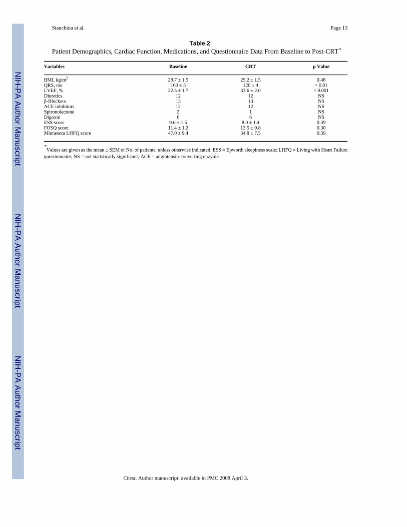
ResultsBetween April 2004 and August 2005, 65 patients who were being considered for implantationof CRT devices were approached about participation in this study. Of these, 36 patients declinedto participate, 3 patients did not ultimately require CRT, decompen-sated CHF requiringhospitalization developed in 1 patient prior to study enrollment, 1 patient left the state prior tostudy enrollment, and 2 patients were found to have OSA that had been previously treated(ie, prior to study enrollment). The remaining 24 patients were screened, and 13 patients (54%)met the inclusion criteria, including an AHI of > 5 events per hour of sleep. Demographics andpatient cardiac medications from baseline and after 6 months of CRT are presented in Tables1 and 2. OSA was predominant among all enrolled patients. One patient also had a Cheyne-Stokes respiration (CSR) pattern as well as a predominance of obstructive events. Six patientshad coronary artery disease, and seven patients had cardiomyopathy of nonischemic origin.After a mean (± SEM) duration of 6.6 ± 1.4 months of CRT, there was improvement in themean LVEF (baseline, 22.5 ± 1.7%; post-CRT, 33.6 ± 2.0%; p < 0.001) [Table 2]. The meanheart rate during sleep was 60.5 ± 1.2 beats/min at baseline, 64.4 ± 1.1 beats/min during theCRT-P, and 76.0 ± 1.3 beats/min during the overdrive CRT-P (p < 0.001 [for baseline andCRT-P compared to overdrive CRT-P]).
Sleep architecture showed no significant changes between baseline and the CRT night.However, there was a trend for an increase in slow-wave sleep noted in the overdrive CRTgroup, but the large variability in the measurements did not allow the increase to be statisticallysignificant. The AHI for each individual for each of the three PSGs is shown in Figure 1, and,as can be seen, there was some improvement in the AHI in 9 of 13 patients. CRT-associatedreductions in AHI for the group were characterized by an improvement in both CSAs and OSAs(Table 3). Hypopneas did not substantially change during the CRT-P night (Table 3). Mixedapneas were limited in number and did not change during the CRT-P or overdrive CRT-P. Themean spontaneous arousal index remained unchanged with CRT or overdrive pacing (Table3). The mean percentage of supine sleep was not increased from the baseline to CRT-P oroverdrive CRT-P night. The FOSQ increased insignificantly (11.4 ± 1.2 vs 13.5 ± 0.8,respectively; p = 0.30), and a small insignificant decrease was noted in Minnesota Living withHeart Failure scores with CRT (47.0 ± 9.4 vs 34.8 ± 7.5, respectively; p = 0.39). Epworthsleepiness scale scores were not abnormal at baseline and were not substantially impacted byCRT (9.6 ± 1.5 vs 8.0 ± 1.4, respectively) [Table 2]. Mean LFCT decreased from baseline of22.5 ± 1.5 to 19.6 ± 1.5 s with CRT (p = 0.01 [ANOVA]) but did not further decease withoverdrive CRT (19.1 ± 1.4 s; p = 0.91 [ANOVA]) [Table 3]. Linear regression analysis revealeda strong relationship between change in AHI and the change in LFCT with CRT (r = 0.89; p< 0.001) [Fig 2] but did not correlate as well with the change in LVEF (r = 0.46; p = 0.16).Last, in the six patients who had the greatest improvements in AHI with pacing, compared tothe whole group there were not significant differences in their BMI, LVEF, or sleeparchitecture.
DiscussionIn the present study, we observed in a group of CHF patients undergoing CRT that theprevalence of newly discovered OSA was 54%, which is consistent with previous reports, andthat with improvements in cardiac function and sleep-disordered breathing improved in mostpatients. The improvement in obstructive apneas correlated closely to the change in circulationtime, which is an indirect measure of cardiac output. Similar to studies with traditional (ie,non-CRT) pacing, CRT pacing with an increased rate did not further eliminate apneas andhypopneas. Heart failure symptoms tended toward improvement, but daytime sleepiness didnot. As others have noted, patients with CHF and incidentally diagnosed OSA were not
Stanchina et al. Page 4
Chest. Author manuscript; available in PMC 2008 April 3.
NIH
-PA Author Manuscript
NIH
-PA Author Manuscript
NIH
-PA Author Manuscript
particularly sleepy at trial entry, and there was limited change in these symptoms despiteimprovements in AHI.
The impact of overdrive pacing on sleep-disordered breathing has been highlighted in a seriesof articles with conflicting results. Garrigue and colleagues10 revealed that in bradycardicpatients with implanted right atrial-synchronous ventricular pacemakers and concomitantOSA/CSA atrial pacing at a rate 15 beats/min faster than the mean nocturnal heart rate led toa 60% reduction in AHI. These authors observed similar improvements in both OSAs andCSAs. However, two other studies failed to reproduce these results in patients withpredominantly obstructive apnea. Luthje and colleagues12 reported that, in a population ofpatients with dual-chamber pacemakers or implantable defibrillators, increased-rate pacing hadno impact on AHI, brain natriuretic peptide (BNP) levels, or urinary catechol-amine levels.Similarly, Simantirakis et al11 reported that neither 1 night nor up to 1 month of atrial increased-rate pacing impacted AHI. In the present study, we obtained significant reductions in AHI withCRT, but additional increased-rate pacing similarly made no significant impact on AHI. Thedifference between studies likely relates to the impact of the different interventions on cardiacoutput. In all of the patients in the study by Garrigue et al,10 the pacemaker was placed forsymptomatic bradycardia. Thus, with increased-rate pacing, greater improvements in cardiacfunction20 might be expected compared with those patients in the study by Simantirakis et al,11 in whom only a defibrillator was placed or who had no bradycardia. In addition, a veryslightly reduced ejection fraction (54 ± 11%) was observed in the study by Garrigue et al,10but a more pronounced impairment in LVEF (22 ± 1.7%) was present in our study by design.Therefore, improvements in cardiac function might be seen in both studies, but for differentreasons (ie, with either pacing or CRT)14; both would likely improve AHI. However, it is notclear that cardiac output increased in the studies by Garrigue et al,10 Simantirakis et al,11 orLuthje et al12 as no hemodynamic data were provided. The study by Simantirakas et al11included measures of BNP. However, the utility of BNP for measuring temporal changes incardiac function and filling pressures has been challenged.21 In the current study, theimprovement in AHI strongly correlated with this change in circulatory time, which is anindirect measure of cardiac output, and suggests that if cardiac output can be increased duringthe night, then apneas possibly may be reduced. Based on this logic, it was somewhat surprisingthat the overdrive CRT, utilizing a higher heart rate, did not further reduce the AHI. However,as noted above, the LFCT also did not change with use of the combined modality, suggestingthat circulatory delay/cardiac output was not further improved. In addition, mechanisms otherthan circulatory delay are also important for apnea generation in CHF patients.2
The etiology of the strong relationship between LFCT and sleep-disordered breathing in thisstudy remains unclear. As suggested by others,22,23 improvements in cardiac output mayimprove both CSA and OSA events by improving respiratory system control loop gain. Loopgain is the ratio of a response to the perturbation in systems controlled by negative feedbackloops. When the response of the system is greater than the initial stimulus, the system becomesunstable and oscillates. Reduced cardiac output increases ventilatory loop gain by prolonginglung-to-chemoreceptor delay, thus promoting respiratory system instability (ie, apneas).Mixing gain, which is a component of overall loop gain, is influenced by circulatory delay.24,25 This lung-to-chemoreceptor delay can be estimated by the LECT, which is a measurevalidated against cardiac output.18 We observed that our surrogate, LFCT, was closelyassociated with an improved change in AHI. There was also a relationship between LVEF andour LFCT, albeit a weak relationship that was likely limited by the study sample size. Althoughcirculation time is only one component of the respiratory system loop gain measure,8fluctuations in output from the central pattern generator in the brainstem are likely to lead topeaks and troughs in the output to the upper airway dilator muscles in these patients.Presumably, during the nadirs in upper airway dilator activity the susceptible airway isvulnerable to collapse, leading to obstructive apneas. This mechanism is also important in the
Stanchina et al. Page 5
Chest. Author manuscript; available in PMC 2008 April 3.
NIH
-PA Author Manuscript
NIH
-PA Author Manuscript
NIH
-PA Author Manuscript
development of CSR, a situation in which ventilatory loop gain is known to be increased. Sinhaand colleagues6 observed in patients with heart failure that CRT devices led to the eliminationof CSA/CSR. Increases in cardiac output were suggested by a significant decrease in theventilatory response to exercise measured by the minute ventilation/carbon dioxide outputratio, which is a surrogate for circulation time. Thus, circulatory delay and cardiac outputappear to be important in both OSA and CSA.
The lack of baseline sleepiness and the limited changes in both Epworth sleepiness scale andFOSQ data are not entirely surprising for the following reasons. First, previous studies havereported low (normal) mean Epworth sleepiness scale scores in their CHF patients with OSA,3 confirming the results of other studies26 reporting a low prevalence of sleepiness (24%) inCHF patients with sleep-disordered breathing. The mechanisms underlying this lack ofsleepiness are unclear but may include a lack of sensitivity for Epworth sleepiness scale scorein sedentary CHF patients, a gradual induction and tolerance to somnogenic cytokines (eg,interleukin-6 and tumor necrosis factor-α), or possibly an alerting influence ofcounterregulatory hormones (eg, cat-echolamines). Second, although there was a small butinsignificant change in the FOSQ and Epworth sleepiness scale scores with CRT, the residualOSAs may have impeded further improvement in the sleepiness scores. Although the changein sleepiness does correlate with the change in AHI, this is typically observed in studies inwhich the AHI is more normalized, which was not the case in most individuals in this study.Because residual AHI persisted, we do not advocate CRT as a “cure” for sleep apnea; rather,we believe that our findings point to interesting and potentially robust underlying physiologicmechanisms.
The impact of overdrive CRT on sleep architecture, arousals, and SaO2 should be noted. First,no significant change was seen in total sleep time, or in the percentages of stage 1/2, stage 3/4,or REM sleep between conditions. However, the amount of stage 3/4 sleep did trend upwardfrom the CRT-P to the overdrive CRT-P nights. The etiology or significance of this finding isunclear, but it was not associated with a decline in the AHI between the CRT-P and overdriveCRT-P nights. We speculate that with habituation to the polysomnography equipment, by thethird night the stage 3/4 sleep increased (analogous to the “first-night effect” in healthy controlsubjects). It is also possible that patients were mildly sleep deprived prior to the final study,potentially explaining the subtle changes in sleep stage distribution. Second, the spontaneousarousal index decreased from baseline to CRT polysomnography, but was not statisticallysignificant and thus did not likely contribute to the changes in apnea severity noted. Last, thelack of improvement in SaO2 nadir was somewhat unexpected. With improvements in LVfunction and declines in AHI, a higher nadir SaO2 might be expected. However, a moderatedegree of OSA remained in many of these individuals, which likely explains the persistenceof hypoxemia.
There are some limitations to this study. First, the order of our CRT and overdrive CRT PSGswere consecutive because of the nature of the CRT defibrillator being placed. The biasintroduced by order effects was likely small, as the impact on sleep architecture, arousals, andfunctional outcomes of sleep did not substantially change between studies. Second, the lung-to-finger measure was a modification of the previously validated LECT. However, this measurewas obtained using the same finger in each person for each study and was repeated in eachpatient during each study. Thus, we believe that the change in values over time withinindividuals is not biased and reflects a true change in circulation time. We have also comparedvalues derived from ear oximetry with those from finger oximetry, and have found only minorand predictable changes in circulatory time with the site of measurement. Third, repeat sleepstudies were not performed on the 11 patients who underwent an initial screeningpolysomnography but did not meet the study inclusion criteria. The lack of an untreated controlgroup possibly introduced regression to the mean bias on the patients studied. However, we
Stanchina et al. Page 6
Chest. Author manuscript; available in PMC 2008 April 3.
NIH
-PA Author Manuscript
NIH
-PA Author Manuscript
NIH
-PA Author Manuscript
feel that this is not a substantial problem for three reasons. First, the cut point for exclusionfrom the study was an AHI of 5 events per hour of sleep, and the observed drop in AHI was11 events per hour. Thus, our findings cannot be completely explained by regression to themean bias. Second, the correlation with circulation time supports the presence of a biologicalmechanism for the decrease in AHI, rather than a statistical artifact. Third, patients served astheir own control subjects, and the trend in AHI reduction was quite consistent rather than afunction of night-to-night variability.
A final limitation that should be addressed is our limited sample size. Because we regard thisas a pilot study, we strongly believe that larger controlled studies are necessary in the future.However, because we believe our findings are novel, we hope that they will encourage furtherresearch in this area, particularly with regard to potential new therapeutic targets (eg, mixinggain) in patients with OSA. Regardless, because our findings are significant for all of ourprimary outcome measures, we do not believe that our studies are underpowered for ourprespecified hypotheses. Indeed, we have calculated > 80% power with α = 0.05 to show adifference in AHI based on the observed sample size, mean values, and variance. However,we acknowledge that some of our exploratory analyses such as effects on sleep architectureand influences of ejection fraction may well yield different results in subsequent larger studies.
In conclusion, we found that CRT improved cardiac function and reduced AHI in patients withCHF and ventricular desynchrony, but yielded limited improvements in daytime sleepiness.The combination of CRT and increased-rate pacing had no further impact on the severity ofsleep-disordered breathing. These findings add to our understanding of OSA pathogenesis inCHF patients and suggest potential new therapeutic targets in OSA patients.
Acknowledgements
All authors had full access to all of the data in the study, and take responsibility for the integrity of the data and theaccuracy of the data analysis.
Drs. Stanchina and Ellison have received grant support from St. Jude Medical for this work. Dr. Malhotra is fundedby a National Institute of Aging Beeson Award (AG024837–01), National Institutes of Health grant RO1-HL73146–01, and SCOR Project 1; and serves as a consultant to Restore Medical, Inspiration Medical, Respironics, Inc, NMTMedical, and Pfizer. All other authors have reported to the ACCP that no significant conflicts of interest exist withany companies/organizations whose products or services may be discussed in this article.
AbbreviationsAHI
apnea-hypopnea index
ANOVA analysis of variance
BMI body mass index
BNP brain natriuretic peptide
CHF congestive heart failure
CRT cardiac resynchronization therapy
CRT-P
Stanchina et al. Page 7
Chest. Author manuscript; available in PMC 2008 April 3.
NIH
-PA Author Manuscript
NIH
-PA Author Manuscript
NIH
-PA Author Manuscript
cardiac resynchronization therapy with repeat polysomnography
CSA central sleep apnea
CSR Cheyne-Stokes respiration
FOSQ functional outcomes of sleep questionnaire
LECT lung-to-ear circulation time
LFCT lung-to-finger circulation time
LV left ventricular
LVEF left ventricular ejection fraction
OSA obstructive sleep apnea
PSG polysomnogram
SaO2 arterial oxygen saturation
References1. Shahar E, Whitney CW, Redline S, et al. Sleep-disordered breathing and cardiovascular disease: cross-
sectional results of the Sleep Heart Health Study. Am J Respir Crit Care Med 2001;163:19–25.[PubMed: 11208620]
2. Leung RST, Bradley TD. Sleep apnea and cardiovascular disease. Am J Respir Crit Care Med2001;164:2147–2165. [PubMed: 11751180]
3. Kaneko Y, Floras J, Usui K, et al. Cardiovascular effects of continuous positive airway pressure inpatients with heart failure and obstructive sleep apnea. N Engl J Med 2003;348:1233–1241. [PubMed:12660387]
4. Bradley T, Logan A, Kimoff R, et al. Continuous positive airway pressure for central sleep apnea andheart failure. N Engl J Med 2005;353:2025–2033. [PubMed: 16282177]
5. Solin P, Bergin P, Richardson M, et al. Influence of pulmonary capillary wedge pressure on centralapnea in heart failure. Circulation 1999;99:1574–1579. [PubMed: 10096933]
6. Sinha AM, Skobel EC, Breithardt OA, et al. Cardiac resynchronization therapy improves central sleepapnea and Cheyne-Stokes respiration in patients with chronic heart failure. J Am Coll Cardiol2004;44:68–71. [PubMed: 15234409]
7. Wellman A, Jordan AS, Malhotra A, et al. Ventilatory control and airway anatomy in obstructive sleepapnea. Am J Respir Crit Care Med 2004;170:1225–1232. [PubMed: 15317668]
8. Younes M. Contributions of upper airway mechanics and control mechanisms to severity of obstructiveapnea. Am J Respir Crit Care Med 2003;168:645–658. [PubMed: 12773321]
9. Younes M, Ostrowski M, Thompson W, et al. Chemical control stability in patients with obstructivesleep apnea. Am J Respir Crit Care Med 2001;163:1181–1190. [PubMed: 11316657]
Stanchina et al. Page 8
Chest. Author manuscript; available in PMC 2008 April 3.
NIH
-PA Author Manuscript
NIH
-PA Author Manuscript
NIH
-PA Author Manuscript
10. Garrigue S, Bordier P, Jais P, et al. Benefit of atrial pacing in sleep apnea syndrome. N Engl J Med2002;346:404–412. [PubMed: 11832528]
11. Simantirakis EN, Schiza SE, Chrysostomakis SI, et al. Atrial overdrive pacing for the obstructivesleep apnea-hypopnea syndrome. N Engl J Med 2005;353:2568–2577. [PubMed: 16354893]
12. Luthje L, Unterberg-Buchwald C, Dajani D, et al. Atrial overdrive pacing in patients with sleep apneaand implanted pacemaker. Am J Respir Crit Care Med 2005;172:118–122. [PubMed: 15750043]
13. Abraham WT, Fisher WG, Smith AL, et al. Cardiac resynchronization in chronic heart failure. N EnglJ Med 2002;346:1845–1853. [PubMed: 12063368]
14. Bristow MR, Saxon LA, Boehmer J, et al. Cardiac resynchronization therapy with or without animplantable defibrillator in advanced chronic heart failure. N Engl J Med 2004;350:2140–2150.[PubMed: 15152059]
15. Rechtschaffen, A.; Kales, A. A manual of standardized terminology, techniques and scoring systemfor sleep stages of human subjects. Los Angeles, CA: Brain Information Service/ Brain ResearchInstitute, UCLA; 1968.
16. American Sleep Disorders Association. EEG arousals: scoring rules and examples: a preliminaryreport from the Sleep Disorders Atlas Task Force of the American Sleep Disorders Association. Sleep1992;15:173–184. [PubMed: 11032543]
17. American Academy of Sleep Medicine. Sleep-related breathing disorders in adults: recommendationsfor syndrome definition and measurement techniques in adults. Sleep 1999;22:667–689. [PubMed:10450601]
18. Hall MJ, Xie A, Rutherford R, et al. Cycle length of periodic breathing in patients with and withoutheart failure. Am J Respir Crit Care Med 1996;154:376–381. [PubMed: 8756809]
19. Ryan CM, Bradley TD. Periodicity of obstructive sleep apnea in patients with and without heartfailure. Chest 2005;127:536–542. [PubMed: 15705993]
20. Stein E, Damato AN, Kosowsky BD, et al. The relation of heart rate to cardiovascular dynamics:pacing by atrial electrodes. Circulation 1966;33:925–932. [PubMed: 5940519]
21. O’Neill JO, Bott-Silverman CE, McRae AT, et al. B-type natriuretic peptide levels are not a surrogatemarker for invasive hemodynamics during management of patients with severe heart failure. AmHeart J 2005;149:363–369. [PubMed: 15846278]
22. Wellman A, Malhotra A, White DP. Atrial pacing in sleep apnea syndrome. N Engl J Med2002;347:445–446. [PubMed: 12167690]
23. Floras J, Bradley TD. Atrial overdrive pacing for sleep apnea: a door now closed? Am J Respir CritCare Med 2005;172:1–3. [PubMed: 15980105]
24. Malhotra A, Jordan AS. Did fat boy Joe need hormone replacement therapy? Sleep 2006;29:16–18.[PubMed: 16453975]
25. Eckert D, Jordan AS, Merchia P, et al. Central sleep apnea: pathophysiology and treatment. Chest2007;131:595–607. [PubMed: 17296668]
26. Javaheri S, Parker TJ, Liming JD, et al. Sleep apnea in 81 ambulatory male patients with stable heartfailure. Circulation 1998;97:2154–2159. [PubMed: 9626176]
Stanchina et al. Page 9
Chest. Author manuscript; available in PMC 2008 April 3.
NIH
-PA Author Manuscript
NIH
-PA Author Manuscript
NIH
-PA Author Manuscript
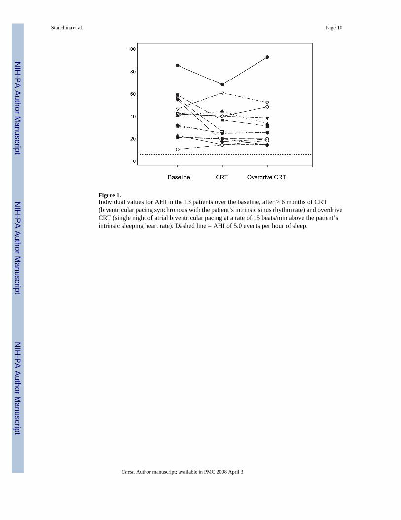
Figure 1.Individual values for AHI in the 13 patients over the baseline, after > 6 months of CRT(biventricular pacing synchronous with the patient’s intrinsic sinus rhythm rate) and overdriveCRT (single night of atrial biventricular pacing at a rate of 15 beats/min above the patient’sintrinsic sleeping heart rate). Dashed line = AHI of 5.0 events per hour of sleep.
Stanchina et al. Page 10
Chest. Author manuscript; available in PMC 2008 April 3.
NIH
-PA Author Manuscript
NIH
-PA Author Manuscript
NIH
-PA Author Manuscript
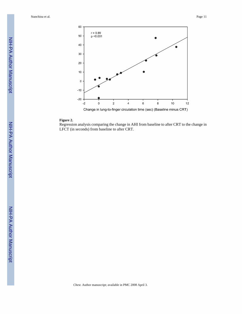
Figure 2.Regression analysis comparing the change in AHI from baseline to after CRT to the change inLFCT (in seconds) from baseline to after CRT.
Stanchina et al. Page 11
Chest. Author manuscript; available in PMC 2008 April 3.
NIH
-PA Author Manuscript
NIH
-PA Author Manuscript
NIH
-PA Author Manuscript

NIH
-PA Author Manuscript
NIH
-PA Author Manuscript
NIH
-PA Author Manuscript
Stanchina et al. Page 12
Table 1Baseline Characteristics/Demographics*
Demographics Values (n = 13)
Age, yr 68.6 ± 3.7Gender Male 11 Female 2CMP Ischemic 6 Nonischemic 7
*Values are given as the mean ± SEM or No. of patients. CMP = cardiomyopathy.
Chest. Author manuscript; available in PMC 2008 April 3.
NIH
-PA Author Manuscript
NIH
-PA Author Manuscript
NIH
-PA Author Manuscript
Stanchina et al. Page 13
Table 2Patient Demographics, Cardiac Function, Medications, and Questionnaire Data From Baseline to Post-CRT*
Variables Baseline CRT p Value
BMI, kg/m2 28.7 ± 1.5 29.2 ± 1.5 0.48QRS, ms 160 ± 5 120 ± 4 < 0.01LVEF, % 22.5 ± 1.7 33.6 ± 2.0 < 0.001Diuretics 12 12 NSβ-Blockers 13 13 NSACE inhibitors 12 12 NSSpironolactone 2 1 NSDigoxin 6 6 NSESS score 9.6 ± 1.5 8.0 ± 1.4 0.39FOSQ score 11.4 ± 1.2 13.5 ± 0.8 0.30Minnesota LHFQ score 47.0 ± 9.4 34.8 ± 7.5 0.39
*Values are given as the mean ± SEM or No. of patients, unless otherwise indicated. ESS = Epworth sleepiness scale; LHFQ = Living with Heart Failure
questionnaire; NS = not statistically significant; ACE = angiotensin-converting enzyme.
Chest. Author manuscript; available in PMC 2008 April 3.
NIH
-PA Author Manuscript
NIH
-PA Author Manuscript
NIH
-PA Author Manuscript
Stanchina et al. Page 14
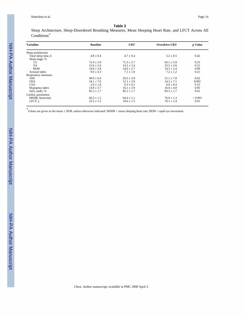
Table 3Sleep Architecture, Sleep-Disordered Breathing Measures, Mean Sleeping Heart Rate, and LFCT Across AllConditions*
Variables Baseline CRT Overdrive CRT p Value
Sleep architecture Total sleep time, h 4.8 ± 0.4 4.7 ± 0.4 5.2 ± 0.5 0.42 Sleep stage, % 1/2 72.4 ± 3.9 71.3 ± 3.7 63.1 ± 5.9 0.35 3/4 12.6 ± 2.6 14.5 ± 3.4 23.5 ± 5.6 0.21 REM 14.9 ± 2.8 14.6 ± 2.7 14.5 ± 2.4 0.99 Arousal index 9.0 ± 4.3 7.1 ± 1.0 7.2 ± 1.2 0.21Respiratory measures AHI 40.9 ± 6.4 29.5 ± 5.9 31.1 ± 7.8 0.02 OSA 24.1 ± 7.0 12.1 ± 5.0 14.3 ± 7.1 0.001 CSA 2.0 ± 1.0 0.3 ± 0.1 0.6 ± 0.4 0.15 Hypopnea index 14.8 ± 3.7 16.1 ± 3.9 16.0 ± 4.0 0.95 SaO2 nadir, % 82.3 ± 1.7 82.1 ± 1.7 83.3 ± 1.7 0.61Cardiac parameters MSHR, beats/min 60.5 ± 1.2 64.4 ± 1.1 76.0 ± 1.3 < 0.001 LFCT, s 22.5 ± 1.5 19.6 ± 1.5 19.1 ± 1.4 0.01
*Values are given as the mean ± SEM, unless otherwise indicated. MSHR = mean sleeping heart rate; REM = rapid eye movement.
Chest. Author manuscript; available in PMC 2008 April 3.
Related Documents


