Tumor cell proliferation and immunotheray of cancers Professor M. C. Bansal M.B.B.S. , M.S. , F.I.C.O.G , M.I.C.O.G

Welcome message from author
This document is posted to help you gain knowledge. Please leave a comment to let me know what you think about it! Share it to your friends and learn new things together.
Transcript

Tumor cell proliferation and immunotheray of cancers
Professor M. C. BansalM.B.B.S. , M.S. , F.I.C.O.G , M.I.C.O.G

CELL CYCLE
M: MITOTIC PHASE i.e. cell division
G1: post mitotic phase
( protein & RNA synthesis)
S: sythetic phase ( DNA synthesis)
G2: post synthetic phase (Nucleus has diploid no. chromosomes)
G0: resting phase ( cell may go in or go out of mitosis)

Cell Cycle Information on growth patterns and doubling times
relates to the growth of the tumor mass as a whole. The kinetic behavior of individual tumor cells has been well described, and a classic cell cycle model has been produced .
M phase (mitotic phase) of the cell cycle is the phase of cell division.
G1 phase (postmitotic phase) is a period of variable duration when cellular activities and protein and RNA synthesis continue. These G1 cells can differentiate or continue in the proliferative cycle.

S phase (DNA synthetic phase) is the period in which new DNA replication occurs.
G2 phase (postsynthetic phase) is the period in which the cell has a diploid number of chromosomes and twice the DNA content of the normal cell. The cell remains in this phase for a relatively short time and then enters the mitotic phase again.
G0 phase (the resting phase) is the time during which cells do not divide. Cells may move in and out of the G0 phase.

The generation time is the duration of the cycle from M phase to M phase. Variation occurs in all phases of the cell cycle, but the variation is greatest during the G1 period. The reasons for this variation are complex and not completely understood.

These cell cycle events have important implications for the cancer therapist.
Differential sensitivities to chemotherapy and radiation therapy are associated with different proliferative states.
Dividing cancer cells that are actively traversing the cell cycle are very sensitive to chemotherapeutic agents.

Cells in a resting state (G0) are relatively insensitive to chemotherapeutic agents, although they occupy space and contribute to the bulk of the tumor.

Cell Kinetics
In cell kinetic studies performed on human tumors, the duration of the S phase (DNA synthesis phase) is relatively similar for most human tumors, ranging from a low of 10 hours to a high of approximately 31 hours.
The length of the cell cycle in human tumors varies from slightly more than half a day to perhaps 5 days.
With cell cycle times in the range of 24 hours and doubling times in the range of 10 to 1,000 days, it is clear that only a small proportion of tumor cells are in active cell division at any one time.

Two major factors that affect the rate at which tumorsgrow are the growth fraction and cell death.
The growth fraction is the number of cells in the tumor mass that are actively undergoing cell division.
There is a marked variation in the growth fraction of tumors in human beings, ranging from 25% to almost 95%.

In the past, it was thought that human tumorscontained billions of cells, all growing slowly.
In actuality, only a small fraction of cells in a tumormass are rapidly proliferating; the remainder are out of the cell cycle and quiescent.
Cancer “stem cells” are a very small population of cells that appear to be relatively chemoresistant; these play a major role in the development and progression of cancers.
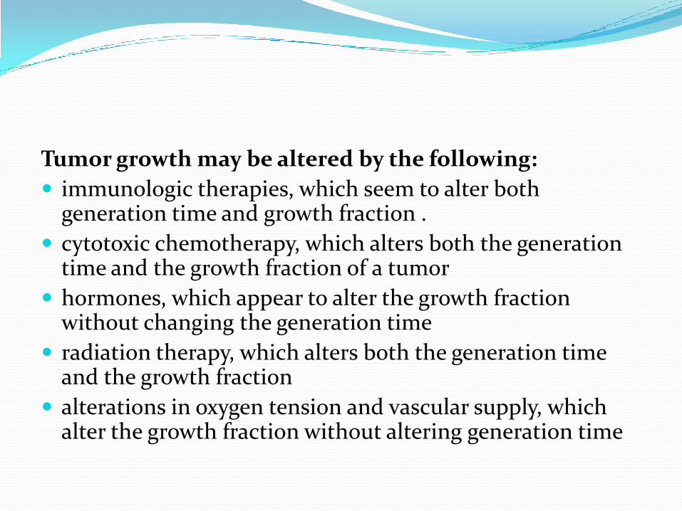
Tumor growth may be altered by the following:
immunologic therapies, which seem to alter both generation time and growth fraction .
cytotoxic chemotherapy, which alters both the generation time and the growth fraction of a tumor
hormones, which appear to alter the growth fraction without changing the generation time
radiation therapy, which alters both the generation time and the growth fraction
alterations in oxygen tension and vascular supply, which alter the growth fraction without altering generation time


INTRODUCTION Cancer is caused by a series of events that include the
accumulation of successive molecular lesions and alterations in the tumor microenvironment .
Molecular lesions include
overexpression,
amplification,
mutations of oncogenes;
deletion of tumor suppressor genes;
inappropriate expression of growth factors
and their cellular receptors.

In addition to these molecular changes, angiogenesis and the lack of effective host antitumor immune responses create a microenvironment that supports the growth of cancer .
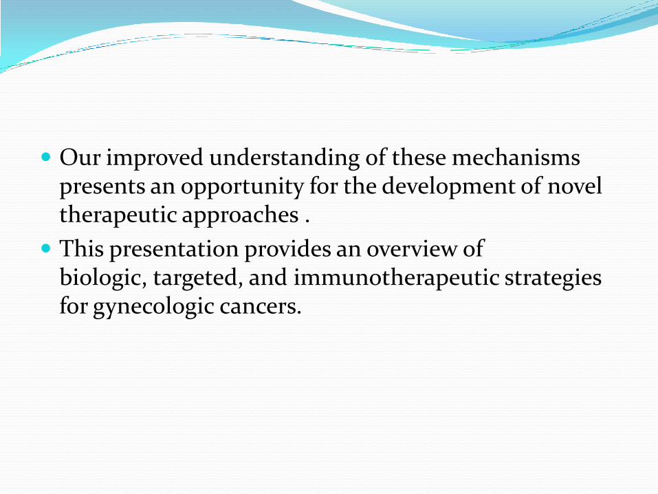
Our improved understanding of these mechanisms presents an opportunity for the development of novel therapeutic approaches .
This presentation provides an overview of biologic, targeted, and immunotherapeutic strategies for gynecologic cancers.

Biologic and Targeted Therapies The growth of cancer cells is crucially dependent on
oncogenic signal transduction pathways.
Extracellular signals are transmitted to the cancer cell via transmembrane receptors.
Activation of the epidermal growth factor receptors (EGFR, HER2, HER3, and HER4), for example, stimulates a cascade of intracellular proteins that ultimately lead to changes in gene expression.
Novel therapeutics are targeted to modulate these signal transduction pathways by blocking the extracellular transmembrane receptors or interfering with intracellular proteins such as tyrosine kinases further downstream.

This novel therapeutic approach is also termed molecular targeting .
It is accomplished by either monoclonal antibodies that bind to transmembrane receptors and serum proteins such as vascular endothelial growth factor (VEGF) or chemical, small-molecule inhibitors that prevent activation of signal transduction proteins.
Targeting the signaling cascade inhibits the proliferation of cancer cells, induces apoptosis, and blocks metastasis.
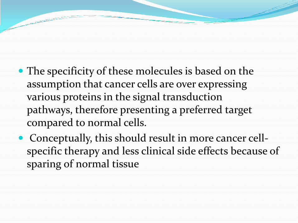
The specificity of these molecules is based on the assumption that cancer cells are over expressing various proteins in the signal transduction pathways, therefore presenting a preferred target compared to normal cells.
Conceptually, this should result in more cancer cell-specific therapy and less clinical side effects because of sparing of normal tissue


Angiogenesis
The formation of new blood vessels (neoangiogenesis) is a normal process during embryonic development, tissue remodeling, and wound healing .
Malignant tumors are able to induce angiogenesis by secreting paracrine factors that promote the formation of new blood vessels.
Angiogenesis is a complex process that is influenced by various pro-and antiangiogenic factors, including VEGF, interleukin 8, platelet-derived endothelial cell growth factor, and angiopoietins.
Overexpression of these angiogenic factors leads to neovascularization and increased supply of nutrients and oxygen to the tumor.

Three main therapeutic strategies that target angiogenesis are currently being explored for the treatment of cancer patients .
One group of agents targets VEGF (e.g., bevacizumab, VEGF-Trap), the second group prevents VEGF from binding to its receptor (pertuzumab), and a third group of agents inhibits tyrosine kinase activation and downstream signalingin the angiogenesis signaling cascade (valatanib, sunitenib).

Vascular Endothelial Growth Factor
VEGF is overexpressed in gynecologicmalignancies, therefore presenting an excellent target for therapy .
Inhibition of VEGF-induced angiogenic signalingdecreases tumor microvascular density and causes death of solid tumors in various preclinical models.
Several agents are now available for clinical use; all target the VEGF signaling pathway.
The most widely used agent at this time is bevacizumab, a humanized, recombinant monoclonal antibody that binds to all isoforms of VEGF-A.
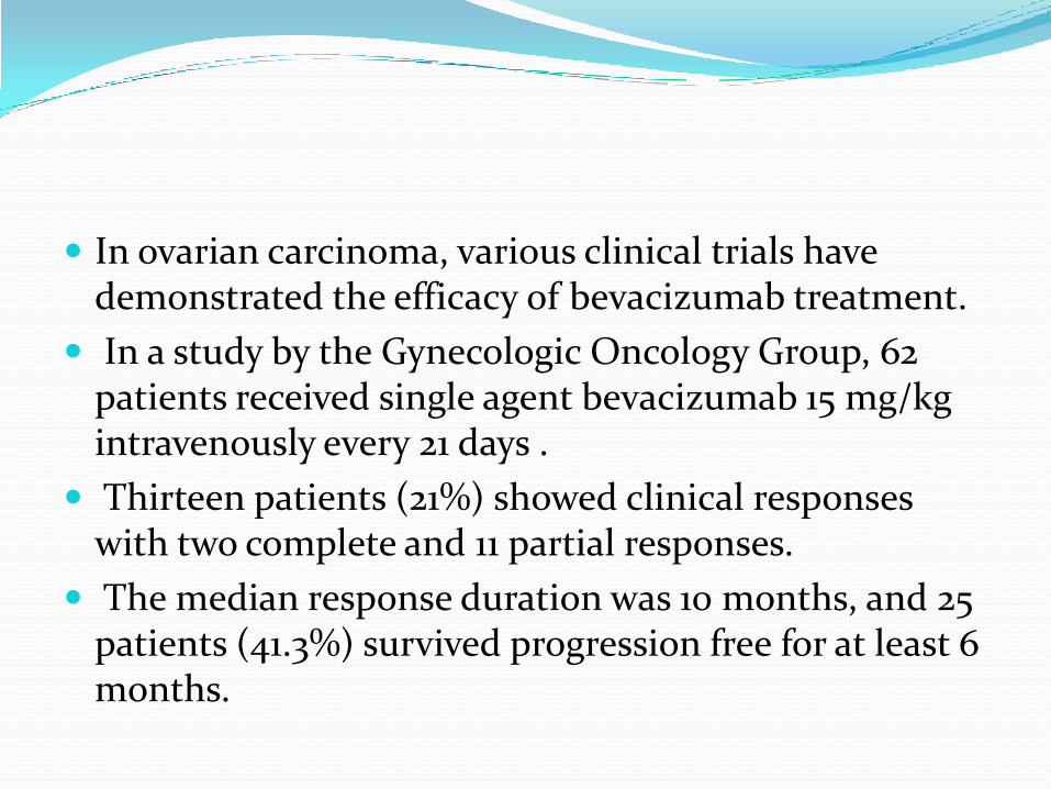
In ovarian carcinoma, various clinical trials have demonstrated the efficacy of bevacizumab treatment.
In a study by the Gynecologic Oncology Group, 62 patients received single agent bevacizumab 15 mg/kg intravenously every 21 days .
Thirteen patients (21%) showed clinical responses with two complete and 11 partial responses.
The median response duration was 10 months, and 25 patients (41.3%) survived progression free for at least 6 months.
Bevacizumab has also been used in combination with other agents.
In a phase II study of 13 patients with recurrent ovarian or primary peritoneal carcinoma, combination treatment with bevacizumab (15 mg/kg i.v. every 21 days) and erlotinib (150 mg/day orally) resulted in one complete response and one partial response for a total response rate of 15% .
Seven patients had stable disease.
Another trial investigated the combination of bevacizumab(10 mg/kg every 14 days) and oral cyclophosphamide (50 mg/day orally) in 70 patients with recurrent ovarian cancer.

The Gynecologic Oncology Group has initiated a clinical trial that will evaluate the addition of bevacizumab to first-line chemotherapy after primary tumor debulking.
A similar trial by the Gynecologic Cancer InterGroup is designed to evaluate the safety and efficacy of adding bevacizumab to standard chemotherapy (carboplatinand paclitaxel) in patients with advanced epithelial ovarian or primary peritoneal cancer .

Epidermal Growth Factor Receptor Inhibitors The epidermal growth factor receptor family consists
of four members including EGFR (HER1), HER2, HER3, and HER4 .
EGFR overexpression has been reported in 35% to 70% of patients with epithelial ovarian cancer .
In endometrial cancer, EGFR is overexpressed in 43% to 67% of tumors and is associated with shortened disease-free and overall survival .
In addition, amplification of the HER2 gene is commonly found in endometrial carcinoma.

Various agents directed against epidermal growth factor receptors are available .
Trastuzumab is a humanized monoclonal antibody that binds to the extracellular domain of HER2 .
Blockade of HER2 affects various molecules that ultimately decreases cell proliferation.
Pertuzumab is another humanized monoclonal antibody that binds to a different epitope of HER2 compared to trastuzumab.
Binding to HER2 prevents dimerization of the receptor, which is required for its function .


Epidermal Growth Factor Receptor Inhibition of EGFR signaling is accomplished by using
either monoclonal antibodies against the extracellular receptor or small-molecule inhibitors against the intracellular kinase domain.
Both strategies results in inhibition of phosphorylation or receptor activation.
Erlotinib is a potent reversible inhibitor of EGFR tyrosine kinase that blocks receptor autophosphorylation and has been used for the treatment of ovarian carcinoma.

Erlotinib has been used in combination with docetaxel and carboplatin as first-line treatment after surgical cytoreduction in patients with ovarian, fallopian tube, and primary peritoneal cancers .
Cetuximab (C225, Erbitux) is a chimerized monoclonal antibody against EGFR.
Cetuximab in combination with carboplatin resulted in three complete (10.7%) and six partial (21.4%) responses in 28 patients with recurrent ovarian cancer.
Twenty-six of these 28 patients (92.8%) had EGFR-positive tumors.
The combination of paclitaxel, carboplatin, and cetuximab for first-line chemotherapy of stage III ovarian cancer patients resulted in progression-free survival of 14.4 months and was therefore not significantly prolonged compared to historical data.
Gefitinib (ZD1839 Iressa) is a low molecular weight quinazoline derivative that inhibits the activation of EGFR tyrosine kinase via competitive binding of the ATP-binding domain of the receptor.
Treatment of patients with recurrent ovarian cancer using the combination of gefitinib, carboplatin, and paxitaxelresulted in a high overall response rate of 63% .
Interestingly, antitumor responses were observed in 35% of patients with platinum-resistant disease compared to a 73% response rate in patients with platinum-sensitive disease.

Gefitinib has also been used in combination with tamoxifen.
In squamous and adenocarcinoma of the cervix, gefitinib(500 mg/day) treatment resulted in disease stabilization in six of 28 patients (20%) but no clinical responses .
Lapatinib is a small-molecule inhibitor of both the HER2 and EGFR tyrosine kinase receptor.
The rationale for using lapatinib in endometrial carcinoma is supported mainly by studies in human cancer cell lines. Its efficacy in endometrial cancer is being investigated currently in clinical trials .

HER-2/neu The HER-2/neu receptor is activated by homo- or
heterodimerization, resulting in tyrosine phosphorylationand subsequent activation of various downstream signals that among other functions control cellular proliferation, migration, and invasion.
Trastuzumab is a recombinant, humanized IgG1 monoclonal antibody that is specific for the extracellular domain of HER-2/neu.
Binding of the antibody to HER-2/neu prevents activation of the receptor with a subsequent increase of apoptosis in vitro and in vivo, impaired DNA damage repair, and inhibition of tumor neovascularization.
The HER-2/neu oncogene is overexpressed in several gynecologic malignancies, including 20% to 30% of ovarian cancers.
HER2/neu overexpression is infrequent in cervical cancer.
In uterine papillary serous carcinoma, 12 of 68 (18%) tumors showed HER2/neu overexpression; this was associated with a worse overall prognosis .
Mitogen-Activated Protein Kinase Pathways
The mitogen-activated protein (MAP) kinase cascades are activated by various cofactors, inflammatory cytokines, and stress.
Sorafenib is among the first of the agents with clinically proven efficacy. Sorafenib is a competitive inhibitor of raf that has been approved for treatment of renal cell carcinoma and hepatocellular carcinoma.
Besides targeting raf, sorafenib also inhibits VEGFR2 and VEGFR3, FT3, c-kit, and PDGFR-β.
The PI3-kinase/Akt/mTOR Pathway The phosphoinositide3-kinase (PI3-kinase)/Akt/mTOR
pathway is a major oncogenic signaling pathway in various cancers.
Activation of this pathway can be demonstrated in more than 80% of endometrial cancers, 50% to 70% of epithelial ovarian cancers, and approximately 50% of cervical cancers.
Several inhibitors of PI3-kinase/Akt/mTOR signaling are currently in clinical trials.
Rapamycin or rapamycin analogues, for example, block the activity of mTOR, a protein complex responsible for increasing protein synthesis and cellular proliferation.

Several mTOR inhibitors, including RAD001 and CCI779, and specific PI3-kinase inhibitors are currently under development in preclinical models and clinical trials. PI3-kinase/Akt/mTOR inhibitors have been used in endometrial cancer with limited benefit.

Immunotherapy Failure of functional immunity contributes to the
genesis of virus-associated cancers, such as those caused by human papilloma virus (HPV) or Epstein-Barr virus.
The greatest success story involving the enhancement of immunity to combat gynecologic cancer is the development of vaccines against HPV, which are highly effective for the prevention of cervical dysplasia and cancer .
Some researchers suggest that immune responses are mainly involved in protection from virus-associated cancers but not other forms of cancer .
Cancer is a common disease, and overt immune deficiency certainly is not necessary for its development.
However, recent studies have shown that many cancers, including those that are not known to have a viral etiology, are seen with increased frequency in patients who have dysfunctional immunity like as in patients of HIV.

In a recent metaanalysis of cancer incidence in populations known to be immune deficient (e.g., organ-transplant recipients, patients with HIV infection), Grulich and co-workers found an increased incidence of several common cancers, suggesting that impaired immunity can contribute to the development of cancer.

Components of the Immune System Involved in Antitumor Responses Various types of human immune responses can target
tumor cells.
Immune responses can be categorized as humoral or cellular, a distinction based on the observation in experimental systems that some immune responses could be transferred by serum (humoral) and others by cells (cellular).
In general, humoral responses refer to antibody responses; antibodies are antigen-reactive, soluble, bifunctional molecules composed of specific antigen-binding sites.

Cellular immune responses generally refer to cytotoxicresponses mediated directly by activated immune cells rather than by the production of antibodies .
Nearly all immune responses involve both humoraland cellular components and require the coordinated activities of populations of lymphocytes operating in concert with each other and with antigen-presenting cells.


Therapeutic Strategies There is great interest in developing effective biologic
and immune therapies for gynecologic malignancies.
For example, patients with small-volume or microscopic residual peritoneal ovarian cancer are attractive candidates for immunotherapy or biologic therapy, especially approaches based on regional peritoneal immunotherapy or biotherapy.
Also, many patients with advanced disease are immunocompromised, suggesting a role for immuneenhancing therapeutic approaches.

Dysplastic and cancerous cervical epithelial cells infected with HPV, an oncogenic virus, also present an attractive target for immune enhancement-based therapeutic strategies, including the development of therapeutic vaccines for HPV.
Advances in molecular biology, biotechnology, immunology, and cytokine biology have resulted in the availability of many new, promising immunotherapeutic approaches for gynecologic cancers.

Human Papillomaviruses and Cervical Neoplasia
Extensive molecular biologic and epidemiologic research confirms certain HPV types to be carcinogenic in humans (13,14,15,79,80,81,82,83,84,85).
Infections with HPV cause approximately 5% of the global burden of human cancers and at least 500,000 deaths annually.
Infection with specific HPV types is necessary for the development of the vast majority of cervical cancers (>99.7%) and the immediate precursor lesion (CIN 3) .
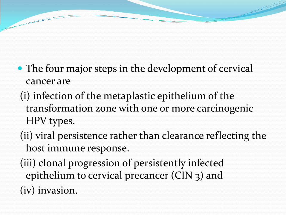
The four major steps in the development of cervical cancer are
(i) infection of the metaplastic epithelium of the transformation zone with one or more carcinogenic HPV types.
(ii) viral persistence rather than clearance reflecting the host immune response.
(iii) clonal progression of persistently infected epithelium to cervical precancer (CIN 3) and
(iv) invasion.

Figure; Major steps in the development of cervical cancer. Incident HPV infection is best measured by molecular tests. Most HPV infections show no concurrent cytological abnormality. Approximately 30% of infections produce concurrent cytopathology, usually non-classical (equivocal) changes. Most HPV infections clear within 2 years. Ten percent persist for 2 years and are highly linked to development of precancer.
Human Papillomavirus and Cervical Cancer: A Causal or Casual Association
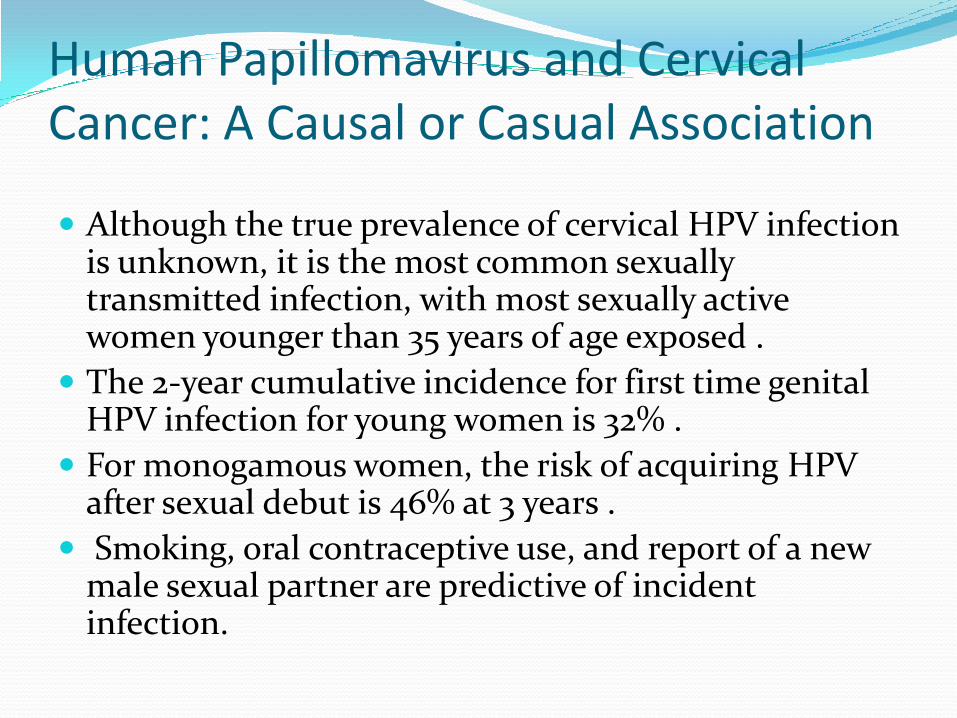
Although the true prevalence of cervical HPV infection is unknown, it is the most common sexually transmitted infection, with most sexually active women younger than 35 years of age exposed .
The 2-year cumulative incidence for first time genital HPV infection for young women is 32% .
For monogamous women, the risk of acquiring HPV after sexual debut is 46% at 3 years .
Smoking, oral contraceptive use, and report of a new male sexual partner are predictive of incident infection.

The median time to clearance of HPV infection in an immunocompetent woman is 6 to 18 months (average of 12 months) with 90% of women clearing a specific HPV-type after 2 years of observation .
Viral clearance is not often associated with reappearance of the same HPV type.
Occasionally the same HPV type will reappear .
It is unclear whether this represents reinfection or resurgence from a latent state in the basal cells of the epithelium.
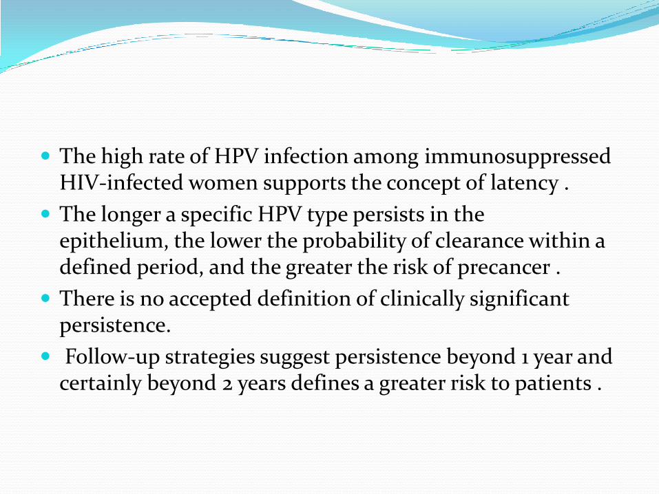
The high rate of HPV infection among immunosuppressedHIV-infected women supports the concept of latency .
The longer a specific HPV type persists in the epithelium, the lower the probability of clearance within a defined period, and the greater the risk of precancer .
There is no accepted definition of clinically significant persistence.
Follow-up strategies suggest persistence beyond 1 year and certainly beyond 2 years defines a greater risk to patients .
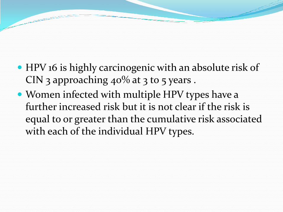
HPV 16 is highly carcinogenic with an absolute risk of CIN 3 approaching 40% at 3 to 5 years .
Women infected with multiple HPV types have a further increased risk but it is not clear if the risk is equal to or greater than the cumulative risk associated with each of the individual HPV types.
Cervical neoplasia can be viewed as the result of a complex interplay between a “seed,” that is, high-risk HPV types, and a “soil,” that is, the immature, metaplasticepithelium of the cervical transformation zone.
Exposure to specific high-risk HPV types, in the presence of cofactor activity, may deviate the metaplastic process along a neoplastic pathway.
Disease expression begins at the new squamocolumnarjunction.
The initial abnormality produced is usually a low-grade cervical lesion.

The most critical step in cervical carcinogenesis is not acquisition of an HPV infection but progression to CIN 3.
HPV infection alone is necessary but not sufficient to induce carcinoma in an immunocompetent host.
HPV infection with oncogenic viral types is much more common than cervical neoplasia, indicating the necessity of cofactors in the process of cervical carcinogenesis .

Cofactor Interaction with Human Papillomavirus Cigarrete smoking
Infection with other microbial agents - An increased incidence of other sexually transmitted diseases has been reported in association with genital HPV infection and cervical neoplasia.
Sex hormone influence -Condylomata acuminataincrease rapidly in size and number in pregnancy. This could suggest that maternal estrogen status is permissive for HPV replication.CIN and cervical cancer are more frequently found in women with increased parity and in women on oral contraceptives independent of sexual activity .

Exogenous and Endogenous Immunosuppression-Iatrogenic induction of immunosuppression in renal transplant recipients increases the rate of CIN to 16 times that of the general community .
Dietary Factors -Dietary deficiencies of vitamin A or beta-carotene may increase the risk of CIN and cervical cancer .

The HPV genome consists of approximately 8,000 base pairs of single-stranded, circular DNA.
HPV genes are designated as E or L according to their expression in early or late differentiation stage of the epithelium: E1, E2, E5, E6, and E7 are expressed early in the differentiation,
E4 is expressed throughout
L1 and L2 are expressed during the final stages of differentiation.

The viral genome is maintained at the basal layer of the epithelium, where HPV infection is established.
Early proteins are expressed at low levels for genome maintenance (raising the possibility of a latent state) and cell proliferation.
As the basal epithelial cells differentiate, the viral life cycle enters successive stages of genome amplification, virus assembly and virus release, with a concomitant shift in expression patterns from early genes to late genes, including L1 and L2, which assemble into viral capsid.


The proteins encoded by the E6 and E7 genes of high-risk HPV types, particularly HPV 16 and 18 , are directly involved in cellular transformation in the presence of an active oncogene .
E6 and E7 are the primary HPV oncoproteins with numerous cellular targets .
Both E6 and E7 proteins can immortalize primary keratinocytes from cervical epithelium and can influence transcription from viral and cellular promoters .
The activity of these viral oncoproteins results in genomic instability, leading to the malignant phenotype.
E6 proteins of high-risk HPV types bind the tumorsuppressor protein p53 .
This induces ubiquitination and degradation of p53, removing the p53-dependent control of the host cell cycle .
The role of E6 as an antiapoptotic protein is of key significance in the development of cervical cancer as it compromises the effectiveness of the cellular DNA damage response and allows the accumulation of secondary mutations to go unchecked.
E6 also increases telomerase activity in keratinocytesthrough increased transcription of the telomerase catalytic subunit gene (hTERT) through induction of c-myc .
E7 is responsible for inactivation of product of Rb gene (tumor supressor)
Degradation of p53 by E6 and the functional inactivation of pRb by E7 represent the main mechanisms leading to malignant changes.

Human Papillomavirus Vaccines
The HPV vaccine is a major scientific and public health advance in the prevention of cervical cancer .
Most invasive cervical cancers and CIN lesions are attributable to high-risk HPV infection .
A prophylactic or therapeutic vaccine against HPV has the potential to have a substantial impact on HPV infection and rates of CIN and cervical cancer .
HPVs cannot be grown in the laboratory so standard approaches to vaccine development, such as inactivation of live virus or development of attenuated virus, are not possible.

Prophylactic vaccines have been produced using recombinant DNA technology.
HPV prophylactic vaccines, designed to prevent HPV infection, are based on viruslike particle (VLP) technology developed through the pioneering research of Zhou and Fraser in Brisbane, Australia .

VLPs are three-dimensional structures similar to papillomavirus particles and produced by expression of L1 and L2 HPV viral capsid proteins.
These DNA-free VLPs are empty capsids and contain no oncogenic or infectious materials.
Viruslike particles resemble the virus immunologically and induce HPV type-specific antibody on administration .
The immunogenicity of HPV involves presentation of the major capsid protein L1 to the immune system.
L1 VLP vaccines induce strong cell-mediated as well as humoral immune responses .

Two vaccine products have been developed, a quadrivalent vaccine incorporating HPVs 16, 18, 6, and 11 (Gardasil; approved by the U.S. Food and Drug Administration [FDA] in June 2006)
and a bivalent vaccine incorporating HPVs 16 and 18 (Cervarix; currently under review by FDA).