Immunostimulation in the era of the metagenome Amy D. Proal, Murdoch University (Perth, Australia) Paul J. Albert, Weill Cornell Medical College (New York, NY) Greg P. Blaney, Stillpoint Centre (Vancouver, BC) Inge A. Lindseth, 4M-klinikken (Oslo, Norway) Chris Benediktsson, Autoimmunity Research Foundation (Thousand Oaks, CA) Trevor G. Marshall, Murdoch University (Perth, Australia) ....................................................................................................................................................................................... Abstract 1 .............................................................................................................................................................................. Introduction 1 ............................................................................................................ The human body is an ecosystem of microbes 1 ............................................................................................. Communities of microbes drive autoimmune disease 2 ............................................................................................................................................................... Immunostimulation 3 ............................................................................................... Key metabolic changes within the nucleated cells 4 .......................................................................................................................................................... Successive infection 5 ................................................................................................................................................................. Immunopathology 5 ........................................................................................................................................................................................... IRIS 6 .............................................................................................................................................. Our therapeutic approach 7 ....................................................... Olmesartan appears to potentiate pulsed subinhibitory antibiotics 9 ................................................................................................................................... Neurological comorbidities 10 ................................................................................................................................................. Subclinical infection 10 ........................................................................................................... Potential severity of immunopathology 11 ................................................................................................................................ Recently diagnosed patients 12 .................................................................................. Surrogate outcomes for disease must be carefully chosen 12 .......................................................................................................................................................... Markers of anemia 13 ........................................................................................................................................ 25-hydroxyvitamin D (25-D) 13 ................................................................................................................................................................. Blood pressure 14 ......................................................................................................................... Blood urea nitrogen and creatinine 14 ................................................................................................... Immunostimulative therapies need further study 16 .................................................................................................................................................... Accepting discomfort 16 ........................................................................................................... Blinding, randomization, and study design 17 ................................................................................................................................................................................ Discussion 17 ............................................................................................................................................................................... References 18 This is a preprint of a paper published by Nature Publishing Group, (c) Copyright, 2011, NPG. 'Cellular & Molecular Immunology': received 10 Dec 2010, published first online: 31 Jan 2011

Welcome message from author
This document is posted to help you gain knowledge. Please leave a comment to let me know what you think about it! Share it to your friends and learn new things together.
Transcript

Immunostimulation in the era of the metagenome
Amy D. Proal, Murdoch University (Perth, Australia)
Paul J. Albert, Weill Cornell Medical College (New York, NY)
Greg P. Blaney, Stillpoint Centre (Vancouver, BC)
Inge A. Lindseth, 4M-klinikken (Oslo, Norway)
Chris Benediktsson, Autoimmunity Research Foundation (Thousand Oaks, CA)
Trevor G. Marshall, Murdoch University (Perth, Australia)
.......................................................................................................................................................................................Abstract 1..............................................................................................................................................................................Introduction 1
............................................................................................................The human body is an ecosystem of microbes 1.............................................................................................Communities of microbes drive autoimmune disease 2
...............................................................................................................................................................Immunostimulation 3...............................................................................................Key metabolic changes within the nucleated cells 4
..........................................................................................................................................................Successive infection 5.................................................................................................................................................................Immunopathology 5
...........................................................................................................................................................................................IRIS 6..............................................................................................................................................Our therapeutic approach 7
.......................................................Olmesartan appears to potentiate pulsed subinhibitory antibiotics 9...................................................................................................................................Neurological comorbidities 10
.................................................................................................................................................Subclinical infection 10...........................................................................................................Potential severity of immunopathology 11
................................................................................................................................Recently diagnosed patients 12..................................................................................Surrogate outcomes for disease must be carefully chosen 12
..........................................................................................................................................................Markers of anemia 13........................................................................................................................................25-hydroxyvitamin D (25-D) 13
.................................................................................................................................................................Blood pressure 14.........................................................................................................................Blood urea nitrogen and creatinine 14
...................................................................................................Immunostimulative therapies need further study 16....................................................................................................................................................Accepting discomfort 16
...........................................................................................................Blinding, randomization, and study design 17................................................................................................................................................................................Discussion 17...............................................................................................................................................................................References 18
This is a preprint of a paper published by Nature Publishing Group, (c) Copyright, 2011, NPG.'Cellular & Molecular Immunology': received 10 Dec 2010, published first online: 31 Jan 2011

Abstract
Microbes are increasingly being implicated in autoimmune disease. This calls for a re-evaluation of how these chronic inflammatory illnesses are routinely treated. The standard of care for autoimmune disease remains the use of medications that slow the immune response, while treatments aimed at eradicating microbes seek the exact opposite – stimulation of the innate immune response. Immunostimulation is complicated by a cascade of sequelae, including exacerbated inflammation, which occur in response to microbial death. Over the past eight years, we have collaborated with U.S. and international clinical professionals to research a model-based treatment for inflammatory disease. This intervention, designed to stimulate the innate immune response, has required a reevaluation of disease progression and amelioration. Paramount is the inherent conflict between palliation and microbicidal efficacy. Increased microbicidal activity was experienced as immunopathology - a temporary worsening of symptoms. Further studies are needed, but they will require careful planning to manage this immunopathology.
Introduction
Ten years ago, the first draft of the human genome was published, opening a window into the detailed operation of the healthy human body, which, even today, is only just beginning to reveal its secrets. However, it is the subsequent understanding of microbial genomes, the emerging field of metagenomics, which is allowing us to start deciphering many of the secrets of human disease.(1)
While the extent of the relationship between microbes and disease has yet to be fully characterized, provocative data is accumulating which suggests a complete re-examination of the factors driving chronic inflammatory disease. We can now begin reappraisal of key assumptions that have guided the assessment, management, and treatment of autoimmune conditions.
The human body is an ecosystem of microbes
A decade ago, Chiller et al. concluded, “The skin is a poor media for bacteria given the large number of inherent defense mechanisms.”(2) This assessment was undermined seven years later by Fierer et al.’s work, which found that the average human palm harbors at least 150 bacterial species – an order of magnitude greater than previous estimates.(3) A 2009 Science study expanded on this understanding of microbial diversity in skin, showing that forearms and underarms, though located just a short distance apart, are as “ecologically dissimilar as rainforests are to deserts.”(4)
Until quite recently, efforts to characterize the human microbiota, such as those of Chiller, had to rely upon in vitro cultivation of microbial species. Today we understand that these conditions scarcely mimic those of the human body. In order to obtain his results, Fierer used 454 pyrosequencing, one of several novel molecular tools that today allow researchers to identify microorganisms based on their DNA signatures with a very high degree of accuracy. These tools, which also include 16S rRNA sequencing, shotgun sequencing, and single cell sampling, are revolutionizing microbiology, providing researchers with unprecedented capability to perform hypothesis-driven analyses of uncultured micro-organisms. They have even allowed researchers to understand the interactions among individual microbes in communities within living tissues.
It is now understood that microbial cells vastly outnumber our own human cells, by a factor of at least 10:1. The genes of these microbes number in the millions, dwarfing the paltry 23,000 genes comprising the human genome.(5) Many microbiologists have begun to replace the concept of “human being” with a “human superorganism” in an effort to reflect the emerging reality that the human genome is one of myriad genomes dictating the human experience in both health and disease.(6)
Viruses (comprising the virome) and phages are also key components of the human microbiome. Like bacteria, many viruses have yet to be fully characterized by high-throughput genome sequencing. However, molecular analysis has revealed that nearly all humans acquire
Proal et al. Immunostimulation in the era of the metagenome 1
multiple persistent viruses within the first years of life, viruses that generally remain with them throughout life. A team led by Gordon recently analyzed the fecal virome of monozygotic twins and their mothers. This study found that 81% of the reads generated from this virome do not match any known viruses.(7)
In concert with a number of privately funded groups, two major multi-site collaborations, the U.S.-based NIH Human Microbiome Project (HMP) and MetaHIT, an E.U. consortium, have begun the process of detailing the human microbiota.(5, 8) Thus far, the HMP has committed itself to collecting sequence data from several key body sites: the gastrointestinal tract, oral cavity, urogenital/vaginal tract, skin, respiratory tract and, to a lesser extent, the blood.(5)
One of the primary goals of these studies has been to compare populations of microbes in healthy individuals with equivalent populations of microbes in their diseased counterparts. Such studies have quickly shown that patients with a given autoimmune or inflammatory diagnosis tend to present with microbial profiles that differ substantially from those of healthy subjects.
A 2008 study of psoriatic skin not only found 84 novel species never before known to persist in skin, but also double the proportion of microbes from the Firmicutes phylum in psoriatic patients, as compared to healthy controls.(9) Distinct microbial profiles have been further demonstrated in obesity(10) as well as inflammatory bowel conditions. For example, the presence of methanogenic bacteria has now been shown to be an independent predictor of higher body mass index (BMI) in obese adults.(11) Communities of bacteria in the gut of patients with diabetes mellitus type 2 were recently reported to differ substantially from those of their healthy counterparts. Using real-time quantitative PCR (qPCR) researchers determined that the proportions of phylum Firmicutes and class Clostridia were significantly reduced in the diabetic group compared to the control group, among other differences.(12) Further, the ratio of Bacteroidetes to Firmicutes as well as the proportion of Bacteroides-Prevotella group to C. coccoides-E. rectale group correlated positively and significantly with plasma glucose concentration but not with BMI. Gophna et al. showed that Crohn’s patients had a significantly higher
proportion of Proteobacteria and Bacteroidetes in their gut as compared to healthy subjects.(13) Yap et al. showed that autistic children had several urinary metabolites that were highly significant as compared to controls.(14) In a murine model, Lee and her California Institute of Technology colleagues recently found that specific intestinal bacteria have a significant role in affecting the nervous system during multiple sclerosis.(15)
However, the human microbiome is not confined to mucosal surfaces. An increasing number of scientific teams are using molecular techniques to re-evaluate the sterility of internal body cavities – with eye-opening results. The amniotic fluid, previously considered completely sterile, was shown to harbor uncultivated, previously-uncharacterized taxa of bacteria, the presence of which was robustly correlated with premature birth.(16) Molecular characterization of prosthetic hip joints removed from body tissues were found to harbor a plethora of diverse bacteria, including species such as hydrothermal vent eubacteria never previously thought capable of persisting in man.(17) We now know there's a microbiome in the lungs, the composition of which differs in health and disease.(18)
Communities of microbes drive autoimmune disease
Discrete pathogens such as HHV6, cytomegalovirus, Epstein-Barr virus (EBV), and Chlamydia pneumoniae have been repeatedly identified in association with autoimmune disease. However, none of these microbes has been detected in 100% of patients with any single autoimmune disease state. The evidence for causality has been lacking. We now understand how these conditions can be polymicrobial in nature.(1) Pathogens are capable of working in concert to cause disease and entire ecosystems of microbes can become dysregulated by the pathogenic genomes. These discoveries have caused a total reevaluation of Koch’s postulates, which, over a century ago, theorized that one microbe would cause one disease.(1)
These and other findings additionally challenge the traditional view that a largely sterile human body can create antibodies against self. Instead it is becoming
Proal et al. Immunostimulation in the era of the metagenome 2

increasingly likely that, in autoimmune disease, the body is not targeting its own tissues, but is generating antibodies against fragments of these metagenomic communities of microbes.(1)
One of the most effective survival mechanisms involves pathogens that enter host cells, especially the phagocytic cells.(19) Earlier work has demonstrated that intracellular pathogens are indeed present in patients with a variety of autoimmune conditions.(20-22) Intracellular microbes living within nucleated cells can interfere with DNA transcription and repair mechanisms, which allows them to create much of the systemic dysfunction often associated with autoimmune diagnoses.
An increasing number of studies are providing support for the view that “autoantibodies” can be generated in response to the persistent presence of a pathogenic microbiota. While high titers of rheumatoid factor (RF) are associated with severe rheumatoid arthritis, they also appear in a number of other diseases including viral, bacterial, and parasitic infections.(23) Maturation of RF can be initiated by chronic infections.(24) For example, patients with subacute bacterial endocarditis, which is frequently tied to the presence of Streptococcus, also often present with high levels of RF.(25) A 2007 study examined blood sera from 88 patients with acute infections (41 bacterial, 23 viral, 17 parasitic, and 7 rikettsial).(26) Elevated titers of autoantibodies including annexin-V, prothrombin, ASCA, ANA, or antiphospholipid antibodies were detected in about 50% of the subjects, with 34 individuals harboring elevated titers of at least two autoantibodies.
Many proteins from pathogens share significant sequence or structural similarities with human proteins, and these can also contribute to autoantibody production. Lekakh et al. found that autoantibodies with polyspecific activity in the serum of healthy donors were able to cross-react with DNA and lipopolysaccharides (LPS) of widespread species of bacteria including E. coli, P. aeruginosa, Shigella boydii, and Salmonella.(27) Furthermore, since human antibodies are polyspecific, it is likely that some antibodies created to target pathogens may mistakenly target human proteins, causing “collateral damage.”(1)
Immunostimulation
While the standard of care for chronic inflammatory disease remains the use of medications that slow the immune response, our bodies themselves seek to do the exact opposite; they strive to stimulate the immune system (immunostimulation) when they sense intra-cellular pathogens. Current exogenous intervention is focused on immunosuppression. It therefore seems prudent to re-evaluate the manner in which inflammatory conditions, including autoimmune conditions, are routinely treated.
A fully activated immune response should be capable of clearing common pathogens from the body, yet, in autoimmune disease, this does not appear to be the case. The answer may lie in the way that pathogens have evolved to slow the defenses of the innate immune system – the very branch of the immune response that would otherwise work to kill them. Indeed, some of these persistent pathogens have long been implicated in autoimmune disease.
One of the key mechanisms by which microbes achieve this immunosuppression is by subverting one of the body’s most prolific nuclear receptors, the VDR. Defects in VDR signaling transduction have previously been linked to bacterial infection and chronic inflammation.(28)
This is not surprising as the VDR is responsible for expression of several families of key endogenous antimicrobials, including cathelicidin and the beta-Defensins. These play a vital role in allowing the innate immune system to target intracellular pathogens. Auvynet and Rosenstein have argued that antimicrobial peptides “seem to participate in every facet of it [modulating immunity] by boosting the immune response to prevent infection, and also by suppressing other proinflammatory responses to avoid uncontrolled inflammation.”(29) Indeed, the activated VDR, which also increases CD14 and TLR2 synthesis, has been described as a critical regulator of the innate immune response.(30) Perversion of VDR function would clearly ease pressure on intracellular microbial communities, thus making it an obvious evolutionary selection.
It should be noted that the AmPs also play a role in mitigating the virulence of the virome and other non-
Proal et al. Immunostimulation in the era of the metagenome 3
bacterial infectious agents. In addition to its antibacterial activity, alpha defensin HNP1 inhibits HIV and influenza virus entry into target cells.(31) It diminishes HIV replication and can inactivate cytomegalovirus, herpes simplex virus, vesicular stomatitis virus, and adenovirus.(29) In addition to killing both gram positive and gram-negative bacteria, human beta defensins HBD-1, HDB-2, and HBD-3 have also been shown to kill the opportunistic yeast species Candida albicans.(32) Cathelicidin also possesses antiviral and antifungal activity.(33, 34)
In 2005, Wang et al. demonstrated that the VDR expresses at least 913 genes, many connected to autoimmune conditions and cancers.(35) Last year a UK-based team used chromatin immunoprecipitation followed by massively parallel DNA sequencing (ChIP-seq) to identify 2,776 binding sites for the VDR along the length of the human genome.(36) Significantly, the binding sites were unusually concentrated near a number of genes associated with susceptibility to autoimmune conditions. Such genes include IRF8 (multiple sclerosis), and PTPN2 (Crohn's disease and type 1 diabetes).
In 2007, Marshall used in silico emulation to show that the sulfonolipid Capnine, which is created by biofilm bacterial species in the genera Cytophaga, Capnocytophaga, Sporocytophaga, and Flexibacter,(37) could bind to and slow the activity of the VDR.(38) This work suggested that microbes may be able to directly alter VDR ligand binding pocket occupancy, and subsequently VDR expression, in order to gain a survival advantage.
Yongzhong et al. used an early cDNA microarray to study cellular gene expression altered by Mycobacterium tuberculosis infection. VDR expression was downregulated.(39) This was not unexpected as the VDR expresses TACO, a protein critical to intraphagocytic survival of M. tuberculosis.(40) Borrelia burgdorferi, another obligate pathogen, also reduces expression of VDR.(41) HIV uses the VDR to recognize its own LTR promoter region in order to transcribe its own genome.(42)
In addition, EBV, which has been associated with many autoimmune diseases, very effectively downregulates expression of VDR in immature lymphablastoid cell lines.(43) This is an especially elegant persistence mechanism.
Key metabolic changes within the nucleated cells
When microbial ligands dysregulate the VDR, the Receptor fails not only to transcribe key antimicrobials but also CYP24A1, a well-studied enzyme which breaks down excess 1,25-dihydroxyvitamin D (1,25-D) into 25-hydroxyvitamin D. Thus, when activity of the receptor is thwarted, 1,25-D levels rise.
Indeed, Bell has pointed out that a number of infectious diseases – tuberculosis, AIDS with Pneumocystis carinii pneumonia, and AIDS with cytomegalovirus infection, disseminated candidiasis – have high levels of 1,25-D leaking into the bloodstream.(44)
A cross-sectional analysis of 100 patients with autoimmune disease showed that a similar dynamic seems to occur in autoimmune disease.(45) Confirmation of this observation has been demonstrated in Crohn’s(46) and rheumatoid arthritis,(44) with Kavathia et al. tying higher levels of 1,25-D to greater disease severity in sarcoidosis patients,(47) and Mawer et al. finding that 1,25-D levels were particularly elevated in the synovial fluid surrounding the joints of subjects with rheumatoid arthritis.(48)
We have previously predicted, based on molecular in silico emulation, that at higher concentrations, 1,25-D interferes with expression of several of the body’s other key nuclear receptors, including the glucocorticoid receptor, the androgen receptor and the thyroid receptor.(49) These receptors also express various families of AmPs – 20, 17 and 15 families respectively, out of the 22 analyzed by Brahmachary.(50) As the concentration of 1,25-D accumulates within the nucleated cells, our model predicts it would increasingly occupy the ligand binding pockets (LBP) of these receptors, displacing their endogenous ligands. For example, in the case of alpha thyroid, the agonist T3 would have to compete with the antagonist 1,25-D for access to the receptor LBP. As the levels of 1,25-D continue to rise, expression of the AmPs by alpha thyroid would be downregulated. GCR and androgen receptors would be similarly affected, leading to a profound suppression of the innate immune system’s ability to respond to the intracellular attack.
Thus, dysregulation of the VDR by pathogenic components of the microbiota could cause flow-on
Proal et al. Immunostimulation in the era of the metagenome 4

effects that effectively disable the bulk of the body’s AmPs, leaving the host increasingly immunocompro-mised. The same phenomenon could explain, at least in part, why many autoimmune diseases are characterized by dysregulated hormonal expression – a symptom that often becomes exacerbated as the disease progresses.
The complete set of mechanisms by which persistent intracellular microbes slow innate immune activity has yet to be definitively determined. However, it likely occurs on a much broader scale than previously supposed, as most of the human microbiome is still under-studied. Each pathogen that decreases VDR expression makes it easier for other pathogens to themselves slow immune activity even further, creating a snowball effect.
Successive infection
We refer to this dynamic state, in which the host microbiome shifts further and further away from a natural homeostatic state, as successive infection. Human genes are upregulated or downregulated by acquired components of the microbiota, and infected cells progressively struggle to correctly produce human metabolites in the presence of the numerous proteins, enzymes and metabolites generated by the pathogenic genomes. Indeed, Kanchwala et al. showed that patients with sarcoidosis expressed the antimicrobial peptide cathelicidin less than healthy subjects, and that the sickest sarcoidosis patients expressed it least of all.(51) In Crohn’s patients, Wang also demonstrated a decline in levels of cathelicidin(52) while Wiken et al. showed reduced TLR2 mRNA expression in patients with Lofgren's syndrome.(53)
After a certain level of dysbiosis has occurred, people may well reach the point where they can be diagnosed with an autoimmune/inflammatory condition. Many, however, incrementally present with aches and pains often attributed to “normal aging.” For example, mice lacking the cathelicidin gene, which is robustly transcribed by the VDR, have longer periods of wound healing than their wild-type counterparts.(29) The absence of this key AmP in a murine model might be compared with impaired wound healing among the elderly.(54) The term “inflammaging” has been coined to explain “the now widely accepted phenomenon that aging is accompanied by a low-grade chronic, systemic upregulation of the
inflammatory response, and that the underlying inflammatory changes are common to most age-associated diseases.”(55)
Further support for successive infection comes from the recent metagenomic studies that show that there does not appear to be a core microbiome across people.(56) Even among relatively homogeneous populations of fewer than 100 individuals, only a “tiny fraction” of the microbial species inhabiting the gut are shared by other community members.(56, 57) Similar variability has also been identified in the skin.(3) These unanticipated discrepancies in microbial inhabitants parallels the variability in presentation of chronic inflammatory disease.
Over 100 years ago, Theobald Smith, M.D. commented, “It is what bacteria do rather than what they are that commands attention, since our interest centers in the host rather than in the parasite.”(58) That many of the pathogens driving the autoimmune disease state may survive by gradually slowing the immune response adds additional weight to the contention that immuno-stimulation rather than immunosuppression is more likely to facilitate reversal of these chronic conditions.
Immunopathology
Unfortunately, immunostimulation in infectious disease is complicated by a cascade of reactions, including inflammation, that occur as part of the immune response to microbial death.(59) As others have done, we use the term immunopathology to refer to a systemic inflammatory response consistent with elevated immune activation.(60, 61)
It is well understood that the symptoms of the flu, or any other acute microbial illness, stem from an inevitable battle between man and microbe, a clash that ensues as the immune system releases a host of cytokines and chemokines in an attempt to eradicate offending infectious agents.(62) Additionally, the dead microbial debris must be cleaned up, placing an additional load on the immune system.(63)
Thus, pathogens driving an infectious disease state cannot be killed without, at the very least, a temporary increase in patients’ symptoms, inflammatory markers, or
Proal et al. Immunostimulation in the era of the metagenome 5

both. While patients may be administered some palliative medications, they must endure the burden of inflammation if the host immune system is to prevail.
This is consistent with the autoimmune disease process being driven largely by the presence of chronic pathogens. Autoimmune diseases are characterized by a relatively continuous inflammatory process. This suggests that the uninterrupted effort by the immune system to secrete cytokines and chemokines is an attempt to keep pathogenic load under control.(64) Microbial death leads to the release of toxins and debris into the bloodstream. The death of intracellular pathogens is particularly difficult for the host to manage, as the body must deal with both the byproducts of entire human cells undergoing phagocytosis and apoptosis, as well as the microbes that once inhabited them. In addition, innate immune activity is signaled to the adaptive immune system, initiating the generation of antibodies from the scraps of both cellular and pathogenic debris.
In chronic inflammatory disease, the conflict between man and microbe rarely ends. Perhaps, because chronic microbes appear so effective at progressively and cumulatively slowing the innate immune response, the body ultimately seems unable to reverse the disease state. What results is a stalemate, where the immune system strives to target the persistent microbes but never fully succeeds, and the initial low-grade inflammation becomes continuous. As far back as 1929, Boas and Michelson commented, “When the battle waged between the invading organism and the body's resistive forces becomes a stalemate, chronicity results.”(65)
Therefore, once a patient with autoimmune disease has accumulated a high enough microbial load, periods of relief may paradoxically correspond to times when the immune system is most compromised, unable to mount an effective immune response against pathogens. Autoimmune diseases are often characterized by patterns of relapse punctuated by periods of remission. Indeed, remission may actually signal a kind of exhaustion on the part of the immune system. On the other hand, relapse, which is often accompanied by a new infection or stress, may represent the immune system’s best effort at a response.
This suggests that, if efforts are made to restore the immune response in these immunocompromised patients, any subsequent renewed attack against pathogens will lead to symptom exacerbation.
The literature offers a number of examples of therapies that deliberately stimulate the immune response in an effort to target chronic pathogens, and, in the process, generate an increase in symptoms as part of a microbial die-off response. This reaction was first described by Jarisch and Herxheimer during therapy of secondary syphilis using mercury, and became known as the Jarisch-Herxheimer reaction.(66) In the 100 years since Jarisch and Herxheimer, researchers have noted this reaction in a broad spectrum of infectious diseases such as relapsing fever, Lyme disease, leptospirosis, brucellosis, tuberculosis, Vincent’s angina, and African trypanosomiasis.(1, 60, 66) Symptom exacerbation varies depending on the the nature of the pathogens targeted, but is generally accompanied by a complex clinical reaction including reports of abrupt onset of fever, chills, myalgias, headache, tachycardia, hyperventilation, and hypo-tension.
IRIS
More recently, a type of immunopathology has been observed in HIV/AIDS patients. During immune reconstitution inflammatory syndrome (IRIS), HIV/AIDS patients experience the worsening or onset of systemic inflammatory clinical signs and symptoms following treatment with highly active antiretroviral therapy (HAART). This syndrome results when HAART allows for partial recovery of the immune response. This causes renewed and exuberant host immunological responses towards opportunistic infectious agents, agents that the host accumulated during prior periods of immuno-suppression.(67)
A number of well-known readily-cultured pathogens have been conclusively linked to IRIS: the herpes viruses, cytomegalovirus, hepatitis B and C, M. tuberculosis, Mycobacterium avium complex, and Cryptococcus neoformans.(68) However, many more microbes likely contribute to the reaction since AIDS clinicians do not yet have access to the metagenomic tools. Certainly, the existence of IRIS in culture-negative HAART patients
Proal et al. Immunostimulation in the era of the metagenome 6

suggests more microbes may be present than the few that have already been isolated.(69)
Interestingly, patients experiencing IRIS often “develop” autoimmune conditions as a manifestation of immune restoration. These include sarcoidosis and other granulomatous reactions,(70, 71) diabetes mellitus, rheumatoid arthritis,(72) systemic lupus erythematosus (SLE),(73) Guillain-Barre syndrome (GBS),(74) Graves disease,(75) and autoimmune thyroid disease.(68, 76) This suggests that these patients accumulated microbes that are directly involved in the pathogenesis of these disease states.
Our therapeutic approach
Over the past eight years we have developed a therapy for autoimmune disease that appears to strongly activate the innate immune response. Treatment is based on the use of a putative VDR agonist, olmesartan medoxomil, which, by re-activating the receptor, appears to gradually restore expression of the numerous AmPs, TLR, and other antimicrobials expressed by the VDR.
Olmesartan medoxomil was developed as a mild hypotensive, an Angiotensin II Type 1 receptor antagonist. Typically it is dosed 20-40mg once a day. However, this drug has a unique affinity for the VDR nuclear receptor, for which it is most probably a partial agonist.(38, 77) In order to be effective in this targeting, the dosing has to be more frequent as the VDR’s half-life is only 4-6 hours before it is broken apart by Caspase-3, and protease activity.(78) Thus, when dosed at 4-8 hour intervals, VDR stimulation persists between doses.
Olmesartan has at least two identified effects on the immune system. By inhibiting Angiotensin II binding to its receptor, which occurs under most dosing regimes, the expression of Nuclear-Factor-kappaB is reduced.(79) This lowers the cell’s production of inflammatory cytokines. We have found that as the dosing interval shortens, immune activation becomes noticeable above about 20mg every 8 hours, achieving saturation at about 40mg every 6 hours. Patients have reported a further palliative effect at even higher doses, but the exact mechanism for this has not yet been validated.
It should be noted that olmesartan is considered a very safe drug,(80-83) for which the U.S. FDA has not dictated any unsafe dosing level. However, there are definite sequelae that some might consider to be “adverse events” – autoimmune patients initiating this therapy appear to experience immunopathology, sometimes severe immunopathology. They generally report consistent increases in overall malaise, particularly those related to the specific symptoms of their disease. At the same time, markers of inflammation rise. It should be noted that healthy people administered the same medications experience no such reaction.(80)
After months, or sometimes years of dealing with these symptomatic flares, the very symptoms that wax and wane in accordance with administration of olmesartan begin to disappear, resulting in reports of symptomatic improvement and, in some cases, eventual resolution of the symptoms. Inflammatory markers generally return to their normal range.
For example, L.Z. is a 58-year old female diagnosed with rheumatoid arthritis in 1996. In the 5 years that followed, she was administered high-dose antibiotics along with frequent cortisone injections. Despite treatment, her disease progressed and she had joint damage in hands and feet. In 2001, L.Z. began 2,000-5,000 IU of vitamin D daily, DHEA, armour thyroid, hydrocortisone, and bioidentical hormone supplementation. In August 2004, L.Z.’s measured levels of antinuclear antibodies (ANA) were 1:160. Following the test, patient stopped vitamin D and was administered 40mg olmesartan 4 times daily. Over the course of several years, she was prescribed rotating combinations of certain subinhibitory antibiotics including minocycline, azithromycin, and clindamycin. This caused transient increases in symptoms of depression, gastrointestinal distress, and joint pain. In March 2005, ANA antibodies were measured at 1:320 while in August of the same year, this measure declined to 1:160. By August 2006, L.Z. was able to discontinue both Celebrex and all hormone therapy. One year later, L.Z. reported being able to hike with reduced joint pain. In November 2006 and in 8 subsequent tests, the patient tested negative for ANA antibodies (Figure 1). In December 2007, L.Z. discontinued all antibiotics but continues to take olmesartan.
Proal et al. Immunostimulation in the era of the metagenome 7

Consistent with an activated immune response to microbes, L.Z. clearly experienced increased discomfort, particularly during the earlier stages of treatment. Because the medications she was administered have benign safety profiles, it would be difficult to attribute her symptom fluctuations to treatment side effects, particularly when these “adverse events” diminish over time.
The same was true for J.H. This 50-year old male was diagnosed with ankylosing spondylitis (AS) in 1984. J.H.was subsequently diagnosed with chronic prostatitis, and irritable bowel syndrome (IBS) and complained of depression and fatigue. In February 2005, J.H. commenced 40 mg of olmesartan 4 times daily and subinhibitory doses of combinations of minocycline, clindamycin, and azithromycin. His erythrocyte sedimentation rate (ESR) was 25 mm/hr, his C-reactive protein (CRP) was 17.1 mg/L, and his BASDAI (Bath Ankylosing Spondylitis Disease Activity Index) was 8.8 (see Figure 2). After 26 months (April 2007), the two markers and index rose. ESR went up to 25 mm/hr, CRP climbed to 21.6 mg/L, and his BASDAI was 9.2. Three years later in April 2010, a total of more than five years since starting the therapy, ESR declined to 4 mm/hr, CRP fell to 6.7 mg/L, and BASDAI descended to 5.3. In addition to improvement in markers of ankylosing spondylitis, J.H.
reported a decline in AS symptoms, as well as less depression and improved IBS.
It may seem unrealistic that a VDR agonist could cause what appears to be immunopathology let alone eventual improvement in patients with so widely differing disease states. Yet, we have been collecting many reports of improvement of patients with a wide range of autoimmune and inflammatory conditions.
The antimicrobial peptides activated by the therapy are able to target vastly different pathogens under very different circumstances. Some even have activity against certain species of antibiotic-resistant bacteria.(84) Zasloff concludes, “Acquisition of resistance by a sensitive microbial strain against antimicrobial peptides is surprisingly improbable.”(85)
Because it can be differentially spliced, the cathelicidin protein itself can respond to a range of very different microbial challenges. In humans, the cathelicidin anti-microbial peptide gene encodes an inactive precursor protein (hCAP18) that is processed to release a 37 amino acid peptide (LL-37) from the C-terminus. LL-37 is susceptible to proteolitic processing by a variety of enzymes, generating many different cathelicidin-derived peptides, each of which has specific targets. For example, LL-37 is generated in response to Staphylococcus aureus, yet LL-37 represents <20% of the cathelicidin-derived
Proal et al. Immunostimulation in the era of the metagenome 8
Figure 1. Antinuclear antibodies (ANA) in a 58-year old female with rheumatoid arthritis.

peptides, with the smaller peptides being much more abundant and able to target even more diverse microbial forms.(29)
B-defensin expression is modulated in response to bacteria-derived molecules and/or cytokines and chemokines produced by the immune system and damaged cells. For example, in immune cells, its production is upregulated following exposure to bacteria, lipopolysaccharides, interferon-gamma, and interleukin-beta among others.(29) So again, the beta-defensin response will differ depending on the presence and abundance of these and other factors that are, in turn, determined by the unique nature of every individual’s microbiota.
Olmesartan appears to potentiate pulsed subinhibitory antibiotics
Antibiotics may be generally ineffective at generating immunopathology if a patient is immunocompromised. Under these conditions, the immune system may not be able to potentiate the actions of the antibiotics in a manner that would allow them to generate significant microbial die-off. The following case history illustrates
how, when certain subinhibitory antibiotics are taken in conjunction with the immunostimulant olmesartan, patients generally become much more sensitive to these antibiotics.
B.G. is a 56-year old male who was first diagnosed with rheumatoid arthritis in June 2002. He also complained of fatigue and depression. In February 2004, B.G.was administered 200mg of minocycline every other day, 200mg of Celebrex daily, and Advil as needed. B.G. reported improvement in all major symptoms within weeks. In April 2005, Celebrex was lowered to 100mg every day. At this point, B.G. reported being “unaware” of rheumatoid arthritis symptoms. On a scale of 1-10, with 10 being the most severe, he rated his overall well-being as a 1. In September 2005, he was administered 40mg of olmesartan four times daily. His symptom levels remained constant. After two weeks, 25mg of minocycline every other day was introduced. Within 48 hours, B.G. reported exquisite photosensitivity, complaining that daylight "hurt his eyes" and "made him feel ill." Over the course of several weeks, his symptoms increased greatly to the point where he rated his overall well-being as an 8.5. After five weeks, B.G. discontinued olmesartan and resumed 200mg of
Proal et al. Immunostimulation in the era of the metagenome 9
Figure 2. BASDAI, ESR, and CRP in a 50-year old male with ankylosing spondylitis

minocycline every other day. He reported immediate relief. In September 2005, B.G. resumed olmesartan four times daily and 25mg minocycline on alternate days. He experienced a spike in symptoms once more. Over a few months, immunopathology gradually decreased on this dose. At present, B.G. has been on the treatment for over 4 years. In September 2010, B.G. reported overall well-being at a 2.
Neurological comorbidities
Since our therapy was originally developed, an increasing number of mental diseases have been tied to microbes. In a seminal 2010 study, a team of Harvard researchers showed that amyloid beta can act as an antimicrobial peptide, having antimicrobial activity against eight common microorganisms including Streptococcus, Staphylococcus aureus, and Listeria.(86) This led study author Rudolph E. Tanzi to conclude that amyloid beta is “the brain's protector.” A large subset of autism spectrum disorder patients show evidence of bacterial and/or viral infections(87) with Nicholson’s group showing unique urinary metabolites associated with the disorder.(14) Thus, it is not implausible that an immunostimulatory treatment could cause mental in addition to physical immuno-pathology.
For example, A.W. is a 59-year old male who was diagnosed with severe depression in 2000. In 2004, he went on total disability due to severe depression. A.W. also suffered from several comorbidities including chronic fatigue syndrome (CFS). CFS/ME is now believed by many to be an autoimmune illness.(88) Between 2000 and 2007, A.W. was administered the psychotropic medications Celexa and Ritalin with limited clinical improvement. He was weaned from Celexa and Ritalin in January 2007. The patient began 40mg 4 times daily olmesartan in October 2007. In the following month, A.W. noted increased symptoms associated with his CFS. In the following years, symptoms of depression and fatigue were further exacerbated upon administration of clindamycin and azithromycin. A.W. temporarily discontinued treatment starting in November 2009 through June 2010. In June 2010, he started olmesartan again, 40mg 4 times daily. A.W.’s CFS symptoms persisted but symptoms of depression improved. The patient’s supervising
psychologist reported in June 2010 that A.W.’s “functioning and emotional adjustment has improved considerably” which the psychologist “attributed to treatment of underlying medical issues.” In September 2010, A.W. remains on the treatment and disability due to his CFS but no longer complains of depression.
When our treatment was first administered in 2001, we were surprised to receive reports of significant neurological immunopathology. However, we have now grown accustomed to receiving frequent reports in which not just depression, but obsessive compulsive disorder, anxiety, dyslexia, cognitive dysfunction, and mania all fluctuate in the same manner as physical symptoms upon administration of olmesartan and, in some cases, pulsed subinhibitory antibiotics.
Subclinical infection
Clinicians have long reported a phenomenon known as “minocycline-induced lupus” in which certain patients administered minocycline appear to develop the autoimmune condition.(89) In fact, there is no plausible mechanism that explains how minocycline can cause lupus – or any other disease.(90) A more logical explanation may be that certain patients harbor persistent bacterial species that predispose for subclinical lupus. When minocycline is administered, some of these bacteria are killed, resulting in immunopathological reactions that are mistakenly interpreted as clinical manifestation of the disease. As Krawitt has argued, the same is likely true for “minocycline-induced hepatitis.”(91) Many of the patients on our immunostimulative therapy have also reported the temporary development of new symptoms, suggesting that the unmasked subclinical infections may be more common than currently supposed.
J.M. is a 54-year old female diagnosed with endometriosis (diagnosed in 1986), chronic fatigue syndrome (2000), and a number of comorbidities. In January 2006, J.M.was administered 40mg of olmesartan four times daily. In April, she was also administered 25mg minocycline every other day. J.M. reported increases in symptoms including but not limited to the following: body pain, fatigue, lightheadedness, insomnia, photosensitivity, anxiety, and depression. In August 2009, she developed acute shingles with distribution of the left greater occipital nerve branch.
Proal et al. Immunostimulation in the era of the metagenome 10

Shingles were managed with oral and topical Valtrex. By November 2009, J.M.’s symptoms were stable and tolerable, although she reported an increase in fatigue after beginning 125mg of Bactrim DS every other day. In March 2010, J.M. discontinued taking all antibiotics but remained on 40mg olmesartan four times daily. By April 2010, the J.M. reported global improvement.
Most of the symptoms that J.M. found exacerbated upon olmesartan and subinhibitory antibiotic administration were symptoms that she had previously experienced before starting therapy. However, J.M. had never reported any history of shingles infections. It is likely that her activated immune response unmasked a previously subclinical infection. This same phenomenon, including treatment-induced appearance of shingles, has also been reported in IRIS.(92)
Potential severity of immunopathology
We have found very strong immunopathology to be quite common, particularly among patients who have been ill for decades and/or have taken immunosuppressants for extended periods of time. Physicians must be aware that helping severely ill patients manage immunopathology may present a significant clinical challenge.
P.F. is a 65-year old female diagnosed with metabolic syndrome (diagnosed in 1995), osteopenia (2004), fibromyalgia (2002), and undiagnosed gastrointestinal symptoms. By 2004, she was taking 1200 IU of vitamin D a day for several years, which she discontinued in March 2005. P.F. was administered four times daily olmesartan starting in March 2005. In June 2005, she was also administered 50mg clindamycin every other day. In November 2008, P.F. experienced acute and severe diarrhea and dehydration, which required hospitalization for several weeks. Tests were negative for acute infections including Clostridium difficile. Due to these severe bowel symptoms, olmesartan was discontinued. P.F. was finally stabilized on ramipril and losartan after 2 weeks. After taking 20mg of olmesartan, she experienced nausea, vomiting, and diarrhea within two hours.
This patient was not able to stay on our immuno-stimulatory treatment despite the fact that she was able to tolerate the immunopathology associated with her diabetes and CFS. Instead, her physician was forced to
terminate therapy because of her severe bowel immunopathology. P.F. is an example of a patient who was simply too ill to tolerate immunopathology that at a lower level might otherwise have allowed for eventual improvement or recovery. Even so, over the course of the treatment, P.F. did experience significant improvement in bone density. Between June 2004 and the June 2010, the patient’s total hip bone mineral density increased 4.8% while anterior/posterior spine (L1-L4) decreased 1.3% since baseline.
Cases like that of P.F. emphasize the importance of actively managing immunopathology with the goal that a patient’s symptoms remain in a tolerable range. Patients would almost certainly die from sepsis if it were somehow possible that their entire pathogenic microbiota could be targeted at once. Indeed the literature has several reports of fatal reactions among patients with syphilis in which too strong a Jarisch-Herxheimer was induced.(93-95) Our treatment protocol encourages physician and patient to work together to adjust levels of olmesartan and antibiotics in order to continually achieve a tolerable level of immunopathology.
Many patients experience an inflammatory reaction for several years before reporting significant improvement. While we expected immunopathology as a result of olmesartan administration to occur for at least several months, we did not anticipate how profound and prolonged the reaction could be. In our experience, patients with severe illness often manage immuno-pathology for 4-7 years before presenting with objective markers indicating significant improvement or disease resolution.
The length of time it takes seriously ill patients to report symptom remission on this therapy has some medical precedent. The preferred regimen for the treatment of latent tuberculosis is nine months of isoniazid – which is also intended to kill intracellular pathogens.(96) Notably, both treatments may involve an immunopathological-style reaction.(97, 98) However, isoniazid treatment is aimed at killing only one major pathogen whereas patients with autoimmune disease may be targeting multiple phyla of pathogenic microbes. By contrast, treatments like HAART in HIV patients target poly-microbial communities of opportunistic infections
Proal et al. Immunostimulation in the era of the metagenome 11

including viruses, fungi, and bacteria. Beatty has noted that symptoms of IRIS among these patients can occur as long as three years after initiating treatment.(99)
The long periods of immunopathology experienced by some patients on our treatment could speak to the possible inadequacies associated with the therapy. However, it may also reflect the sheer number and virulence of the pathogenic microbes present in autoimmune disease.
Recently diagnosed patients
Conversely, patients who start our treatment early after disease onset and have not previously been administered immunosuppressants often find their immunopathology is much easier to tolerate and experience faster symptom improvement.
At the age of 34, in January 2007 A.C. was diagnosed with her first inflammatory condition, mixed connective tissue disease. Several months later, she had ANA of 1:2520 and rheumatoid factor (RF) of 12. Several other diagnoses followed in the next two years including neuropathy, Sjögren's and Raynaud’s syndromes, muscle fasciculation, vulvodynia, dermatitis, and cervical dysplasia. A.C. began 40mg of olmesartan 4 times daily in September 2009, less than three years after her initial diagnosis. Prior to beginning treatment with olmesartan, A.C. reported that on a scale of 1-10, with 10 being the most severe, high levels of muscle (7), joint (8), and vulva (10) pain. Six months into treatment, those scores declined: muscle (3), joint (3), and vulva(3). In September 2010, one year after beginning treatment, she rates those symptoms, respectively, as 1, 1, and 2. In February 2010, the patient’s bloodwork was negative for ANA. RF has not been retested since. A.C. has reported similar 12-month improvements in dry mouth (5 to 0), dry eye (6 to 3), burning tongue (8 to 0), and noise intolerance (5 to 0). Only fissured tongue (9 to 8) has remained unchanged at this point in therapy.
Cases like A.C.’s strongly suggest that the sooner an immunostimulative treatment is started, the less immunopathology a patient may have to manage, and the quicker the potential recovery. This underscores the importance of immunostimulatory therapies being researched in further depth so that they might become
increasingly used as first-line treatment options for autoimmune disease.
Surrogate outcomes for disease must be carefully chosen
Since microbes seem to play a significant role in the autoimmune disease process, we must necessarily reconsider the role of the various metabolites associated with the markers we use to assess those conditions. Many inflammatory disease states are marked by both metabolic fluctuations and physical presentations that would not be considered “normal.” We observe shifts in white blood cell count, cholesterol levels, blood pressure and measures of kidney function (such as BUN and creatinine) to be common.
In responding to elevated or depressed markers associated with inflammatory disease, physicians have a broad range of available therapeutic strategies. Statins, diuretics, hypoglycemics, TNF-alpha antagonists, vitamin D, and thyroid hormones each target a particular surrogate outcome associated with disease. Six of the world’s ten top-selling drugs in 2010 are marketed at targeting outcomes that are surrogate outcomes.
Some physicians argue that autoimmune diseases should be treated until laboratory abnormalities resolve.(100) But, while surrogate outcomes have some utility in signaling the presence of inflammatory disease, it has become increasingly less clear the extent to which changing a marker associated with disease alters the course of the disease itself; “few surrogate markers” have been shown to “capture the effect of a treatment.”(101)
More and more, researchers and physicians routinely deprecate evidence devoid of outcomes that matter most to patients and their caregivers (i.e. patient-important outcomes). This evolution in approach is borne out of experience. According to Grimes and Schulz, “thousands of useless and misleading reports on surrogate end points litter the medical literature.”(101)
While a number of drugs are highly effective at altering measurable metabolites they barely affect the progression of disease. For example, in low-risk individuals with high cholesterol, statins have a marginal, if not
Proal et al. Immunostimulation in the era of the metagenome 12
absent, effect in protecting against cardiovascular disease.(102)
There are two possibilities in metabolite and disease interaction, cause and effect. It is critical that we do not try to intervene to drive purely associative metabolites back into range, as that may disrupt systemic homeostasis and possibly delay disease resolution.
Markers of anemia
The low levels of blood cells characteristic of anemia of chronic disease (ACD) are relatively common among autoimmune conditions(103) and obesity(104). A related hallmark of ACD is increased uptake and retention of iron within cells.(105) In their New England Journal of Medicine review, Weiss and Goodnough write that despite treatment guidelines, “anemia of chronic disease remains underrecognized and undertreated.” Anemia should be actively managed, they put forth, because the condition “has been associated with a relatively poor prognosis” and is associated with suboptimal oxygen delivery.(105)
However, Zarychanski and Houston state ACD is fundamentally an adaptive physiologic response which benefits the patient during times of infection(103) with Baker and Ghio offering a similar argument.(106) As a nutrient that is essential for the survival of many microbes, increased iron availability promotes microbial growth.(107) In fact, the ability of a particular species of bacteria to glean iron from its host is often a good indicator of its virulence. It is logical then that the body sequesters iron in response to an infection. Kemna et al. showed that injecting human volunteers with lipopolysaccharides, a component of the cell walls of gram-negative bacteria, leads to a significant decrease in serum iron.(108) In iron-deficient conditions, blood plasma is moderately effective at inhibiting bacterial growth.(107, 109, 110)
It has been our experience that both white and red blood cell counts surge and wane during therapy, tending to normalize as the inflammation resolves. So while it might otherwise seem reasonable for a clinician to directly manage ACD using iron supplements, doing so may actually lead to microbial proliferation.
25-hydroxyvitamin D (25-D)
Lower than normal levels of the metabolite 25-D, which is widely believed to serve as a marker of vitamin D status, have been independently associated with all-cause mortality(111) and increased prevalence of at least 40 different chronic inflammatory diseases. Over the last decade, low levels of 25-hydroxyvitamin D (25-D) have generated interest among physicians, with many concerned that failing to supplement puts the patient at greater risk for further disease. Proponents of heavy supplementation have stated that adult humans can take extraordinary levels of vitamin D for prolonged periods of time without risk of adverse effects.(112) But the issue of what serum level of the secosteroid is optimal for health may be less conclusive than some have suggested.(113)
As previously discussed, microbes including M. tuberculosis, Borrelia, and EBV have been shown to downregulate the activity of the Vitamin D Receptor (VDR). As expression of CYP24A1 diminishes, 1,25-D levels rise. When the hormone/secosteroid rises above a normal range, it may downregulate, via the PXR Nuclear Receptor, the amount of previtamin D converted into 25-D.(38) The result is that 25-D levels drop.
Thus, in patients suffering from VDR dysregulation, there is a strong possibility that a low 25-D level is a result rather than a cause of the disease process. A similar pattern has been demonstrated in VDR knockout mice. While there are clear biological differences between humans and rodents, VDR knockout mice also show a marked increase, by a factor of ten, in serum 1,25-D and a clear reduction in serum 25-D to almost undetectable levels.(114)
This challenges the entire concept of vitamin D “deficiency” and helps explain why many patients with inflammatory disease present with low levels of 25-D even when they are consuming large amounts of the secosteroid or are exposed to abundant sunlight.(115)
Supplemental vitamin D has been widely lauded for conferring immunosuppressive effects: Arnson et al. writes, “Vitamin D affects the immune system at many levels and by a number of mechanisms.... Vitamin D has multiple immunosuppressant properties.... On the whole, vitamin D confers an immunosuppressive effect.”(116) Indeed, in a 2010 study of pregnancy-associated breast
Proal et al. Immunostimulation in the era of the metagenome 13
cancer, higher levels of 25-D were positively correlated with serum antibodies to EBV, suggesting that EBV is able to better proliferate in patients who take vitamin D.(117) Further evidence for vitamin D’s activity as an immunosuppressant comes in the range of reports of short-term symptom resolution in autoimmune patients taking vitamin D.(118) In animal models, administration of vitamin D has been shown to effectively inhibit signs of autoimmunity, even when animals were vitamin D “sufficient.”(118)
Vitamin D may be a case where a substance has inadvertently become popular in autoimmune disease because of its immunosuppressive properties and subsequent abil ity to lower inflammation and immunopathology. Ingestion of an immunosuppressant would counteract an immunostimulatory therapy, leading to treatment failure even while a patient experiences modest symptomatic improvement in the short-term.
Blood pressure
Low blood pressure is often taken to be a cause of disease, but in many circumstances, hypotension is one of the net results of microbial death. Hudgins found that injecting human volunteers with a small amount of endotoxin – which typically enters the blood stream when gram-negative bacteria are destroyed - not only produces fever and activates coagulatory and inflammatory processes, but leads to a drop in blood pressure.(63) One lab worker ingested very large amounts of Salmonella endotoxin and found his blood pressure drop to 42/20 mm Hg.(119)
Unfortunately, artificially raising a patient’s blood pressure back to the range normally correlated with health does not alter the bioavailability of endotoxins or the underlying disease state. In many cases, intervention to raise or lower the blood pressure is unnecessary, especially because additional medications may affect immune homeostasis. We find that blood pressure surges and wanes during treatment, but usually settles into the normal range as the inflammation wanes.
An illustrative case is that of A.Y., a 54-year old female who began treatment with a diagnosis of chronic fatigue syndrome in 1993 and a number of comorbidities. In November 2004, prior to treatment, A.Y.’s blood pressure
was measured as 75/45. That month, she began four times daily dosing of 40mg olmesartan. Initially, symptoms of CFS and photosensitivity increased. Over the subsequent year, A.Y.’s blood pressure ranged between 65/45 and 75/50. In January 2005, the patient was administered 25mg of minocycline every other day. The dose was slowly increased until she reached a maximum of 100mg every other day. Symptoms intensified including several episodes of syncope. Starting in March 2005 the patient continued rotating combinations of minocycline, clindamycin, and azithromycin, all taken in pulsed subinhibitory doses, which resulted in an increase in nearly all disease symptoms. However, in January 2006, A.Y.’s blood pressure rose to 90/60. In August 2006, she reported increased tolerance to light and noise, less insomnia, increased exercise tolerance but still limited functionality. In March 2008, A.Y. became able to travel and reported greatly decreased photosensitivity. Blood pressure was measured at 100/65. In January 2009, the patient’s blood pressure read 110/65. In April 2010, A.Y. discontinued all antibiotics, remaining on 40mg of olmesartan taken three times daily. A.Y. reported being able to exercise vigorously. The latest blood pressure was taken at this time and read 120/75.
It should be noted that A.Y. was taking the same dose of olmesartan when her blood pressure was 75/45 as when it later stabilized to 120/75, further suggesting that her original low blood pressure was not directly related to treatment medications.
Blood urea nitrogen and creatinine
Several studies have pointed to elevated markers of kidney function as a risk factor for disease.(120) In the absence of further context, a physician administering an immunostimulatory therapy might be tempted to withdraw treatment in the face of such measures. However, increases in nitrogenous waste may also reflect host-defensive measures.(121-124)
During acute infections, proinflammatory cytokines and interferon-gamma (IFN-γ) stimulate the production of nitric oxide(123) as do bacterial lipopolysaccharides(125). Nitric oxide acts as a highly potent microbicidal and tumoricidal agent,(121) and has immunomodulatory functions.(126)
Proal et al. Immunostimulation in the era of the metagenome 14
Research has also shown that nitric oxide acts as an effector of the innate immune system targeting adenoviruses and other similar viruses.(127) As a result, during acute infections we see a sudden and dramatic increase in excretion of urinary nitrite,(128) a stable metabolite of nitric oxide(129). Fever, the body’s evolutionarily conserved response to infection, is also accompanied by urinary excretion of creatinine, urea and ammonia.(130)
A 2003 study found that among 117 hemodialysis patients, those who had high serum values of urea nitrogen (BUN) were less likely to have the acute infection, Helicobacter pylori.(131) This clinical work suggests that temporary markers of kidney stress may correlate with a robust and successful immune response. In certain cases, elevated kidney metabolites are associated with improved clinical outcomes. A recent Japanese four-year follow up study of 877 men found that lower serum creatinine was significantly associated with an increased risk of type 2 diabetes.(132)
Studies of immunopathology indicate that the kidneys and liver must deal with the burden of toxins released after microbial death. This, in addition to the factors
described above, may lead to a decline in markers of kidney function. While the kidneys are under a certain amount of stress, bringing such metabolites back into range would require interfering with the immuno-pathological reaction. This defeats the purpose of administering the treatment in the first place. Therefore, to a certain extent, physicians may want to consider allowing BUN, creatinine, or other kidney markers to remain out of range provided these metabolites are carefully monitored and immunopathology is tolerable. As with blood pressure, we typically see that, if left alone in this fashion, kidney metabolites will come back into range as immunopathology eventually subsides.
E.J. is a 53-year old female diagnosed with CFS. She also complains of musculoskeletal pain. E.J. began olmesartan 40mg 4 times daily in May 2007. After starting subinhibitory doses of minocycline and azithromycin, E.J. reported increases in pain and fatigue while, around the same time, her eGFR plummeted from 86 to 38mL/min. After only normal readings, E.J.’s creatinine first measured out of range 7 months into therapy at 1.53mg/dL. However, in the course of the next year, her eGFR climbed to 53 and eventually 80.3. During the same period of time,
Proal et al. Immunostimulation in the era of the metagenome 15
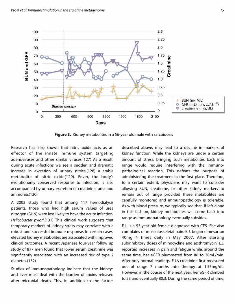
Figure 3. Kidney metabolites in a 56-year old male with sarcoidosis

E.J.’s creatinine dropped to 0.76. As of September 2010, E.J. reports her musculoskeletal pain as 0, but still complains of fatigue (5-7) and cognitive problems (6-7). Her latest kidney readings are a creatinine of 0.79 and eGFR of 75.9.
Although E.J.’s creatinine and eGFR fluctuated substantially out of the normal range during some of the time on the treatment, the metabolites stabilized without intervention. As expected, her most pronounced drop in eGFR corresponded to a period in which she experienced very high levels of immunopathology.
A more extreme example is that of B.B., a 56-year old male with a diagnosis of sarcoidosis. He began 40mg of olmesartan 4 times daily in December 2005 and began rotating combinations of subinhibitory minocycline, azithromycin, and clindamycin shortly thereafter. As seen in Figure 3, in the several months after starting treatment, measures of renal function initially declined sharply, reaching out-of-range levels in BUN (18 of 21 measures), creatinine (19 of 22), and GFR (14 previous tests). However, he is now 95% free of his previous symptoms and no longer takes oral or inhaled steroids.
This case is interesting in that typically a patient with kidney metabolites such as those of B.B. would likely be preparing for dialysis or experiencing symptoms of kidney failure. Instead, B.B. is relatively symptom-free. Again, his test results fluctuate in a way that suggests they are tied to immunopathology. This implies that in B.B.’s case, factors other than progressive kidney deterioration may be affecting his BUN, creatinine, and GFR. It is important to note that even though we have received many case histories in which patients’ kidney metabolites fluctuate out of range, we have had no reports of patients needing dialysis.
Immunostimulative therapies need further study
In studying therapeutic approaches designed to induce immunopathology, we must grapple with several ethical issues that have received minimal attention thus far.
Accepting discomfort
Physicians and researchers, especially in the context of clinical studies, feel an acute imperative to relieve pain
and discomfort whenever possible. Anything less, many have argued, would be unethical or immoral. Pullman writes, “The duties to relieve pain and suffering are clearly matters of moral obligation.”(133) Lohman writes that access to pain treatment is a “human right”(134) while Rollin states that necessary suffering is any suffering which is “impossible to alleviate.”(135)
However, medical providers have limited reservations with administering painful treatments or conducting uncomfortable procedures that lead to well-characterized positive outcomes: sentinel node biopsy in suspected cancer, major shoulder surgery, certain dental procedures, etc. Nor are most clinicians categorically opposed to using medicines that have a range of serious side effects, chemotherapy being an obvious example. Given sufficient reason for causing near-term discomfort, many patients and physicians are perfectly willing to seriously consider these types of trade-offs.
Therapies are typically thought of as having two categories of effects: therapeutic effects and adverse effects. The former is intentional and makes a patient feel better, and the latter is unintended and makes a patient feel worse. Yet, certain therapies that induce immunopathology have the potential to be a hybrid of the two: by necessity, the treatment is intended to make a patient feel worse. In other words, in the context of an immunopathology-inducing therapy, an adverse effect is not so much a collateral effect of the treatment, but the treatment itself.
As we have discussed, our immunopathology-inducing protocol can cause a sustained exacerbation in symptoms over at least several years’ time. But, in spite of the treatment’s length, we have found many patients are more than willing, considering the gravity of the prognoses they face, to commit themselves to such therapy.
It should be noted that the immune system may become healthier over the course of treatment. As microbes dysregulating the nuclear receptors are increasingly eliminated, and infected cells are replaced by their healthy counterparts, the immune response gains substantial strength. Thus, in some cases, immunopathology may actually become more severe several years into treatment.
Proal et al. Immunostimulation in the era of the metagenome 16

Physicians and patients should be aware that symptomatic improvement on an immunostimulatory treatment is rarely linear.
Blinding, randomization, and study design
The randomized controlled trial is regarded as the gold standard of measuring the efficacy of a therapy. Blinding the intervention to participants and physicians further reduces the effect of treatment bias. However, in the case of an immunopathology-inducing therapy, the severity of the symptom response would invariably make blinding impossible – patient and physician would know in short order the treatment arm to which a participant had been assigned. Given that therapies that generate immunopathology seem to require at least several years before sicker patients report substantial improvement, randomization also poses a challenge. To be sure, subjects could be assigned to the immunopathology-inducing group, but the potential for early withdrawal is clearly much higher for multi-year treatments that necessitate symptom exacerbation, even if only in the short term.
The number of patients who decline to participate and withdraw early may make an intention to treat analysis untenable. In any case, any study design for a therapy which makes patients feel significantly uncomfortable has the potential to incur self-selection bias: people who commit themselves to a challenging multi-year therapy are qualitatively different from people who decline the treatment.
Another key consideration for study design is the length of the trial. Trials that assess overall patient outcomes, such as improvement in activities of daily living, would need to last several years. Alternatively, trials of efficacy (i.e. those looking at microbial death) could be concluded in a month or two. It has been reported that HIV/AIDS patients on anti-retroviral drugs experience immune reconstitution inflammatory syndrome (IRIS) within weeks of beginning therapy.(136) This is consistent with our experience when using immunostimulation in auto-immune disease.
An essential feature of the design of any clinical trial is informed consent, and this is especially important in therapies that generate immunopathology. Patients need
to expect to experience temporary well-defined increases in symptoms, markers of inflammation, and disease state. They must also appreciate why such increases are necessary, and how they would work with their physician to manage their symptoms.
Discussion
Over the past decade, molecular sequencing technology and metagenomic analyses have revolutionized the field of microbiology. The human body, once deemed largely sterile, is now known to harbor thousands of genomes – many of which have still to be named and characterized. These genomes continually interact with the human genome in both health and disease. Not just single pathogens, but entire communities of microbes differ substantially between healthy individuals and those with autoimmune disease. The body is likely not creating antibodies against self, but instead in response to these pathogenic genomes. Indeed, many “autoantibodies” have already been linked to the presence of pathogens.
Those pathogens that can persist intracellularly have access to DNA translation and repair mechanisms, and can also affect gene expression. This activity mandates a fundamental reconsideration of how chronic diseases are treated. Rather than immunosuppression, immuno-stimulation may be the true key to reversal of these conditions.
The symptoms of an acute infection result from a battle between man and microbe. A similar battle, in which the immune system releases cytokines and microbes form endotoxins or debris, would occur if pathogens were killed in chronic disease. Thus, if chronic microbes are successfully targeted, patients would necessarily be expected to experience symptom exacerbation. In effect, patients recovering from autoimmune disease may need to feel worse before they feel better.
Interestingly, these responses have been described in the literature, generally as being paradoxical.(137-141) Yet symptom exacerbation resulting from an immuno-stimulative approach towards pathogens is paradoxical only because it defies previously held intuition.
The immunostimulatory approach we have studied uses a putative VDR agonist to reactivate the innate immune
Proal et al. Immunostimulation in the era of the metagenome 17

response. Case histories suggest that patients with a variety of autoimmune diagnoses experience prolonged immunopathology. Many subjects additionally exper-ienced neurological immunopathology, often for several years before improvement. This emphasizes the systemic nature of autoimmune disease processes and the significant number of pathogens that seem to be involved in the development of these disease states.
A broad array of medications and supplements are effective at reducing discomfort of patients with chronic disease. Our reports suggest that many of these drugs, whose actions at the molecular level are not fully understood, may generate feelings of clinical and subjective improvement precisely because they suppress and slow activity of the immune system.
In addition, accumulating evidence from metagenomic studies is pushing physicians and researchers to reflect upon the wisdom of using interventions in an attempt to alter the body’s metabolites. The more prominently microbes figure in our understanding of the pathogenesis of chronic disease, the more likely it is that therapies should be tailored to support the body as it responds to infection. In many cases, out of range markers seem to be a necessary part of the healing process.
Nevertheless, there remains a need to identify microbe-specific markers. Metabolomics is the emerging field which studies microbial metabolites. As metabolomics and metagenomics evolve, we should expect the emergence of technologies capable of more optimally defining a treatment regime based on examination of specimens from blood, urine and swabs.
There needs to be more collaboration between researchers and clinicians in order to more tightly define the immunopathology we have observed.
References
1. Proal AD, Albert PJ, Marshall TG. Autoimmune disease and the human metagenome. In: Nelson KE, (ed.). Metagenomics of the Human Body: Springer, 2010. In Press.
2. Chiller K, Selkin BA, Murakawa GJ. Skin microflora and bacterial infections of the skin. J Investig Dermatol Symp Proc. 2001; 6: 170-4.
3. Fierer N, Hamady M, Lauber CL, Knight R. The influence of sex, handedness, and washing on the diversity of hand surface bacteria. Proc Natl Acad Sci U S A. 2008; 105: 17994-9.
4. Grice EA, Kong HH, Conlan S, Deming CB, Davis J, Young AC, et al. Topographical and temporal diversity of the human skin microbiome. Science. 2009; 324: 1190-2.
5. Qin J, Li R, Raes J, Arumugam M, Burgdorf KS, Manichanh C, et al. A human gut microbial gene catalogue established by metagenomic sequencing. Nature. 2010; 464: 59-65.
6. Eberl G. A new vision of immunity: homeostasis of the superorganism. Mucosal Immunol. 2010; 3: 450-60.
7. Reyes A, Haynes M, Hanson N, Angly FE, Heath AC, Rohwer F, et al. Viruses in the faecal microbiota of monozygotic twins and their mothers. Nature. 2010; 466: 334-8.
8. Turnbaugh PJ, Ley RE, Hamady M, Fraser-Liggett CM, Knight R, Gordon JI. The human microbiome project. Nature. [10.1038/nature06244]. 2007; 449: 804-10.
9. Paulino LC, Tseng CH, Strober BE, Blaser MJ. Molecular analysis of fungal microbiota in samples from healthy human skin and psoriatic lesions. J Clin Microbiol. 2006; 44: 2933-41.
10. Ley RE, Turnbaugh PJ, Klein S, Gordon JI. Microbial ecology: human gut microbes associated with obesity. Nature. 2006; 444: 1022-3.
11. Basseri RJ, Basseri B, Chong K, Youdim A, Low K, Hwang LJ, et al. Intestinal Methane Production in Obese Humans Is Associated With Higher Body Mass Index. Digestive Disease Week (DDW) 2010; New Orleans, LA2010.
12. Larsen N, Vogensen FK, van den Berg FW, Nielsen DS, Andreasen AS, Pedersen BK, et al. Gut microbiota in human adults with type 2 diabetes differs from non-diabetic adults. PLoS One. 2010; 5: e9085.
13. Gophna U, Sommerfeld K, Gophna S, Doolittle WF, Veldhuyzen van Zanten SJ. Differences between tissue-associated intestinal microfloras of patients with Crohn's disease and ulcerative colitis. J Clin Microbiol. 2006; 44: 4136-41.
14. Yap IK, Angley M, Veselkov KA, Holmes E, Lindon JC, Nicholson JK. Urinary Metabolic Phenotyping Differentiates Children with Autism from Their Unaffected Siblings and Age-Matched Controls. J Proteome Res. 2010.
15. Lee YK, Menezes JS, Umesaki Y, Mazmanian SK. Microbes and Health Sackler Colloquium: Proinflammatory T-cell responses to gut microbiota promote experimental autoimmune encephalomyelitis. Proc Natl Acad Sci U S A. 2010.
16. DiGiulio DB, Romero R, Amogan HP, Kusanovic JP, Bik EM, Gotsch F, et al. Microbial prevalence, diversity and abundance in amniotic fluid during preterm labor: a molecular and culture-based investigation. PLoS ONE. 2008; 3: e3056.
17. Dempsey KE, Riggio MP, Lennon A, Hannah VE, Ramage G, Allan D, et al. Identification of bacteria on the surface of clinically infected and non-infected prosthetic hip joints removed during revision arthroplasties by 16S rRNA gene sequencing and by microbiological culture. Arthritis Res Ther. 2007; 9: R46.
Proal et al. Immunostimulation in the era of the metagenome 18

18. Erb-Downward JR, Thompson D, Han MK, Freeman CM, Chensue SW, Arenberg DA, et al. Analysis Of The Microbiome Of The Normal And COPD Lung. Am J Respir Crit Care Med. 2010; 181: A5628-.
19. Glickman MS, Pamer EG. Cell-mediated defense against infection. In: Mandell GL, Bennett JE, Dolin R, (eds.). Mandell, Douglas, and Bennett's principles and practice of infectious diseases. Philadelphia, PA: Churchill Livingstone/Elsevier, 2010.
20. Wirostko E, Johnson L, Wirostko B. Sarcoidosis associated uveitis. Parasitization of vitreous leucocytes by mollicute-like organisms. Acta Ophthalmol (Copenh). 1989; 67: 415-24.
21. Wirostko E, Johnson L, Wirostko W. Chronic leucocytoclastic bacterial vitritis. A lymphocyte transmission electron microscopic study. J Submicrosc Cytol. 1987; 19: 651-6.
22. Wirostko E, Johnson L, Wirostko B. Ulcerative colitis associated chronic uveitis. Parasitization of intraocular leucocytes by mollicute-like organisms. J Submicrosc Cytol Pathol. 1990; 22: 231-9.
23. Posnett DN, Edinger J. When do microbes stimulate rheumatoid factor? J Exp Med. 1997; 185: 1721-3.
24. Djavad N, Bas S, Shi X, Schwager J, Jeannet M, Vischer T, et al. Comparison of rheumatoid factors of rheumatoid arthritis patients, of individuals with mycobacterial infections and of normal controls: evidence for maturation in the absence of an autoimmune response. Eur J Immunol. 1996; 26: 2480-6.
25. Russell MW, Wu HY, White PL, Kilian M, Henrichsen J. Serum antibody responses to Streptococcus mutans antigens in humans systemically infected with oral streptococci. Oral Microbiol Immunol. 1992; 7: 321-5.
26. Berlin T, Zandman-Goddard G, Blank M, Matthias T, Pfeiffer S, Weis I, et al. Autoantibodies in nonautoimmune individuals during infections. Ann N Y Acad Sci. 2007; 1108: 584-93.
27. Lek ak h IV, Rott GM, Poverennyi AM. ["Masked" autoantibodies from the serum of healthy blood donors cross-reacting with DNA and bacterial lipopolysaccharides]. Biull Eksp Biol Med. 1991; 111: 516-8.
28. Sun J. Vitamin D and mucosal immune function. Curr Opin Gastroenterol. 2010.
29. Auvynet C, Rosenstein Y. Multifunctional host defense peptides: antimicrobial peptides, the small yet big players in innate and adaptive immunity. FEBS J. 2009; 276: 6497-508.
30. Froicu M, Cantorna MT. Vitamin D and the vitamin D receptor are critical for control of the innate immune response to colonic injury. BMC Immunol. 2007; 8: 5.
31. Wu Z, Cocchi F, Gentles D, Ericksen B, Lubkowski J, Devico A, et al. Human neutrophil alpha-defensin 4 inhibits HIV-1 infection in vitro. FEBS Lett. 2005; 579: 162-6.
32. Krishnakumari V, Rangaraj N, Nagaraj R. Antifungal activities of human beta-defensins HBD-1 to HBD-3 and their C-terminal analogs Phd1 to Phd3. Antimicrob Agents Chemother. 2009; 53: 256-60.
33. Howell MD, Wollenberg A, Gallo RL, Flaig M, Streib JE, Wong C, et al. Cathelicidin deficiency predisposes to eczema herpeticum. J Allergy Clin Immunol. 2006; 117: 836-41.
34. Benincasa M, Scocchi M, Pacor S, Tossi A, Nobili D, Basaglia G, et al. Fungicidal activity of five cathelicidin peptides against clinically isolated yeasts. J Antimicrob Chemother. 2006; 58: 950-9.
35. Wang TT, Tavera-Mendoza LE, Laperriere D, Libby E, MacLeod NB, Nagai Y, et al. Large-scale in silico and microarray-based identification of direct 1,25-dihydroxyvitamin D3 target genes. Mol Endocrinol. 2005; 19: 2685-95.
36. Ramagopalan SV, Heger A, Berlanga AJ, Maugeri NJ, Lincoln MR, Burrell A, et al. A ChIP-seq defined genome-wide map of vitamin D receptor binding: Associations with disease and evolution. Genome Res. 2010.
37. Abbanat DR, Godchaux W, 3rd, Polychroniou G, Leadbetter ER. Biosynthesis of a sulfonolipid in gliding bacteria. Biochem Biophys Res Commun. 1985; 130: 873-8.
38. Marshall TG. Vitamin D discovery outpaces FDA decision making. BioEssays: News and Reviews in Molecular, Cellular and Developmental Biology. [10.1002/bies.20708]. 2008; 30: 173-82.
39. Xu Y, Xie J, Li Y, Yue J, Chen J, Chunyu L, et al. Using a cDNA microarray to study cellular gene expression altered by Mycobacterium tuberculosis. Chinese Medical Journal. 2003; 116: 1070-3.
40. Anand PK, Kaul D, Sharma M. Synergistic action of vitamin D and retinoic acid restricts invasion of macrophages by pathogenic mycobacteria. J Microbiol Immunol Infect. 2008; 41: 17-25.
41. Salazar JC, Duhnam-Ems S, La Vake C, Cruz AR, Moore MW, Caimano MJ, et al. Activation of human monocytes by live Borrelia burgdorferi generates TLR2-dependent and -independent responses which include induction of IFN-beta. PLoS Pathog. 2009; 5: e1000444.
42. Nevado J, Tenbaum SP, Castillo AI, Sanchez-Pacheco A, Aranda A. Activation of the human immunodeficiency virus type I long terminal repeat by 1 alpha,25-dihydroxyvitamin D3. J Mol Endocrinol. 2007; 38: 587-601.
43. Yenamandra SP, Lundin A, Arulampalam V, Yurchenko M, Pettersson S, Klein G, et al. Expression profile of nuclear receptors upon Epstein -- Barr virus induced B cell transformation. Experimental Oncology. 2009; 31: 92-6.
44. Bell NH. Renal and nonrenal 25-hydroxyvitamin D-1alpha-hydroxylases and their clinical significance. J Bone Miner Res. 1998; 13: 350-3.
45. Blaney GP, Albert PJ, Proal AD. Vitamin D metabolites as clinical markers in autoimmune and chronic disease. Ann N Y Acad Sci. 2009; 1173: 384-90.
46. Abreu MT, Kantorovich V, Vasiliauskas EA, Gruntmanis U, Matuk R, Daigle K, et al. Measurement of vitamin D levels in inflammatory bowel disease patients reveals a subset of Crohn's disease patients with elevated 1,25-dihydroxyvitamin D and low bone mineral density. Gut. 2004; 53: 1129-36.
Proal et al. Immunostimulation in the era of the metagenome 19

47. Kavathia D, Buckley JD, Rao D, Rybicki B, Burke R. Elevated 1, 25-dihydroxyvitamin D levels are associated with protracted treatment in sarcoidosis. Respir Med. 2010; 104: 564-70.
48. Mawer EB, Hayes ME, Still PE, Davies M, Lumb GA, Palit J, et al. Evidence for nonrenal synthesis of 1,25-dihydroxyvitamin D in patients with inflammatory arthritis. J Bone Miner Res. 1991; 6: 733-9.
49. Proal AD, Albert PJ, Marshall TG. Dysregulation of the vitamin D nuclear receptor may contribute to the higher prevalence of some autoimmune diseases in women. Ann N Y Acad Sci. 2009; 1173: 252-9.
50. Brahmachary M, Schonbach C, Yang L, Huang E, Tan SL, Chowdhary R, et al. Computational promoter analysis of mouse, rat and human antimicrobial peptide-coding genes. BMC Bioinformatics. 2006; 7 Suppl 5: S8.
51. Kanchwala A, Barna B, Singh R, Culver D, Malur A, Abraham S, et al., editors. Deficiencies of cathelicidin and vitamin D accompany disease severity in sarcoidosis2009.
52. Wang TT, Dabbas B, Laperriere D, Bitton AJ, Soualhine H, Tavera-Mendoza LE, et al. Direct and indirect induction by 1,25-dihydroxyvitamin D3 of the NOD2/CARD15-defensin beta2 innate immune pathway defective in Crohn disease. J Biol Chem. 2010; 285: 2227-31.
53. Wiken M, Idali F, Al Hayja M, Grunewald J, Eklund A, Wahlstrom J. No evidence of altered alveolar macrophage polar izat ion , but reduced express ion of TLR2, in bronchoalveolar lavage cells in sarcoidosis. Respiratory Research. 2010; 11: 121.
54. Gosain A, DiPietro LA. Aging and wound healing. World J Surg. 2004; 28: 321-6.
55. Giunta S. Is inflammaging an auto[innate]immunity subclinical syndrome? Immun Ageing. 2006; 3: 12.
56. Kuczynski J, Costello EK, Nemergut DR, Zaneveld J, Lauber CL, Knights D, et al. Direct sequencing of the human microbiome readily reveals community differences. Genome Biol. 2010; 11: 210.
57. Turnbaugh PJ, Hamady M, Yatsunenko T, Cantarel BL, Duncan A, Ley RE, et al. A core gut microbiome in obese and lean twins. Nature. 2009; 457: 480-4.
58. Smith T. Some Problems in the Life-history of Pathogenic Microörganisms. Amer Med. 1904: 711-8.
59. Jenssen H, Hancock RE. Therapeutic potential of HDPs as immunomodulatory agents. Methods Mol Biol. 2010; 618: 329-47.
60. Meintjes G, Rabie H, Wilkinson RJ, Cotton MF. Tuberculosis-associated immune reconstitution inflammatory syndrome and unmasking of tuberculosis by antiretroviral therapy. Clin Chest Med. 2009; 30: 797-810, x.
61. Chen F, Day SL, Metcalfe RA, Sethi G, Kapembwa MS, Brook MG, et al. Characteristics of autoimmune thyroid disease occurring as a late complication of immune reconstitution in patients with advanced human immunodeficiency virus (HIV) disease. Medicine (Baltimore). 2005; 84: 98-106.
62. Schaffner A. [Fever--useful or noxious symptom that should be treated?]. Ther Umsch. 2006; 63: 185-8.
63. Hudgins LC, Parker TS, Levine DM, Gordon BR, Saal SD, Jiang XC, et al. A single intravenous dose of endotoxin rapidly alters serum lipoproteins and lipid transfer proteins in normal volunteers. J Lipid Res. 2003; 44: 1489-98.
64. O'Connor SM, Taylor CE, Hughes JM. Emerging infectious determinants of chronic diseases. Emerging Infectious Diseases. 2006; 12: 1051-7.
65. Boas EP, Michelson N, DOANE JC. The Challenge of Chronic Diseases. AJN The American Journal of Nursing. 1930; 30: 372-3.
66. Nau R, Eiffert H. Modulation of release of proinflammatory bacterial compounds by antibacterials: potential impact on course of inflammation and outcome in sepsis and meningitis. Clin Microbiol Rev. 2002; 15: 95-110.
67. Cabral RF, Valle Bahia PR, Gasparetto EL, Chimelli L. Immune reconstitution inflammatory syndrome and cerebral toxoplasmosis. AJNR Am J Neuroradiol. 2010; 31: E65-6.
68. Murdoch DM, Venter WD, Van Rie A, Feldman C. Immune reconstitution inflammatory syndrome (IRIS): review of common infectious manifestations and treatment options. AIDS Res Ther. 2007; 4: 9.
69. Bicanic T, Meintjes G, Rebe K, Williams A, Loyse A, Wood R, et al. Immune reconstitution inflammatory syndrome in HIV-associated cryptococcal meningitis: a prospective study. J Acquir Immune Defic Syndr. 2009; 51: 130-4.
70. Naccache JM, Antoine M, Wislez M, Fleury-Feith J, Oksenhendler E, Mayaud C, et al. Sarcoid-like pulmonary disorder in human immunodeficiency virus-infected patients receiving antiretroviral therapy. Am J Respir Crit Care Med. 1999; 159: 2009-13.
71. Mirmirani P, Maurer TA, Herndier B, McGrath M, Weinstein MD, Berger TG. Sarcoidosis in a patient with AIDS: a manifestation of immune restoration syndrome. J Am Acad Dermatol. 1999; 41: 285-6.
72. Bell C, Nelson M, Kaye S. A case of immune reconstitution rheumatoid arthritis. Int J STD AIDS. 2002; 13: 580-1.
73. Behrens G, Knuth C, Schedel I, Mendila M, Schmidt RE. Highly active antiretroviral therapy. Lancet. 1998; 351: 1057-8; author reply 8-9.
74. Piliero PJ, Fish DG, Preston S, Cunningham D, Kinchelow T, Salgo M, et al. Guillain-Barre syndrome associated with immune reconstitution. Clin Infect Dis. 2003; 36: e111-4.
75. Sereti I, Sarlis NJ, Arioglu E, Turner ML, Mican JM. Alopecia universalis and Graves' disease in the setting of immune restoration after highly active antiretroviral therapy. AIDS. 2001; 15: 138-40.
76. Calabrese LH, Kirchner E, Shrestha R. Rheumatic complications of human immunodeficiency virus infection in the era of highly active antiretroviral therapy: emergence of a new syndrome of immune reconstitution and changing patterns of disease. Semin Arthritis Rheum. 2005; 35: 166-74.
Proal et al. Immunostimulation in the era of the metagenome 20

77. Marshall TG. VDR nuclear receptor is key to recovery from cognitive dysfunction. Days of Molecular Medicine; April 17-19; Stockholm, Sweden2008.
78. Malloy PJ, Feldman D. Inactivation of the Human Vitamin D Receptor by Caspase-3. Endocrinology. 2008.
79. Brasier AR. The nuclear factor-kappaB-interleukin-6 signalling pathway mediating vascular inflammation. Cardiovasc Res. 2010; 86: 211-8.
80. Schwocho LR, Masonson HN. Pharmacokinetics of CS-866, a new angiotensin II receptor blocker, in healthy subjects. J Clin Pharmacol. 2001; 41: 515-27.
81. Brunner HR. Clinical efficacy and tolerability of olmesartan. Clin Ther. 2004; 26 Suppl A: A28-32.
82. Brunner HR. The new oral angiotensin II antagonist olmesartan medoxomil: a concise overview. J Hum Hypertens. 2002; 16 Suppl 2: S13-6.
83. Yoshida K, Kohzuki M. Clinical and experimental aspects of olmesartan medoxomil, a new angiotensin II receptor antagonist. Cardiovasc Drug Rev. 2004; 22: 285-308.
84. Saiman L, Tabibi S, Starner TD, San Gabriel P, Winokur PL, Jia HP, et al. Cathelicidin peptides inhibit multiply antibiotic-resistant pathogens from patients with cystic fibrosis. Antimicrob Agents Chemother. 2001; 45: 2838-44.
85. Zasloff M. Antimicrobial peptides of multicellular organisms. Nature. 2002; 415: 389-95.
86. Soscia SJ, Kirby JE, Washicosky KJ, Tucker SM, Ingelsson M, Hyman B, et al. The Alzheimer's disease-associated amyloid beta-protein is an antimicrobial peptide. PLoS One. 2010; 5: e9505.
87. Nicolson GL, Gan R, Nicolson NL, Haier J. Evidence for Mycoplasma ssp., Chlamydia pneunomiae, and human herpes virus-6 coinfections in the blood of patients with autistic spectrum disorders. J Neurosci Res. 2007; 85: 1143-8.
88. Ortega-Hernandez OD, Shoenfeld Y. Infection, vaccination, and autoantibodies in chronic fatigue syndrome, cause or coincidence? Ann N Y Acad Sci. 2009; 1173: 600-9.
89. Geddes R. Minocycline-induced lupus in adolescents: clinical implications for physical therapists. J Orthop Sports Phys Ther. 2007; 37: 65-71.
90. Lefebvre N, Forestier E, Farhi D, Mahsa MZ, Remy V, Lesens O, et al. Minocycline-induced hypersensitivity syndrome presenting with meningitis and brain edema: a case report. J Med Case Reports. 2007; 1: 22.
91. Krawitt EL. Autoimmune hepatitis. N Engl J Med. 2006; 354: 54-66.
92. Martinez E, Gatell J, Moran Y, Aznar E, Buira E, Guelar A, et al. High incidence of herpes zoster in patients with AIDS soon after therapy with protease inhibitors. Clin Infect Dis. 1998; 27: 1510-3.
93. Melkert PW. Fatal-Jarisch Herxheimer reaction in a case of relapsing fever misdiagnosed as lobar pneumonia. Trop Geogr Med. 1987; 39: 92-3.
94. Diefenbach WC. Fatal Jarish-Herxheimer reaction with sudden aneurysmal dilatation and complete bronchial occlusion following penicillin therapy. N Engl J Med. 1949; 241: 95.
95. Stark FR, Crast F, Clemmer T, Ramirez R. Letter: Fatal Herxheimer reaction after pentamidine in Pneumocystis pneumonia. Lancet. 1976; 1: 1193-4.
96. Blumberg HM, Leonard MK, Jr., Jasmer RM. Update on the treatment of tuberculosis and latent tuberculosis infection. JAMA. 2005; 293: 2776-84.
97. Cheung CMG, Chee SP. Jarisch-Herxheimer reaction: paradoxical worsening of tuberculosis chorioretinitis following initiation of antituberculous therapy. Eye. 2008; 23: 1472-3.
98. Thwaites GE, Nguyen DB, Nguyen HD, Hoang TQ, Do TT, Nguyen TC, et al. Dexamethasone for the treatment of tuberculous meningitis in adolescents and adults. N Engl J Med. 2004; 351: 1741-51.
99. Beatty GW. Immune reconstitution inflammatory syndrome. Emerg Med Clin North Am. 2010; 28: 393-407, Table of Contents.
100. Montano-Loza AJ, Carpenter HA, Czaja AJ. Consequences of treatment withdrawal in type 1 autoimmune hepatitis. Liver Int. 2007; 27: 507-15.
101. Grimes DA, Schulz KF. Surrogate end points in clinical research: hazardous to your health. Obstet Gynecol. 2005; 105: 1114-8.
102. Shepherd J, Cobbe SM, Ford I, Isles CG, Lorimer AR, MacFarlane PW, et al. Prevention of coronary heart disease with pravastatin in men with hypercholesterolemia. West of Scotland Coronary Prevention Study Group. N Engl J Med. 1995; 333: 1301-7.
103. Zarychanski R, Houston DS. Anemia of chronic disease: a harmful disorder or an adaptive, beneficial response? CMAJ. 2008; 179: 333-7.
104. Brotanek JM, Gosz J, Weitzman M, Flores G. Iron deficiency in early childhood in the United States: risk factors and racial/ethnic disparities. Pediatrics. 2007; 120: 568-75.
105. Weiss G, Goodnough LT. Anemia of chronic disease. N Engl J Med. 2005; 352: 1011-23.
106. Baker JF, Ghio AJ. Iron homoeostasis in rheumatic disease. Rheumatology (Oxford). 2009; 48: 1339-44.
107. Weinberg ED. Iron depletion: a defense against intracellular infection and neoplasia. Life Sci. 1992; 50: 1289-97.
108. Kemna E, Pickkers P, Nemeth E, van der Hoeven H, Swinkels D. Time-course analysis of hepcidin, serum iron, and plasma cytokine levels in humans injected with LPS. Blood. 2005; 106: 1864-6.
109. Afzali B, Goldsmith DJ. Intravenous iron therapy in renal failure: friend and foe? J Nephrol. 2004; 17: 487-95.
110. Bullen JJ, Ward CG, Rogers HJ. The critical role of iron in some clinical infections. Eur J Clin Microbiol Infect Dis. 1991; 10: 613-7.
Proal et al. Immunostimulation in the era of the metagenome 21

111. Melamed ML, Michos ED, Post W, Astor B. 25-hydroxyvitamin D levels and the risk of mortality in the general population. Arch Intern Med. 2008; 168: 1629-37.
112. Vieth R. Vitamin D and cancer mini-symposium: the risk of additional vitamin D. Ann Epidemiol. 2009; 19: 441-5.
113. Albert PJ, Proal AD, Marshall TG. Vitamin D: the alternative hypothesis. Autoimmunity Reviews. [10.1016/j.autrev.2009.02.011]. 2009; 8: 639-44.
114. Yoshizawa T, Handa Y, Uematsu Y, Takeda S, Sekine K, Yoshihara Y, et al. Mice lacking the vitamin D receptor exhibit impaired bone formation, uterine hypoplasia and growth retardation after weaning. Nat Genet. 1997; 16: 391-6.
115. Crew KD, Shane E, Cremers S, McMahon DJ, Irani D, Hershman DL. High prevalence of vitamin D deficiency despite supplementation in premenopausal women with breast cancer undergoing adjuvant chemotherapy. J Clin Oncol. 2009; 27: 2151-6.
116. Arnson Y, Amital H, Shoenfeld Y. Vitamin D and autoimmunity: new aetiological and therapeutic considerations. Ann Rheum Dis. 2007; 66: 1137-42.
117. Agborsangaya CB, Lehtinen T, Toriola AT, Pukkala E, Surcel HM, Tedeschi R, et al. Association between Epstein-Barr virus infection and risk for development of pregnancy-associated breast cancer: Joint effect with vitamin D? Eur J Cancer. 2010.
118. Cutolo M, Otsa K, Uprus M, Paolino S, Seriolo B. Vitamin D in rheumatoid arthritis. Autoimmun Rev. 2007; 7: 59-64.
119. Taveira da Silva AM, Kaulbach HC, Chuidian FS, Lambert DR, Suffredini AF, Danner RL. Brief report: shock and multiple-organ dysfunction after self-administration of Salmonella endotoxin. N Engl J Med. 1993; 328: 1457-60.
120. Faisst M, Wellner UF, Utzolino S, Hopt UT, Keck T. Elevated blood urea nitrogen is an independent risk factor of prolonged intensive care unit stay due to acute necrotizing pancreatitis. J Crit Care. 2010; 25: 105-11.
121. Stamler JS, Singel DJ, Loscalzo J. Biochemistry of nitric oxide and its redox-activated forms. Science. 1992; 258: 1898-902.
122. Stuehr DJ, Nathan CF. Nitric oxide. A macrophage product responsible for cytostasis and respiratory inhibition in tumor target cells. J Exp Med. 1989; 169: 1543-55.
123. Drapier JC, Wietzerbin J, Hibbs JB, Jr. Interferon-gamma and tumor necrosis factor induce the L-arginine-dependent cytotoxic effector mechanism in murine macrophages. Eur J Immunol. 1988; 18: 1587-92.
124. Wagner DA, Tannenbaum SR. Enhancement of nitrate biosynthesis by E.coli LPS. In: Magee PN, (ed.). Nitrosamines and Human Cancer: Banbury Rep., 1982. 437-43.
125. O'Donnell C, Liew E. Immunological aspects of nitric oxide. The Biochemist. 1994; 16: 19-22.
126. Mannick JB. Immunoregulatory and antimicrobial effects of nitrogen oxides. Proc Am Thorac Soc. 2006; 3: 161-5.
127. Cao W, Baniecki ML, McGrath WJ, Bao C, Deming CB, Rade JJ, et al. Nitric oxide inhibits the adenovirus proteinase in vitro and viral infectivity in vivo. FASEB J. 2003; 17: 2345-6.
128. Olteanu A, Feigin RD, Beisel WR. Metabolic response of the host to infecitons. In: Feigin RD, (ed.). Feigin & Cherry's textbook of pediatric infectious diseases. 6th ed. Philadelphia, PA: Saunders/Elsevier, 2009. 65-81.
129. Goggins MG, Shah SA, Goh J, Cherukuri A, Weir DG, Kelleher D, et al. Increased urinary nitrite, a marker of nitric oxide, in active inflammatory bowel disease. Mediators Inflamm. 2001; 10: 69-73.
130. Beisel WR, Sawyer WD, Ryll ED, Crozier D. Metabolic effects of intracellular infections in man. Ann Intern Med. 1967; 67: 744-79.
131. Tsukada K, Miyazaki T, Katoh H, Yoshikawa M, Masuda N, Ojima H, et al. Helicobacter pylori infection in hemodialysis patients. Hepatogastroenterology. 2003; 50: 2255-8.
132. Harita N, Hayashi T, Sato KK, Nakamura Y, Yoneda T, Endo G, et al. Lower serum creatinine is a new risk factor of type 2 diabetes: the Kansai healthcare study. Diabetes Care. 2009; 32: 424-6.
133. Pullman D. Human dignity and the ethics and aesthetics of pain and suffering. Theor Med Bioeth. 2002; 23: 75-94.
134. Lohman D, Schleifer R, Amon JJ. Access to pain treatment as a human right. BMC Med. 2010; 8: 8.
135. Gaynor JS, Muir W. Handbook of veterinary pain management. St. Louis, Mo.: Mosby, 2002.
136. Mohanty K. Immune reconstitution inflammatory syndrome after initiation of highly active anti-retroviral therapy in HIV/AIDS. Indian J Dermatol Venereol Leprol. 2010; 76: 301-4.
137. Brown ST. Adverse reactions in syphilis therapy. J Am Vener Dis Assoc. 1976; 3: 172-6.
138. Oliveira JB, Gupta S. Disorders of apoptosis: mechanisms for autoimmunity in primary immunodeficiency diseases. J Clin Immunol. 2008; 28 Suppl 1: S20-8.
139. Hurley JC. Antibiotic-induced release of endotoxin. A therapeutic paradox. Drug Safety: An International Journal of Medical Toxicology and Drug Experience. 1995; 12: 183-95.
140. Jindal G, Ghosh D. Therapeutic paradox in CNS tuberculosis. Journal of Pediatric Neurosciences. 2009; 4: 133-4.
141. Cheung CM, Chee SP. Jarisch-Herxheimer reaction: paradoxical worsening of tuberculosis chorioretinitis following initiation of antituberculous therapy. Eye. 2008.
Proal et al. Immunostimulation in the era of the metagenome 22
Related Documents