I M M U N O L O G Y I M M U N O L O G Y A brief survey Naira Renault (former Naira Roland Matevosyan) MD, PhD, MSJ Seton Hall Law School. Emory University. CDC 10.01.2017

Welcome message from author
This document is posted to help you gain knowledge. Please leave a comment to let me know what you think about it! Share it to your friends and learn new things together.
Transcript

I M M U N O L O G YI M M U N O L O G YA brief survey
Naira Renault (former Naira Roland Matevosyan) MD, PhD, MSJ
Seton Hall Law School. Emory University. CDC
10.01.2017
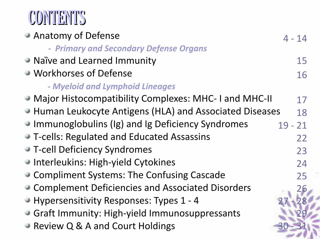
CONTENTSCONTENTSAnatomy of Defense
- Primary and Secondary Defense OrgansNaȉve and Learned ImmunityWorkhorses of Defense
- Myeloid and Lymphoid LineagesMajor Histocompatibility Complexes: MHC- I and MHC-IIHuman Leukocyte Antigens (HLA) and Associated DiseasesImmunoglobulins (Ig) and Ig Deficiency SyndromesT-cells: Regulated and Educated AssassinsT-cell Deficiency SyndromesInterleukins: High-yield CytokinesCompliment Systems: The Confusing CascadeComplement Deficiencies and Associated DisordersHypersensitivity Responses: Types 1 - 4Graft Immunity: High-yield ImmunosuppressantsReview Q & A and Court Holdings
4 - 14
1516
1718
19 - 212223242526
27 - 2829
30 - 31
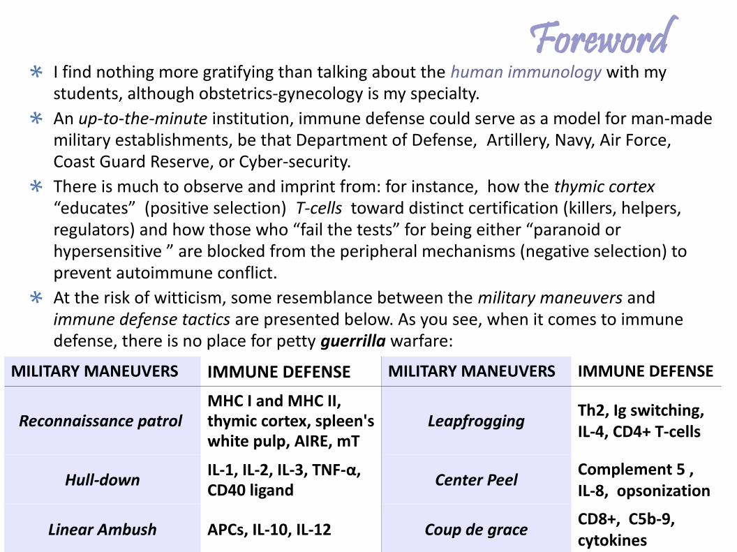
ForewordI find nothing more gratifying than talking about the human immunology with my students, although obstetrics-gynecology is my specialty.An up-to-the-minute institution, immune defense could serve as a model for man-made military establishments, be that Department of Defense, Artillery, Navy, Air Force, Coast Guard Reserve, or Cyber-security.There is much to observe and imprint from: for instance, how the thymic cortex “educates” (positive selection) T-cells toward distinct certification (killers, helpers, regulators) and how those who “fail the tests” for being either “paranoid or hypersensitive ” are blocked from the peripheral mechanisms (negative selection) to prevent autoimmune conflict. At the risk of witticism, some resemblance between the military maneuvers and immune defense tactics are presented below. As you see, when it comes to immune defense, there is no place for petty guerrilla warfare:
MILITARY MANEUVERS IMMUNE DEFENSE MILITARY MANEUVERS IMMUNE DEFENSE
Reconnaissance patrol MHC I and MHC II, thymic cortex, spleen's white pulp, AIRE, mT
Leapfrogging Th2, Ig switching, IL-4, CD4+ T-cells
Hull-down IL-1, IL-2, IL-3, TNF-α, CD40 ligand Center Peel Complement 5 ,
IL-8, opsonization
Linear Ambush APCs, IL-10, IL-12 Coup de grace CD8+, C5b-9, cytokines
Anatomy of DefensePRIMARY LYMPHOID ORGANS (bone marrow and thymus) function as “boarding schools” for immature progenitor cells to generate, mature, and educate young lymphocytes in an antigen-independent manner.
– Bone Marrow: A critical primary defense organ, it consists of red marrow (the bone parenchyma, containing hematopoietic stem-cells which generate all blood cell lines, including B and T cells), and yellow marrow (the stroma, a supportive adipose tissue).
– Thymus: The high cellular-density cortex is for positive selection of immature T-cells, the cortico-medullar junction for negative selection of T-cells through apoptoric signal, and the low-cellular density medulla for housing both positively and negatively selected mature thymocites.
SECONDARY LYMPOID ORGANS (peripheral lymph nodes, jugular and subclavian trunks, parotid nodes or tonsils, occipital, mastoid and mediastinal nodes, adenoid, appendix, spleen*, mucosa-associated lymphoid tissue or MALT, etc) are where lymphocytes differentiate and undergo clonal expansion (quantitative growth) in an antigen-dependent fashion (i.e. to act only when there is a foreign invader). *See slide 11.
4
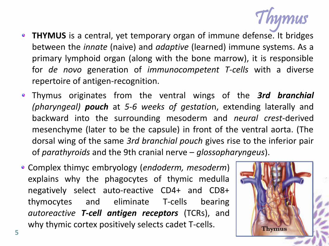
ThymusTHYMUS is a central, yet temporary organ of immune defense. It bridges between the innate (naive) and adaptive (learned) immune systems. As a primary lymphoid organ (along with the bone marrow), it is responsible for de novo generation of immunocompetent T-cells with a diverse repertoire of antigen-recognition.
Thymus originates from the ventral wings of the 3rd branchial (pharyngeal) pouch at 5-6 weeks of gestation, extending laterally and backward into the surrounding mesoderm and neural crest-derived mesenchyme (later to be the capsule) in front of the ventral aorta. (The dorsal wing of the same 3rd branchial pouch gives rise to the inferior pair of parathyroids and the 9th cranial nerve – glossopharyngeus).
5
Complex thimyc embryology (endoderm, mesoderm) explains why the phagocytes of thymic medulla negatively select auto-reactive CD4+ and CD8+ thymocytes and eliminate T-cells bearing autoreactive T-cell antigen receptors (TCRs), and why thymic cortex positively selects cadet T-cells.
Thymus (continued)
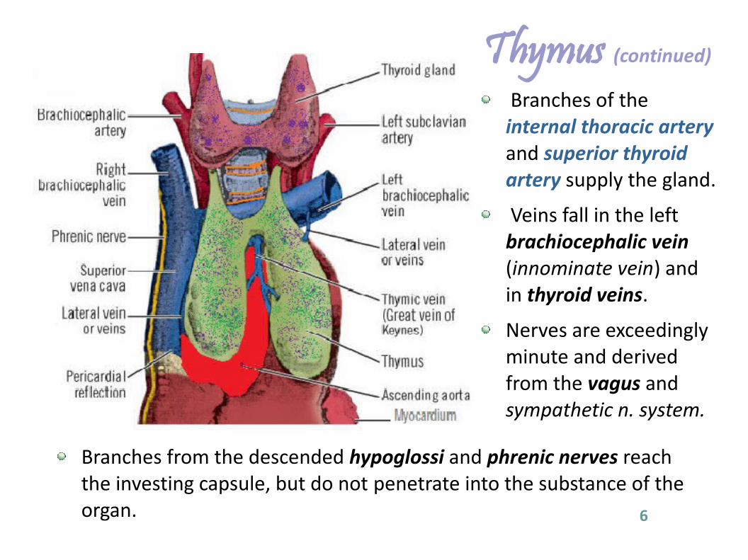
Branches of the internal thoracic artery and superior thyroid artery supply the gland.
Veins fall in the left brachiocephalic vein (innominate vein) and in thyroid veins.
Nerves are exceedingly minute and derived from the vagus and sympathetic n. system.
Branches from the descended hypoglossi and phrenic nerves reach the investing capsule, but do not penetrate into the substance of the organ. 6
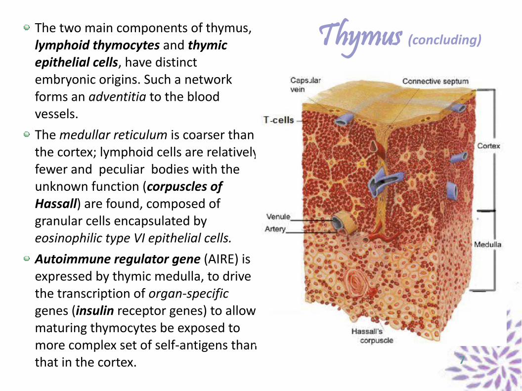
Thymus (concluding)The two main components of thymus, lymphoid thymocytes and thymic epithelial cells, have distinct embryonic origins. Such a network forms an adventitia to the blood vessels.The medullar reticulum is coarser than the cortex; lymphoid cells are relatively fewer and peculiar bodies with the unknown function (corpuscles of Hassall) are found, composed of granular cells encapsulated by eosinophilic type VI epithelial cells.Autoimmune regulator gene (AIRE) is expressed by thymic medulla, to drive the transcription of organ-specific genes (insulin receptor genes) to allow maturing thymocytes be exposed to more complex set of self-antigens than that in the cortex. 7
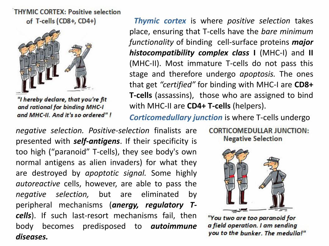
Thymic cortex is where positive selection takes place, ensuring that T-cells have the bare minimum functionality of binding cell-surface proteins major histocompatibility complex class I (MHC-I) and II (MHC-II). Most immature T-cells do not pass this stage and therefore undergo apoptosis. The ones that get “certified” for binding with MHC-I are CD8+ T-cells (assassins), those who are assigned to bind with MHC-II are CD4+ T-cells (helpers).Corticomedullary junction is where T-cells undergo
negative selection. Positive-selection finalists are presented with self-antigens. If their specificity is too high (“paranoid” T-cells), they see body's own normal antigens as alien invaders) for what they are destroyed by apoptotic signal. Some highly autoreactive cells, however, are able to pass the negative selection, but are eliminated by peripheral mechanisms (anergy, regulatory T-cells). If such last-resort mechanisms fail, then body becomes predisposed to autoimmune diseases.
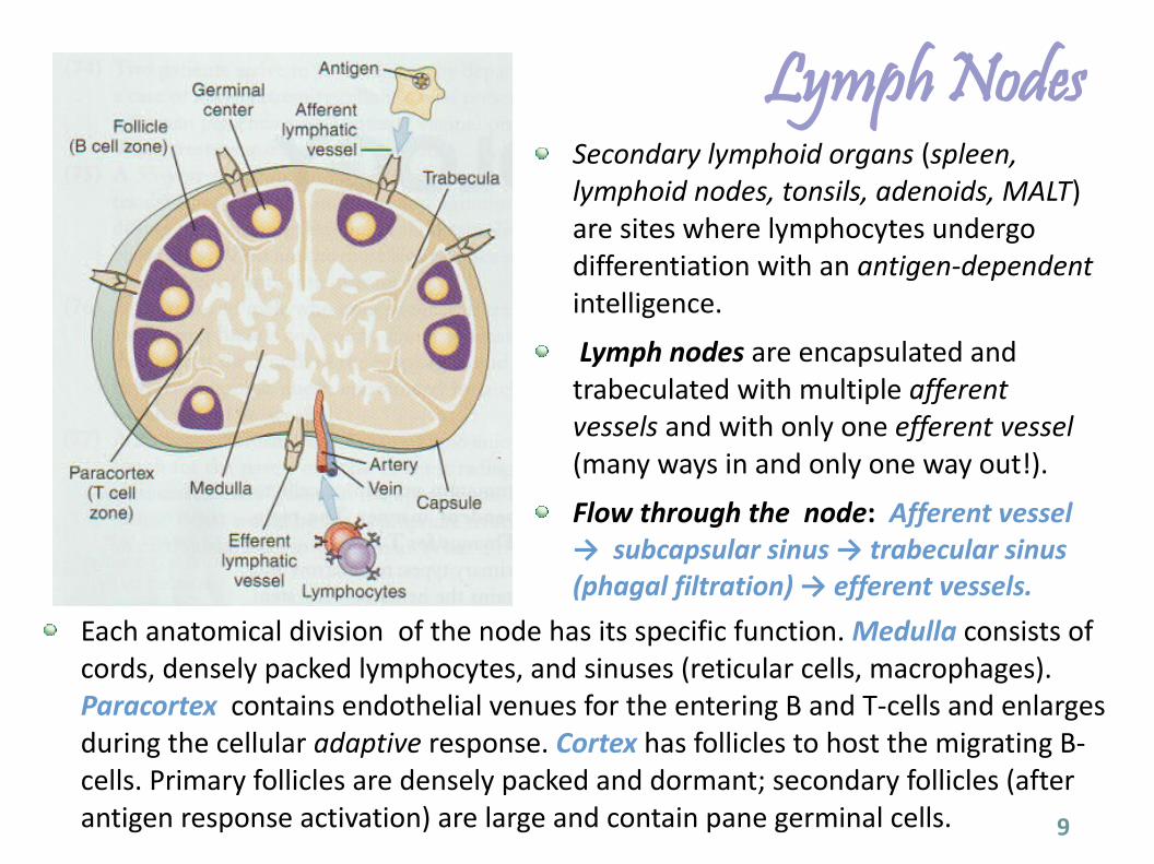
Lymph NodesSecondary lymphoid organs (spleen, lymphoid nodes, tonsils, adenoids, MALT) are sites where lymphocytes undergo differentiation with an antigen-dependent intelligence.
Lymph nodes are encapsulated and trabeculated with multiple afferent vessels and with only one efferent vessel (many ways in and only one way out!).
Flow through the node: Afferent vessel → subcapsular sinus → trabecular sinus (phagal filtration) → efferent vessels.
9
Each anatomical division of the node has its specific function. Medulla consists of cords, densely packed lymphocytes, and sinuses (reticular cells, macrophages). Paracortex contains endothelial venues for the entering B and T-cells and enlarges during the cellular adaptive response. Cortex has follicles to host the migrating B-cells. Primary follicles are densely packed and dormant; secondary follicles (after antigen response activation) are large and contain pane germinal cells.
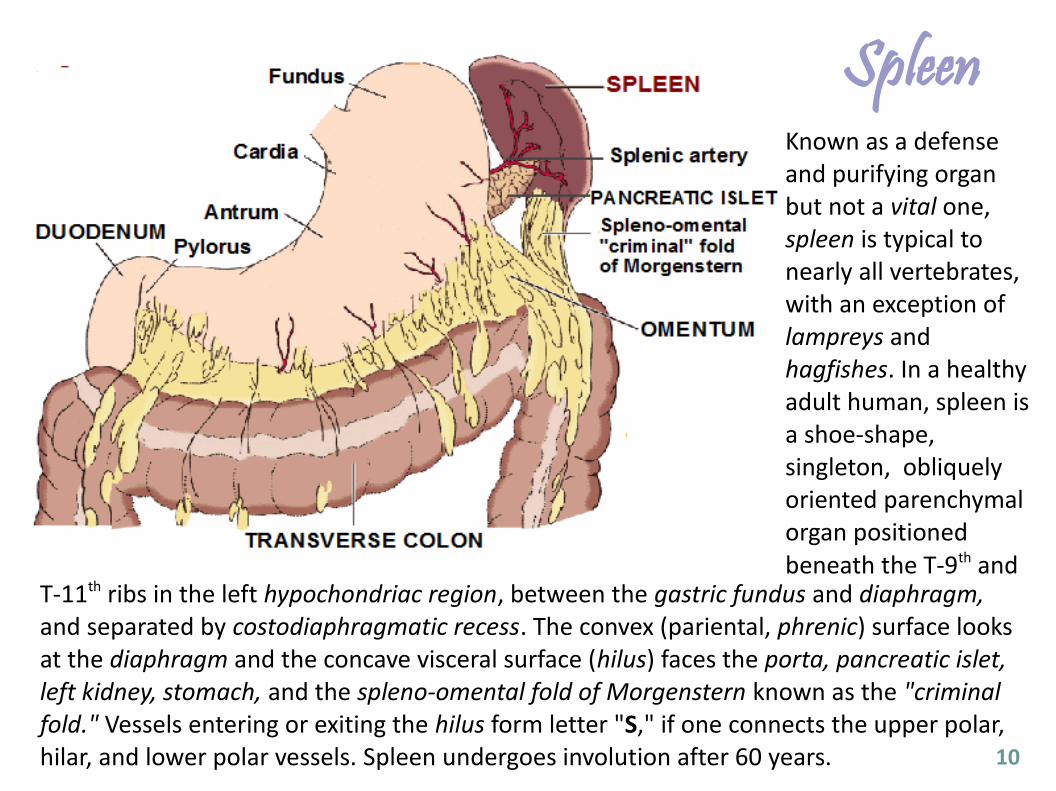
SpleenKnown as a defense and purifying organ but not a vital one, spleen is typical to nearly all vertebrates, with an exception of lampreys and hagfishes. In a healthy adult human, spleen is a shoe-shape, singleton, obliquely oriented parenchymal organ positioned beneath the T-9th and
10
T-11th ribs in the left hypochondriac region, between the gastric fundus and diaphragm, and separated by costodiaphragmatic recess. The convex (pariental, phrenic) surface looks at the diaphragm and the concave visceral surface (hilus) faces the porta, pancreatic islet, left kidney, stomach, and the spleno-omental fold of Morgenstern known as the "criminal fold." Vessels entering or exiting the hilus form letter "S," if one connects the upper polar, hilar, and lower polar vessels. Spleen undergoes involution after 60 years.
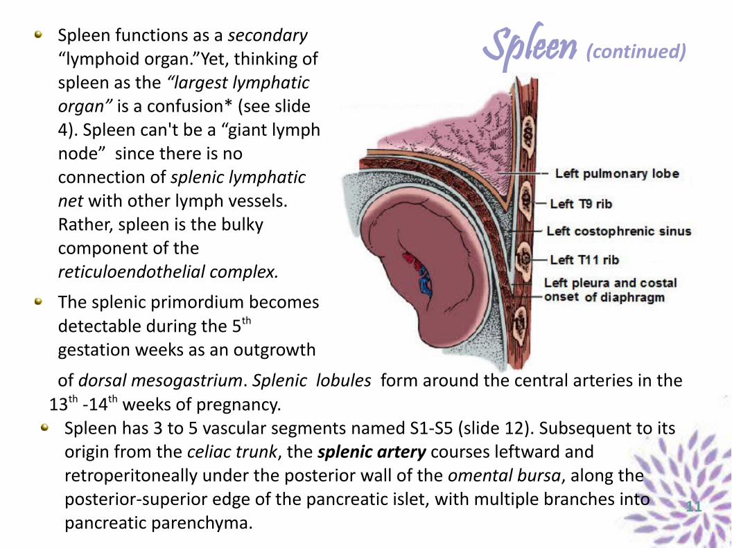
Spleen (continued)Spleen functions as a secondary “lymphoid organ.”Yet, thinking of spleen as the “largest lymphatic organ” is a confusion* (see slide 4). Spleen can't be a “giant lymph node” since there is no connection of splenic lymphatic net with other lymph vessels. Rather, spleen is the bulky component of the reticuloendothelial complex.
The splenic primordium becomes detectable during the 5th gestation weeks as an outgrowth
11
of dorsal mesogastrium. Splenic lobules form around the central arteries in the 13th -14th weeks of pregnancy.
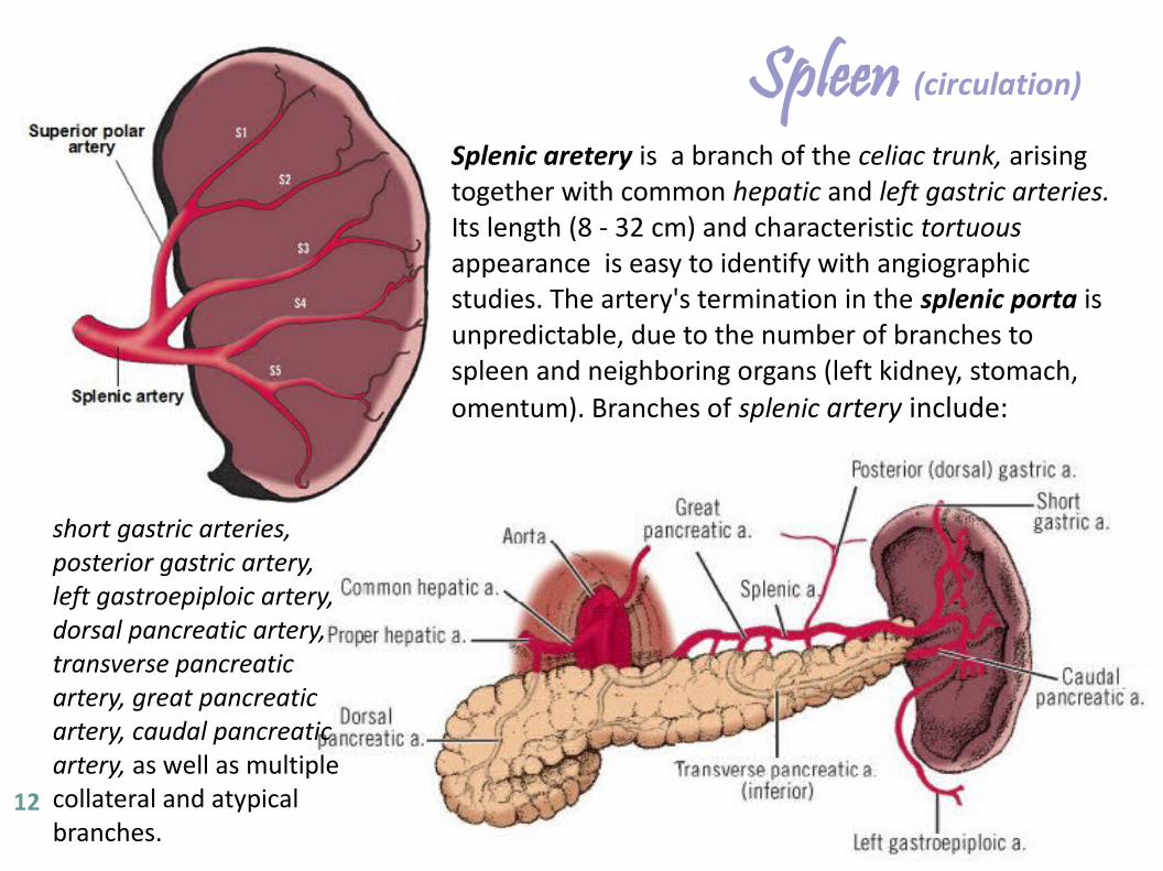
Spleen has 3 to 5 vascular segments named S1-S5 (slide 12). Subsequent to its origin from the celiac trunk, the splenic artery courses leftward and retroperitoneally under the posterior wall of the omental bursa, along the posterior-superior edge of the pancreatic islet, with multiple branches into pancreatic parenchyma.
Spleen (circulation)
Splenic aretery is a branch of the celiac trunk, arising together with common hepatic and left gastric arteries. Its length (8 - 32 cm) and characteristic tortuous appearance is easy to identify with angiographic studies. The artery's termination in the splenic porta is unpredictable, due to the number of branches to spleen and neighboring organs (left kidney, stomach, omentum). Branches of splenic artery include:
12
short gastric arteries, posterior gastric artery, left gastroepiploic artery, dorsal pancreatic artery, transverse pancreatic artery, great pancreatic artery, caudal pancreatic artery, as well as multiple collateral and atypical branches.
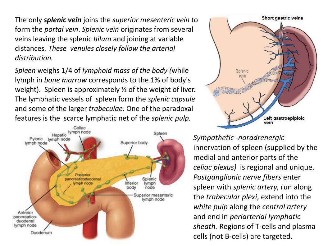
The only splenic vein joins the superior mesenteric vein to form the portal vein. Splenic vein originates from several veins leaving the splenic hilum and joining at variable distances. These venules closely follow the arterial distribution.
Spleen weighs 1/4 of lymphoid mass of the body (while lymph in bone marrow corresponds to the 1% of body's weight). Spleen is approximately ½ of the weight of liver. The lymphatic vessels of spleen form the splenic capsule and some of the larger trabeculae. One of the paradoxal features is the scarce lymphatic net of the splenic pulp.
Sympathetic -noradrenergic innervation of spleen (supplied by the medial and anterior parts of the celiac plexus) is regional and unique. Postganglionic nerve fibers enter spleen with splenic artery, run along the trabecular plexi, extend into the white pulp along the central artery and end in periarterial lymphatic sheath. Regions of T-cells and plasma cells (not B-cells) are targeted.
Spleen (concluding)
Most of the blood flow passes through the splenic marginal zone and directly via the white pulp, ensuring an efficient monitor. While the white pulp is mainly for the adaptive immunity, the marginal zone is involved in both innate and adaptive responses through specific metallomorphic macrophages and B-cells. In addition to pattern-recognition receptors (Toll-like receptors) expressed by most tissue macrophages, marginal-zone metallomorphic macrophages express a C-type lectin SIGNR1 and type-I scavenger receptor MARCO. SIGNR-1 efficiently binds polysaccharide antigens (Micobacterium Tuberculosae, Streptococcus Pneumoniae, E-coli, Staphylococcus aureus). Marginal-zone macrophages lack the expression of MHC class II molecules and subsequent activation of marginal-zone-B cells occurs through shedding of pathogen-degradation products that are opsonized by complement.When signaling through LT-β receptors (LT-βR) or TNF receptor 1 (TNFR1) is lacking, levels of homeostatic chemokines CXCL13, CCL19 and CCL21 are reduced in spleen, resultant in disorganization of the white-pulp. Chemokines CCL19, CCL21 (produced by the stromal cells of T-cell zone) play a crucial role in regulating T-cell zone integrity.Splenectomy leads to mild thrombocytosis (spleen can store 1/3 of total body platelets, so removal allows more circulate in plasma), Howell-Jolly bodies (RBC remnants), poorer response to vaccines, and higher risk of certain infections: Haemophilus influenzae, Neisseria meningitidis, Salmonella typhi - “SHiNS”. 14
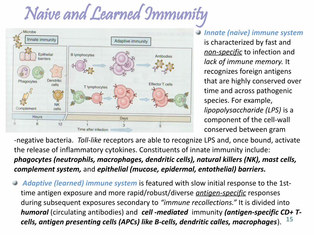
Naive and Learned ImmunityInnate (naive) immune system is characterized by fast and non-specific to infection and lack of immune memory. It recognizes foreign antigens that are highly conserved over time and across pathogenic species. For example, lipopolysaccharide (LPS) is a component of the cell-wall conserved between gram
15
-negative bacteria. Toll-like receptors are able to recognize LPS and, once bound, activate the release of inflammatory cytokines. Constituents of innate immunity include: phagocytes (neutrophils, macrophages, dendritic cells), natural killers (NK), mast cells, complement system, and epithelial (mucose, epidermal, entothelial) barriers.
Adaptive (learned) immune system is featured with slow initial response to the 1st-time antigen exposure and more rapid/robust/diverse antigen-specific responses during subsequent exposures secondary to “immune recollections.” It is divided into humoral (circulating antibodies) and cell -mediated immunity (antigen-specific CD+ T-cells, antigen presenting cells (APCs) like B-cells, dendritic calles, macrophages).
Workhorses of DefenseAll cells of immune system originate from hematopoietic stem-cells of the bone marrow. These are multipotent cells (are able to form all blood cell lines) and have the capacity of self-renewal. They further differentiate to form myeloid and lymphoid cell-lines:
MYELOID LINEAGE➔ Basophil: Mature cell with bilobed nucleus and large blue granules.➔ Dendritic cell: Have long cytoplasmic arms capable of efficient antigen presentation to
lymphocytes (professional antigen-presenting cells [APCs]).➔ Eosinophil: Mature cell with bilobed nucleus and large pink granules containing major
basic protein that attacks parasitic and helminthic agents.➔ Macrophage: Tissue histocyte (differentiated monocyte) capable for phagocytosis, as well
as synthesis and secretion of various cytokines (IL-1, IL-6, IL8, IL-12, TNF-α).➔ Mast cell: Has small nucleus and large cytoplasmatic granules containing histamine and
other allergic mediators in response to allergies, hives, anaphylaxis.➔ Monocyte: Circulating phagocytic cell to be further stimulated and differentiated to the
tissue macrophage. LYMPHOID LINEAGE➔ B-lymphocytes: Cells that undergo differentiation into either memory B-cells or plasma cells
(that produce antibodies).➔ T-lymphocytes: Cells that further differentiate to either CD4+ helpers, CD8+ cytotoxic cells,
regulatory T-cells, and memory T-cells.➔ Natural Killers (NK): CD56+ lymphocytes containing cytoplasmic toxic granules (granzymes)
and are able to kill malignant, virus-infected, and antibody-coated (opsonized) cells. 16
Major Histocompatibility Complexes: MHC- 1, MHC-2 MHC system helps us discern ourselves from everything else, as well as detect when body's own cells are either infected or undergo malignant changes. Two structurally and functionally distinct classes of MHC are involved:MHC Class I is present in all nucleated cells of our body and is encoded by human leukocyte antigen genes HLA-A, HLA-B, HLA-C. MHC is a cell-surface protein that displays peptide fragments from inside the cell to out. Normally, the antigen loaded onto MHC-I is an autoreactive antigen and cytotoxic CD8+ T-cells will not react to it. However, if a virus infects a cell, it produces viral proteins using the host's cellular machinery. These viral proteins too, are loaded onto MHC-I. This makes cytotixic CD8+ confer immunity to viral infection, and by recognizing the viral antigen they target to destroy it – if a costimulatory signal (for anergy) isn't present (discussed in slide 23).MHC Class II is only present on antigen-coated cells (macrophages, dendritic cells), is encoded by the HLA genes HLA-DP, HLA-DQ, HLA-DR, and composed of two α- and β- subunits. After phagocytosing the microbe, the APCs process and load antigens onto MHC-II. Then, MHC-II is inserted into the cell membrane to make it recognizable by CD4+ T-cells (helpers) which after activate B-cells and trigger local inflammation. 17
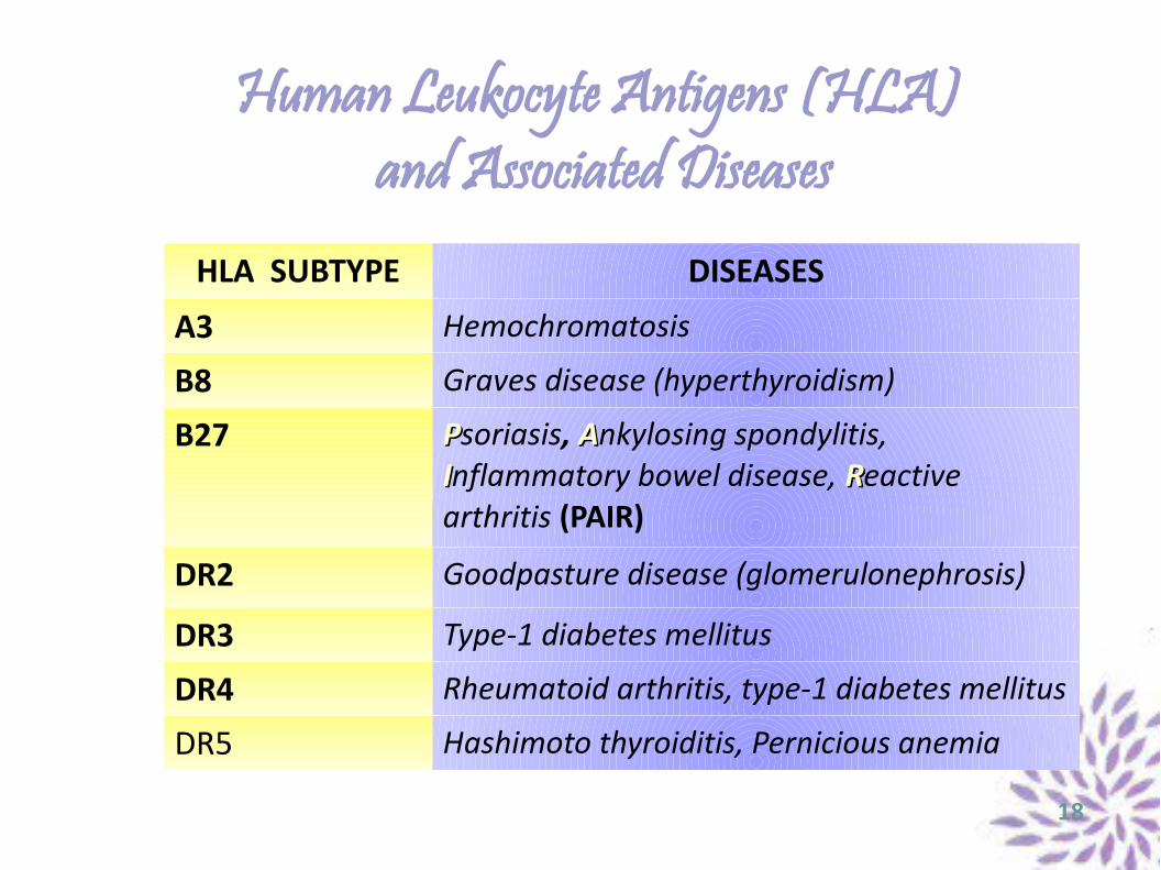
Human Leukocyte Antigens (HLA) and Associated Diseases
HLA SUBTYPE DISEASES
A3 Hemochromatosis
B8 Graves disease (hyperthyroidism)
B27 PPsoriasis, AAnkylosing spondylitis, IInflammatory bowel disease, RReactive arthritis (PAIR)
DR2 Goodpasture disease (glomerulonephrosis)
DR3 Type-1 diabetes mellitus
DR4 Rheumatoid arthritis, type-1 diabetes mellitus
DR5 Hashimoto thyroiditis, Pernicious anemia
18
Immunoglobulins Antibody formation is accomplished by mature plasma B-cells which synthesize and release immunoglobulins (Ig) after being activated by the appropriate mechanisms of antigen stimulation. The number of antigens is unlimited; so too the number of activation mechanisms. This is called antibody diversity and is built on four main processes:
1) Random recombination of VJ (light chain) or V(D)J (heavy chain) genes
2) Random combination of various heavy chains with light chains
3) Somatic hypermutation in germinal centers (after antigen stimulation)
4) Terminal deoxynucleotidyl transferase (TdT) addition of DNA to the heavy and light chains.
The common antibody isotypes are IgM and IgG. Isotope switching occurs after antigen stimulation and activation of B-cells, resulting in alternative splicing of messenger - mRNA. The resultant post-translational modification of mRNA dictates the isotype of plasma B-cells (IgA, IgG, IgE, IgM, etc).
19
Ig and Ig Deficiency SyndromesIgA: Occurs as a monomer in bloodstram and as a diamer when secreted (epithelial cell component). IgA is secreted onto mucosal surfaces (GI, GUI, respiratory) to block attachment of pathogens to mucose membranes.
IgD: Found on the surface of mature B-cells. Function is unclear.
IgE: Implicated in allergic response (type-1 hypersensitivity) because it binds with both mast cells and basophils and undergoes cross-linking after exposure to appropriate antigen.
IgG: The main antibody in the secondary (slow) antigenic response. Occurs as a monomer in complement fixing job, cross the placenta to provide passive immunity to developing fetus, opsonize bacteria, neutralize various toxins and viruses. IgD does not make multimers and therefore, it does cross the placenta.
IgM: Found on the surface of mature B-cells. Produced in the primary, fast antigen response. Occurs as a monomer and commonly as a pentamer for more efficient trapping and complement fixing. As a pentamer is does not cross the placenta. 20
B-cell Deficiency and Ig Deficiency Syndromes● COMMON VARIABLE IMMUNODEFICIENCY: Most common form of
primary B-cell deficiency with distinct low levels of IgG and IgA (rarely IgM) resultant in high rates of lymphomas and gastric cancer.
● HYPER IgM SYNDROME: Normal level of B-cells, with diminished levels of IgG and IgA and higher levels of IgM. Associated with increased risk for Pneumocystis infections (fungi). Conditioned with inability of isotype switching and sequential secondary deficiency in CD40 ligand on Th2 cells.
● SELECTIVE IgA DEFICIENCY: Most common type of Ig deficiency, associated with increased respiratory, GI, GUI infections, and high risk of anaphylaxis from blood-product transfusions.
● X-LINKED AGAMMAGOBULINEMIA (Bruton agammaglobulinemia): Results from a mutation in the receptor tyrosine kinase (BTK). Present exclusively in males. Children with XLA are usually healthy for the first months of infancy, as they are protected by the maternal antibodies. After, they begin to develop recurrent infections, resistant to anti-microbal treatment. 21
T-cells: Regulated and Educated AssassinsAs noted before, T-cells originate from the lymphoid lineage of hematopoietic differentiation. They are “born” in the bone-marrow and “trained” in the thymus. It is in thumys where they get “certifications” of helpers (CD4) or assassins (CD8).
CD4+ T Cells: These “helpers” (Th) will undergo further differentiation after appropriate stimulation by interleukins to become either Th1 or Th2 with specific functions to help regulate both humoral and cell-mediated immunity. - Th1 are involved in regulation of cell-mediated response, and are activated by APCs and secret interferon-gamma (IFN-γ) which in turn activates APCs for efficient killing. They also secrete IL-2 which activates CD8+ to kill virally infected cells. - Th2 are involved in activating B-cells and enhancing isotype switching by secreting IL-4, IL-5, and IL-6.
CD8+ T Cells: These cytotoxic cells are responsible for seeking out and eliminating virus/parasite-infected cells, cancer cells, and other foreign cells.
22
T-cell Deficiency SyndromesViral infection has unique activation arrays. When an APC (dendritic cell, macrophage) is exposed to viral antigen, it will load the latter onto MHC-II for presentation to the CD4+ T-cell. It will also express co-stimulatory signal. The T-cell receptor (TCR) then interacts with antigen-positive MHC-II on the APC. Yet, a single signal is not enough. Immune system's checks and balances require a second signal for an appropriate activation. This B7 costimulatory signal on the APC must interact with CD28 on CD4+ T-cell while the TCR-MHC II interaction is occurring. If these conditions are met, then CD4+ T-cell will also release IL-2 to activate CD8+ killers and differentiate CD4+T-cell in autocrine manner. If TCR, weirdly sees and binds to host antigens (autoreactivity), immune system tackles with this issue by anergy command, i.e. deactivating self-reactive T-cells. If this process fails as well, then autoimmune disorders occur.
T-CELL DEFICIENCT SYNROMES: - Acquired Immunodeficiency Syndrome (AIDS): The final stage of the decremental quantity and quality of T-cells (CD4+) caused by HIV. - Ataxia Telangiectasia: A T-cell deficiency along with cerebellar ataxia and increased risk for various types of cancer (impaired double-strand DNA repair). - DiGeorge Syndrome: 22q11.2 deletion syndrome resultant in CATCH-22 (Cardiac defects, Abnormal facies, Thymic hypoplasia, Cleft palate, Hypocalciemia). - Severe Combined Immunodeficiency (SCID): The common form of X-linked disorder with suspectibility to numerous pathogenic infections with diarrhea, pneumonia, otitis, sinusitis.
23
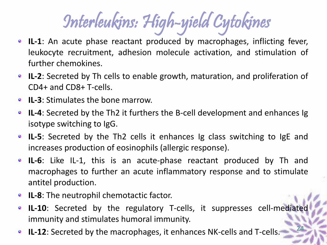
Interleukins: High-yield CytokinesIL-1: An acute phase reactant produced by macrophages, inflicting fever, leukocyte recruitment, adhesion molecule activation, and stimulation of further chemokines.IL-2: Secreted by Th cells to enable growth, maturation, and proliferation of CD4+ and CD8+ T-cells.IL-3: Stimulates the bone marrow.IL-4: Secreted by the Th2 it furthers the B-cell development and enhances Ig isotype switching to IgG.IL-5: Secreted by the Th2 cells it enhances Ig class switching to IgE and increases production of eosinophils (allergic response).IL-6: Like IL-1, this is an acute-phase reactant produced by Th and macrophages to further an acute inflammatory response and to stimulate antitel production.IL-8: The neutrophil chemotactic factor. IL-10: Secreted by the regulatory T-cells, it suppresses cell-mediated immunity and stimulates humoral immunity.IL-12: Secreted by the macrophages, it enhances NK-cells and T-cells. 24
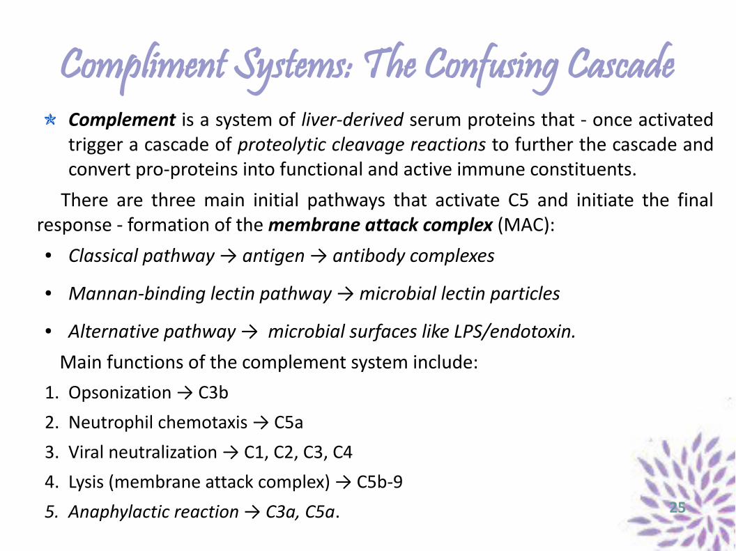
Compliment Systems: The Confusing CascadeComplement is a system of liver-derived serum proteins that - once activated trigger a cascade of proteolytic cleavage reactions to further the cascade and convert pro-proteins into functional and active immune constituents.
There are three main initial pathways that activate C5 and initiate the final response - formation of the membrane attack complex (MAC):
● Classical pathway → antigen → antibody complexes
● Mannan-binding lectin pathway → microbial lectin particles
● Alternative pathway → microbial surfaces like LPS/endotoxin. Main functions of the complement system include:1. Opsonization → C3b
2. Neutrophil chemotaxis → C5a
3. Viral neutralization → C1, C2, C3, C4
4. Lysis (membrane attack complex) → C5b-9
5. Anaphylactic reaction → C3a, C5a. 25
Complement Deficiency and Associated Conditions
26
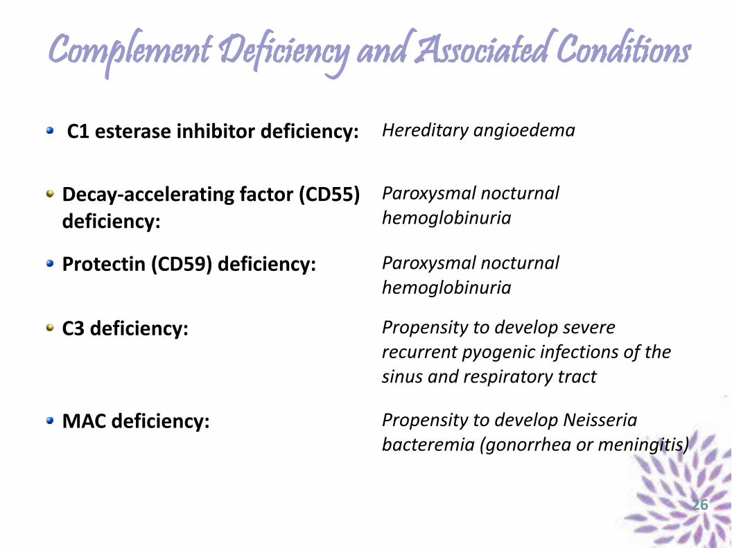
C1 esterase inhibitor deficiency: Hereditary angioedema
Decay-accelerating factor (CD55) deficiency:
Paroxysmal nocturnal hemoglobinuria
Protectin (CD59) deficiency: Paroxysmal nocturnal hemoglobinuria
C3 deficiency: Propensity to develop severe recurrent pyogenic infections of the sinus and respiratory tract
MAC deficiency: Propensity to develop Neisseria bacteremia (gonorrhea or meningitis)
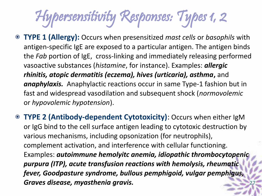
Hypersensitivity Responses: Types 1, 2TYPE 1 (Allergy): Occurs when presensitized mast cells or basophils with antigen-specific IgE are exposed to a particular antigen. The antigen binds the Fab portion of IgE, cross-linking and immediately releasing performed vasoactive substances (histamine, for instance). Examples: allergic rhinitis, atopic dermatitis (eczema), hives (urticaria), asthma, and anaphylaxis. Anaphylactic reactions occur in same Type-1 fashion but in fast and widespread vasodilation and subsequent shock (normovolemic or hypovolemic hypotension).
TYPE 2 (Antibody-dependent Cytotoxicity): Occurs when either IgM or IgG bind to the cell surface antigen leading to cytotoxic destruction by various mechanisms, including opsonization (for neutrophils), complement activation, and interference with cellular functioning. Examples: autoimmune hemolyitc anemia, idiopathic thrombocytopenic purpura (ITP), acute transfusion reactions with hemolysis, rheumatic fever, Goodpasture syndrome, bullous pemphigoid, vulgar pemphigus, Graves disease, myasthenia gravis. 27
Hypersensitivity Responses: Types 3, 4
TYPE 3 (Immune Complex Disease): Occurs when antigen-antibody (mainly IgG) complexes are formed and deposited in tissues, resulting in activation of complement systems and recruitment of neutrophils leading to the tissue injury. Examples: systemic lupus erythematosus (SLE), rheumatoid arthritis, Arthus reaction, serum sickness, post-streptococcal glomerulonephritis.
TYPE 4 (Delayed, Antibody-dependent Cytotoxicity): Being not antibody related, these are the only reactions that are not transferred by serum. These are slow, cell-mediated reactions occurring when the learned T-cell interacts with the same antigen, resulting in lymphokine production and activation of other players (macrophages). Examples: contact dermatitis, nickel allergy, PPD test,, GVHD test, multiple sclerosis, Gullian-Barré syndrome.
28
Graft Immunity: High-yield ImmunosuppressantsTYPES OF TISSUE TRANSPLANTANTS
● Autograft: Transplanting tissue back to the same host but in a different location.● Allograft: Transplanting tissue/organ/fluid/cell from one human to another.● Xenograft: Transplanting tissue from animals to a human being.
TYPES OF REJECTIONHyperacute rejection: Rapid failure resultant from complement activation and neutrophil migration into the donor organ. Acute rejection: T-cell-mediation rejection occurring within weeks to months, reversible with immunusuppressants.Chronic rejection: Long-lasting failure with progressive loss of function of the transplant – secondary to vascular fibrosis.
HIGH-YIELD IMMUNOSUPPRESSANTSAzathioprine: A prodrug converted in vivo to 6-mercaptopurine (6-MP) to inhibit purine metabolism and proliferation of T and B cells. Used in acute rejection and autoimmune disorders. Cyclosporine: Binds to cyclophilin, which inhibits calcineurin and therefore prevents transcription of IL-2 in T-cells. Used in tissue transplantation only.Tacrolimus: Binds to FK-binding protein, inhibits calcineurin (and IL-2). Used as ciclosporine replacement.Microphenolate mofetil: Inhibits inosine monophosphate dehydrogenase, the rate-limiting enzyme in guanosine monophosphate (GMP) synthesis. Used in organ transplantation only. 29
Review Q & A Q1. Which type of immunization a patient with a compliment disorder involving deficiency of the membrane attack complex (MAC) should specifically receive?
A1. That patient is at risk for Neisseria infections, therefore is at high risk for meningococcal infections. S/he should receive meningococcal (MCV-4) vaccine.
Q2. A teenage African male with sickle-cell anemia presents for a regular exam. The peripheral smear shows sickle cells together with RBCs and large numbers of platelets. Your guess.
A2. The peripheral smear with thrombocytosis and Howell-Jolly bodies suggests that the patient is after splenectomy or autosplenectomy (from the chronic splenic infections).
Q3: Which cell type in the human body does not express MHC class I?
A3: MHC-I is expressed by all nucleated cells. Therefore, mature RBC (erythrocytes) no longer express MHC class I on their membranes. 30
Kennedy v. Collagen Corp. (1998)1
● PROCEDURAL HISTORY: Plaintiff alleges injuries sustained by defendant (Collagen Corporation) and its employees for negligently injecting her Zyderm, a substance made from the skin, tendons, and connective tissue of bovine animals, for purposes of cosmetically curing her facial wrinkles. The claimed side-effect is systemic lupus erythematosus (SLE), for what strict liability, breach of express and implied warranty, battery, and conspiracy are incorporated in this malpractice claim. Former two summary judgments, entered for defendant, were based on insufficient expert testimony (a poor cause-effect relationship affidavit) on part of plaintiff.
● HOLDING: The district court abused its discretion by improperly applying the Daubert test, as it failed to consider relevant scientific evidence presented by plaintiff's expert witness.
● JURISPRUDENCE: Daubert v. Merrell Dow Pharmaceuticals (1993) 2
● DISPOSITION: Judgment of the former court is reversed and remanded.● REASONING: Daubert's court has established that the trial judge, in making
initial assessment as to the admission of evidence, must determine whether the expert's testimony reflects (1) "scientific knowledge," and (2) will assist the trier of fact to understand or determine a material fact at issue. This requires a preliminary assessment of a number of factors. 1) 161 F. 3d 1226 - Court of Appeals, 9th Circuit 1998; 2) 509 US 579 - Supreme Court 1993
DISCLAIMERDISCLAIMERThis presentation does not constitute a medical or
legal advice. The burden for determining its completeness, suitability or appropriateness for
intended use or purpose rests solely on the reader accessing this information.
The author declares no commercial, strategic, or financial interest or trusteeship with the names of
entities either used or omitted.
32
Related Documents