ARTICLE Immunolocalization of Periostin-like Factor and Periostin During Embryogenesis Shimei Zhu, Mary F. Barbe, 1 Neilay Amin, Shobha Rani, Steven N. Popoff, Fayez F. Safadi, and Judith Litvin 1 Department of Anatomy and Cell Biology (SZ,MFB,NA,SR,SNP,FFS,JL), Musculoskeletal Research Group (MFB,SNP,FFS,JL), and Department of Physical Therapy, College of Health Professions (MFB), Temple Medical School, Philadelphia, Pennsylvania SUMMARY Periostin-like factor (PLF) and Periostin are alternatively spliced mRNAs. Our findings are the first to show similarities and differences between PLF and Periostin location using isoform-specific antibodies. The differences in when and where they are present during mouse embryogenesis suggest that they may have different functions. Using immunostaining techniques, we observed that PLF was highly expressed at 12.5 days postconception (dpc) in the intermediate and outer zones of most brain regions, spinal cord, cranial and spinal nerves, and chondrocytes in developing bone and in the heart wall. By 16.5 dpc, PLF was also present in ameloblasts and odontoblasts in developing teeth, and by 19.5 dpc, PLF was present at low levels only in vagal nerve bundles, discrete white matter bundles in the brain, and chondrocytes of developing ribs. Periostin, on the other hand, was absent at 12.5 dpc from dorsal spinal cord and from cranial and spinal nerves. By 16.5 dpc, Periostin was present in many spinal nerves, but absent thereafter, and at 19.5 dpc, Periostin was present in chondrocytes in developing bone but not in neural tissues. The different spatial and temporal location of PLF and Periostin in cartilage and bone cells suggests different roles for these proteins in endochondral bone formation. The early expression of PLF in brain differentiation zones and in developing axon bundles and nerves suggests that it may facilitate axon growth. (J Histochem Cytochem 56:329–345, 2008) KEY WORDS Periostin-like factor embryogenesis heart brain spinal cord nerves PERIOSTIN-LIKE FACTOR (PLF) was first identified in cardiac tissue and is highly homologous to Periostin and bIG-H3 (Litvin et al. 2004,2005). PLF is ex- pressed in the heart during embryogenesis and during neonatal development. In the adult, it is upregulated in the heart in patients with cardiomyopathy, and our findings suggest that Periostin isoforms play a crucial role in adult cardiac myocyte growth after mechanical overload (Litvin et al. 2005,2006). Recent findings suggest a role for Periostin in cardiac hyper- trophy and ventricular remodeling (Oka et al. 2007). In vascular smooth muscle cells (VSMCs), PLF levels increased in response to mitogen stimulation, and its ability to promote VSMC proliferation and migra- tion suggests a role in vascular proliferative disease (Litvin et al. 2007). In both the heart and vasculature, PLF seems to be present in the adult only under con- ditions of overload or injury; only very low levels are detected normally. Sequence analysis of PLF identified an N-terminal signal sequence, suggesting that the protein is secreted; a putative nuclear localization sequence (NLS), suggest- ing that it may be translocated to the nucleus; one potential N-linked glycosylation site; and four fasciclin domains each containing 150 amino acids (Litvin et al. 2004). Proteins that contain fasciclin domains are re- lated to Fasciclin, identified in insects (Zinn et al. 1988). In both Drosophila and grasshoppers, fasciclin I is expressed on the surface of a subset of commissural axon pathways in the embryonic central nervous sys- tem (CNS) and on sensory axonal pathways in the peripheral nervous system (PNS) (McAllister et al. Correspondence to: Judith Litvin, PhD, Department of Anatomy and Cell Biology, Temple Medical School, 3420 N. Broad Street, MRB 615, Philadelphia, PA 19140. E-mail: [email protected] 1 These authors contributed equally to this work. Received for publication July 23, 2007; accepted November 15, 2007 [DOI: 10.1369/jhc.7A7321.2007]. The Journal of Histochemistry & Cytochemistry C The Histochemical Society, Inc. 0022-1554/07/$3.30 329 Volume 56(4): 329–345, 2008 Journal of Histochemistry & Cytochemistry http://www.jhc.org

Welcome message from author
This document is posted to help you gain knowledge. Please leave a comment to let me know what you think about it! Share it to your friends and learn new things together.
Transcript
ARTICLE
Immunolocalization of Periostin-like Factor and PeriostinDuring Embryogenesis
Shimei Zhu, Mary F. Barbe,1 Neilay Amin, Shobha Rani, Steven N. Popoff, Fayez F. Safadi,and Judith Litvin1
Department of Anatomy and Cell Biology (SZ,MFB,NA,SR,SNP,FFS,JL), Musculoskeletal Research Group (MFB,SNP,FFS,JL),and Department of Physical Therapy, College of Health Professions (MFB), Temple Medical School,Philadelphia, Pennsylvania
SUMMARY Periostin-like factor (PLF) and Periostin are alternatively spliced mRNAs. Ourfindings are the first to show similarities and differences between PLF and Periostin locationusing isoform-specific antibodies. The differences in when and where they are present duringmouse embryogenesis suggest that theymay have different functions. Using immunostainingtechniques, we observed that PLF was highly expressed at 12.5 days postconception (dpc) inthe intermediate and outer zones of most brain regions, spinal cord, cranial and spinal nerves,and chondrocytes in developing bone and in the heart wall. By 16.5 dpc, PLF was also presentin ameloblasts and odontoblasts in developing teeth, and by 19.5 dpc, PLF was present atlow levels only in vagal nerve bundles, discrete white matter bundles in the brain, andchondrocytes of developing ribs. Periostin, on the other hand, was absent at 12.5 dpc fromdorsal spinal cord and from cranial and spinal nerves. By 16.5 dpc, Periostin was presentin many spinal nerves, but absent thereafter, and at 19.5 dpc, Periostin was present inchondrocytes in developing bone but not in neural tissues. The different spatial and temporallocation of PLF and Periostin in cartilage and bone cells suggests different roles for theseproteins in endochondral bone formation. The early expression of PLF in brain differentiationzones and in developing axon bundles and nerves suggests that it may facilitate axongrowth. (J Histochem Cytochem 56:329–345, 2008)
KEY WORDS
Periostin-like factor
embryogenesis
heart
brain
spinal cord
nerves
PERIOSTIN-LIKE FACTOR (PLF) was first identified incardiac tissue and is highly homologous to Periostinand bIG-H3 (Litvin et al. 2004,2005). PLF is ex-pressed in the heart during embryogenesis and duringneonatal development. In the adult, it is upregulatedin the heart in patients with cardiomyopathy, andour findings suggest that Periostin isoforms play acrucial role in adult cardiac myocyte growth aftermechanical overload (Litvin et al. 2005,2006). Recentfindings suggest a role for Periostin in cardiac hyper-trophy and ventricular remodeling (Oka et al. 2007).In vascular smooth muscle cells (VSMCs), PLF levels
increased in response to mitogen stimulation, and itsability to promote VSMC proliferation and migra-tion suggests a role in vascular proliferative disease(Litvin et al. 2007). In both the heart and vasculature,PLF seems to be present in the adult only under con-ditions of overload or injury; only very low levels aredetected normally.
Sequence analysis of PLF identified an N-terminalsignal sequence, suggesting that the protein is secreted;a putative nuclear localization sequence (NLS), suggest-ing that it may be translocated to the nucleus; onepotential N-linked glycosylation site; and four fasciclindomains each containing 150 amino acids (Litvin et al.2004). Proteins that contain fasciclin domains are re-lated to Fasciclin, identified in insects (Zinn et al. 1988).In both Drosophila and grasshoppers, fasciclin I isexpressed on the surface of a subset of commissuralaxon pathways in the embryonic central nervous sys-tem (CNS) and on sensory axonal pathways in theperipheral nervous system (PNS) (McAllister et al.
Correspondence to: Judith Litvin, PhD, Department of Anatomyand Cell Biology, Temple Medical School, 3420 N. Broad Street,MRB 615, Philadelphia, PA 19140. E-mail: [email protected]
1These authors contributed equally to this work.Received for publication July 23, 2007; accepted November 15,
2007 [DOI: 10.1369/jhc.7A7321.2007].
The
Journ
alof
His
toch
emis
try
&C
ytoch
emis
try
C The Histochemical Society, Inc. 0022-1554/07/$3.30 329
Volume 56(4): 329–345, 2008
Journal of Histochemistry & Cytochemistry
http://www.jhc.org
1992). Although the details on the molecular mecha-nisms involved are unclear, fasciclin I mediates inter-actions between cell surfaces in the nervous system.Therefore, in examining the temporal and spatial loca-tion of PLF, we paid particular attention to the devel-oping nervous system.
We rely on data obtained from studies on bIG-H3and Periostin to provide clues about the role of PLF inthe structure and function of cells. This is the first re-port to examine differences between PLF and Periostinlocalization during embryogenesis using isoform-specific antibodies.bIG-H3was first identified in adeno-carcinoma cells treated with transforming growthfactor-b (Skonier et al. 1992). It has a signal sequenceat the N terminus, an Arg-Gly-Asp sequence at the Cterminus, and four Fas domains (Skonier et al. 1992).Transcripts of bIG-H3 are detected in connective tis-sues including cartilage during embryogenesis (Fergusonet al. 2003). bIG-H3 is expressed in preosteoblasts,mediates osteoblast adhesion, and inhibits osteoblastdifferentiation (Thapa et al. 2005). In addition, it issecreted from various cell types and is detected innuclei of human bladder smooth muscle cells andfibroblasts (Billings et al. 2000). The major function ofbIG-H3 as a secreted protein is to mediate cell spread-ing, adhesion, proliferation, andmigration (reviewed byLitvin et al. 2005).
Periostinwas first identified inMC3T3-E1 osteoblast-like cells. The major difference between PLF andPeriostin is at the C-terminal region (Litvin et al. 2004).Periostin is expressed in osteoblasts in vitro and inperiosteum and periodontal ligament tissues in vivo. Ithas several isoforms (PLF being one of them). Periostinis secreted and supports MC3T3-E1 cell adhesion andspreading (Horiuchi et al. 1999). Periostin is expressedin the teeth and its surrounding tissues during devel-opment but not in CNS tissues (Goetsch et al. 2003;Suzuki et al. 2004). It is also expressed and functions inother cell types and tissues, as reviewed by Litvin et al.(2005). Mice deficient in Periostin display dwarfism,incisor enamel defects, and an early-onset periodontaldisease–like phenotype (Rios et al. 2005). Moreover,it has been shown that Periostin is regulated by Twist,a transcription factor, which is essential for intramem-branous ossification (el Ghouzzi et al. 1997; Oshimaet al. 2002).
In our previous study, we showed using in situhybridization that Periostin isoform mRNAs are de-tected in cartilage and developing bone at 16.5 dayspostconception (dpc) (Litvin et al. 2004). However,we have yet to explore the localization of eachisoform, Periostin, and PLF protein in other tissues.The primary objective of this paper was to examinethe temporal and spatial expression of PLF andPeriostin immunohistochemically using PLF andPeriostin-specific antibodies in developing mouse em-
bryonic tissues and to compare their spatial andtemporal location.
Materials and Methods
Tissue Collection
Mouse embryos that were 12.5, 13.5, 16.5, and19.5 dpc were collected from mouse dams after eutha-nasia using sodium pentobarbital (120 mg/kg bodyweight). Three to five embryos were collected per agegroup. The embryos were immersion fixed in 4% para-formaldehyde in phosphate buffer (pH 7.4), processedfor paraffin embedding, and sectioned. Sections were5 mm thick, were cut in a cephalic to caudal directionalong the long axis, and were mounted on chargedand coated glass slides (Fisher Plus; Fisher Scientific,Fair Lawn, NJ). Animals (dams) were maintained andused according to the principles in the National Insti-tutes of Health Guide for the Care and Use of Labo-ratory Animals (US Department of Health and HumanServices, Publication No. 86-23, 1985) and guidelinesestablished by the Institutional Care and Animal UseCommittee of Temple University.
Western Blot Analysis
Tissues were homogenized in radioimmunoprecip-itation assay (RIPA) buffer (50 mM HEPES, pH 7.5,150 mmol NaCl, and 0.1% Tritron X-100) containingprotease inhibitors (Sigma; St Louis, MO). Cells inculture were washed in PBS, and proteins were ex-tracted in RIPA buffer containing protease inhibitors.Lysates were incubated on ice for 20 min, passedthrough an 18-gauge needle twice, and centrifuged at3000 rpm for 10 min at 4C. Protein concentration wasdetermined using the BCA kit (Pierce; Rockford, IL).Equal protein from each sample (100 mg/lane) wasseparated by PAGE, and the proteins were transferredto nitrocellulose. The blots were probed with affinitypurified polyclonal antibodies specific for either Perios-tin or PLF. The Periostin-specific antibody was directedagainst a peptide specific to Periostin (KFIEGGDGHL-FEDEEIKR aa 764–781; Invitrogen, Carlsbad, CA), andthe PLF-specific antibody was directed against a peptidespecific to PLF (EPKIKVIQGSLQPIIKTEG aa 681–699;Litvin et al. 2006). Primary antibody binding was de-tected with horseradish peroxidase–conjugated goatanti-rabbit secondary antibody and visualized by en-hanced chemiluminescence (Amersham; ArlingtonHeights, IL).
Immunohistochemistry
Paraffin-embedded mouse embryo sections at 12.5,13.5, 16.5, and 19.5 dpc were deparaffinized and incu-bated in 1.5% H2O2 for 15 min at room temperature.
The
Journ
alof
His
toch
emis
try
&C
ytoch
emis
try
330 Zhu, Barbe, Amin, Rani, Popoff, Safadi, Litvin
The slides were blocked with 5% goat serum in PBS for1 hr and incubated with PLF antibody at a 1:1000 dilu-tion overnight at 4C or in Periostin antibody at a1:750 dilution overnight at 4C. The PLF-specific anti-body is described by Litvin et al. (2006). Subsequentsecondary antibody and substrate-chromogen devel-opment was carried out using a diaminobenzidineperoxidase kit (Vector Laboratories; Burlingame, CA).Sections were counterstained with nuclear stains, suchas hematoxylin, nuclear red, or 4¶,6-Diamidino-2-phenylindole, dihydrochloride (the latter as a fluores-cent mounting media from Vector Laboratories), orwith 1% Alcian blue (a cartilage counterstain). Somesections were also co-immunostained using immuno-fluorescent techniques after the PLF staining forPGP9.5, a general nerve marker used here to identifynerve fascicles (cat. no. ab8189; Abcam, Cambridge,MA). Sections were first incubated in 10% goat serumwith 0.3% triton in PBS for 1 hr and then in thePGP9.5 antibody at a 1:50 dilution in PBS overnight atroom temperature. A goat anti-mouse Cy3 (red fluores-cent tag; Jackson ImmunoResearch Laboratories, Inc.,West Grove, PA) was used for detection. Omission of theprimary antibody or replacement of the antibody withnormal serum was used as negative controls.
Results
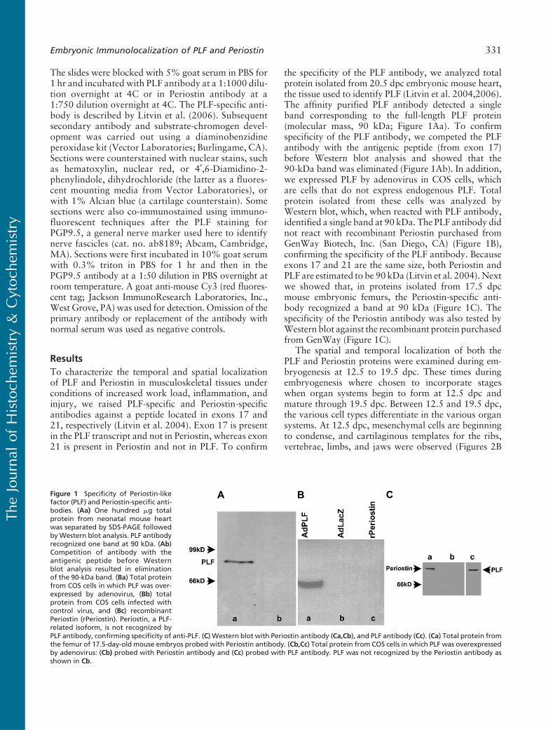
To characterize the temporal and spatial localizationof PLF and Periostin in musculoskeletal tissues underconditions of increased work load, inflammation, andinjury, we raised PLF-specific and Periostin-specificantibodies against a peptide located in exons 17 and21, respectively (Litvin et al. 2004). Exon 17 is presentin the PLF transcript and not in Periostin, whereas exon21 is present in Periostin and not in PLF. To confirm
the specificity of the PLF antibody, we analyzed totalprotein isolated from 20.5 dpc embryonic mouse heart,the tissue used to identify PLF (Litvin et al. 2004,2006).The affinity purified PLF antibody detected a singleband corresponding to the full-length PLF protein(molecular mass, 90 kDa; Figure 1Aa). To confirmspecificity of the PLF antibody, we competed the PLFantibody with the antigenic peptide (from exon 17)before Western blot analysis and showed that the90-kDa band was eliminated (Figure 1Ab). In addition,we expressed PLF by adenovirus in COS cells, whichare cells that do not express endogenous PLF. Totalprotein isolated from these cells was analyzed byWestern blot, which, when reacted with PLF antibody,identified a single band at 90 kDa. The PLF antibody didnot react with recombinant Periostin purchased fromGenWay Biotech, Inc. (San Diego, CA) (Figure 1B),confirming the specificity of the PLF antibody. Becauseexons 17 and 21 are the same size, both Periostin andPLF are estimated to be 90 kDa (Litvin et al. 2004). Nextwe showed that, in proteins isolated from 17.5 dpcmouse embryonic femurs, the Periostin-specific anti-body recognized a band at 90 kDa (Figure 1C). Thespecificity of the Periostin antibody was also tested byWestern blot against the recombinant protein purchasedfrom GenWay (Figure 1C).
The spatial and temporal localization of both thePLF and Periostin proteins were examined during em-bryogenesis at 12.5 to 19.5 dpc. These times duringembryogenesis where chosen to incorporate stageswhen organ systems begin to form at 12.5 dpc andmature through 19.5 dpc. Between 12.5 and 19.5 dpc,the various cell types differentiate in the various organsystems. At 12.5 dpc, mesenchymal cells are beginningto condense, and cartilaginous templates for the ribs,vertebrae, limbs, and jaws were observed (Figures 2B
Figure 1 Specificity of Periostin-likefactor (PLF) and Periostin-specific anti-bodies. (Aa) One hundred mg totalprotein from neonatal mouse heartwas separated by SDS-PAGE followedbyWestern blot analysis. PLF antibodyrecognized one band at 90 kDa. (Ab)Competition of antibody with theantigenic peptide before Westernblot analysis resulted in eliminationof the 90-kDa band. (Ba) Total proteinfrom COS cells in which PLF was over-expressed by adenovirus, (Bb) totalprotein from COS cells infected withcontrol virus, and (Bc) recombinantPeriostin (rPeriostin). Periostin, a PLF-related isoform, is not recognized byPLF antibody, confirming specificity of anti-PLF. (C) Western blot with Periostin antibody (Ca,Cb), and PLF antibody (Cc). (Ca) Total protein fromthe femur of 17.5-day-old mouse embryos probed with Periostin antibody. (Cb,Cc) Total protein from COS cells in which PLF was overexpressedby adenovirus: (Cb) probed with Periostin antibody and (Cc) probed with PLF antibody. PLF was not recognized by the Periostin antibody asshown in Cb.
The
Journ
alof
His
toch
emis
try
&C
ytoch
emis
try
Embryonic Immunolocalization of PLF and Periostin 331
The
Journ
alof
His
toch
emis
try
&C
ytoch
emis
try
332 Zhu, Barbe, Amin, Rani, Popoff, Safadi, Litvin
and 2H). As development proceeds, at 16.5 dpc, endo-chondral bone formation is typified by the presence ofossification centers, allowing us to examine the expres-sion of both isoforms in differentiating chondrocytesat various stages of maturation. In addition, we presentdata on the presence of PLF and Periostin in central andperipheral neural tissues to ascertain whether it has anearly and transient expression pattern that may suggesta role in neurogenesis or axonogenesis.
At 12.5 dpc
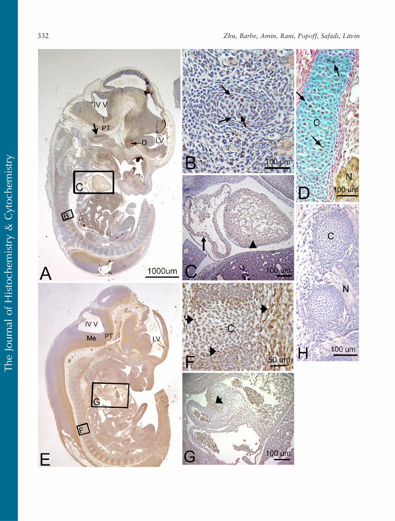
PLF immunoreactivity in mouse embryonic vertebralcartilage at 12.5 dpc was weak (Figures 2B and 2D).Relatively higher staining was detected in the atrium
and ventricle of the heart (Figure 2C). In the nervoussystem, at a stage associated with the onset of neuro-genesis and axon outgrowth, intense staining wasdetected throughout the brain and cord and in pro-cesses associated with cranial and spinal nerves (sum-marized in Table 1). For example, the upper border ofthe cochlear lumen contained PLF-stained cells andprocesses, as did the diencephalon region of the opticstalk (Figure 2A). The presence of PLF in the ganglioniceminence (Figures 3A and 3B), medulla, and pons wasrestricted to the outer differentiating zones (Figure 3C).PLF was absent from the ventricular zone of theganglionic eminence, presumptive pons, and medulla(Figures 3A–3C), a region that consists of progenitor-
’
Figure 2 Localization of PLF and Periostin proteins in developing cartilage and myocardium in a mouse embryo at 12.5 days postconception(dpc). Sectioned embryo was probedwith PLF antibody (A–D) or Periostin antibody (E–G) and counterstained with hematoxylin (a nuclear stain,A–H), nuclear red (a nuclear stain, D), or Alcian blue (a cartilage stain, D). (A) Sagittal section of embryo at low magnification showing fourth(IV V) and lateral (LV) ventricles and positive PLF staining in the presumptive pyramidal track (PT). Large arrow indicates PLF stained cells liningcochlear lumen (brown); small arrow indicates PLF staining in optic stalk region of diencephalon (D). (B) Higher magnification of correspondingarea in A showing cells weakly stained for PLF in developing vertebra (arrows). (C) Higher magnification of corresponding area in A showingPLF staining in the atrium (arrow) and ventricle (arrowhead) of a developing heart. (D) Arrows indicate PLF-stained cells (brown) in rib cartilage(C, blue) and in nearby peripheral nerve (N). (E) Sagittal section of embryo at low magnification showing Periostin staining in the medullarydifferentiating field (Me) and presumptive pyramidal track (PT). (F) Higher magnification of corresponding area in E showing mesenchymalcells that are stained weakly for Periostin in and around the developing vertebral cartilage (C, arrows). (G) Higher magnification ofcorresponding area in E showing Periostin staining in the atrium (arrow) of a developing heart. (H) Photo of negative antibody control slide (noprimary antibody) showing an absence of PLF staining in cartilage (C) and nerve (N).
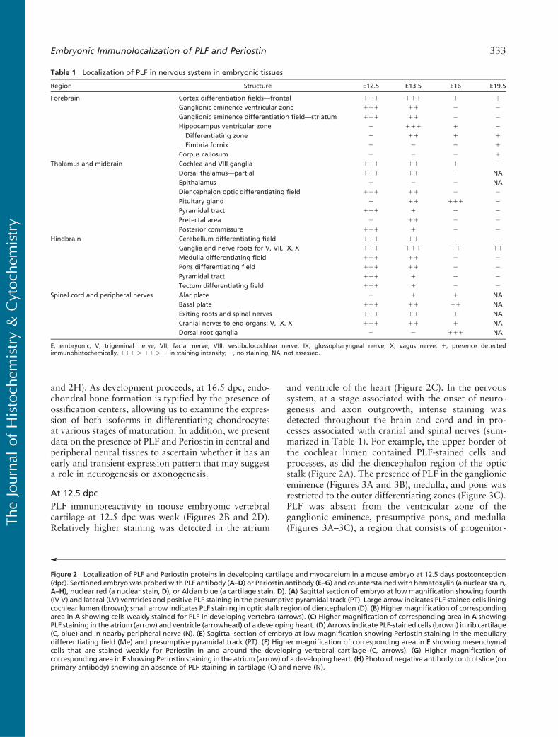
Table 1 Localization of PLF in nervous system in embryonic tissues
Region Structure E12.5 E13.5 E16 E19.5
Forebrain Cortex differentiation fields—frontal 111 111 1 1
Ganglionic eminence ventricular zone 111 11 2 2
Ganglionic eminence differentiation field—striatum 111 11 2 2
Hippocampus ventricular zone 2 111 1 2
Differentiating zone 2 11 1 1
Fimbria fornix 2 2 2 1
Corpus callosum 2 2 2 1
Thalamus and midbrain Cochlea and VIII ganglia 111 11 1 2
Dorsal thalamus—partial 111 11 2 NAEpithalamus 1 2 2 NADiencephalon optic differentiating field 111 11 2 2
Pituitary gland 1 11 111 2
Pyramidal tract 111 1 2 2
Pretectal area 1 11 2 2
Posterior commissure 111 1 2 2
Hindbrain Cerebellum differentiating field 111 11 2 2
Ganglia and nerve roots for V, VII, IX, X 111 111 11 11
Medulla differentiating field 111 11 2 2
Pons differentiating field 111 11 2 2
Pyramidal tract 111 1 2 2
Tectum differentiating field 111 1 2 2
Spinal cord and peripheral nerves Alar plate 1 1 1 NABasal plate 111 11 11 NAExiting roots and spinal nerves 111 11 1 NACranial nerves to end organs: V, IX, X 111 11 1 NADorsal root ganglia 2 2 111 NA
E, embryonic; V, trigeminal nerve; VII, facial nerve; VIII, vestibulocochlear nerve; IX, glossopharyngeal nerve; X, vagus nerve; 1, presence detectedimmunohistochemically, 111 . 11 . 1 in staining intensity; 2, no staining; NA, not assessed.
The
Journ
alof
His
toch
emis
try
&C
ytoch
emis
try
Embryonic Immunolocalization of PLF and Periostin 333
The
Journ
alof
His
toch
emis
try
&C
ytoch
emis
try
334 Zhu, Barbe, Amin, Rani, Popoff, Safadi, Litvin
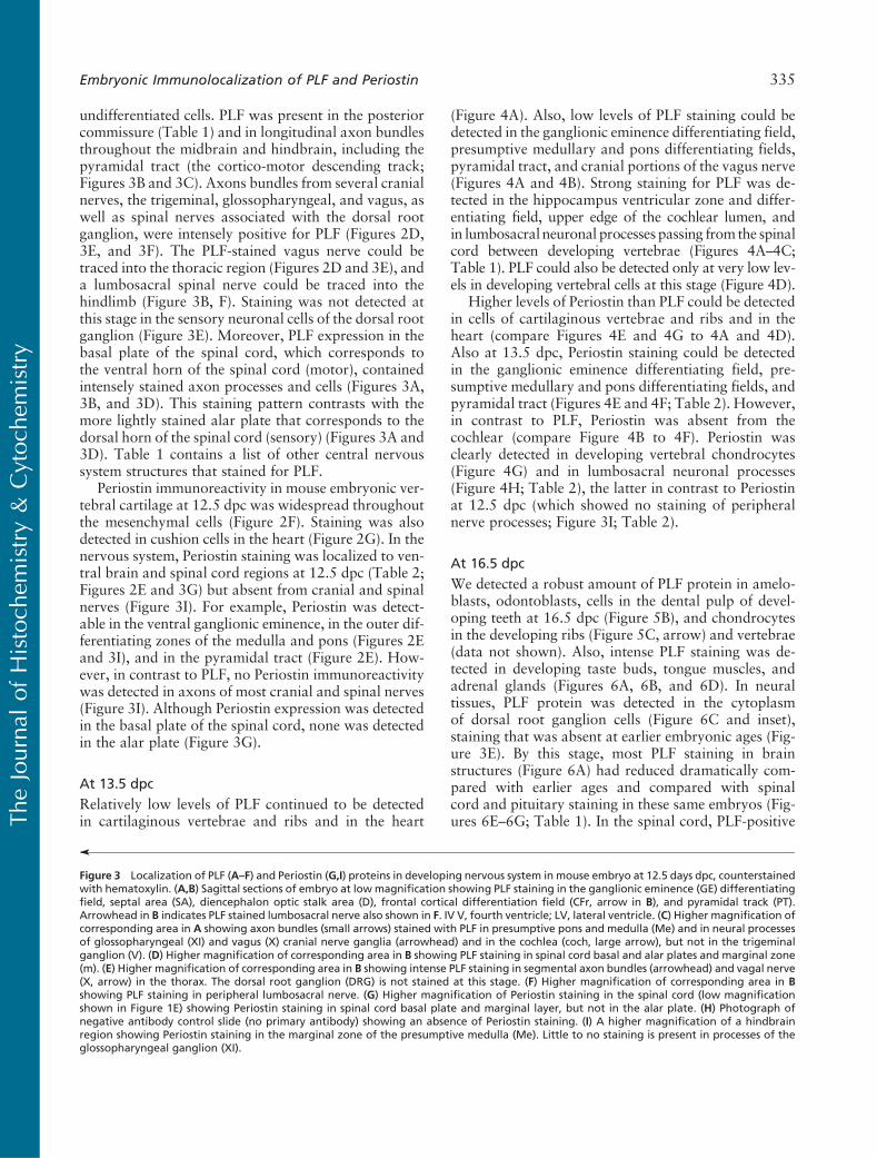
undifferentiated cells. PLF was present in the posteriorcommissure (Table 1) and in longitudinal axon bundlesthroughout the midbrain and hindbrain, including thepyramidal tract (the cortico-motor descending track;Figures 3B and 3C). Axons bundles from several cranialnerves, the trigeminal, glossopharyngeal, and vagus, aswell as spinal nerves associated with the dorsal rootganglion, were intensely positive for PLF (Figures 2D,3E, and 3F). The PLF-stained vagus nerve could betraced into the thoracic region (Figures 2D and 3E), anda lumbosacral spinal nerve could be traced into thehindlimb (Figure 3B, F). Staining was not detected atthis stage in the sensory neuronal cells of the dorsal rootganglion (Figure 3E). Moreover, PLF expression in thebasal plate of the spinal cord, which corresponds tothe ventral horn of the spinal cord (motor), containedintensely stained axon processes and cells (Figures 3A,3B, and 3D). This staining pattern contrasts with themore lightly stained alar plate that corresponds to thedorsal horn of the spinal cord (sensory) (Figures 3A and3D). Table 1 contains a list of other central nervoussystem structures that stained for PLF.
Periostin immunoreactivity in mouse embryonic ver-tebral cartilage at 12.5 dpc was widespread throughoutthe mesenchymal cells (Figure 2F). Staining was alsodetected in cushion cells in the heart (Figure 2G). In thenervous system, Periostin staining was localized to ven-tral brain and spinal cord regions at 12.5 dpc (Table 2;Figures 2E and 3G) but absent from cranial and spinalnerves (Figure 3I). For example, Periostin was detect-able in the ventral ganglionic eminence, in the outer dif-ferentiating zones of the medulla and pons (Figures 2Eand 3I), and in the pyramidal tract (Figure 2E). How-ever, in contrast to PLF, no Periostin immunoreactivitywas detected in axons of most cranial and spinal nerves(Figure 3I). Although Periostin expression was detectedin the basal plate of the spinal cord, none was detectedin the alar plate (Figure 3G).
At 13.5 dpc
Relatively low levels of PLF continued to be detectedin cartilaginous vertebrae and ribs and in the heart
(Figure 4A). Also, low levels of PLF staining could bedetected in the ganglionic eminence differentiating field,presumptive medullary and pons differentiating fields,pyramidal tract, and cranial portions of the vagus nerve(Figures 4A and 4B). Strong staining for PLF was de-tected in the hippocampus ventricular zone and differ-entiating field, upper edge of the cochlear lumen, andin lumbosacral neuronal processes passing from the spinalcord between developing vertebrae (Figures 4A–4C;Table 1). PLF could also be detected only at very low lev-els in developing vertebral cells at this stage (Figure 4D).
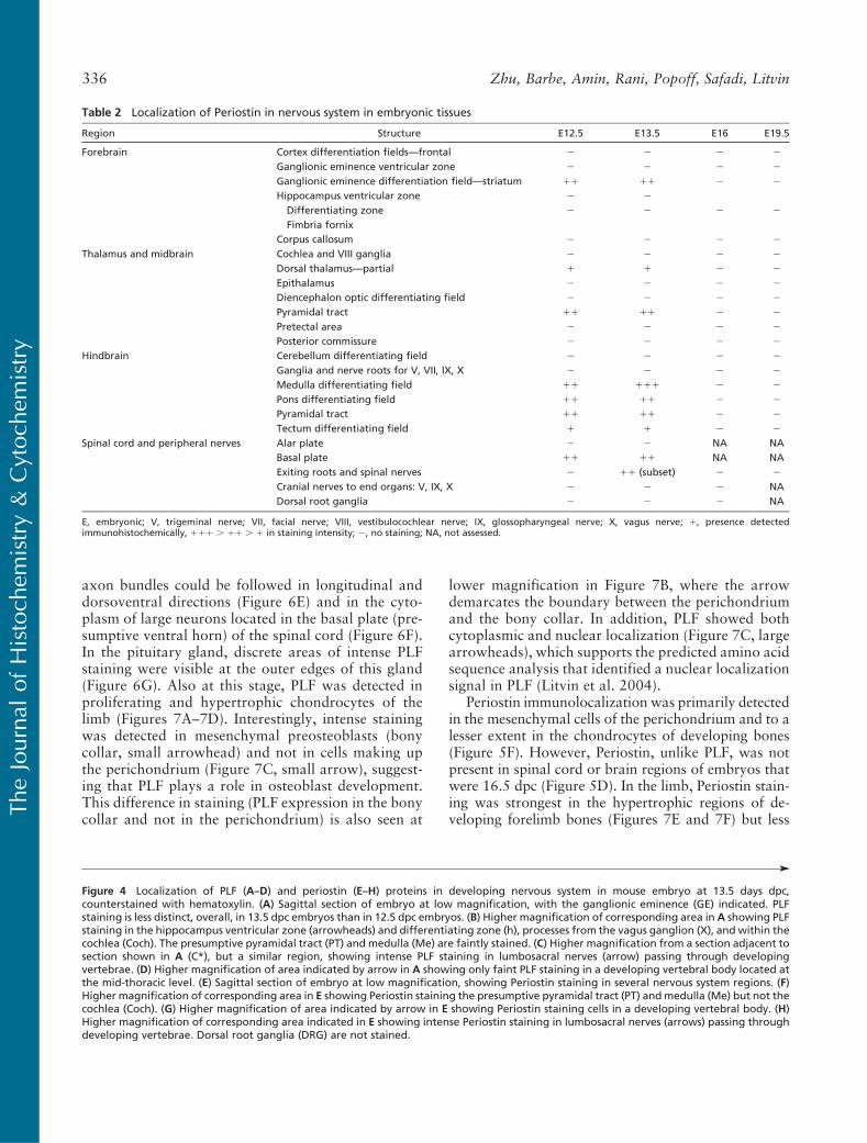
Higher levels of Periostin than PLF could be detectedin cells of cartilaginous vertebrae and ribs and in theheart (compare Figures 4E and 4G to 4A and 4D).Also at 13.5 dpc, Periostin staining could be detectedin the ganglionic eminence differentiating field, pre-sumptive medullary and pons differentiating fields, andpyramidal tract (Figures 4E and 4F; Table 2). However,in contrast to PLF, Periostin was absent from thecochlear (compare Figure 4B to 4F). Periostin wasclearly detected in developing vertebral chondrocytes(Figure 4G) and in lumbosacral neuronal processes(Figure 4H; Table 2), the latter in contrast to Periostinat 12.5 dpc (which showed no staining of peripheralnerve processes; Figure 3I; Table 2).
At 16.5 dpc
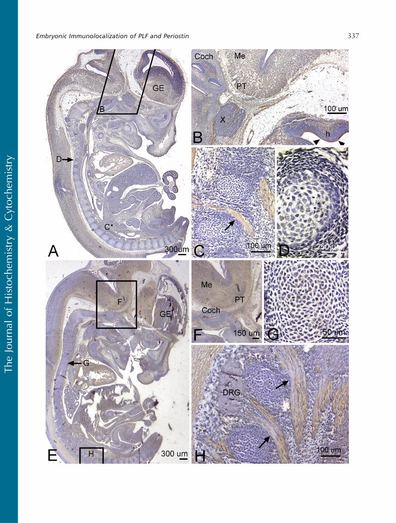
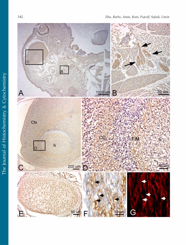
We detected a robust amount of PLF protein in amelo-blasts, odontoblasts, cells in the dental pulp of devel-oping teeth at 16.5 dpc (Figure 5B), and chondrocytesin the developing ribs (Figure 5C, arrow) and vertebrae(data not shown). Also, intense PLF staining was de-tected in developing taste buds, tongue muscles, andadrenal glands (Figures 6A, 6B, and 6D). In neuraltissues, PLF protein was detected in the cytoplasmof dorsal root ganglion cells (Figure 6C and inset),staining that was absent at earlier embryonic ages (Fig-ure 3E). By this stage, most PLF staining in brainstructures (Figure 6A) had reduced dramatically com-pared with earlier ages and compared with spinalcord and pituitary staining in these same embryos (Fig-ures 6E–6G; Table 1). In the spinal cord, PLF-positive
’
Figure 3 Localization of PLF (A–F) and Periostin (G,I) proteins in developing nervous system in mouse embryo at 12.5 days dpc, counterstainedwith hematoxylin. (A,B) Sagittal sections of embryo at low magnification showing PLF staining in the ganglionic eminence (GE) differentiatingfield, septal area (SA), diencephalon optic stalk area (D), frontal cortical differentiation field (CFr, arrow in B), and pyramidal track (PT).Arrowhead in B indicates PLF stained lumbosacral nerve also shown in F. IV V, fourth ventricle; LV, lateral ventricle. (C) Higher magnification ofcorresponding area in A showing axon bundles (small arrows) stained with PLF in presumptive pons and medulla (Me) and in neural processesof glossopharyngeal (XI) and vagus (X) cranial nerve ganglia (arrowhead) and in the cochlea (coch, large arrow), but not in the trigeminalganglion (V). (D) Higher magnification of corresponding area in B showing PLF staining in spinal cord basal and alar plates and marginal zone(m). (E) Higher magnification of corresponding area in B showing intense PLF staining in segmental axon bundles (arrowhead) and vagal nerve(X, arrow) in the thorax. The dorsal root ganglion (DRG) is not stained at this stage. (F) Higher magnification of corresponding area in Bshowing PLF staining in peripheral lumbosacral nerve. (G) Higher magnification of Periostin staining in the spinal cord (low magnificationshown in Figure 1E) showing Periostin staining in spinal cord basal plate and marginal layer, but not in the alar plate. (H) Photograph ofnegative antibody control slide (no primary antibody) showing an absence of Periostin staining. (I) A higher magnification of a hindbrainregion showing Periostin staining in the marginal zone of the presumptive medulla (Me). Little to no staining is present in processes of theglossopharyngeal ganglion (XI).
The
Journ
alof
His
toch
emis
try
&C
ytoch
emis
try
Embryonic Immunolocalization of PLF and Periostin 335
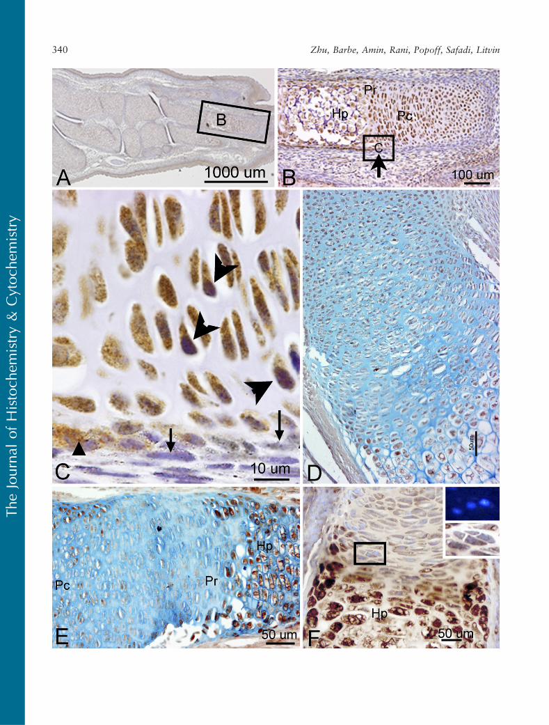
axon bundles could be followed in longitudinal anddorsoventral directions (Figure 6E) and in the cyto-plasm of large neurons located in the basal plate (pre-sumptive ventral horn) of the spinal cord (Figure 6F).In the pituitary gland, discrete areas of intense PLFstaining were visible at the outer edges of this gland(Figure 6G). Also at this stage, PLF was detected inproliferating and hypertrophic chondrocytes of thelimb (Figures 7A–7D). Interestingly, intense stainingwas detected in mesenchymal preosteoblasts (bonycollar, small arrowhead) and not in cells making upthe perichondrium (Figure 7C, small arrow), suggest-ing that PLF plays a role in osteoblast development.This difference in staining (PLF expression in the bonycollar and not in the perichondrium) is also seen at
lower magnification in Figure 7B, where the arrowdemarcates the boundary between the perichondriumand the bony collar. In addition, PLF showed bothcytoplasmic and nuclear localization (Figure 7C, largearrowheads), which supports the predicted amino acidsequence analysis that identified a nuclear localizationsignal in PLF (Litvin et al. 2004).
Periostin immunolocalization was primarily detectedin the mesenchymal cells of the perichondrium and to alesser extent in the chondrocytes of developing bones(Figure 5F). However, Periostin, unlike PLF, was notpresent in spinal cord or brain regions of embryos thatwere 16.5 dpc (Figure 5D). In the limb, Periostin stain-ing was strongest in the hypertrophic regions of de-veloping forelimb bones (Figures 7E and 7F) but less
’
Figure 4 Localization of PLF (A–D) and periostin (E–H) proteins in developing nervous system in mouse embryo at 13.5 days dpc,counterstained with hematoxylin. (A) Sagittal section of embryo at low magnification, with the ganglionic eminence (GE) indicated. PLFstaining is less distinct, overall, in 13.5 dpc embryos than in 12.5 dpc embryos. (B) Higher magnification of corresponding area in A showing PLFstaining in the hippocampus ventricular zone (arrowheads) and differentiating zone (h), processes from the vagus ganglion (X), and within thecochlea (Coch). The presumptive pyramidal tract (PT) and medulla (Me) are faintly stained. (C) Higher magnification from a section adjacent tosection shown in A (C*), but a similar region, showing intense PLF staining in lumbosacral nerves (arrow) passing through developingvertebrae. (D) Higher magnification of area indicated by arrow in A showing only faint PLF staining in a developing vertebral body located atthe mid-thoracic level. (E) Sagittal section of embryo at low magnification, showing Periostin staining in several nervous system regions. (F)Higher magnification of corresponding area in E showing Periostin staining the presumptive pyramidal tract (PT) andmedulla (Me) but not thecochlea (Coch). (G) Higher magnification of area indicated by arrow in E showing Periostin staining cells in a developing vertebral body. (H)Higher magnification of corresponding area indicated in E showing intense Periostin staining in lumbosacral nerves (arrows) passing throughdeveloping vertebrae. Dorsal root ganglia (DRG) are not stained.
Table 2 Localization of Periostin in nervous system in embryonic tissues
Region Structure E12.5 E13.5 E16 E19.5
Forebrain Cortex differentiation fields—frontal 2 2 2 2
Ganglionic eminence ventricular zone 2 2 2 2
Ganglionic eminence differentiation field—striatum 11 11 2 2
Hippocampus ventricular zone 2 2
Differentiating zone 2 2 2 2
Fimbria fornixCorpus callosum 2 2 2 2
Thalamus and midbrain Cochlea and VIII ganglia 2 2 2 2
Dorsal thalamus—partial 1 1 2 2
Epithalamus 2 2 2 2
Diencephalon optic differentiating field 2 2 2 2
Pyramidal tract 11 11 2 2
Pretectal area 2 2 2 2
Posterior commissure 2 2 2 2
Hindbrain Cerebellum differentiating field 2 2 2 2
Ganglia and nerve roots for V, VII, IX, X 2 2 2 2
Medulla differentiating field 11 111 2 2
Pons differentiating field 11 11 2 2
Pyramidal tract 11 11 2 2
Tectum differentiating field 1 1 2 2
Spinal cord and peripheral nerves Alar plate 2 2 NA NABasal plate 11 11 NA NAExiting roots and spinal nerves 2 11 (subset) 2 2
Cranial nerves to end organs: V, IX, X 2 2 2 NADorsal root ganglia 2 2 2 NA
E, embryonic; V, trigeminal nerve; VII, facial nerve; VIII, vestibulocochlear nerve; IX, glossopharyngeal nerve; X, vagus nerve; 1, presence detectedimmunohistochemically, 111 . 11 . 1 in staining intensity; 2, no staining; NA, not assessed.
The
Journ
alof
His
toch
emis
try
&C
ytoch
emis
try
336 Zhu, Barbe, Amin, Rani, Popoff, Safadi, Litvin
The
Journ
alof
His
toch
emis
try
&C
ytoch
emis
try
Embryonic Immunolocalization of PLF and Periostin 337
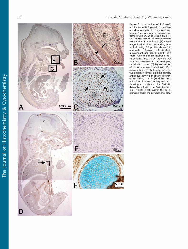
Figure 5 Localization of PLF (A–C)and Periostin (D,F) protein in cartilageand developing teeth of a mouse em-bryo at 16.5 dpc, counterstained withhematoxylin (A–E) or Alcian blue (F).(A) Sagittal section of mouse embryoreacted with PLF antibody. (B) Highermagnification of corresponding areain A showing PLF protein (brown) inameloblasts (arrow), odontoblasts(arrowhead), and dental pulp (P) in atooth. (C) Higher magnification of cor-responding area in A showing PLFlocalized to cells within the developingvertebrae (arrows). (D) Sagittal sectionof mouse embryo reacted with Peri-ostinantibody. (E) Photographofnega-tive antibody control slide (no primaryantibody) showing an absence of Peri-ostin staining in a rib. (F) Higher mag-nification of corresponding area in Dshowing a rib stained for Periostin(brown) andAlcianblue. Periostin stain-ing is visible in cells within the devel-oping rib and in the perichondrial area.
The
Journ
alof
His
toch
emis
try
&C
ytoch
emis
try
338 Zhu, Barbe, Amin, Rani, Popoff, Safadi, Litvin
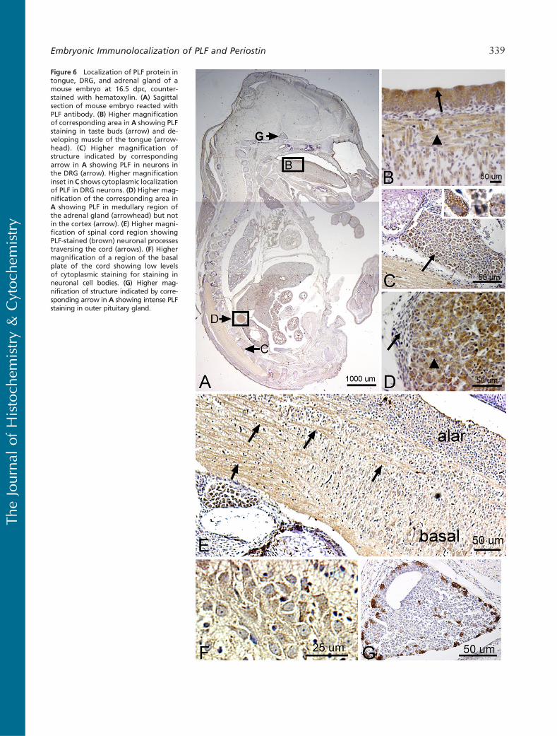
Figure 6 Localization of PLF protein intongue, DRG, and adrenal gland of amouse embryo at 16.5 dpc, counter-stained with hematoxylin. (A) Sagittalsection of mouse embryo reacted withPLF antibody. (B) Higher magnificationof corresponding area in A showing PLFstaining in taste buds (arrow) and de-veloping muscle of the tongue (arrow-head). (C) Higher magnification ofstructure indicated by correspondingarrow in A showing PLF in neurons inthe DRG (arrow). Higher magnificationinset in C shows cytoplasmic localizationof PLF in DRG neurons. (D) Higher mag-nification of the corresponding area inA showing PLF in medullary region ofthe adrenal gland (arrowhead) but notin the cortex (arrow). (E) Higher magni-fication of spinal cord region showingPLF-stained (brown) neuronal processestraversing the cord (arrows). (F) Highermagnification of a region of the basalplate of the cord showing low levelsof cytoplasmic staining for staining inneuronal cell bodies. (G) Higher mag-nification of structure indicated by corre-sponding arrow in A showing intense PLFstaining in outer pituitary gland.
The
Journ
alof
His
toch
emis
try
&C
ytoch
emis
try
Embryonic Immunolocalization of PLF and Periostin 339
The
Journ
alof
His
toch
emis
try
&C
ytoch
emis
try
340 Zhu, Barbe, Amin, Rani, Popoff, Safadi, Litvin
visible in the prehypertrophic and proliferating regions(Figures 7E and 7F).
At 19.5 dpc
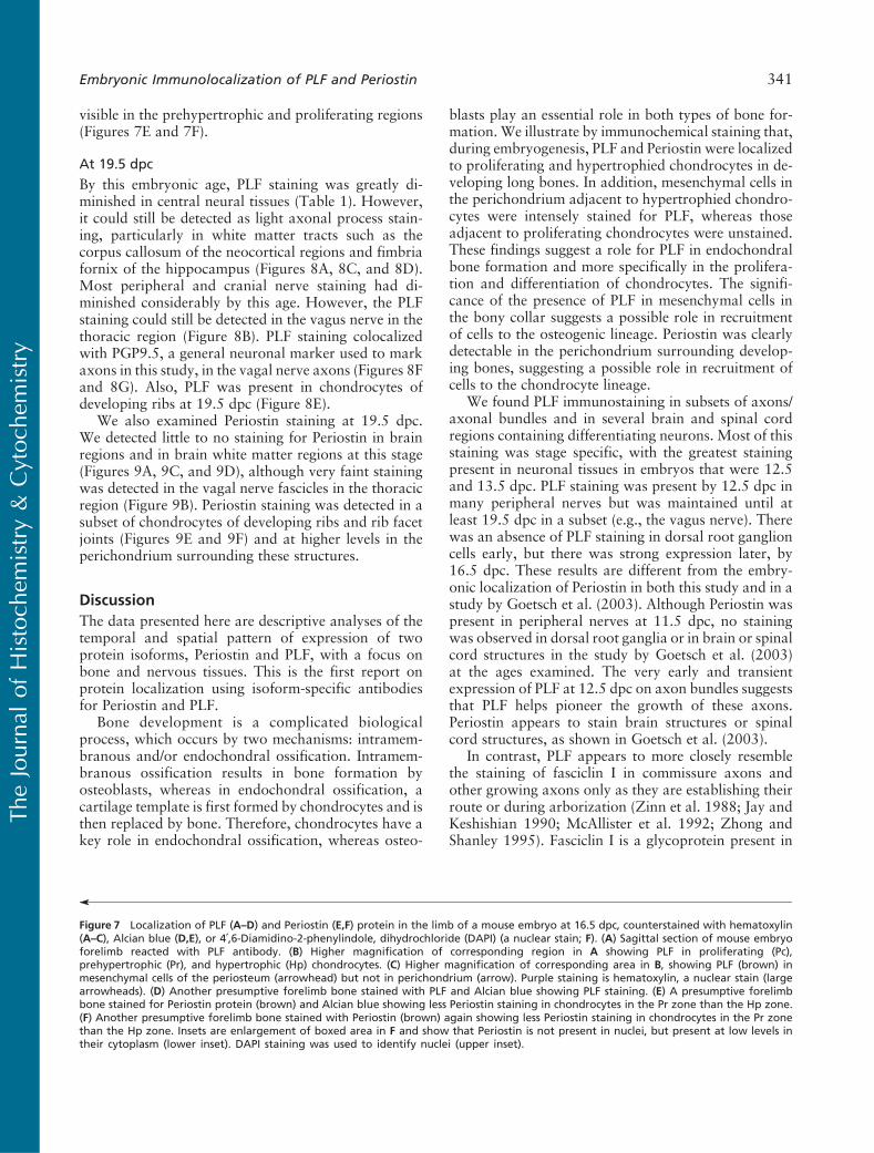
By this embryonic age, PLF staining was greatly di-minished in central neural tissues (Table 1). However,it could still be detected as light axonal process stain-ing, particularly in white matter tracts such as thecorpus callosum of the neocortical regions and fimbriafornix of the hippocampus (Figures 8A, 8C, and 8D).Most peripheral and cranial nerve staining had di-minished considerably by this age. However, the PLFstaining could still be detected in the vagus nerve in thethoracic region (Figure 8B). PLF staining colocalizedwith PGP9.5, a general neuronal marker used to markaxons in this study, in the vagal nerve axons (Figures 8Fand 8G). Also, PLF was present in chondrocytes ofdeveloping ribs at 19.5 dpc (Figure 8E).
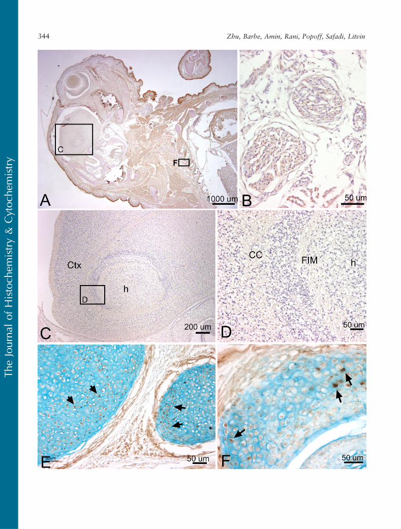
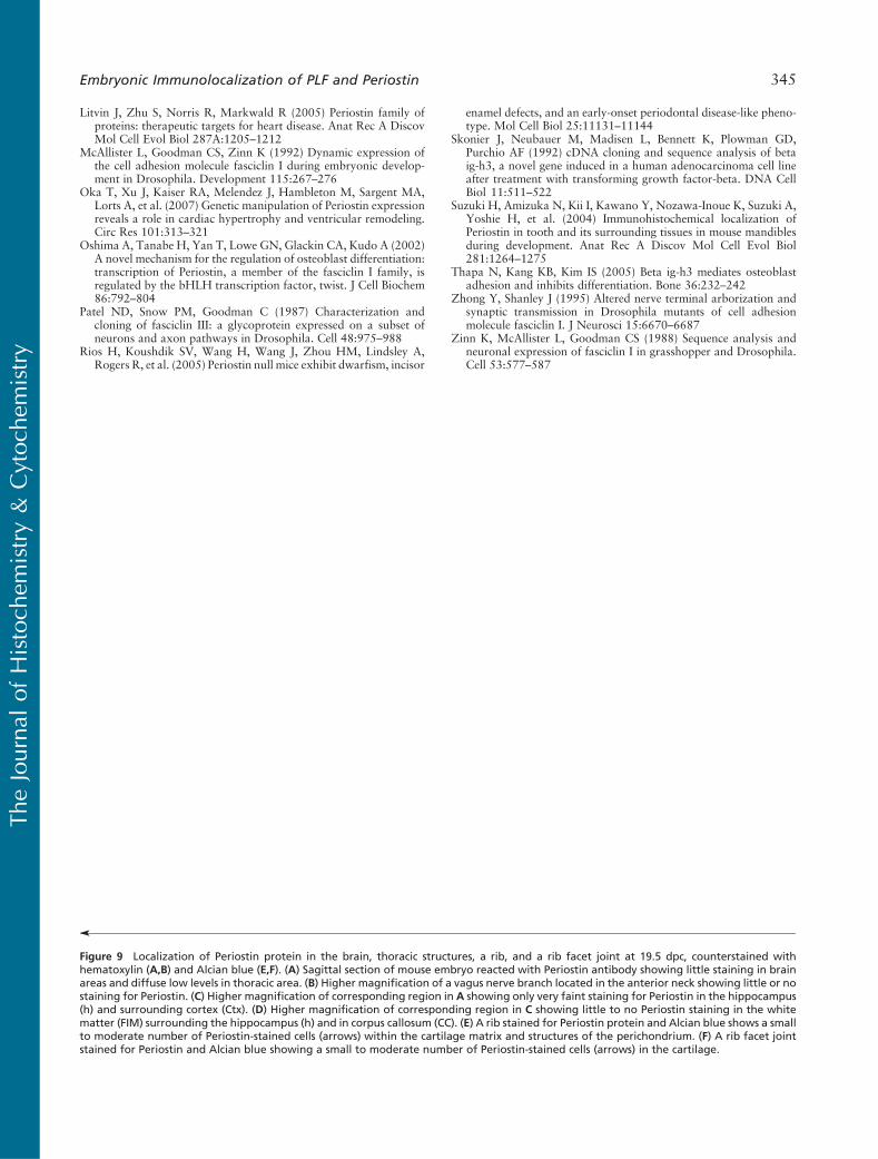
We also examined Periostin staining at 19.5 dpc.We detected little to no staining for Periostin in brainregions and in brain white matter regions at this stage(Figures 9A, 9C, and 9D), although very faint stainingwas detected in the vagal nerve fascicles in the thoracicregion (Figure 9B). Periostin staining was detected in asubset of chondrocytes of developing ribs and rib facetjoints (Figures 9E and 9F) and at higher levels in theperichondrium surrounding these structures.
Discussion
The data presented here are descriptive analyses of thetemporal and spatial pattern of expression of twoprotein isoforms, Periostin and PLF, with a focus onbone and nervous tissues. This is the first report onprotein localization using isoform-specific antibodiesfor Periostin and PLF.
Bone development is a complicated biologicalprocess, which occurs by two mechanisms: intramem-branous and/or endochondral ossification. Intramem-branous ossification results in bone formation byosteoblasts, whereas in endochondral ossification, acartilage template is first formed by chondrocytes and isthen replaced by bone. Therefore, chondrocytes have akey role in endochondral ossification, whereas osteo-
blasts play an essential role in both types of bone for-mation. We illustrate by immunochemical staining that,during embryogenesis, PLF and Periostin were localizedto proliferating and hypertrophied chondrocytes in de-veloping long bones. In addition, mesenchymal cells inthe perichondrium adjacent to hypertrophied chondro-cytes were intensely stained for PLF, whereas thoseadjacent to proliferating chondrocytes were unstained.These findings suggest a role for PLF in endochondralbone formation and more specifically in the prolifera-tion and differentiation of chondrocytes. The signifi-cance of the presence of PLF in mesenchymal cells inthe bony collar suggests a possible role in recruitmentof cells to the osteogenic lineage. Periostin was clearlydetectable in the perichondrium surrounding develop-ing bones, suggesting a possible role in recruitment ofcells to the chondrocyte lineage.
We found PLF immunostaining in subsets of axons/axonal bundles and in several brain and spinal cordregions containing differentiating neurons. Most of thisstaining was stage specific, with the greatest stainingpresent in neuronal tissues in embryos that were 12.5and 13.5 dpc. PLF staining was present by 12.5 dpc inmany peripheral nerves but was maintained until atleast 19.5 dpc in a subset (e.g., the vagus nerve). Therewas an absence of PLF staining in dorsal root ganglioncells early, but there was strong expression later, by16.5 dpc. These results are different from the embry-onic localization of Periostin in both this study and in astudy by Goetsch et al. (2003). Although Periostin waspresent in peripheral nerves at 11.5 dpc, no stainingwas observed in dorsal root ganglia or in brain or spinalcord structures in the study by Goetsch et al. (2003)at the ages examined. The very early and transientexpression of PLF at 12.5 dpc on axon bundles suggeststhat PLF helps pioneer the growth of these axons.Periostin appears to stain brain structures or spinalcord structures, as shown in Goetsch et al. (2003).
In contrast, PLF appears to more closely resemblethe staining of fasciclin I in commissure axons andother growing axons only as they are establishing theirroute or during arborization (Zinn et al. 1988; Jay andKeshishian 1990; McAllister et al. 1992; Zhong andShanley 1995). Fasciclin I is a glycoprotein present in
’
Figure 7 Localization of PLF (A–D) and Periostin (E,F) protein in the limb of a mouse embryo at 16.5 dpc, counterstained with hematoxylin(A–C), Alcian blue (D,E), or 4¶,6-Diamidino-2-phenylindole, dihydrochloride (DAPI) (a nuclear stain; F). (A) Sagittal section of mouse embryoforelimb reacted with PLF antibody. (B) Higher magnification of corresponding region in A showing PLF in proliferating (Pc),prehypertrophic (Pr), and hypertrophic (Hp) chondrocytes. (C) Higher magnification of corresponding area in B, showing PLF (brown) inmesenchymal cells of the periosteum (arrowhead) but not in perichondrium (arrow). Purple staining is hematoxylin, a nuclear stain (largearrowheads). (D) Another presumptive forelimb bone stained with PLF and Alcian blue showing PLF staining. (E) A presumptive forelimbbone stained for Periostin protein (brown) and Alcian blue showing less Periostin staining in chondrocytes in the Pr zone than the Hp zone.(F) Another presumptive forelimb bone stained with Periostin (brown) again showing less Periostin staining in chondrocytes in the Pr zonethan the Hp zone. Insets are enlargement of boxed area in F and show that Periostin is not present in nuclei, but present at low levels intheir cytoplasm (lower inset). DAPI staining was used to identify nuclei (upper inset).
The
Journ
alof
His
toch
emis
try
&C
ytoch
emis
try
Embryonic Immunolocalization of PLF and Periostin 341
The
Journ
alof
His
toch
emis
try
&C
ytoch
emis
try
342 Zhu, Barbe, Amin, Rani, Popoff, Safadi, Litvin
insects and involved in the mediation of homopholiccell adhesion, axonal arborization, and development ofpresynaptive functions (Elkins et al. 1990; Zhong andShanley 1995). It is expressed in a stage-specific mannerin the developing nervous system of grasshopperembryos on the surfaces of all cell bodies and axonsin the PNS, commissural axons, bilateral longitudinalaxonal tracts extending the length of the embryo, andseveral specialized sensory structures of the head (Jayand Keshishian 1990; McAllister et al. 1992). Theduration of fasciclin I expression varied, with expres-sion on commissural cell bodies and axons detectedonly during their formation, but expression was main-tained on tracts such as the anterior corpus callosum.The related proteins, fasciclin II and III, are also ex-pressed transiently on subsets of axonal cells, axons,and bundles, and appear on other pathways later indevelopment (Patel et al. 1987; Grenningloh et al.1991). Because PLF, like fasciclin I, is present in mul-tiple tissues, it is possible that it is involved in mor-phogenesis in a manner similar to fasciclin proteins.The similarity between PLF and fasciclin staining, bothtemporally and spatially, suggests that PLF may play arole in neuronal morphogenesis.
PLF was also detected in the adrenal gland andpituitary. The adrenal gland is made up of two parts:the outer region is called the adrenal cortex and theinner region is called the adrenal medulla. The adrenalcortex is of mesodermal origin and derived fromthe proliferation of coelomic epithelium, whereas themedulla is of ectodermal origin and derived from theneural crest. The medulla of the adrenal gland consistsof sympathetic neural cells. Therefore, a strong signal ofPLF in the medulla of the adrenal gland is consistentwith localization of PLF in neural tissues. The pituitaryis the master gland of the endocrine system, whichcontrols the functions of the other endocrine glands.It is located at the base of the brain and is dividedinto two lobes: the anterior (adenohypophysis) and theposterior (neurohypophysis). The neurohypophysisconsists mainly of axons extending from the hypothal-amus. The detection of PLF in the neurohypophysispossibly suggests that PLF acts as a guidance moleculeto direct these projecting axons from the hypothalamusto the neurohypophysis. Given that cells in the anteriorhypophysis produce hormones necessary for the func-
tion of a variety of organs, the biologic function of PLFin this region requires further study.
In summary, the spatial and temporal pattern oflocalization of Periostin and PLF differ, suggesting dif-ferent roles for these proteins. PLF may be secretedbased on the presence of a signal peptide. It may also bepresent in the nucleus as suggested by the presence ofa nuclear localization signal and as we have shown inembryonic tissues. Therefore, understanding mecha-nisms by which PLF mediates its effects will requirestudying its function as a secreted, as well as an intra-cellular, protein.
Literature Cited
Billings PC, Herrick DJ, Kucich U, Engelsberg BN, Abrams WR,Macarak EJ, Rosenbloom J, et al. (2000) Extracellular matrixand nuclear localization of beta ig-h3 in human bladder smoothmuscle and fibroblast cells. J Cell Biochem 79:261–273
el Ghouzzi V, Le Merrer M, Perrin-Schmitt F, Lajeunie E, Benit P,Renier D, Bourgeois P, et al. (1997) Mutations of the TWIST genein the Saethre-Chotzen syndrome. Nat Genet 15:42–46
Elkins R, Hortsch M, Beiber AJ, Snow PM, Goodman CS (1990)Drosophila fasciclin I is a novel homophilic adhesion moleculethat along with fasciclin III can mediate cell sorting. J Cell Biol110:1825–1832
Ferguson JW, Mikesh MF, Wheeler EF, LeBaron RG (2003)Developmental expression patterns of Beta-ig (betaIG-H3) andits function as a cell adhesion protein. Mech Dev 120:851–864
Goetsch SC, Hawke TJ, Gallardo TD, Richardson JA, Garry DJ(2003) Transcriptional profiling and regulation of the extracel-lular matrix during muscle regeneration. Physiol Genomics 14:261–271
Grenningloh G, Rehm EJ, Goodman CS (1991) Genetic analysis ofgrowth cone guidance in Drosophila: fasciclin II functions as aneuronal recognition molecule. Cell 67:45–57
Horiuchi K, Amizuka N, Takeshita S, Takamatsu H, Katsuura M,Ozawa H, Toyama Y, et al. (1999) Identification and character-ization of a novel protein, Periostin, with restricted expression toperiosteum and periodontal ligament and increased expression bytransforming growth factor beta. J Bone Miner Res 14:1239–1249
Jay DG, Keshishian H (1990) Laser inactivation of fasciclin I dis-rupts axon adhesion of grasshopper pioneer neurons. Nature 348:548–550
Litvin J, Blagg A, Mu A, Matiwala S, Montgomery M, Berretta R,Houser S, et al. (2006) Periostin and Periostin-like factor in thehuman heart: possible therapeutic targets. Cardiovasc Pathol15:24–32
Litvin J, Chen X, Keleman S, Zhu S, Autieri M (2007) Expression andfunction of Periostin-like factor in vascular smooth muscle cells.Am J Physiol Cell Physiol 292:C1672–C1680
Litvin J, Selim AH, Montgomery MO, Lehmann K, Rico MC, DevlinH, Bednarik DP, et al. (2004) Expression and function of Periostin-isoforms in bone. J Cell Biochem 92:1044–1061
’
Figure 8 Localization of PLF protein in the brain, peripheral nerves in the thorax, and a rib at 19.5 dpc, counterstained with hematoxylin (A–D)and PGP9.5 (an axonal marker, G). (A) Sagittal section of mouse embryo reacted with PLF antibody. PLF staining is much lighter overall than inprevious embryonic ages. (B) Higher magnification of corresponding region in A showing PLF (brown) in vagus nerve bundles (arrows) in theupper thorax and neck. (C) Higher magnification of corresponding region in A showing PLF in the hippocampus (h) and surrounding cortex(Ctx). (D) Higher magnification of corresponding region in C showing PLF stained processes in the white matter [Fimbria fornix (FIM)]surrounding the hippocampus (h) and in the corpus callosum (CC). (E) A rib stained for PLF protein shows stained cells within the cartilagematrix. (F) Higher magnification of vagus nerve from an adjacent section, showing staining of axonal processes for PLF protein (arrows). (G)Same nerve as shown in F showing colocalization of PLF with PGP9.5 (arrows).
The
Journ
alof
His
toch
emis
try
&C
ytoch
emis
try
Embryonic Immunolocalization of PLF and Periostin 343
The
Journ
alof
His
toch
emis
try
&C
ytoch
emis
try
344 Zhu, Barbe, Amin, Rani, Popoff, Safadi, Litvin
Litvin J, Zhu S, Norris R, Markwald R (2005) Periostin family ofproteins: therapeutic targets for heart disease. Anat Rec A DiscovMol Cell Evol Biol 287A:1205–1212
McAllister L, Goodman CS, Zinn K (1992) Dynamic expression ofthe cell adhesion molecule fasciclin I during embryonic develop-ment in Drosophila. Development 115:267–276
Oka T, Xu J, Kaiser RA, Melendez J, Hambleton M, Sargent MA,Lorts A, et al. (2007) Genetic manipulation of Periostin expressionreveals a role in cardiac hypertrophy and ventricular remodeling.Circ Res 101:313–321
Oshima A, Tanabe H, Yan T, Lowe GN, Glackin CA, Kudo A (2002)A novel mechanism for the regulation of osteoblast differentiation:transcription of Periostin, a member of the fasciclin I family, isregulated by the bHLH transcription factor, twist. J Cell Biochem86:792–804
Patel ND, Snow PM, Goodman C (1987) Characterization andcloning of fasciclin III: a glycoprotein expressed on a subset ofneurons and axon pathways in Drosophila. Cell 48:975–988
Rios H, Koushdik SV, Wang H, Wang J, Zhou HM, Lindsley A,Rogers R, et al. (2005) Periostin null mice exhibit dwarfism, incisor
enamel defects, and an early-onset periodontal disease-like pheno-type. Mol Cell Biol 25:11131–11144
Skonier J, Neubauer M, Madisen L, Bennett K, Plowman GD,Purchio AF (1992) cDNA cloning and sequence analysis of betaig-h3, a novel gene induced in a human adenocarcinoma cell lineafter treatment with transforming growth factor-beta. DNA CellBiol 11:511–522
Suzuki H, Amizuka N, Kii I, Kawano Y, Nozawa-Inoue K, Suzuki A,Yoshie H, et al. (2004) Immunohistochemical localization ofPeriostin in tooth and its surrounding tissues in mouse mandiblesduring development. Anat Rec A Discov Mol Cell Evol Biol281:1264–1275
Thapa N, Kang KB, Kim IS (2005) Beta ig-h3 mediates osteoblastadhesion and inhibits differentiation. Bone 36:232–242
Zhong Y, Shanley J (1995) Altered nerve terminal arborization andsynaptic transmission in Drosophila mutants of cell adhesionmolecule fasciclin I. J Neurosci 15:6670–6687
Zinn K, McAllister L, Goodman CS (1988) Sequence analysis andneuronal expression of fasciclin I in grasshopper and Drosophila.Cell 53:577–587
’
Figure 9 Localization of Periostin protein in the brain, thoracic structures, a rib, and a rib facet joint at 19.5 dpc, counterstained withhematoxylin (A,B) and Alcian blue (E,F). (A) Sagittal section of mouse embryo reacted with Periostin antibody showing little staining in brainareas and diffuse low levels in thoracic area. (B) Higher magnification of a vagus nerve branch located in the anterior neck showing little or nostaining for Periostin. (C) Higher magnification of corresponding region in A showing only very faint staining for Periostin in the hippocampus(h) and surrounding cortex (Ctx). (D) Higher magnification of corresponding region in C showing little to no Periostin staining in the whitematter (FIM) surrounding the hippocampus (h) and in corpus callosum (CC). (E) A rib stained for Periostin protein and Alcian blue shows a smallto moderate number of Periostin-stained cells (arrows) within the cartilage matrix and structures of the perichondrium. (F) A rib facet jointstained for Periostin and Alcian blue showing a small to moderate number of Periostin-stained cells (arrows) in the cartilage.
The
Journ
alof
His
toch
emis
try
&C
ytoch
emis
try
Embryonic Immunolocalization of PLF and Periostin 345
Related Documents