Immunogenicity of Novel Mumps Vaccine Candidates Generated by Genetic Modification Pei Xu, a,b Zhenhai Chen, a Shannon Phan, a Adrian Pickar, a Biao He a Department of Infectious Diseases, College of Veterinary Medicine, University of Georgia, Athens, Georgia, USA a ; Intercollege Graduate Program in Cell and Developmental Biology, Pennsylvania State University, University Park, Pennsylvania, USA b Mumps is a highly contagious human disease, characterized by lateral or bilateral nonsuppurative swelling of the parotid glands and neurological complications that can result in aseptic meningitis or encephalitis. A mumps vaccination program imple- mented since the 1960s reduced mumps incidence by more than 99% and kept the mumps case numbers as low as hundreds of cases per year in the United States before 2006. However, a large mumps outbreak occurred in vaccinated populations in 2006 and again in 2009 in the United States, raising concerns about the efficacy of the vaccination program. Previously, we have shown that clinical isolate-based recombinant mumps viruses lacking expression of either the V protein (rMuVV) or the SH protein (rMuVSH) are attenuated in a neurovirulence test using newborn rat brains (P. Xu et al., Virology 417:126 –136, 2011, http://dx.doi.org/10.1016/j.virol.2011.05.003; P. Xu et al., J. Virol. 86:1768 –1776, 2012, http://dx.doi.org/10.1128/JVI.06019-11) and may be good candidates for vaccine development. In this study, we examined immunity induced by rMuVSH and rMuVV in mice. Furthermore, we generated recombinant mumps viruses lacking expression of both the V protein and the SH protein (rMuVSHV). Analysis of rMuVSHV indicated that it was stable in tissue culture cell lines. Importantly, rMuVSHV was immunogenic in mice, indicating that it is a promising candidate for mumps vaccine development. M umps is a human infectious disease characterized by lateral or bilateral nonsuppurative swelling of the parotid glands. In severe cases, mumps can lead to orchitis in postpuberty male patients and damage to the central nervous system. In the prevac- cine era, 90% of the population turned seropositive for mumps virus (MuV) by 14 to 15 years of age, reflecting its highly conta- gious nature. Mumps virus is neurotropic and was one of the most common causes of aseptic meningitis before the implementation of mass mumps vaccination programs. At present, the Jeryl Lynn (JL) vaccine is the most commonly used mumps vaccine, administered as lyophilized live virus with measles and rubella vaccine components. The JL vaccine strain originated from an infectious isolate from a mumps patient in 1963 (1). The virus was attenuated through continuous passages in embryonic hen eggs and chicken embryos/chicken embryo cell cultures (1). The JL vaccine was licensed in the United States in 1967 and has been used for over 40 years. This vaccine has been efficacious and safe overall (2–6). However, several large mumps outbreaks have occurred re- cently in the United States and worldwide in populations that have been vaccinated with the JL vaccine (7–10). Major mumps outbreaks in the United States include the 2006 multistate mumps outbreak, reporting 6,584 suspected cases originating from the state of Iowa (11, 12) and the 2009 –2010 New York and New Jersey mumps outbreaks with a total of 2,078 suspected cases reported in 2010 (13). Both of the outbreaks occurred among highly vaccinated populations, raising questions about the efficacy of the current vaccination program in the United States. One possible causality is the antigenic differences be- tween the genotype A vaccine strain and the genotype G circulating wild-type mumps viruses. In this study, we seek to develop a mumps vaccine candidate through genetic modification of a clinically isolated mumps virus. Mumps virus is a member of the family Paramyxoviridae, subfamily Paramyxovirinae, and genus Rubulavirus (6, 14). It is an enveloped virus enclosing a negative-sense, single-stranded, non- segmented RNA genome of 15,384 nucleotides in length which encodes 9 viral proteins (15–17). Studies of the function of the Paramyxovirus SH protein reveal that it blocks tumor necrosis factor alpha (TNF-) induction, signaling, caspase activation, and NF-B nuclear translocation in transfected and virus-infected cells (18–23). The V protein is an accessory protein translated from the authentic transcript of the V/P gene (24, 25). Mumps V protein is an antagonist of antiviral innate immunity. It interferes with type I interferon (IFN) induction by disrupting the recogni- tion of intracellular viral double-stranded RNA (dsRNA) by MDA5 (26–28). It also blocks IFN signaling by targeting STAT proteins for proteasome-mediated degradation (29–35). Recom- binant mumps viruses with either the V protein deletion (rMuVV) or the SH protein deletion (rMuVSH) are attenu- ated in neurotoxicity in intracerebrally (IC) infected rats (21, 36). In this study, we tested the immunogenicity of rMuVV and rMuVSH in mice. Furthermore, we generated a recombinant MuV lacking expression of both the SH and V proteins (rMuVSHV) and examined antibody and cellular immune re- sponses in mice. MATERIALS AND METHODS Plasmids, viruses, and cells. The MuV strain was obtained from a pa- tient during the 2005–2006 Midwest mumps outbreak in the United States. A full-length cDNA clone of the virus (pMuV) was constructed as previously described (21). Recombinant MuV lacking the V protein (rMuVV), recombinant MuV lacking the SH protein, and recombinant Received 23 September 2013 Accepted 9 December 2013 Published ahead of print 18 December 2013 Editor: D. S. Lyles Address correspondence to Biao He, [email protected]. Copyright © 2014, American Society for Microbiology. All Rights Reserved. doi:10.1128/JVI.02778-13 2600 jvi.asm.org Journal of Virology p. 2600 –2610 March 2014 Volume 88 Number 5

Welcome message from author
This document is posted to help you gain knowledge. Please leave a comment to let me know what you think about it! Share it to your friends and learn new things together.
Transcript

Immunogenicity of Novel Mumps Vaccine Candidates Generated byGenetic Modification
Pei Xu,a,b Zhenhai Chen,a Shannon Phan,a Adrian Pickar,a Biao Hea
Department of Infectious Diseases, College of Veterinary Medicine, University of Georgia, Athens, Georgia, USAa; Intercollege Graduate Program in Cell andDevelopmental Biology, Pennsylvania State University, University Park, Pennsylvania, USAb
Mumps is a highly contagious human disease, characterized by lateral or bilateral nonsuppurative swelling of the parotid glandsand neurological complications that can result in aseptic meningitis or encephalitis. A mumps vaccination program imple-mented since the 1960s reduced mumps incidence by more than 99% and kept the mumps case numbers as low as hundreds ofcases per year in the United States before 2006. However, a large mumps outbreak occurred in vaccinated populations in 2006and again in 2009 in the United States, raising concerns about the efficacy of the vaccination program. Previously, we haveshown that clinical isolate-based recombinant mumps viruses lacking expression of either the V protein (rMuV�V) or the SHprotein (rMuV�SH) are attenuated in a neurovirulence test using newborn rat brains (P. Xu et al., Virology 417:126 –136, 2011,http://dx.doi.org/10.1016/j.virol.2011.05.003; P. Xu et al., J. Virol. 86:1768 –1776, 2012, http://dx.doi.org/10.1128/JVI.06019-11)and may be good candidates for vaccine development. In this study, we examined immunity induced by rMuV�SH andrMuV�V in mice. Furthermore, we generated recombinant mumps viruses lacking expression of both the V protein and the SHprotein (rMuV�SH�V). Analysis of rMuV�SH�V indicated that it was stable in tissue culture cell lines. Importantly,rMuV�SH�V was immunogenic in mice, indicating that it is a promising candidate for mumps vaccine development.
Mumps is a human infectious disease characterized by lateralor bilateral nonsuppurative swelling of the parotid glands.
In severe cases, mumps can lead to orchitis in postpuberty malepatients and damage to the central nervous system. In the prevac-cine era, 90% of the population turned seropositive for mumpsvirus (MuV) by 14 to 15 years of age, reflecting its highly conta-gious nature. Mumps virus is neurotropic and was one of the mostcommon causes of aseptic meningitis before the implementationof mass mumps vaccination programs.
At present, the Jeryl Lynn (JL) vaccine is the most commonly usedmumps vaccine, administered as lyophilized live virus with measlesand rubella vaccine components. The JL vaccine strain originatedfrom an infectious isolate from a mumps patient in 1963 (1). Thevirus was attenuated through continuous passages in embryonic heneggs and chicken embryos/chicken embryo cell cultures (1). The JLvaccine was licensed in the United States in 1967 and has been usedfor over 40 years. This vaccine has been efficacious and safe overall(2–6). However, several large mumps outbreaks have occurred re-cently in the United States and worldwide in populations that havebeen vaccinated with the JL vaccine (7–10). Major mumps outbreaksin the United States include the 2006 multistate mumps outbreak,reporting 6,584 suspected cases originating from the state of Iowa (11,12) and the 2009–2010 New York and New Jersey mumps outbreakswith a total of 2,078 suspected cases reported in 2010 (13). Both of theoutbreaks occurred among highly vaccinated populations, raisingquestions about the efficacy of the current vaccination program in theUnited States. One possible causality is the antigenic differences be-tween the genotype A vaccine strain and the genotype G circulatingwild-type mumps viruses.
In this study, we seek to develop a mumps vaccine candidatethrough genetic modification of a clinically isolated mumpsvirus. Mumps virus is a member of the family Paramyxoviridae,subfamily Paramyxovirinae, and genus Rubulavirus (6, 14). It is anenveloped virus enclosing a negative-sense, single-stranded, non-segmented RNA genome of 15,384 nucleotides in length which
encodes 9 viral proteins (15–17). Studies of the function of theParamyxovirus SH protein reveal that it blocks tumor necrosisfactor alpha (TNF-�) induction, signaling, caspase activation, andNF-�B nuclear translocation in transfected and virus-infectedcells (18–23). The V protein is an accessory protein translatedfrom the authentic transcript of the V/P gene (24, 25). Mumps Vprotein is an antagonist of antiviral innate immunity. It interfereswith type I interferon (IFN) induction by disrupting the recogni-tion of intracellular viral double-stranded RNA (dsRNA) byMDA5 (26–28). It also blocks IFN signaling by targeting STATproteins for proteasome-mediated degradation (29–35). Recom-binant mumps viruses with either the V protein deletion(rMuV�V) or the SH protein deletion (rMuV�SH) are attenu-ated in neurotoxicity in intracerebrally (IC) infected rats (21, 36).In this study, we tested the immunogenicity of rMuV�V andrMuV�SH in mice. Furthermore, we generated a recombinantMuV lacking expression of both the SH and V proteins(rMuV�SH�V) and examined antibody and cellular immune re-sponses in mice.
MATERIALS AND METHODSPlasmids, viruses, and cells. The MuV strain was obtained from a pa-tient during the 2005–2006 Midwest mumps outbreak in the UnitedStates. A full-length cDNA clone of the virus (pMuV) was constructed aspreviously described (21). Recombinant MuV lacking the V protein(rMuV�V), recombinant MuV lacking the SH protein, and recombinant
Received 23 September 2013 Accepted 9 December 2013
Published ahead of print 18 December 2013
Editor: D. S. Lyles
Address correspondence to Biao He, [email protected].
Copyright © 2014, American Society for Microbiology. All Rights Reserved.
doi:10.1128/JVI.02778-13
2600 jvi.asm.org Journal of Virology p. 2600 –2610 March 2014 Volume 88 Number 5
MuV expressing a Renilla luciferase protein have been described before(21). A plasmid containing the MuV genome but lacking both V and SHwas constructed by combining the SH open reading frame (ORF) deletionwith the plasmid encoding the rMuV�V genome. Primer sequences, de-tailed cloning strategies, and entire cDNA sequences of MuV are availableupon request. Jeryl Lynn (JL) vaccine, isolated from the measles, mumps,and rubella (MMR) vaccine, was a gift from Paul Rota at the CDC.
To rescue an infectious virus, plasmid pMuV�SH�V (5 �g), alongwith plasmids pCAGGS-L (1 �g), pCAGGS-NP (1.5 �g), and pCAGGS-P(200 ng), were transfected into BSRT-7 cells. Three days later, transfectedBSRT-7 cells were mixed with Vero cells at a 1:1 ratio. Ten to 14 days later,when syncytium formation was observed, supernatants containing puta-tive rMuV�SH�V were collected and plaque purified in Vero cells.Plaques (developing 4 to 7 days postinfection [dpi]) were amplified inVero cells once (P0), and their genomes were sequenced. All recombinantviruses used for the following experiments were expanded once in Verocells from the P0 amplification (P1). The rescue procedure was repeated toproduce independent stocks of rMuV�SH�V, resulting in 6 isolates ofindependently rescued rMuV�SH�V viruses (PX64-1, PX64-4, PX64-61,PX64-67, and PX64-84).
All mumps viruses were grown in Vero cells and harvested at 4 to 7 dpi.Virus titers were measured in Vero cells by plaque assay as describedpreviously (37, 38). JL virus was grown in Vero cells and concentrated toachieve a working titer. Harvested virus stock was cushioned onto 20%sucrose using ultracentrifugation at 37,500 rpm (Thermo Scientific Sor-vall RC 6 plus centrifuge). Pelleted viruses were resuspended in 1% bovineserum albumin (BSA)–Dulbecco’s modified Eagle medium (DMEM) andstored at �80°C. Concentrated JL virus was retitrated in Vero cells byplaque assay.
Vero cells were maintained in DMEM with 10% fetal bovine serum(FBS) and 1% penicillin-streptomycin (P/S) (Mediatech Inc., Holu Hill,FL). BSRT-7 cells were maintained in DMEM supplemented with 10%FBS, 1% P/S, 10% tryptose phosphate broth (TPB), and 400 �g/ml Gene-ticin G418 antibiotic. Cells were cultured at 37°C with 5% CO2 and passedthe day before infection or transfection at appropriate dilution factors toachieve 80 to 90% confluence the next day. For virus infection, cells wereinoculated with viruses in DMEM plus 1% BSA at an multiplicity of in-fection (MOI) of 0.01, 3, or 5 and incubated for 1 to 2 h at 37°C with 5%CO2. The inocula were then replaced with DMEM supplemented with 2%FBS and 1% P/S. Cells were transfected with plasmids using Plus andLipofectamine reagents (Invitrogen, Carlsbad, CA) by following the man-ufacturer-provided protocols.
Sequencing of viruses. Viral RNA was extracted from cell culture su-pernatants using the QIAamp viral RNA extraction minikit (Qiagen Inc.,Valencia, CA) by following the manufacturer’s protocol. Isolated viralRNA was reverse transcribed into cDNA using Super Script III reversetranscriptase with random hexamers (Invitrogen). Synthesized cDNAthen served as templates for PCR using mumps virus genome-specificprimers and Taq polymerase (Invitrogen). Fifteen sets of primers, eachcontaining a forward and reverse primer, were designed to divide thegenome into 15 overlapping fragments. The primers were then used forthe subsequent sequencing of the PCR products (39). Primer sequencesare available upon request.
Immunoblotting. Vero cells in 6-well plates at approximately 90%confluence were mock infected or infected with rMuV or rMuV�SH�V atan MOI of 0.5. Cells were lysed and collected at different time pointspostinfection in 0.5 ml WCEB buffer (50 mM Tris-HCl, pH 8.0, 120 mMNaCl, 0.5% NP-40, 0.00076% EGTA, 0.2 mM EDTA, 10% glycerol) witha mixture of protease inhibitors as described previously (30, 31). Celllysates were briefly centrifuged to remove cell debris and loaded onto a10% or 17.5% polyacrylamide gel and subjected to SDS-PAGE. Proteinswere transferred to an Immobilon-FL transfer membrane (Millipore, Bil-lerica, MA), incubated with primary antibody (anti-MuV V, 1:500; anti-MuV NP, 1:5,000; anti-MuV P, 1:2,000; and anti-MuV SH, 1:200) (21)and corresponding secondary antibodies conjugated to horseradish per-
oxidase (1:1,000) (KPL, Inc.) and detected using an Amersham ECL West-ern blotting detection kit (GE Healthcare Bioscience, Piscataway, NJ).
Multicycle growth curve in Vero cells. Vero cells in 6-cm plates or6-well plates were mock infected or infected with rMuV, JL, rMuV�V,rMuV�SH, or rMuV�SH�V (multiple isolates) at an MOI of 0.01. Oneml (6-cm plate) or 100 �l (6-well plate) of supernatant was collected at 1,2, 3, 4, 5, and 6 dpi, supplemented with 1% BSA, and stored at �80°C.Virus titers were determined by plaque assay in triplicate using Vero cellsin 6-well plates. After 1 to 2 h of incubation with the viruses, the growthmedium was changed to DMEM with 2% FBS, 1% P/S, and 1% low-melting-point agarose. Four to 7 dpi, the Vero cells were stained withGiemsa stain and plaques were counted.
Immunization of mice. BALB/c mice (female, 6 to 8 weeks old) werepurchased from Charles River Laboratories (Frederick, MD). Mice wereimmunized with 1 � 106 PFU of rMuV, JL, rMuV�V, rMuV�SH, orrMuV�SH�V in a volume of 100 �l for intranasal (i.n.) vaccination. Forintramuscular (i.m.) vaccination, mice were injected with 25 �l of inoc-ulum into each side of the caudal thigh bilaterally (106 PFU). i.n.- ori.m.-vaccinated mice were boosted with the same amount of virus inoculaas the primary vaccination on the 21st or 22nd day after primary vaccina-tion. Blood samples were obtained from mock or recombinant MuV-vaccinated mice through tail vein puncture. At the termination of eachexperiment, mice were euthanized with 500 �l of Avertin (2,2,2-tribro-moethanol) (Sigma-Aldrich) followed by cervical dislocation. Spleenswere removed from the mice for splenocyte isolation and in vitro analysis.All mouse immunizations and studies with mumps viruses were per-formed in enhanced biosafety level 2 facilities with HEPA-filtered isola-tors and were conducted by following protocols reviewed and approvedby the Institutional Animal Care and Use Committee of the University ofGeorgia.
The ELISPOT assay. Splenocytes were isolated from mouse spleens atthe time of euthanasia. Spleens were ground, filtered through cell strainers(BD Falcon), and washed once with 50 ml of Hanks’ balanced salt solution(Life Technologies) per spleen. Washed splenocytes from each spleenwere treated with 3 ml of Gey’s solution (ammonium chloride, 8.29 g/li-ter; potassium bicarbonate, 1 g/liter) for 5 min at room temperature (RT)to lyse red blood cells. The residual splenocytes were washed once with 50ml Hanks balanced salt solution (HBSS) per spleen and resuspended in 10ml complete tumor medium (CTM) containing 0.75 g/liter D-glucose(Sigma), 7.5 ml/liter essential amino acids (50�) (Invitrogen), 14 ml/liternonessential amino acids (100�) (Invitrogen), 10 ml/liter sodium pyru-vate (100�) (Gibco), 10 ml/liter L-glutamine (100�) (Gibco), 0.85 g/litersodium bicarbonate (Sigma), 1% gentamicin-penicillin G-streptomycinsulfate (Sigma), and 3.4 �l/liter 2-mercaptoethanol (Fisher) in minimumessential medium, Spinner modification (S-MEM; Sigma). Splenocyteswere counted and reconstituted to a concentration of 3 � 106 cells/ml and1.5 � 106 cells/ml in CTM. One hundred �l of splenocytes was plated ontoprepared enzyme-linked immunosorbent spot (ELISPOT) plates (Multi-Screen-IP without underdrain; 0.45 �m, white, sterile; Millipore). TheELISPOT plates were precoated with anti-mouse IFN-� (AN-18;MABTECH) overnight, washed with sterile PBS five times, and incu-bated with CTM for 1 h at RT. One hundred �l of CTM containingeither mock-infected or MuV-infected Vero cell lysates at 50 �g/mlwas overlaid onto splenocytes as a stimulant. Vero cell lysates wereprepared by rounds of sonication and several freeze-thaw cycles to in-activate any infectious viral particles. The mixture of splenocytes and viralantigens was incubated for 40 to 48 h at 37°C with 5% CO2. The plateswere washed after incubation, blotted with biotinylated anti-mouseIFN-� antibody (MAb R4-6A2; MABTECH) and streptavidin-alkalinephosphatase (MABTECH), and developed in 5-bromo-4-chloro-3-in-dolylphosphate/nitroblue tetrazolium (KPL).
ELISA. Enzyme-linked immunosorbent assay (ELISA) was performedas previously described (40). Briefly, immulon 2 HB 96-well microtiterplates (ThermoLab Systems) were coated with MuV proteins at 2 �g/mland incubated at 4°C overnight. Plates were then washed with KPL wash
Novel Mumps Vaccine
March 2014 Volume 88 Number 5 jvi.asm.org 2601
solution (KPL, Inc.), and each well was blocked with 200 �l KPL washsolution with 5% nonfat dry milk and 0.5% BSA (Blotto) for 1 h at RT.Serum samples were inactivated by heating at 56°C for 0.5 h and wereserially diluted 2-fold or 4-fold in Blotto. One hundred �l of dilutedserum samples was transferred to the coated plate and incubated for 1 h atRT. To detect anti-MuV specific antibodies, alkaline phosphatase (AP)-labeled, goat anti-mouse IgG (KPL, Inc.) was diluted in Blotto accordingto the manufacturer’s instructions, added to each well, and incubated for1 h at RT. Plates were washed and developed by adding 100 �l pNPPphosphatase substrate (KPL, Inc.) per well. Optical density (OD) wasmeasured at 405 nm on a Bio-Tek Powerwave XS plate reader.
Luciferase activity-based neutralization assay. Serum samples wereserially diluted 2-fold starting from 1:10 or 1:40 up to 1:20,480. Recom-binant virus expressing a Renilla luciferase protein (rMuV-Luc) was di-luted to 2,000 PFU/ml in 1% BSA-DMEM. One portion of serum (40 �l)was mixed with an equal volume of rMuV-Luc virus (80 PFU/40 �l) intoeach well of a 96-well plate and incubated at 37°C with 5% CO2. Each96-well plate contained five serum samples and one standard in duplicate.After 1 h of incubation, trypsinized Vero cells in 4% FBS, 2% P/S inDMEM were added to each well of the 96-well plates. At 48 to 72 h postin-fection, infected Vero cells were lysed and analyzed for total luciferaseactivity per well using the Renilla Luciferase assay system (Promega) anda Veritas microplate luminometer (Promega). The neutralizing titer wascalculated as the highest dilution level with luciferase readings exceedingthat produced by 40 PFU of rMuV-Luc virus in standard control wells.
Statistics. P values were calculated using Student’s t test (two-tailed,type 2). Correlations of titers determined by luciferase activity-based neu-tralization assay to that determined by plaque reduction neutralizationassay were calculated by R2.
RESULTSImmunogenicity of recombinant mumps viruses lacking eitherthe V protein or the SH protein in mice. To analyze the immu-nogenicity of the current mumps vaccine, JL, and the clinical MuVisolate from the 2006 outbreak (referred to as MuV), mice werevaccinated with JL or MuV via the intranasal (i.n.) or intramus-cular (i.m.) route and boosted at 22 days postprimary vaccinationwith the same virus, dose, and route as the primary vaccination.
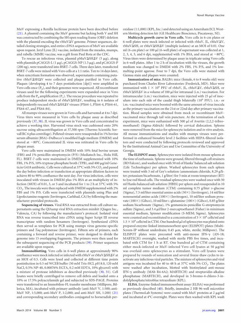
Serum samples were collected at 14 days postboost (dpb). As ex-pected, JL generated higher neutralizing antibody titers against JLthan MuV, and MuV generated higher anti-MuV titers than JL,regardless of the route of immunization (Fig. 1). This result isconsistent with a previous report that sera from JL-vaccinatedhumans had higher anti-JL neutralizing titers than anti-MuV neu-tralizing titers (41).
Previous studies have shown that rMuV�V (lacking V proteinexpression) or rMuV�SH (lacking SH protein expression) are atten-uated in a neurovirulency potency test in rat brains (21, 36), suggest-ing these viruses are good candidates for vaccine development. Toinvestigate the immunogenicity and vaccine potential of rMuV�Vand rMuV�SH in mice, BALB/c mice were mock vaccinated (PBS) orvaccinated with rMuV, JL, rMuV�SH, or rMuV�V and boosted at 22days postprimary vaccination through i.m. injection with the samevirus and dose as the primary vaccination. We chose the i.m. routebecause the trivalent MMR vaccine is usually administered by intra-muscular (i.m.) or deep subcutaneous injection (42), but mostly viai.m. administration. Serum samples were collected at 14 dpb. Thetotal IgG antibody titer against MuV was measured by ELISA usingplates coated with purified MuV (Fig. 2). We found that all groupsgenerated robust anti-MuV antibody responses.
Mumps virus is a human respiratory virus transmitted via re-spiratory secretions such as saliva and nose and throat discharge(6). i.n. vaccination induces both local immunity in the respira-tory tract and systemic immunity. Mucosal immunity providesdirect and rapid protection against virus challenge. To examinethe immunogenicity of rMuV�V and rMuV�SH compared tothat of rMuV and JL, BALB/c mice were mock vaccinated (PBS) orvaccinated with rMuV, JL, rMuV�SH, or rMuV�V intranasallyand boosted at 22 days postprimary vaccination with the samevirus type and dose as the primary i.n. vaccination. Serum sampleswere collected at 14 dpb. Total IgG antibody titers against MuVwere measured by ELISA (Fig. 3). All groups generated robustanti-MuV antibody responses.
Rescue of recombinant viruses lacking both V and SH pro-teins. To further enhance the safety of vaccine candidates, we con-structed a recombinant virus lacking expression of both the V and SHproteins. The genome length of the newly synthesized cDNA(pMuV�SH�V) complied with the rule of six for Paramyxovirus
FIG 1 Cross-reactivity of JL and MuV. BALB/c mice were i.n. or i.m. immu-nized with PBS, MuV, or JL at 106 PFU/mouse and boosted at 22 days post-vaccination with the same virus at 106 PFU/mouse. Serum samples were col-lected at 14 days postboost. Heat-inactivated serum samples of individual micefrom the same group were pooled to perform the plaque reduction neutraliza-tion test (PRNT). Serum samples were 2-fold serial diluted from 1:30 to1:3,840. A volume of 120 �l diluted serum was mixed with 120 �l diluted viruscontaining 80 PFU of either JL or rMuV virus and incubated at 37°C for 1 h.The count of residual unneutralized PFU per 100 �l was determined by plaqueassay in 6-well plates of Vero cells. PRNT titer is determined as the first dilutionlevel with residual PFU of more than half of the input per 100 �l.
FIG 2 i.m. immunization with rMuV�SH or rMuV�V induced antibodyresponses in mice. BALB/c mice were i.m. vaccinated with PBS, rMuV, JL,rMuV�SH, and rMuV�V at 106 PFU and boosted 22 days postvaccinationwith 106 PFU. Serum samples were collected at 14 dpb, and total antibodytiters in these samples were measured by ELISA coated with MuV viral pro-teins.
Xu et al.
2602 jvi.asm.org Journal of Virology

(43). Infectious recombinant viruses (rMuV�SH�V) were rescuedfrom BSRT-7 cells transfected with pMuV�SH�V and helper plas-mids as described before (21). To confirm rescue of the virus, viralRNA was extracted from cell culture medium containing rescuedviruses (Fig. 4B). The SH gene and the V/P gene region were ampli-fied using reverse transcription-PCR and sequenced. As shown in Fig.4, the SH ORF truncation as well as the V deletion was confirmed(Fig. 4C and D).
To confirm that genomic changes in rMuV�SH�V abolish Vand SH expression, Vero cells were mock infected or infected withrMuV or rescued rMuV�SH�V (PX64-67 strain). Expression lev-els of MuV NP, P, V, and SH proteins were examined using West-ern blotting. While NP and P were detected in both rMuV- andrMuV�SH�V-infected cells, expression of V or SH protein wasonly detected in rMuV-infected Vero cells (Fig. 4E).
Analysis of rMuV�SH�V in tissue culture cells. To select anrMuV�SH�V virus that replicates well for vaccine production pur-poses, the replication capability of rMuV�SH�V viruses from 6 in-
FIG 3 i.n. immunization with rMuV�SH or rMuV�V induced antibody re-sponses in mice. BALB/c mice were i.n. vaccinated with PBS, rMuV, JL,rMuV�SH, and rMuV�V with 106 PFU and boosted 22 days postvaccinationwith 106 PFU. Serum samples were collected at 14 dpb, and total antibodytiters of these samples were measured by ELISA coated with MuV viral pro-teins.
FIG 4 Generation of recombinant MuV lacking V and SH proteins (rMuV�SH�V). (A) Schematics of pMuV�SH�V. A 156-bp section was removedfrom the SH gene of pMuV�V, a cDNA genome of mumps virus lacking expression of V protein. (B) Reverse transcription-PCR confirmed the mutationin the SH ORF in rescued rMuV�SH�V. Recombinant viruses (rMuV�SH�V) were rescued from pMuV�SH�V through transfection of BSRT-7 cellswith pMuV�SH�V, together with the helper plasmids (pCAGGS-L, pCAGGS-NP, and pCAGGS-P). RNA was extracted from rMuV�SH�V-infectedVero cells. Two primers, PX47F and PX48R (sequences are available upon request), were used to amplify the SH gene region. (C and D) Sequenceconfirmation of the mutated regions in the SH ORF and the V/P editing site. The reverse transcription-PCR product of the SH gene was sent forsequencing. Sequencing results confirmed the mutation was successfully introduced into rMuV�SH�V. (E) Western blot confirmation of the deletion ofV and SH proteins in rMuV�SH�V viruses. Vero cells were mock infected or infected with rMuV or rMuV�SH�V at an MOI of 0.5. Cell lysates werecollected at 48 hpi and were blotted against MuV NP, P, V, and SH proteins.
Novel Mumps Vaccine
March 2014 Volume 88 Number 5 jvi.asm.org 2603
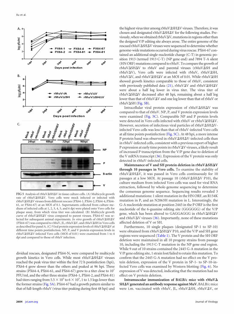
dividual rescues, designated PX64-N, were compared by multicyclegrowth kinetics in Vero cells. While most rMuV�SH�V virusesreached the peak virus titer within the first 72 h postinfection (hpi),PX64-4 grew slower than the others and peaked at 96 hpi. Threestrains (PX64-4, PX64-61, and PX64-67) grew to a titer close to 107
PFU/ml, and the other three strains (PX64-1, PX64-2, and PX64-81)had titers ranging from 5.5 � 105 to 6 � 105, 1 to 1.5 logs lower thanthe former strains (Fig. 5A). PX64-67 had a growth pattern similar tothat of full-length rMuV (virus titer peaking during first 48 hpi) and
the highest virus titer among rMuV�SH�V viruses. Therefore, it waschosen and designated rMuV�SH�V for the following studies. Pre-viously, when we obtained rMuV�V, mutations in regions other thanthe designed V/P editing site always arose. The entire genome of therescued rMuV�SH�V viruses were sequenced to determine whethergenome-wide mutations occurred during virus rescue. PX64-67 con-tained an additional single-nucleotide change (C-T) in genomic po-sition 1913 (termed 1913 C-T) (NP gene end) and 7894 T-A silent(HN ORF) mutations compared to rMuV. To compare the growth ofrMuV�SH�V to rMuV and parental viruses (rMuV�SH andrMuV�V), Vero cells were infected with rMuV, rMuV�SH,rMuV�V, and rMuV�SH�V at an MOI of 0.01. While rMuV�SHshowed growth kinetics comparable to those of rMuV, consistentwith previously published data (21), rMuV�V and rMuV�SH�Vwere about a half log lower in virus titer. The virus titer ofrMuV�SH�V decreased after 48 hpi, remaining about a half loglower than that of rMuV�V and one log lower than that of rMuV orrMuV�SH (Fig. 5B).
Intracellular viral protein expression of rMuV�SH�V wascompared to that of rMuV. NP, P, and V protein expression levelswere examined (Fig. 5C). Comparable NP and P protein levelswere detected in Vero cells infected with rMuV or rMuV�SH�V.However, secretion of infectious viral particles of rMuV�SH�V-infected Vero cells was less than that of rMuV-infected Vero cellsat all time points postinfection (Fig. 5C). At 48 hpi, a more intenseP protein band was observed in rMuV�SH�V-infected cells thanin rMuV-infected cells, consistent with a previous report of higherP expression at early time points in rMuV�V viruses, a likely resultof increased P transcription from the V/P gene due to deletion ofthe V mRNA transcript (36). Expression of the V protein was onlydetected in rMuV-infected cells.
Maintenance of V and SH protein deletion in rMuV�SH�Vthrough 10 passages in Vero cells. To examine the stability ofrMuV�SH�V, it was passed in Vero cells continuously for 10passages at a low MOI. At passage 10 (rMuV�SH�V P10), theculture medium from infected Vero cells was used for viral RNAextraction, followed by whole-genome sequencing to determinethe consensus genome sequence. Sequencing results revealed 3additional mutations: 1 silent mutation in the HN ORF, an R154Kmutation in P, and an N2063H mutation in L. Interestingly, theG-A nucleotide mutation at position 2445 in the P ORF is the firstnucleotide of the 6-guanine editing site (GGGGGG) of the V/Pgene, which has been altered to GAGGAGGG in rMuV�SH�Vand rMuV�V viruses (36). Importantly, none of these mutationsaffected deletion of V or SH.
Furthermore, 10 single plaques (designated SP-1 to SP-10)were obtained from rMuV�SH�V P10, and the V/P and SH generegions were sequenced (Table 1). The V protein and the SH ORFdeletion were maintained in all 10 progeny strains from passage10, including the 1913 C-T mutation in the NP gene end region.While 9 out of 10 strains contained the 2445 G-A mutation in theV/P gene editing site, 1 strain lost/failed to retain this mutation. Toconfirm that the 2445 G-A mutation had no effect on the V pro-tein deletion, expression of the V protein in SP-1- to SP-10-in-fected Vero cells was examined by Western blotting (Fig. 6). Noexpression of V was detected, indicating that the mutation had noeffect on V protein deletion.
Intramuscular immunization of BALB/c mice with rMuV�SH�V generated an antibody response against MuV. BALB/c micewere i.m. vaccinated with rMuV, JL, rMuV�SH, rMuV�V, or
FIG 5 Analysis of rMuV�SH�V in tissue culture cells. (A) Multicycle growthrate of rMuV�SH�V. Vero cells were mock infected or infected withrMuV�SH�V viruses from different rescues (PX64-1, PX64-2, PX64-4, PX64-61, or PX64-67) at an MOI of 0.1. Supernatants collected from culture me-dium of infected cells at 1, 2, 3, 4, 5, and 6 dpi were plated onto Vero cells forplaque assay, from which virus titer was calculated. (B) Multicycle growthcurve of rMuV�SH�V virus compared to parent viruses. PX64-67 was se-lected for subsequent animal experiments. In vitro growth of rMuV�SH�V(PX64-67) was compared to rMuV, JL, rMuV�V, and rMuV�SH in Vero cellsas described for panel A. (C) Viral protein expression levels of rMuV�SH�V atdifferent time points postinfection. NP, P, and V protein expression levels inrMuV�SH�V-infected Vero cells (MOI of 0.01) were examined from 1 to 4dpi and compared to those of rMuV infection.
Xu et al.
2604 jvi.asm.org Journal of Virology

rMuV�SH�V as described above. Serum samples were collectedas described previously. Serum IgG antibody titers against MuVwere measured using ELISA with plates coated with MuV viralproteins (lysed virions). Neutralizing antibody titers against MuVwere measured by an rMuV-Luc-based neutralization assay as de-scribed in Materials and Methods (linear correlation of rMuV-Luc-based neutralization assay to traditional plaque reductionneutralization assay was confirmed by an R2 value of 0.9317 usingferret serum samples [data not shown]). Similar approaches havebeen used for adenovirus, measles virus, and respiratory syncytialvirus (RSV) to substitute for the traditional plaque reduction neu-tralization test (PRNT) (44–47). We used rMuV-Luc, which wasconstructed based on the genetic background of MuV, as the tar-geting virus to compare the potentials of humoral responses in-duced by the vaccine candidates as well as the JL strain in mice toprotect against the circulating mumps virus in the United States.No significant differences were detected among the groups fortotal antibody titers (Fig. 7A). However, the neutralizing antibodytiters of serum samples showed some differences. The averageneutralizing titer of JL-inoculated mice was significantly lowerthan that of rMuV-infected mice, which had the highest neutral-izing titer. Mice inoculated with rMuV�SH, rMuV�V, orrMuV�SH�V had similar average titers (Fig. 7B).
Intranasal immunization of BALB/c mice with rMuV�SH�V generated an antibody response against MuV. BALB/cmice were i.n. inoculated with rMuV, JL, rMuV�SH, rMuV�V, orrMuV�SH�V at 106 PFU, and serum samples were collected formeasurement of both total antibody titer and neutralizing titer
against MuV. rMuV�SH-inoculated mice developed the highesttotal antibody titer, and the rMuV group had a higher titer thanthe JL group. No significant differences were detected among theJL group, rMuV�V group, and rMuV�SH�V groups (Fig. 8A).The rMuV group had the highest neutralizing titer, the JL grouphad the lowest neutralizing titer, and the other three groups(rMuV�SH, rMuV�V, and rMuV�SH�V) ranked between them(Fig. 8B). Although i.n.-immunized mice exhibited neutralizingantibody titer patterns like those observed in the i.m. groups, sta-tistically significant differences were found between the rMuV andJL groups (P 0.001), JL and rMuV�SH groups (P 0.001), JLand rMuV�SH�V groups (P 0.038), and rMuV�V andrMuV�SH groups (P 0.034).
Adaptive T cell responses were induced in mice vaccinatedwith rMuV�SH�V. To investigate the cellular immune re-sponses induced by rMuV�SH�V, i.m.- or i.n.-inoculated micewere euthanized at 28 dpb and splenocytes were isolated forELISPOT assay. In i.m.-inoculated mice, the JL group had thehighest T cell response levels and rMuV�SH�V had the lowest Tcell response levels, with no distinguishable differences among therMuV, rMuV�SH, and rMuV�V groups (Fig. 9A). Differenceswere significant between JL and rMuV�SH, JL and rMuV�SH�V,rMuV�SH and rMuV�SH�V, and rMuV�V and rMuV�SH�V.In i.n.-inoculated mouse groups, rMuV- and rMuV�V-immu-
TABLE 1 V/P gene and SH gene sequences of rMuV�SH�V P10 singleplaque-purified virusesa
VirusV proteindeletion
NP GE or V/P GSmutation
V/P editing sitemutation
SH ORFdeletion
SP-1 Yes 1913 C-T 2445 G-A YesSP-2 Yes 1913 C-T 2445 G-A YesSP-3 Yes 1913 C-T 2445 G-A YesSP-4 Yes 1913 C-T 2445 G-A YesSP-5 Yes 1913 C-T 1578 A-C YesSP-6 Yes 1913 C-T 2445 G-A YesSP-7 Yes 1913 C-T 2445 G-A YesSP-8 Yes 1913 C-T 2445 G-A YesSP-9 Yes 1913 C-T 2445 G-A YesSP-10 Yes 1913 C-T 2445 G-A Yesa rMuV�SH�V was passed continuously in Vero cells for 10 passages. Ten plaques wererandomly obtained from rMuV�SH�V at passage 10 and grown in Vero cells (SP-1 toSP-10). The V/P and SH gene regions of SP-1 to SP-10 were sequenced. Mutationsfound in these regions are shown.
FIG 6 Lack of V expression in isolates from the 10th passage of rMuV�SH�V.Vero cells were mock infected or infected with rMuV or SP-1 to SP-10. Onehundred �l of infected Vero cell lysates was subjected to Western blotting todetect NP and V protein expression levels.
FIG 7 Evaluation of antibody responses in mice i.m. vaccinated withrMuV�SH�V. BALB/c mice were i.m. immunized with PBS, rMuV, JL,rMuV�SH, rMuV�V, or rMuV�SH�V at 106 PFU/mouse and boosted at 22days postvaccination with the same virus at 106 PFU/mouse. (A) ELISA resultsmeasuring total antibody titer at 14 dpb. Serum samples were collected at 14dpb. The total antibody titer against MuV was measured through ELISA. (B)Neutralizing antibody titer at 14 dpb. Neutralizing antibody titers in serumsamples collected at 14 dpb were measured through an rMuV-Luc-based neu-tralization assay. P values of 0.05 are shown.
Novel Mumps Vaccine
March 2014 Volume 88 Number 5 jvi.asm.org 2605

nized mice had the lowest responding cell counts (Fig. 9B). Signif-icant differences were observed between JL and rMuV�V, rMuVand rMuV�SH, rMuV and rMuV�SH�V, rMuV�SH andrMuV�V, and rMuV�V and rMuV�SH�V groups.
DISCUSSION
The JL vaccine is one of the most successful vaccines developedduring the third quarter of the last century. It was produced by thepropagation of mumps virus in embryonated hen’s eggs that re-sulted in attenuation (48–51). Introduction of the in vitro tissue/cell culture technique into vaccinology facilitated the develop-ment and production of the majority of currently licensed live-attenuated vaccines in the United States against viral infections(52–56). For mumps vaccine candidates, JL is the great success,but unfortunately there were many failures. Different passages ofattenuated viruses were tested in animal models or in field trials inorder to select a vaccine seed with the most reduced virulence andgreatest immunogenicity (1). Selected vaccine candidates need tobe biologically characterized in order to be distinguished from
virulent strains. There are currently no standardized attenuationmarkers for mumps vaccines, partially due to the semirational andsemiempirical nature of the traditional attenuation method (1,57–60). The rate of aseptic meningitis following vaccination withJL (estimated one case per 1.8 million doses) is below backgroundlevels (61). However, other live attenuated mumps virus vaccineshave had much higher incidences of vaccine-associated meningi-tis. The Urabe vaccine, which was widely distributed in Japan,Europe, and Canada, is estimated to cause one case of meningitisin every 1,000 to 11,000 doses distributed in the United Kingdomand one case of meningitis in every 62,000 doses distributed inCanada. The Urabe vaccine has been withdrawn due to safetyconcerns.
In this study, based on the establishment of reverse genetictechnology of negative-sensed, nonsegmented RNA viruses
FIG 8 Evaluation of antibody responses in mice i.n. vaccinated withrMuV�SH�V. BALB/c mice were i.n. immunized with PBS, rMuV, JL,rMuV�SH, rMuV�V, or rMuV�SH�V at 106 PFU and boosted at 22 dayspostvaccination with the same virus at 106 PFU. (A) ELISA results measuringtotal antibody titer at 14 dpb. Serum samples were collected at 14 dpb. Totalantibody titers against MuV were measured through ELISA. (B) Neutralizingantibody titer at 14 dpb. Neutralizing antibody titers in serum samples col-lected at 14 dpb were measured through an rMuV-Luc-based neutralizationassay. P values for significantly different groups were found for rMuV and JLgroups (0.001), rMuV and rMuV�V groups (0.016), JL and rMuV�SH groups(0.001), JL and rMuV�SH�V groups (0.038), and rMuV�V and rMuV�SHgroups (0.034). For simplicity, P values comparing the rMuV and JL groupsand JL and rMuV�SH�V groups are shown.
FIG 9 Cellular immune responses induced by rMuV�SH�V vaccination inmice. (A) Memory T cell responses in mumps virus i.m.-immunized mice.BALB/c mice were i.m. immunized with PBS, rMuV, JL, rMuV�SH, rMuV�V,or rMuV�SH�V at 106 PFU and boosted at 22 days postvaccination with thesame virus at 106 PFU. Splenocytes were extracted from mouse spleens andused for ELISPOT assay. Splenocytes were stimulated with MuV-infected Verocell lysates or with mock-infected Vero cell lysates at 50 �g/ml. P values of0.05 are shown. (B) Memory T cell responses in mumps virus i.n.-immu-nized mice. BALB/c mice were i.m. immunized with PBS, rMuV, JL,rMuV�SH, rMuV�V, or rMuV�SH�V at 106 PFU and boosted at 22 dayspostvaccination with the same virus at 106 PFU. Splenocytes were extractedfrom mouse spleens and used for ELISPOT assay. Splenocytes were stimulatedwith MuV-infected Vero cell lysates or with mock-infected Vero cell lysates at50 �g/ml. P values of 0.05 are shown.
Xu et al.
2606 jvi.asm.org Journal of Virology

(62–66), we examined the possibility of generating a new mumpsvaccine candidate through deletion of V and SH protein expres-sion from a clinical isolate from a 2006 Iowa mumps epidemic(MuV, genotype G). Deletion of either of the proteins has beenpreviously reported to reduce mumps neurotoxicity in IC-in-fected rats. Importantly, since deletion of the V protein alone(from the MuV strain) is sufficient to reduce the neurovirulencepotential of the recombinant virus to a level comparable to that ofthe JL vaccine (21, 36), the lack of V protein expression can beused as an attenuation marker for our vaccine candidates.
Attenuation based on targeted genetic modification has severaladvantages. The most commonly used mumps vaccine, JL, is amixture of at least two well-distinguished components (67–69).Surveillance of the compositional balance between the two com-ponents during vaccine preparation and propagation has beenproposed (68). However, cDNA-derived recombinant viruseshave defined consensus sequences and higher homogeneity. Theyare plaque purified, whole genome sequenced, and passed in Verocells only for the purpose of amplification. Vaccine candidates canbe continuously rescued from the cDNA plasmids with a definedconsensus sequence and clear genetic markers for attenuation. Allprocesses are cell culture based, bypassing the necessity of high-quality pathogen-free chickens, chicken eggs, or any other animalsused for in vivo adaption (70–72). Omission of the serial passagessaves time for vaccine development and avoids potential adap-tion-induced antigen shifts of the vaccine strains; therefore, it re-tains the maximum amount of immunogenic epitopes.
Mumps viruses are classified into 12 genotypes based on ge-netic variability of the SH gene (73, 74). Different subtypes ofmumps viruses exhibit distinguished geographic distributionworldwide. Although the driving force of such distribution re-mains unclear, emergence of new subclusters of circulatingmumps viruses within a genotype (75, 76) indicates evolution ofwild-type mumps viruses under various selection forces. Failureto detect genotype A wild-type mumps viruses in countries/re-gions immunized with genotype A vaccine in recent studies maybe due to a vaccine-based selection pressure. This pressure mayselect for genotypes with increased virulence and heterogeneitycompared to current vaccines (77–82). Decreased neutralizationcapabilities against heterogenotypes among subtypes of mumpsviruses (83–86) and lack of cross protection between differentsubtypes (genotype D against genotype A) in human natural in-fection have been reported (87). It would be ideal to use a geno-type-matched vaccine candidate (genotype G), which elicits morespecific immune responses that effectively protect against the cir-culating mumps viruses in the United States (genotype G) (11, 13,78, 79).
Although additional mutations occurred during rMuV�SH�Vvirus rescue and during passages of rMuV�SH�V in Vero cells, noregaining of the V protein or the SH protein was observed in anyrMuV�SH�V viruses analyzed, indicating that rMuV�SH�V isstable in tissue culture cells. Interestingly, besides two silent mu-tations in the HN and L ORFs, rMuV�SH�V (PX64-67) pos-sessed one nucleotide change (C-T) in genomic position 1913 inthe V/P GS region, which has been previously seen in the course ofrMuV�V virus rescue (36). This mutation is believed to be impor-tant in regulating the transcription/translation level of P protein,emphasizing the significance of a proper ratio between NP and Pprotein during virus growth.
One challenge of developing a new mumps vaccine is the lack
of correlation between protection and immune responses. While aneutralization titer is thought to be essential in protection againstmumps infection (79), investigations of serum samples of patientsversus nonpatients during recent mumps outbreaks revealed nodefined cutoff neutralizing antibody titer against mumps virus,indicating a potential role for cellular immunity in effective pro-tection against mumps challenge (41, 86). In this study, the inves-tigation of immunogenicity of rMuV�SH�V in i.n.- and i.m.-vaccinated mice showed that rMuV�SH�V was able to induce aneutralizing titer comparable to those induced by rMuV�SH andrMuV�V and a higher titer than that induced by JL vaccine. Fur-thermore, rMuV�SH�V vaccination also stimulated T cell re-sponses in mice, although the role of cell-mediated immunity inmumps disease protection remains to be demonstrated. We alsoobserved that rMuV�SH induced slightly higher total antibodytiters than those induced by rMuV, and rMuV�SH�V inducedhigher antibody titers than those induced by rMuV�V, suggestingthat deletion of SH leads to better antigen presentation. Similarresults have been reported for a closely related virus, parainfluenzavirus 5 (PIV5), in which PIV5 lacking SH is more immunogenicthan PIV5 (88). The mouse models have been widely used to testvaccine efficacy for various human viruses (89–92). However, it isnot a good model for mumps virus infection. The efficacy ofrMuV�SH�V in nonhuman primates, which is a good model formumps virus infection (40), should be examined before testingthis candidate in humans. In summary, rMuV�SH�V was able toelicit both antibody and cellular responses against MuV in i.n.-and i.m.-vaccinated mice, providing a safe and immunogenicmumps vaccine candidate.
ACKNOWLEDGMENTS
We appreciate the comments, suggestions, and technical help from mem-bers of the He laboratory. We thank Paul Rota for providing the JL vaccinestrain. We are grateful to Kaori Sakamoto for carefully reading the man-uscript prior to submission.
This work has been supported by a grant from the NIH (AI097368 toB.H.).
REFERENCES1. Buynak EB, Hilleman MR. 1966. Live attenuated mumps virus vaccine. 1.
Vaccine development. Proc. Soc. Exp. Biol. Med. 123:768 –775. http://dx.doi.org/10.3181/00379727-123-31599.
2. Pagano JS, Levine RH, Sugg WC, Finger JA. 1967. Clinical trial of newattenuated mumps virus vaccine (Jeryl Lynn strain): preliminary report.Prog. Immunobiol. Stand. 3:196 –202.
3. Weibel RE, Stokes J, Jr, Buynak EB, Leagus MB, Hilleman MR. 1968.Jeryl Lynn strain live attenuated mumps virus vaccine. Durability of im-munity following administration. JAMA 203:14 –18.
4. Young ML, Dickstein B, Weibel RE, Stokes J, Jr, Buynak EB, HillemanMR. 1967. Experiences with Jeryl Lynn strain live attenuated mumps virusvaccine in a pediatric outpatient clinic. Pediatrics 40:798 – 803.
5. Rubin SA, Afzal MA, Powell CL, Bentley ML, Auda GR, Taffs RE,Carbone KM. 2005. The rat-based neurovirulence safety test for the as-sessment of mumps virus neurovirulence in humans: an internationalcollaborative study. J. Infect. Dis. 191:1123–1128. http://dx.doi.org/10.1086/428098.
6. Carbone KM, Wolinsky JS. 2001. Mumps virus, p 1381–1400. In KnipeDM, Howley PM, Griffin DE, Lamb RA, Martin MA, Roizman B, StrausSE (ed), Fields virology, 4 ed, vol 1. Lippincott Williams and Wilkins,Philadelphia, PA.
7. Smits G, Mollema L, Hahne S, de Melker H, Tcherniaeva I, Waaijen-borg S, van Binnendijk R, van der Klis F, Berbers G. 2013. Seropreva-lence of mumps in the Netherlands: dynamics over a decade with highvaccination coverage and recent outbreaks. PLoS One 8:e58234. http://dx.doi.org/10.1371/journal.pone.0058234.
Novel Mumps Vaccine
March 2014 Volume 88 Number 5 jvi.asm.org 2607
8. Mahamud A, Fiebelkorn AP, Nelson G, Aguon A, McKenna J, Villar-ruel G, Gallagher K, Ortega-Sanchez IR. 2012. Economic impact of the2009 –2010 Guam mumps outbreak on the public health sector and af-fected families. Vaccine 30:6444 – 6448. http://dx.doi.org/10.1016/j.vaccine.2012.08.001.
9. Rajcevic S, Seguljev Z, Petrovic V, Medic S, Nedelijkovic J, Milosevic V,Turo L, Ristic M. 2012. Ongoing mumps outbreak in Novi Sad, theautonomous province of Vojvodina, Serbia, January to April 2012. EuroSurveill. 17:20169.
10. Hassan J, Dean J, Moss E, Carr MJ, Hall WW, Connell J. 2012.Seroepidemiology of the recent mumps virus outbreaks in Ireland. J. Clin.Virol. 53:320 –324. http://dx.doi.org/10.1016/j.jcv.2011.12.022.
11. Marin M, Quinlisk P, Shimabukuro T, Sawhney C, Brown C, LebaronCW. 2008. Mumps vaccination coverage and vaccine effectiveness in alarge outbreak among college students–Iowa, 2006. Vaccine 26:3601–3607. http://dx.doi.org/10.1016/j.vaccine.2008.04.075.
12. Barskey AE, Glasser JW, LeBaron CW. 2009. Mumps resurgences in theUnited States: a historical perspective on unexpected elements. Vaccine27:6186 – 6195. http://dx.doi.org/10.1016/j.vaccine.2009.06.109.
13. Anonymous. 2009. Mumps outbreak–New York, New Jersey, Quebec,2009. MMWR Morb. Mortal. Wkly. Rep. 58:1270 –1274.
14. Gordon JE, Kilham L. 1949. Ten years in the epidemiology of mumps.Am. J. Med. Sci. 218:338 –359. http://dx.doi.org/10.1097/00000441-194909000-00013.
15. Rima BK, Roberts MW, McAdam WD, Martin SJ. 1980. Polypeptidesynthesis in mumps virus-infected cells. J. Gen. Virol. 46:501–505. http://dx.doi.org/10.1099/0022-1317-46-2-501.
16. Elango N, Varsanyi TM, Kovamees J, Norrby E. 1988. Molecular cloningand characterization of six genes, determination of gene order and intergenicsequences and leader sequence of mumps virus. J. Gen. Virol. 69(Part 11):2893–2900. http://dx.doi.org/10.1099/0022-1317-69-11-2893.
17. Hosaka Y, Shimizu K. 1968. Lengths of the nucleocapsids of Newcastledisease and mumps viruses. J. Mol. Biol. 35:369 –373. http://dx.doi.org/10.1016/S0022-2836(68)80031-2.
18. Fuentes S, Tran KC, Luthra P, Teng MN, He B. 2007. Function of therespiratory syncytial virus small hydrophobic protein. J. Virol. 81:8361–8366. http://dx.doi.org/10.1128/JVI.02717-06.
19. Lin Y, Bright AC, Rothermel TA, He B. 2003. Induction of apoptosis byparamyxovirus simian virus 5 lacking a small hydrophobic gene. J. Virol.77:3371–3383. http://dx.doi.org/10.1128/JVI.77.6.3371-3383.2003.
20. Wilson RL, Fuentes SM, Wang P, Taddeo EC, Klatt A, Henderson AJ, HeB. 2006. Function of small hydrophobic proteins of paramyxovirus. J. Virol.80:1700–1709. http://dx.doi.org/10.1128/JVI.80.4.1700-1709.2006.
21. Xu P, Li Z, Sun D, Lin Y, Wu J, Rota PA, He B. 2011. Rescue ofwild-type mumps virus from a strain associated with recent outbreakshelps to define the role of the SH ORF in the pathogenesis of mumps virus.Virology 417:126 –136. http://dx.doi.org/10.1016/j.virol.2011.05.003.
22. Li Z, Xu J, Patel J, Fuentes S, Lin Y, Anderson D, Sakamoto K, WangLF, He B. 2011. Function of the small hydrophobic protein of J paramyxo-virus. J. Virol. 85:32– 42. http://dx.doi.org/10.1128/JVI.01673-10.
23. He B, Lin GY, Durbin JE, Durbin RK, Lamb RA. 2001. The SH integralmembrane protein of the paramyxovirus simian virus 5 is required toblock apoptosis in mdbk cells. J. Virol. 75:4068 – 4079. http://dx.doi.org/10.1128/JVI.75.9.4068-4079.2001.
24. Elliott GD, Yeo RP, Afzal MA, Simpson EJ, Curran JA, Rima BK. 1990.Strain-variable editing during transcription of the P gene of mumps virusmay lead to the generation of non-structural proteins NS1 (V) and NS2. J.Gen. Virol. 71(Part 7):1555–1560. http://dx.doi.org/10.1099/0022-1317-71-7-1555.
25. Paterson RG, Lamb RA. 1990. RNA editing by G-nucleotide insertion inmumps virus P-gene mRNA transcripts. J. Virol. 64:4137– 4145.
26. Motz C, Schuhmann KM, Kirchhofer A, Moldt M, Witte G, Conzel-mann KK, Hopfner KP. 2013. Paramyxovirus V proteins disrupt the foldof the RNA sensor MDA5 to inhibit antiviral signaling. Science 339:690 –693. http://dx.doi.org/10.1126/science.1230949.
27. Parisien JP, Bamming D, Komuro A, Ramachandran A, Rodriguez JJ,Barber G, Wojahn RD, Horvath CM. 2009. A shared interface mediatesparamyxovirus interference with antiviral RNA helicases MDA5 andLGP2. J. Virol. 83:7252–7260. http://dx.doi.org/10.1128/JVI.00153-09.
28. Ramachandran A, Horvath CM. 2010. Dissociation of paramyxovirusinterferon evasion activities: universal and virus-specific requirements forconserved V protein amino acids in MDA5 interference. J. Virol. 84:11152–11163. http://dx.doi.org/10.1128/JVI.01375-10.
29. Nishio M, Garcin D, Simonet V, Kolakofsky D. 2002. The carboxylsegment of the mumps virus V protein associates with stat proteins in vitrovia a tryptophan-rich motif. Virology 300:92. http://dx.doi.org/10.1006/viro.2002.1509.
30. Poole E, He B, Lamb RA, Randall RE, Goodbourn S. 2002. The Vproteins of simian virus 5 and other paramyxoviruses inhibit induction ofinterferon-beta. Virology 303:33– 46. http://dx.doi.org/10.1006/viro.2002.1737.
31. Ulane CM, Rodriguez JJ, Parisien JP, Horvath CM. 2003. STAT3 ubiq-uitylation and degradation by mumps virus suppress cytokine and onco-gene signaling. J. Virol. 77:6385– 6393. http://dx.doi.org/10.1128/JVI.77.11.6385-6393.2003.
32. Yokosawa N, Yokota S, Kubota T, Fujii N. 2002. C-terminal region ofSTAT-1alpha is not necessary for its ubiquitination and degradationcaused by mumps virus V protein. J. Virol. 76:12683–12690. http://dx.doi.org/10.1128/JVI.76.24.12683-12690.2002.
33. Ulane CM, Horvath CM. 2002. Paramyxoviruses SV5 and HPIV2 assem-ble STAT protein ubiquitin ligase complexes from cellular components.Virology 304:160 –166. http://dx.doi.org/10.1006/viro.2002.1773.
34. Andrejeva J, Poole E, Young DF, Goodbourn S, Randall RE. 2002. Thep127 subunit (DDB1) of the UV-DNA damage repair binding protein isessential for the targeted degradation of STAT1 by the V protein of theparamyxovirus simian virus 5. J. Virol. 76:11379 –11386. http://dx.doi.org/10.1128/JVI.76.22.11379-11386.2002.
35. Parisien JP, Lau JF, Rodriguez JJ, Ulane CM, Horvath CM. 2002.Selective STAT protein degradation induced by paramyxoviruses requiresboth STAT1 and STAT2 but is independent of alpha/beta interferon signaltransduction. J. Virol. 76:4190 – 4198. http://dx.doi.org/10.1128/JVI.76.9.4190-4198.2002.
36. Xu P, Luthra P, Li Z, Fuentes S, D’Andrea JA, Wu J, Rubin S, Rota PA,He B. 2012. The V protein of mumps virus plays a critical role in patho-genesis. J. Virol. 86:1768 –1776. http://dx.doi.org/10.1128/JVI.06019-11.
37. He B, Lamb RA. 1999. Effect of inserting paramyxovirus simian virus5 gene junctions at the HN/L gene junction: analysis of accumulationof mRNAs transcribed from rescued viable viruses. J. Virol. 73:6228 –6234.
38. He B, Paterson RG, Ward CD, Lamb RA. 1997. Recovery of infectiousSV5 from cloned DNA and expression of a foreign gene. Virology 237:249 –260. http://dx.doi.org/10.1006/viro.1997.8801.
39. Li Z, Yu M, Zhang H, Magoffin DE, Jack PJ, Hyatt A, Wang HY, WangLF. 2006. Beilong virus, a novel paramyxovirus with the largest genome ofnon-segmented negative-stranded RNA viruses. Virology 346:219 –228.http://dx.doi.org/10.1016/j.virol.2005.10.039.
40. Xu P, Huang Z, Gao X, Michel FJ, Hirsch G, Hogan RJ, Sakamoto K,Ho W, Wu J, He B. 2013. Infection of mice, ferrets, and rhesus macaqueswith a clinical mumps virus isolate. J. Virol. 87:8158 – 8168. http://dx.doi.org/10.1128/JVI.01028-13.
41. Cortese MM, Barskey AE, Tegtmeier GE, Zhang C, Ngo L, Kyaw MH,Baughman AL, Menitove JE, Hickman CJ, Bellini WJ, Dayan GH,Hansen GR, Rubin S. 2011. Mumps antibody levels among studentsbefore a mumps outbreak: in search of a correlate of immunity. J. Infect.Dis. 204:1413–1422. http://dx.doi.org/10.1093/infdis/jir526.
42. Carter H, Campbell H. 1993. Rational use of measles, mumps and rubella(MMR) vaccine. Drugs 45:677– 683. http://dx.doi.org/10.2165/00003495-199345050-00005.
43. Calain P, Roux L. 1993. The rule of six, a basic feature for efficientreplication of Sendai virus defective interfering RNA. J. Virol. 67:4822–4830.
44. Chen M, Chang JS, Nason M, Rangel D, Gall JG, Graham BS, Ledger-wood JE. 2010. A flow cytometry-based assay to assess RSV-specific neu-tralizing antibody is reproducible, efficient and accurate. J. Immunol.Methods 362:180 –184. http://dx.doi.org/10.1016/j.jim.2010.08.005.
45. Haralambieva IH, Ovsyannikova IG, Vierkant RA, Poland GA. 2008.Development of a novel efficient fluorescence-based plaque reduction mi-croneutralization assay for measles virus immunity. Clin. Vaccine Immu-nol. 15:1054 –1059. http://dx.doi.org/10.1128/CVI.00008-08.
46. Sprangers MC, Lakhai W, Koudstaal W, Verhoeven M, Koel BF, VogelsR, Goudsmit J, Havenga MJ, Kostense S. 2003. Quantifying adenovirus-neutralizing antibodies by luciferase transgene detection: addressing pre-existing immunity to vaccine and gene therapy vectors. J. Clin. Microbiol.41:5046 –5052. http://dx.doi.org/10.1128/JCM.41.11.5046-5052.2003.
47. Fuentes S, Crim RL, Beeler J, Teng MN, Golding H, Khurana S. 2013.Development of a simple, rapid, sensitive, high-throughput luciferase re-
Xu et al.
2608 jvi.asm.org Journal of Virology
porter based microneutralization test for measurement of virus neutraliz-ing antibodies following respiratory syncytial virus vaccination and infec-tion. Vaccine 31:3987–3994. http://dx.doi.org/10.1016/j.vaccine.2013.05.088.
48. Goodpasture EW, Woodruff AM, Buddingh GJ. 1931. The cultivation ofvaccine and other viruses in the chorioallantoic membrane of chick embryos.Science 74:371–372. http://dx.doi.org/10.1126/science.74.1919.371.
49. Habel K. 1946. Preparation of mumps vaccines and immunization ofmonkeys against experimental mumps infection. Public Health Rep. 61:1655–1664. http://dx.doi.org/10.2307/4585906.
50. Levens JH, Enders JF. 1945. The hemoagglutinative properties of amni-otic fluid from embryonated eggs infected with mumps virus. Science102:117–120. http://dx.doi.org/10.1126/science.102.2640.117.
51. Enders JF, Levens JH, et al. 1946. Attenuation of virulence with retentionof antigenicity of mumps virus after passage in the embryonated egg. J.Immunol. 54:283–291.
52. Hilleman MR. 2002. Overview of the needs and realities for developingnew and improved vaccines in the 21st century. Intervirology 45:199 –211.http://dx.doi.org/10.1159/000067911.
53. Hilleman MR. 2002. Overview: past and future of immunologic interven-tion in the pathogenesis, prophylaxis and therapeusis of hepatitis B. J.Gastroenterol. Hepatol. 17(Suppl):S449 –S451. http://dx.doi.org/10.1046/j.1440-1746.17.s4.8.x.
54. U.S. FDA. 2013. Complete list of vaccines licensed for immunization anddistribution in the US. U.S. FDA, Washington, DC.
55. Malik H, Khan FH, Ahsan H. 2013. Human papillomavirus: currentstatus and issues of vaccination. Arch. Virol.[Epub ahead of print.] http://dx.doi.org/10.1007/s00705-013-1827-z.
56. Doan HQ, Ung B, Ramirez-Fort MK, Khan F, Tyring SK. 2013. Zos-tavax: a subcutaneous vaccine for the prevention of herpes zoster. ExpertOpin. Biol. Ther. 13:1467–1477. http://dx.doi.org/10.1517/14712598.2013.830101.
57. St Geme JW, Hawley LM, Davis WC. 1976. Comparative replication ofnatural, attenuated, and laboratory strains of mumps virus. Life Sci. 18:129 –134. http://dx.doi.org/10.1016/0024-3205(76)90283-6.
58. Yamanishi K, Hosai H, Ueda S, Takahashi M, Okuno Y. 1970. Studieson live attenuated mumps virus vaccine. II. Biological characteristics ofthe strains adapted to the amniotic and chorioallantoic cavity of develop-ing chick embryos. Biken J. 13:127–132.
59. Kaptsova TI, Alekseeva AK, Gordienko NM, Rozina EE, Ermakova MN.1976. Results of a study of live mumps vaccine from strain L-3 producedby the Moscow research institute for viral preparations. Characteristics ofthe vaccine. Voprosy Virusologii 1976:674 – 685.
60. Gluck R, Hoskins JM, Wegmann A, Just M, Germanier R. 1986. Rubini,a new live attenuated mumps vaccine virus strain for human diploid cells.Dev. Biol. Stand. 65:29 –35.
61. Nalin DR. 1989. Mumps vaccine complications: which strain? Lancetii:1396.
62. Schnell MJ, Mebatsion T, Conzelmann KK. 1994. Infectious rabiesviruses from cloned cDNA. EMBO J. 13:4195– 4203.
63. Collins PL, Hill MG, Camargo E, Grosfeld H, Chanock RM, MurphyBR. 1995. Production of infectious human respiratory syncytial virusfrom cloned cDNA confirms an essential role for the transcription elon-gation factor from the 5=proximal open reading frame of the M2 mRNA ingene expression and provides a capability for vaccine development. Proc.Natl. Acad. Sci. U. S. A. 92:11563–11567. http://dx.doi.org/10.1073/pnas.92.25.11563.
64. Radecke F, Spielhofer P, Schneider H, Kaelin K, Huber M, Dotsch C,Christiansen G, Billeter MA. 1995. Rescue of measles viruses from clonedDNA. EMBO J. 14:5773–5784.
65. Neumann G, Watanabe T, Ito H, Watanabe S, Goto H, Gao P, Hughes M,Perez DR, Donis R, Hoffmann E, Hobom G, Kawaoka Y. 1999. Generationof influenza A viruses entirely from cloned cDNAs. Proc. Natl. Acad. Sci.U. S. A. 96:9345–9350. http://dx.doi.org/10.1073/pnas.96.16.9345.
66. Fodor E, Devenish L, Engelhardt OG, Palese P, Brownlee GG, Garcia-Sastre A. 1999. Rescue of influenza A virus from recombinant DNA. J.Virol. 73:9679 –9682.
67. Afzal MA, Pickford AR, Forsey T, Heath AB, Minor PD. 1993. The JerylLynn vaccine strain of mumps virus is a mixture of two distinct isolates. J.Gen. Virol. 74(Part 5):917–920. http://dx.doi.org/10.1099/0022-1317-74-5-917.
68. Amexis G, Rubin S, Chizhikov V, Pelloquin F, Carbone K, ChumakovK. 2002. Sequence diversity of Jeryl Lynn strain of mumps virus: quanti-
tative mutant analysis for vaccine quality control. Virology 300:171–179.http://dx.doi.org/10.1006/viro.2002.1499.
69. Chambers P, Rima BK, Duprex WP. 2009. Molecular differences betweentwo Jeryl Lynn mumps virus vaccine component strains, JL5 and JL2. J. Gen.Virol. 90:2973–2981. http://dx.doi.org/10.1099/vir.0.013946-0.
70. Barrett PN, Mundt W, Kistner O, Howard MK. 2009. Vero cellplatform in vaccine production: moving towards cell culture-basedviral vaccines. Expert Rev. Vaccines 8:607– 618. http://dx.doi.org/10.1586/erv.09.19.
71. Clark HF, Offit PA, Plotkin SA, Heaton PM. 2006. The new pentavalentrotavirus vaccine composed of bovine (strain WC3)-human rotavirus re-assortants. Pediatr. Infect. Dis. J. 25:577–583. http://dx.doi.org/10.1097/01.inf.0000220283.58039.b6.
72. Bernstein DI. 2006. Live attenuated human rotavirus vaccine, Rotarix.Semin. Pediatr. Infect. Dis. 17:188 –194. http://dx.doi.org/10.1053/j.spid.2006.08.006.
73. Jin L, Rima B, Brown D, Orvell C, Tecle T, Afzal M, Uchida K,Nakayama T, Song JW, Kang C, Rota PA, Xu W, Featherstone D. 2005.Proposal for genetic characterisation of wild-type mumps strains: prelim-inary standardisation of the nomenclature. Arch. Virol. 150:1903–1909.http://dx.doi.org/10.1007/s00705-005-0563-4.
74. Santos CL, Ishida MA, Foster PG, Sallum MA, Benega MA, Borges DB,Correa KO, Constantino CR, Afzal MA, Paiva TM. 2008. Detection of anew mumps virus genotype during parotitis epidemic of 2006 –2007 in thestate of Sao Paulo, Brazil. J. Med. Virol. 80:323–329. http://dx.doi.org/10.1002/jmv.21068.
75. Tecle T, Mickiene A, Johansson B, Lindquist L, Orvell C. 2002. Molec-ular characterisation of two mumps virus genotypes circulating during anepidemic in Lithuania from 1998 to 2000. Arch. Virol. 147:243–253. http://dx.doi.org/10.1007/s705-002-8317-y.
76. Utz S, Richard JL, Capaul S, Matter HC, Hrisoho MG, Muhlemann K.2004. Phylogenetic analysis of clinical mumps virus isolates from vacci-nated and non-vaccinated patients with mumps during an outbreak, Swit-zerland 1998 –2000. J. Med. Virol. 73:91–96. http://dx.doi.org/10.1002/jmv.20064.
77. Greenland K, Whelan J, Fanoy E, Borgert M, Hulshof K, Yap KB,Swaan C, Donker T, van Binnendijk R, de Melker H, Hahne S. 2012.Mumps outbreak among vaccinated university students associated with alarge party, the Netherlands, 2010. Vaccine 30:4676 – 4680. http://dx.doi.org/10.1016/j.vaccine.2012.04.083.
78. Anonymous. 2012. Mumps outbreak on a university campus–California,2011. MMWR Morb. Mortal. Wkly. Rep. 61:986 –989.
79. Hviid A, Rubin S, Muhlemann K. 2008. Mumps. Lancet 371:932–944.http://dx.doi.org/10.1016/S0140-6736(08)60419-5.
80. Echevarria JE, Castellanos A, Sanz JC, Perez C, Palacios G, Martinez deAragon MV, Pena Rey I, Mosquera M, de Ory F, Royuela E. 2010.Circulation of mumps virus genotypes in Spain from 1996 to 2007. J. Clin.Microbiol. 48:1245–1254. http://dx.doi.org/10.1128/JCM.02386-09.
81. Echevarria AC, Sanz JEJC, Martinez de Aragon MV, Pena Rey I,Mosquera M, Fde Ory Royuela E. 2010. Mumps virus genotyping: basisand known circulating genotypes. Open Vaccine J. 3:37– 41. http://dx.doi.org/10.2174/1875035401003020037.
82. Muhlemann K. 2004. The molecular epidemiology of mumps virus. Infect.Genet. Evol. 4:215–219. http://dx.doi.org/10.1016/j.meegid.2004.02.003.
83. Santak M, Lang-Balija M, Ivancic-Jelecki J, Kosutic-Gulija T, Ljubin-Sternak S, Forcic D. 2013. Antigenic differences between vaccine andcirculating wild-type mumps viruses decreases neutralization capacity ofvaccine-induced antibodies. Epidemiol. Infect. 141:1298 –1309. http://dx.doi.org/10.1017/S0950268812001896.
84. Orvell C, Tecle T, Johansson B, Saito H, Samuelson A. 2002. Antigenicrelationships between six genotypes of the small hydrophobic proteingene of mumps virus. J. Gen. Virol. 83:2489 –2496.
85. Rubin SA, Qi L, Audet SA, Sullivan B, Carbone KM, Bellini WJ, RotaPA, Sirota L, Beeler J. 2008. Antibody induced by immunization with theJeryl Lynn mumps vaccine strain effectively neutralizes a heterologouswild-type mumps virus associated with a large outbreak. J. Infect. Dis.198:508 –515. http://dx.doi.org/10.1086/590115.
86. Rubin SA, Link MA, Sauder CJ, Zhang C, Ngo L, Rima BK, Duprex WP.2012. Recent mumps outbreaks in vaccinated populations: no evidence ofimmune escape. J. Virol. 86:615– 620. http://dx.doi.org/10.1128/JVI.06125-11.
87. Nojd J, Tecle T, Samuelsson A, Orvell C. 2001. Mumps virus neutraliz-ing antibodies do not protect against reinfection with a heterologous
Novel Mumps Vaccine
March 2014 Volume 88 Number 5 jvi.asm.org 2609

mumps virus genotype. Vaccine 19:1727–1731. http://dx.doi.org/10.1016/S0264-410X(00)00392-3.
88. Li Z, Gabbard JD, Mooney A, Chen Z, Tompkins SM, He B. 2013.Efficacy of parainfluenza virus 5 mutants expressing hemagglutinin fromH5N1 influenza A virus in mice. J. Virol. 87:9604 –9609. http://dx.doi.org/10.1128/JVI.01289-13.
89. Boyoglu-Barnum S, Gaston KA, Todd SO, Boyoglu C, Chirkova T,Barnum TR, Jorquera P, Haynes LM, Tripp RA, Moore ML, AndersonLJ. 2013. A respiratory syncytial virus (RSV) anti-G protein F(ab=)2monoclonal antibody suppresses mucous production and breathing effortin RSV rA2-line19F-infected Balb/c mice. J. Virol. 87:10955–10967. http://dx.doi.org/10.1128/JVI.01164-13.
90. Xu L, Bao L, Deng W, Zhu H, Chen T, Lv Q, Li F, Yuan J, Xiang Z, Gao
K, Xu Y, Huang L, Li Y, Liu J, Yao Y, Yu P, Yong W, Wei Q, Zhang L,Qin C. 2013. The mouse and ferret models for studying the novel avian-origin human influenza A (H7N9) virus. Virol. J. 10:253. http://dx.doi.org/10.1186/1743-422X-10-253.
91. Li Z, Gabbard JD, Mooney A, Gao X, Chen Z, Place RJ, Tompkins SM, HeB. 2013. Single-dose vaccination of a recombinant parainfluenza virus 5 ex-pressing NP from H5N1 virus provides broad immunity against influenza Aviruses. J. Virol. 87:5985–5993. http://dx.doi.org/10.1128/JVI.00120-13.
92. Li Z, Mooney AJ, Gabbard JD, Gao X, Xu P, Place RJ, Hogan RJ,Tompkins SM, He B. 2013. Recombinant parainfluenza virus 5 express-ing hemagglutinin of influenza A virus H5N1 protected mice against lethalhighly pathogenic avian influenza virus H5N1 challenge. J. Virol. 87:354 –362. http://dx.doi.org/10.1128/JVI.02321-12.
Xu et al.
2610 jvi.asm.org Journal of Virology
Related Documents