Immunodominant “Asymptomatic” Herpes Simplex Virus 1 and 2 Protein Antigens Identified by Probing Whole-ORFome Microarrays with Serum Antibodies from Seropositive Asymptomatic versus Symptomatic Individuals Gargi Dasgupta, a Aziz A. Chentoufi, a * Mina Kalantari, b * Payam Falatoonzadeh, a Sookhee Chun, b Chang Hyun Lim, a Philip L. Felgner, b D. Huw Davies, b and Lbachir BenMohamed a,c Laboratory of Cellular and Molecular Immunology, Gavin Herbert Eye Institute, University of California—Irvine School of Medicine, Irvine, California, USA a ; Department of Medicine, Division of Infectious Diseases, University of California—Irvine, Irvine, California, USA b ; and Institute for Immunology, University of California—Irvine School of Medicine, Irvine, California, USA c Herpes simplex virus 1 (HSV-1) and HSV-2 are medically significant pathogens. The development of an effective HSV vaccine remains a global public health priority. HSV-1 and HSV-2 immunodominant “asymptomatic” antigens (ID-A-Ags), which are strongly recognized by B and T cells from seropositive healthy asymptomatic individuals, may be critical to be included in an effective immunotherapeutic HSV vaccine. In contrast, immunodominant “symptomatic” antigens (ID-S-Ags) may exacerbate herpetic disease and therefore must be excluded from any HSV vaccine. In the present study, proteome microarrays of 88 HSV-1 and 84 HSV-2 open reading frames(ORFs) (ORFomes) were constructed and probed with sera from 32 HSV-1-, 6 HSV-2-, and 5 HSV-1/HSV-2-seropositive individuals and 47 seronegative healthy individuals (negative controls). The proteins detected in both HSV-1 and HSV-2 proteome microarrays were further classified according to their recognition by sera from HSV-seroposi- tive clinically defined symptomatic (n 10) and asymptomatic (n 10) individuals. We found that (i) serum antibodies recog- nized an average of 6 ORFs per seropositive individual; (ii) the antibody responses to HSV antigens were diverse among HSV-1- and HSV-2-seropositive individuals; (iii) panels of 21 and 30 immunodominant antigens (ID-Ags) were identified from the HSV-1 and HSV-2 ORFomes, respectively, as being highly and frequently recognized by serum antibodies from seropositive in- dividuals; and (iv) interestingly, four HSV-1 and HSV-2 cross-reactive asymptomatic ID-A-Ags, US4, US11, UL30, and UL42, were strongly and frequently recognized by sera from 10 of 10 asymptomatic patients but not by sera from 10 of 10 symptomatic patients (P < 0.001). In contrast, sera from symptomatic patients preferentially recognized the US10 ID-S-Ag (P < 0.001). We have identified previously unreported immunodominant HSV antigens, among which were 4 ID-A-Ags and 1 ID-S-Ag. These newly identified ID-A-Ags could lead to the development of an efficient “asymptomatic” vaccine against ocular, orofacial, and genital herpes. H erpes simplex virus 1 (HSV-1) and HSV-2 are infectious pathogens that cause serious diseases at every stage of life, from fatal disseminated disease in newborns to cold sores, genital ulcerations, eye disease, and fatal encephalitis in adults (14, 15, 18, 82, 83). HSV-1 infects 60% of the U.S. population, who develop painful recurrent orolabial infections, causing a significant cumu- lative health care burden (37). For example, infection of the brain and eyes can lead to irreversible brain damage and blindness (37). Over 400,000 adults in the United States have a history of recur- rent ocular disease capable of causing a loss of vision (62, 63, 65, 67, 68, 83). Virtually all herpetic orolabial disease is caused by HSV-1 (42). HSV-1 infection is responsible for approximately 50% of clinical first episodes of genital herpes in the United States. The geographic distribution of HSV-1 is worldwide, with infec- tion occurring in both developed and underdeveloped countries. The virus is transmitted from infected to susceptible individuals during close personal contact only. There is no seasonal variation in the incidence of infection. HSV-1 infection is rarely fatal and establishes latency in the trigeminal ganglia after primary infec- tion. Over one-third of the world’s population has recurrent HSV-1 infections, and hence, the probability of transmitting HSV-1 is during the episodes of productive infection and not dur- ing latent infection. As such, recurrent herpes labialis is the largest reservoir of HSV-1 infections in the community. Recurrent geni- tal herpes infection (primarily by HSV-2) also leads to an immu- nopathological response that develops into genital ulcerations and scarring (8, 61). The global prevalences of HSV-2-seropositive individuals of 15 years of age and older are estimated to be at least 45 million within the United States (24, 84) and well over 530 million worldwide, with a greater frequency of infection in women (53). The shedding of reactivated HSV-1 is estimated to occur at rates of 3 to 28% in adults who harbor latent HSV-1 in their sensory neurons (44, 78–80). However, the vast majority of these Received 17 December 2011 Accepted 30 January 2012 Published ahead of print 8 February 2012 Address correspondence to Lbachir BenMohamed, [email protected]. * Present address: Aziz A. Chentoufi, Pathology and Clinical Laboratory Medicine, Department of Immunology, King Fahad Medical City, Riyadh, Kingdom of Saudi Arabia; Mina Kalantari, Chao Family Cancer Center, Sprague Hall, University of California, Irvine, Irvine, California, USA. Copyright © 2012, American Society for Microbiology. All Rights Reserved. doi:10.1128/JVI.07107-11 4358 jvi.asm.org 0022-538X/12/$12.00 Journal of Virology p. 4358 – 4369

Welcome message from author
This document is posted to help you gain knowledge. Please leave a comment to let me know what you think about it! Share it to your friends and learn new things together.
Transcript

Immunodominant “Asymptomatic” Herpes Simplex Virus 1 and 2Protein Antigens Identified by Probing Whole-ORFome Microarrayswith Serum Antibodies from Seropositive Asymptomatic versusSymptomatic Individuals
Gargi Dasgupta,a Aziz A. Chentoufi,a* Mina Kalantari,b* Payam Falatoonzadeh,a Sookhee Chun,b Chang Hyun Lim,a Philip L. Felgner,b
D. Huw Davies,b and Lbachir BenMohameda,c
Laboratory of Cellular and Molecular Immunology, Gavin Herbert Eye Institute, University of California—Irvine School of Medicine, Irvine, California, USAa; Department ofMedicine, Division of Infectious Diseases, University of California—Irvine, Irvine, California, USAb; and Institute for Immunology, University of California—Irvine School ofMedicine, Irvine, California, USAc
Herpes simplex virus 1 (HSV-1) and HSV-2 are medically significant pathogens. The development of an effective HSV vaccineremains a global public health priority. HSV-1 and HSV-2 immunodominant “asymptomatic” antigens (ID-A-Ags), which arestrongly recognized by B and T cells from seropositive healthy asymptomatic individuals, may be critical to be included in aneffective immunotherapeutic HSV vaccine. In contrast, immunodominant “symptomatic” antigens (ID-S-Ags) may exacerbateherpetic disease and therefore must be excluded from any HSV vaccine. In the present study, proteome microarrays of 88 HSV-1and 84 HSV-2 open reading frames(ORFs) (ORFomes) were constructed and probed with sera from 32 HSV-1-, 6 HSV-2-, and 5HSV-1/HSV-2-seropositive individuals and 47 seronegative healthy individuals (negative controls). The proteins detected inboth HSV-1 and HSV-2 proteome microarrays were further classified according to their recognition by sera from HSV-seroposi-tive clinically defined symptomatic (n � 10) and asymptomatic (n � 10) individuals. We found that (i) serum antibodies recog-nized an average of 6 ORFs per seropositive individual; (ii) the antibody responses to HSV antigens were diverse among HSV-1-and HSV-2-seropositive individuals; (iii) panels of 21 and 30 immunodominant antigens (ID-Ags) were identified from theHSV-1 and HSV-2 ORFomes, respectively, as being highly and frequently recognized by serum antibodies from seropositive in-dividuals; and (iv) interestingly, four HSV-1 and HSV-2 cross-reactive asymptomatic ID-A-Ags, US4, US11, UL30, and UL42,were strongly and frequently recognized by sera from 10 of 10 asymptomatic patients but not by sera from 10 of 10 symptomaticpatients (P < 0.001). In contrast, sera from symptomatic patients preferentially recognized the US10 ID-S-Ag (P < 0.001). Wehave identified previously unreported immunodominant HSV antigens, among which were 4 ID-A-Ags and 1 ID-S-Ag. Thesenewly identified ID-A-Ags could lead to the development of an efficient “asymptomatic” vaccine against ocular, orofacial, andgenital herpes.
Herpes simplex virus 1 (HSV-1) and HSV-2 are infectiouspathogens that cause serious diseases at every stage of life,
from fatal disseminated disease in newborns to cold sores, genitalulcerations, eye disease, and fatal encephalitis in adults (14, 15, 18,82, 83). HSV-1 infects 60% of the U.S. population, who developpainful recurrent orolabial infections, causing a significant cumu-lative health care burden (37). For example, infection of the brainand eyes can lead to irreversible brain damage and blindness (37).Over 400,000 adults in the United States have a history of recur-rent ocular disease capable of causing a loss of vision (62, 63, 65,67, 68, 83). Virtually all herpetic orolabial disease is caused byHSV-1 (42). HSV-1 infection is responsible for approximately50% of clinical first episodes of genital herpes in the United States.The geographic distribution of HSV-1 is worldwide, with infec-tion occurring in both developed and underdeveloped countries.The virus is transmitted from infected to susceptible individualsduring close personal contact only. There is no seasonal variationin the incidence of infection. HSV-1 infection is rarely fatal andestablishes latency in the trigeminal ganglia after primary infec-tion. Over one-third of the world’s population has recurrentHSV-1 infections, and hence, the probability of transmittingHSV-1 is during the episodes of productive infection and not dur-ing latent infection. As such, recurrent herpes labialis is the largest
reservoir of HSV-1 infections in the community. Recurrent geni-tal herpes infection (primarily by HSV-2) also leads to an immu-nopathological response that develops into genital ulcerations andscarring (8, 61). The global prevalences of HSV-2-seropositiveindividuals of 15 years of age and older are estimated to be at least45 million within the United States (24, 84) and well over 530million worldwide, with a greater frequency of infection inwomen (53).
The shedding of reactivated HSV-1 is estimated to occur atrates of 3 to 28% in adults who harbor latent HSV-1 in theirsensory neurons (44, 78–80). However, the vast majority of these
Received 17 December 2011 Accepted 30 January 2012
Published ahead of print 8 February 2012
Address correspondence to Lbachir BenMohamed, [email protected].
* Present address: Aziz A. Chentoufi, Pathology and Clinical Laboratory Medicine,Department of Immunology, King Fahad Medical City, Riyadh, Kingdom of SaudiArabia; Mina Kalantari, Chao Family Cancer Center, Sprague Hall, University ofCalifornia, Irvine, Irvine, California, USA.
Copyright © 2012, American Society for Microbiology. All Rights Reserved.
doi:10.1128/JVI.07107-11
4358 jvi.asm.org 0022-538X/12/$12.00 Journal of Virology p. 4358–4369

individuals do not experience recurrent herpetic disease and aredesignated asymptomatic patients (32, 52, 80). In contrast, forsome individuals (symptomatic patients), the reactivation of la-tent virus leads to the induction of ineffective or “symptomatic”HSV-specific CD4� and CD8� T cells (25, 32, 80). While somepeople have frequent recurrences of herpes disease (i.e., symp-tomatic patients, with 1 to 5 episodes of recurrent disease/year),others have less frequent recurrent disease to no history of recur-rent disease (i.e., asymptomatic patients, with 0 to 1 episodes ofrecurrent disease/year). Interestingly, the difference between thesymptomatic and asymptomatic groups is not a result of howoften the latent herpesvirus reactivates, as both groups shed thevirus at similar rates (75, 80). Instead, the difference is very likelyrelated to variations in the number and nature of HSV antigens(Ags) that are targeted. In animal models, HSV antigens have beenreported (i) to be protective against the disease (7, 28–30, 69, 73)and (ii) to cause/exacerbate the disease (11, 27, 31, 70). However,the number and nature of “protective” (i.e., asymptomatic) and“pathogenic” (i.e., “symptomatic”) antigens remain largely to bedetermined (18). Regardless of the number and nature of theseantigens, it is logical that an efficient vaccine must include immu-nodominant asymptomatic antigens (ID-A-Ags) but not immu-nodominant symptomatic antigens (ID-S-Ags), to avoid inducingor exacerbating herpes disease. While an enzyme-linked immu-nosorbent assay (ELISA) is often used to identify one or severalantigens targeted by antibodies (Abs), new promising antibody-profiling technologies, such as protein microarrays, are emergingand can now be used to simultaneously identify hundreds, or eventhousands, of pathogen-derived protein Ags (1, 2, 9).
In the present study, proteome microarrays of 88 HSV-1 and84 HSV-2 open reading frames (ORFs) expressed in Escherichiacoli-based in vitro transcription-translation reactions (ORFomes)were constructed and probed with sera from 32 HSV-1-, 6 HSV-2-, and 5 HSV-1/HSV-2-seropositive individuals and 47 seroneg-ative healthy controls. Twenty-one and 30 immunodominant an-tigens (ID-Ags) were identified from the HSV-1 and HSV-2ORFomes, respectively. Interestingly, using sera from asymptom-atic versus symptomatic individuals, we identified four HSV-1and HSV-2 cross-reactive ID-A-Ags that are highly and frequentlyrecognized by serum antibodies from HSV-1-infected and HSV-2-infected asymptomatic, but not symptomatic, individuals. Theidentification of HSV-1 and HSV-2 ID-A-Ags should provide newinsights into immune mechanisms that correlate with protectionand may lead to the development of an effective immunothera-peutic vaccine against ocular, orofacial, and genital herpes.
MATERIALS AND METHODSStudy population. From August 2003 to August 2011, we screened 345HSV-1- and/or HSV-2-seropositive individuals. Among these, a cohort of43 immunocompetent individuals, with an age range of 18 to 63 years(median, 31 years), who were seropositive or seronegative for HSV-1and/or HSV-2 were enrolled in the present study. Table 1 shows the char-acteristics of this study population with respect to sex, age, HSV serology,and HSV disease. Thirty-two patients were HSV-1 seropositive andHSV-2 seronegative, among which 22 patients were healthy and asymp-tomatic (no history of recurrent HSV disease). The other 10 patients weredefined as HSV-1/2-seropositive symptomatic individuals who sufferedfrequent and severe recurrent ocular and/or orofacial lesions, with twopatients having had well-characterized herpes stromal keratitis (HSK). Sixpatients were HSV-2 seropositive and HSV-1 seronegative, 5 of whomwere healthy and asymptomatic (no history of recurrent HSV disease).
One patient was defined as an HSV-2-seropositive symptomatic individ-ual who suffered frequent and severe recurrent genital lesions. Five indi-viduals tested positive for both HSV-1 and HSV-2. Control individuals(n � 47) were seronegative for both HSV-1 and HSV-2 and had no history ofocular HSK, genital lesions, or orofacial herpes disease. Fifty-one individualswere Caucasian, 39 were non-Caucasian (African, Asian, Hispanic, and oth-ers), 51 were females, and 39 were males. All patients were negative for HIVand hepatitis B virus (HBV) and had no history of immunodeficiency. Allsubjects were enrolled at the University of California—Irvine (UCI) under aninstitutional review board-approved protocol (protocol no. 2009-6963). Allsubjects provided written informed consent.
HSV-1 and HSV-2 seropositivity screening. Sera that had beenbanked at �80°C were coded and supplied for serological analysis withoutpatient identifiers or clinical information. In addition to probing againstprotein arrays, the sera were assayed by FDA-approved commercialHSV-1 and HSV-2 ELISAs (FocuSelect 1 and 2) according to the manu-facturer’s instructions (Focus Diagnostics, Cypress, CA) (23). The sensi-tivity and specificity of these ELISAs were 91.2% (HSV-1) to 96.1%(HSV-2) and 92.3% (HSV-1) to 97% (HSV-2), respectively. Although thisassay generally gives a clear-cut result, in some instances, the stereotypingwas also validated by Western blotting, as previously described (72).
Construction of HSV-1 and HSV-2 proteome microarrays. The con-struction of HSV-1 and HSV-2 proteome microarrays was described indetail in a previous report (39); proteome microarrays were fabricated, asdescribed previously (19, 20, 54), by the PCR amplification of codingsequences in genomic DNA, followed by the insertion of amplicons into aT7 expression vector by homologous recombination and expression incoupled in vitro transcriptions-translations (IVTT) prior to printing ontomicroarrays. Gene sequences for PCR primer design were obtained fromthe NCBI (accession no. NC001806 and NC001798 for HSV-1 strain 17and HSV-2 strain HG52, respectively). The gene nomenclature used isthat published in the Oral Pathogen Genome Sequence Databases (OR-ALGEN) at the Los Alamos National Laboratory (http://www.oralgen.lanl.gov/). HSV-1 strain 17 DNA was supplied as 5 overlapping genomicfragments cloned into cosmids. HSV-2 strain HG52 DNA was supplied as
TABLE 1 Demographics and clinical features of the study populationa
Subject-level characteristicValue for allsubjects (n � 90)
No. (%) of subjects of genderFemale 51 (56)Male 39 (44)
No. (%) of subjects of raceCaucasian 51 (57)Non-Caucasian 39 (43)
Median age (yr) (range) 31 (18–63)
No. (%) of subjects with HSV status of:HSV-1 seropositive 32 (35)HSV-2 seropositive 06 (07)HSV-1 and HSV-2 seropositive 05 (06)HSV seronegative 47 (52)
No. (%) of subjects with herpes disease status of:Seropositive symptomatic 10 (11)Seropositive asymptomatic 33 (37)
a Sera from a total of 90 individuals attending the UCI Medical Center were used toprobe the chips displaying the HSV-1 and HSV-2 protein microarrays. These 90individuals were comprised of 32 HSV-1-, 6 HSV-2-, and 5 HSV-1/HSV-2-seropositiveand 47 HSV-1- and HSV-2-seronegative individuals. Sera from all 90 individuals wereserotyped by a using commercial gG1 and gG2 ELISA (FocuSelect 1 and 2 IgG). Theseronegative individuals were used as negative controls and were used to establishbaseline responses for each antigen.
Asymptomatic HSV B-Cell Antigens
April 2012 Volume 86 Number 8 jvi.asm.org 4359

virion-extracted DNA, and primers used for PCR amplification contained20-bp nucleotides specific for each gene, with an extension of 20 bp com-plementary to ends of the linear pXT7 vector at the 5= ends (19, 20, 54).The genome of herpes simplex viruses are CG rich (68% for HSV-1 and70% for HSV-2). For PCR, genes were amplified by using AccuPrine GC-rich DNA polymerase (catalog no. 12337-016; Invitrogen) or 2� Phusionhigh-fidelity PCR master mix with GC buffer (catalog no. F-532S;Finnzymes/Thermo Scientific), with the addition of dimethyl sulfoxide(DMSO) (final concentration, 2%) and 8 ng/�l bovine serum albumin(BSA), using touchdown PCR with cycling conditions of an initial dena-turation step at 98°C for 1 min followed by 20 cycles of 98°C for 10 s, 68°Cfor 20 s with a decremental temperature of 0.5°C/cycle, and 72°C for 30s/kb, followed by 20 cycles of 98°C for 10 s, 58°C for 20 s, and 72°C for 30s/kb. In vivo homologous recombination takes place between the PCRproduct and the pXT7 vector in competent DH5� cells. The recombinantplasmids were isolated from this culture by using a QIAprep 96 Turbo kit(Qiagen). Cloned genes were sequenced, and it was verified that the cor-rect sequence was inserted.
For array fabrication, purified minipreparations of DNA were ex-pressed in the E. coli-based in vitro transcription-translation expressionsystem (RTS-100; Roche). Ten-microliter reaction mixtures were set up insealed 384-well plates and incubated for 16 h at 24°C in a platform shakerat 300 rpm. A protease inhibitor cocktail (Complete; Roche) and Tween20 at a final concentration of 0.05% were then added prior to printing. Therapid translation system (RTS) reactions were printed in singlicate with-out further purification onto 8-pad nitrocellulose-coated Fast slides(Whatman) by using a Gene Machine OmniGrid Accent microarrayprinter (Digilabs Inc.) in a 1-by-4 subarray format. Each subarray in-cluded multiple negative-control spots comprising “mock” RTS reactionmixtures lacking a DNA template. Each subarray also included positive-control spots of 4 serial dilutions of mouse, rat, and human whole immu-noglobulin G (IgG) and 2 serial dilutions of human IgM and mouse IgM.Together, these positive and negative controls were used to normalize thedata from different arrays (see below). Also included were 4 serial dilu-tions of purified recombinant Epstein-Barr virus nuclear antigen 1(EBNA-1; DevaTal Inc., Hamilton, NJ), which is recognized by the ma-jority of humans and which serves as a useful guide for serum quality.
To monitor the protein expression in each spot, we used antibodiesagainst the N-terminal poly-His (clone His-1; Sigma) and the C-terminalhemagglutinin (HA) (clone 3F10; Roche) tags engineered into each pro-tein. Arrays were first blocked for 30 min in protein array blocking buffer(Whatman) at room temperature (RT) and then probed for 1 h withanti-tag antibodies diluted 1/1,000 in blocking buffer. The slides werethen washed six times in Tris-buffered saline (TBS) containing 0.05%(vol/vol) Tween 20 (T-TBS) and incubated with biotinylated secondaryantibodies (Jackson ImmunoResearch). After washing the slides sixtimes in T-TBS, bound antibodies were detected by incubation withstreptavidin-conjugated SureLight P-3 (Columbia Biosciences). Theslides were then washed three times each in T-TBS followed by TBS anddipped in distilled water prior to air drying by brief centrifugation. Slideswere scanned with a Perkin-Elmer ScanArray confocal laser scanner, anddata were acquired by using ScanArrayExpress software.
For probing with human sera, samples were diluted 1/200 in proteinarray blocking buffer supplemented with E. coli lysate (Antigen DiscoveryInc.) at a final concentration of 10 mg/ml protein to block anti-E. coliantibodies and incubated at 37°C for 30 min with constant mixing. Mean-while, the arrays were incubated in protein array blocking buffer for 30min and probed with the pretreated sera overnight at 4°C with gentlerocking. The slides were then washed six times in T-TBS and incubatedwith biotinylated anti-human IgG(H�L) (Jackson ImmunoResearch) di-luted 1/400 in protein array blocking buffer. After the slides were washedthree times each with T-TBS and TBS, bound antibodies were visualizedas described above.
ELISA. Anti-HSV IgG responses was measured in serum samples by asolid-phase ELISA on microtiter plates, as we previously described (6, 64).
Plates were coated overnight at 4°C with 50 �l of a 5-�g/ml solution of aprotein (i.e., RTS) per well in 0.1 M carbonate-bicarbonate buffer (pH9.6) per well. The plates were blocked with 1% BSA in phosphate-bufferedsaline (PBS) (pH 7.3). Serial 2-fold dilutions of sera in PBS– 0.05% Tween20 –1% BSA (final volume, 50 �l) were added to the plates, which wereincubated at 37°C for 1 h and then washed. Fifty microliters of a 1:2,000dilution of peroxidase-conjugated mouse anti-human IgG (heavy andlight chains) was then added to each well, and the plates were incubatedfor 1 h at 37°C. The unbound conjugate was removed by washing, and 50�l of 0.04% o-phenylenediamine– hydrogen peroxidase in citrate-phos-phate buffer was added to detect the bound enzyme. The reaction wasstopped after 10 min by the addition of 25 �l of 2 M sulfuric acid per well,and the absorbance (A450) was determined with an automatic plate reader.Titers of anti-RTS antibodies are expressed as optical densities at 450 nm(OD450) as an ELISA ratio calculated as follows: OD450 of postimmunesera divided by OD450 of preimmune sera.
Array data analysis and statistical treatment. Raw array data werecollected as the mean pixel signal intensity data for each spot. To stabilizethe variance of the raw data, a variant of the log transformation (asinh)was used (22, 33), and negative- and positive-control spots (the “no-DNA” and IgG spots, respectively) were used to normalize the data byuse of the “VSN” package in R from the Bioconductor suite (http://Bioconductor.org/). Reactive antigens were defined as positive when thenormalized log signal intensity was above the mean plus 2 standard devi-ations (SD) of the average no-DNA control spots. We then calculated Pvalues for the log-normalized data by comparing signals between groupsof donors by using a Bayes-regularized t test adapted from Cyber-T for usewith protein arrays (22, 35). To account for multiple test conditions, wecalculated P value adjustments by the Benjamini-Hochberg method (33,43). Positive antigens were classified as type specific or cross-reactive ac-cording to significance (P values of �0.05 and �0.05, respectively). Re-ceiver operator characteristic (ROC) analyses were performed with log-transformed/normalized array data for single antigens by testing signalsfor each donor as a threshold cutoff to discriminate HSV-1 and HSV-2infections. Sensitivity and specificity values were calculated for each cut-off, and the data were plotted to give an ROC curve. The area under thecurve (AUC) was used as a relative measure of each antigen’s ability todiscriminate between HSV-1 and -2 infections. For frequency-of-recog-nition (FR) analyses, a cutoff was defined for each antigen on the array byusing the log-transformed/normalized data and was set as the averagesignal plus 3 SD of the seronegative population (as defined by a commer-cial ELISA). The numbers of individuals above the cutoff in each of theseropositive groups were determined and expressed as a percentage.
RESULTSConstruction and verification of HSV-1 and HSV-2 proteomemicroarrays. The construction of HSV-1 and HSV-2 proteomemicroarrays was described in detail in a previous report (39).Briefly, HSV-1 strain 17 template DNA was supplied as 5 overlap-ping genomic fragments cloned into cosmids (17). HSV-2 strain333 DNA was prepared from virion-extracted DNA. Open read-ing frames were amplified and cloned into the T7 vector pXi, asdescribed previously (19, 54). Recombinant plasmids were veri-fied by “QC-PCR” (in which the correct size of the insert wasverified by PCR) and/or by sequencing. Plasmids were expressedin vitro and printed onto nitrocellulose glass slides, and proteinexpression was verified by using monoclonal antibodies to termi-nal polyhistidine and hemagglutinin epitope tags, as describedpreviously (19). Seroreactivity was defined as positive if averagetriplicate signals were above a threshold set as the average of con-trol spots (IVTT reaction mixtures lacking a template plasmid)plus 2 standard deviations (SD). A total of 99% of HSV-1 and 97%of HSV-2 proteins were reactive with one or both of the antitagantibodies, and the remaining 1% and 3% were negative for both.
Dasgupta et al.
4360 jvi.asm.org Journal of Virology

To ascertain the sensitivity and specificity of our proteomic mi-croarray, we compared the screening of either a panel of HSVORFs or the HSV US4 ORF by the array to an ELISA. Figure 1Ashows that the proteomic array is highly sensitive and specificcompared to the ELISA. A strong correlation exists between thearray and ELISA results when tested against a single antigen (i.e.,US4/glycoprotein G [gG]) or against a panel of HSV antigens(Fig. 1B).
The following paragraphs describe the identification of im-munodominant antigens from HSV-1 and HSV-2 using theproteome-wide microarray. Details on the potential use of theidentified antigens for diagnostic purposes at a point of care(POC) were discussed in a previous report (39).
Broad and diverse immunodominant HSV antigens targetedby serum antibodies from HSV-1- and HSV-2-seropositive in-dividuals. Protein microarray chips displaying either HSV-1 orHSV-2 ORFomes were used to identify immunodominant proteinantigens (ID-Ags) that are recognized by serum antibodies from
individuals infected with HSV-1, HSV-2, or both HSV-1 andHSV-2. A total of 90 individuals were enrolled, and their serumantibodies were used to probe the chips displaying the HSV-1 andHSV-2 protein microarrays. These individuals included 32 HSV-1-, 6 HSV-2-, and 5 HSV-1/HSV-2-seropositive individuals thatwere serotyped by use of commercial gG-1 and gG-2 ELISAs(FocuSelect). FocuSelect is an ELISA that is based on the recom-binant glycoprotein G from HSV-1 (gG1) and HSV-2 (gG2). Acontrol group, comprising 47 HSV-1- and HSV-2-seronegativeindividuals, was also enrolled to establish baseline responses foreach antigen. All individuals were hepatitis and HIV negative. Ta-ble 1 shows demographics and clinical features of this study pop-ulation with respect to gender, age, HSV serology, and HSV dis-ease. At the time of blood drawing, none of the 43 HSV-1- and/orHSV-2-seropositive individuals had ongoing acute ocular, orofa-cial, or genital herpes disease. Each serum sample was probedagainst the microarrays in a single application. A serum samplewas defined as being seropositive for a particular antigen if the
FIG 1 Validation of the microarray. (A) The protein microarray signals for HSV-1 US4/gG were compared to OD450 readings by a FocuSelect ELISA using sera from90 patients and 21 randomly selected general population controls, The patients were grouped according to serodiagnosis based on the commercial ELISA results andranked within each group by the signal to HSV-1 US4/gG on the array. (B) Linear regression analysis between the sum of the top 10 HSV-1 antigens recognized on thearray by HSV-1-seropositive individuals and ODs for patient sera (left) and population controls (right) determined by a FocuSelect ELISA.
Asymptomatic HSV B-Cell Antigens
April 2012 Volume 86 Number 8 jvi.asm.org 4361

signal was �C plus 2 SD (where C is the average signal of 16control spots of IVTT reaction mixtures lacking template DNA).Reactive antigens were ranked by the normalized signal intensityand by Benjamini-Hochberg-corrected P values after comparingthe seropositive and seronegative donors by using Bayesian t tests,as we described previously (39). The intensity of antibody re-sponses, specific to the products of the HSV-1 and HSV-2 OR-Fomes, was determined for each individual and ranked as follows:strong responses, with an average signal intensity (ASI) of�20,000; medium responses, with an ASI of between �10,000and �20,000; low responses, with an ASI of between �5,000 and�10,000; and no responses, with an ASI of �5,000. Positive anti-body responses were compared with negative-control wells with-out Ag.
Figure 2A shows a heat map that provides an overview of thereactivities of serum antibodies against the top 15 seroreactiveantigens from the HSV-1 and HSV-2 ORFomes. An antigen wasdefined as being seroreactive if the average signal intensity of oneor more of the 5 donor groups was above a cutoff defined as C plus2 SD. By this criterion, 26 different HSV-1 antigens and 30 differ-ent HSV-2 antigens were seroreactive. As expected, many ID-Agsbelonged to the envelope proteins, but many tegument proteinswere also reactive to serum antibodies. The heat map shows thatthe seronegative samples were minimally reactive against fewHSV-1 or HSV-2 ORFs. The sera from a single seropositive indi-vidual targeted an average of 6 ORFs, with a maximum of 11ORFs. These data were analyzed for type-specific and type-com-mon antigens in our previous report (39). Figure 2B shows repre-sentative examples of array signal intensities of the antibody re-sponses of two HSV-2-seropositive individuals. Overall, thereactivity of donor 6 against the individual HSV-2 antigens wasstronger than the corresponding signal of donor 27.
Altogether, these results showed that (i) serum antibodiesfrom HSV-1- and/or HSV-2-seropositive individuals fre-quently recognized many HSV antigens with a high to mediumintensity compared to those from seronegative individuals; (ii)while, as expected, the envelope proteins appeared to be highlyrecognized by serum antibodies, many tegument proteins werealso reactive to serum antibodies; (iii) interestingly, serum an-tibodies from a single HSV-seropositive individual appeared tobroadly target HSV antigens, and serum antibodies recognizedan average of 6 ORFs per seropositive individual; and (iv) theantibody responses to HSV antigens were diverse amongHSV-1- and HSV-2-seropositive individuals. Whether the di-versity in the antibody responses among HSV-1- and HSV-2-seropositive individuals is associated with the frequency ofviral reactivation or with symptomatic and asymptomatic sta-tuses warrants further investigation.
Immunodominant HSV-1 and HSV-2 antigens confirmed bythe frequency of antibody responses from seropositive individ-uals. To ascertain the immunodominance of the above-identifiedHSV-1 and HSV-2 ID-Ags, the frequency of antibody responsesspecific to each HSV-1 and HSV-2 ORFome was determined. Thefrequency of antibody responses was ranked as follows: frequentresponses, with �75% average responders (ARs); medium fre-quency, with between �75% and �50% ARs; low frequency, withbetween �50% and �10% ARs; and no responses, with �10%ARs. An antigen was designated immunodominant when it wasrecognized by serum antibodies with a high to medium frequencyfrom seropositive individuals with a high to medium intensity.
The intensity was ranked as described above (i.e., strong re-sponses, with an ASI of �20,000; medium responses, with an ASIof between �10,000 and �20,000 ASI; low responses, with an ASIof between �5,000 and �10,000; and no responses, with an ASI of�5,000). The various signals for a particular antigen probed withdifferent sera (i.e., reading left to right across the heat map in Fig.2) are directly correlated with antibody titers, since the concentra-tion of a given protein was constant between different arrays.Thus, to identify a positive antigen, we derived P values by com-paring seropositive and seronegative individuals using Bayesian ttests.
Based on these criteria, we confirmed that a panel of 21 ID-Agsfrom the HSV-1 ORFome was frequently recognized by serumantibodies from 32 HSV-1-seropositive individuals: US11, US4,US8, UL30, UL42, US11, UL26, UL46, US10, UL26.5, UL22,UL44, UL14, US7, UL10, UL39, UL49, UL1, UL25, UL21, andUL51, in descending order of average signal intensity. Anotherpanel of 30 ID-Ags from the HSV-2 ORFome was recognized byserum antibodies from 6 HSV-2-seropositive individuals, US9,UL44, UL42, UL34, US11, US6, UL26.5, UL45, RL2 (ICP0), UL1,UL6, UL23, UL26, UL17, UL32, UL18, UL3, US8, UL51, UL28,UL14, UL10, UL49, UL41, UL7, UL50, US7, UL54, UL27, andUL5, in descending order of frequency. Some of the antigens wereoverlapping between the HSV-1 and HSV-2 ID-Ags. We also con-firmed that the human antibody response to HSV-1 and HSV-2antigens is remarkably broad, targeting an average of 6 ORFs perindividual.
Although, as expected, the surface glycoproteins were amongthe most recognized antigens, sera from HSV-1/HSV-2-sero-positive individuals also frequently recognized many tegumentand regulatory proteins. The pie chart in Fig. 3 summaries theproportions of glycoproteins, teguments proteins, regulatory pro-teins, and capsid proteins, derived from either HSV-1 or HSV-2,that were recognized by serum antibodies from HSV-1- and/orHSV-2-seropositive individuals. The detection of a higher num-ber of HSV-2 ID-Ags than HSV-1 ID-Ags might be attributed to(i) the few HSV-2-seropositive individuals enrolled in this study,compared to the relatively higher number of HSV-1-seropositiveindividuals (i.e., 6 versus 32 individuals), and (ii) the frequency ofHSV-1 and HSV-2 reactivation.
Next, we hypothesized that the screening of HSV-1 andHSV-2 proteomes with sera from symptomatic versus asymp-tomatic individuals would uncover “symptomatic” and“asymptomatic” ID-Ags.
Definition of symptomatic and asymptomatic individuals. A“black-and-white” definition of symptomatic and asymptomaticHSV patients is difficult, mainly because the spectrum of ocular,orofacial, and genital disease is broad, complex, and multifaceted(18). For simplicity, we concentrated on extreme disease situa-tions, with the following definitions. Symptomatic individualswere defined as seropositive individuals with known HSV-1and/or HSV-2 infection, as determined by a FocuSelect test and byphysician examination or self-report, with 1 or more recurrentepisodes of ocular, orofacial, and/or genital herpes per year for thepast 2 years. Symptoms that typically define genital herpes includea burning feeling or pain in the genital area, dysuria, itching, tin-gling, sores, genital lesions, and vaginal discharge. Symptoms thattypically define orofacial herpes are an eruption of painful sores inand around the oral cavity. This includes lips (cold sores), cheeks,nose, chin, roof of the mouth, tongue, and gums (51). Symptoms
Dasgupta et al.
4362 jvi.asm.org Journal of Virology

that typically define ocular herpes include a clinically well-docu-mented history of recurrent HSV-1 ocular disease (HSK), such asherpetic lid lesions, herpetic conjunctivitis, dendritic or geo-graphic keratitis, stromal keratitis, decreased corneal sensation,
and iritis consistent with HSK. Asymptomatic individuals weredefined as seropositive individuals with known HSV-1 and/orHSV-2 infection based on physician examination or self-reportbut who never had symptoms typical of any herpes disease. Sero-
FIG 2 Intensities of antibody responses from HSV-1- and HSV-2-seropositive individuals against HSV-1 and HSV-2 antigens. (A) Heat map overview of broadlytargeted HSV-1 and HSV-2 immunodominant antigens determined by the intensities of antibody responses. Columns correspond to sera used to probe the array,and rows are arrayed antigens. Sera were serotyped by using FocuSelect 1 and 2 IgG ELISAs (Focus Diagnostics), as shown at the top, and were used as thereference for sample categorizations. The patient sera were thus classified into seronegative (n � 47), HSV-1-seropositive-only (n � 32), HSV-2-seropositive-only (n � 6), and HSV-1- and HSV-2-seropositive (n � 5) groups. For comparison, sera from the general population were probed (n � 21). Only those antigensthat were reactive against sera from the HSV-1- or HSV-2-seropositive populations are shown. An antigen was defined as reactive when the average signalintensity for a donor population was more than the mean plus 2 SD of the control spots consisting of IVTT reaction mixtures lacking a DNA template (C plus2 SD). The HSV-1 antigens are ranked by descending average signal intensity of the HSV-1-seropositive population, and the HSV-2 antigens are similarly rankedby the HSV-2-positive population. In each case, only the top 15 antigens are shown. The sera are also ranked from left to right within each group by the increasingsum of the signals. The heat map was generated from log-normalized data that were retransformed to approximate raw values, and the signal was converted intoa color (red, high; green, low). (B) Representative data for serum antibodies from two HSV-2-seropositive individuals recognizing protein microarrays derivedfrom HSV-2 ORFs. IgG antibodies from seropositive individuals 6 and 27 reacted strongly to 7 and 11 ORFs, respectively.
Asymptomatic HSV B-Cell Antigens
April 2012 Volume 86 Number 8 jvi.asm.org 4363

negative individuals were defined as healthy individuals who wereseronegative for both HSV-1 and HSV-2 infection and had nohistory of genital, orofacial, or ocular herpes disease.
ProteomicanalysisofserumfromHSV-1-andHSV-2-seropositiveasymptomatic versus symptomatic individuals reveals potentialsymptomatic and asymptomatic antigens. Sera from clinically de-fined symptomatic and asymptomatic HSV-seropositive individ-uals were used to screen HSV-1 and HSV-2 immunodominantantigens. The serum proteomic analysis was performed in ablinded study. Sera from symptomatic (n � 10) and asymptom-atic (n � 10) individuals were collected from patients attendingthe UCI Institute for Clinical and Translational Science (ICTS)and clinics and were confirmed to be seropositive by a FocusHerpeSelect test, a single-antigen-based ELISA using recombinantgG antigens from HSV-1 and HSV-2. None of the sera were fromdonors with acute herpetic disease, thereby ruling out the param-eter for ongoing acute HSV-1 and HSV-2 infections. Negative sera(n � 10) were obtained from seronegative individuals and werealso confirmed to be negative by the FocuSelect test. To rank anantigen as seroreactive, both the frequency and the intensity ofantibody responses, specific to the antigens of each HSV-1 andHSV-2 ORFome, were determined by using the same criteria de-scribed above. An antigen was designated asymptomatic (ID-A-Ag) when it was recognized with a high to medium frequency byserum antibodies from seropositive asymptomatic individualswith a high to medium intensity. Inversely, an antigen was desig-nated as an immunodominant symptomatic antigen (ID-S-Ag)when it is was recognized with a high to medium frequency byserum antibodies from seropositive symptomatic individuals witha high to medium intensity.
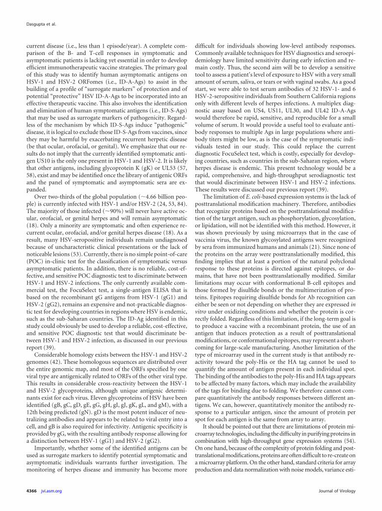
As shown in Table 2, four HSV ID-A-Ags (US4, US11, US10,and UL42) were frequently recognized by serum antibodies from
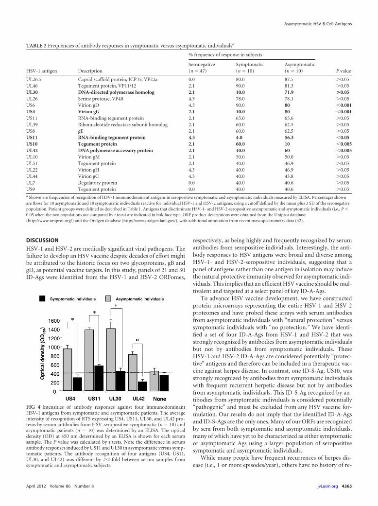
10 out of 10 asymptomatic patients but not by serum antibodiesfrom 10 symptomatic patients (P � 0.001 by an analysis of vari-ance [ANOVA] posttest). In contrast, serum antibodies fromsymptomatic patients preferentially recognized the US10 anti-gen (P � 0.001). The high intensity of recognition of the four HSVID-A-Ags by serum antibodies from HSV-1- and HSV-2-seropos-itive asymptomatic individuals was confirmed by an ELISA. Fig-ure 4 shows the average intensities of recognition of RTS express-ing US4, US11, UL30, and UL42 proteins by HSV-seropositivesymptomatic (n � 10) and asymptomatic (n � 10) patients, con-firmed by an ELISA. The P value was calculated by using theANOVA two-tailed test. Note the difference in serum antibodyresponses induced by US11 and UL30 in asymptomatic versussymptomatic patients. The antibody recognitions of four antigens(US4, US11, UL30, and UL42) were different by �2-fold betweensymptomatic and asymptomatic serum samples. Elevated anti-body responses were confirmed by immunohistochemistry andimmunoblotting (not shown). Altogether, these data identified,for the first time, novel ID-A-Ags that are recognized specifi-cally by antibodies from symptomatic versus asymptomaticindividuals.
Interestingly, the surface gG encoded by US4 appeared to bethe only ID-A-Ag recognized by serum antibodies from asymp-tomatic individuals (i.e., gG with an unknown function) (Table2). The other three ID-A-Ags, US11, UL30, and UL42, are eithertegument or regulatory proteins (Table 2). US11 is a tegument-associated phosphoprotein involved in RNA binding and post-transcriptional regulation. Both UL30 and UL42 are polymeraseaccessory proteins involved in DNA replication. The only detectedID-S-Ag, US10, is a capsid/tegument-associated phosphoproteinwith an unknown function.
FIG 3 Pie chart summarizing the proportions of HSV-1 and HSV-2 envelope glycoproteins, tegument proteins, capsid proteins, and regulatory proteinsrecognized by serum antibodies from HSV-1- and/or HSV-2-seropositive individuals. Note that HSV-2 US4/gG was not present on the array.
Dasgupta et al.
4364 jvi.asm.org Journal of Virology
DISCUSSION
HSV-1 and HSV-2 are medically significant viral pathogens. Thefailure to develop an HSV vaccine despite decades of effort mightbe attributed to the historic focus on two glycoproteins, gB andgD, as potential vaccine targets. In this study, panels of 21 and 30ID-Ags were identified from the HSV-1 and HSV-2 ORFomes,
respectively, as being highly and frequently recognized by serumantibodies from seropositive individuals. Interestingly, the anti-body responses to HSV antigens were broad and diverse amongHSV-1- and HSV-2-seropositive individuals, suggesting that apanel of antigens rather than one antigen in isolation may inducethe natural protective immunity observed for asymptomatic indi-viduals. This implies that an efficient HSV vaccine should be mul-tivalent and targeted at a select panel of key ID-A-Ags.
To advance HSV vaccine development, we have constructedprotein microarrays representing the entire HSV-1 and HSV-2proteomes and have probed these arrays with serum antibodiesfrom asymptomatic individuals with “natural protection” versussymptomatic individuals with “no protection.” We have identi-fied a set of four ID-A-Ags from HSV-1 and HSV-2 that wasstrongly recognized by antibodies from asymptomatic individualsbut not by antibodies from symptomatic individuals. TheseHSV-1 and HSV-2 ID-A-Ags are considered potentially “protec-tive” antigens and therefore can be included in a therapeutic vac-cine against herpes disease. In contrast, one ID-S-Ag, US10, wasstrongly recognized by antibodies from symptomatic individualswith frequent recurrent herpetic disease but not by antibodiesfrom asymptomatic individuals. This ID-S-Ag recognized by an-tibodies from symptomatic individuals is considered potentially“pathogenic” and must be excluded from any HSV vaccine for-mulation. Our results do not imply that the identified ID-A-Agsand ID-S-Ags are the only ones. Many of our ORFs are recognizedby sera from both symptomatic and asymptomatic individuals,many of which have yet to be characterized as either symptomaticor asymptomatic Ags using a larger population of seropositivesymptomatic and asymptomatic individuals.
While many people have frequent recurrences of herpes dis-ease (i.e., 1 or more episodes/year), others have no history of re-
TABLE 2 Frequencies of antibody responses in symptomatic versus asymptomatic individualsa
HSV-1 antigen Description
% frequency of response in subjects
P valueSeronegative(n � 47)
Symptomatic(n � 10)
Asymptomatic(n � 10)
UL26.5 Capsid scaffold protein, ICP35, VP22a 0.0 80.0 87.5 �0.05UL46 Tegument protein, VP11/12 2.1 90.0 81.3 �0.05UL30 DNA-directed polymerase homolog 2.1 10.0 71.9 >0.05UL26 Serine protease, VP40 4.3 78.0 78.1 �0.05US6 Virion gD 4.3 90.0 80 �0.001US4 Virion gG 2.1 10.0 80 �0.001US11 RNA-binding tegument protein 2.1 65.0 65.6 �0.05UL39 Ribonucleotide reductase subunit homolog 2.1 60.0 62.5 �0.05US8 gE 2.1 60.0 62.5 �0.05US11 RNA-binding tegument protein 4.3 4.0 56.3 �0.05US10 Tegument protein 2.1 60.0 10 �0.005UL42 DNA polymerase accessory protein 2.1 10.0 60 �0.005UL10 Virion gM 2.1 50.0 50.0 �0.05UL51 Tegument protein 2.1 40.0 46.9 �0.05UL22 Virion gH 4.3 40.0 46.9 �0.05UL44 Virion gC 4.3 40.0 43.8 �0.05UL7 Regulatory protein 0.0 40.0 40.6 �0.05US9 Tegument protein 0.0 40.0 40.6 �0.05a Shown are frequencies of recognition of HSV-1 immunodominant antigens in seropositive symptomatic and asymptomatic individuals measured by ELISA. Percentages shownare those for 10 asymptomatic and 10 symptomatic individuals reactive for individual HSV-1 and HSV-2 antigens, using a cutoff defined by the mean plus 3 SD of the seronegativepopulation. Patient groups were defined as described in Table 1. Antigens that discriminate HSV-1- and HSV-2-seropositive asymptomatic and symptomatic individuals (i.e., P �0.05 when the two populations are compared by t tests) are indicated in boldface type. ORF product descriptions were obtained from the Uniprot database(http://www.uniprot.org/) and the Oralgen database (http://www.oralgen.lanl.gov/), with additional annotation from recent mass spectrometry data (42).
FIG 4 Intensities of antibody responses against four immunodominantHSV-1 antigens from symptomatic and asymptomatic patients. The averageintensity of recognition of RTS expressing US4, US11, UL30, and UL42 pro-teins by serum antibodies from HSV-seropositive symptomatic (n � 10) andasymptomatic patients (n � 10) was determined by an ELISA. The opticaldensity (OD) at 450 nm determined by an ELISA is shown for each serumsample. The P value was calculated by t tests. Note the difference in serumantibody responses induced by US11 and UL30 in asymptomatic versus symp-tomatic patients. The antibody recognition of four antigens (US4, US11,UL30, and UL42) was different by �2-fold between serum samples fromsymptomatic and asymptomatic subjects.
Asymptomatic HSV B-Cell Antigens
April 2012 Volume 86 Number 8 jvi.asm.org 4365
current disease (i.e., less than 1 episode/year). A complete com-parison of the B- and T-cell responses in symptomatic andasymptomatic patients is lacking yet essential in order to developefficient immunotherapeutic vaccine strategies. The primary goalof this study was to identify human asymptomatic antigens onHSV-1 and HSV-2 ORFomes (i.e., ID-A-Ags) to assist in thebuilding of a profile of “surrogate markers” of protection and ofpotential “protective” HSV ID-A-Ags to be incorporated into aneffective therapeutic vaccine. This also involves the identificationand elimination of human symptomatic antigens (i.e., ID-S-Ags)that may be used as surrogate markers of pathogenicity. Regard-less of the mechanism by which ID-S-Ags induce “pathogenic”disease, it is logical to exclude those ID-S-Ags from vaccines, sincethey may be harmful by exacerbating recurrent herpetic disease(be that ocular, orofacial, or genital). We emphasize that our re-sults do not imply that the currently identified symptomatic anti-gen US10 is the only one present in HSV-1 and HSV-2. It is likelythat other antigens, including glycoprotein K (gK) or UL53 (57,58), exist and may be identified once the library of antigenic ORFsand the panel of symptomatic and asymptomatic sera are ex-panded.
Over two-thirds of the global population (�4.66 billion peo-ple) is currently infected with HSV-1 and/or HSV-2 (24, 53, 84).The majority of those infected (�90%) will never have active oc-ular, orofacial, or genital herpes and will remain asymptomatic(18). Only a minority are symptomatic and often experience re-current ocular, orofacial, and/or genital herpes disease (18). As aresult, many HSV-seropositive individuals remain undiagnosedbecause of uncharacteristic clinical presentations or the lack ofnoticeable lesions (53). Currently, there is no simple point-of-care(POC) in-clinic test for the classification of symptomatic versusasymptomatic patients. In addition, there is no reliable, cost-ef-fective, and sensitive POC diagnostic test to discriminate betweenHSV-1 and HSV-2 infections. The only currently available com-mercial test, the FocuSelect test, a single-antigen ELISA that isbased on the recombinant gG antigens from HSV-1 (gG1) andHSV-2 (gG2), remains an expensive and not-practicable diagnos-tic test for developing countries in regions where HSV is endemic,such as the sub-Saharan countries. The ID-Ag identified in thisstudy could obviously be used to develop a reliable, cost-effective,and sensitive POC diagnostic test that would discriminate be-tween HSV-1 and HSV-2 infection, as discussed in our previousreport (39).
Considerable homology exists between the HSV-1 and HSV-2genomes (42). These homologous sequences are distributed overthe entire genomic map, and most of the ORFs specified by oneviral type are antigenically related to ORFs of the other viral type.This results in considerable cross-reactivity between the HSV-1and HSV-2 glycoproteins, although unique antigenic determi-nants exist for each virus. Eleven glycoproteins of HSV have beenidentified (gB, gC, gD, gE, gG, gH, gI, gJ, gK, gL, and gM), with a12th being predicted (gN). gD is the most potent inducer of neu-tralizing antibodies and appears to be related to viral entry into acell, and gB is also required for infectivity. Antigenic specificity isprovided by gG, with the resulting antibody response allowing fora distinction between HSV-1 (gG1) and HSV-2 (gG2).
Importantly, whether some of the identified antigens can beused as surrogate markers to identify potential symptomatic andasymptomatic individuals warrants further investigation. Themonitoring of herpes disease and immunity has become more
difficult for individuals showing low-level antibody responses.Commonly available techniques for HSV diagnostics and seroepi-demiology have limited sensitivity during early infection and re-main costly. Thus, the second aim will be to develop a sensitivetool to assess a patient’s level of exposure to HSV with a very smallamount of serum, saliva, or tears or with vaginal swabs. As a goodstart, we were able to test serum antibodies of 32 HSV-1- and 6HSV-2-seropositive individuals from Southern California regionsonly with different levels of herpes infections. A multiplex diag-nostic assay based on US4, US11, UL30, and UL42 ID-A-Agswould therefore be rapid, sensitive, and reproducible for a smallvolume of serum. It would provide a useful tool to evaluate anti-body responses to multiple Ags in large populations where anti-body titers might be low, as is the case of the symptomatic indi-viduals tested in our study. This could replace the currentdiagnostic FocuSelect test, which is costly, especially for develop-ing countries, such as countries in the sub-Saharan region, whereherpes disease is endemic. This present technology would be arapid, comprehensive, and high-throughput serodiagnostic testthat would discriminate between HSV-1 and HSV-2 infections.These results were discussed our previous report (39).
The limitation of E. coli-based expression systems is the lack ofposttranslational modification machinery. Therefore, antibodiesthat recognize proteins based on the posttranslational modifica-tion of the target antigen, such as phosphorylation, glycosylation,or lipidation, will not be identified with this method. However, itwas shown previously by using microarrays that in the case ofvaccinia virus, the known glycosylated antigens were recognizedby sera from immunized humans and animals (21). Since none ofthe proteins on the array were posttranslationally modified, thisfinding implies that at least a portion of the natural polyclonalresponse to these proteins is directed against epitopes, or do-mains, that have not been posttranslationally modified. Similarlimitations may occur with conformational B-cell epitopes andthose formed by disulfide bonds or the multimerization of pro-teins. Epitopes requiring disulfide bonds for Ab recognition caneither be seen or not depending on whether they are expressed invitro under oxidizing conditions and whether the protein is cor-rectly folded. Regardless of this limitation, if the long-term goal isto produce a vaccine with a recombinant protein, the use of anantigen that induces protection as a result of posttranslationalmodifications, or conformational epitopes, may represent a short-coming for large-scale manufacturing. Another limitation of thetype of microarray used in the current study is that antibody re-activity toward the poly-His or the HA tag cannot be used toquantify the amount of antigen present in each individual spot.The binding of the antibodies to the poly-His and HA tags appearsto be affected by many factors, which may include the availabilityof the tags for binding due to folding. We therefore cannot com-pare quantitatively the antibody responses between different an-tigens. We can, however, quantitatively monitor the antibody re-sponse to a particular antigen, since the amount of protein perspot for each antigen is the same from array to array.
It should be pointed out that there are limitations of protein mi-croarray technologies, including the difficulty in purifying proteins incombination with high-throughput gene expression systems (54).On one hand, because of the complexity of protein folding and post-translational modifications, proteins are often difficult to re-create ona microarray platform. On the other hand, standard criteria for arrayproduction and data normalization with noise models, variance esti-
Dasgupta et al.
4366 jvi.asm.org Journal of Virology

mation, and differential expression analysis techniques have becomepowerful tools for the interpretation of results. Based on this progress,successful attempts have been made using protein microarrays forprofiling the antibody responses to numerous infectious pathogens(3, 21, 40, 54, 66).
Homologous sequences exist between the HSV-1 and HSV-2genomes and are distributed over the entire genomic map (42).Most of the ORFs specified by one viral type are antigenicallyrelated to ORFs of the other viral type. This indicates that consid-erable cross-reactivity does exist between the HSV-1 and HSV-2antigens, although unique antigenic determinants might be pres-ent for each virus, as we recently described (39). Therefore, it isunlikely that the HSV-1 US4 antigen does not cross-react with itsHSV-2 US4 ortholog. If the US4 asymptomatic antigen is absentin the HSV-2 ORF, we will not be able to determine whether theantibodies induced in HSV-1-seropositive individuals specificallyrecognize HSV-2 US4. However, sera from HSV-2-seropositiveindividuals did not recognize HSV-1 US4. Our approach appearsbe very specific, given the appropriate antigen, as in the case ofHSV-1 US4. The specificity of US4 is already well established, andit was our intention to discover additional useful antigens. Notobtaining the HSV-2 US4 was unfortunate, but it does not auto-matically rule out others of the 80� HSV-2 antigens that are po-tentially type specific.
Considering the wealth of data addressing the role of protectiveversus pathogenic immune effectors in animal models, it is sur-prising how there are few reports that have explored the immuno-logical basis of symptomatic and asymptomatic HSV infections inhumans. The identification of these immune mechanisms bywhich asymptomatic patients control herpes disease and symp-tomatic patients do not, or at least the antigens involved, is criticalfor the advancement of HSV vaccine development. The symptom-atic (i.e., pathogenic) and asymptomatic (i.e., protective) antigensidentified in this study may be key to an understanding of themechanisms of pathogenicity versus protection in HSV-infectedhumans. Multiple and complex mechanisms might be in play,including (i) differences in precursor frequencies, proliferative ca-pacities, and functional properties of symptomatic versus asymp-tomatic epitope-specific B cells; (ii) the possibility that asymp-tomatic antigens might trigger neutralizing protective antibodies,while symptomatic antigens might trigger nonneutralizing patho-genic antibodies; (iii) the possibility that symptomatic antigensmay direct antibody responses away from those that are bestsuited to neutralize the viral infection with a minimal pathogenicreaction; and (iv) finally, the possibility that B-cell cross-reactivitywith antigens and epitopes from other viral pathogens, within oroutside the herpesvirus family, may also play a role in protectiveimmunity versus damaging immunopathology (41, 81).
T cells, mainly of the CD4� subsets, rather than antibodiesappear to mediate herpetic disease (26, 74, 77). Until our recentstudies with gB and gD (13), no symptomatic or asymptomaticHSV T-cell epitopes had been identified. The symptomatic andasymptomatic B-cell antigens identified in this study would cer-tainly help speed up the identification of more symptomatic andasymptomatic T-cell antigens by serodiagnosing more symptom-atic and asymptomatic individuals. In addition, many HSV andnon-HSV antigens tend to contain both B- and T-cell epitopes(36, 59, 60). It was suggested previously that the most immuno-dominant B-cell antigens in HSV may be those of envelope pro-teins, such as the envelope glycoproteins UL27/gB and US6/gD
(10). However, many CD4� and CD8� T-cell epitopes have beenrecently identified, by our laboratory and others, for gB and gD (5,13, 16, 45, 47, 48, 50, 55, 56, 71). Tegument proteins such as thoseencoded by HSV-1/2 UL39, UL41, UL46/VP11/12, UL47/VP13/14, UL48/VP16, and UL49 and immediate-early (IE) proteinssuch as RL2/ICP0 and RS1/ICP4 have also been identified as ma-jor targets for effector T cells (12, 34, 37, 46, 49). Of these, thearrays detected strong antibody signals against US6/gD (fromboth HSV-1 and -2) and UL48 of HSV-2, both of which are alsoCD4� target antigens. The overlap between the targets of antibodyand CD4� cells, rather than CD8� cells, is also consistent withrecent findings for vaccinia virus antigens (36, 59, 60). Furtherinvestigations of T-cell responses to ID-A-Ags versus ID-S-Agsmay be needed to break new ground in our understanding of theprotective versus pathogenic immune mechanisms against ocular,orofacial, and genital herpes. We should, however, emphasize thatT- and B-cell epitopes have frequently been observed in othersystems to cluster within a limited region of Ags (6, 38, 76). We arecurrently using a genomic approach to detect HSV-specific T-celldominance in symptomatic versus asymptomatic individuals.
Whether the ID-A-Ags identified in this study are truly protec-tive and whether the ID-S-Ags are pathogenic remain to be deter-mined. A logical extension of this screening study is to constructand test their protective efficacy in animal models of HSV infec-tion (mice, guinea pigs, and rabbits) using vaccine formulationsthat will exclusively incorporate the ID-A-Ags (but exclude thesymptomatic antigens). We expect that the next immunogenicityand protective preclinical studies that use the four identified ID-A-Ags (US4, US11, UL30, and UL42) individually and in combi-nation will confirm their protective function. Inversely, we alsoexpect that the ID-S-Ag (UL10) identified in this study will exac-erbate herpes disease following the vaccination of latently infectedanimals. Results from these studies will be the subject of futurereports.
In conclusion, the present study (i) validates the use of a high-throughput microarray analysis for the characterization of the Abresponse to HSV-1 and HSV-2 antigens; (ii) shows that the anti-body responses to HSV-1 and HSV-2 antigens among seropositiveindividuals were broad and diverse; (iii) reports 21 and 30 immu-nodominant HSV-1 and HSV-2 antigens, respectively; and (iv)characterizes four previously unreported ID-A-Ags that, consid-ering the recent unsuccessful clinical HSV vaccine (4), are poten-tially useful to be included in a novel asymptomatic vaccineagainst HSV.
ACKNOWLEDGMENTS
This work was supported by NIH Public Health Service grants EY14900and EY019896 (L.B.M.), R44AI058365 (D.H.D.), and U01AI078213(P.L.F.); the Discovery Eye Foundation; the Henry L. Guenther Founda-tion; and a Research To Prevent Blindness challenge grant.
We thank Steven L. Wechsler and Dale Carpenter for providing theHSV-2 DNA.
REFERENCES1. Anderson KS, et al. 2008. Application of protein microarrays for multi-
plexed detection of antibodies to tumor antigens in breast cancer. J. Pro-teome Res. 7:1490 –1499.
2. Anderson KS, et al. 2011. Protein microarray signature of autoantibodybiomarkers for the early detection of breast cancer. J. Proteome Res. 10:85–96.
3. Barbour AG, et al. 2008. A genome-wide proteome array reveals a limited
Asymptomatic HSV B-Cell Antigens
April 2012 Volume 86 Number 8 jvi.asm.org 4367

set of immunogens in natural infections of humans and white-footed micewith Borrelia burgdorferi. Infect. Immun. 76:3374 –3389.
4. Belshe PB, et al. 2012. Efficacy results of a trial of a herpes simplexvaccine. N. Engl. J. Med. 366:34 – 43.
5. BenMohamed L, et al. 2003. Identification of novel immunodominantCD4� Th1-type T-cell peptide epitopes from herpes simplex virus glyco-protein D that confer protective immunity. J. Virol. 77:9463–9473.
6. BenMohamed L, et al. 1997. Lipopeptide immunization without adju-vant induces potent and long-lasting B, T helper, and cytotoxic T lympho-cyte responses against a malaria liver stage antigen in mice and chimpan-zees. Eur. J. Immunol. 27:1242–1253.
7. Bernstein DI, Spruance SL, Arora SS, Schroeder JL, Meng TC. 2005.Evaluation of imiquimod 5% cream to modify the natural history of her-pes labialis: a pilot study. Clin. Infect. Dis. 41:808 – 814.
8. Bettahi I, Zhang X, Afifi RE, BenMohamed L. 2006. Protective immu-nity to genital herpes simplex virus type 1 and type 2 provided by self-adjuvanting lipopeptides that drive dendritic cell maturation and elicit apolarized Th1 immune response. Viral Immunol. 19:220 –236.
9. Burbelo PD, Ching KH, Bush ER, Han BL, Iadarola MJ. 2010. Anti-body-profiling technologies for studying humoral responses to infectiousagents. Expert Rev. Vaccines 9:567–578.
10. Burbelo PD, et al. 2009. Serological diagnosis of human herpes simplexvirus type 1 and 2 infections by luciferase immunoprecipitation systemassay. Clin. Vaccine Immunol. 16:366 –371.
11. Cantin E, Tanamachi B, Openshaw H, Mann J, Clarke K. 1999. Gammainterferon (IFN-gamma) receptor null-mutant mice are more susceptibleto herpes simplex virus type 1 infection than IFN-gamma ligand null-mutant mice. J. Virol. 73:5196 –5200.
12. Carmack MA, et al. 1996. T cell recognition and cytokine productionelicited by common and type-specific glycoproteins of herpes simplexvirus type 1 and type 2. J. Infect. Dis. 174:899 –906.
13. Chentoufi AA, et al. 2008. Asymptomatic human CD4� cytotoxic T-cellepitopes identified from herpes simplex virus glycoprotein B. J. Virol.82:11792–11802.
14. Chentoufi AA, et al. 2010. A novel HLA (HLA-A*0201) transgenic rabbitmodel for preclinical evaluation of human CD8� T cell epitope-basedvaccines against ocular herpes. J. Immunol. 184:2561–2571.
15. Chentoufi AA, et al. 2010. Nasolacrimal duct closure modulates ocularmucosal and systemic CD4(�) T-cell responses induced following topicalocular or intranasal immunization. Clin. Vaccine Immunol. 17:342–353.
16. Chentoufi AA, et al. 2008. HLA-A*0201-restricted CD8� cytotoxic Tlymphocyte epitopes identified from herpes simplex virus glycoprotein D.J. Immunol. 180:426 – 437.
17. Cunningham C, Davison AJ. 1993. A cosmid-based system for construct-ing mutants of herpes simplex virus type 1. Virology 197:116 –124.
18. Dasgupta G, Nesburn AB, Wechsler SL, BenMohamed L. 2010. Devel-oping an asymptomatic mucosal herpes vaccine: the present and the fu-ture. Future Microbiol. 5:1– 4.
19. Davies DH, et al. 2005. Profiling the humoral immune response to infec-tion by using proteome microarrays: high-throughput vaccine and diag-nostic antigen discovery. Proc. Natl. Acad. Sci. U. S. A. 102:547–552.
20. Davies DH, et al. 2005. Vaccinia virus H3L envelope protein is a majortarget of neutralizing antibodies in humans and elicits protection againstlethal challenge in mice. J. Virol. 79:11724 –11733.
21. Davies DH, et al. 2008. Antibody profiling by proteome microarrayreveals the immunogenicity of the attenuated smallpox vaccine modifiedvaccinia virus Ankara is comparable to that of Dryvax. J. Virol. 82:652–663.
22. Durbin BP, Hardin JS, Hawkins DM, Rocke DM. 2002. A variance-stabilizing transformation for gene-expression microarray data. Bioinfor-matics 18(Suppl 1):S105–S110.
23. Field PR, Ho DW, Irving WL, Isaacs D, Cunningham AL. 1993. Thereliability of serological tests for the diagnosis of genital herpes: a critique.Pathology 25:175–179.
24. Fleming DT, et al. 1997. Herpes simplex virus type 2 in the United States,1976 to 1994. N. Engl. J. Med. 337:1105–1111.
25. Freeman ML, Sheridan BS, Bonneau RH, Hendricks RL. 2007. Psycho-logical stress compromises CD8� T cell control of latent herpes simplexvirus type 1 infections. J. Immunol. 179:322–328.
26. Gangappa S, Deshpande SP, Rouse BT. 1999. Bystander activation ofCD4(�) T cells can represent an exclusive means of immunopathology ina virus infection. Eur. J. Immunol. 29:3674 –3682.
27. Geiger KD, et al. 1997. Interferon-gamma protects against herpes simplexvirus type 1-mediated neuronal death. Virology 238:189 –197.
28. Ghiasi H, Cai S, Slanina S, Nesburn AB, Wechsler SL. 1995. Vaccinationof mice with herpes simplex virus type 1 glycoprotein D DNA produceslow levels of protection against lethal HSV-1 challenge. Antiviral Res. 28:147–157.
29. Ghiasi H, Kaiwar R, Nesburn AB, Wechsler SL. 1992. Baculovirus-expressed glycoprotein G of herpes simplex virus type 1 partially protectsvaccinated mice against lethal HSV-1 challenge. Virology 190:233–239.
30. Ghiasi H, Perng GC, Cai S, Nesburn AB, Wechsler SL. 1996. The UL3open reading frame of herpes simplex virus type 1 codes for a phospho-protein. Virus Res. 44:137–142.
31. Ghiasi H, Slanina S, Nesburn AB, Wechsler SL. 1994. Characterizationof baculovirus-expressed herpes simplex virus type 1 glycoprotein K. J.Virol. 68:2347–2354.
32. Herpetic Eye Disease Study Group. 1998. Acyclovir for the prevention ofrecurrent herpes simplex virus eye disease. N. Engl. J. Med. 339:300 –306.
33. Hochberg Y, Benjamini Y. 1990. More powerful procedures for multiplesignificance testing. Stat. Med. 9:811– 818.
34. Hosken N, et al. 2006. Diversity of the CD8� T-cell response to herpessimplex virus type 2 proteins among persons with genital herpes. J. Virol.80:5509 –5515.
35. Huber W, von Heydebreck A, Sultmann H, Poustka A, Vingron M.2002. Variance stabilization applied to microarray data calibration and tothe quantification of differential expression. Bioinformatics 18(Suppl 1):S96 –S104.
36. Jing L, et al. 2008. An extremely diverse CD4 response to vaccinia virus inhumans is revealed by proteome-wide T-cell profiling. J. Virol. 82:7120 –7134.
37. Jing L, et al. 2012. Cross-presentation and genome-wide screening revealcandidate T cells antigens for a herpes simplex virus type 1 vaccine. J. Clin.Invest. 122:654 – 673.
38. Kabilan L, et al. 1988. T-cell epitopes in Pf155/RESA, a major candidatefor a Plasmodium falciparum malaria vaccine. Proc. Natl. Acad. Sci.U. S. A. 85:5659 –5663.
39. Kalantari-Dehaghi M, et al. 2012. Discovery of potential diagnostic andvaccine antigens in herpes simplex virus 1 and 2 by proteome-wide anti-body profiling. J. Virol. 86:4328 – 4339.
40. Kalantari-Dehaghi M, et al. 2011. New targets of pemphigus vulgarisantibodies identified by protein array technology. Exp. Dermatol. 20:154 –156.
41. Kim SK, et al. 2005. Private specificities of CD8 T cell responses controlpatterns of heterologous immunity. J. Exp. Med. 201:523–533.
42. Kimberlin DW, et al. 2011. Oral acyclovir suppression and neurodevel-opment after neonatal herpes. N. Engl. J. Med. 365:1284 –1292.
43. Klipper-Aurbach Y, et al. 1995. Mathematical formulae for the predictionof the residual beta cell function during the first two years of disease inchildren and adolescents with insulin-dependent diabetes mellitus. Med.Hypotheses 45:486 – 490.
44. Knaup B, Schunemann S, Wolff MH. 2000. Subclinical reactivation ofherpes simplex virus type 1 in the oral cavity. Oral Microbiol. Immunol.15:281–283.
45. Koelle DM. 2003. Expression cloning for the discovery of viral antigensand epitopes recognized by T cells. Methods 29:213–226.
46. Koelle DM, Abbo H, Peck A, Ziegweid K, Corey L. 1994. Direct recoveryof herpes simplex virus (HSV)-specific T lymphocyte clones from recur-rent genital HSV-2 lesions. J. Infect. Dis. 169:956 –961.
47. Koelle DM, et al. 2001. CD8 CTL from genital herpes simplex lesions:recognition of viral tegument and immediate early proteins and lysis ofinfected cutaneous cells. J. Immunol. 166:4049 – 4058.
48. Koelle DM, Corey L. 2003. Recent progress in herpes simplex virusimmunobiology and vaccine research. Clin. Microbiol. Rev. 16:96 –113.
49. Koelle DM, et al. 1994. Antigenic specificities of human CD4� T-cellclones recovered from recurrent genital herpes simplex virus type 2 le-sions. J. Virol. 68:2803–2810.
50. Koelle DM, et al. 2003. Immunodominance among herpes simplex virus-specific CD8 T cells expressing a tissue-specific homing receptor. Proc.Natl. Acad. Sci. U. S. A. 100:12899 –12904.
51. Kriesel JD, et al. 2011. C21orf91 genotypes correlate with herpes simplexlabialis (cold sore) frequency: description of a cold sore susceptibilitygene. J. Infect. Dis. 204:1654 –1662.
52. Leigh JF, Acharya N, Cevallos V, Margolis TP. 2008. Does asymptomatic
Dasgupta et al.
4368 jvi.asm.org Journal of Virology

shedding of herpes simplex virus on the ocular surface lead to false-positive diagnostic PCR results? Br. J. Ophthalmol. 92:435– 436.
53. Looker KJ, Garnett GP, Schmid GP. 2008. An estimate of the globalprevalence and incidence of herpes simplex virus type 2 infection. Bull.World Health Organ. 86:805– 812.
54. Luevano M, et al. 2010. High-throughput profiling of the humoral im-mune responses against thirteen human papillomavirus types by pro-teome microarrays. Virology 405:31– 40.
55. Mikloska Z, Cunningham AL. 1998. Herpes simplex virus type 1 glyco-proteins gB, gC and gD are major targets for CD4 T-lymphocyte cytotox-icity in HLA-DR expressing human epidermal keratinocytes. J. Gen. Virol.79(Pt 2):353–361.
56. Mikloska Z, Kesson AM, Penfold ME, Cunningham AL. 1996. Herpessimplex virus protein targets for CD4 and CD8 lymphocyte cytotoxicity incultured epidermal keratinocytes treated with interferon-gamma. J. In-fect. Dis. 173:7–17.
57. Mott KR, et al. 2009. The role of a glycoprotein K (gK) CD8� T-cellepitope of herpes simplex virus on virus replication and pathogenicity.Invest. Ophthalmol. Vis. Sci. 50:2903–2912.
58. Mott KR, et al. 2007. Role of anti-glycoproteins D (anti-gD) and K(anti-gK) IgGs in pathology of herpes stromal keratitis in humans. Invest.Ophthalmol. Vis. Sci. 48:2185–2193.
59. Moutaftsi M, et al. 2007. Vaccinia virus-specific CD4� T cell responsestarget a set of antigens largely distinct from those targeted by CD8� T cellresponses. J. Immunol. 178:6814 – 6820.
60. Moutaftsi M, et al. 2010. Uncovering the interplay between CD8, CD4and antibody responses to complex pathogens. Future Microbiol. 5:221–239.
61. Nesburn AB, et al. 2006. Topical/mucosal delivery of sub-unit vaccinesthat stimulate the ocular mucosal immune system. Ocul. Surf. 4:178 –187.
62. Nesburn AB, Burke RL, Ghiasi H, Slanina SM, Wechsler SL. 1998.Therapeutic periocular vaccination with a subunit vaccine induces higherlevels of herpes simplex virus-specific tear secretory immunoglobulin Athan systemic vaccination and provides protection against recurrent spon-taneous ocular shedding of virus in latently infected rabbits. Virology252:200 –209.
63. Nesburn AB, Burke RL, Ghiasi H, Slanina SM, Wechsler SL. 1998. Atherapeutic vaccine that reduces recurrent herpes simplex virus type 1corneal disease. Invest. Ophthalmol. Vis. Sci. 39:1163–1170.
64. Nesburn AB, et al. 2005. Local and systemic B cell and Th1 responsesinduced following ocular mucosal delivery of multiple epitopes of herpessimplex virus type 1 glycoprotein D together with cytosine-phosphate-guanine adjuvant. Vaccine 23:873– 883.
65. Nesburn AB, et al. 1998. Local periocular vaccination protects against eyedisease more effectively than systemic vaccination following primary oc-ular herpes simplex virus infection in rabbits. J. Virol. 72:7715–7721.
66. Nnedu ON, et al. 2011. Humoral immune responses to Plasmodiumfalciparum among HIV-1 infected Kenyan adults. Proteomics Clin. Appl.5:613– 623.
67. O’Farrell N. 1999. Increasing prevalence of genital herpes in developing
countries: implications for heterosexual HIV transmission and STI con-trol programmes. Sex. Transm. Infect. 75:377–384.
68. O’Farrell N, Tovey SJ. 1994. High cumulative incidence of genital herpesamongst HIV-1 seropositive heterosexuals in south London. Int. J. STDAIDS 5:415– 418.
69. Perng GC, et al. 1994. The latency-associated transcript gene of herpessimplex virus type 1 (HSV-1) is required for efficient in vivo spontaneousreactivation of HSV-1 from latency. J. Virol. 68:8045– 8055.
70. Perng GC, et al. 1999. Herpes simplex virus type 1 serum neutralizingantibody titers increase during latency in rabbits latently infected withlatency-associated transcript (LAT)-positive but not LAT-negative vi-ruses. J. Virol. 73:9669 –9672.
71. Posavad CM, et al. 2003. T cell immunity to herpes simplex viruses inseronegative subjects: silent infection or acquired immunity? J. Immunol.170:4380 – 4388.
72. Schmid DS, et al. 1999. Limits in reliability of glycoprotein G-basedtype-specific serologic assays for herpes simplex virus types 1 and 2. J. Clin.Microbiol. 37:376 –379.
73. Tengvall S, O’Hagan D, Harandi AM. 2008. Rectal immunization gen-erates protective immunity in the female genital tract against herpes sim-plex virus type 2 infection: relative importance of myeloid differentiationfactor 88. Antiviral Res. 78:202–214.
74. Thomas J, Rouse BT. 1998. Immunopathology of herpetic stromal ker-atitis: discordance in CD4� T cell function between euthymic host andreconstituted SCID recipients. J. Immunol. 160:3965–3970.
75. Tronstein E, et al. 2011. Genital shedding of herpes simplex virus amongsymptomatic and asymptomatic persons with HSV-2 infection. JAMA305:1441–1449.
76. Troye-Blomberg M, et al. 1988. T cell reactivity of defined peptides froma major Plasmodium falciparum vaccine candidate: the Pf155/RESA anti-gen. Immunol. Lett. 19:229 –233.
77. Verjans GM, et al. 1998. Identification and characterization of herpessimplex virus-specific CD4� T cells in corneas of herpetic stromal kera-titis patients. J. Infect. Dis. 177:484 – 488.
78. Wald A, et al. 1997. Frequent genital herpes simplex virus 2 shedding inimmunocompetent women. Effect of acyclovir treatment. J. Clin. Invest.99:1092–1097.
79. Wald A, et al. 2002. Genital shedding of herpes simplex virus among men.J. Infect. Dis. 186(Suppl 1):S34 –S39.
80. Wald A, et al. 2000. Reactivation of genital herpes simplex virus type 2infection in asymptomatic seropositive persons. N. Engl. J. Med. 342:844 –850.
81. Welsh RM, Selin LK. 2002. No one is naive: the significance of heterolo-gous T-cell immunity. Nat. Rev. Immunol. 2:417– 426.
82. Whitley RJ, Miller RL. 2001. Immunologic approach to herpes simplexvirus. Viral Immunol. 14:111–118.
83. Whitley RJ, Roizman B. 2001. Herpes simplex virus infections. Lancet357:1513–1518.
84. Xu F, et al. 2006. Trends in herpes simplex virus type 1 and type 2seroprevalence in the United States. JAMA 296:964 –973.
Asymptomatic HSV B-Cell Antigens
April 2012 Volume 86 Number 8 jvi.asm.org 4369
Related Documents


![hma.org.tw · the control group for seropositive participants (1-88%[95% CI 1.54-2.31] in seropositive controls vs 0.38% [0-26—0.54] in seropositive vaccinees), and increased by](https://static.cupdf.com/doc/110x72/5f180af091496b79e1655a71/hmaorgtw-the-control-group-for-seropositive-participants-1-8895-ci-154-231.jpg)