JOURNAL OF VIROLOGY, July 2002, p. 7187–7202 Vol. 76, No. 14 0022-538X/02/$04.000 DOI: 10.1128/JVI.76.14.7187–7202.2002 Copyright © 2002, American Society for Microbiology. All Rights Reserved. Immunization of Rhesus Macaques with a DNA Prime/Modified Vaccinia Virus Ankara Boost Regimen Induces Broad Simian Immunodeficiency Virus (SIV)-Specific T-Cell Responses and Reduces Initial Viral Replication but Does Not Prevent Disease Progression following Challenge with Pathogenic SIVmac239 Helen Horton, 1 Thorsten U. Vogel, 1 Donald K. Carter, 1 Kathy Vielhuber, 1 Deborah H. Fuller, 1,2 Tim Shipley, 2 James T. Fuller, 2 Kevin J. Kunstman, 3 Gerd Sutter, 4 David C. Montefiori, 5 Volker Erfle, 4 Ronald C. Desrosiers, 6 Nancy Wilson, 1 Louis J. Picker, 7 Steven M. Wolinsky, 3 Chenxi Wang, 8, 9 David B. Allison, 8,9 and David I. Watkins 1,10 * Wisconsin Regional Primate Research Center 1 and Department of Pathology and Laboratory Medicine, 10 University of Wisconsin, Madison, Wisconsin 53715; PowderJect Vaccines, Inc., Madison, Wisconsin 53711 2 ; Northwestern University Medical School, Chicago, Illinois 60611-3008 3 ; GSF-Institute for Molecular Virology, D-81675 Munich, Germany 4 ; Center for AIDS Research, Department of Surgery, Duke University Medical Center, Durham, North Carolina 27710 5 ; New England Regional Primate Research Center, Harvard Medical School, Southborough, Massachusetts 01772-9102 6 ; Vaccine and Gene Therapy Institute, Oregon Regional Primate Research Center, Oregon Health Sciences University, Beaverton, Oregon 97006 7 ; and Section on Statistical Genetics, Department of Biostatistics, 8 and Clinical Nutrition Research Center, Department of Nutrition Sciences, 9 University of Alabama at Birmingham, Birmingham, Alabama 35294 Received 8 January 2002/Accepted 9 April 2002 Producing a prophylactic vaccine for human immunodeficiency virus (HIV) has proven to be a challenge. Most biological isolates of HIV are difficult to neutralize, so that conventional subunit-based antibody-inducing vaccines are unlikely to be very effective. In the rhesus macaque model, some protection was afforded by DNA/recombinant viral vector vaccines. However, these studies used as the challenge virus SHIV-89.6P, which is neutralizable, making it difficult to determine whether the observed protection was due to cellular immunity, humoral immunity, or a combination of both. In this study, we used a DNA prime/modified vaccinia virus Ankara boost regimen to immunize rhesus macaques against nearly all simian immunodeficiency virus (SIV) proteins. These animals were challenged intrarectally with pathogenic molecularly cloned SIVmac239, which is resistant to neutralization. The immunization regimen resulted in the induction of virus-specific CD8 and CD4 responses in all vaccinees. Although anamnestic neutralizing antibody responses against laboratory- adapted SIVmac251 developed after the challenge, no neutralizing antibodies against SIVmac239 were detect- able. Vaccinated animals had significantly reduced peak viremia compared with controls (P < 0.01). However, despite the induction of virus-specific cellular immune responses and reduced peak viral loads, most animals still suffered from gradual CD4 depletion and progressed to disease. It is estimated that in excess of 35 million individuals are infected with human immunodeficiency virus (HIV), and the epidemic does not appear to be slowing (AIDS Epidemic Update, 2000, http://wwwunaidsorg/epidemic_update/report _dec00/index_dechtml). The catastrophic implications of such a worldwide pandemic have placed enormous pressure on the scientific community to produce a prophylactic or therapeutic vaccine as quickly as possible. The infection of rhesus macaques with simian immunodefi- ciency virus (SIV) is used routinely as a model for AIDS (43, 95). In this model, the greatest degree of protection has been achieved with attenuated viral vaccines (7, 26, 62, 70, 102, 103). However, attenuated viruses have inherent risks (27, 28, 35, 85, 101) and, furthermore, have been shown to cause disease in infants (11, 12, 22). Therefore, the use of attenuated retrovi- ruses as an AIDS vaccine is controversial (66, 84). So far, the mechanism of protection induced by attenuated viruses is still unknown (6, 8, 69, 94, 98). The degree of protection achieved with attenuated viruses is variable and depends on the degree of attenuation of the vaccine virus (44), the time point of challenge (25), and the challenge virus chosen (102); all of these factors make determining the mechanism of protection very difficult. Depending on the challenge virus used, some studies have suggested a role for neutralizing antibodies (21, 103), cytotoxic T lymphocytes (CTL) (44), or perhaps both (21) as protective mechanisms. Due to the present inability to generate neutralizing anti- bodies against a wide variety of HIV strains (19, 55, 61), it is believed that current HIV vaccine candidates will have to rely * Corresponding author. Mailing address: Wisconsin Regional Pri- mate Research Center, 1220 Capitol Ct., Madison, WI 53715. Phone: (608) 265-3380. Fax: (608) 265-8084. E-mail: [email protected] .edu. 7187

Welcome message from author
This document is posted to help you gain knowledge. Please leave a comment to let me know what you think about it! Share it to your friends and learn new things together.
Transcript
JOURNAL OF VIROLOGY, July 2002, p. 7187–7202 Vol. 76, No. 140022-538X/02/$04.00�0 DOI: 10.1128/JVI.76.14.7187–7202.2002Copyright © 2002, American Society for Microbiology. All Rights Reserved.
Immunization of Rhesus Macaques with a DNA Prime/ModifiedVaccinia Virus Ankara Boost Regimen Induces Broad Simian
Immunodeficiency Virus (SIV)-Specific T-Cell Responsesand Reduces Initial Viral Replication but Does Not
Prevent Disease Progression following Challengewith Pathogenic SIVmac239
Helen Horton,1 Thorsten U. Vogel,1 Donald K. Carter,1 Kathy Vielhuber,1 Deborah H. Fuller,1,2
Tim Shipley,2 James T. Fuller,2 Kevin J. Kunstman,3 Gerd Sutter,4 David C. Montefiori,5Volker Erfle,4 Ronald C. Desrosiers,6 Nancy Wilson,1 Louis J. Picker,7
Steven M. Wolinsky,3 Chenxi Wang,8, 9 David B. Allison,8,9
and David I. Watkins1,10*Wisconsin Regional Primate Research Center1 and Department of Pathology and Laboratory Medicine,10 University of Wisconsin,
Madison, Wisconsin 53715; PowderJect Vaccines, Inc., Madison, Wisconsin 537112; Northwestern University Medical School,Chicago, Illinois 60611-30083; GSF-Institute for Molecular Virology, D-81675 Munich, Germany4; Center for AIDS Research,
Department of Surgery, Duke University Medical Center, Durham, North Carolina 277105; New England Regional PrimateResearch Center, Harvard Medical School, Southborough, Massachusetts 01772-91026; Vaccine and Gene Therapy Institute,
Oregon Regional Primate Research Center, Oregon Health Sciences University, Beaverton, Oregon 970067; andSection on Statistical Genetics, Department of Biostatistics,8 and Clinical Nutrition Research Center,
Department of Nutrition Sciences,9 University of Alabama at Birmingham,Birmingham, Alabama 35294
Received 8 January 2002/Accepted 9 April 2002
Producing a prophylactic vaccine for human immunodeficiency virus (HIV) has proven to be a challenge.Most biological isolates of HIV are difficult to neutralize, so that conventional subunit-based antibody-inducingvaccines are unlikely to be very effective. In the rhesus macaque model, some protection was afforded byDNA/recombinant viral vector vaccines. However, these studies used as the challenge virus SHIV-89.6P, whichis neutralizable, making it difficult to determine whether the observed protection was due to cellular immunity,humoral immunity, or a combination of both. In this study, we used a DNA prime/modified vaccinia virusAnkara boost regimen to immunize rhesus macaques against nearly all simian immunodeficiency virus (SIV)proteins. These animals were challenged intrarectally with pathogenic molecularly cloned SIVmac239, whichis resistant to neutralization. The immunization regimen resulted in the induction of virus-specific CD8� andCD4� responses in all vaccinees. Although anamnestic neutralizing antibody responses against laboratory-adapted SIVmac251 developed after the challenge, no neutralizing antibodies against SIVmac239 were detect-able. Vaccinated animals had significantly reduced peak viremia compared with controls (P < 0.01). However,despite the induction of virus-specific cellular immune responses and reduced peak viral loads, most animalsstill suffered from gradual CD4 depletion and progressed to disease.
It is estimated that in excess of 35 million individuals areinfected with human immunodeficiency virus (HIV), and theepidemic does not appear to be slowing (AIDS EpidemicUpdate, 2000, http://wwwunaidsorg/epidemic_update/report_dec00/index_dechtml). The catastrophic implications of sucha worldwide pandemic have placed enormous pressure on thescientific community to produce a prophylactic or therapeuticvaccine as quickly as possible.
The infection of rhesus macaques with simian immunodefi-ciency virus (SIV) is used routinely as a model for AIDS (43,95). In this model, the greatest degree of protection has beenachieved with attenuated viral vaccines (7, 26, 62, 70, 102, 103).
However, attenuated viruses have inherent risks (27, 28, 35, 85,101) and, furthermore, have been shown to cause disease ininfants (11, 12, 22). Therefore, the use of attenuated retrovi-ruses as an AIDS vaccine is controversial (66, 84). So far, themechanism of protection induced by attenuated viruses is stillunknown (6, 8, 69, 94, 98). The degree of protection achievedwith attenuated viruses is variable and depends on the degreeof attenuation of the vaccine virus (44), the time point ofchallenge (25), and the challenge virus chosen (102); all ofthese factors make determining the mechanism of protectionvery difficult. Depending on the challenge virus used, somestudies have suggested a role for neutralizing antibodies (21,103), cytotoxic T lymphocytes (CTL) (44), or perhaps both (21)as protective mechanisms.
Due to the present inability to generate neutralizing anti-bodies against a wide variety of HIV strains (19, 55, 61), it isbelieved that current HIV vaccine candidates will have to rely
* Corresponding author. Mailing address: Wisconsin Regional Pri-mate Research Center, 1220 Capitol Ct., Madison, WI 53715. Phone:(608) 265-3380. Fax: (608) 265-8084. E-mail: [email protected].
7187

on virus-specific T-cell-mediated responses. T-cell-mediatedresponses have been associated with the control of viral repli-cation after infection (17, 28, 42, 47, 57, 71, 82, 87, 106).Virus-specific cellular immune responses can be induced in avariety of ways by using DNA vaccines (13, 15, 20, 29, 36, 46,99), recombinant viral vector vaccines (14, 16, 64, 67, 72, 91),or a combination of both (5, 9, 40, 45, 80). However, the degreeof protection achieved against infection with pathogenic im-munodeficiency viruses by these approaches has been variable.This result may be explained in part by the differences in thevirulence of the challenge viruses used in these studies and/orthe different routes used to challenge the animals, since bothvariables can influence the outcome of a challenge (16, 44, 60,102).
In this study, we immunized rhesus macaques with nearly allthe proteins of SIV to maximize the breadth of immune re-sponse induction against the virus. We used a DNA prime/modified vaccinia virus Ankara (MVA) boost regimen to max-imize the induction of CD4�- and CD8�-T-cell responses. Wemeasured the virus-specific cellular immune responses inducedby the vaccine against the entire peptide sequence of SIV andthen mucosally challenged animals with SIVmac239 to evalu-ate the potential role of these responses in the control of viralreplication. This molecular clone is difficult to neutralize andis, in this respect, similar to most field strains of HIV. Despitethe elicitation of both CD8� and CD4� virus-specific re-sponses and a reduction in peak viral loads, most animalssuffered from gradual CD4�-T-cell loss and progressed to dis-ease.
MATERIALS AND METHODS
Animals. Rhesus macaques (Macaca mulatta) were maintained in accordancewith the Guide for the Care and Use of Laboratory Animals (67a) and under theapproval of the University of Wisconsin Research Animal Resource Centerreview committee.
Peptides. Overlapping peptides (20-mer and 15-mer peptides) were synthe-sized at Chiron (Raleigh, N.C.) or the Natural and Medical Science Institute(University of Tubingen, Tubingen, Germany) on the basis of SIVmac239 pro-tein sequences, with the exception of Pol peptides, which corresponded to SIV-mac251 sequences. Lyophilized peptides were resuspended in phosphate-buff-ered saline (PBS) with 10% dimethyl sulfoxide (Sigma Chemical Co., St. Louis,Mo.). Consecutive 20-mer and 15-mer peptides overlapped by 10 and 11 aminoacids, respectively. Pools of peptides contained 10 peptides each at a finalconcentration of 1 mg/ml.
Generation of the SIVmac17E-Fr gag-pol-env DNA vaccine. The macrophage-tropic clone SIVmac17E-Fred (SIVmac17E-Fr) is closely related to SIVmac239(10, 92). Molecularly cloned SIVmac17E-Fr gag coding sequences were isolatedby using StuI and BamHI sites and cloned into pCMV-BGHpA/AMP, a vectorcontaining the pUC19 origin of replication, the ampicillin resistance gene, thecytomegalovirus immediate-early promoter, and the polyadenylation signal fromthe bovine growth hormone gene. This cloning yielded intermediate plasmidp185-31. The cytomegalovirus-gag-bovine growth hormone sequences fromp185-31 were then subcloned into a plasmid containing the pBR322 origin ofreplication, resulting in intermediate plasmid pSIVgag-int. pol-env sequencesthat included vif, vpx, vpr, tat, and rev were isolated from SIVmac17E-Fr andligated into pSIVgag-int by using BsiEI and DraIII sites to generate vaccineplasmid pSIV17E-Fr gag-pol-env. The final plasmid thus encodes SIVmac17E-Frgag-pol-env, including vif, vpx, vpr, tat, and rev, except that the 5� LTR is deletedand the 3� LTR is truncated by 360 bp. SIV nef was truncated at the sequence foramino acid 93 by insertion of a stop codon.
Generation of the SIVmac17E-Fr nef and SIVmac239 rev DNA vaccines. Thetemplate DNA used for PCR of the SIV nef gene was pSIV17E-Fr gag-pol-env(see above), which expresses most of the SIV proteins, except for full-length Nef.Two PCRs were performed to create a full-length Nef coding fragment. Tem-plate pSIV17E-Fr gag-pol-env was PCR amplified with primers A (5�-GGA GCTAGC ATG GGT GGA GCT ATT TCC ATG-3�) and B (5�-C TAC CAA GTC
ATC ATC CTC ATC TAT ATC-3�) and with primers C (5�-GAT ATA GATGAG GAT GAT GAC TTG GTA G-3�) and D (5�-GGA TCC TCA GCG AGTTTC CTT CTT GTC AGC-3�) under standard PCR conditions (PCR core bufferwith 15 mM MgCl2 [Promega], 0.400 �M each sense and antisense primers, 200�M each deoxynucleoside triphosphate, 2.5 U of Taq polymerase [Promega], 1.0ng of template DNA, water to 100 �l, mineral oil overlay). Thermocycler con-ditions were an initial denaturation step at 95°C for 4 min followed by 30 cyclesof 1 min of denaturation at 95°C, 1 min 15 s of annealing at 55°C, and 1 min ofextension at 72°C. A final 10-min extension step at 72°C followed before reactionmixtures were stored at 4°C.
PCR products AB and CD were electrophoresed on a 1% agarose gel, stained,cut out of the gel, and soaked for 30 min at 65°C in 100 �l of water to elute thePCR fragments. A second PCR was performed under standard conditions withprimers A and D and with 1 �l of eluates from products AB and CD as templatesin the reaction. The PCR fragment was phenol-chloroform extracted, ethanolprecipitated, and resuspended in water. An aliquot was cut with NheI and BamHI(New England Biolabs) to prepare an insert fragment. A vector fragment wasprepared by removing the hepatitis B core antigen from plasmid pHBcAg (49) bycutting with NheI and BamHI. The two fragments were ligated, resulting inpSIVNef-TPA. The Nef insert in pSIVNef-TPA was sequenced, and one change(isoleucine to asparagine) at position 140 was discovered. The pSIVNef-TPAvector has the leader sequence of the human tissue of plasminogen activatorprotein (TPA) in front of the nef gene, allowing for secretion of the product. Asecond, very similar vector for nef was constructed by using a vector fragment ofa TPA leader-less version of plasmid pHBcAg, resulting in plasmid pSIVNef,which promotes the retention of the antigen and may enhance major histocom-patibility complex (MHC) class I presentation.
The SIVmac239 rev vector (pSIVrev) was constructed in a similar fashion asdescribed previously (31).
DNA vaccinations. Animals received DNA immunizations at 4- to 9-weekintervals. A particle delivery device was used to deliver DNA directly into cellsof the epidermis as described previously (24, 105). A 10th DNA immunizationwas administered following a 23-week rest period (Fig. 1). Four vaccine plasmidsencoding SIV Gag, Pol, Env, Vif, Vpr, Vpx, Tat, and Rev (pSIV17E-Fr gag-pol-env), SIV Nef (pSIVNef-TPA and pSIVNef), and SIV Rev (pSIVrev) werecoadministered and included all the proteins of SIV. All protein sequences werederived from the macrophage-tropic clone SIVmac17E-Fr, except for that ofRev, which was derived from SIVmac239 (see above) (10, 76, 92). The macro-phage-tropic clone SIVmac17E-Fr is closely related to SIVmac239 (10, 92).Plasmid DNA was precipitated onto 1 to 3 �M gold particles (Degussa, Plain-field, N.J.) in the presence of 0.1 M spermidine (Sigma) and 2.5 M CaCl2(Fujisawa, Inc., Melrose Park, Ill.) at a rate of 4.4 �g of DNA per mg of gold. Thefour plasmids were coprecipitated onto gold beads at a rate of 2.0 �g ofpSIV17E-Fr gag-pol-env, 1.0 �g of pSIVNef-TPA, 1.0 �g of pSIVNef, and 0.4 �gof pSIVrev DNA per mg of gold. Abdominal fur was clipped from rhesusmacaques, the skin was swabbed with 70% alcohol, and DNA-coated gold par-ticles were introduced into the abdominal epidermis by use of a particle deliverydevice at a constant helium pressure of 500 lb/in2. For the primary immunization,a total dose of 8.8 �g of DNA was administered by delivering 0.25 mg of gold/siteinto each of eight sites. For all subsequent immunizations, a total dose of 35.2 �gof DNA per immunization was administered by delivering 1 mg of gold/site intoeach of eight sites.
Generation and inoculation of MVA vector vaccines. MVA recombinant vi-ruses used in this study separately express the env, gag-pol, tat, rev, or nef codingsequences of SIVmac251 32H clone pJ5 (83) under the transcriptional control ofvaccinia virus-specific early and late promoters P7.5 (gag-pol, tat, rev, and nef) andsP (env). To generate vaccine preparations, MVA recombinant and nonrecom-binant viruses were amplified on chicken embryo fibroblast (CEF) cells derivedfrom embryonated eggs of a specific-pathogen-free stock. CEF cells were grownin minimal essential medium (Biochrom, Berlin, Germany) supplemented with10% fetal bovine serum (Biochrom) and were maintained in a humidified air–5%CO2 atmosphere at 37°C. Viruses were purified by ultracentrifugation through acushion of 36% (wt/vol) sucrose in 10 mM Tris-Cl (pH 8.0) and reconstituted inPBS, and titers were determined by immunostaining of virus-infected cell foci onCEF cell monolayers by using a vaccinia virus-specific rabbit polyclonal antibody(Biogenesis Ltd., Poole, United Kingdom). Virus preparations were divided intoaliquots to contain 5 � 108 infectious units/ml and stored at �70°C. The vectorvaccine preparations were tested in vitro for their capacity to synthesize SIVmactarget antigens by monitoring for Gag, Env, Rev, and Nef proteins by Westernblot analyses and assaying the trancriptional activation of HIV LTR-controlledluciferase reporter gene expression to confirm Tat production (data not shown).
Approximately 1 year after the last DNA vaccination, all animals in the vaccinegroup were inoculated twice with recombinant MVA vaccines within a 13-week
7188 HORTON ET AL. J. VIROL.
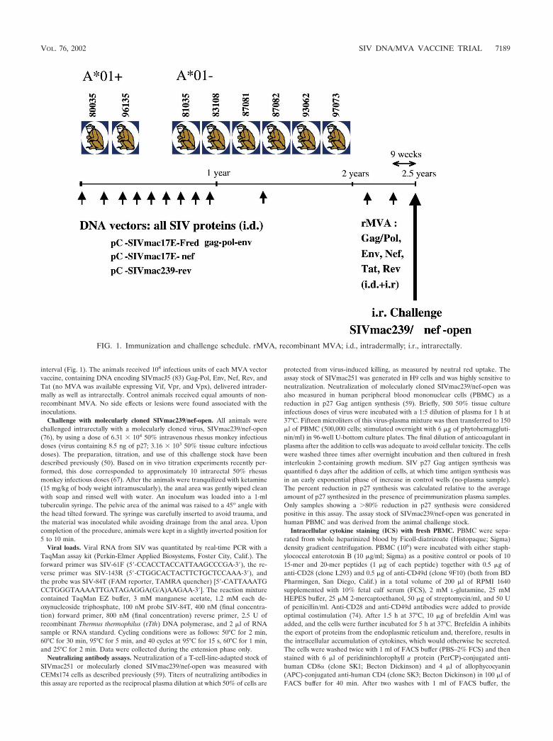
interval (Fig. 1). The animals received 108 infectious units of each MVA vectorvaccine, containing DNA encoding SIVmacJ5 (83) Gag-Pol, Env, Nef, Rev, andTat (no MVA was available expressing Vif, Vpr, and Vpx), delivered intrader-mally as well as intrarectally. Control animals received equal amounts of non-recombinant MVA. No side effects or lesions were found associated with theinoculations.
Challenge with molecularly cloned SIVmac239/nef-open. All animals werechallenged intrarectally with a molecularly cloned virus, SIVmac239/nef-open(76), by using a dose of 6.31 � 104 50% intravenous rhesus monkey infectiousdoses (virus containing 8.5 ng of p27; 3.16 � 103 50% tissue culture infectiousdoses). The preparation, titration, and use of this challenge stock have beendescribed previously (50). Based on in vivo titration experiments recently per-formed, this dose corresponded to approximately 10 intrarectal 50% rhesusmonkey infectious doses (67). After the animals were tranquilized with ketamine(15 mg/kg of body weight intramuscularly), the anal area was gently wiped cleanwith soap and rinsed well with water. An inoculum was loaded into a 1-mltuberculin syringe. The pelvic area of the animal was raised to a 45o angle withthe head tilted forward. The syringe was carefully inserted to avoid trauma, andthe material was inoculated while avoiding drainage from the anal area. Uponcompletion of the procedure, animals were kept in a slightly inverted position for5 to 10 min.
Viral loads. Viral RNA from SIV was quantitated by real-time PCR with aTaqMan assay kit (Perkin-Elmer Applied Biosystems, Foster City, Calif.). Theforward primer was SIV-61F (5�-CCACCTACCATTAAGCCCGA-3�), the re-verse primer was SIV-143R (5�-CTGGCACTACTTCTGCTCCAAA-3�), andthe probe was SIV-84T (FAM reporter, TAMRA quencher) [5�-CATTAAATGCCTGGGTAAAATTGATAGAGGA(G/A)AAGAA-3�]. The reaction mixturecontained TaqMan EZ buffer, 3 mM manganese acetate, 1.2 mM each de-oxynucleoside triphosphate, 100 nM probe SIV-84T, 400 nM (final concentra-tion) forward primer, 800 nM (final concentration) reverse primer, 2.5 U ofrecombinant Thermus thermophilus (rTth) DNA polymerase, and 2 �l of RNAsample or RNA standard. Cycling conditions were as follows: 50°C for 2 min,60°C for 30 min, 95°C for 5 min, and 40 cycles at 95°C for 15 s, 60°C for 1 min,and 25°C for 2 min. Data were collected during the extension phase only.
Neutralizing antibody assays. Neutralization of a T-cell-line-adapted stock ofSIVmac251 or molecularly cloned SIVmac239/nef-open was measured withCEMx174 cells as described previously (59). Titers of neutralizing antibodies inthis assay are reported as the reciprocal plasma dilution at which 50% of cells are
protected from virus-induced killing, as measured by neutral red uptake. Theassay stock of SIVmac251 was generated in H9 cells and was highly sensitive toneutralization. Neutralization of molecularly cloned SIVmac239/nef-open wasalso measured in human peripheral blood mononuclear cells (PBMC) as areduction in p27 Gag antigen synthesis (59). Briefly, 500 50% tissue cultureinfectious doses of virus were incubated with a 1:5 dilution of plasma for 1 h at37°C. Fifteen microliters of this virus-plasma mixture was then transferred to 150�l of PBMC (500,000 cells; stimulated overnight with 6 �g of phytohemaggluti-nin/ml) in 96-well U-bottom culture plates. The final dilution of anticoagulant inplasma after the addition to cells was adequate to avoid cellular toxicity. The cellswere washed three times after overnight incubation and then cultured in freshinterleukin 2-containing growth medium. SIV p27 Gag antigen synthesis wasquantified 6 days after the addition of cells, at which time antigen synthesis wasin an early exponential phase of increase in control wells (no-plasma sample).The percent reduction in p27 synthesis was calculated relative to the averageamount of p27 synthesized in the presence of preimmunization plasma samples.Only samples showing a �80% reduction in p27 synthesis were consideredpositive in this assay. The assay stock of SIVmac239/nef-open was generated inhuman PBMC and was derived from the animal challenge stock.
Intracellular cytokine staining (ICS) with fresh PBMC. PBMC were sepa-rated from whole heparinized blood by Ficoll-diatrizoate (Histopaque; Sigma)density gradient centrifugation. PBMC (106) were incubated with either staph-ylococcal enterotoxin B (10 �g/ml; Sigma) as a positive control or pools of 1015-mer and 20-mer peptides (1 �g of each peptide) together with 0.5 �g ofanti-CD28 (clone L293) and 0.5 �g of anti-CD49d (clone 9F10) (both from BDPharmingen, San Diego, Calif.) in a total volume of 200 �l of RPMI 1640supplemented with 10% fetal calf serum (FCS), 2 mM L-glutamine, 25 mMHEPES buffer, 25 �M 2-mercaptoethanol, 50 �g of streptomycin/ml, and 50 Uof penicillin/ml. Anti-CD28 and anti-CD49d antibodies were added to provideoptimal costimulation (74). After 1.5 h at 37°C, 10 �g of brefeldin A/ml wasadded, and the cells were further incubated for 5 h at 37°C. Brefeldin A inhibitsthe export of proteins from the endoplasmic reticulum and, therefore, results inthe intracellular accumulation of cytokines, which would otherwise be secreted.The cells were washed twice with 1 ml of FACS buffer (PBS–2% FCS) and thenstained with 6 �l of peridininchlorophyll a protein (PerCP)-conjugated anti-human CD8� (clone SK1; Becton Dickinson) and 4 �l of allophycocyanin(APC)-conjugated anti-human CD4 (clone SK3; Becton Dickinson) in 100 �l ofFACS buffer for 40 min. After two washes with 1 ml of FACS buffer, the
FIG. 1. Immunization and challenge schedule. rMVA, recombinant MVA; i.d., intradermally; i.r., intrarectally.
VOL. 76, 2002 SIV DNA/MVA VACCINE TRIAL 7189
cells were fixed with 2% paraformaldehyde (PFA)–PBS solution overnight at4°C. The cells were then washed once with FACS buffer, treated with perme-abilization buffer (0.1% saponin in FACS buffer) for 10 min at room tempera-ture, washed once more with 0.1% saponin buffer, and resuspended in 100 �l of0.1% saponin buffer. Then, 1 �l of an anti-human gamma interferon (IFN-�)–fluorescein isothiocyanate (FITC) monoclonal antibody (MAb) (clone 4S.B3;Pharmingen) and either 6 �l of an anti-CD69–phycoerythrin (PE) MAb (cloneL78; Becton Dickinson) or 1 �l of an anti-human tumor necrosis factor alpha–PEMAb (clone MAb11; Pharmingen) were added. After 50 min of incubation atroom temperature, the cells were washed two times with 0.1% saponin buffer,with a 10-min incubation before the last spin, and then fixed with 2% PFA–PBS.Samples were stored in the dark at 4°C, and the acquisition of 100,000 to 200,000lymphocyte-gated events was performed with a FACSCalibur flow cytometer(Becton Dickinson) and analyzed with FlowJo software (Treestar). The back-ground level of IFN-� staining in PBMC (induced by a control influenza viruspeptide, SNEGSYFFG) varied from animal to animal but was typically below0.05% in the CD8� lymphocytes and below 0.02% in the CD4� lymphocytes.Only samples in which IFN-� staining was at least twice that of the backgroundor in which there was a distinct population of IFN-� (bright)-positive cells (alsopositive for CD69 or tumor necrosis factor alpha) were considered positive. Onthe basis of these criteria, PBMC samples taken from all control animals 1 weekafter the injection of empty MVA did not produce any positive responses afterstimulation with pools of peptides. All values are reported after the subtractionof background staining.
Lymphocyte subset determination. PBMC (5 � 105) were stained with 10 �l ofanti-human CD8–phycoerythrin-Cy5 (PECy5) (clone B9.11; Beckman Coulter),10 �l of anti-human CD4–APC (clone SK3; Becton Dickinson), 5 �l of anti-human CD3ε–FITC (clone SP34; Pharmingen), and 5 �l of anti-humanCD20–PE (clone L27; Becton Dickinson) in 200 �l of RPMI 1640 mediumcontaining 10% FCS. Cells stained with immunoglobulins of matching isotypeswere used as negative controls. Samples were incubated in the presence ofantibodies for 45 min at an ambient temperature, washed, and fixed with 1%PFA–PBS solution for at least 30 min before the sample data were acquired witha FACSCalibur flow cytometer. The data were analyzed by using CellQuestsoftware (Becton Dickinson). Absolute numbers of major lymphocyte subsetswere determined by multiplying the percentage for each lymphocyte subset(percentages of CD3� CD4�, CD3� CD8�, or CD20� lymphocytes) by the totallymphocyte count per microliter of blood, as obtained from a complete bloodcount. The complete blood count was determined by using an AcT10 CoulterCounter (Beckman Coulter).
Tetramer staining. Fresh unstimulated PBMC (106) were washed two timeswith FACS buffer. In a 100-�l volume, cells were stained in the dark for 40 minat room temperature with Mamu-A*01/CM9 or Mamu-A*01/SL8 tetramer la-beled with either PE or APC (5 �g/ml), anti-human CD3ε–FITC (SP34), andanti-CD8�–PerCP (clone SK1). The cells were then washed two times with 1 mlof FACS buffer and fixed with 2% PFA–PBS solution. Sample data were ac-quired with a FACSCalibur instrument and analyzed by using CellQuest soft-ware. Background tetramer staining of fresh unstimulated PBMC from naiveMamu-A*01-positive animals was routinely less than 0.08%.
Statistical analysis. Differences in the levels of plasma SIV RNAs betweengroups were tested for statistical significance by using t tests after log transfor-mation of the data to improve normality and homoscedasticity. In addition, theLevene test for homoscedasticity was conducted; if the results were found to besignificant, the Welch correction for unequal variances was used. Finally, tofurther examine the robustness of the results, a nonparametric test, the Mann-Whitney U test, was performed. The P values for the nonparametric tests werecalculated by exact methods. Associations between T-cell responses and viralloads were tested by Pearson correlation analysis and the Spearman correlationtest, a nonparametric test, after log transformation of the data. All P valuesreported are two tailed. Survival analysis was conducted via Cox regression.
RESULTS
Cellular immune responses induced by vaccination and elic-
ited after viral challenge. Eight rhesus monkeys were immu-nized 10 times with vaccines containing DNA encoding all theproteins from SIVmac. When tetramer staining was used, noCTL against the well-documented immunodominant Mamu-A*01-restricted epitopes Gag181–189CM9 (4) and Tat28–35SL8(3) were detectable after the last DNA boost in fresh PBMCfrom Mamu-A*01-positive animals 80035 and 96135. However,CTL against these epitopes were detectable after in vitro stim-ulation (data not shown). To boost the cellular immune re-sponses (5, 37, 38, 89), these animals were then immunizedtwice with recombinant MVA expressing Gag-Pol, Env, Nef,Rev, and Tat but not Vif, Vpr, and Vpx (Fig. 1). The cellularimmune responses elicited following each MVA boost weredetermined by ICS. One week after the first MVA boost,freshly isolated PBMC were stimulated with pools of peptides(10 overlapping 15-mer and 20-mer peptides per pool) span-ning the proteins Gag, Pol, Env, Nef, Tat, and Rev and wereanalyzed for the presence of virus-specific lymphocytes thatproduce cytokines. All animals showed both virus-specificCD8� and virus-specific CD4� responses after the first immu-nization with recombinant MVA (Fig. 2A and 3A), althoughthe numbers of different proteins recognized and the frequen-cies of responding T cells differed greatly from animal to ani-mal. The total frequency of SIV-specific CD8� lymphocytesranged from 0.05% to 1.26% of all CD8� lymphocytes (Fig.2A), with an average of 0.5%. The total frequency of SIV-specific CD4� lymphocytes ranged from 0.04% to 1.23% of allCD4� lymphocytes (Fig. 3A), with an average of 0.51%. Mostanimals mounted Gag-specific CD8� (Fig. 2A) and CD4�
(Fig. 3A) responses. In particular, the two Mamu-A*01-posi-tive animals (80035 and 96135) exhibited CD8�-T-cell re-sponses against the well-documented immunodominantGag181–189CM9 (4) and Tat28–35SL8 (3) epitopes, which werealso detected by tetramer staining (Fig. 4A). Both tetramerstaining and ICS revealed similar frequencies of CD8� lym-phocytes specific for these two Mamu-A*01-restrictedepitopes, confirming that ICS is as sensitive as tetramer stain-ing (5, 97). Four animals (81035, 83108, 87081, and 97073)showed only low-frequency CD8� responses against a few pep-tide pools after the first immunization with recombinant MVA(Fig. 2A), but two of these four animals, 87081 and 97073,exhibited strong CD4� responses (Fig. 3A).
All animals were boosted a second time with the same re-combinant MVA via the same routes. However, an elevation inthe overall SIV-specific immune response was not observed.Moreover, not all of the responses previously detected in theperipheral blood following the first boost were detectable afterthe second boost (data not shown). The low frequencies ofCD8� responses against the two Mamu-A*01-restrictedepitopes, Gag181–189CM9 and Tat28–35SL8, in the two Mamu-A*01-positive animals were also confirmed by tetramer stain-ing (data not shown). Nevertheless, at 9 weeks after the lastMVA boost (the day of challenge), both Mamu-A*01-positive
FIG. 2. ICS data for virus-specific CD8�-lymphocyte responses in animals vaccinated with DNA/MVA. The frequency of responding CD8�
cells against each protein is shown. These frequencies were obtained by adding the frequencies detected with peptide pools containing overlappingpeptides from a single protein (after subtraction of the background). CD8� responses after the first MVA boost (prior to infection), 2 weekspostinfection (acute phase of infection), and 12 weeks postinfection (chronic phase of infection) are shown.
7190 HORTON ET AL. J. VIROL.

7191

7192
animals still showed detectable tetramer-positive CD8� re-sponses against the two Mamu-A*01-restricted epitopes,Gag181–189CM9 (80035, 0.1% of CD3/8� lymphocytes; 96135,0.19% of CD3� CD8� lymphocytes) and Tat28–35SL8 (80035,0.3% of CD3/8� lymphocytes; 96135, 0.14% of CD3/8� lym-phocytes), as detected by tetramer staining. Only one otheranimal, 93062, showed a detectable CD8� response againstGag (0.8% of all CD8� lymphocytes) at this time point, asdetected by ICS (data not shown).
Nine weeks after the last MVA boost, all vaccinated animalsand 5 naive control animals were challenged intrarectally withmolecularly cloned SIVmac239/nef-open. All animals becameinfected, as determined by positive detection of plasma SIVRNA by real-time PCR (Fig. 5) (see below). After challenge,cellular immune responses were measured by ICS with peptidepools spanning all SIV proteins, including Vpr, Vpx, and Vif,which were included in the DNA vaccine but not in the MVAboost. With the exception of two animals (81035 and 87081), inwhich no CD8� responses were detectable 2 weeks postchal-lenge (acute phase), SIV-specific CD8� lymphocytes increasedin all animals to total frequencies ranging from 5.8% to 36.1%of all CD8� lymphocytes (Fig. 2B), with an average of 16.6%.In contrast, CD4� responses were low to undetectable 2 weekspostchallenge in all eight animals. Animal 80035 showed aborderline response against Gag, and animal 97073 mountedlow but significant CD4� responses against Pol, Env, and Nef(Fig. 3B). CD4� responses were below the limits of detectionin the remaining six animals. In the two Mamu-A*01-positiveanimals, the majority of the CD8� responses were directedagainst the two Mamu-A*01-restricted epitopes, Gag181–189CM9and Tat28–35SL8, as confirmed by tetramer staining (Fig. 4B).The frequency of Gag181–189CM9-specific CTL increased moreslowly but remained high (7 to 11% of all CD3� CD8� lym-phocytes) longer than that of Tat28–35SL8-specific CTL. Tat28–
35SL8-specific CTL reached a high frequency at week 2 (11 to12% of all CD3/8� lymphocytes) but declined afterward (Fig.4B). A similar rapid decline of Tat28–35SL8-specific CTL inMamu-A*01-positive animals was previously shown to be dueto the rapid selection of escape variants (3), which also oc-curred in the two Mamu-A*01-positive animals tested here(data not shown). Tetramer staining for two additional Mamu-A*01-restricted epitopes (Env235–243CL9 and Env622–630TL9)(2) revealed only very low frequencies of CD8� T cells againstthese epitopes (Fig. 4B). CD8� responses against these twoepitopes were undetectable by ICS.
By 12 weeks of infection (chronic phase), the frequencies ofvirus-specific CD8� T cells in the vaccinees declined but per-sisted at significant levels, 0.17 to 4.1% of all CD8� lympho-cytes (Fig. 2C), with an average of 1.68%. Interestingly, signif-icant CD8� responses were detected at 12 weeks postinfectionin animals 81035 and 87081, which did not show any detectableCD8� responses at week 2 (Fig. 2C). Moreover, in contrast tothe absence of CD4� responses in most animals at week 2, fiveof the seven vaccinees showed detectable CD4� responses by
week 12, with frequencies ranging from 0.04% to 0.53% of allCD4� lymphocytes (Fig. 3C), with an average of 0.18%.
In contrast to the strong virus-specific CD8� responses de-tected in most vaccinees, analysis by ICS showed that mostcontrol animals exhibited very weak virus-specific CD8� re-sponses at 3 weeks postchallenge, with frequencies of all CD8�
lymphocytes ranging between 0.28 and 0.6% in four of fivecontrol animals. Only control animal 92050 demonstrated sig-nificant SIV-specific CD8� responses at this time point, with2.8% of all CD8� lymphocytes being specific for SIV se-quences (overlapping peptides), as measured by ICS (data notshown) (65). At 3 weeks postchallenge, control animals alsoshowed low to undetectable SIV-specific CD4� responses (to-tal frequencies of between 0 and 0.2%; average, 0.06%) (datanot shown).
Consistent with the results seen in the acute phase, thecontrol animals also showed weaker cellular immune responsesthan the vaccinees by 12 weeks postchallenge (chronic phase).Control animals 90131 and 92050, which died at weeks 16 and38, respectively, showed no detectable SIV-specific CD8� re-sponses (data not shown), and the remaining three controlanimals showed only very weak CD8� responses at this timepoint (total frequencies of between 0.1 and 0.48%; average,0.34%) (data not shown). At this time point, only the controlanimal which was able to control its virus replication, animal92080, showed detectable CD4� responses (total frequency of0.14%) (data not shown).
Neutralizing antibodies. Even though SIVmac239 is ex-tremely difficult to neutralize (48, 59), we assessed neutralizingantibody levels in all animals on the day of challenge and at 3,6, and 10 weeks postchallenge, since the vaccine was capable ofinducing an antibody response (data not shown). None of thevaccinated animals or control animals had SIVmac239-specificneutralizing antibody activity against a PBMC-grown stock ofSIVmac239, as measured with CEMx174 target cells, at any ofthe time points tested (Tables 1 and 2). In addition, all plasmasamples were negative for neutralizing activity against thisPBMC-grown stock of SIVmac239, as measured with humanPBMC (80% reduction in p27 Gag antigen synthesis), evenat low plasma dilutions (1:5) (data not shown). However, allthe vaccinees demonstrated a low-level neutralizing antibodyresponse against the laboratory-adapted virus strain, SIV-mac251, on the day of challenge, as assessed with CEMx174cells (Table 1). This response increased rapidly after infection(Table 1). The neutralizing antibody titers against SIVmac251in control animals increased much more slowly or remainedundetectable; when detected, titers observed in control ani-mals were lower than those observed in the vaccinees (Tables1 and 2).
Viral loads. Plasma viral RNA levels after challenge weremeasured by real-time PCR. The control monkeys had 1.0 �107 to 1.0 � 108 copies of virus per ml of plasma at the time ofpeak viremia at 2 weeks postchallenge (Fig. 5A). In the vacci-nated animals, peak viremia was measured at 4.5 � 105 to 4.5
FIG. 3. ICS data for virus-specific CD4�-lymphocyte responses in animals vaccinated with DNA/MVA. The frequency of responding CD4�
cells against each protein is shown. These frequencies were obtained as described in the legend to Fig. 2. CD4� responses after the first MVA boost(prior to infection), 2 weeks postinfection (acute phase of infection), and 12 weeks postinfection (chronic phase of infection) are shown.
VOL. 76, 2002 SIV DNA/MVA VACCINE TRIAL 7193
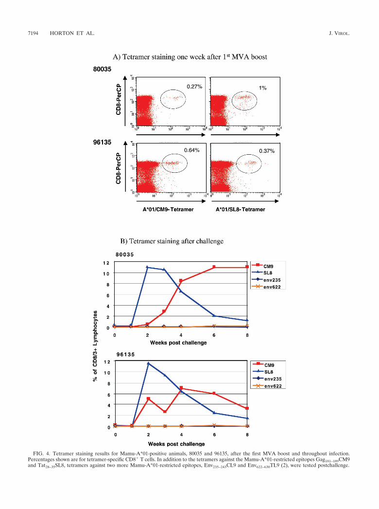
FIG. 4. Tetramer staining results for Mamu-A*01-positive animals, 80035 and 96135, after the first MVA boost and throughout infection.Percentages shown are for tetramer-specific CD8� T cells. In addition to the tetramers against the Mamu-A*01-restricted epitopes Gag181–189CM9and Tat28–35SL8, tetramers against two more Mamu-A*01-restricted epitopes, Env235–243CL9 and Env622–630TL9 (2), were tested postchallenge.
7194 HORTON ET AL. J. VIROL.

FIG. 5. Viral load data for all animals up to 28 weeks postinfection, as determined by real-time PCR. (A) Control animals 90069, 90131, 92050,92080, and 97086 were given empty MVA boosts only before intrarectal infection with SIVmac239. (B) Animals 80035, 81035, 83108, 87081, 87082,93062, 96135, and 97073 were vaccinated 10 times with DNA and twice with recombinant MVA before intrarectal infection with SIVmac239.(C) Average viral loads of control animals versus vaccinees. A dagger indicates euthanasia of the animal.
VOL. 76, 2002 SIV DNA/MVA VACCINE TRIAL 7195

� 106 copies/ml (Fig. 5B). One vaccinated animal, 83108, died3 weeks postinfection due to a non-SIV-related illness (seebelow) and was excluded from the analysis. On average, thevaccinees had a 1.5-log-lower viral load during peak viremiathan the controls (Fig. 5C). This difference was significant (P 0.001 [t test]; P 0.003 [nonparametric test]). However, by 12weeks postinfection, the viral loads in all vaccinees increased tolevels comparable to those in the controls, and the significanceof the difference in viral loads between the two groups was lostby week 6 or 8, depending on which statistical test was used.
Clinical disease progression. Postchallenge CD4�-T-lym-phocyte counts were determined over time in all animals as anindicator of disease progression (Fig. 6). With the exception oftwo vaccinees (96135 and 97073), all vaccinated animals dem-onstrated progressive declining CD4�-T-cell counts. Two vac-cinees, 81035 and 87082, were euthanized with an AIDS-likedisease at 28 and 32 weeks postinfection, respectively (Table 3and Fig. 6B). Vaccinee 83108 was euthanized at 3 weekspostinfection due to diabetes (Table 3) and was thereforeexcluded from all analyses. Four of the five control animals
demonstrated various degrees of postinfection CD4�-T-celldecline (Fig. 6A). The fifth control animal, 92080, showed virusreplication to low levels (10,000 copies per ml) for 24 weeks(Fig. 5A) and maintained CD4�-T-cell counts (Fig. 6A) for upto 36 weeks postinfection. However, at 32 weeks postchallenge,the viral load in this animal began to progressively increase; by44 weeks postchallenge, the viral load was �400,000 copies/ml(data not shown). Control animal 90131, which showed thehighest viral load (Fig. 5A), also demonstrated the most dra-matic decline in CD4�-T-cell counts (Fig. 6A) and had to beeuthanized due to an AIDS-related disease at week 16 (Table3). Another control animal, 92050, was also euthanized at week38 due to an AIDS-related disease (Table 3). There was nostatistical difference (P � 0.05 [Cox regression]) between thesurvival rates of vaccinees and control animals (Fig. 7).
Analysis of correlation between cell-mediated immunity andviral load. Associations between total CD8 and CD4 responsesdetected after vaccination (preinfection), during the acutephase of infection or the chronic phase of infection, with eitherthe viral load peak (week 2 or 3) or the set point (average ofthe viral loads between weeks 16 and 28) were tested by bothPearson and Spearman correlation analyses. Interestingly, asignificant inverse correlation was found only between the totalvirus-specific CD8� responses detected after vaccination andthe viral load set point by Spearman analysis (P 0.003) butnot by Pearson analysis (P 0.065). No correlation was foundbetween the virus-specific CD4� responses and viral load.
There was also no correlation between either the strength orthe breadth of the virus-specific CD8� responses detected ineach animal and its ability to maintain CD4 counts. In fact, onevaccinee, 97073, maintained its CD4 counts throughout the
TABLE 1. Neutralizing antibody data from vaccinated animals atvarious weeks after challenge with SIVmac239
Animal wk postchallengeNeutralizing antibody titera to:
SIVmac251 SIVmac239
80035 0 94 203 10,419 206 23,097 20
10 11,600 20
81035 0 36 204 9,795 206 6,996 20
10 12,947 20
83108 0 37 203 2,466 20
87081 0 34 203 6,297 206 1,369 20
10 2,412 20
87082 0 52 203 6,941 206 2,024 20
10 1,822 20
93062 0 282 203 12,917 206 5,630 20
10 4,703 20
96135 0 82 203 8,436 206 8,333 20
10 2,366 20
97073 0 211 203 11,135 206 7,274 20
10 13,967 20
a Reciprocal serum dilution at which 50% of CEMx174 cells were protectedfrom virus-induced killing, as measured by neutral red uptake. SIVmac251 waslaboratory adapted.
TABLE 2. Neutralizing antibody data from control animals atvarious weeks after challenge with SIVmac239
Animal wk postchallengeNeutralizing antibody titera to:
SIVmac251 SIVmac239
90069 0 20 203 20 206 856 20
10 1,755 20
90131 0 20 203 20 206 20 20
10 20 20
92050 0 20 203 42 206 919 20
10 651 20
92080 0 20 203 341 206 2,139 20
10 1,641 20
97086 0 25 203 419 206 8,010 20
10 9,952 20
a Reciprocal serum dilution at which 50% of CEMx174 cells were protectedfrom virus-induced killing, as measured by neutral red uptake. SIVmac251 waslaboratory adapted.
7196 HORTON ET AL. J. VIROL.

study (Fig. 6B) but demonstrated only low virus-specific CD8�
responses throughout the study (Fig. 2). In contrast, animal80035 demonstrated the strongest virus-specific CD8� re-sponses throughout the study (Fig. 2) but failed to maintain itsCD4 counts throughout the study (Fig. 6B).
The magnitude of the virus-specific CD4� responses did notappear to correlate with an ability to mount strong CD8�
cellular immune responses. Animal 87081 demonstrated strongvirus-specific CD4� responses prechallenge and during thechronic phase of infection (Fig. 3A and C) but showed very lowto undetectable CD8� responses throughout the study (Fig. 2).This animal also suffered a loss of its CD4� T cells (Fig. 6B).In one case, the presence of a strong virus-specific CD4�
response during the chronic phase of infection also did notpredict a better outcome, since animal 81035, which had dem-onstrated the strongest virus-specific CD4� responses through-out the study (Fig. 3C), still progressed to AIDS.
DISCUSSION
We have demonstrated here that a DNA prime/recombinantMVA boost regimen induced strong SIV-specific CD8� andCD4� cellular immune responses and resulted in a significantreduction in viral replication during the acute phase after in-
trarectal challenge with SIVmac239. Nevertheless, the immu-nization did not protect the animals against the pathogenicconsequences of infection with this virus. Most animals dem-onstrated a progressive loss of CD4� cells, and two of sevenvaccinees had to be euthanized by weeks 28 and 32 postchal-lenge due to progression to simian AIDS. These results aresomewhat disappointing, since recent studies with similarDNA prime/viral vector boost approaches protected rhesusmacaques from the pathogenic consequences of infection withSHIV-89.6P (9, 80). Even a cytokine-augmented DNA vacci-nation alone (15) and a vaccination with recombinant MVAalone (14) protected animals from disease progression afterchallenge with SHIV-89.6P. These results suggest that the dis-crepancy between our results with SIVmac239 and the resultsof previous studies, where similar regimens have afforded sig-nificant protection against SHIV-89.6P, may lie in critical dif-ferences between the challenge viruses used. It was previouslydocumented that the virulence of a challenge virus can influ-
FIG. 6. Postchallenge CD4�-T-lymphocyte counts in control andvaccinated animals. A dagger indicates euthanasia of the animal.
FIG. 7. Postchallenge mortality in vaccinated and control animalsthrough 38 weeks postinfection.
TABLE 3. Significant clinical events attributable to SIVmac239infection or subsequent immunodeficiency in control and
vaccinated animals through 38 weeks postinfection
Monkey Disease-attributable clinical event(s)
Controls90069 ...............None90131 ...............Moderate anorexia, moderate diarrhea, opportunistic
infections (Staphylococcus aureus, Giardia spp., Crypto-sporidium spp.); euthanasia at wk 14
92050 ...............Severe anemia, moderate thrombocytopenia; euthanasiaat wk 38
92080 ...............None97086 ...............Moderate diarrhea
Vaccinees80035 ...............Mild weight loss81035 ...............Mild anorexia, peritonitis; euthanasia at wks 2883108 ...............Euthanasia due to clinical diabetes87081 ...............Mild gingivitis87082 ...............Moderate weight loss, moderate anorexia, suppurative
lymphadenitis; euthanasia at wk 3293062 ...............None96135 ...............None97073 ...............None
VOL. 76, 2002 SIV DNA/MVA VACCINE TRIAL 7197

ence the outcome of a challenge (60, 102). SIVmac239 andSHIV-89.6P are both highly pathogenic viruses (50, 53, 76–79).However, each has some distinguishing characteristics. SHIV-89.6P induces a rapid CD4�-T-cell decline within 2 to 3 weeksafter infection, depending on the route of challenge (52, 77–79), an outcome that is not seen with field strains of HIV. Incontrast, infection with SIVmac239 results in slow, inexorableCD4 destruction (50, 53), an outcome that is similar to thatseen with HIV infection (86). More importantly, SIVmac239 isvery difficult to neutralize with antibodies (48, 59), much likemany field strains of HIV (19, 55, 61).
A previous study demonstrated that longer survival afterinfection with SHIV-89.6P was associated with the productionof antiviral antibodies (52), indicating that antibodies againstthis virus may play a significant role in protection againstSHIV-89.6P. Indeed, this protective capacity of neutralizingantibodies against infection with SHIV-89.6P or disease devel-opment after infection has been demonstrated by passive im-munization (54, 56). It appears, therefore, that if animals canbe protected against the acute CD4 depletion caused by SHIV-89.6P, then the development of effective antibody responsesmay ameliorate the disease course. Even animals without anydetectable virus-specific immune responses after vaccinationwere still protected against the pathogenic consequences of achallenge with SHIV-89.6P (81), indicating that it may bemuch easier to protect against a challenge with this virus thanit is to protect against SIVmac239. Because SIVmac239 is sodifficult to neutralize and no neutralizing antibodies againstthis virus were detectable in our animals (Tables 1 and 2), it isunlikely that neutralizing antibodies played any role in thereduced viral loads observed during the acute phase of infec-tion in all of our vaccinees, even though the neutralizing anti-body titers against laboratory-adapted SIVmac251 increased(Tables 1 and 2). However, no correlation between vaccine-induced immune responses and reduced peak viral loads wasdetectable either. Interestingly, we detected a trend toward aninverse correlation between the total frequency of SIV-specificCD8� responses detected after vaccination and the viral loadset point, as determined by the average of the viral loadsbetween weeks 16 and 28. Vaccinated animals demonstratedstronger and more sustained CD8�-T-cell responses postinfec-tion than control animals. However, the viral load set point inthe vaccinated animals was not significantly lower than that inthe control animals. No correlation was observed between thestrength and/or the breadth of the virus-specific CD8� re-sponses and the ability to maintain CD4 counts.
Although early viral replication dynamics have been corre-lated with disease progression (41, 72, 93), reduced peak viralreplication alone is apparently not sufficient to protect animalsfrom disease progression after infection with certain viruses,such as SIVmac 251 (51, 93) or SIVmac239 (this study). Inaddition to the reduced peak level of viral RNA in plasma, allvaccinees demonstrated some transient suppression of viralreplication at weeks 6 to 12 postchallenge compared with non-vaccinated controls. It is unclear why the plasma virus concen-trations started to rise again after this point. However, a pre-vious study demonstrated a functional defect in SIV-specificCTL after 16 weeks of infection with SIVmac239 (97). Othergroups have also described such a functionally impaired cellu-lar immune system in SIVmac-infected animals (68, 104, 107),
which could result in inefficient control of the virus by thecellular immune system in the absence of effective antibodies.Another explanation for the increase in viral replication couldbe escape of the virus from the cellular immune responsethrough mutation of MHC class I-restricted epitopes, as hasbeen indicated for HIV (18, 34, 75). Several studies have in-dicated that SIV can also accumulate mutations in MHC classI-restricted epitopes, abrogating recognition by CTL (3, 30,63). We are currently exploring whether this property couldexplain the lack of control of viral replication after 12 weeks inthese animals by sequencing regions of the virus against whichcellular immune responses were directed at several time pointsafter challenge.
The significant reduction in acute viral loads in the vacci-nated animals and the inverse correlation between viral loadsand CD8� responses following vaccination reported here sug-gest that vaccines that elicit strong virus-specific CTL may becapable of controlling the replication of even highly pathogenicviruses. However, the failure of this vaccine strategy to achievelong-term protection from CD4�-T-cell decline following chal-lenge with SIVmac239 is consistent with previous studies dem-onstrating that it is very difficult to protect against SIVmac239(1, 26, 33, 58, 67, 99). Indeed, although several vaccines havebeen tested for the ability to protect against SIVmac239 (1, 26,33, 58, 67, 99), only a few studies have reported similar vaccine-induced control of this virus postchallenge (67, 99). AlthoughMamu-A*01 had been associated with a better outcome afterchallenge with SIVmac251 (73), the two Mamu-A*01-positiveanimals selected for this study did not stand out with regard totheir viral loads or disease progression after challenge withSIVmac239 in comparison to the non-Mamu-A*01 vaccinees.Therefore, the inclusion of the two Mamu-A*01-positive ani-mals as vaccinees did not affect the outcome of this study.
Unfortunately, it is not possible to compare the levels ofimmune responses induced by our vaccine regimen to thosethat provided protection from disease progression after chal-lenge with SIVmac239 (67, 99) or SHIV-89.6P (9, 14, 15)because of the different assay systems used. With regard totetramer staining, however, the frequencies of Gag181–189CM9-specific CTL in our two Mamu-A*01-positive animals after thefirst MVA boost were comparable to those reported afterDNA vaccination alone (15) and were approximately half thefrequencies obtained after cytokine-augmented DNA vaccina-tion, which resulted in the prevention of clinical AIDS afterchallenge with SHIV-89.6P (15). Nevertheless, the levels ofGag181–189CM9-specific CD8� T cells that we obtained weresimilar to the levels obtained with recombinant MVA vaccina-tion alone, which also prevented the development of diseaseafter challenge with SHIV-89.6P (14). Much higher frequen-cies of Mamu-A*01/Gag181–189CM9-specific responses havebeen observed after DNA/MVA vaccination (5; T. U. Vogel etal., unpublished data); however, most of those animals weremuch younger than the ones used in this study. It is possiblethat immune responses in older animals may not be as strongas those in younger animals (32, 88, 96). However, with thelimitations of having a small, diverse group of animals of dif-ferent ages in this study, our data do not directly indicate suchan influence of age on cellular immune responses.
The failure of our immunization strategy could be due toseveral other factors. The expression of certain proteins from
7198 HORTON ET AL. J. VIROL.
immunodeficiency viruses might have had negative effects onthe induction of immune responses. For example, Nef has beendocumented to downregulate the surface expression of MHCclass I molecules (90) and, therefore, may result in less efficientantigen presentation by the MHC class I pathway (23). How-ever, a study by Robinson et al. also included Nef as an im-munogen and, nevertheless, protection against SHIV challengewas achieved (80). In addition, Env has been reported to beimmunosuppressive (100). However, Env was also included asan immunogen in multiple studies, and it protected monkeysagainst the pathogenic consequences of a SHIV-89.6P chal-lenge (9, 14, 15, 80, 81). Nevertheless, by preventing the im-munosuppressive potential of some of our immunogens, wemay be able to induce more effective immune responses.
In summary, we have demonstrated that the DNA prime/MVA boost regimen, which has proven to be safe and capableof inducing strong cellular immune responses against SIV inrhesus monkeys (5, 9, 38–40), suppressed viral replication dur-ing the acute phase of infection with SIVmac239. However, thevaccine regimen was unable to induce protection against thepathogenic consequences of an infection with SIVmac239. Thisfailure may be related in part to the neutralization resistanceand highly pathogenic potential of SIVmac239, against whichprotection has proven to be very difficult (1, 26, 33, 58, 67, 99).
ACKNOWLEDGMENTS
Helen Horton and Thorsten U. Vogel contributed equally to thiswork.
We thank Sarah Fuenger, Max Liebl, and Edward Dunphy for as-sistance in performing ICS and data collection; Marion Ohlmann forexpert MVA vaccine preparation; Dagna Sheerar and Eva Rakasz forassistance in determining lymphocyte subsets by flow cytometry; andJason Reed and Jason Weinfurter for assistance in tissue processingfrom necropsies. We also thank Jody Helgeland for assistance withblood processing, Jacque Mitchen for coordinating all animal proce-dures, Carol L. Emerson for performing all animal procedures, andAmy L. Usborne for help in evaluating all clinical signs and necropsyreports.
This work was supported by NIH grants RR1537, AI46366, AI45461,and RR00167 (awarded to David I. Watkins) and AI85343 (awarded toDavid C. Montefiori) and by the European Community (ProgrammeEVA, grant EU QLK2-2000-1040 awarded to Volker Erfle and GerdSutter). David I. Watkins is a recipient of an Elizabeth Glaser scientistaward.
REFERENCES
1. Ahmad, S., B. Lohman, M. Marthas, L. Giavedoni, Z. el-Amad, N. L.Haigwood, C. J. Scandella, M. B. Gardner, P. A. Luciw, and T. Yilma. 1994.Reduced virus load in rhesus macaques immunized with recombinant gp160and challenged with simian immunodeficiency virus. AIDS Res. Hum. Ret-rovir. 10:195–204.
2. Allen, T. M., B. R. Mothe, J. Sidney, P. Jing, J. L. Dzuris, M. E. Liebl, T. U.Vogel, D. H. O’Connor, X. Wang, M. C. Wussow, J. A. Thomson, J. D.Altman, D. I. Watkins, and A. Sette. 2001. CD8� lymphocytes from simianimmunodeficiency virus-infected rhesus macaques recognize 14 differentepitopes bound by the major histocompatibility complex class I moleculemamu-A*01: implications for vaccine design and testing. J. Virol. 75:738–749.
3. Allen, T. M., D. H. O’Connor, J. Peicheng, J. L. Dzuris, B. R. Mothe, T. U.Vogel, E. Dunphy, M. E. Liebl, C. Emerson, N. Wilson, K. J. Kunstman, X.Wang, D. B. Allison, A. L. Hughes, R. C. Desrosiers, J. D. Altman, S. M.Wolinsky, A. Sette, and D. I. Watkins. 2000. Tat-specific cytotoxic T lym-phocytes select for SIV escape variants during resolution of primary vire-mia. Nature 407:386–390.
4. Allen, T. M., J. Sidney, M. F. del Guercio, R. L. Glickman, G. L. Lensmeyer,D. A. Wiebe, R. DeMars, C. D. Pauza, R. P. Johnson, A. Sette, and D. I.Watkins. 1998. Characterization of the peptide binding motif of a rhesusMHC class I molecule (Mamu-A*01) that binds an immunodominant CTLepitope from simian immunodeficiency virus. J. Immunol. 160:6062–6071.
5. Allen, T. M., T. U. Vogel, D. H. Fuller, B. R. Mothe, S. Steffen, J. E. Boyson,T. Shipley, J. Fuller, T. Hanke, A. Sette, J. D. Altman, B. Moss, A. J.McMichael, and D. I. Watkins. 2000. Induction of AIDS virus-specific CTLactivity in fresh, unstimulated peripheral blood lymphocytes from rhesusmacaques vaccinated with a DNA prime/modified vaccinia virus Ankaraboost regimen. J. Immunol. 164:4968–4978.
6. Almond, N., T. Corcoran, R. Hull, B. Walker, J. Rose, R. Sangster, K.Silvera, P. Silvera, M. Cranage, E. Rud, and E. J. Stott. 1997. Mechanismsof protection induced by attenuated simian immunodeficiency virus. IV.Protection against challenge with virus grown in autologous simian cells.J. Med. Primatol. 26:34–43.
7. Almond, N., K. Kent, M. Cranage, E. Rud, B. Clarke, and E. J. Stott. 1995.Protection by attenuated simian immunodeficiency virus in macaquesagainst challenge with virus-infected cells. Lancet 345:1342–1344.
8. Almond, N., J. Rose, R. Sangster, P. Silvera, R. Stebbings, B. Walker, andE. J. Stott. 1997. Mechanisms of protection induced by attenuated simianimmunodeficiency virus. I. Protection cannot be transferred with immuneserum. J. Gen. Virol. 78:1919–1922.
9. Amara, R. R., F. Villinger, J. D. Altman, S. L. Lydy, S. P. O’Neil, S. I.Staprans, D. C. Montefiori, Y. Xu, J. G. Herndon, L. S. Wyatt, M. A.Candido, N. L. Kozyr, P. L. Earl, J. M. Smith, H. L. Ma, B. D. Grimm, M. L.Hulsey, J. Miller, H. M. McClure, J. M. McNicholl, B. Moss, and H. L.Robinson. 2001. Control of a mucosal challenge and prevention of AIDS bya multiprotein DNA/MVA vaccine. Science 292:69–74.
10. Anderson, M. G., D. Hauer, D. P. Sharma, S. V. Joag, O. Narayan, M. C.Zink, and J. E. Clements. 1993. Analysis of envelope changes acquired bySIVmac239 during neuroadaption in rhesus macaques. Virology 195:616–626.
11. Baba, T. W., Y. S. Jeong, D. Pennick, R. Bronson, M. F. Greene, and R. M.Ruprecht. 1995. Pathogenicity of live, attenuated SIV after mucosal infec-tion of neonatal macaques. Science 267:1820–1825.
12. Baba, T. W., V. Liska, A. H. Khimani, N. B. Ray, P. J. Dailey, D. Penninck,R. Bronson, M. F. Greene, H. M. McClure, L. N. Martin, and R. M.Ruprecht. 1999. Live attenuated, multiply deleted simian immunodefi-ciency virus causes AIDS in infant and adult macaques. Nat. Med. 5:194–203.
13. Barouch, D. H., A. Craiu, S. Santra, M. A. Egan, J. E. Schmitz, M. J.Kuroda, T. M. Fu, J. H. Nam, L. S. Wyatt, M. A. Lifton, G. R. Krivulka,C. E. Nickerson, C. I. Lord, B. Moss, M. G. Lewis, V. M. Hirsch, J. W.Shiver, and N. L. Letvin. 2001. Elicitation of high-frequency cytotoxicT-lymphocyte responses against both dominant and subdominant simian-human immunodeficiency virus epitopes by DNA vaccination of rhesusmonkeys. J. Virol. 75:2462–2467.
14. Barouch, D. H., S. Santra, M. J. Kuroda, J. E. Schmitz, R. Plishka, A.Buckler-White, A. E. Gaitan, R. Zin, J. H. Nam, L. S. Wyatt, M. A. Lifton,C. E. Nickerson, B. Moss, D. C. Montefiori, V. M. Hirsch, and N. L. Letvin.2001. Reduction of simian-human immunodeficiency virus 89.6P viremia inrhesus monkeys by recombinant modified vaccinia virus Ankara vaccina-tion. J. Virol. 75:5151–5158.
15. Barouch, D. H., S. Santra, J. E. Schmitz, M. J. Kuroda, T. M. Fu, W.Wagner, M. Bilska, A. Craiu, X. X. Zheng, G. R. Krivulka, K. Beaudry,M. A. Lifton, C. E. Nickerson, W. L. Trigona, K. Punt, D. C. Freed, L. Guan,S. Dubey, D. Casimiro, A. Simon, M. E. Davies, M. Chastain, T. B. Strom,R. S. Gelman, D. C. Montefiori, and M. G. Lewis. 2000. Control of viremiaand prevention of clinical AIDS in rhesus monkeys by cytokine-augmentedDNA vaccination. Science 290:486–492.
16. Benson, J., C. Chougnet, M. Robert-Guroff, D. Montefiori, P. Markham, G.Shearer, R. C. Gallo, M. Cranage, E. Paoletti, K. Limbach, D. Venzon, J.Tartaglia, and G. Franchini. 1998. Recombinant vaccine-induced protec-tion against the highly pathogenic simian immunodeficiency virus SIV-(mac251): dependence on route of challenge exposure. J. Virol. 72:4170–4182.
17. Borrow, P., H. Lewicki, B. H. Hahn, G. M. Shaw, and M. B. A. Oldstone.1994. Virus-specific CD8� cytotoxic T-lymphocyte activity associated withcontrol of viremia in primary human immunodeficiency virus type 1 infec-tion. J. Virol. 68:6103–6110.
18. Borrow, P., H. Lewicki, X. Wei, M. S. Horwitz, N. Peffer, H. Meyers, J. A.Nelson, J. E. Gairin, B. H. Hahn, M. B. Oldstone, and G. M. Shaw. 1997.Antiviral pressure exerted by HIV-1-specific cytotoxic T lymphocytes(CTLs) during primary infection demonstrated by rapid selection of CTLescape virus. Nat. Med. 3:205–211.
19. Bures, R., A. Gaitan, T. Zhu, C. Graziosi, K. M. McGrath, J. Tartaglia, P.Caudrelier, R. El Habib, M. Klein, A. Lazzarin, D. M. Stablein, M. Deers,L. Corey, M. L. Greenberg, D. H. Schwartz, and D. C. Montefiori. 2000.Immunization with recombinant canarypox vectors expressing membrane-anchored glycoprotein 120 followed by glycoprotein 160 boosting fails togenerate antibodies that neutralize R5 primary isolates of human immu-nodeficiency virus type 1. AIDS Res. Hum. Retrovir. 16:2019–2035.
20. Cafaro, A., F. Titti, C. Fracasso, M. T. Maggiorella, S. Baroncelli, A.Caputo, D. Goletti, A. Borsetti, M. Pace, E. Fanales-Belasio, B. Ridolfi,D. R. Negri, L. Sernicola, R. Belli, F. Corrias, I. Macchia, P. Leone, Z.Michelini, P. ten Haaft, S. Butto, P. Verani, and B. Ensoli. 2001. Vaccina-
VOL. 76, 2002 SIV DNA/MVA VACCINE TRIAL 7199
tion with DNA containing tat coding sequences and unmethylated CpGmotifs protects cynomolgus monkeys upon infection with simian/humanimmunodeficiency virus (SHIV89.6P). Vaccine 19:2862–2877.
21. Clements, J. E., R. C. Montelaro, M. C. Zink, A. M. Amedee, S. Miller,A. M. Trichel, B. Jagerski, D. Hauer, L. N. Martin, and R. P. Bohm. 1995.Cross-protective immune responses induced in rhesus macaques by immu-nization with attenuated macrophage-tropic simian immunodeficiency vi-rus. J. Virol. 69:2737–2744.
22. Cohen, J. 1997. Weakened SIV vaccine still kills. Science 278:24–25.23. Collins, K. L., B. K. Chen, S. A. Kalams, B. D. Walker, and D. Baltimore.
1998. HIV-1 Nef protein protects infected primary cells against killing bycytotoxic T lymphocytes. Nature 391:397–401.
24. Condon, C., S. C. Watkins, C. M. Celluzzi, K. Thompson, and L. D. Falo,Jr. 1996. DNA-based immunization by in vivo transfection of dendriticcells. Nat. Med. 2:1122–1128.
25. Connor, R. I., D. C. Montefiori, J. M. Binley, J. P. Moore, S. Bonhoeffer, A.Gettie, E. A. Fenamore, K. E. Sheridan, D. D. Ho, P. J. Dailey, and P. A.Marx. 1998. Temporal analyses of virus replication, immune responses, andefficacy in rhesus macaques immunized with a live, attenuated simian im-munodeficiency virus vaccine. J. Virol. 72:7501–7509.
26. Daniel, M. D., F. Kirchhoff, S. C. Czajak, P. K. Sehgal, and R. C. Desro-siers. 1992. Protective effects of a live attenuated SIV vaccine with a dele-tion in the nef gene. Science 258:1938–1941.
27. Desrosiers, R. C. 1994. Safety issues facing development of a live-attenu-ated, multiply deleted HIV-1 vaccine. AIDS Res. Hum. Retrovir. 10:331–332.
28. Dittmer, U., T. Nisslein, W. Bodemer, H. Petry, U. Sauermann, C. Stahl-Hennig, and G. Hunsmann. 1995. Cellular immune response of rhesusmonkeys infected with a partially attenuated nef deletion mutant of thesimian immunodeficiency virus. Virology 212:392–397.
29. Egan, M. A., W. A. Charini, M. J. Kuroda, J. E. Schmitz, P. Racz, K.Tenner-Racz, K. Manson, M. Wyand, M. A. Lifton, C. E. Nickerson, T. Fu,J. W. Shiver, and N. L. Letvin. 2000. Simian immunodeficiency virus (SIV)gag DNA-vaccinated rhesus monkeys develop secondary cytotoxic T-lym-phocyte responses and control viral replication after pathogenic SIV infec-tion. J. Virol. 74:7485–7495.
30. Evans, D. T., D. H. O’Connor, P. Jing, J. L. Dzuris, J. Sidney, J. da Silva,T. M. Allen, H. Horton, J. E. Venham, R. A. Rudersdorf, T. Vogel, C. D.Pauza, R. E. Bontrop, R. DeMars, A. Sette, A. L. Hughes, and D. I. Watkins.1999. Virus-specific cytotoxic T-lymphocyte responses select for amino-acidvariation in simian immunodeficiency virus Env and Nef. Nat. Med. 5:1270–1276.
31. Fuller, D. H., L. Simpson, K. S. Cole, J. E. Clements, D. L. Panicali, R. C.Montelaro, M. Murphey-Corb, and J. R. Haynes. 1997. Gene gun-basednucleic acid immunization alone or in combination with recombinant vac-cinia vectors suppresses virus burden in rhesus macaques challenged with aheterologous SIV. Immunol. Cell Biol. 75:389–396.
32. Gardner, E. M., E. D. Bernstein, M. Dorfman, E. Abrutyn, and D. M.Murasko. 1997. The age-associated decline in immune function of healthyindividuals is not related to changes in plasma concentrations of beta-carotene, retinol, alpha-tocopherol or zinc. Mech. Ageing Dev. 94:55–69.
33. Giorgi, J. V., L. E. Hultin, and R. C. Desrosiers. 1996. The immunopatho-genesis of retroviral diseases: no immunophenotypic alterations in T, B, andNK cell subsets in SIVmac239-challenged rhesus macaques protected bySIV delta nef vaccination. J. Med. Primatol. 25:186–191.
34. Goulder, P. J., R. E. Phillips, R. A. Colbert, S. McAdam, G. Ogg, M. A.Nowak, P. Giangrande, G. Luzzi, B. Morgan, A. Edwards, A. J. McMichael,and S. Rowland-Jones. 1997. Late escape from an immunodominant cyto-toxic T-lymphocyte response associated with progression to AIDS. Nat.Med. 3:212–217.
35. Gundlach, B. R., M. G. Lewis, S. Sopper, T. Schnell, J. Sodroski, C.Stahl-Hennig, and K. Uberla. 2000. Evidence for recombination of live,attenuated immunodeficiency virus vaccine with challenge virus to a morevirulent strain. J. Virol. 74:3537–3542.
36. Haigwood, N. L., C. C. Pierce, M. N. Robertson, A. J. Watson, D. C.Montefiori, M. Rabin, J. B. Lynch, L. Kuller, J. Thompson, W. R. Morton,R. E. Benveniste, S. L. Hu, P. Greenberg, and S. P. Mossman. 1999.Protection from pathogenic SIV challenge using multigenic DNA vaccines.Immunol. Lett. 66:183–188.
37. Hanke, T., T. J. Blanchard, J. Schneider, C. M. Hannan, M. Becker, S. C.Gilbert, A. V. Hill, G. L. Smith, and A. McMichael. 1998. Enhancement ofMHC class I-restricted peptide-specific T cell induction by a DNA prime/MVA boost vaccination regime. Vaccine 16:439–445.
38. Hanke, T., and A. McMichael. 1999. Pre-clinical development of a multi-CTL epitope-based DNA prime MVA boost vaccine for AIDS. Immunol.Lett. 66:177–181.
39. Hanke, T., V. C. Neumann, T. J. Blanchard, P. Sweeney, A. V. Hill, G. L.Smith, and A. McMichael. 1999. Effective induction of HIV-specific CTL bymulti-epitope using gene gun in a combined vaccination regime. Vaccine17:589–596.
40. Hanke, T., R. V. Samuel, T. J. Blanchard, V. C. Neumann, T. M. Allen, J. E.Boyson, S. A. Sharpe, N. Cook, G. L. Smith, D. I. Watkins, M. P. Cranage,
and A. J. McMichael. 1999. Effective induction of simian immunodeficiencyvirus-specific cytotoxic T lymphocytes in macaques by using a multiepitopegene and DNA prime-modified vaccinia virus Ankara boost vaccinationregimen. J. Virol. 73:7524–7532.
41. Hirsch, V. M., T. R. Fuerst, G. Sutter, M. W. Carroll, L. C. Yang, S.Goldstein, M. Piatak, Jr., W. R. Elkins, W. G. Alvord, D. C. Montefiori, B.Moss, and J. D. Lifson. 1996. Patterns of viral replication correlate withoutcome in simian immunodeficiency virus (SIV)-infected macaques: effectof prior immunization with a trivalent SIV vaccine in modified vacciniavirus Ankara. J. Virol. 70:3741–3752.
42. Jin, X., D. E. Bauer, S. E. Tuttleton, S. Lewin, A. Gettie, J. Blanchard, C. E.Irwin, J. T. Safrit, J. Mittler, L. Weinberger, L. G. Kostrikis, L. Zhang, A. S.Perelson, and D. D. Ho. 1999. Dramatic rise in plasma viremia afterCD8(�) T cell depletion in simian immunodeficiency virus-infected ma-caques. J. Exp. Med. 189:991–998.
43. Johnson, R. P. 1996. Macaque models for AIDS vaccine development.Curr. Opin. Immunol. 8:554–560.
44. Johnson, R. P., J. D. Lifson, S. C. Czajak, K. S. Cole, K. H. Manson, R.Glickman, J. Yang, D. C. Montefiori, R. Montelaro, M. S. Wyand, and R. C.Desrosiers. 1999. Highly attenuated vaccine strains of simian immunodefi-ciency virus protect against vaginal challenge: inverse relationship of degreeof protection with level of attenuation. J. Virol. 73:4952–4961.
45. Kent, S. J., A. Zhao, S. J. Best, J. D. Chandler, D. B. Boyle, and I. A.Ramshaw. 1998. Enhanced T-cell immunogenicity and protective efficacy ofa human immunodeficiency virus type 1 vaccine regimen consisting ofconsecutive priming with DNA and boosting with recombinant fowlpoxvirus. J. Virol. 72:10180–10188.
46. Kim, J. J., J. S. Yang, L. K. Nottingham, D. J. Lee, M. Lee, K. H. Manson,M. S. Wyand, J. D. Boyer, K. E. Ugen, and D. B. Weiner. 2001. Protectionfrom immunodeficiency virus challenges in rhesus macaques by multicom-ponent DNA immunization. Virology 285:204–217.
47. Koup, R. A., J. T. Safrit, Y. Cao, C. A. Andrews, G. McLeod, W. Borkowsky,C. Farthing, and D. D. Ho. 1994. Temporal association of cellular immuneresponses with the initial control of viremia in primary human immunode-ficiency virus type 1 syndrome. J. Virol. 68:4650–4655.
48. Langlois, A. J., R. C. Desrosiers, M. G. Lewis, V. N. KewalRamani, D. R.Littman, J. Y. Zhou, K. Manson, M. S. Wyand, D. P. Bolognesi, and D. C.Montefiori. 1998. Neutralizing antibodies in sera from macaques immu-nized with attenuated simian immunodeficiency virus. J. Virol. 72:6950–6955.
49. Lesinski, G. B., S. L. Smithson, N. Srivastava, D. Chen, G. Widera, andM. A. Westerink. 2001. A DNA vaccine encoding a peptide mimic ofStreptococcus pneumoniae serotype 4 capsular polysaccharide induces spe-cific anti-carbohydrate antibodies in Balb/c mice. Vaccine 19:1717–1726.
50. Lewis, M. G., S. Bellah, K. McKinnon, J. Yalley-Ogunro, P. M. Zack, W. R.Elkins, R. C. Desrosiers, and G. A. Eddy. 1994. Titration and characteriza-tion of two rhesus-derived SIVmac challenge stocks. AIDS Res. Hum.Retrovir. 10:213–220.
51. Lu, S., J. Arthos, D. C. Montefiori, Y. Yasutomi, K. Manson, F. Mustafa, E.Johnson, J. C. Santoro, J. Wissink, J. I. Mullins, J. R. Haynes, N. L. Letvin,M. Wyand, and H. L. Robinson. 1996. Simian immunodeficiency virus DNAvaccine trial in macaques. J. Virol. 70:3978–3991.
52. Lu, Y., C. D. Pauza, X. Lu, D. C. Montefiori, and C. J. Miller. 1998. Rhesusmacaques that become systemically infected with pathogenic SHIV 89.6-PDafter intravenous, rectal, or vaginal inoculation and fail to make an antiviralantibody response rapidly develop AIDS. J. Acquir. Immune Defic. Syndr.Hum. Retrovirol. 19:6–18.
53. Luciw, P. A., K. E. Shaw, R. E. Unger, V. Planelles, M. W. Stout, J. E.Lackner, E. Pratt-Lowe, N. J. Leung, B. Banapour, and M. L. Marthas.1992. Genetic and biological comparisons of pathogenic and nonpathogenicmolecular clones of simian immunodeficiency virus (SIVmac). AIDS Res.Hum. Retrovir. 8:395–402.
54. Mascola, J. R., M. G. Lewis, G. Stiegler, D. Harris, T. C. VanCott, D. Hayes,M. K. Louder, C. R. Brown, C. V. Sapan, S. S. Frankel, Y. Lu, M. L. Robb,H. Katinger, and D. L. Birx. 1999. Protection of macaques against patho-genic simian/human immunodeficiency virus 89.6PD by passive transfer ofneutralizing antibodies. J. Virol. 73:4009–4018.
55. Mascola, J. R., S. W. Snyder, O. S. Weislow, S. M. Belay, R. B. Belshe, D. H.Schwartz, M. L. Clements, R. Dolin, B. S. Graham, G. J. Gorse, M. C.Keefer, M. J. McElrath, M. C. Walker, K. F. Wagner, J. G. McNeil, F. E.McCutchan, D. S. Burke, et al. 1996. Immunization with envelope subunitvaccine products elicits neutralizing antibodies against laboratory-adaptedbut not primary isolates of human immunodeficiency virus type 1. J. Infect.Dis. 173:340–348.
56. Mascola, J. R., G. Stiegler, T. C. VanCott, H. Katinger, C. B. Carpenter,C. E. Hanson, H. Beary, D. Hayes, S. S. Frankel, D. L. Birx, and M. G.Lewis. 2000. Protection of macaques against vaginal transmission of apathogenic HIV-1/SIV chimeric virus by passive infusion of neutralizingantibodies. Nat. Med. 6:207–210.
57. Matano, T., R. Shibata, C. Siemon, M. Connors, H. C. Lane, and M. A.Martin. 1998. Administration of an anti-CD8 monoclonal antibody inter-
7200 HORTON ET AL. J. VIROL.
feres with the clearance of chimeric simian/human immunodeficiency virusduring primary infections of rhesus macaques. J. Virol. 72:164–169.
58. Miller, C. J., M. B. McChesney, X. Lu, P. J. Dailey, C. Chutkowski, D. Lu,P. Brosio, B. Roberts, and Y. Lu. 1997. Rhesus macaques previously in-fected with simian/human immunodeficiency virus are protected from vag-inal challenge with pathogenic SIVmac239. J. Virol. 71:1911–1921.
59. Montefiori, D. C., T. W. Baba, A. Li, M. Bilska, and R. M. Ruprecht. 1996.Neutralizing and infection-enhancing antibody responses do not correlatewith the differential pathogenicity of SIVmac239delta3 in adult and infantrhesus monkeys. J. Immunol. 157:5528–5535.
60. Mooij, P., W. M. Bogers, H. Oostermeijer, W. Koornstra, P. J. Ten Haaft,B. E. Verstrepen, G. Van Der Auwera, and J. L. Heeney. 2000. Evidence forviral virulence as a predominant factor limiting human immunodeficiencyvirus vaccine efficacy. J. Virol. 74:4017–4027.
61. Moore, J. P., P. W. Parren, and D. R. Burton. 2001. Genetic subtypes,humoral immunity, and human immunodeficiency virus type 1 vaccine de-velopment. J. Virol. 75:5721–5729.
62. Mori, K., Y. Yasutomi, S. Ohgimoto, T. Nakasone, S. Takamura, T. Shioda,and Y. Nagai. 2001. Quintuple deglycosylation mutant of simian immuno-deficiency virus SIVmac239 in rhesus macaques: robust primary replication,tightly contained chronic infection, and elicitation of potent immunityagainst the parental wild-type strain. J. Virol. 75:4023–4028.
63. Mortara, L., F. Letourneur, H. Gras-Masse, A. Venet, J. G. Guillet, and I.Bourgault-Villada. 1998. Selection of virus variants and emergence of virusescape mutants after immunization with an epitope vaccine. J. Virol. 72:1403–1410.
64. Mossman, S. P., F. Bex, P. Berglund, J. Arthos, S. P. O’Neil, D. Riley, D. H.Maul, C. Bruck, P. Momin, A. Burny, P. N. Fultz, J. I. Mullins, P.Liljestrom, and E. A. Hoover. 1996. Protection against lethal simian immu-nodeficiency virus SIVsmmPBj14 disease by a recombinant Semliki Forestvirus gp160 vaccine and by a gp120 subunit vaccine. J. Virol. 70:1953–1960.
65. Mothe, B. R., H. Horton, D. K. Carter, T. M. Allen, M. E. Liebl, P. Skinner,T. U. Vogel, S. Fuenger, K. Vielhuber, W. Rehrauer, N. Wilson, G.Franchini, J. D. Altman, A. Haase, L. J. Picker, D. B. Allison, and D. I.Watkins. 2002. Dominance of CD8 responses specific for epitopes bound bya single major histocompatibility complex class I molecule during the acutephase of viral infection. J. Virol. 76:875–884.
66. Murphey-Corb, M. 1997. Live-attenuated HIV vaccines: how safe is safeenough? Nat. Med. 3:17–18.
67. Murphy, C. G., W. T. Lucas, R. E. Means, S. Czajak, C. L. Hale, J. D.Lifson, A. Kaur, R. P. Johnson, D. M. Knipe, and R. C. Desrosiers. 2000.Vaccine protection against simian immunodeficiency virus by recombinantstrains of herpes simplex virus. J. Virol. 74:7745–7754.
67a.National Research Council. 1996. Guide for the care and use of laboratoryanimals. National Academy Press, Washington, D.C. .
68. Neben, K., M. Heidbreder, J. Muller, A. Marxer, H. Petry, A. Didier, A.Schimpl, T. Hunig, and T. Kerkau. 1999. Impaired thymopoietic potentialof immature CD3(�)CD4(�)CD8(�) T cell precursors from SIV-infectedrhesus monkeys. Int. Immunol. 11:1509–1518.
69. Nixon, D. F., S. M. Donahoe, W. M. Kakimoto, R. V. Samuel, K. J. Metzner,A. Gettie, T. Hanke, P. A. Marx, and R. I. Connor. 2000. Simian immuno-deficiency virus-specific cytotoxic T lymphocytes and protection againstchallenge in rhesus macaques immunized with a live attenuated simianimmunodeficiency virus vaccine. Virology 266:203–210.
70. Norley, S., B. Beer, D. Binninger-Schinzel, C. Cosma, and R. Kurth. 1996.Protection from pathogenic SIVmac challenge following short-term infec-tion with a nef-deficient attenuated virus. Virology 219:195–205.
71. Ogg, G. S., X. Jin, S. Bonhoeffer, P. R. Dunbar, M. A. Nowak, S. Monard,J. P. Segal, Y. Cao, S. L. Rowland-Jones, V. Cerundolo, A. Hurley, M.Markowitz, D. D. Ho, D. F. Nixon, and A. J. McMichael. 1998. Quantitationof HIV-1-specific cytotoxic T lymphocytes and plasma load of viral RNA.Science 279:2103–2106.
72. Ourmanov, I., C. R. Brown, B. Moss, M. Carroll, L. Wyatt, L. Pletneva, S.Goldstein, D. Venzon, and V. M. Hirsch. 2000. Comparative efficacy ofrecombinant modified vaccinia virus Ankara expressing simian immunode-ficiency virus (SIV) Gag-Pol and/or Env in macaques challenged withpathogenic SIV. J. Virol. 74:2740–2751.
73. Pal, R., D. Venzon, N. L. Letvin, S. Santra, D. C. Montefiori, N. R. Miller,E. Tryniszewska, M. G. Lewis, T. C. VanCott, V. Hirsch, R. Woodward, A.Gibson, M. Grace, E. Dobratz, P. D. Markham, Z. Hel, J. Nacsa, M. Klein,J. Tartaglia, and G. Franchini. 2002. ALVAC-SIV-gag-pol-env-based vac-cination and macaque major histocompatibility complex class I (A*01)delay simian immunodeficiency virus SIVmac-induced immunodeficiency.J. Virol. 76:292–302.
74. Picker, L. J., M. K. Singh, Z. Zdraveski, J. R. Treer, S. L. Waldrop, P. R.Bergstresser, and V. C. Maino. 1995. Direct demonstration of cytokinesynthesis heterogeneity among human memory/effector T cells by flowcytometry. Blood 86:1408–1419.
75. Price, D. A., P. J. Goulder, P. Klenerman, A. K. Sewell, P. J. Easterbrook,M. Troop, C. R. Bangham, and R. E. Phillips. 1997. Positive selection ofHIV-1 cytotoxic T lymphocyte escape variants during primary infection.Proc. Natl. Acad. Sci. USA 94:1890–1895.
76. Regier, D. A., and R. C. Desrosiers. 1990. The complete nucleotide se-quence of a pathogenic molecular clone of simian immunodeficiency virus.AIDS Res. Hum. Retrovir. 6:1221–1231.
77. Reimann, K. A., J. T. Li, R. Veazey, M. Halloran, I. W. Park, G. B.Karlsson, J. Sodroski, and N. L. Letvin. 1996. A chimeric simian/humanimmunodeficiency virus expressing a primary patient human immunodefi-ciency virus type 1 isolate env causes an AIDS-like disease after in vivopassage in rhesus monkeys. J. Virol. 70:6922–6928.
78. Reimann, K. A., J. T. Li, G. Voss, C. Lekutis, K. Tenner-Racz, P. Racz, W.Lin, D. C. Montefiori, D. E. Lee-Parritz, Y. Lu, R. G. Collman, J. Sodroski,and N. L. Letvin. 1996. An env gene derived from a primary human immu-nodeficiency virus type 1 isolate confers high in vivo replicative capacity toa chimeric simian/human immunodeficiency virus in rhesus monkeys. J. Vi-rol. 70:3198–3206.
79. Reimann, K. A., A. Watson, P. J. Dailey, W. Lin, C. I. Lord, T. D. Steenbeke,R. A. Parker, M. K. Axthelm, and G. B. Karlsson. 1999. Viral burden anddisease progression in rhesus monkeys infected with chimeric simian-hu-man immunodeficiency viruses. Virology 256:15–21.
80. Robinson, H. L., D. C. Montefiori, R. P. Johnson, K. H. Manson, M. L.Kalish, J. D. Lifson, T. A. Rizvi, S. Lu, S. L. Hu, G. P. Mazzara, D. L.Panicali, J. G. Herndon, R. Glickman, M. A. Candido, S. L. Lydy, M. S.Wyand, and H. M. McClure. 1999. Neutralizing antibody-independent con-tainment of immunodeficiency virus challenges by DNA priming and re-combinant pox virus booster immunizations. Nat. Med. 5:526–534.
81. Rose, N. F., P. A. Marx, A. Luckay, D. F. Nixon, W. J. Moretto, S. M.Donahoe, D. Montefiori, A. Roberts, L. Buonocore, and J. K. Rose. 2001.An effective AIDS vaccine based on live attenuated vesicular stomatitisvirus recombinants. Cell 106:539–549.
82. Rosenberg, E. S., J. M. Billingsley, A. M. Caliendo, S. L. Boswell, P. E. Sax,S. A. Kalams, and B. D. Walker. 1997. Vigorous HIV-1-specific CD4� Tcell responses associated with control of viremia. Science 278:1447–1450.
83. Rud, E. W., M. Cranage, J. Yon, J. Quirk, L. Ogilvie, N. Cook, S. Webster,M. Dennis, and B. E. Clarke. 1994. Molecular and biological characteriza-tion of simian immunodeficiency virus macaque strain 32H proviral clonescontaining nef size variants. J. Gen. Virol. 75:529–543.
84. Ruprecht, R. M., T. W. Baba, and V. Liska. 1996. Attenuated HIV vaccine:caveats. Science 271:1790–1792.
85. Sawai, E. T., M. S. Hamza, M. Ye, K. E. Shaw, and P. A. Luciw. 2000.Pathogenic conversion of live attenuated simian immunodeficiency virusvaccines is associated with expression of truncated Nef. J. Virol. 74:2038–2045.
86. Schellekens, P. T., M. Tersmette, M. T. Roos, R. P. Keet, F. de Wolf, R. A.Coutinho, and F. Miedema. 1992. Biphasic rate of CD4� cell count declineduring progression to AIDS correlates with HIV-1 phenotype. AIDS 6:665–669.
87. Schmitz, J. E., M. J. Kuroda, S. Santra, V. G. Sasseville, M. A. Simon, M. A.Lifton, P. Racz, K. Tenner-Racz, M. Dalesandro, B. J. Scallon, J. Ghrayeb,M. A. Forman, D. C. Montefiori, E. P. Rieber, N. L. Letvin, and K. A.Reimann. 1999. Control of viremia in simian immunodeficiency virus infec-tion by CD8� lymphocytes. Science 283:857–860.
88. Schmucker, D. L., K. Thoreux, and R. L. Owen. 2001. Aging impairsintestinal immunity. Mech. Ageing Dev. 122:1397–1411.
89. Schneider, J., S. C. Gilbert, T. J. Blanchard, T. Hanke, K. J. Robson, C. M.Hannan, M. Becker, R. Sinden, G. L. Smith, and A. V. Hill. 1998. Enhancedimmunogenicity for CD8� T cell induction and complete protective efficacyof malaria DNA vaccination by boosting with modified vaccinia virus An-kara. Nat. Med. 4:397–402.
90. Schwartz, O., V. Marechal, G. S. Le, F. Lemonnier, and J. M. Heard. 1996.Endocytosis of major histocompatibility complex class I molecules is in-duced by the HIV-1 Nef protein. Nat. Med. 2:338–342.
91. Seth, A., I. Ourmanov, J. E. Schmitz, M. J. Kuroda, M. A. Lifton, C. E.Nickerson, L. Wyatt, M. Carroll, B. Moss, D. Venzon, N. L. Letvin, andV. M. Hirsch. 2000. Immunization with a modified vaccinia virus expressingsimian immunodeficiency virus (SIV) Gag-Pol primes for an anamnesticGag-specific cytotoxic T-lymphocyte response and is associated with reduc-tion of viremia after SIV challenge. J. Virol. 74:2502–2509.
92. Sharma, D. P., M. C. Zink, M. Anderson, R. Adams, J. E. Clements, S. V.Joag, and O. Narayan. 1992. Derivation of neurotropic simian immunode-ficiency virus from exclusively lymphocytetropic parental virus: pathogene-sis of infection in macaques. J. Virol. 66:3550–3556.
93. Staprans, S. I., P. J. Dailey, A. Rosenthal, C. Horton, R. M. Grant, N.Lerche, and M. B. Feinberg. 1999. Simian immunodeficiency virus diseasecourse is predicted by the extent of virus replication during primary infec-tion. J. Virol. 73:4829–4839.
94. Stebbings, R., J. Stott, N. Almond, R. Hull, J. Lines, P. Silvera, R. Sangster,T. Corcoran, J. Rose, S. Cobbold, F. Gotch, A. McMichael, and B. Walker.1998. Mechanisms of protection induced by attenuated simian immunode-ficiency virus. II. Lymphocyte depletion does not abrogate protection.AIDS Res. Hum. Retrovir. 14:1187–1198.
95. Stott, J., and N. Almond. 1995. Assessing animal models of AIDS. Nat.Med. 1:295–297.
96. Taylor, L. D., C. K. Daniels, and D. L. Schmucker. 1992. Ageing compro-
VOL. 76, 2002 SIV DNA/MVA VACCINE TRIAL 7201

mises gastrointestinal mucosal immune response in the rhesus monkey.Immunology 75:614–618.
97. Vogel, T. U., T. M. Allen, J. D. Altman, and D. I. Watkins. 2001. Functionalimpairment of simian immunodeficiency virus-specific CD8� T cells duringthe chronic phase of infection. J. Virol. 75:2458–2461.
98. Vogel, T. U., J. Fournier, A. Sherring, D. Ko, M. Parenteau, D. Bogdanovic,J. Mihowich, and E. W. Rud. 1998. Presence of circulating CTL induced byinfection with wild-type or attenuated SIV and their correlation with pro-tection from pathogenic SHIV challenge. J. Med. Primatol. 27:65–72.
99. Wang, S. W., P. A. Kozlowski, G. Schmelz, K. Manson, M. S. Wyand, R.Glickman, D. Montefiori, J. D. Lifson, R. P. Johnson, M. R. Neutra, and A.Aldovini. 2000. Effective induction of simian immunodeficiency virus-specificsystemic and mucosal immune responses in primates by vaccination withproviral DNA producing intact but noninfectious virions. J. Virol. 74:10514–10522.
100. Weinhold, K. J., H. K. Lyerly, S. D. Stanley, A. A. Austin, T. J. Matthews,and D. P. Bolognesi. 1989. HIV-1 GP120-mediated immune suppressionand lymphocyte destruction in the absence of viral infection. J. Immunol.142:3091–3097.
101. Whatmore, A. M., N. Cook, G. A. Hall, S. Sharpe, E. W. Rud, and M. P.Cranage. 1995. Repair and evolution of nef in vivo modulates simian im-munodeficiency virus virulence. J. Virol. 69:5117–5123.
102. Wyand, M. S., K. Manson, D. C. Montefiori, J. D. Lifson, R. P. Johnson,and R. C. Desrosiers. 1999. Protection by live, attenuated simian immu-nodeficiency virus against heterologous challenge. J. Virol. 73:8356–8363.
103. Wyand, M. S., K. H. Manson, M. M. Garcia, D. Montefiori, and R. C.Desrosiers. 1996. Vaccine protection by a triple deletion mutant of simianimmunodeficiency virus. J. Virol. 70:3724–3733.
104. Xiong, Y., M. A. Luscher, J. D. Altman, M. Hulsey, H. L. Robinson, M.Ostrowski, B. H. Barber, and K. S. MacDonald. 2001. Simian immunode-ficiency virus (SIV) infection of a rhesus macaque induces SIV-specificCD8� T cells with a defect in effector function that is reversible on ex-tended interleukin-2 incubation. J. Virol. 75:3028–3033.
105. Yang, N. S., J. Burkholder, B. Roberts, B. Martinell, and D. McCabe. 1990.In vivo and in vitro gene transfer to mammalian somatic cells by particlebombardment. Proc. Natl. Acad. Sci. USA 87:9568–9572.
106. Yasutomi, Y., K. A. Reimann, C. I. Lord, M. D. Miller, and N. L. Letvin.1993. Simian immunodeficiency virus-specific CD8� lymphocyte responsein acutely infected rhesus monkeys. J. Virol. 67:1707–1711.
107. Zeitz, M., R. Ullrich, T. Schneider, S. Kewenig, and E. Riecken. 1998.Mucosal immunodeficiency in HIV/SIV infection. Pathobiology 66:151–157.
7202 HORTON ET AL. J. VIROL.
Related Documents


