Immunity, neuroglia and neuroinflammation in autism CARLOS A. PARDO 1,2,3 , DIANA L. VARGAS 1,2 , & ANDREW W. ZIMMERMAN 1,4 1 Department of Neurology, 2 Division of Neuroimmunology and Infectious Disorders, 3 Department of Pathology, Johns Hopkins University School of Medicine, Baltimore, and 4 Kennedy Krieger Institute, Baltimore, Maryland, USA Summary Autism is a complex neurodevelopmental disorder of early onset that is highly variable in its clinical presentation. Although the causes of autism in most patients remain unknown, several lines of research support the view that both genetic and environmental factors influence the development of abnormal cortical circuitry that underlies autistic cognitive processes and behaviors. The role of the immune system in the development of autism is controversial. Several studies showing peripheral immune abnormalities support immune hypotheses, however until recently there have been no immune findings in the CNS. We recently demonstrated the presence of neuroglial and innate neuroimmune system activation in brain tissue and cerebrospinal fluid of patients with autism, findings that support the view that neuroimmune abnormalities occur in the brain of autistic patients and may contribute to the diversity of the autistic phenotypes. The role of neuroglial activation and neuroinflammation are still uncertain but could be critical in maintaining, if not also in initiating, some of the CNS abnormalities present in autism. A better understanding of the role of neuroinflammation in the pathogenesis of autism may have important clinical and therapeutic implications. Introduction Autism is the most severe and devastating condition in the broad spectrum of developmental disorders called ‘pervasive developmental disorders’ (Rapin, 1997). Autistic disorders are characterized by marked impairment in social skills, verbal commu- nication, behavior, and cognitive function (Rapin, 1997; Lord et al., 2000). Abnormalities in language development, mental retardation, and epilepsy are frequent problems in the clinical profile of patients with autism (Rapin, 1997). The syndrome is clinically heterogeneous and can be associated in up to 10% of patients with well-described neurologi- cal and genetic disorders, such as tuberous sclerosis, fragile X, Rett and Down syndromes, although in most patients the causes are still unknown (Rapin & Katzman, 1998; Newschaffer et al., 2002; Cohen et al., 2005). The importance of autism as a public health problem has been recognized in recent years, as epidemiological studies have suggested that the age-adjusted incidence of research-identified autism has increased from 5.5 (95% confidence interval, 1.4–9.5) per 100 000 children in the period 1980–1983 to 44.9 (95% confidence interval, 32.9–56.9) in the period 1995–1997 (8.2-fold increase) (Barbaresi et al., 2005) while the prevalence of autistic syndromes has increased to 3–6 per 1000 children, with a male to female ratio of 3:1 (Fombonne, 2003; Yeargin- Allsopp et al., 2003). In addition to complex multigenic factors (Folstein & Rosen-Sheidley, 2001), several researchers have also hypothesized important roles for environmental factors, pre- or perinatal injuries, vaccines, mercury toxicity, or persistent viral infections (Wing & Potter, 2002; Larsson et al., 2005). These recent observations on the epidemiology of autism strongly suggest that an interplay between genetic and as yet undefined environmental factors may increase the risk of autism, to a level greater than one would expect from genetic causes alone. Neurobiology of autism Clinical and epidemiological aspects of autism Although the neurobiological basis for autism remains poorly understood, several lines of research now support the view that genetic, environmental, neurological, and immunological factors contribute to its development (Rapin & Katzman, 1998; Newschaffer et al., 2002; Folstein & Rosen- Sheidley, 2001; Korvatska et al., 2002). Several different genetic factors and/or other risk factors may combine during development to produce complex Correspondence: Carlos A. Pardo, MD, Department of Neurology, Johns Hopkins University School of Medicine, Pathology 627, 600 North Wolfe Street, Baltimore, Maryland 21287, USA. Tel: þ410 614 5757. Fax: þ410 502 7609. E-mail: [email protected] International Review of Psychiatry, December 2005; 17(6): 485–495 ISSN 0954–0261 print/ISSN 1369–1627 online/05/060485–11 ß Institute of Psychiatry DOI: 10.1080/02646830500381930

Welcome message from author
This document is posted to help you gain knowledge. Please leave a comment to let me know what you think about it! Share it to your friends and learn new things together.
Transcript
Immunity, neuroglia and neuroinflammation in autism
CARLOS A. PARDO1,2,3, DIANA L. VARGAS1,2, & ANDREW W. ZIMMERMAN1,4
1Department of Neurology, 2Division of Neuroimmunology and Infectious Disorders, 3Department of Pathology,
Johns Hopkins University School of Medicine, Baltimore, and 4Kennedy Krieger Institute, Baltimore, Maryland, USA
SummaryAutism is a complex neurodevelopmental disorder of early onset that is highly variable in its clinical presentation. Althoughthe causes of autism in most patients remain unknown, several lines of research support the view that both genetic andenvironmental factors influence the development of abnormal cortical circuitry that underlies autistic cognitive processes andbehaviors. The role of the immune system in the development of autism is controversial. Several studies showing peripheralimmune abnormalities support immune hypotheses, however until recently there have been no immune findings in the CNS.We recently demonstrated the presence of neuroglial and innate neuroimmune system activation in brain tissue andcerebrospinal fluid of patients with autism, findings that support the view that neuroimmune abnormalities occur in the brainof autistic patients and may contribute to the diversity of the autistic phenotypes. The role of neuroglial activation andneuroinflammation are still uncertain but could be critical in maintaining, if not also in initiating, some of the CNSabnormalities present in autism. A better understanding of the role of neuroinflammation in the pathogenesis of autism mayhave important clinical and therapeutic implications.
Introduction
Autism is the most severe and devastating condition
in the broad spectrum of developmental disorders
called ‘pervasive developmental disorders’ (Rapin,
1997). Autistic disorders are characterized by
marked impairment in social skills, verbal commu-
nication, behavior, and cognitive function (Rapin,
1997; Lord et al., 2000). Abnormalities in language
development, mental retardation, and epilepsy are
frequent problems in the clinical profile of patients
with autism (Rapin, 1997). The syndrome is
clinically heterogeneous and can be associated in
up to 10% of patients with well-described neurologi-
cal and genetic disorders, such as tuberous
sclerosis, fragile X, Rett and Down syndromes,
although in most patients the causes are still
unknown (Rapin & Katzman, 1998; Newschaffer
et al., 2002; Cohen et al., 2005). The importance of
autism as a public health problem has been
recognized in recent years, as epidemiological studies
have suggested that the age-adjusted incidence of
research-identified autism has increased from 5.5
(95% confidence interval, 1.4–9.5) per 100 000
children in the period 1980–1983 to 44.9 (95%
confidence interval, 32.9–56.9) in the period
1995–1997 (8.2-fold increase) (Barbaresi et al.,
2005) while the prevalence of autistic syndromes
has increased to 3–6 per 1000 children, with a male
to female ratio of 3:1 (Fombonne, 2003; Yeargin-
Allsopp et al., 2003). In addition to complex
multigenic factors (Folstein & Rosen-Sheidley,
2001), several researchers have also hypothesized
important roles for environmental factors, pre- or
perinatal injuries, vaccines, mercury toxicity, or
persistent viral infections (Wing & Potter, 2002;
Larsson et al., 2005). These recent observations on
the epidemiology of autism strongly suggest that an
interplay between genetic and as yet undefined
environmental factors may increase the risk of
autism, to a level greater than one would expect
from genetic causes alone.
Neurobiology of autism
Clinical and epidemiological aspects of autism
Although the neurobiological basis for autism
remains poorly understood, several lines of research
now support the view that genetic, environmental,
neurological, and immunological factors contribute
to its development (Rapin & Katzman, 1998;
Newschaffer et al., 2002; Folstein & Rosen-
Sheidley, 2001; Korvatska et al., 2002). Several
different genetic factors and/or other risk factors may
combine during development to produce complex
Correspondence: Carlos A. Pardo, MD, Department of Neurology, Johns Hopkins University School of Medicine,Pathology 627, 600 North Wolfe Street, Baltimore, Maryland 21287, USA. Tel: þ410 614 5757. Fax: þ410 502 7609.E-mail: [email protected]
International Review of Psychiatry, December 2005; 17(6): 485–495
ISSN 0954–0261 print/ISSN 1369–1627 online/05/060485–11 � Institute of PsychiatryDOI: 10.1080/02646830500381930
changes in CNS organization that translate into
abnormalities of neuronal and cortical cytoarchitec-
ture that are responsible for the complex language
and behavioral problems that characterize the autistic
phenotype. The core symptoms of autism include
abnormal communication, social relatedness, beha-
vior, and cognition (Rapin, 1997; Lord et al., 2000).
The majority of children show abnormalities during
infant development that may not become apparent
until the second year of life. Approximately 30–50%
of children undergo regression, with a loss of skills,
including language, between 16 and 25 months of
age (Lord et al., 2004). In the medical evaluation of
autism, specific etiologies can be found in <10%
of children, including fragile X, tuberous sclerosis,
and other rare diseases (Cohen et al., 2005).
Epilepsy occurs in up to 40% of patients, and
epileptic discharges may occur on EEGs early in
childhood, even in the absence of clinical seizures
(Tuchman & Rapin, 2002). Although children with
autism present with a wide spectrum of symptoms
that vary in severity and clinical progression, it
is possible to define these features in affected
individuals and follow them over time (Aman et al.,
2004).
Neuroanatomical abnormalities in autism
A wide range of anatomical and structural brain
abnormalities have been observed in autistic patients
by longitudinal clinical and magnetic resonance
imaging studies. The most remarkable observation
is that the clinical onset of autism appears to be
preceded by two phases of brain growth abnormal-
ities: a reduced head size at birth and a sudden and
excessive increase in head size between 1–2 months
and 6–14 months (Courchesne et al., 2004). These
studies have also shown that the most abnormal
pattern of brain overgrowth occurs in areas of
the frontal lobe, cerebellum, and limbic structures
between 2–4 years of age, a pattern that is followed
by abnormal slowness and an arrest in brain growth
(Courchesne et al., 2004; Courchesne & Pierce,
2005). Other studies of high-functioning autistic
patients have shown an overall enlargement of
brain volume associated with increased cerebral
white matter and decrease in cerebral cortex and
hippocampal-amygdala volumes (Herbert et al., 2003;
Herbert et al., 2004). One of the most puzzling issues
in the neuroanatomical observations in autism is the
lack of an acceptable explanation for the cause of this
dissociation or patterns of abnormal brain growth.
However, it is likely that disruption of white matter
tracts and disconnection between brain regions are
present in autistic patients, as demonstrated by
new techniques such as diffusion tensor imaging.
This approach has demonstrated reduced fractional
anisotropy values in white matter adjacent to the
ventromedial prefrontal cortices, anterior cingulate
gyrus, and superior temporal regions, findings
suggestive of the disruption in white matter tracts
in brain regions involved in social functioning
that has been described in autistic patients
(Barnea-Goraly et al., 2004).
In addition to abnormal growth patterns of the
brain, one of the most consistent findings of
neuroimaging studies in autism is the presence of
abnormalities in the cerebellum. Reduction in the
size of cerebellar regions such as the vermis
(Hashimoto et al., 1995; Kaufmann et al., 2003),
an increase in white matter volume, and reduction in
the gray/white matter ratio (Courchesne & Pierce,
2005) are the most prominent changes observed
in the cerebellum. In one of these studies, the
cerebellar changes appeared to be specific to autism,
in contrast to other neurodevelopmental disorders
such as Down syndrome, Down syndrome with
autism, fragile X and fragile X with autism
(Kaufmann et al., 2003). These observations
concur with: (1) the findings from neuropathological
studies describing abnormalities in the cerebellum,
such as a decreased number of Purkinje cells
(Kemper & Bauman, 1998; Bailey et al., 1998)
and, most recently, (2) observation of increased
microglial activation and astroglial reactions in both
the granular cell and white matter layers and a
reduction in Purkinje and granular cells (Vargas
et al., 2005).
Neuropathology of autism
Cytoarchitectural organizational abnormalities of the
cerebral cortex, cerebellum, and other subcortical
structures appear to be the most prominent neuro-
pathological changes in autism (Kemper & Bauman,
1998; Bailey et al., 1998). An unusual laminar
cytoarchitecture with packed small neurons has
been described in the classical neuropathological
studies by Kemper and Bauman (1998), but no
abnormalities in the external configuration of the
cerebral cortex were noted. Cerebellar and brainstem
pathology was prominent, with a loss and atrophy
of Purkinje cells, predominantly in the posterolateral
neocerebellar cortex. Kemper and Bauman (1998)
have delineated at least three different types of
pathological abnormalities in autism: (1) a curtail-
ment of the normal development of neurons in the
forebrain limbic system; (2) an apparent decrease
in the cerebellar Purkinje cell population; and (3) age-
related changes in neuronal size and number in the
nucleus of the diagonal band of Broca, the cerebellar
nuclei, and the inferior olive. These observations
suggest that delays in neuronal maturation
are important component in the spectrum of
486 Carlos A. Pardo et al.
neuropathological changes in autism (Kemper &
Bauman, 1998). In addition to these cytoarchitec-
tural abnormalities, the number of cortical mini-
columns, the narrow chain of neurons that extend
vertically across layers 2–6 to form anatomical and
functional units, appeared to be more numerous,
smaller, and less compact in their cellular configura-
tion in the frontal and temporal regions of the brain
of autistic patients, as compared with controls
(Casanova et al., 2002). Pathological evidence of
immunological reactions within the CNS, such as
lymphocyte infiltration and microglial nodules, has
been described in a few case reports (Bailey et al.,
1998; Guerin et al., 1996).
Immunological factors associated with autism
Immunological abnormalities in autism
Reports of differences in systemic immune findings
over the past 30 years have led to speculation that
autism may represent, in some patients, an immune
mediated or autoimmune disorder (Ashwood & de
Water, 2004). Recent reviews of immune dysfunc-
tion in autism have sought to understand these
findings in the clinical context of the syndrome
(Korvatska et al., 2002; Ashwood & de Water, 2004;
Zimmerman, 2005). Abnormalities of both humoral
and cellular immune functions have been described
in small studies of children with autism (N¼ 10–36),
and include decreased production of immunoglobu-
lins or B and T-cell dysfunction (Warren et al.,
1986). Early studies suggested that prenatal viral
infections might damage the immature immune
system and induce viral tolerance (Stubbs &
Crawford, 1977), while later studies showed altered
T-cell subsets and activation, consistent with the
possibility of an autoimmune pathogenesis (Gupta
et al., 1998). Odell et al. (2005) recently confirmed
earlier reports of a four-fold increase in the serum
complement (C4B) null allele (i.e., no protein
produced) in 85 children with autism, compared to
controls.
Studies of peripheral blood have shown a range
of abnormalities, including T-cell, B-cell, and
NK-cell dysfunction; autoantibody production; and
increased pro-inflammatory cytokines (Gupta et al.,
1998; Singh et al., 1997; Singh et al., 2002; Vojdani
et al., 2002; Jyonouchi et al., 2001). Shifts observed
in Th1 to Th2 lymphocyte subsets and cytokines and
associations with human leukocyte antigen (HLA)-
DR4 have suggested the possibility that autoimmu-
nity against brain antigens may contribute to the
neuropathology of autism (van Gent et al., 1997).
Decreases in immunoglobulin subsets and comple-
ment, the presence of auto-antibodies against
CNS antigens, and an effect of maternal antibodies
have also been proposed as pathogenic factors
(Dalton et al., 2003). In most of these studies,
phenotyping was limited to descriptions of the
subjects as ‘autistic’ based on criteria of the
Diagnostic and Statistical Manual of the American
Psychiatric Association. ‘Abnormal’ immune find-
ings varied from 15–60% of children with autism.
For some parameters, unaffected siblings showed
intermediate values, and a background of such
‘abnormalities’ was noted in normal controls as
well. In all studies, measurements have been
reported at single time points and among subjects
of different ages. Since these differences in systemic
immune findings in autism have not been followed in
the same patients over time, it is not clear whether
they reflect true immune dysfunction or may
represent dysmaturation that changes with age
(Zimmermann, 2005). Also, no clinical immune
deficiency states have been reported in association
with unusual infections or reactions to immuniza-
tions, despite widespread interest in the possibility of
such relationships (Halsey & Hyman, 2001).
Autoimmunity and autism
Circulating auto-antibodies directed against CNS
antigens have been described in patients with autism,
reacting to myelin basic protein (Singh, Lin, &
Tang, 1998), frontal cortex (Todd et al., 1988),
cerebral endothelial cells (Connolly et al., 1999),
and neurofilament proteins (Singh et al., 1997).
Autoreactivity to a human protein with molecular
weight in the range (but distinct from) myelin basic
protein has been reported (Silva et al., 2004). Recent
findings suggest reactivity in sera from children with
autism to a 73 Kd epitope in the cingulate gyrus and
cerebellum. The significance of auto-antibodies in
serum from patients with autism has been difficult to
determine. Their presence might imply that autism
is an autoimmune disorder. However, several
criteria, including the necessity to demonstrate the
autoimmune disease after passive transfer of anti-
bodies into animals, would be necessary to establish
the role of these auto-antibodies as pathogenic
effectors (Rose & Bona, 1993), and this evidence is
still lacking. Even though several antibodies in
autism serum have been demonstrated to react
against human brain tissue, their pathogenicity has
not been demonstrated in autism postmortem brain
tissue. Of equal interest to serum reactivity in the
children, however, have been studies in maternal
sera. Warren et al. (1990) demonstrated reactivity
of mothers’ sera to their autistic children’s lympho-
cytes. Maternal serum has also been shown to cause
antibody binding to fetal Purkinje cells when it
was injected into pregnant mice (Dalton et al.,
2003). Maternal antibodies may therefore be relevant
Immunity, neuroglia and neuroinflammation in autism 487
to prenatal brain development (Dalton et al., 2003),
by interfering with cell signaling in the developing
brain, and (perhaps) disturbing patterns of CNS
organization.
Other autoimmune disorders, such as rheumatoid
arthritis, lupus and thyroid disorders, have been
found at increased rates in surveys of family
members of children with autism, rather than in
the children themselves, compared to controls. This
was first observed in one family by Money (Money,
Bobrow, & Clarke, 1971), and subsequently in three
clinical surveys (Comi et al., 1999; Sweeten et al.,
2003; Molloy CA, personal communication).
However, these associations were not found in
another study after review of medical records
(Micali, Chakrabarti, & Fombonne, 2004). A
recent study of mothers with autistic children
reported an association with psoriasis but not other
autoimmune disorders, and a two-fold increased
risk of having an autistic child for those mothers
with asthma and allergies during the second trime-
ster (Croen et al., 2005). The meaning of these
studies for autism is still not clear, but they suggest
that maternal immunological effects might be
important during gestation. They are also consistent
with reported increases in frequencies of HLA
DR4 and related alleles in children with autism
and their mothers (Daniels et al., 1995; Torres
et al., 2002).
Immunogenetics in autism
Some of the most promising studies that link the
immune system to autism come from the study
of the HLA genes, which are important genetic
determinants of immune function within the major
histocompatibility complex (MHC) and could reflect
important antigenic differences between parents and
their affected children. Other genetic loci associated
with autoimmune and inflammatory disorders
appear to cluster with those for autism (as well as
Tourette’s syndrome) and suggest a genetic
relationship based on immune dysregulation
(Becker, Freidlin, & Simon, 2003). In the case of
HLA genes, the association of specific antigens/
alleles with autoimmunity suggests that autistic
patients may exhibit a similar pattern of association.
Immunogenetic studies have shown an increased
frequency of HLA-DR4 in children with autism
and their mothers, a finding that is consistent
with clinical observations of increased frequencies
of autoimmune disorders in families with autism
(although not in the children themselves) (Comi
et al., 1999). These observations are important,
as HLA-DR4, a class II antigen, has been identified
as one of the susceptibility markers for certain
autoimmune diseases, such as rheumatoid arthritis,
and is strongly associated with others such as
hypothyroidism and autoimmune diabetes (Levin
et al., 2004). These disorders have a higher incidence
among families, especially mothers, of autistic
children than of controls (Comi et al., 1999;
Sweeten et al., 2003). These findings were further
supported by a recent report that DR4 alleles occur
in individuals with autism with higher frequency than
in controls recruited from the National Marrow
Donor Program (Torres et al., 2002). These
observations have led researchers to investigate the
possible expression of HLA-DR4 in the families of
some children with autism. To confirm this possible
association between HLA-DR4 and autism, we
studied HLA-DR4 and its subtypes in single-birth
and multiplex families with autism (Zimmerman,
Tyler, & Matteson, 2001). Among 17 single-birth
families with an autistic child in the East Tennessee
region, the mothers were 4.62 times more likely
(95% CI: 1.54, 14.34), and the children were
3.6 times more likely to have an HLA-DR4
haplotype than were controls (Lee et al., 2004).
Infections and autism
Infections have been associated with autism
in small numbers of children, and include prenatal
rubella (Chess, Fernandez, & Korn, 1978) and
cytomegalovirus (Sweeten et al., 2003; Yamashita
et al., 2003), and postnatal herpes encephalitis
(DeLong, Bean, & Brown, 1981). Given the variety
of viruses and their pathogenic effects that can be
associated with autism, the location of the pathology
and the neural networks affected appear to be more
important than the specific types of viruses. For
example, reversible symptoms of autism have been
reported with bilateral temporal lobe involvement in
herpes simplex virus encephalitis (DeLong, Bean, &
Brown, 1981). Autism rarely results from known
infectious causes, and the immune abnormalities or
variants described in autism studies have not been
consistent with typical immune deficiency states that
would predispose to such infections. Furthermore,
there have been no documented increased rates
of infection in children with autism (Comi et al.,
1999). And, although persistence of measles virus in
the GI tract and peripheral mononuclear cells has
been reported in children with autism (Kawashima
et al., 2000), replication and further study of its
possible relevance to autism in CSF and brain tissue
are needed. Animal models of autism using prenatal
infections (Patterson, 2002) lend credence to the
importance of gestational effects on fetal brain
development, as in the association of maternal
influenza and the increased risk of schizophrenia
(Shi et al., 2003). Autistic behaviors also have been
488 Carlos A. Pardo et al.
induced experimentally in a rat model using neonatal
Borna disease virus (Carbone et al., 2002).
Neuroglia responses and neuroinflammationin autism
Neuroglia and CNS function
Neuroglial cells such as astrocytes and microglia,
along with perivascular macrophages and endothelial
cells, play important roles in neuronal function
and homeostasis (Aloisi, 2001; Dong & Benveniste,
2001). Both microglia and astroglia are fundamen-
tally involved in cortical organization, neuroaxonal
guidance and synaptic plasticity (Fields & Stevens-
Graham, 2002). Neuroglial cells contribute in a
number of ways to the regulation of immune
responses in the CNS. Astrocytes, for example,
play an important role in the detoxification of
excess excitatory amino acids (Nedergaard,
Takano, & Hansen, 2002), maintenance of the
integrity of the blood-brain barrier (Prat et al.,
2001), and production of neurotrophic factors
(Bauer, Rauschka, & Lassmann, 2001). In normal
homeostatic conditions, astrocytes facilitate neuronal
survival by producing growth factors and mediating
uptake/removal of excitotoxic neurotransmitters,
such as glutamate, from the synaptic microenviron-
ment (Nedergaard, Takano, & Hansen, 2002).
However, during astroglial activation secondary to
injury or in response to neuronal dysfunction,
astrocytes can produce several factors that may
modulate inflammatory responses; they secrete pro-
inflammatory cytokines, chemokines, and metallo-
proteinases that can magnify immune reactions
within the CNS (Bauer, Rauschka, & Lassmann,
2001; Rosenberg, 2002). Similarly, microglial
activation is an important factor in the neuroglial
responses to injury or dysfunction. Microglia are
involved in synaptic stripping, cortical plasticity, and
immune surveillance (Aloisi, 2001). Changes in
astroglia and microglia can therefore produce
marked neuronal and synaptic changes that are
likely to contribute to CNS dysfunction or modify
CNS homeostasis during disease processes.
Neuronal dysfunction and abnormalities in cortical
organization such as those seen in autism may also be
responsible for pathophysiological responses that
may lead to neuroglial activation, reactions that may
subsequently increase the magnitude of neuronal
dysfunction.
Neuroglia responses in autism
The role of neuroglia in autism has been ignored in
the past several years and previous neuropathological
studies did not show evidence of astrogliosis or
microglial reactions (Kemper & Bauman, 1998).
Evidence of neuroglial activation and a role for
neuroimmune responses mediated by innate immu-
nity in the neuropathology of autism, recently has
been demonstrated by our laboratory (Vargas et al.,
2005). Based on neuropathological analysis of
postmortem brain tissues from 11 autistic patients
(age range 5–44 years), we have demonstrated the
presence of an active and ongoing neuroinflamma-
tory process in the cerebral cortex and white matter,
and notably in the cerebellum. Immunocytochemical
studies of brain tissues from these 11 autistic patients
showed marked activation of microglia and astroglia
as compared with controls. The neuroglial activation
was particularly prominent in the granular cell layer
and white matter of the cerebellum. An assessment
of the magnitude of astrogliosis using immunocyto-
chemistry for glial fibrillary acidic protein (GFAP)
in the midfrontal (MFG) and anterior cingulate
gyrus (ACG) and cerebellum (CBL) of the autistic
brains revealed increased astroglial reactions char-
acterized by an increase in the volume of perikarya
and glial processes. In the brains of autistic patients,
GFAP immunostaining of the cerebellum showed a
marked reactivity of the Bergmann’s astroglia in
areas of neuronal loss within the Purkinje cell layer,
as well as a marked astroglial reaction in the granular
cell layer and cerebellar white matter. In the MFG
and ACG, astroglial reactions were prominent in
the subcortical white matter, and in some cases
panlaminar astrogliosis was observed. Quantitative
assessment of astroglial immunoreactivity by
fractional area methods showed a significant increase
in GFAP immunoreactivity in the GCL (P¼ 0.000)
and white matter (P¼ 0.007) compartments of the
cerebellum. Further analysis by western blotting of
GFAP expression in protein homogenates obtained
from a subset of autistic (n¼ 7) and control patients
(n¼ 7) from whom fresh-frozen brain tissue had
been obtained, showed a significantly increased
expression of GFAP in the cerebellum (P¼ 0.001),
MFG (P¼ 0.001) and ACG (P¼ 0.038) of autistic
patients, as compared to controls, findings that
demonstrate the presence of a marked astroglial
reaction in autism.
The pattern of microglial activation in autistic
brains was further characterized by immunocyto-
chemical staining for MHC class II markers
(HLA-DR). Marked microglial activation was
observed in the cerebellum, cortical regions and
white matter of autistic patients. The most
prominent microglial reaction was observed in the
cerebellum, where the immunoreactivity
for HLA-DR showed a significantly higher frac-
tional area of immunoreactivity in both the
GCL (P < 0.0001) and cerebellar white matter
(P < 0.0001) of autistic subjects than in controls
Immunity, neuroglia and neuroinflammation in autism 489

(Figure 1). At present, it is still unclear what the role
of neuroglial responses in autism is or how these
responses are involved in pathogenic mechanisms.
The microglial and astroglial activation in the CNS
may then have a dichotomous role in the inflamma-
tory responses of the brain: as a direct effector of
injury and on the other hand as neuroprotectant
(Nguyen, Julien, & Rivest, 2002). It is unclear how
and when microglia and astroglia become activated
in the brain of autistic patients. Neuroglial activation
in autism may be part of both primary (intrinsic)
responses that result from disturbances of neuroglial
function or neuronal–neuroglial interactions during
brain development and secondary (extrinsic) effects,
resulting from unknown factors that disturb prenatal
or postnatal CNS development. It is possible that the
presence of activated microglia in the brain in autism
may reflect abnormal persistence of fetal patterns
of development in response to genetic or environ-
mental (e.g., intrauterine, maternal) factors. Our
findings may indicate that at some point during
cortical and neuronal organization, unknown factors
influence both neuronal and neuroglial cell popula-
tions, disturbing neurodevelopment and producing
the neurocytoarchitectural changes seen in autism
as well as inducing CNS dysfunction that results
in neuroinflammation. Another potential explanation
is that extrinsic etiological factors (e.g., non-genetic,
neurotoxic or environmental) involved in the
pathogenesis of autism may produce neuronal and
cortical abnormalities, to which neuroglial reactions
are only secondary responses.
Cytokine profile in the brain of autistic patients
Cytokines and chemokines play important roles
as mediators of inflammatory reactions in the CNS
and in processes of neuronal–neuroglial interactions
that modulate the neuroimmune system. Cytokines
may contribute to neuroinflammation as mediators
of pro-inflammatory or anti-inflammatory responses
within the CNS. Our laboratory has focused on
studies to characterize the profiles of cytokines and
chemokines in autistic brains by assessing the relative
expression of these proteins in tissue homogenates
from MFG, ACG, and CBL of autistic (n¼ 7) and
control (n¼ 7) patients by using cytokine protein
array methodology (Huang, 2004). A statistical
analysis of the relative expression of cytokines in
autistic and control tissues showed a consistent and
significantly higher level of subsets of cytokines in the
brains of autistic patients: the anti-inflammatory
cytokine transforming growth factor �1 (TGF-�1)
was increased in the MFG (P¼ 0.026), ACG
(P¼ 0.011) and CBL (P¼ 0.035) and the pro-
inflammatory chemokines macrophage chemo-
attractant protein-1 (MCP-1) and thymus and
activation-regulated chemokine (TARC), were
increased in the ACG (P¼ 0.026 and 0.035,
respectively) and CBL (P¼ 0.026 and 0.035, respec-
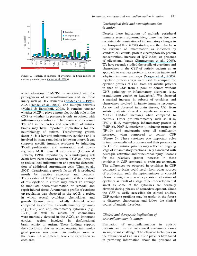
tively). Interestingly, a larger spectrum of increases
in pro-inflammatory and modulatory cytokines was
seen in the ACG, an important cortical structure
in autism, where there was a significant increase in
pro-inflamamtory cytokines such as interleukin-6
(IL-6), interleukin-10 (IL-10), macrophage
chemoattractant protein-3 (MCP-3), eotaxin,
eotaxin 2, macrophage-derived chemokine (MDC),
chemokine-�8 (Ck�8.1), neutrophil activating
peptide-2 (NAP-2), monokine induced by
interferon-� (MIG) and B-lymphocyte chemo-
attractant (BLC) (Figure 2).
The presence of MCP-1 is of particular interest,
since it facilitates the infiltration and accumulation
of monocytes and macrophages in inflammatory
CNS disease (Mahad & Ransohoff, 2003). Chemo-
attractant protein-1 is produced by activated and
reactive astrocytes, a finding that demonstrate the
effector role of these cells in the disease process
in autism. The increase in MCP-1 expression has
relevance to the pathogenesis of autism as we believe
its elevation in the brain is linked to pathways of
microglial activation and perhaps to the recruitment
of monocytes/macrophages to areas of neuronal-
cortical abnormalities. Our observations resemble
findings in other neurological disorders in
Figure 1. Neuropathology of cerebellum in autism. (A) Normal
appearance of the cerebellum in a control patient; (B–C) atrophic
folia and marked loss of Purkinje and granular cells in the
cerebellum of an autistic patient (H&E stain); (D) microglia
activation seen with anti-MHC class II immunostaining (from
Vargas et al., 2005).
490 Carlos A. Pardo et al.
which elevation of MCP-1 is associated with the
pathogenesis of neuroinflammation and neuronal
injury such as HIV dementia (Kelder et al., 1998),
ALS (Henkel et al., 2004), and multiple sclerosis
(Mahad & Ransohoff, 2003). It remains unclear
whether MCP-1 plays a more pleotrophic role in the
CNS or whether its presence is only associated with
inflammatory conditions. The presence of increased
TGF-�1 in the cortex and cerebellum of autistic
brains may have important implications for the
neurobiology of autism. Transforming growth
factor �1 is a key anti-inflammatory cytokine and is
involved in tissue remodeling following injury. It can
suppress specific immune responses by inhibiting
T-cell proliferation and maturation and down-
regulates MHC class II expression (Letterio &
Roberts, 1998). Importantly, cells undergoing cell
death have been shown to secrete TGF-�1, possibly
to reduce local inflammation and prevent degenera-
tion of additional surrounding cells (Chen et al.,
2001). Transforming growth factor �1 is produced
mostly by reactive astrocytes and neurons.
The elevation of TGF-�1 suggests that the elevation
of this cytokine in autism may reflect an attempt
to modulate neuroinflammation or remodel and
repair injured tissue. A remarkable profile of cytokine
up-regulation was observed in the ACG, a region
in which several cytokines, chemokines, and
growth factors were markedly elevated when
compared to controls. Pro-inflammatory cytokines
(e.g., IL-6) and anti-inflammatory cytokines (e.g.,
IL-10) as well as subsets of chemokines
were markedly elevated in the ACG, an important
cortical region involved in dysfunctional
brain activity in autism. These findings support
the conclusion that an active, ongoing immunolo-
gical process was present in multiple areas of
the brain but at different levels of expression in
each area.
Cerebrospinal fluid and neuroinflammationin autism
Despite these indications of multiple peripheral
immune system abnormalities, there has been no
consistent demonstration of inflammatory changes in
cerebrospinal fluid (CSF) studies, and there has been
no evidence of inflammation as indicated by
standard cell counts, protein electrophoresis, protein
concentration, increase of IgG index, or presence
of oligoclonal bands (Zimmerman et al., 2005).
We have recently studied the profile of cytokines and
chemokines in the CSF of autistic patients as an
approach to evaluate proteins involved in innate and
adaptive immune pathways (Vargas et al., 2005).
Cytokine protein arrays were used to compare the
cytokine profiles of CSF from six autistic patients
to that of CSF from a pool of donors without
CNS pathology or inflammatory disorders (e.g.,
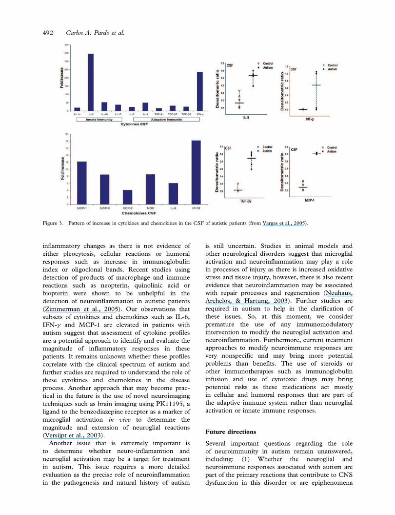
pseudotumor cerebri or headaches). We observed
a marked increase in subsets of cytokines and
chemokines involved in innate immune responses.
As we had observed in brain tissues, CSF from
autistic patients showed a significant increase in
MCP-1 (12-fold increase) when compared to
controls. Other pro-inflammatory such as IL-6,
IFN-�, IL-8, macrophage inflammatory protein-1�(MIP1�), NAP-2, interferon-� inducing protein-10
(IP-10) and angiogenin were all significantly
increased when compared to control CSF
(Figure 3). These cytokines play important roles
in immune-mediated processes and their presence in
the CSF in autistic patients may reflect an ongoing
stage of inflammatory reactions likely associated with
neuroglial activation and/or neuronal injury. Reasons
for the relatively greater increases in these
cytokines in CSF compared to brain are unknown.
The differences we observed in cytokines in CSF
compared to brain could result from other sources
of production, such the leptomeninges or choroid
plexus or might represent a persistent elevation of
cytokines as result of a stage of neurodevelopmental
arrest as some of the cytokines are normally
elevated during phases of neurodevelopment. Since
the CSF is easily accessible for clinical studies,
CSF cytokine profiling may be useful in the future
to diagnose, characterize and follow the clinical
course of autistic disorders.
Clinical and therapeutic implications ofneuroinflammation in autism
Evaluation of neuroinflammation in autistic
patients and its use in clinical assessment raises
an important challenge. The classical techniques in
the evaluation of CSF in autistic patients have failed
in providing information about the presence of
IL-1 α
IL-1 β
IL-6
IL-10
IL-15
TNF-α
TNF- β
IL-2
IL-4
IL-5
IL-13
IL-16
TGF- β1
−1
1
3
5
7
9
11Fo
ld In
crea
se
Cerebellum
Ant. cingulate gyrus
Midfrontal gyrus
Innate ImmunityAdaptive Immunity
Figure 2. Pattern of increase of cytokines in brain regions of
autistic patients (from Vargas et al., 2005).
Immunity, neuroglia and neuroinflammation in autism 491
inflammatory changes as there is not evidence of
either pleocytosis, cellular reactions or humoral
responses such as increase in immunoglobulin
index or oligoclonal bands. Recent studies using
detection of products of macrophage and immune
reactions such as neopterin, quinolinic acid or
biopterin were shown to be unhelpful in the
detection of neuroinflammation in autistic patients
(Zimmerman et al., 2005). Our observations that
subsets of cytokines and chemokines such as IL-6,
IFN-� and MCP-1 are elevated in patients with
autism suggest that assessment of cytokine profiles
are a potential approach to identify and evaluate the
magnitude of inflammatory responses in these
patients. It remains unknown whether these profiles
correlate with the clinical spectrum of autism and
further studies are required to understand the role of
these cytokines and chemokines in the disease
process. Another approach that may become prac-
tical in the future is the use of novel neuroimaging
techniques such as brain imaging using PK11195, a
ligand to the benzodiazepine receptor as a marker of
microglial activation in vivo to determine the
magnitude and extension of neuroglial reactions
(Versijpt et al., 2003).
Another issue that is extremely important is
to determine whether neuro-inflamamtion and
neuroglial activation may be a target for treatment
in autism. This issue requires a more detailed
evaluation as the precise role of neuroinflammation
in the pathogenesis and natural history of autism
is still uncertain. Studies in animal models and
other neurological disorders suggest that microglial
activation and neuroinflammation may play a role
in processes of injury as there is increased oxidative
stress and tissue injury, however, there is also recent
evidence that neuroinflammation may be associated
with repair processes and regeneration (Neuhaus,
Archelos, & Hartung, 2003). Further studies are
required in autism to help in the clarification of
these issues. So, at this moment, we consider
premature the use of any immunomodulatory
intervention to modify the neuroglial activation and
neuroinflammation. Furthermore, current treatment
approaches to modify neuroimmune responses are
very nonspecific and may bring more potential
problems than benefits. The use of steroids or
other immunotherapies such as immunoglobulin
infusion and use of cytotoxic drugs may bring
potential risks as these medications act mostly
in cellular and humoral responses that are part of
the adaptive immune system rather than neuroglial
activation or innate immune responses.
Future directions
Several important questions regarding the role
of neuroimmunity in autism remain unanswered,
including: (1) Whether the neuroglial and
neuroimmune responses associated with autism are
part of the primary reactions that contribute to CNS
dysfunction in this disorder or are epiphenomena
Figure 3. Pattern of increase in cytokines and chemokines in the CSF of autistic patients (from Vargas et al., 2005).
492 Carlos A. Pardo et al.
resulting from reactions to CNS dysfunction;
(2) the nature of the relationship of cytokines
and chemokines to immune and neurobiological
processes in the brain of autistic patients; (3) whether
the cerebellar pathology in autism is primarily the
result of neuroimmune processes or primary
abnormalities in neuronal function; (4) how analysis
of CSF may help us identify markers of immune
reactions within the CNS; and (5) whether the
immunogenetic background of the host influences
the development of neuroimmune reactions or
determines patterns of susceptibility to autism.
Conclusions
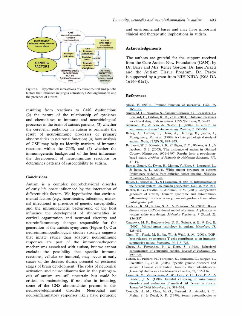
Autism is a complex neurobehavioral disorder
of early life onset influenced by the interaction of
different risk factors. We hypothesize that environ-
mental factors (e.g., neurotoxins, infections, mater-
nal infections) in presence of genetic susceptibility
and the immunogenetic background of the host
influence the development of abnormalities in
cortical organization and neuronal circuitry and
neuroinflammatory changes responsible for the
generation of the autistic symptoms (Figure 4). Our
neuroimmunopathological studies strongly suggests
that innate rather than adaptive neuroimmune
responses are part of the immunopathogenic
mechanisms associated with autism, but we cannot
exclude the possibility that specific immune
reactions, cellular or humoral, may occur at early
stages of the disease, during prenatal or postnatal
stages of brain development. The roles of neuroglial
activation and neuroinflammation in the pathogen-
esis of autism are still uncertain but could be
critical in maintaining, if not also in initiating,
some of the CNS abnormalities present in this
neurodevelopmental disorder. Neuroglial and
neuroinflammatory responses likely have polygenic
and environmental bases and may have important
clinical and therapeutic implications in autism.
Acknowledgements
The authors are grateful for the support received
from the Cure Autism Now Foundation (CAN), by
Dr. Barry and Mrs. Renee Gordon, Dr. Jane Pickett
and the Autism Tissue Program. Dr. Pardo
is supported by a grant from NIH-NIDA (K08-DA
16160-01a1).
References
Aloisi, F. (2001). Immune function of microglia. Glia, 36,
165–179.
Aman, M. G., Novotny, S., Samango-Sprouse, C., Lecavalier, L.,
Leonard, E., Gadow, K. D., et al. (2004). Outcome measures
for clinical drug trials in autism. CNS Spectrums, 9, 36–47.
Ashwood, P., & Van de Water, J. (2004). Is autism an
autoimmune disease? Autoimmunity Reviews, 3, 557–562.
Bailey, A., Luthert, P., Dean, A., Harding, B., Janota, I.,
Montgomery, M., et al. (1998). A clinicopathological study of
autism. Brain, 121(Pt 5), 889–905.
Barbaresi, W. J., Katusic, S. K., Colligan, R. C., Weaver, A. L., &
Jacobsen, S. J. (2005). The incidence of autism in Olmsted
County, Minnesota, 1976–1997: Results from a population-
based study. Archives of Pediatric & Adolescent Medicine, 159,
37–44.
Barnea-Goraly, N., Kwon, H., Menon, V., Eliez, S., Lotspeich, L.,
& Reiss, A. L. (2004). White matter structure in autism:
Preliminary evidence from diffusion tensor imaging. Biological
Psychiatry, 55, 323–326.
Bauer, J., Rauschka, H., & Lassmann, H. (2001). Inflammation in
the nervous system: The human perspective. Glia, 36, 235–243.
Becker, K. G., Freidlin, B., & Simon, R. M. (2003). Comparative
genomics of autism, Tourette syndrome and autoimmune/
inflammatory disorders. www.grc.nia.nih.gov/branches/rrb/dna/
pubs/cgoatad.pdf.
Carbone, K. M., Rubin, S. A., & Pletnikov, M. (2002). Borna
disease virus (BDV)-induced model of autism: Application to
vaccine safety test design. Molecular Psychiatry, 7 (Suppl. 2),
S36–S37.
Casanova, M. F., Buxhoeveden, D. P., Switala, A. E., & Roy, E.
(2002). Minicolumnar pathology in autism. Neurology, 58,
428–432.
Chen, W., Frank, M. E., Jin, W., & Wahl, S. M. (2001). TGF-
beta released by apoptotic T cells contributes to an immuno-
suppressive milieu. Immunity, 14, 715–725.
Chess, S., Fernandez, P., & Korn, S. (1978). Behavioral
consequences of congenital rubella. Journal of Pediatrics, 93,
699–703.
Cohen, D., Pichard, N., Tordjman, S., Baumann, C., Burglen, L.,
Excoiffier, E., et al. (2005). Specific genetic disorders and
autism: Clinical contribution towards their identification.
Journal of Autism & Developmental Disorders, 35, 103–116.
Comi, A. M., Zimmerman, A. W., Frye, V. H., Law, P. A., &
Peeden, J. N. (1999). Familial clustering of autoimmune
disorders and evaluation of medical risk factors in autism.
Journal of Child Neurology, 14, 388–394.
Connolly, A. M., Chez, M. G., Pestronk, A., Arnold, S. T.,
Mehta, S., & Deuel, R. K. (1999). Serum autoantibodies to
Figure 4. Hypothetical interactions of environmental and genetic
factors that influence neuroglia activation, CNS organization and
the presence of autism.
Immunity, neuroglia and neuroinflammation in autism 493
brain in Landau-Kleffner variant, autism, and other neurologic
disorders. Journal of Pediatrics, 134, 607–613.
Courchesne, E., & Pierce, K. (2005). Brain overgrowth in autism
during a critical time in development: Implications for frontal
pyramidal neuron and interneuron development and connec-
tivity. International Journal of Developmental Neuroscience, 23,
153–170.
Courchesne, E., Redcay, E., & Kennedy, D. P. (2004). The
autistic brain: Birth through adulthood. Current Opinions
in Neurology, 17, 489–496.
Croen, L. A., Grether, J. K., Yoshida, C. K., Odouli, R., &
Van de, W. J. (2005). Maternal autoimmune diseases, asthma
and allergies, and childhood autism spectrum disorders: A case-
control study. Archives in Pediatric & Adolescent Medicine, 159,
151–157.
Dalton, P., Deacon, R., Blamire, A., Pike, M., McKinlay, I.,
Stein, J., et al. (2003). Maternal neuronal antibodies associated
with autism and a language disorder. Annal of Neurology, 53,
533–537.
Daniels, W. W., Warren, R. P., Odell, J. D., Maciulis, A.,
Burger, R. A., Warren, W. L., et al. (1995). Increased
frequency of the extended or ancestral haplotype B44-SC30-
DR4 in autism. Neuropsychobiology, 32, 120–123.
DeLong, G. R., Bean, S. C., & Brown, F. R. III. (1981).
Acquired reversible autistic syndrome in acute encephalopathic
illness in children. Archives in Neurology, 38, 191–194.
Dong, Y., & Benveniste, E. N. (2001). Immune function of
astrocytes. Glia, 36, 180–190.
Fields, R. D., & Stevens-Graham, B. (2002). New insights into
neuron-glia communication. Science, 298, 556–562.
Folstein, S. E., & Rosen-Sheidley, B. (2001). Genetics of autism:
Complex aetiology for a heterogeneous disorder. Nature Reviews
Genetics, 2, 943–955.
Fombonne, E. (2003). Epidemiological surveys of autism
and other pervasive developmental disorders: An update.
Journal of Autism & Developmental Disorders, 33, 365–382.
Guerin, P., Lyon, G., Barthelemy, C., Sostak, E., Chevrollier, V.,
Garreau, B., et al. (1996). Neuropathological study of a case of
autistic syndrome with severe mental retardation. Developmental
Medicine & Child Neurology, 38, 203–211.
Gupta, S., Aggarwal, S., Rashanravan, B., & Lee, T. (1998).
Th1- and Th2-like cytokines in CD4þ and CD8þ T cells in
autism. Journal of Neuroimmunology, 85, 106–109.
Halsey, N. A., & Hyman, S. L. (2001). Measles-mumps-rubella
vaccine and autistic spectrum disorder: Report from the New
Challenges in Childhood Immunizations Conference convened
in Oak Brook, Illinois, June 12–13, 2000. Pediatrics, 107, E84.
Hashimoto, T., Tayama, M., Murakawa, K., Yoshimoto, T.,
Miyazaki, M., Harada, M., et al. (1995). Development of the
brainstem and cerebellum in autistic patients. Journal of Autism
& Developmental Disorders, 25, 1–18.
Henkel, J. S., Engelhardt, J. I., Siklos, L., Simpson, E. P.,
Kim, S. H., Pan, T., et al. (2004). Presence of dendritic cells,
MCP-1, and activated microglia/macrophages in amyotrophic
lateral sclerosis spinal cord tissue. Annals of Neurology, 55,
221–235.
Herbert, M. R., Ziegler, D. A., Deutsch, C. K., O’Brien, L. M.,
Lange, N., Bakardjiev, A., et al. (2003). Dissociations of
cerebral cortex, subcortical and cerebral white matter volumes
in autistic boys. Brain, 126, 1182–1192.
Herbert, M. R., Ziegler, D. A., Makris, N., Filipek, P. A.,
Kemper, T. L., Normandin, J. J., et al. (2004). Localization of
white matter volume increase in autism and developmental
language disorder. Annals of Neurology, 55, 530–540.
Huang, R. P. (2004). Cytokine protein arrays. Methods in
Molecular Biology, 278, 215–232.
Jyonouchi, H., Sun, S., & Le, H. (2001). Pro-inflammatory and
regulatory cytokine production associated with innate and
adaptive immune responses in children with autism spectrum
disorders and developmental regression. Journal of
Neuroimmunology, 120, 170–179.
Kaufmann, W. E., Cooper, K. L., Mostofsky, S. H., Capone,
G. T., Kates, W. R., Newschaffer, C. J., et al. (2003).
Specificity of cerebellar vermian abnormalities in autism: A
quantitative magnetic resonance imaging study. Journal of Child
Neurology, 18, 463–470.
Kawashima, H., Mori, T., Kashiwagi, Y., Takekuma, K.,
Hoshika, A., & Wakefield, A. (2000). Detection and sequencing
of measles virus from peripheral mononuclear cells from
patients with inflammatory bowel disease and autism. Digestive
Diseases & Science, 45, 723–729.
Kelder, W., McArthur, J. C., Nance-Sproson, T., McClernon, D.,
& Griffin, D. E. (1998). Beta-chemokines MCP-1 and
RANTES are selectively increased in cerebrospinal fluid of
patients with human immunodeficiency virus-associated
dementia. Annals of Neurology, 44, 831–835.
Kemper, T. L., & Bauman, M. (1998). Neuropathology of
infantile autism. Journal of Neuropathology & Experimental
Neurology, 57, 645–652.
Korvatska, E., Van de, W. J., Anders, T. F., & Gershwin, M. E.
(2002). Genetic and immunologic considerations in autism.
Neurobiology of Disease, 9, 107–125.
Larsson, H. J., Eaton, W. W., Madsen, K. M., Vestergaard, M.,
Olesen, A. V., Agerbo, E., et al. (2005). Risk factors for autism:
Perinatal factors, parental psychiatric history, and socioeco-
nomic status. American Journal of Epidemiology, 161, 916–925.
Lee, L.-C., Zachary, A. A., Leffell, M. S., Newschaffer, C. J.,
Matteson, K. J., Tyler, J. D., et al. (2004). Increased incidence
of maternal HLA-DR4 in single-birth, but not multiplex
families with autism. International Meeting for Autism
Research, Sacramento, CA. May 2004 (abstr).
Letterio, J. J., & Roberts, A. B. (1998). Regulation of immune
responses by TGF-beta. Annual Review of Immunology, 16,
137–161.
Levin, L., Ban, Y., Concepcion, E., Davies, T. F., Greenberg,
D. A., & Tomer, Y. (2004). Analysis of HLA genes in families
with autoimmune diabetes and thyroiditis. Human Immunology,
65, 640–647.
Lord, C., Cook, E. H., Leventhal, B. L., & Amaral, D. G. (2000).
Autism spectrum disorders. Neuron, 28, 355–363.
Lord, C., Shulman, C., & DiLavore, P. (2004). Regression and
word loss in autistic spectrum disorders. Journal of Child
Psychology & Psychiatry, 45, 936–955.
Mahad, D. J., & Ransohoff, R. M. (2003). The role of MCP-1
(CCL2) and CCR2 in multiple sclerosis and experimental
autoimmune encephalomyelitis (EAE). Seminars in Immunology,
15, 23–32.
Micali, N., Chakrabarti, S., & Fombonne, E. (2004). The broad
autism phenotype: Findings from an epidemiological survey.
Autism, 8, 21–37.
Money, J., Bobrow, N. A., & Clarke, F. C. (1971). Autism and
autoimmune disease: A family study. Journal of Autism & Child
Schizophrenia, 1, 146–160.
Nedergaard, M., Takano, T., & Hansen, A. J. (2002). Beyond the
role of glutamate as a neurotransmitter. Nature Reviews
Neuroscience, 3, 748–755.
Neuhaus, O., Archelos, J. J., & Hartung, H. P. (2003).
Immunomodulation in multiple sclerosis: From immuno-
suppression to neuroprotection. Trends in Pharmacological
Science, 24, 131–138.
Newschaffer, C. J., Fallin, D., & Lee, N. L. (2002). Heritable and
non-heritable risk factors for autism spectrum disorders.
Epidemiology Reviews, 24, 137–153.
Nguyen, M. D., Julien, J. P., & Rivest, S. (2002). Innate
immunity: The missing link in neuroprotection and neuro-
degeneration? Nature Reviews Neuroscience, 3, 216–227.
494 Carlos A. Pardo et al.
Odell, D., Maciulis, A., Cutler, A., Warren, L., McMahon,
W. M., Coon, H., et al. (2005). Confirmation of the association
of the C4B null allelle in autism. Human Immunology, 66,
140–145.
Patterson, P. H. (2002). Maternal infection: Window on
neuroimmune interactions in fetal brain development and
mental illness. Current Opinions in Neurobiology, 12, 115–118.
Prat, A., Biernacki, K., Wosik, K., & Antel, J. P. (2001). Glial cell
influence on the human blood-brain barrier. Glia, 36, 145–155.
Rapin, I. (1997). Autism. New England Journal of Medicine, 337,
97–104.
Rapin, I., & Katzman, R. (1998). Neurobiology of autism.
Annals of Neurology, 43, 7–14.
Rose, N. R., & Bona, C. (1993). Defining criteria for autoimmune
diseases (Witebsky’s postulates revisited). Immunology Today,
14, 426–430.
Rosenberg, G. A. (2002). Matrix metalloproteinases in
neuroinflammation. Glia, 39, 279–291.
Shi, L., Fatemi, S. H., Sidwell, R. W., & Patterson, P. H. (2003).
Maternal influenza infection causes marked behavioral
and pharmacological changes in the offspring. Journal of
Neuroscience, 23, 297–302.
Silva, S. C., Correia, C., Fesel, C., Barreto, M., Coutinho, A. M.,
Marques, C., et al. (2004). Autoantibody repertoires to brain
tissue in autism nuclear families. Journal of Neuroimmunology,
152, 176–182.
Singh, V. K., Lin, S. X., Newell, E., & Nelson, C. (2002).
Abnormal measles-mumps-rubella antibodies and CNS auto-
immunity in children with autism. Journal of Biomedical Science,
9, 359–364.
Singh, V. K., Lin, S. X., & Yang, V. C. (1998). Serological
association of measles virus and human herpesvirus-6
with brain auto-antibodies in autism. Clinical Immunology &
Immunopathology, 89, 105–108.
Singh, V. K., Warren, R., Averett, R., & Ghaziuddin, M. (1997).
Circulating autoantibodies to neuronal and glial filament
proteins in autism. Pediatric Neurology, 17, 88–90.
Stubbs, E. G., & Crawford, M. L. (1977). Depressed lymphocyte
responsiveness in autistic children. Journal of Autism & Child
Schizophrenia, 7, 49–55.
Sweeten, T. L., Bowyer, S. L., Posey, D. J., Halberstadt, G. M., &
McDougle, C. J. (2003). Increased prevalence of familial
autoimmunity in probands with pervasive developmental
disorders. Pediatrics, 112, e420.
Todd, R. D., Hickok, J. M., Anderson, G. M., & Cohen, D. J.
(1988). Antibrain antibodies in infantile autism. Biological
Psychiatry, 23, 644–647.
Torres, A. R., Maciulis, A., Stubbs, E. G., Cutler, A., & Odell, D.
(2002). The transmission disequilibrium test suggests that
HLA-DR4 and DR13 are linked to autism spectrum disorder.
Human Immunology, 63, 311–316.
Tuchman, R., & Rapin, I. (2002). Epilepsy in autism.
Lancet Neurology, 1, 352–358.
van Gent, T., Heijnen, C. J., & Treffers, P. D. (1997). Autism and
the immune system. Journal of Child Psychology & Psychiatry,
38, 337–349.
Vargas, D. L., Nascimbene, C., Krishnan, C., Zimmerman, A. W.,
& Pardo, C. A. (2005). Neuroglial activation and neuroin-
flammation in the brain of patients with autism. Annals of
Neurology, 57, 67–81.
Versijpt, J. J., Dumont, F., Van Laere, K. J., Decoo, D.,
Santens, P., Audenaert, K., et al. (2003). Assessment of
neuroinflammation and microglial activation in Alzheimer’s
disease with radiolabelled PK11195 and single photon emission
computed tomography. A pilot study. European Journal of
Neurology, 50, 39–47.
Vojdani, A., Campbell, A. W., Anyanwu, E., Kashanian, A.,
Bock, K., & Vojdani, E. (2002). Antibodies to neuron-specific
antigens in children with autism: Possible cross-reaction with
encephalitogenic proteins from milk, Chlamydia pneumoniae
and Streptococcus group A. Journal of Neuroimmunology, 129,
168–177.
Warren, R. P., Cole, P., Odell, J. D., Pingree, C. B., Warren,
W. L., White, E., et al. (1990). Detection of maternal antibodies
in infantile autism. Journal of the American Academy of Child &
Adolescent Psychiatry, 29, 873–877.
Warren, R. P., Margaretten, N. C., Pace, N. C., & Foster, A.
(1986). Immune abnormalities in patients with autism. Journal
of Autism & Developmental Disorders, 16, 189–197.
Wing, L., & Potter, D. (2002). The epidemiology of
autistic spectrum disorders: Is the prevalence rising? Mental
Retardation & Developmental Disabilities Research Reviews,
8, 151–161.
Yamashita, Y., Fujimoto, C., Nakajima, E., Isagai, T., &
Matsuishi, T. (2003). Possible association between congenital
cytomegalovirus infection and autistic disorder. Journal of
Autism & Developmental Disorders, 33, 455–459.
Yeargin-Allsopp, M., Rice, C., Karapurkar, T., Doernberg, N.,
Boyle, C., & Murphy, C. (2003). Prevalence of autism in a
US metropolitan area. Journal of the American Medical
Association, 289, 49–55.
Zimmerman, A. W., Jyonouchi, H., Comi, A. M., Connors, S. L.,
Milstien, S., Varsou, A., et al. (2005). Cerebrospinal fluid and
serum markers of inflammation in autism. Pediatric Neurology,
33, 195–201.
Zimmerman, A. W., Tyler, J. D., & Matteson, K. J. (2001).
Increased incidence of HLA-B60 and maternal DR4 in autism.
Annals of Neurology, 50, S122–S123.
Zimmerman, A. W. (2005). The immune system. In M. Bauman
& T. L. Kemper (Eds.), The neurobiology of autism
(pp. 371–386). Baltimore: The Johns Hopkins University Press.
Immunity, neuroglia and neuroinflammation in autism 495
Related Documents