IMMUNITY IN PARASITIC INFECTIONS General Features (Trichinella spiralis). Leishmania (Plasmodium vivax) Effector Mechanisms T. spiralis, Schistosoma mansoni Immunity in Parasitic, Viral, Bacterial and Fungal Infections 18

Welcome message from author
This document is posted to help you gain knowledge. Please leave a comment to let me know what you think about it! Share it to your friends and learn new things together.
Transcript
IMMUNITY IN PARASITIC INFECTIONS
General Features
Parasites infect, very large number of people and present major medical problems, es-pecially in tropical countries. The diseases caused are diverse and the immune respons-es, which are effective against the different parasites vary considerably. Parasitic infec-tions do, however, share a number of com-mon features.
Protozoan parasites and the worms are considerably larger than the bacteria and viruses, not only more in quantity, but also in variety. The parasites, unlike bacteria and viruses, undergo a life cycle in the host and exhibit different antigenicity at different stag-es of life, besides some species also change their surface antigens, a process known as antigenic variation (Trichinella spiralis). Pro-tozoa evolve different mechanisms to enter inside the cell to have their intracellular exis-tence. The invasive merozoite attached itself to the receptor on erythrocytes and uses the cell. On the other hand, Leishmania species use the complement receptor on macrophage to be engulfed by macrophage, where ulti-mately they reside and multiply.
The host resistance to parasite infec-tion may be genetic and controlled by im-mune response genes situated in the major histocompatibility complex class II (MHC II)
area. Certain individuals are genetically less susceptible to certain parasites. People, with sickle-cell trait, are resistant to malaria. For example, merozoites of the malaria parasite (Plasmodium vivax) use a particular blood group substance on to erythrocyte surface, the Duffy antigen, as a receptor to their entry into the cell. Certain African populations lack this antigen and are totally resistant to infec-tion by parasite.
Parasitic infections are generally chronic and show marked host specificity. For ex-ample, the malarial parasites of birds' ro-dents and man can multiply only in its own particular kind of host. Some exceptions are there. The tapeworm of pig can also infect humans.
Effector Mechanisms
After the entry of the parasite into the host, before it faces the specific immune response, it has to overcome the host's pre-existing defense mechanisms. Complement plays role in eliminating or causing lysis of many parasites, including certain adult worms and infective larva of T. spiralis, schistosomules of Schistosoma mansoni. Natural killer (NK) cell also may be active in imparting innate immunity against parasitic infection initially.
Various kinds of effector cells such as mac-rophages, neutrophils, eosinophils and even platelets helps to defend the host against the
Immunity in Parasitic, Viral, Bacterial and
Fungal Infections18
242 Textbook of Immunology
invasion of the parasites and act to control the multiplication and spread of the parasites, already in the residence. They act as the cells of the first line of defense.
Macrophage
Apart from acting as antigen processing and presenting cells in initiating immune response, macrophage affects the course of parasitic infection in two ways:
1. They secrete molecules, which act to regulate the inflammatory response. In-terleukin-1 (IL-1), tumor necrosis factor (TNF) and colony-stimulating factor (CSF) may enhance immunity by activating other cells or stimulating their prolifera-tion. Macrophage releases pro-staglan-dins, which may be immunosuppressive.
2. They act as effector cells, which inhibit the multiplication of the parasite or they may destroy them.
Activation of macrophages is a general feature of the early stages of infection. Ac-tivation of macrophages is brought about by the cytokines produced by T helper (T
h1)
cell [interferon- (IFN- , granulocyte-mac-rophage (GM)-CSF, IL-3, etc.]. Some products of the parasite such as Trypanosoma brucei and malarial parasites can cause activation of macrophage, independent of T cells, per-haps directly or by inducing macrophages to secrete TNF- , which then activate other macrophages.
Granuloma formation in liver and fibrous en-capsulation: In some parasitic infections in which the immune system cannot completely eliminate the parasite, the body reduces dam-age by walling of the parasite behind a cap-sule of inflammatory cells. This reaction is T cell dependent, is a chronic cell-mediated immune response to released antigen. Mac-rophages accumulate, release fibrogenic fac-tor of granuloma tissue. This sort of granuloma formation occurs in schistosomal infection in which the eggs are trapped in liver.
Phagocytosis: It is one of the primary func-tions of macrophage and is important in the defense against smaller parasite invaders. The effectiveness is markedly enhanced by opsonization of the organisms to be ingested. In addition, activated macrophages may ex-press more Fc and C3 receptors, which also tend to enhance their phagocytic function. African trypanosomes are quickly taken up by phagocytic cells in the liver.
Parasite killing properties: Macrophages se-crete scores of soluble factors, many of which can be cytotoxic and consequently they can also kill parasites by process that do not de-pend upon ingestion.
Activated macrophages are important in the control of infections caused by, for ex-ample, Trypanosoma cruzi, Leishmania and Plasmodium species. Macrophages are ca-pable of killing not only, relatively small in-tracellular parasite such as erythrocytic stage of malarial parasite, but also, when activat-ed, larger parasite such as the larval stage of schistosomes. They can also act as the killer cells by antibody-dependent cell-mediated cytotoxicity (ADCC). Specific immunoglobu-lin G (IgG) and IgE for instance can enhance their ability to kill schistosomules.
Granulocytes
Phagocytic properties: They can kill by both oxygen (O
2)-dependent and O
2-independent
mechanisms. They produce more intense oxidative bursts than macrophages and their secretory granules contain highly cytotoxic protein.
Extracellular destruction: The neutrophils also can be activated by cytokines (IFN- , GM-CSF, TNF- ). Extracellular destruction of T. cruzi is mediated by superoxide and hydro-gen peroxide.
Neutrophils: They also bear Fc receptor and receptor for complement that renders them (S. mansoni and T. spiralis) effective partici-pants in ADCC.
Immunity in Parasitic, Viral, Bacterial and Fungal Infections 243
Eosinophils
Infiltration of eosinophils and the production of high level of IgE are the common conse-quences of infection by parasitic worms and the eosinophils appear as major effector cell against helminths. It has been argued that the eosinophil has evolved, especially, as a defense against the tissue stage of the para-sites that are too large to be phagocytosed (e.g. helminths). The IgE-dependent mast cell reaction has evoked, primarily, to local-ize eosinophils near the parasite and then enhances the antiparasitic function through eosinophilic chemotactic factor (ECF). The increase in the number of eosinophils in schistosomiasis and ascariasis is also T cell dependent. The recruitment of eosinophils is mediated by a specific factor, eosinophilic stimulation promoter, which is a product of T cell. The eosinophils function in a various ways:
1. In association with specific antibody, they kill the worm by ADCC. Damage to helminths (schistosomulae) can be caused by the major basic protein liber-ated after degranulation.
2. Eosinophils are less phagocytic than neutrophils, but like neutrophils they can kill parasites by both O
2-dependent
and O2-independent mechanisms.
3. Enzymes released from the granules ex-ert a controlling effect on the substances released from the mast cells. The mast cell-derived factors are important in controlling the permeability of the local blood vessels and inflammation at the site of infection.
Mast Cells
Mast cell mediators enhance the activity of other effector processes in worm infection:
1. They interact with eosinophils as described.
2. They play an important role in acceler-ating the expulsion of worms.
In response to local release of antigen, T cells produce cytokines, which induce a vast increase in the number of inflammatory cells including monocytes, mucosal mast cells and goblet cells.
Mast cells also promote enhanced secre-tion of mediators and of mucus and provide help for the accumulation of IgG, IgA and IgE. Mast cell products including proteinase, cause change in permeability of the gut and shedding of the epithelium, which may help ejecting certain protozoan parasites.
Platelets
Platelets are capable of killing various types of parasites, including Schistosoma, Toxo-plasma gondii and T. cruzi. Further, they also bear Fc receptors for IgE on their surface membrane by which they mediate ADCC as-sociated with IgE.
T Cells
The T cells play important role in counteract-ing protozoan infections. It has been shown that nude mice, depleted of Tcells, have re-duced capacity to control trypanosomal and malarial infection. The transfer of splenic cells, especially T cells from immune ani-mals, gives protection against most parasitic infection.
Both CD4+ and CD8+ cells are needed for protection against some parasites. The type of T cells responsible for controlling infec-tion varies depending on the type of parasite, the stages of infection and the cytokines they produce. The CD4+ and CD8+ T cells protect against the different phases of Plasmodium infection. The CD4+ cells mediate immunity against blood stage. The CD8+ cells protect against the liver stage. The CD8+ cells act in two ways. They secrete IFN- , which inhibits the multiplication of the parasite in the liver cells and also they destroy hepatocytes by cytotoxic action, because of the presence of

244 Textbook of Immunology
MHC I in hepatocytes. The CD8+ cells cannot affect blood stage parasite as the erythrocytes do not possess MHC I molecule.
The immune response against T. cruzi and T. gondii depend not only upon CD4+ and CD8+ cells, but also NK cells and anti-body production.
The CD4+ cells act in different ways in dif-ferent infections. T
h cells are of two types T
h1
and Th2. The T
h1 and T
h2 cytokines are mutu-
ally antagonists. Th1 and T
h2 cells both coun-
ter malarial infections. Th1 cells act against
the liver stage of malaria. Administration of IFN- (T
h1 cytokines) directly to chimpanzee,
infected with sporozoites of vivax diminishes parasitemia. T
h2 cells through IL-4 activate B
cells to produce specific antibody. Elimina-tion of blood stage of malaria occurs in the spleen via the activated effector cells and by antibody-dependent cytotoxicity.
T helper type 1 subset enhances protec-tive response against intracellular protozoa. T
h1 subsets produce IFN- , which activate
macrophages to kill protozoa that live within them (L. donovani, T. gondii). It also enhances the effector responses against other parasites.
Both Th1 and T
h2 responses are important
in helminthic infection. IgE and eosinophils play vital role in the immunity against most of the helminthic infections and depend on the cytokines production by T
h2 cells. The
role of Th1 cells also cannot be ignored. In
schistosomiasis, the immune responses differ in mice, rats and in human beings. T
h2 re-
sponse is more important as resistance after drug treatment is correlated with the produc-tion of IgE. In the mouse, T
h1 cells and IFN-
are needed for vaccine induced protection and T
h2 cells are associated with egg-related
immunopathology. The switch to Th2 is trig-
gered by egg-related antigens.
The secretion of IFN- by Th1 cells acti-
vates effector cells that destroy lung-stage larvae, via the production of nitric oxide, but
when the adult schistosomes (S. mansoni) lay eggs, a soluble antigen is released. The antigen reduces T
h1 cells function, as well as
the level of IFN- and increases Th2 cell pro-
duction and IL-5.
In some parasites, when the immune sys-tem will fail to completely eliminate the or-ganism, the damage to body is reduced by granuloma formation by inflammatory cells and fibroblast surrounding the remnant of parasite. This T
h1-mediated reaction is a
chronic cell-mediated response to locally re-leased antigen and is mediated by cytokines, especially TNF and IFN- .
The immune response induced against various worms depend upon the anatomi-cal sites (gut, tissue), where they are present and also the antigens they gain, while pass through their life cycles.
Antibodies
In addition to the rise in specific antibod-ies, many parasitic infections provoke non-specific hypergammaglobulinemia. While specific responses are mostly T cell depen-dent, much of the non-specific antibody pro-duction is probably due to antigens released from the parasites acting as B cell mitogens.
Specific antibody is particularly important in the control of extracellular parasites. The antibody is effective in preventing the rein-vasion of the cells by blood-borne parasites, but ineffective once the parasite enters the host cells. The mechanism by which specific antibody controls the parasitic infections are summarized (Fig. 18.1).
Antibody is also involved in ADCC. Cytotoxic cells such as macrophages, neu-trophils and eosinophils adhere to worms coated with antibody by means of Fc and C3 receptors. Damage to schistosomes caused by the major basic protein (MBP) of the eosinophils crystalloid core. The re-lease of MBP into a small space between the

Immunity in Parasitic, Viral, Bacterial and Fungal Infections 245
eosinophils and the schistosomes surface localizes its action on the teguments of the worm and kill them.
Escape Mechanisms
There are various mechanisms, how the parasites evade the host immune system and establish infections.
Intrinsic Resistance
Anatomical inaccessibility (seclusion): Many parasites are protected from the host defense by their anatomical inaccessibility. For exam-ple, intracellular parasites avoid the effect of antibody being in accessible (T. cruzi, Plas-modium species and leishmanial species). Intracellular parasites residing inside the macrophages also evoke different strategies
of avoiding being killed by O2 metabolites
and lysosomal enzymes. Some parasites (En-tamoeba histolytica) are protected by chang-ing to cystic form.
Ability to resist destruction by complement: L. donovani offers more resistance to comple-ment-mediated lysis than L. tropica.
Avoidance of Recognition
Certain parasites undergo antigenic varia-tion (African trypanosomes) and change the antigens of their surface coat. Each variant possess antigenically distinct glycoproteins, which form its surface coat. The surface coat, presumably, protects the underlying surface membrane from the host's defense mechanisms. Malaria parasites also show antigenic variation.
Fig. 18.1: 1. Antibody-mediated defense of parasitic infections, direct damage: Antibody activates the classical complement pathway, causing damage to the parasite membrane and increasing susceptibility to other mediators; 2. Neutralization: Parasites such as Plasmodium species spread to new cells by specific receptor attachment; blocking the merozoite binding site with antibody prevents attachment to the recep-tors on the erythrocyte surface and hence prevents further multiplication; 3. Enhancement of phagocytosis: Complement C3b deposited on parasite membrane opsonizes it for phagocytosis by cells with C3b recep-tors (for example, macrophages). Macrophages also have Fc receptors; 4. Eosinophils, neutrophils, plate-lets and macrophages may be cytotoxic for some parasites when they recognize the parasite via specific antibody (ADCC). The reaction is enhanced by complement.
246 Textbook of Immunology
Other parasites, for example, Schistoso-ma species acquire a surface layer of host's antigens, so that the host defense does not distinguish them from the self.
Suppression of the Host’s Immune Response
1. Parasites can cause disruption of lymph-oid cells or tissue directly (the soluble lymphotoxic factor of T. spiralis). Schis-
tosoma can cleave a peptide from IgG. 2. Soluble parasite antigens, which can oc-
cur in enormous quantities, may reduce the effectiveness of host's response by a process known as immune distraction.
3. Non-specific immunosuppression is a universal phenomenon of parasitic in-fection as a whole.
4. Macrophage dysfunction may occur due to antigenic load. Antigen processing capacity is reduced. There will be de-creased production of IL-1. Macrophage also releases prostaglandins, which sup-press the immune reaction.
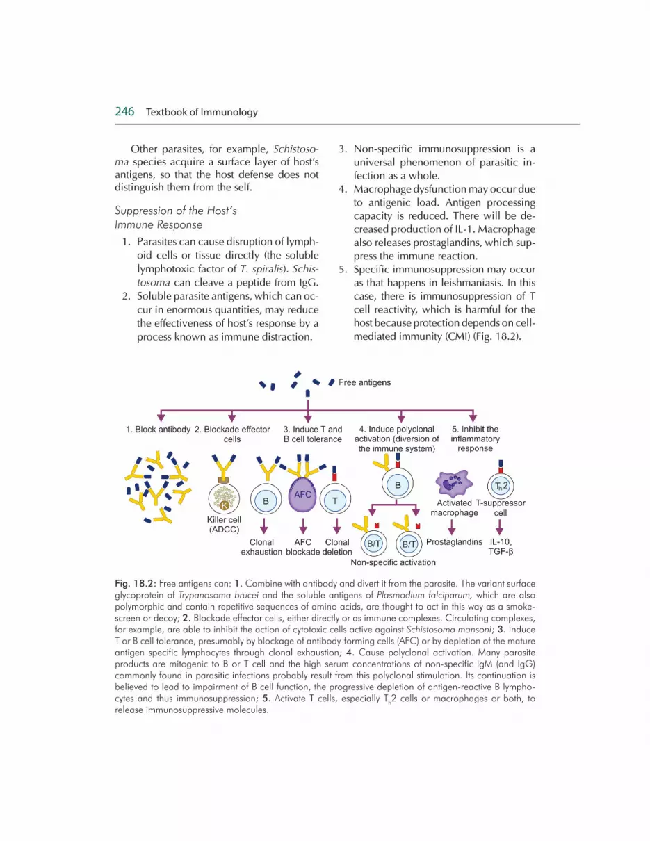
5. Specific immunosuppression may occur as that happens in leishmaniasis. In this case, there is immunosuppression of T cell reactivity, which is harmful for the host because protection depends on cell-mediated immunity (CMI) (Fig. 18.2).
Fig. 18.2: Free antigens can: 1. Combine with antibody and divert it from the parasite. The variant surface glycoprotein of Trypanosoma brucei and the soluble antigens of Plasmodium falciparum, which are also polymorphic and contain repetitive sequences of amino acids, are thought to act in this way as a smoke-screen or decoy; 2. Blockade effector cells, either directly or as immune complexes. Circulating complexes, for example, are able to inhibit the action of cytotoxic cells active against Schistosoma mansoni; 3. Induce T or B cell tolerance, presumably by blockage of antibody-forming cells (AFC) or by depletion of the mature antigen specific lymphocytes through clonal exhaustion; 4. Cause polyclonal activation. Many parasite products are mitogenic to B or T cell and the high serum concentrations of non-specific IgM (and IgG) commonly found in parasitic infections probably result from this polyclonal stimulation. Its continuation is believed to lead to impairment of B cell function, the progressive depletion of antigen-reactive B lympho-cytes and thus immunosuppression; 5. Activate T cells, especially T
h2 cells or macrophages or both, to
release immunosuppressive molecules.

Immunity in Parasitic, Viral, Bacterial and Fungal Infections 247
Immunopathological Consequences of Parasitic Infections
Apart from the directly destructive effects of some parasites and their products, many immune responses, themselves, have some pathological effects.
1. The IgE liberated in response to worm infections have serious effects on the hosts through mast cell mediators.
Examples:
a. Anaphylactic shock may occur when the hydatid cyst ruptures.
b. Asthma like reaction occurs in Toxocara canis infection.
c. Tropical eosinophilia occurs when filarial worms migrate through the lungs.
2. The formation of immune complex is common in parasitic infection.
Examples:
a. Immune complex may be depos-ited in the kidney, as in the neph-rotic syndrome of quartan malaria.
b. Immune complex containing para-sitic antigen may bind to unpara-sitized cells and accelerate their phagocytosis by the phagocytic cell of the spleen and liver.
3. Autoantibodies have been detected against red cells, lymphocytes and de-oxyribonucleic acid (DNA) (e.g. in try-panosomiasis and malaria).
4. Cross-reacting antigens: Antibodies against parasite may cross-react with host tissues.
Example: a. Cardiomyopathy, enlarged esopha-
gus and megacolon that occurs in the Chagas' disease are thought to result from the autoimmune effects of antibody or cytotoxic T cells on nerve ganglia that cross-reacts with T. cruzi.
5. There will be increase in the numbers and activity of macrophages and lym-phocytes leading to splenomegaly and hepatomegaly in malaria, sleeping sick-ness, visceral leishmaniasis.
6. Granuloma formation around the worm eggs leads to enlarged liver and fibrosis.
7. Excess production of cytokines may contribute to the sum of the manifesta-tions of the disease. Thus the fever, ane-mia, diarrhea and pulmonary changes of acute malaria closely resemble the symptoms of endotoxemia and prob-ably caused by TNF- .
8. Many immunological mechanisms may be combined in their pathological ef-fects to cause anemia in malaria.
9. Non-specific immunosuppression ex-plains, why people with parasitic infec-tions are, especially, perceptible to bac-terial and viral infection (measles).
IMMUNITY IN VIRAL INFECTION
The host response to invading virus de-pends upon the infectious agents and where it is encountered. In response to virus entry there may not be, always, an overt reaction leading to clinical manifestation. There may be simply, subclinical infection, which would protect the individual from later ex-posure. A number of specific immune effec-tor mechanisms together with non-specific defense mechanism play role in eliminating an infective virus (Table 18.1). At the same time the virus also finds out ways to subvert the defense mechanism and establish the infection.
Innate Immune Response
The interferon-alpha and beta (IFN- and - ) and NK cells play vital role in imparting innate immune response to viral infection. IFN- and IFN- are produced in response to the presence of viruses and certain intracel-lular bacteria. Double-stranded ribonucleic

248 Textbook of Immunology
acid (dsRNA) may be the important inducer. Macrophages, monocytes and fibroblasts are also capable of synthesizing these cytokines. IFN- and IFN- can induce an antiviral re-sponse or resistance to viral replication by binding to IFN receptors. Following binding of these IFNs with IFN-receptor, there is induc-tion of the synthesis of both 2-5 oligoadenyl-ate synthetase, (2-5A synthetase) and protein kinase-RNA (PKR). The action of 2-5A synthe-tase results in the activation of ribonucleaseL (RNaseL), which can degrade messenger-RNA (mRNA) protein kinase, inactivates the trans-lation initiation factor by phosphorylating it. Both pathways thus, result in the inhibition of protein synthesis and there by, effectively block viral replication (Fig. 18.3).
Certain cell-mediated reactions are also the part of the innate defenses against viral infections. NK cells play important role in this aspect. The formation of a close conjugate between the NK cell and the target, induces the effector cell to produce toxic molecules, which bring about the destruction of target cell. Natural killing is increased by IFNs, both the number of effector cells and their killing
potential. Therefore, these two innate defense mechanisms appear to work together to pro-tect the host from viral infection.
Humoral Immunity
Humoral immunity are various ways, how the antibodies against the viral components protect the host. Antibodies have no action against the latent viruses, as well as the vi-ruses those spread from cell to cell. Small amount of antibody in the blood, can neu-tralize the virus before it reaches its target cells in the nervous system. Most viruses express surface receptor molecules that en-able them to initiate infection by binding to these molecules on the complementary part on the tissues [sialic acid residues in cell membrane glycoprotein and glycolipid for influenza virus, intercellular adhesion mol-ecules (ICAMs) for rhinovirus, type 2 com-plement receptors on B cell for Epstein-Barr (EB) virus]. The antibodies block the recep-tor molecules on the viruses, thus, prevent-ing their attachment to the complementary tissues. Secretory IgA can neutralize viruses by similar mechanism on mucosal surfaces.
Table 18.1 Mechanisms of humoral and cell-mediated immune responses to viruses
Response type E�ector molecule or cell Activity
Humoral Antibody (especially, secretory IgA)
Blocks binding of virus to host cells, thus preventing infection or reinfection
IgG, IgM and IgA antibody Blocks fusion of viral envelope with host-cell plasma membrane
IgG and IgM antibody Enhances phagocytosis of viral particles (opsonization)
IgM antibody Agglutinates viral particles
Complement activated by IgG or IgM antibody
Mediates opsonization by C3b and lysis of enveloped viral particles by membrane-attack complex and direct antiviral activity
Cell mediated IFN secreted by Th or T
c cells Kill virus-infected self-cells
Cytotoxic T lymphocytes (CTLs), NK cells and macrophages
Kill virus-infected cells by antibody-dependent cell-mediated cytotoxicity (ADCC)

Immunity in Parasitic, Viral, Bacterial and Fungal Infections 249
In some cases, antibodies may block viral penetration by binding to epitope that are nec-essary, to mediate fusion of the viral envelope with the plasma membrane. Antibodies also can work at stages after penetration. Uncoat-ing with its release of viral nucleic acid, into the cytoplasm, can be inhibited if the virion is covered by antibody. If the induced antibody is of complement-activating isotypes, lysis of enveloped virions can ensue. Antibody can also cause aggregation of virus particles, thus,
Fig. 18.3: Induction of antiviral activity by IFN- and - . These IFNs bind to the IFN receptor, which in turn induces the synthesis of both 2-5A synthetase and protein kinase RNA (PKR). The action of 2-5A synthetase results in the activation of RNAse L, which can degrade mRNA. PKR inactivates the translation initiation fac-tor elF-2 by phosphorylating it. Both pathways thus result in the inhibition of protein synthesis and thereby effectively block viral replication.
limiting the spread of the infectious particles and forming a complex that is readily phago-cytosed. Antibody and complement can also function as an opsonizing agent to facilitate Fc or C3b receptor-mediated phagocytosis of the viral particles.
Humoral immunity does play a major pro-tective role in polio and in number of other viral infections. Passively administered anti-body can protect humans against several in-fections including measles, hepatitis A and B

250 Textbook of Immunology
and chickenpox, if given before or very soon after exposure. The immunity to many viral infections is lifelong.
Cell-mediated Immunity
As long as the virus is extracellular and the infection is not established, the antibody plays major role either eliminating the virus by different mechanisms or preventing the entry by blocking the receptor site. Antibody plays no role, once the virus is intracellular and the DNA of virus is integrated into the host DNA.
Once the infection is established CMI is imperative to deal with the virus. In general, CD8+ [cytotoxic T lymphocytes (CTL) cells] and CD4+ (T
h1) cells are the main cell types,
which take part in the defense mechanisms. Activated CD4+ (T
h1) cells produce a number
of cytokines such as IFN- , IL-2 and TNF- , which defend against virus infection directly or indirectly. IFN- acts directly on the virus infected cell and produce antiviral state. IL-2 activates CTLs and potentiates the lytic ac-tion on viral infected cells. Both IFN- and IL-2 activate NK cells, which play important role in causing lysis of the viral infected cells by ADCC mechanism in the beginning of the infection, when specific CTLs have not de-veloped. The role of CTLs in defense against viruses is demonstrated by the ability of virus specific CTLs to confer protection for the spe-cific virus on non-immune recipient by adop-tive transfer.
Viral Evasion of Host Defense Mechanism
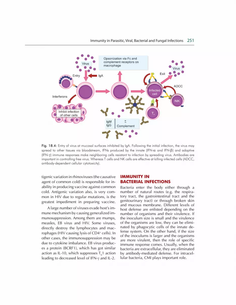
Despite adequate immune response pro-duced against the viral components, virus evades the defense mechanism of the host and establish infection (Fig. 18.4).
In many viruses, additional proteins are produced that interfere at various levels with specific and non-specific defenses. The ad-vantage of such proteins is that they enable
viruses to replicate more effectively amidst host antiviral defenses (Fig. 18.4). There are certain viruses, which evolve the strategies to evade the action of IFN- and IFN- . These include hepatitis �C' virus, which has been shown to overcome the antiviral effect of IFNs by blocking the action of PKR. Her-pes simplex virus (HSV) both I and II, evade host defense mechanism by inhibition of antigen presentation by infected host cells. Both HSV-I and HSV-II express an immedi-ate early protein called infected cell protein (ICP) 47, which very effectively inhibits the human transporter molecule (TAP) needed for antigen processing. The targeting on MHC molecule is not unique to HSV. In ad-enovirus and cytomegalovirus (CMV) also there is down regulation of MHC I expres-sion inhibiting antigen presentation to CD8+ T lymphocytes. In some viral infections such as CMV, measles and human immunodefi-ciency virus (HIV) infection, there is also re-duction of MHC II molecule expression on the surface of antigen-presenting cell (APC), thus blocking the function of antigen-specif-ic antiviral T
h cells.
Antibody-mediated destruction of the vi-rus depends on the activation of the comple-ment leading to lysis or phagocytosis follow-ing opsonization, but certain viruses develop strategies, to overcome complement-medi-ated destruction. For example, vaccinia vi-rus secretes protein that binds to C4b com-ponent of the complement and inhibit the classical complement pathway. Similarly, the glycoprotein component of HSV virus binds to C3b component inhibiting both classical and alternative pathway.
Many other viruses evolve the host's de-fense mechanism by adopting, antigenic vari-ation. The best example is influenza virus, which undergoes antigenic variation, produc-ing a different strain. The antibody produced against earlier strain becomes ineffective. An-
Immunity in Parasitic, Viral, Bacterial and Fungal Infections 251
tigenic variation in rhinoviruses (the causative agent of common cold) is responsible for in-ability in producing vaccine against common cold. Antigenic variation also, is very com-mon in HIV due to regular mutations, is the greatest impediment in preparing vaccine.
A large number of viruses evade host's im-mune mechanism by causing generalized im-munosuppression. Among them are mumps, measles, EB virus and HIV. Some viruses, directly destroy the lymphocytes and mac-rophages (HIV causing lysis of CD4+ cells). In other cases, the immunosuppression may be due to cytokine imbalance. EB virus produc-es a protein (BCRF1), which has got similar action as IL-10, which suppresses T
h1 action
leading to decreased level of IFN- and IL-2.
Fig. 18.4: Entry of virus at mucosal surfaces inhibited by IgA. Following the initial infection, the virus may
spread to other tissues via bloodstream, IFNs produced by the innate (IFN- and IFN- ) and adaptive
(IFN- ) immune responses make neighboring cells resistant to infection by spreading virus. Antibodies are important in controlling free virus. Whereas T cells and NK cells are effective at killing infected cells (ADCC, antibody-dependent cellular cytotoxicity).
IMMUNITY IN BACTERIAL INFECTIONS
Bacteria enter the body either through a number of natural routes (e.g. the respira-tory tract, the gastrointestinal tract and the genitourinary tract) or through broken skin and mucous membrane. Different levels of host defense are enlisted depending on the number of organisms and their virulence. If the inoculum size is small and the virulence of the organisms are low, they can be elimi-nated by phagocytic cells of the innate de-fense system. On the other hand, if the size of the inoculums is larger and the organisms are more virulent, then the role of specific immune response comes. Usually, when the bacteria are extracellular, they are eliminated by antibody-mediated defense. For intracel-lular bacteria, CMI plays important role.

252 Textbook of Immunology
Host Defenses
Very few organisms can penetrate the in-tact skin, because of various innate defense mechanisms. Whenever, the bacteria gain access to the tissues, their ability to fight the organisms and to eliminate depends upon the immune response generated against the mi-crobial antigens. In most cases, the immune reponse is generated against the components of the bacteria and the molecules secreted by them. Immune response is also gener-ated against the organ of motility (flagella), the organ of adhesion (fimbriae) and also the capsules. Specific antibodies to flagella and fimbriae also affect their ability to function properly. Antibodies also can inactivate vari-ous bacterial enzymes and toxins.
The bacteria after successfully evading the innate defense mechanisms (mechanical barrier, antibacterial substances, phagocyto-sis, etc.), proliferate in the tissue liberating the toxic products, which trigger inflammation. The resulting increased vascular permeability leads to the exudation of serum, which con-tains complement components, antibodies, clotting factors, as well as phagocytic cells. Chemotactic factors attract the phagocytic cells to the site of inflammation. Anaphyla-toxins (C3a, C5a) generated by complement activations, further increase the vascular per-meability increasing blood flow to the area.
Two types of adaptive immunity play roles against bacterial infection. They are hu-moral immunity and CMI.
Humoral Immunity
Attachment and invasion are important pro-cesses, which pathogenic bacteria adopt to establish the infection. Certain antibodies such as secretory IgA, interfere with the at-tachment molecule (agresin) and prevent colonization of pathogenic bacteria. Many organisms produce disease through their exotoxins (diphtheria, tetanus, botulism, etc).
Antibodies acquired by either immuniza-tion or previous infection or given passively, neutralize the bacterial exotoxins. Subsequent-ly the toxin-antitoxin complexes are phagocy-tosed. Many bacterial exotoxins are enzymes. The antibody against enzymes interferes with the ability of the enzyme to interact with substrates.
Antibody can interfere the normal func-tioning of bacteria, if in various ways when it binds directly to bacteria. Direct binding af-fects the activity of specific transport systems, there by depriving the bacteria of its energy supply and other essential chemicals. Inva-sion of the bacteria can also be inhibited by restricting the motility, when antiflagellar an-tibodies are produced. Specific antibody can agglutinate the bacteria, thus restricting the dissemination of the organism.
Antibody that binds to accessible antigens on the surface of a bacterium together with C3b component of complement, act as an opsonin and increases the phagocytosis and clearance of the bacterium (Fig. 18.5).
In some bacteria, notably gram-nega-tive bacteria, complement activations can lead directly to the lysis of the organism. Antibody-mediated complement activation also produce certain localized effector mol-ecules, which help to amplify the inflamma-tory response. For example, the complement split products (C3a, C4a and C5a) act as ana-phylatoxin to induce mast cell degranulation and thus, vasodilation and extravasation of neutrophils and lymphocytes from blood to tissues.
Cell-mediated Immunity
Ultimately, all bacteria will be engulfed by macrophages either to kill the bacteria or to remove after extracellular killing. The microbial products (muramyl dipeptide and trehalose dimycolate) and chemotactic factors (formyl methionyl peptide) are the

Immunity in Parasitic, Viral, Bacterial and Fungal Infections 253
Fig. 18.5: Antibody-mediated mechanisms for combating infection by extracellular bacteria. 1. Antibody neutralizes bacterial toxins; 2. Complement activation on bacterial surface leads to complement-mediated lysis of bacteria; 3. Antibody and the complement split product C3b bind to bacteria, serving as opsonins to increase phagocytosis; 4. C3a and C5a, generated by antibody-initiated complement activation, in-duce local mast cell degranulation, releasing substances that mediate vasodilation and extravasation of lymphocytes and neutrophils; 5. Other complement split products are chemotactic for neutrophils and macrophages.

254 Textbook of Immunology
stimuli to activate macrophages and mono-cytes. The endotoxin present in the cell wall of gram-negative bacteria and various carbo-hydrate polymers, such as -glucans, are also potent macrophage activators.
While innate immunity as well as humor-al immunity are not very effective against in-tracellular bacterial pathogens, intracellular bacteria can activate NK cells, which inturn provide early defense against intracellular bacteria. Intracellular bacteria induce a cell-mediated immune response, specifically de-layed type of hypersensitivity. The cytokines secreted by CD4+ (T
h) cells are important, no-
tably IFN- though TNF- and CSF activate macrophages to kill ingested pathogens more effectively.
Evasion of Host Defense Mechanisms
Establishment of bacterial infection involves four primary steps. They are:
1. Attachment to host cells. 2. Proliferation. 3. Invasion of host cells. 4. Toxin-induced damage to host cells.
Host defense mechanisms act at each of these steps and many bacteria have evolved ways to circumvent some of these host de-fenses (Table 18.2).
Intracellular Bacteria
The bacteria, which can survive and repli-cate inside the cell are in an advantageous condition, because the antibodies have no access on them. M. leprae adopts an intra-cellular environment, so that they can no longer live extracellularly. Listeria monocyto-genes, the causative organism of listeriosis, multiply in normal macrophages, but fail to survive in activated macrophages. Liste-riosis occurs mostly in immunocompromised subjects, pregnant women and neonate in whom probably the T cell-dependent mac-rophage activating factors (IFN- , TNF- , etc)
are deficient. Salmonella species and Bru-cella species can also survive intracellularly. They owe their resistance to glycolipid that is resistant to destruction.
In case of mycobacterial species, there is a waxy cell wall, which is resistant to lyso-somal enzymes. M. tuberculosis secretes cer-tain molecules, which prevents fusion of lyso-some with phagosome, but M. leprae escapes phagosome and grow in the cytoplasm. Be-sides the cell wall of both the mycobacterial species contain lipoarabinomannan, which blocks the action of IFN- on macrophages. T
h1 represents the major host defense against
intracellular bacteria.
In many circumstances, the immune re-sponse brings about the destruction of the host tissue. The accumulation of macrophages, in response to the lymphokines (IFN- , TNF- , GM-CSF) secreted by CD4+ cells, will cause formation of granuloma, which will prevent dissemination of the bacteria to other sites of the body.
IMMUNITY IN FUNGAL INFECTIONS
Fungi are eukaryotes with a rigid cell wall consisting of complex polysaccharides such as chitin, glucans and mannan. Among 70,000 or so species of fungi, only a small numbers are pathogenic for humans. How-ever, the fungi can cause, sometimes, serious life-threatening illness. The fungi can exist as:
1. Single cells (yeasts), which can easily be phagocytosed because of small size.
2. Long sender branching hyphae, which require extracellular killing processes.
Some pathogenic fungi exist in nature as an infectious mould (hyphal form) and invade tis-sue as a yeast (or yeast-like form such as spher-ules and endospores). These fungi are known as dimorphic fungi. Both the phases possess important virulent determinants and pose dif-ferent problems in the immune system.

Immunity in Parasitic, Viral, Bacterial and Fungal Infections 255
Although, some fungal infections can oc-cur in healthy individuals, the immunocom-promised subjects [patients with untreated acquired immunodeficiency syndrome (AIDS), patients undergoing cancer therapy, transplant recipient with immunosuppressive therapy, patients depending on long-term corticosteroid therapy and others] are more prone to suffer from a variety of fungal infec-tions. The immunity against different catego-ries of fungi broadly consists of:
1. Innate immune response. 2. T cell-mediated specific immune re-
sponses.
Innate Immune Response
Intact skin and normal commensal flora
plays important role in preventing the entry
and colonization of fungi.
Certain antifungal and antibacterial sub-
stances such as defensins, mannose-binding
lectin (MBL), surface protein �A' and �D', coats
Table 18.2 Host immune responses to bacterial infection
Infection process Host defenece Bacterial evasion mechanisms
Attachment to host cells Blockage of attachment by secretory
Secretion of proteases that cleave secretory IgA antibodies, IgA dimers (Neisseria
meningitidis, N. gonorrhoeae,
Haemophilus in�uenzae)
Antigenic variation in attach-ment structures (pill of N.
gonorrhoeae)
Cell-mediated Phagocytosis (Ab and C3b-mediated opsonization)
Production of surface structures (polysaccharide capsule, M protein, !brin coat) that inhibit phagocytic cell
Mechanisms for surviving within phagocytic cells induction of apoptosis in macrophages (Shigella �exneri)
Complement-mediated lysis and localized in"ammatory response
Generalized resistance of gram-positive bacteria to complement-mediated lysis
Insertion of membrane attack complex prevented by long side chain in cell wall LPS (some gram-negative bacteria)
Invasion of host tissues Ab-mediated agglutination Secretion of elastase that inactivates C3a and C5a (Pseudomonas)
Toxin-induced damage to host cells
Neutralization of toxin by antibody
Secretion of hyaluronidase, which enhances bacterial invasiveness
256 Textbook of Immunology
over the fungal elements and opsonize them for phagocytosis. Immunity to mycoses is prin-cipally cellular, involving neutrophils, mac-rophages, lymphocytes and probably by NK cells (for extracellular killing). With the pos-sible exception of dermatophytes and Rhizo-pus arrhizus, fungi are immune to antibody-complement-mediated lysis. The phagocytes (neutrophils and macrophages) kill the fungi.
1. Degranulation and release of the toxic materials on to indigestible hyphae.
2. Ingestion of the yeasts or conidia.The oxidative bursts, following ingestion,
play an important role in destruction of fungi. Defect in nicotinamide adenine dinucleotide phosphate (NADPH) oxidase system, as that occurs in patients with chronic granuloma-tous disease (CGD), fails to deal with Asper-gillus species, leading to severe aspergillosis. However, phagocytes from CGD patients, with defective oxygen reduction pathways, are competent enough to kill other yeasts and hyphae with normal efficiency, indicat-ing the role of other mechanisms.
The phagocytic response is dependent on the recognition of (PAMPs) pathogen-associ-ated molecular patterns in the fungal cell wall by either soluble or cell bound pattern recog-nition molecules. Toll-like receptor 2 (TLR2) recognizes fungal phospholipid mannan of Candida albicans, yeasts and A. fumigatus hyphae and conidia. TLR4/CD14 recognizes C. albicans, A. fumigatus and glucuronoxylo-mannan capsule of C. neoformans.
T Cell-mediated Specific Immune Response
Most fungi are highly immunogenic. They in-duce antibody production, as well as T CMI, which can be detected by serology and skin test (delayed hypersensitivity), respectively. Antibodies are seldom protective. Consider-able evidence suggests that T
h1-macrophage
activity plays dominant role in eliminating fungal pathogens.
Immunity against most pathogenic fungi (including dermatophytes and most systemic mycoses such as C. neoformans, H. capsu-latum, etc. but not Aspergillus species) is de-pendent on T CMI particularly, CD4+-T
h1 cell
secreting cytokine (IFN- ).
In some fungal infection, such as para-coccidioidomycosis, the type of immune response depends upon the severity of the lesion. In mild paracoccidioidomycosis, T
h1
response dominates. On the other hand, in severe disseminated paracoccidioidomyco-sis, the T
h1 response is replaced by T
h2 re-
sponse with high level of Th2 cytokines (IL-4,
IL-10) and eosinophils. A reduction of IFN- (with concomitant increase level of IL-10) is also a marker of impaired immunity in sys-temic mycosis such as C. albicans and neu-tropenia-associated aspergillosis.
Evasion Strategies
Many fungi have evolved the ways to cir-cumvent, some of the host defense for their survival:
1. Cryptococcus neoformans, ordinar-ily, inhibits phagocytosis because of its polysaccharide capsule, but can be overcome by the opsonic effect of com-plement and antibody.
2. Dermatophytes suppress host T cell re-sponses and delay the cell-mediated de-struction.
3. Histoplasma capsulatum, an obligatory intracellular pathogen, evades killing by macrophage by entering through CR3 receptor and altering the normal path-way of phagosome maturation.
STUDY QUESTIONS
Essay Questions
1. Discuss the effector mechanism involved in the elimination of parasites from the host.
Immunity in Parasitic, Viral, Bacterial and Fungal Infections 257
2. Briefly discuss the various mechanisms, how the parasites evade the host imm-une system and established infections.
3. Discuss, briefly the humoral and cell-mediated immune responses to viruses.
4. How viruses evade the host defense mechanisms?
5. Discuss the host immune responses to bacterial infections.
6. Discuss the host immune responses to fungal infections.
Short Notes
1. Role of eosinophils in parasitic infection. 2. Immunopathological consequences of
parasitic infections. 3. Immunity againt intracellular bacteria. 4. Mucosal immunity against viral infections. 5. T cell-mediated immune response
against fungal infections.
SUGGESTED READING
1. Black JG. Microbiology: Principles and applications, 3rd edition. USA: Printice Hall College div; 1996.
2. Coligan JE, Kruisbeek AM, Margulies DH, et
al. Current Protocols in Immunology. New
York: Wiley; 1997.
3. Daniel P Stites. Basic and Clinical Immuno-
logy, 8th edition. USA: Lange (Medical
Book); 2007.
4. Gerald L Mandell, John E Bennett, Raphael
Dolin. Principles and Practice of Infectious
Diseases (Volume 1 and 2), 5th edition.
2000.
5. Goldsby RA, Kindt Thomas J, Osborn Barbara
A Kuby. Immunology, 6th edition. New York:
WH Freeman and Company; 2007.
6. Jawetz. Melnick and Adelberg's Medical
Microbiology, 25th edition. USA: McGraw
Hill, Lange Basic Science; 2010.
7. Male David, Brostoff Jonathan, Roitt Ivan, et
al. Immunology, 7th edition; 2006.
8. Tortora Gerard J, Funke Berdel R. Case chri-
stine L. Microbiology: an introduction, 10th
edition. USA; 1998.
9. Thao Doan, Roger Melvold, Susan Viselli, et
al. Lippincott's illustrated reviews: Immuno-
logy. 1st Indian print. Baltimore, USA:
Lippincott Williams and Wilkins; 2008.

Related Documents